lagowski - genetics · in ry and absent in ma-2 flies (forrest, hanly and lagowski 1961). the...
TRANSCRIPT

POLYGENIC CONTROL OF ALDEHYDE OXIDASE IN DROSOPHILA'
JAMES B. COURTRIGHT?
Depariment of Biology, The Johns Hopkins University, Baltimore, Maryland 21218
Received February 20, 1967
HEN extracts of wild-type Drosophila are electrophoresed on agar gels and the gels stained with a tetrazolium solution containing benzaldehyde, a
formazan band is formed at a location not identical to the xanthine dehydro- genase (XDH) band ( COURTRIGHT 1966a). This was a surprising observation since benzaldehyde has been reported to serve as a substrate for XDH (GLASS- MAN and MITCHELL 1959a). Since our new band is detected in extracts of ry flies, but not ma-1 flies, the evidence suggested that benzaldehyde was serving as a substrate for pyridoxal oxidase, a possibly related enzyme which also is present in ry and absent in ma-2 flies (FORREST, HANLY and LAGOWSKI 1961). The present paper presents genetic and biochemical evidence that the enzyme in question is neither pyridoxal oxidase nor XDH, but an aldehyde oxidase under similar genetic control. The enzyme also has some properties in common with the so-called ma-lf factor (GLASSMAN 1965, 1966), although its identity to the ma-l+ complementing factor is questionable.
MATERIALS A N D METHODS
General Procedures: All flies were grown on the cornmeal-agar medium described by GLASS- MAN, KARAM and KELLER (1962), at 25°C. After sterilization of the food bottles, 0.5 ml of 0.018% folic acid (Sigma, neutralized with NaOH) in 20% ethanol was applied to each.
All solutions were prepared using glass distilled water. Homogenates were prepared, stored, and processed at temperatures between 0 and 5°C.
For developmental studies eggs were collected using the method of MITCHELL and MITCHELL (1964.). Eggs and larvae were homogenized separately in an equal volume of 0.3 M KH2P04- K,HPO,, 0.001 M EDTA3 buffer pH 7.5 in a 2 ml Dual1 glass homogenizer.
Genetic stocks: The following stocks were used: (a) Drosophila melanogaster. Beadex3 (Bxs) 1-59.4, blistery ( b y ) 3-48.7, forked (f) 1-56.7, low xanthine dehydrogenase (Ixd) 3-33 (KELLER and GLASSMAN 1964), maroon-like (ma-Z) 1-64.9 (GLASSMAN 1959), maroon-likebrOnZY (ma-lbz), rosy ( r y ) 3-52.35 (CHOVNICK et al. 1964), rough ( ru ) 3-01, vermilion ( U ) 1-33.0, yellow ( y ) 1-0.0, ru lxd by (referred to as Ixd), ru lxd by r y (referred to as Ixd r y ) , U f Bxj ma-1; ru lxd by r y (referred to as ma-1; Ixd r y ) , yf attached-X, and Oregon-R (OreR) wild type. Unless indicated, map positions are taken from BRIDGES and BREHME (1944).
(b) Drosophila simulans. aldehyde oxidase1 (aZdoz1) a fast anodally migrating aldehyde oxidase contained in jv st p e and pe H h ; aldehyde oxidase11 (aldozr') a slower anodally migrating aldehyde oxidase than aldoxl present in wild type, from Lima, Peru; javelin ( j v ) 3-0.0 and
This work was supported by Public EIealth Service Grants GhI-504-07 and 1-F1-Ghf-33, 779-01, and National
This work has been presented in partial fulfillment of the requirements for the degree of Doctor of Philosophy. Abbreviations used in this paper: DCIP-dichloroindophenol, DELE-diethylaminoethyl, EDTA-ethylenediamine
tetraacetic acid. NAD-nicotinamide adenine dinucleotide, NBT-nitro blue tetrazolium, PMS-phenazine methosulfate, Tris-tris( hydroxymethyl) aminomethane.
Genetics 5 7 : 25-39 September 1967.
Science Foundation grant GB-4451.

26 J. B. COURTRIGHT
peach (pe ) 3-101.0 (STURTEVANT and PLUNKET 1926) ; Hairyhair’ess ( H h ) 3-61.0 and scarlet ( s t ) 3-40.0 (STURTEVANT and NOVITSKI 1941).
Spectrophotometic assays: All assays were performed with a Ziess M4 QIII spectrophoto- meter equipped with a Gilford multiple sample absorbance recorder. All buffers used for spec- trophotometic assays were prewarmed to 30°C. The reaction mixtures for the various enzyme reactions are listed below.
Aldehyde oxidase: The following modification of the flavin-linked aldehyde oxidase assay of MAHLER (1955) was used: 0.03 ml of 1 M redistilled acetaldehyde (Eastman), 0.02 ml of 2 mg/ml PMS (Sigma), 0.20 ml of 0.01% aqueous dichloroindophenol (Mann Research Lab), 0.10 ml of enzyme solution and water, 0.65 ml of 0.5 M KH,PO,-K,HPO,, 0.001 M EDTA pH 7.5 containing 0.1 % bovine serum albumin (Sigma). Under these conditions the enzyme activity is linear with time and enzyme concentration, provided that the assay mixture does not contain more than 0.25 units of enzyme. One unit is defined by MAHLER (1955) at that amount of enzyme causing a decrease of 1.03 O D ~ ~ ~ per minute at 30°C. In this paper specific activity is units per mg protein.
Catalase: Catalase was assayed using a solution consisting of 0.95 ml of 0.06% H,O, in 0.01 M KH,PO,-K,HPO, pH 7.5 and 0.05 ml of enzyme and water. One unit is defined by decompo- sition of one micromole of hydrogen peroxide per minute. This corresponds to an optical density decrease of 0.043 per minute at 241) m ~ 1 .
Preparation of aldehyde oxidase: Singlefy ho~mogemtes. For genetic analysis, it was necessary to detect the enzyme in single flies. Routinely, a single fly was homogenized in ,003 ml of 0.1 M Tris-HC1, 0.001 M EDTA buffer, pH 8.08 containing 0.1% bovine serum albumin. The homo- genizer vessels were round bottom 0.5 ml glass tubes (6 x 50 mm) for which soft glass rods had been shaped and ground with carbonmdum powder for a good fit. The homogenates were centri- fuged at room temperature in a “hematocrit” centrifuge at 12,000 x g for 3 minutes. The super- natant solutions were used for electrophoresis.
Preparation of mass homogenates. Two procedures, 1 and 2, were used. (1) Homogenates were prepared by adding 1 ml of 0.1 M Tris-HC1, 0.001 M EDTA, pH 8.0 for each 400 mg of freshly etherized flies. The resulting mixture was homogenized in a Ten Broeck homogenizer and then centrifuged at 48,000 x g in a Servall RC-2B refrigerated centrifuge. Alternatively, the homogenization may be done in 0.3 M KH,PO,-K,HPO,, 0.001 M EDTA, pH 7.5. Such extracts retain aldehyde oxidase activity for several days if stored at 4°C.
( 2 ) Homogenates were prepared by adding 1 ml of 0.1 M Tris-HC1, 0.001 M EDTA, pH 8.0 for each 400 mg of freshly etherized flies. The resulting mixture was homogenized in a Servall Omnimixer operated at full speed for 15 seconds. To the resulting homogenate, 1 M MnC1, was added dropwise with stirring to a final concentration of 0.05 M to precipitate nucleic acids (SMITH and YANOFSKY 1962). The resulting solution was homogenized for an additional 15 seconds at full speed in the Omnimixer, allowed to sit at 0°C for 2 to 3 hours, and then centri- fuged at 48,000 x g for 20 minutes in a Servall RC-BB refrigerated centrifuge. The resulting homogenate was either assayed directly o r subjected to further purification as described below.
Purification of aldehyde oxidase: The pale yellow supernatant solution from the MnC1, precipitation step was applied to a 2.5 x 45 cm column of G-25 Sephadex (coarse), equilibrated with 0.1 M Tris-HC1, 0,001 M EDTA, pH 8.0 in order to remove eye pigments. The pale yellow fraction which came off in the void volume of 75 ml had an O D , ~ ~ / O D , ~ ~ ratio of 1.2, and possessed all of the applied aldehyde oxidase activity. For each ml of this fraction, 313 mg of solid, finely ground ammonium sulfate (Baker) recrystallized from 0.001 M EDTA was added with constant stirring. The final pH was adjusted to 7.1 by dropwise addition of 28% ammonium hydroxide (Baker). The resulting solution was stirred at 4°C for one hour, then centrifuged at 48,000 x g for 10 minutes and the precipitate discarded. An additional 214 mg (NH,), SO, was added per ml solution, the pH adjusted to 7.1 by dropwise addition of NH,OH as before. The resulting precipitate, was dissolved in distilled water to a final concentration of 12 mg protein/ml and dialyzed overnight against BOO volumes of 0.01 M KH,PO,-K,HPO,, 0.001 M
EDTA, pH 7.2. The dialyzed sample (110 mg protein/9.2 ml) was applied to a 1.5 X 25 cm column of DEAE-cellulose (Whatman, 1 .O milliequivalents/g) prepared by the methods of

POLYGENIC CONTROL O F ALDEHYDE OXIDASE 27
PETERSON and SOBER (1962), and was eluted with a liner gradient of KH,PO,-K,HPO,, 0.001 M
EDTA, pH 7.1 from 0.01 to 0.30 M (total volume equals 360 ml), at a flow rate of 0.32 ml/minute. A volume of approximately 0.1 ml of each 5 ml fraction was applied to the wells of a spot plate containing 0.1 ml of the DCIP assay solution. Fractions containing aldehyde oxidase were detected visually by the disappearance of the blue color of DCIP. The fractions were then assayed for aldehyde oxidase activity Those fractions with a specific activity greater than 12.00 and a total activity greater than 5.44 units/ml were combined and 561 mg of (NH,),SO, were added for each ml. The pH was adjusted to 7.2 with NH,OH as before. The precipitate was dissolved in distilled water and dialyzed overnight against 200 volumes of 0.025 M Tris-H,PO,, 0.001 M
EDTA, pH 9.0. The dialyzed sample was applied to a 1.5 x 25 cm DEAE-cellulose column equilibrated with 0.025 M Tris-H,PO,, 0.001 M EDTA, pH 9.0, The aldehyde oxidase was eluted with a linear gradient from 0.025 M to 1.0 M Tris-H,PO,, 0.001 M EDTA, pH 9.0 (total volume equals 400 ml) at a flow rate of 0.32 ml/minute. Five-ml fractions were assayed for aldehyde oxidase activity and those fractions with a specific activity greater than 68.00 and a total activity greater than 4.60 units per ml were concentrated to a volume of 1 ml using suction filtration through an 8-ml collodion bag (Schleicher and Schuell). Approximately 0.2 g sucrose (Baker) was added for each ml, and a t? td of 925 pg protein contained in 1 ml was applied to a 2.5 X 32 cm column of Sephadex G-150 equilibrated with 0.01 M KH,PO,-K,HPO,, 0.001 M EDTA, pH 7.2. The enzyme was eluted with the same buffer at a flow rate of 0.45 ml/minute. The 5-ml fractions were analyzed for enzymatic activity by the spectrophotometric assay. The fractions with a specific activity of 344 or greater were pooled and found to be stable at 4°C for at least three weeks This procedure gives a 200-fold purification from crude extracts, with 20% recovery (Table 1).
Electrophoretic technique: Electrophoresis of extracts was performed as described by URSPRUNG and LEDNE (1965), except that “Agarose” (Nutritional Biochemicals), was substituted for agar in the preparation of the gels.
Immunochemical procedures Antibodies to aldehyde oxidase were elicited by injecting rab- bits supra-scapularly with a total of 128 units of aldehyde oxidase, specific activity 411. in Freund‘s complete adjuvant (Difco) (COHN 1952). Three weeks later the rabbits were injected with the same enzyme preparation emulsified with Freund’s incomplete adjuvant. The rabbits were bled 6 to 8 days after the second injection The gamma globulin was prepared by the method of KEKWICK (1941), except that a 15-18% sodium sulfate cut of whole serum was used, (D. B. ROBERTS, personal communication).
Ouchterlony plates were prepared using a 0.7% agarose solution in 1/20 EBT buffer (URSPRUNG and LEONE 1965). Plates with a well-spacing of 4mm were developed overnight at 37”C, and uncomplexed antigen was washed out by several changes of 0.1 M Tris HCI, 0.001 M
EDTA buffer, pH 8.0 over 12 hours Aldehyde oxidase precipitin lines were detected by staining plates in a solution containing 10 mg NBT (Dajacj, 0.4 mg PMS (Sigma), 2 ml acetaldehyde (Eastman) or 0.5 ml benzaldehyde (Fisher) in 25 m10.2 M Tris HCI buffer, pH 8.0.
TABLE 1
Purification of albhyde ozidase
Step
Crude MnCl, G-25 5 0 4 0 % (NH,),SO, DEAE pH 7.2 DEAE pH 9.0 G-150
Total ml Units/inl Total units ?g Specific
protein/ml activity Recovery Purification X
34.0 30.5 51.0
9.2 51.0 66.3 15.3
48.0 1632 120.0 3660 75.0 3825
126.0 1160 24.0 1225 11.74 778 22.60 345
21.6 2.22 16.0 7.50 8.0 9.38
12.0 10.50 0.68 35.00 0.07 165.00 0.057 396.00
100 225 234
71 75 47 21
1 3.4 4.2 4.7
15.7 74.0
178.0

28 J. B. COURTRIGHT
To titrate a given number of units of enzyme, 0.05 ml of enzyme was incubated with varying amounts of antibody to a final volume of 0.10 ml. This solution was incubated at 4°C overnight and the complexed enzyme was removed by centrifugation at 10,000 x g for 20 minutes at 4°C. Aliquots of the supernatant solution were th-n assayed for enzyme activity.
Protein de/erminations: Protein in crude extracts was determined by the method of LOWRY et al. (1951) using bovine serum albumin (Sigma) as a standard. Protein in ednuent column fractions was determined by the ultraviolet spectrophotometric method of WARBURC and CHRISTIAN (1941 ) .
Sucrose &miry centrifugation. Crude extracts of ry flies were centrifuged on 5-20% linear sucrose gradients by the method of MARTIN and AM= (1961). Gradients were generated simul- taneously in a 20 ml gradient generator (Buchler) and 4.6 ml was collected in each of three 1.27 x 5.08 cm cellulose nitrate tubes, and 0.10 ml of enzyme was carefully layered onto each gradient. To two gradients, 20 units of aldehyde oxidase was applied. Since crude Drosophila extracts contain catalase, 110 units of bovine catalase (Worthington) was applied separately to the third gradient. The tubes were placed in a SW-SOL rotor and centrifuged at 39,000 rpm for 12 hours in a model L-2 centrifuge at 4°C. The tubes were punctured with a piercing unit (Buchler) and ten-drop fractions were collected. At least 80% of all of the applied enzyme activity was recovered.
O R I G I N
( + I GENOTYPE
I
02 @ r 0 '8 'J
O X I D A S E
- X D H
I I
..
rJ rL
"0 5 2
FIGURE 1 .Simultaneous detection of aldehyde oxidase and xanthine dehydrogenase. Extracts of the various genotypes were prepared by method (1) and electrophoresed on agarose gels for 20 minutes a t 31 v/cm and 25 ma at 5-15°C. Gels were stained for XDH activity by incubating with 7 mg hypoxanthine (Sigma), 100 mg NAD (Sigma), and the concentrations of PMS, NBT, and Tris buffer given in METHODS. Aldehyde oxidase was detected by staining as in METHODS.

POLYGENIC CONTROL O F ALDEHYDE OXIDASE 29
Origin FIGURE 2.-Detection of aldehyde oxidase
isxymes of D. mrlanogaster on agarose gels. Major Aldehyde Mass homogenates of ma-I, lxd, and wild type Minor Oxidase were prepared h y method (1). The gels were
electrophorescd 15 minutes at 31 v/cm and 25 ma at 5-15°C. Aldehyde oxidase activity - X D H was detected by immersing the gels in staining solution for one hour.
1
RESULTS
Identification of aldehyde oxidase by gel electrophoresis: When wild-type ex- tracts of D. melanogaster and D. simulans, are electrophoresed and simultane- ously stained for aldehyde oxidase and xanthine dehydrogenase, two zones of formazan deposition are detected (Figure 1). The faster migrating of the two bands was identified as XDH since it is absent when ry extracts are electro- phoresed or when hypoxanthine is omitted from the staining solution. The slower migrating component is aldehyde oxidase. It is present in wild-type and ry extracts when acetaldehyde or benzaldehyde is supplied as substrate. The inde- pendence of aldehyde oxidase and XDH is further indicated by electrophoretic differences in the mobility of aldehyde oxidase in both these two species and their hybrids which are not correlated with equivalent differences in the mobility of the XDH molecules (Figure 1 ) .
When more concentrated extracts are stained, a minor component of aldehyde oxidase is seen on zymograms in addition to the major component (compare Figures 2 and 3 ) . This minor component is under the same genetic control as is
O R l G l l
FIGURE 3.--Electrophoretic variants of alde- hyde oxidase in Drosophila simulans. Extracts of the various genotypes were prepared by method (1) and electrophoresed 20 minutes at 31 v/cm and 25 ma at 5-15°C. Gels were stained for aldehyde oxidase.

30 J. B. COURTRIGHT
TABLE 2
Relative rates of reaction of aldehyde oxidase* with various substrated ~ ~~~~~~~
Dimethylammo- Substrate Acetaldehyde Benzaldehyde benzaldehyde Pyridoxal Blank
Relative rate 1 OO$ 218 108 < I < I
* 200-fold pure aldehyde oxidase (see METHODS). f Reaction rate is the initial rate with excess enzyme in the presence of 0.25 micromoles of each of the substrates in a
i 100 under these conditions equals an O D ~ decrease of O.OR/minute. total volume of 1.0 ml.
the major component (Figure 2). The absence of the major component in ma4 and the small amount present in Zxd flies is paralleled by the absence of pyridoxal oxidase in these genotypes (GLASSMAN et al. 1964). However, the major com- ponent is not pyridoxal oxidase since extracts purified 200-fold with respect to aldehyde oxidase possess only the major component and react with several aldehydes but not pyridoxal (Table 2). In these extracts there is a component which reduces the tetrazolium and which migrates to the same position as XDH (Figure 2). This component may be XDH since it is absent in ma-1 extracts.
Genetic control of aldehyde oxidase: The deficiencies in aldehyde oxidase activ- ity in ma-1 or Izd extracts suggested that either the ma-lf or lxdf locus was the structural gene for aldehyde oxidase. There are three lines of evidence which indicate the polygenic control of this enzyme.
a. Genetic mapping of aldehyde oxidase. Although no electrophoretic variants of aldehyde oxidase have been found in more than 25 strains of D. melanogaster examined, such variants were found in D. simulans. (Figure 3 ) . Reciprocal crosses of iu st pe (aldox:) X aldox:' established that the gene was not sex linked. The backcross recombinants, 4- st p / i v st pe, were homozygous for aldox:, indi- cating that the aldehyde oxidase gene was located in the st pe region (COURT-
TABLE 3
Crosses of aldoxlI x pe Hh
Generation Genotype
Number of flies of aldox type
I I1 1/11.
P pe (aldox:) H h P -t (aldox:') + Fl Backcross
pe (aldol') H h / f (aldox") f pe (aldozI) Hh/pe (aldotd) H h pe (aldod) H h / f (aldox") f pe (aldod) + / p e (aldod) H h pe (aldvx::) f / p e (aldol:) Hh f (aldol:) Hh/pe (aldoz:) H h + (aldod:) Hh/pe (aldox:) H h
13 0 0 9 0
29 0
69 0
0 13 0 0 0 0 0 0 0'
0 0
20 0 8 0
63 0
38
* 1/11 type is the a l d o x ~ / a l d o x ~ ~ heterozygote (Figure 3)
Number of crossovers between p e and aldox
Percent crossing over between aldor and H h Map location 74.5
132 67 34
Number of crossovers between aldox and H h

POLYGENIC CONTROL O F ALDEHYDE OXIDASE 31
RIGHT 1966b). To map the aldox locus, pe H" (aldox:) was crossed to aldox" flies. and the F, heterozygous females were crossed to pe H" males. All parents were routinely checked for their aldox genotype after successful mating. The analysis of the resultant backcross and recombinant progeny (Table 3) indicates that the aldox locus is on the D. simulans third chromosome at 74.5.
On the basis of these mapping experiments. the homologous D. melanogaster aldox locus should be located at about 64 on the third chromosome. Two mutants located in this chromosomal region, glassy (63.1 ) and Hennarecess"e (64.5) were tested and found negative for deficiencies or altered mobility of aldehyde oxidase molecules. However, the existence of a separate aldox+ locus in D. melanogaster was confirmed by crossing ma-1; lxd ry (D. melanogaster) females x aldox: (D. simulans) males. The aldehyde oxidase zymogram obtained from hybrid progeny (Figure 4) is similar to the simple aldox'/aldox" hybrid zymogram. The position of the slowest aldehyde oxidase band in the interspecies hybrid is the same as that in OreR and shows that there is a functional aldoxf locus in ma-I; lxd ry flies.
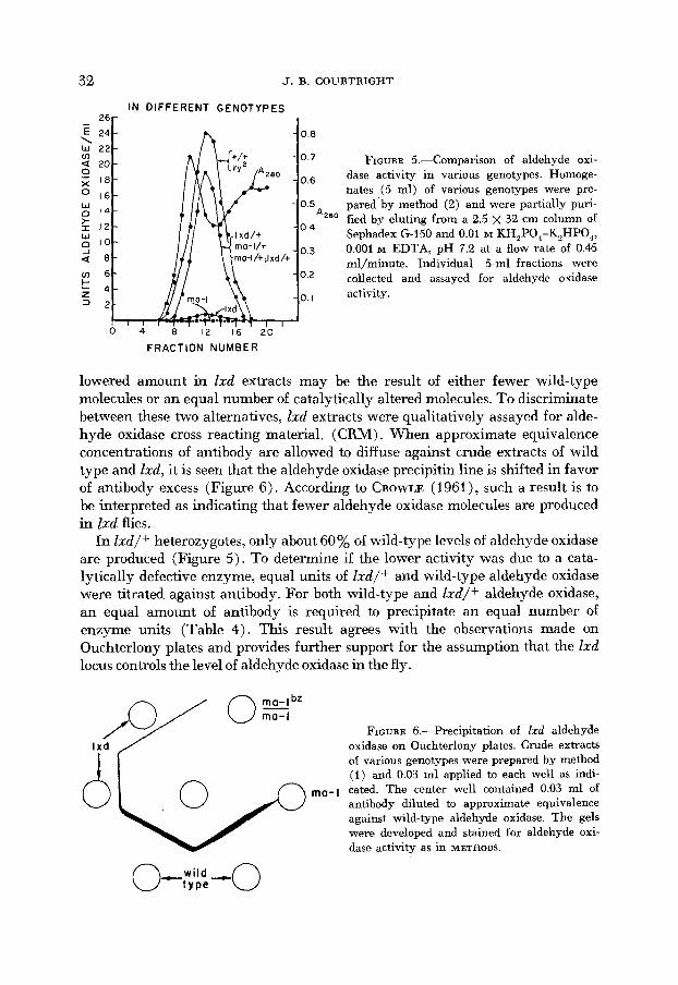
Although the aldehyde oxidase structural gene is not allelic to either ma-1 or lxd, both of these loci affect this enzyme (Figure 2). When partially purified extracts of ma-2 are eluted from a column of G150 Sephadex (Figure 5) no aldehyde oxidase activity is found in any of the fractions, whereas in Ixd extracts there is a small amount of aldehyde oxidase activity which has the same molecular size as the wild-type aldehyde oxidase (Figure 5).
b. Aldehyde oxidase in lxd flies. Aldehyde oxidase in lxd extracts resemble the wild-type enzyme in size and electrophoretic properties (Figures 2, 5). Its
O R I G I N
(+)
GENOTYPE
FIGURE 4.-Aldehyde oxidase in species hybrids. Extracts of D. simulans, D. sirnulam/ D. nieIanogas/er (ma-1; Ixd ry) . and D. melanogaster were prepared by method ( i ) , and electrophoresed 20 minutes at 31 v/cm and 25 ma at 5-15°C. Gels were stained for aldehyde oxidase.
32 J. B. COURTRIGHT
IN DIFFERENT GENOTYPES 26 -
E 24 0 0 \ w 2 2 v) a 20 0 7 FIGURE 5.--Comparison of aldehyde oxi- 0 j; I8 o 6 dase activity in various genotypes. Homoge-
nates (5 ml) of various genotypes were pre- pared by method (2) and were partially puri- fied by eluting from a 2.5 x 32 cm column of Sephadex G-150 and 0.01 M KH,P0,-K2HP0, r 12
J 0.001 M EDTA, pH 7.2 at a flow rate of 0.45 mljminute. Individual 5-ml fractions were a 8
u - 1 6 O 2 collected and assayed for aldehyde oxidase t z 4 I activity. = 2
16
E 14 * 0 5
0 4
0 3 E I O
0 4 8 I 2 I 6 20
FRACTION NUMBER
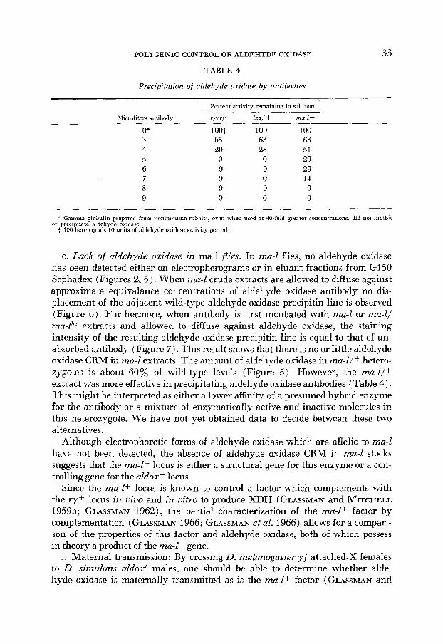
lowered amount in Zxd extracts may be the result of either fewer wild-type molecules or an equal number of catalytically altered molecules. To discriminate between these two alternatives, Ixd extracts were qualitatively assayed for alde- hyde oxidase cross reacting material. (CRM) . When approximate equivalence concentrations of antibody are allowed to diffuse against crude extracts of wild type and Zxd, it is seen that the aldehyde oxidase precipitin line is shifted in favor of antibody excess (Figure 6). According to CROWLE (1961), such a result is to be interpreted as indicating that fewer aldehyde oxidase molecules are produced in lxd flies.
In Zxd/+ heterozygotes, only about 60% of wild-type levels of aldehyde oxidase are produced (Figure 5 ) . To determine if the lower activity was due to a cata- lytically defective enzyme, equal units of I d / + and wild-type aldehyde oxidase were titrated against antibody. For both wild-type and I d / + aldehyde oxidase, an equal amount of antibody is required to precipitate an equal number of enzyme units (Table 4). This result agrees with the observations made on Ouchterlony plates and provides further support for the assumption that the Zxd locus controls the level of aldehyde oxidase in the fly.
J-J/ OsbZ FIGURE 6.-Precipitation of Zxd aldehyde
oxidase on Ouchterlony plates. Crude extracts of various genotypes were prepared by method (1) and 0.03 ml applied to each well as indi-
ma-l cated. The center well contained 0.03 ml of antibody diluted to approximate equivalence against wild-type aldehyde oxidase. The gels were developed and stained for aldehyde oxi- dase activity as in METHODS.
POLYGENIC CONTROL O F ALDEHYDE OXIDASE
TABLE 4
Precipitation of aldehyde oxidase by antibodies
33
Percent activity remaining in solution
Microliters antibody v / r r lxd/ + mad+
0' 3 4 5 6 7 8 9
1 oot 65 20 0 0 0 0 0
100 63 28 0 0 0 0 0
100 63 51 29 29 14 9 0
* Gamma globulin prepared from nonimmune rabbits, even when used at W-fold greater concentrations, did not inhibit
+ 100 here equals 10 units of aldehyde oxidase activity per ml. or precipitate aldehyde oxidase.
c. Lack of aldehyde oxidme in ma-1 flies. In ma4 flies, no aldehyde oxidase has been detected either on electropherograms or in eluant fractions from G150 Sephadex (Figures 2 ,5) . When ma-1 crude extracts are allowed to diffuse against approximate equivalance concentrations of aldehyde oxidase antibody no dis- placement of the adjacent wild-type aldehyde oxidase precipitin line is observed (Figure 6). Furthermore, when antibody is first incubated with ma-1 or ma-l/ ma-P extracts and allowed to diffuse against aldehyde oxidase, the staining intensity of the resulting aldehyde oxidase precipitin line is equal to that of un- absorbed antibody (Figure 7). This result shows that there is no or little aldehyde oxidase CRM in ma-1 extracts. The amount of aldehyde oxidase in ma-l/f hetero- zygotes is about 60% of wild-type levels (Figure 5). However, the mal/+ extract 'was more effective in precipitating aldehyde oxidase antibodies (Table 4). This might be interpreted as either a lower affinity of a presumed hybrid enzyme for the antibody or a mixture of enzymatically active and inactive molecules in this heterozygote. We have not yet obtained data to decide between these two alternatives.
Although electrophoretic forms of aldehyde oxidase which are allelic to ma-l have not been detected, the absence of aldehyde oxidase CRM in mad stocks suggests that the ma-l+ locus is either a structural gene for this enzyme or a con- trolling gene for the aldoxf locus.
Since the ma-lf locus is known to control a factor which complements with the ry+ locus in vivo and in vitro to produce XDH (GLASSMAN and ITCH ELL 1959b; GLASSMAN 1962), the partial characterization of the ma-lf factor by complementation (GLASSMAN 1966; GLASSMAN et al. 1966) allows for a compari- son of the properties of this factor and aldehyde oxidase, both of which possess in theory a product of the ma-Lf gene.
i. Maternal transmission: By crossing D. melanogaster yf attached-X females to D. simulans aldoxI males, one should be able to determine whether alde- hyde oxidase is maternally transmitted as is the m - l + factor (GLASSMAN and

34 J. B. COURTRIGHT
I;IGUIW 7.-Detection of aldehyde oxidase isozymes. Crude extracts of various genotypes pre- parecl by method ( 1 ) were incubated at 37°C for one hour with an equal volume of antibody. Aliquots were then applied to the wells and allowed to diffuse against a wild-type extract pre- pared by method (1). Gels were drvelnped and stained for aldehyde oxidase activity. Reservoirs 1 and 2 contained wild-type extract; reservoirs 3 and 5 contained ma-l/ma-l"' extract; reservoir 4 contained 0.1 M Tris HCl, 0.001 M EDTA, pH 8.0; reservoir 6 contained ma-I; Zrd r y extract all preinculmted with antibody. The center reservoir contained a crude extract of wild type not preincubated with antihody. The reservoirs were spaced at 10 mm, which resolves the single precipitin line of Figure 6 into two components. These lines may represent the aldehyde oxidase isozymes of Figure 2.
MITCHELL 1959b). Analysis of electropherograms of hybrid progeny for ma- ternal and paternal enzyme mobilities shows that the paternal enzyme is not detected until the late third instar (Figure 8). This absence of the D. simulans electrophoretic form in eggs and in first- and second-instar larvae probably indi- cates that neither the paternal nor the maternal aZdox+ locus is producing func- tional product.
When ma-Z females are crossed to OreR males, aldehyde oxidase activity is detected in the eggs, but this activity remains low until third instar (Table 5).
TABLE 5
Aldehyde ozidase ac/ivi/y in ma-I/+ embryos derived from ma-l/ma-1 female parents
Specific activity 0.82 0.52 0.77 1.36 1.92 3.65 7.31
. Developmcntnl slages include a l l indirirlualc. from 0-2+ lionrs for any given slage. i larvae abniit 120 Iinurs after Iiatrliing.

POLYGENIC CONTROL OF ALDEHYDE OXIDASE
( + I
-
Aldehyde oxidase
a
k - z 3 2
V I 6
35
250 c z
- 2 0 0 ; 0
m \
50 3
Or ig in
D.MELANOGASTER/D.SIMULANS FIGURE 8.-Maternal transmission of aldehyde oxidase. Drosophila melanogaster, D . simulans,
and the various embryonic stages of the hybrid progeny of an interspecies cross (see text) were prepared by method ( l ) , electrophoresed for 20 minutes at 31 v/cm and 25 ma at 5-15"C, and stained for aldehyde oxidase.
FRACTION NUMBER
FIGURE 9.-Sedimentation of aldehyde oxidase on a sucrose gradient. Extracts of r y flies and bovine catalase were centrifuged and analyzed. and aldehyde oxidas- and bovine catalase were assayed as in METHODS. Fraction 36 represents the top of the tube.

36 J. B. COURTRIGHT
The stage of development at which this increase in aldehyde oxidase activity occurs correlates well with the time of detection of the hybrid enzyme in inter- species hybrids. : \2
ii. Sedimentation: GLASSMAN et al. (1966) have found that the ma-Zf factor sediments in sucrose gradients at about the same rate as bovine catalase. This is also the case for aldehyde oxidase. When ry extracts are centrifuged at 39,000 rpm in an SW-50 rotor for 12 hours, aldehyde oxidase is found in the same position as bovine catalase in the gradient (Figure 9).
iii. Trypsin and thermal stability: The ma-Zf factor is relatively resistent to trypsin and heat (GLASSMAN 1966). Extracts of ry were prepared and treated with trypsin and exposed to 50" and 60°C according to GLASSMAN (1966). Under these conditions, aldehyde oxidase has similar stabilities to the ma-Zf factor (Table 6). The enzyme is especially thermostable in dilute solutions. This property was not known to us when we initially purified the enzyme; it might be useful in future purification procedures.
DISCUSSION
Benzaldehyde has been listed as one of the substrates of XDH in Drosophila ( GLASSMAN and MITCHELL 1959a). However benzaldehyde is also oxidized by an enzyme 'with a different electrophoretic mobility (Figure 1 ) . This enzyme does not require NAD+ and is present in ry flies, which lack XDH activity; since it reacts with a variety of aldehydes, it has been termed aldehyde oxidase. Its
TABLE 6
A comparison of aldehyde oxidase and ma-lf complzmenting actiuity after various treatments
Percent Percent ma-l+ Aldehgde aldehyde complementing oxidase Protein oxidase activity
Enzyme source units Treatment (mg/ml) remaining remaining
ry (pH 5 supernatant solution) 3 1 .00 15 min, 50°C 1 .oo 15 min, 50°C 1 .oo 15 min, 50°C 1 .oo 15 min, 60°C 1 .oo 15 min, 60°C 1.00 15 min, 60" C 0.25 60 min, Norite
potassium phosphate, pH 7.2) 1.12 15 min, trypsin 1.12 180 min, trypsin 1.12 15 min, 50°C 1.12 15 min, 50°C 1.12 15 min, 50°C 1.12 15 min, 60°C 1.12 15 min, 60°C
r y (Methods (2), in 0.01 M
11 .a0 66 9W 1.18 62 2.18* 92
1.18 21 2.18* 27
11.80 4 5 t
11.80 168 0.123
2.80 100 1 ow 2.80 100 2.80 73 0.28 73 I.@* 86 2.80 59 0.28 54
1.12 15 min, 60°C 1.28* 71
' Dilution into 0.1% bovine serum albumin in 0.3 M potassium phosphate, pH 7.5 t After GLASSMAN (1966). P After GLASSMAN (1962).

POLYGENIC CONTROL OF ALDEHYDE OXIDASE 37
genetic control appears to be quite complicated, and at least three loci are involved in its production.
The structural gene responsible for different electrophoretic forms of aldehyde oxidase has been mapped at 74.5 on the third chromosome of D. simulans (Table 3). When these electrophoretic variants in D. simulans were crossed to ma-1; lxd ry D. melanogaster, a hybrid aldehyde oxidase zymogram was obtained (Figure 4), indicating that there is a homologous aldoxf locus in this genotype.
The ma-1 locus may also be a structural gene for aldehyde oxidase as evidenced by the absence of aldehyde oxidase CRM in ma-l/ma-1 flies (Figures 6, 7) and the altered levels of both enzymatic activity and CRM in ma-l/f heterozygotes (Figure 5 ; Table 4). However, the existence of a second structural gene for aldehyde oxidase should be accepted with caution until electrophoretic variants allelic to ma-1 are found.
The amount of aldehyde oxidase is lowered by a third gene, lxd, which appears to have its effect at the level of enzyme production rather than determination of enzyme structure. Aldehyde oxidase in lxd flies has the same electrophoretic mobility at pH 8.7 and the same molecular size as wild-type aldehyde oxidase (Figures 2, 5 ) . In crude extracts of lxd/lxd and lxd/+flies, the lower amount of aldehyde oxidase CRM suggests that there are fewer wild-type molecules present (Figure 6; Table 4) .
Although aldehyde oxidase and pyridoxal oxidase are controlled by the same loci, ma4 and lxd, the two are known to be separate molecules for several reasons: (1 ) no pyridoxal oxidase has been detected in lxd extracts (GLASSMAN et al. 1964) while small amounts of aldehyde oxidase are present in Ixd extracts (Figures 2, 5 ) ; (2) aldehyde oxidase purified 200-fold does not react with pyridoxal, although it reacts readily with several aldehydes; (Table 2); (3 ) a strain of D. mehno- gaster which has low levels of pyridoxal oxidase (lpo) has normal levels of alde- hyde oxidase (J. COLLINS, personal communication).
The presence of aldehyde oxidase in lxd flies and its absence in ma4 flies sug- gested that it might be the ma-l+ complementing factor (GLASSMAN 1962). Although the enzyme has the same pattern of maternal transmission, sedimenta- tion, thermostability, and trypsin resistence as the ma-l+ factor ( GLASSMAN 1966, GLASSMAN et al. 1966; Figures 8, 9; Table 5) , since it is not inactivated or removed by Norite (Table 5 ) nor is it found in ma-l/ma-lbz flies which are known to contain 5-10% XDH (Figure 6, 7; GLASSMAN and PINKERTON 1960), its iden- tity with the complementing factor is not established. This evidence does suggest, however, that the product of the ma-l+ gene is a component of both aldehyde oxidase and the ma-l+ complementing factor.
Thus there are at least three loci involved in the production and regulation of aldehyde oxidase. Our findings are consistent with the assumption that the aldox+ and ma-I+ loci produce or control subunits of the aldehyde oxidase molecule, while Ixd regulates the functioning of one or both of these loci.
The author is grateful to DR. H. URSPRUNC for valuable suggestions and advice throughout this investigation, and to DR. S. R. SUSKIND for critical evaluation of the immunochemical procedures and results.

38 J. B. COURTRIGHT
SUMMARY
The genetic control and physiological role of aldehyde oxidase have been ex- amined in D. melanogaster and D. simulans. The structural gene for aldehyde oxidase is in chromosome 3. The enzyme is low when the gene Ixd (low xanthine dehydrogenase) is homozygous, and absent when individuals are homozygous for the gene maroon-like (ma-l) . These nonallelic loci do not alter the aldehyde oxidase molecules, but regulate the amount produced.-Aldehyde oxidase re- sembles the mad+ complementing factor in its pattern of maternal transmission, sedimentation, trypsin resistence, and thermostability, but differs from the com- plementing factor since it is not removed by Norite treatment.
LITERATURE CITED
BRIDGES, C. B., and K. S. BREHME, 1944 The mutants of Drosophila meLanogaster. Carnegie Inst. Wash. Publ. 552.
CHOVNICK, A., A. SCHALET, R. P. KERNAGHAN, and M. KRAUSS, 196.F The rosy cistron in Drosophila melaogaster: Genetic fine structure analysis. Genetic 50: 1245-1259.
COHN, M., 1952 Production of antibodies in experimental animals. Methods Med. Res. 5 : 271- 283.
Electrophoretic analysis of xanthine dehydrogenase mutants. Dro- sDphila Inform. Sew. 41: 77-78. - 1966b The genetics of aldehyde oxidase in Dro- sophila. (Abstr.) Genetics 54: 328.
COURTRIGHT, J. B., 1966a
CROWLE, A. J., 1961 FORREST, H. S., E. W. HANLY, and J. M. LAGOWSKI, 1961
GLASSMAN, E., 1962
Immundiffusion. Academic Press, New York. Biochemical differences between
mutants rosy-2 and maroon-like of Drosophila meLanogaster. Genetics 46 : 1455-1463.
In vitro complementation between nonallelic Drosophila mutants deficient in xanthine dehydrogenase. Proc. Natl. Acad. Sci. U.S. 48: 1491-1497. - 1965 Xanthine dehydrogenase of Drosophila melanogaster. J. Elisha Mitchell Sci. Soc. 81 : Suppl. 1: 42-54. - 1966 Complementation in uitro between non-allelic Drosophila mutants deficient in xanthine dehydrogenase. 111. Observatims on heat stabilities. Biochim. Biophys. Acta 117: 342-350.
Mutants of Drosophila melanogaster deficient in xanthine dehydrogenase. Genetics 4: 153-162. - 1959b Maternal effect of ma-l+ on xanthine dehydrogenase of Drosophila melanogaster. Genetics 44: 547-554.
Complementation of the maroon-like eye color locus of Drosophila melanogaster. Science 131: 1810-181 1.
Differential response to gene dosage experiments involving the two loci which control xanthine dehydrogenase of Drosophila melanogaster. Z. Vererb. 93: 399-4Q3.
GLASSMAN, E., T. SHINODA, H. M. MOON, and J. D. KARAM, 1966 In uitro complementation between non-allelic Drosophila mutants deficient in xanthine dehydrogenase. IV. Molecular weights. J. Mol. Biol. 20: 419422.
GLASSMAN, E., E. C. KELLER, JR., J. D. KARAM, J. MCCLFAN, and M. CATES, 1964 I n vitro complementation between non-allelic Drosophila mutants deficient in xanthine dehydro- genase. 11. The absence of the ma-l+ factor in lxd mutant flies. Biochem. Biophys. Res. Comm. 17: 242-247.
The serum proteins in multiple myelomatosis. Biochem. J. 34: 1248- 1256.
GLASSMAN, E., and H. K. MITCHELL, 1959a
GLASSMAN, E., and W. RNKERTON, 1960
GLASSMAN, E., J. D. KARAM, and E. C. KELLER, JR., 1962
KEKWICK, R. A., 1941

POLYGENIC CONTROL O F ALDEHYDE OXIDASE 39
KELLER, E. C., JR., and E. GLASSMAN, 1964 A third locus ( I d ) affecting xanthine dehydrogenase in Drosophila melavogaster. Genetics 49 : 663-668.
LOWRY, 0. H., N. J. ROSENBROUGH, A. L. FARR, and R. J. RANDALL, 1951 Protein measurement with the Folin phenol reagent. J. Biol. Chem. 193: 265-275.
MAHLER, H. R., 1955 Flavin-linked aldehyde oxidase. pp. 523-528. Methods in Enzymology, Vol. 1. Edited by S. P. COLOWICK and N. 0. KAPLAN. Academic Press, New York.
MARTIN, R. G., and B. N. AMES, 1961 A method for determining the sedimentation behavior of enzymes: application to protein mixtures. J. Biol. Chem. 236: 1372-1379.
MITCHELL, H. K., and A. MITCHELL, 1964 Mass culture and age selection in Drosophila. Drosophila Inform. Sew. 39: 135.
PETERSON, E. A., and H. A. SOBER, 1962 Column chromatography of proteins: substituted celluloses. pp. 3-27. Methods in Enzymology, Vol. 5. Edited by S. P. COLOWICK and N. 0. KAPLAN. Academic Press, New York.
Enzymes involved in the biosynthesis of tryptophan. pp. 794406. Methods in Enzymology, Vol. 5. Edited by S. P. COLOWIC'K and N. 0. KAPLAN. Academic Press, New York.
STURTEVANT, A. H.. and E. NOVITSKI, 1941 The homologies of the chromosome elements in the genus Drosophila. Genetics 26 : 51 7-541.
STURTEVANT, A. H., and C. R. PLUNKETT, 1926 Sequence of corresponding third chromosome genes in Drosophila melanogaster and Drosophila simulans. Biol. Bull. 50 : 56-60.
URSPRUNG, H., and J. LEONE, 1965 Alcohol dehydrogenases: A polymorphism in Drosophila melanogaster. J. Exptl. Zool. 160: 147-154.
WARBURG, O., and W. CHRISTIAN, 1941 Isolierung and Kristallisation der Enolase. Biochem. Z. 310: 384-402.
SMITH, 0. H., and C. YANOFSKY, 1962