lack of the light-harvesting complex cp24 affects the structure
TRANSCRIPT

Lack of the Light-Harvesting Complex CP24 Affects theStructure and Function of the Grana Membranes of HigherPlant Chloroplasts OA
Laszlo Kovacs,a,1 Jakob Damkjær,b Sami Kereıche,c Cristian Ilioaia,a Alexander V. Ruban,a,2
Egbert J. Boekema,c Stefan Jansson,b and Peter Hortona,3
a Department of Molecular Biology and Biotechnology, University of Sheffield, Sheffield S10 2TN, United Kingdomb Umea Plant Science Centre, Department of Plant Physiology, Umea University, S-901 87 Umea, Swedenc Department of Biophysical Chemistry, Groningen Biomolecular Sciences and Biotechnology Institute, University of Groningen,
9747 AG Groningen, The Netherlands
The photosystem II (PSII) light-harvesting antenna in higher plants contains a number of highly conserved gene products
whose function is unknown. Arabidopsis thaliana plants depleted of one of these, the CP24 light-harvesting complex, have
been analyzed. CP24-deficient plants showed a decrease in light-limited photosynthetic rate and growth, but the pigment
and protein content of the thylakoid membranes were otherwise almost unchanged. However, there was a major change in
the macroorganization of PSII within these membranes; electron microscopy and image analysis revealed the complete
absence of the C2S2M2 light-harvesting complex II (LHCII)/PSII supercomplex predominant in wild-type plants. Instead, only
C2S2 supercomplexes, which are deficient in the LHCIIb M-trimers, were found. Spectroscopic analysis confirmed the dis-
ruption of the wild-type macroorganization of PSII. It was found that the functions of the PSII antenna were disturbed:
connectivity between PSII centers was reduced, and maximum photochemical yield was lowered; rapidly reversible non-
photochemical quenching was inhibited; and the state transitions were altered kinetically. CP24 is therefore an important
factor in determining the structure and function of the PSII light-harvesting antenna, providing the linker for association of
the M-trimer into the PSII complex, allowing a specific macroorganization that is necessary both for maximum quantum
efficiency and for photoprotective dissipation of excess excitation energy.
INTRODUCTION
Photosynthesis is dependent on the collection of sunlight by
groups of protein-bound chlorophylls and carotenoids, which
make up the light-harvesting antenna. For photosystem II (PSII) in
higher plants, this antenna is composed of the core pigment
protein complexes CP47, CP43, and light-harvesting complex II
(LHCII) (Green and Durnford, 1996). The latter are referred to as
the peripheral antenna since they are located further away from
the reaction center. The peripheral antenna is composed of
a number of different proteins encoded by the Lhcb genes
(Jansson, 1994). The major proteins are Lhcb1-6, which associ-
ate with pigments to form the complexes CP24, CP26, CP29,
and LHCIIb. CP24, CP26, and CP29 are monomeric, but LHCIIb
is a trimeric complex composed of mixtures of the Lhcb1-3
proteins and binds ;60% of the PSII chlorophyll (Peter and
Thornber, 1991). Each PSII reaction center is associated with
between two and four trimers depending on plant material and
light conditions. The LHCII antenna is associated with a dimeric
reaction center core (C2) to give rise to a highly conserved
structural unit, the C2S2 LHCII-PSII supercomplex, consisting of
two copies each of CP26, CP29, and two LHCIIb trimers of the
S type (reviewed in Dekker and Boekema, 2005). Further antenna
complexes associate with this supercomplex to give rise to
C2S2M2 supercomplexes, which contain in addition two copies
of CP24 and a further two LHCIIb trimers of the M type (see
Figure 3A for a diagrammatic representation). In the grana mem-
branes, supercomplexes are associated together into various
megacomplexes, sometimes in higher-order semicrystalline ar-
rays (Boekema et al., 2000). Such macrodomains are thought to
provide extensive and efficient long-range energy transfer be-
tween photosystems that includes interaction even between
domains on opposing pairs of membranes in the granum (Dekker
and Boekema, 2005). This macroorganization of PSII seems to
be functionally important; for example, when the main LHCIIb
trimers are removed by genetic manipulation, the normally minor
monomeric CP26 complex accumulates in larger amounts and is
assembled in trimeric form into the megacomplexes (Ruban
et al., 2003).
1 Current address: Institute of Plant Biology, Biological Research Center,Hungarian Academy of Sciences, H-6726 Szeged, Temesvari krt. 62,Hungary.2 Current address: School of Biological and Chemical Sciences, QueenMary University of London, London E1 4NS, UK.3 To whom correspondence should be addressed. E-mail [email protected]; fax 44-114-222-2712.The author responsible for distribution of materials integral to thefindings presented in this article in accordance with the policy describedin the Instructions for Authors (www.plantcell.org) is: Peter Horton([email protected]).OA Open Access articles can be viewed online without a subscription.www.plantcell.org/cgi/doi/10.1105/tpc.106.045641
The Plant Cell, Vol. 18, 3106–3120, November 2006, www.plantcell.org ª 2006 American Society of Plant Biologists

Considerable attention has been given to the question of why
there are so many different but very similar proteins in the LHCII
antenna. In general terms, it has been hypothesized that this
diversity arises from the requirement of dynamic aspects of PSII
function; because of fluctuations in the spectral quality and
intensity of sunlight and temporal variations in other environ-
mental and metabolic factors, the PSII antenna needs to be able
to harvest light efficiently when it is limiting but dispose of the
excess energy when it is saturating (Horton et al., 1996; Bassi
and Caffarri, 2000; Horton and Ruban, 2005). Thus, plants are
able to not only adjust the composition of the antenna but also to
reversibly switch between a light-harvesting and a dissipative
state. The adjustment in composition involves changes in gene
expression and protein turnover (Anderson et al., 1995; Montane
et al., 1998), whereas covalent modification of LHCII proteins by
phosphorylation modulates their interaction with PSII and pho-
tosystem I (PSI), a process known as the state transitions (Allen
and Forsberg, 2001; Haldrup et al., 2001). Energy dissipation is
observed as the nonphotochemical quenching (NPQ) of chloro-
phyll fluorescence. NPQ is a heterogeneous process. but the
main component, the qE-type of NPQ, is induced in excess light
because of the increase in the thylakoid DpH (Briantais et al.,
1979). The acidification of the thylakoid lumen activates the
enzymatic deepoxidation of LHCII-bound violaxanthin into zea-
xanthin (Demmig-Adams, 1990) and is also thought to result in
protonation of key amino acid residues within the LHCII antenna
(Walters et al., 1996; Li et al., 2004). Together, these two events
activate a process of energy dissipation. The PSII-associated
protein, PsbS, a four-helix member of the Lhc gene family, plays
a vital role in this process (Li et al., 2000).
Therefore, it has been proposed that the different compliment
of Lhcb proteins is needed to enable these regulations of light-
harvesting function to take place. For example, in plants, only
Lhcb1 and Lhcb2 are phosphorylated after the state transition
(Allen, 1992), and in the absence of these proteins, the transition
is inhibited (Andersson et al., 2003). In the case of NPQ, existing
data give a less clear conclusion. Elimination of Lhcb1 and Lhcb2
led to ;30% reduction in NPQ capacity (Andersson et al., 2003),
but interpretation of this result is complicated by the compen-
satory change in CP26. In the absence of CP26, there was
negligible change in NPQ, whereas in the absence of CP29, there
was some small loss of NPQ, but only in low light (Andersson
et al., 2001). In this article, we describe the investigation of plants
in which the level of CP24 has been reduced either with a knock-
out mutation in the Lhcb6 gene or in which Lhcb6 gene ex-
pression has been reduced by expression of the corresponding
antisense gene. The function of this complex had hitherto been
unknown, but the ease of its removal from PSII core (Camm and
Green, 1989) and its location in the more external part of the
megacomplex (Dekker and Boekema, 2005) would support a
hypothesis that its function was entirely regulatory. Moreover,
the absence of this protein had a particularly strong effect on
fitness, observed when plants depleted in this complex were
grown in the field (Ganeteg et al., 2004). Here, we show that not
only does the absence of this protein profoundly alter the macro-
organization of the PSII antenna, but its absence affects both of
the regulatory functions of the LHCII antenna, NPQ, and state
transitions.
RESULTS
A Series of Plants with Lhcb6 Levels Ranging
from 0 to 100%
The Lhcb protein composition of the asLhcb6 line has been de-
termined by Ganeteg et al. (2004), but it was necessary to ensure
that the T-DNA knockout line for the Lhcb6 gene (koLhcb6) had
the same phenotype. Immunoblotting of thylakoids isolated from
the Lhcb6 knockout line (koLhcb6) showed, as expected, that
there was no detectable Lhcb6 protein, and on a chlorophyll
basis, there were no marked changes in the levels of any other
Lhcb protein in koLhcb6 compared with the wild type (Figure 1A).
Only for Lhcb3 was there any difference, there being a 30%
decrease in the mutant compared with the wild type. There was
no change in the level of the PsbS protein or of the PsbA (D1) PSII
reaction center protein. Apparently, the effect of removing CP24
by means of antisense inhibition or T-DNA insertion produced
plants with the same phenotype. However, a large amount of
variation in the various phenotypic characteristics of plants of the
asLhcb6 line was noted (see below). Therefore, a population of
asLhcb6 plants was analyzed for the level of Lhcb6. The extent of
reduction showed considerable variation between plants: some
plants had almost no CP24 protein, whereas others had levels
approaching the wild type (Figures 1B and 1C). Taken together,
we therefore had the possibility to analyze a whole series of
plants, with Lhcb6 levels ranging from 100% (wild type) to 0%
(koLhcb6). All phenotypic effects recorded during the further
photosynthetic characteristics of the plants were stronger in
plants having less CP24, and in the following, only data from anti-
sense plants with strongly reduced levels of CP24 will be re-
ported. However, in some analyses, advantage was made of
plants having different amounts of CP24 depletion, and these will
be mentioned specifically in the text.
Lhcb6 Depletion Caused a Reduction in Growth
and Light-Limited Photosynthetic Rate
The chlorophyll content per leaf area was not changed by the
depletion of CP24 (Table 1). Mature plants could not be distin-
guished visibly, and the photosynthetic rate at saturating light
was the same. However, the photosynthetic rate in limiting light
was slightly depressed in the plants lacking CP24, and there was
a reduction in growth rate of the koLhcb6 plants compared with
the wild type: the rate of increase in rosette diameter was slower,
the average time for flowering increased by several days, and the
fresh weight was lower (Table 1).
Lhbc6 Depletion Did Not Cause a Major Change in
Chloroplast Ultrastructure or the Composition of
Pigments and Pigment Protein Complexes
Electron microscopy of thin sections revealed no significant dif-
ferences in the appearance of the chloroplasts. Grana mem-
branes were present in plants possessing reduced levels of
CP24, and no significant differences in the size or frequency
of appressed membranes was observed (data not shown).
Pigment analysis indicated that the contents of chlorophylls
Role of the CP24 Light-Harvesting Complex 3107

and carotenoids were essentially identical in the plants deficient
in Lhcb6 and the wild type (Table 1). There was no change in the
chlorophyll a/b ratio and no change in the content of various
xanthophylls. There was no difference in the extent light-induced
deepoxidation of the xanthophyll cycle pigments. Since CP24
binds only a small fraction of thylakoid chlorophyll, no measur-
able change is expected from the absence of this complex.
Rather, the data confirm that there are no major changes in the
levels of other pigment protein complexes (e.g., in the PSI/PSII
ratio or in the ratio of antenna over core complexes).
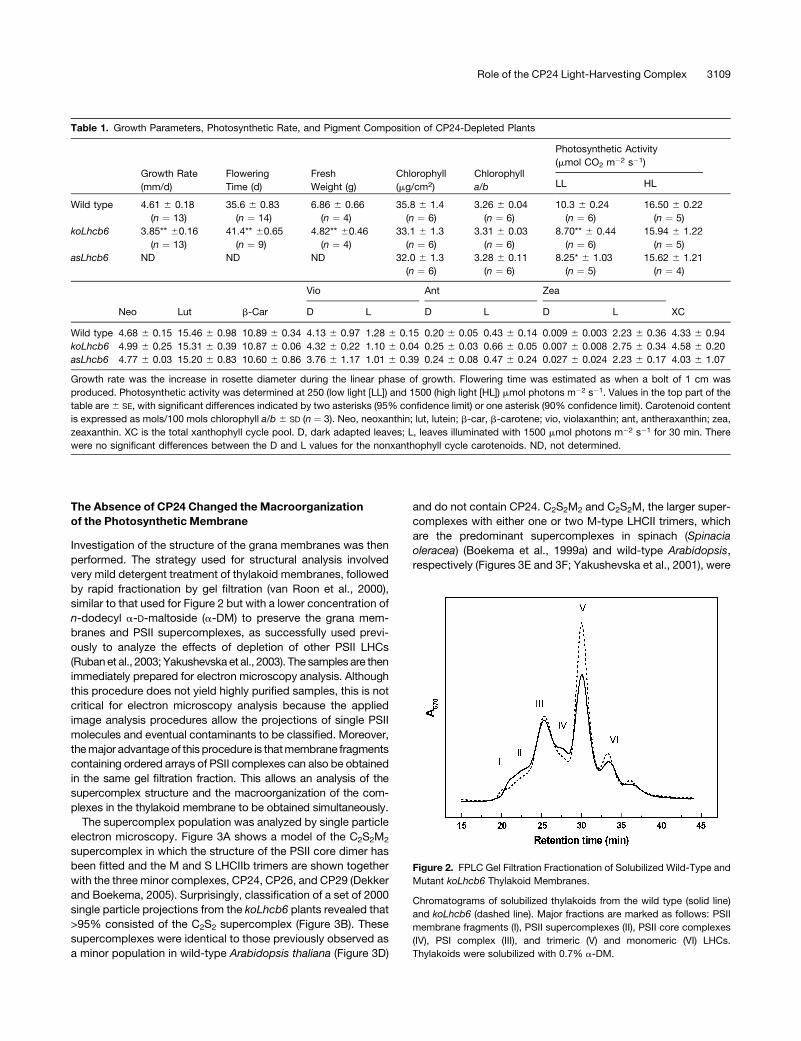
Gel filtration of detergent solubilized thylakoids confirmed the
presence of the major pigment protein complexes in the CP24-
depleted plants. Figure 2 shows the gel filtration chromatograms
of solubilized thylakoid membranes obtained from wild-type and
koLhcb6 plants, recorded at 670 nm to monitor chlorophyll
content. The elution profile of the wild-type plant (solid line)
shows a strong similarity to that obtained previously (Ruban et al.,
2006), and the compositions of these fractions were confirmed
by observation of their absorption spectra (data not shown). The
first two fractions (I and II) are attributed to grana membrane
fragments and PSII-LHCII supercomplexes, respectively. Fraction
III is assigned to monomeric PSI-200 complexes, and Fraction IV
arises from PSII core monomers. Fractions V and VI originate
from the major, trimeric LHCII and from monomeric LHC com-
plexes, respectively. The chromatogram from the koLhcb6
plants was broadly similar to the wild type, although some dif-
ferences were consistently observed in the relative contributions
of some bands (Figure 2, dashed line). There was always an
increase in the amount of chlorophyll in the LHCII trimer Fraction
V in the knockout line, indicating that some LHCII trimers
were bound less tightly to the PSII core. Thus, although the
composition may be almost the same, the CP24-depleted
plants may exhibit some changes in the macroorganization of
LHCII/PSII.
Figure 1. Content of Lhcb6 in Antisense and T-DNA Insertion Mutant Plants.
(A) Protein gel blots of wild-type and koLhcb6 plants against antibodies to a range of PSII proteins. The amount of each protein, expressed as
percentage of wild type, was determined from densitometry of between three and five replicate blots. Gels were loaded with equal chlorophyll. KO,
knockout.
(B) Protein gel blots of different plants in the population of the asLhcb6 line against Lhcb6 antibody. Asterisks indicates a wild-type plant included in
each gel.
(C) Content of Lhcb6 from protein gel blots in the range of antisense plants, expressed as densitometric intensity on an equal chlorophyll basis. Data
were obtained from the three gels shown in (B).
3108 The Plant Cell
The Absence of CP24 Changed the Macroorganization
of the Photosynthetic Membrane
Investigation of the structure of the grana membranes was then
performed. The strategy used for structural analysis involved
very mild detergent treatment of thylakoid membranes, followed
by rapid fractionation by gel filtration (van Roon et al., 2000),
similar to that used for Figure 2 but with a lower concentration of
n-dodecyl a-D-maltoside (a-DM) to preserve the grana mem-
branes and PSII supercomplexes, as successfully used previ-
ously to analyze the effects of depletion of other PSII LHCs
(Ruban et al., 2003; Yakushevska et al., 2003). The samples are then
immediately prepared for electron microscopy analysis. Although
this procedure does not yield highly purified samples, this is not
critical for electron microscopy analysis because the applied
image analysis procedures allow the projections of single PSII
molecules and eventual contaminants to be classified. Moreover,
the major advantage of this procedure is that membrane fragments
containing ordered arrays of PSII complexes can also be obtained
in the same gel filtration fraction. This allows an analysis of the
supercomplex structure and the macroorganization of the com-
plexes in the thylakoid membrane to be obtained simultaneously.
The supercomplex population was analyzed by single particle
electron microscopy. Figure 3A shows a model of the C2S2M2
supercomplex in which the structure of the PSII core dimer has
been fitted and the M and S LHCIIb trimers are shown together
with the three minor complexes, CP24, CP26, and CP29 (Dekker
and Boekema, 2005). Surprisingly, classification of a set of 2000
single particle projections from the koLhcb6 plants revealed that
>95% consisted of the C2S2 supercomplex (Figure 3B). These
supercomplexes were identical to those previously observed as
a minor population in wild-type Arabidopsis thaliana (Figure 3D)
and do not contain CP24. C2S2M2 and C2S2M, the larger super-
complexes with either one or two M-type LHCII trimers, which
are the predominant supercomplexes in spinach (Spinacia
oleracea) (Boekema et al., 1999a) and wild-type Arabidopsis,
respectively (Figures 3E and 3F; Yakushevska et al., 2001), were
Table 1. Growth Parameters, Photosynthetic Rate, and Pigment Composition of CP24-Depleted Plants
Growth Rate
(mm/d)
Flowering
Time (d)
Fresh
Weight (g)
Chlorophyll
(mg/cm2)
Chlorophyll
a/b
Photosynthetic Activity
(mmol CO2 m�2 s�1)
LL HL
Wild type 4.61 6 0.18
(n ¼ 13)
35.6 6 0.83
(n ¼ 14)
6.86 6 0.66
(n ¼ 4)
35.8 6 1.4
(n ¼ 6)
3.26 6 0.04
(n ¼ 6)
10.3 6 0.24
(n ¼ 6)
16.50 6 0.22
(n ¼ 5)
koLhcb6 3.85** 60.16
(n ¼ 13)
41.4** 60.65
(n ¼ 9)
4.82** 60.46
(n ¼ 4)
33.1 6 1.3
(n ¼ 6)
3.31 6 0.03
(n ¼ 6)
8.70** 6 0.44
(n ¼ 6)
15.94 6 1.22
(n ¼ 5)
asLhcb6 ND ND ND 32.0 6 1.3
(n ¼ 6)
3.28 6 0.11
(n ¼ 6)
8.25* 6 1.03
(n ¼ 5)
15.62 6 1.21
(n ¼ 4)
Vio Ant Zea
Neo Lut b-Car D L D L D L XC
Wild type 4.68 6 0.15 15.46 6 0.98 10.89 6 0.34 4.13 6 0.97 1.28 6 0.15 0.20 6 0.05 0.43 6 0.14 0.009 6 0.003 2.23 6 0.36 4.33 6 0.94
koLhcb6 4.99 6 0.25 15.31 6 0.39 10.87 6 0.06 4.32 6 0.22 1.10 6 0.04 0.25 6 0.03 0.66 6 0.05 0.007 6 0.008 2.75 6 0.34 4.58 6 0.20
asLhcb6 4.77 6 0.03 15.20 6 0.83 10.60 6 0.86 3.76 6 1.17 1.01 6 0.39 0.24 6 0.08 0.47 6 0.24 0.027 6 0.024 2.23 6 0.17 4.03 6 1.07
Growth rate was the increase in rosette diameter during the linear phase of growth. Flowering time was estimated as when a bolt of 1 cm was
produced. Photosynthetic activity was determined at 250 (low light [LL]) and 1500 (high light [HL]) mmol photons m�2 s�1. Values in the top part of the
table are 6 SE, with significant differences indicated by two asterisks (95% confidence limit) or one asterisk (90% confidence limit). Carotenoid content
is expressed as mols/100 mols chlorophyll a/b 6 SD (n ¼ 3). Neo, neoxanthin; lut, lutein; b-car, b-carotene; vio, violaxanthin; ant, antheraxanthin; zea,
zeaxanthin. XC is the total xanthophyll cycle pool. D, dark adapted leaves; L, leaves illuminated with 1500 mmol photons m�2 s�1 for 30 min. There
were no significant differences between the D and L values for the nonxanthophyll cycle carotenoids. ND, not determined.
Figure 2. FPLC Gel Filtration Fractionation of Solubilized Wild-Type and
Mutant koLhcb6 Thylakoid Membranes.
Chromatograms of solubilized thylakoids from the wild type (solid line)
and koLhcb6 (dashed line). Major fractions are marked as follows: PSII
membrane fragments (I), PSII supercomplexes (II), PSII core complexes
(IV), PSI complex (III), and trimeric (V) and monomeric (VI) LHCs.
Thylakoids were solubilized with 0.7% a-DM.
Role of the CP24 Light-Harvesting Complex 3109

completely absent. A very small number (;1%) of C2S2 particles
lacking one CP26 subunit were also present (Figure 3C) as well
as some sandwiched supercomplexes in side-view position (data
not shown). In Arabidopsis, almost all the C2S2 supercomplexes
have both CP26 subunits attached, in contrast with spinach
where a substantial number of complexes lack a CP26 subunit
(Boekema et al., 1999b).
Grana membrane fragments were found to be partly com-
posed of semicrystalline areas, which consisted of rows of super-
complexes in a head-to-tail arrangement along the rows (Figures
3G and 3H). This structure is completely different from the crys-
talline array found in the wild type (Figure 3I). This array has been
analyzed previously (Yakushevska et al., 2001, 2003; Ruban
et al., 2003) and was shown to consist of arrays of C2S2M2 super-
complexes. The width of the rows in the image of the CP24-
depleted membranes was 15.5 to 16 nm, which is identical to the
width of the C2S2 supercomplex. Hence, they are considered to
be composed of ordered arrays of C2S2 complexes. Areas of
noncrystalline domains were also observed, such as in the left
part of Figure 3H; these have a granularity similar to PSI domains
found earlier (Boekema et al., 2000), which are quite different
from the LHCII-only domain, which have a very smooth appear-
ance (Boekema et al., 2000). Biochemical analysis indicated that
there was no depletion of LHCII in the koLhcb6 plants (see Figure
1). The crystalline domains were generally smaller in the samples
depleted of CP24 compared with those of the wild type. Hence,
they may be more disrupted than the wild type, and it is sug-
gested that the nonbound LHCII trimers have been selectively
Figure 3. Electron Microscopy of PSII Particles and Grana Membranes.
(A) Model of the C2S2M2 supercomplex showing the fitting of the high-resolution structural model of the PSII core dimer, the CP24, CP26, and CP29
minor complexes (pale green), and the M and S LHCIIb trimers (green) (from Dekker and Boekema, 2005).
(B) Average projection map of best 500 projections of a set of ;2000 C2S2 supercomplexes from the plants lacking CP24.
(C) Average projection map of 10 projections of C2S2 supercomplexes lacking one CP26 subunit at the top left tip from the plants lacking CP24.
(D) to (F) Projection maps of C2S2, C2S2M, and C2S2M2 supercomplexes found in wild-type plants (reproduced from Yakushevska et al., 2001)
(G) and (H) Partially solubilized grana membranes from plants lacking CP24, showing ordered arrays of C2S2 supercomplexes.
(I) Partially solubilized grana membranes from wild-type plants, showing ordered arrays of C2S2M2 supercomplexes.
Thylakoids were solubilized with 0.6% a-DM. Bars ¼ 10 nm in (B) to (F) and 100 nm in (G) to (I).
3110 The Plant Cell

removed from these membranes by the detergent treatment. The
presence of a more loosely associated population of LHCII would
also explain the stronger LHCII trimer band in the gel filtration
profile of the CP24-depleted plants (Figure 2).
Spectroscopy Analysis Confirmed the Alteration in PSII
Macroorganization in the CP24-Depleted Plants
Circular dichroism (CD) spectroscopy is a sensitive noninvasive
method to study the structural changes in the PSII supramolec-
ular organization and was therefore used to further investigate
the effects of CP24 depletion. Figure 4 shows the CD spectra of
leaves (A) and isolated thylakoids (B) of the wild type and plants
lacking Lhcb6. The CD spectra of leaves and isolated thylakoid
suspensions were identical, showing that all the bands observed
in thylakoid were present in intact leaves, and there was no
distortion or artifact from the structural elements of leaf tissues.
The main CD bands of the wild-type leaf and thylakoid spectra
in the red (at 690 nm) and blue (at 510 nm) region are the psi-type
bands (Figures 4Aa and 4Bg). These large, anomalously shaped
CD bands are not explained by short-range interactions of chro-
mophores within individual pigment protein complexes but arise
from the long-range interactions between them in large, chiral
macromolecular domains (Keller and Bustamante, 1986). The
presence of the psi-type CD bands in the CD spectra of wild-type
leaves and thylakoid membranes indicates the expected exis-
tence of chiral LHCII-containing macrodomains of the pigment
protein complexes in the granal membranes, estimated to be
;200 to 400 nm (Garab et al., 1988; Finzi et al., 1989). The minor
bands in the spectrum at (�)653 nm, (þ)485 nm, and (þ)448 nm
are excitonic bands arising from short-range interactions of the
chromophores. Solubilization of the thylakoid membranes com-
pletely abolishes the psi-type CD bands (Figure 4Bh) by disruption
of long-range interactions, revealing the conservatively shaped
split signals attributed only to the short-range excitonic interac-
tions of chlorophyll molecules (Dobrikova et al., 2003).
Depletion of CP24 has a marked effect on the CD spectrum of
the leaves and thylakoid membranes (Figures 4Af and 4Bi). The
(þ)690-nm psi-type band is completely missing from the spectrum
of koLhcb6 plants. This region of the spectrum exhibits only the
split signals similar to that found in the solubilized wild-type
thylakoid membrane. The 690-nm band was also significantly re-
duced in the asLhcb6 leaves, but the extent of decrease showed
a high deviation in different plants, similar to the deviation found
in CP24 content (Figures 4Ab and 4Ae). Some asLhcb6 plants
exhibited a (þ)690-nm band with an amplitude characteristic
of the wild type (Figure 4Ab), while others were similar to the
koLhcb6 plants (Figure 4Ae). Neither the minor excitonic bands
nor the (þ)505-nm psi-type band in the Soret region changed
significantly in either type of CP24-depleted plant modified lines
compared with the wild type. Solubilization of the CP24-depleted
thylakoid membranes with detergent results in CD spectra iden-
tical with the solubilized thylakoid membrane isolated from wild-
type plants (Figure 4Bj), proving that the differences in the CD
spectra measured in situ reflect the altered macroorganization of
the pigment protein complexes of the thylakoid membrane.
The variable content of CP24 protein in asLhcb6 plants pro-
vided the possibility to establish the correlation between the
CP24 content of leaves and the macroorganization of thylakoid
membrane monitored by CD spectroscopy. The amplitude of
the (þ)690-nm psi-type band of normalized CD spectrum plotted
against the relative amount of CP24 protein is shown in Figure 5A.
At lower CP24 levels, the (þ)690-nm psi-type band shows
approximately linear correlation with CP24 content, but at higher
concentrations (;60% of the wild type), it saturates at the
amplitude found in wild-type plants. The amplitude of the 448-nm
excitonic band was independent of CP24 content.
The functional expression of the long-range organization of the
PSII antenna into the macrodomain is the establishment of con-
nectivity between PSII reaction centers. Thus, reaction centers
can share the same light-harvesting system, and energy transfer
can take place between large numbers of LHCII in the macrodo-
main. Connectivity is manifested in the sigmoidal kinetics of the
fluorescence rise that accompanies light-induced primary elec-
tron accepting plastoquinone of PSII (QA) reduction and can be
simply explained by the notion that excitation arriving at a closed
center can transfer to an open one rather than being emitted as
fluorescence (Lavergne and Trissl, 1995). The connectivity pa-
rameter J was found to be ;1.5 in wild-type plants (Table 2), in
agreement with previous results (Nedbal et al., 1999). In the
koLhcb6 plants, the J parameter decreases to ;0.9. In the anti-
sense plants, again there was a range of J values, between those
found for the wild-type and koLhcb6 plants (data not shown).
The maximum quantum efficiency, Fv/Fm, was also lower in the
plants depleted of CP24. This arose in part from the higher mini-
mum level of fuorescence (Fo) in the CP24 mutants.
Low-temperature fluorescence spectroscopy of thylakoid
membranes from wild-type and koLhcb6 plants provided further
Figure 4. CD spectroscopy of Leaves and Thylakoid Preparations from
the Wild Type and Plants Lacking CP24.
(A) Wild-type leaves (a), asLhcb6 leaves with different CP24 contents ([b]
to [e]), and koLhcb6 leaves (f).
(B) Wild-type thylakoids (g), wild-type thylakoids solubilized with 0.01%
b-DM (h), asLhcb6 thylakoids (i), and asLhcb6 thylakoids solubilized with
0.01% b-DM (j).
Role of the CP24 Light-Harvesting Complex 3111

evidence for a difference in the organization of the PSII antenna
(Figure 6). The emission spectrum for wild-type thylakoids
showed the three major bands at 685 and 695 nm (from the
PSII core) and 735 nm from PSI (Figure 6A). A weak shoulder at
;680 nm corresponds to fluorescence directly from LHCIIb. In
the CP24-depleted thylakoids, the spectrum was altered: the
680-nm emission was enhanced, while that from the PSII core
bands was reduced. The excitation spectra for the 695-nm PSII
emission were analyzed to identify the origin of the change
(Figure 6B). The difference spectrum, wild type-minus-koLhcb6,
contained several bands, all of which are diagnostic of LHCIIb
trimers (Ruban et al., 2000): 438 nm (chlorophyll a), 472 nm
(chlorophyll b), 485 (neoxanthin), 495 (lutein 1), and 510 nm (lutein
2). Thus, the changes in the emission spectrum in Figure 6A can
be explained by a decrease in efficiency of energy transfer from
LHCII to PSII and an associated increase in emission directly
from LHCII itself.
Depletion of CP24 Resulted in a Reduction in NPQ
Fluorescence induction curves recorded on leaves are shown in
Figure 7. As expected from previous results, the wild-type leaves
(Figure 7A) showed rapidly forming NPQ, the majority of which
reversed rapidly at the end of the illumination, indicating that it
was the DpH-dependent qE type of NPQ. Illumination of leaves
from plants lacking CP24 revealed a decreased tendency for
NPQ formation (Figures 7B and 7C). Calculation of NPQ values
showed that steady state levels were reached in all cases, and
the difference between wild-type and CP24-deficient plants was
in the extent of qE (Figure 7D). It was noted that the kinetics of
NPQ formation were altered in the koLhcb6 plants compared
with the wild type; in the latter, the rapid rise in NPQ was followed
by a slower phase, whereas in the mutant, the slower phase was
almost completely absent. The full light intensity profile for the
induction of qE is shown in Figure 8. The reduction in qE in the
CP24-depleted plants was observed at all light intensities, with a
maximum reduction of ;60% in the koLhcb6 plants. Differences
in NPQ were observed across a number of antisense plants, and
these could be correlated again with the content of CP24 protein;
plants with 50% of Lhcb6 were found to have wild-type levels of
NPQ (Figure 5B). Analysis of the activity of the xanthophyll cycle
revealed no difference between the wild type and the plants
deficient in CP24; for the NPQ data shown in Figure 7, the
deepoxidation state was 43.7 6 1.9 in the wild type and 47.9 6
5.1 in the CP24-depleted plants.
State Transitions Were Affected in the Absence of CP24
State transitions describe the ability of plants to adjust the relative
rates of excitation of PSII and PSI by reversible dissociation of
LHCII from PSII and its association with PSI (Haldrup et al., 2001).
It is dependent upon the reversible phosphorylation of LHCIIb. The
effect of state transitions on chlorophyll fluorescence in wild-type
Figure 5. Relationship between Lhcb6 Protein Content and Spectro-
scopic Parameters of Thylakoid Structure and Function in asLhcb6
Plants.
(A) Amplitude of CD bands at 690 nm (closed circles) and 448 nm (open
circles) obtained from the spectra recorded in Figure 4, normalized to the
amplitude of the (�)653-nm band and the relative CP24 content, as-
sessed from densitometry of protein gel blots as shown in Figure 1 and
normalized to the maximum amount (¼ 1).
(B) Value of reversible NPQ (qE) obtained as in Figure 7 and the CP24
content.
(C) Correlation between NPQ obtained as in Figure 7 and amplitude of
CD obtained as in Figure 4.
3112 The Plant Cell

plants is shown in Figure 9A. Illumination with red light, absorbed
preferentially by PSII, results in the leaves reaching State II. This is
characterized by a low Fm (FmII) because LHCII has been re-
moved from PSII and a very small decrease in fluorescence when
light absorbed by PSI (far red [FR]) is first applied (because the red
light is almost equally exciting PSII and PSI, and no further
oxidation of QA would be induced). When the illumination with
FR is continued, the fluorescence slowly rises until State I is
reached; here, the Fm (FmI) is now higher (because LHCII has
reassociated with PSII), and turning off the FR light gives an
immediate increase in fluorescence (because red light now ex-
cites PSII more than PSI, causing QA reduction). This is followed
by a fluorescence decrease as the transition to State II is induced.
In plants lacking CP24, the state transition is observed, but there
are important differences (Figure 9B). First, the extent of the state
transition when expressed by the difference between FmI and FmII
was reduced. However, it should be noted that in the plants
lacking CP24, State II was completely reached: a very small
fluorescence decrease was observed when FR light was given.
Second, the state transition was much faster in the plants lacking
CP24. Thus, when the FR light is turned off in State I, the induction
of State II occurs rapidly and is complete within 3 min. By contrast,
in the wild type, this takes at least 10 min. The transition from State
II to State I was also faster in the koLhcb6 plants.
DISCUSSION
CP24 Plays a Role in the Macrostructure of the
Thylakoid Membrane
In the grana membranes of higher plant chloroplasts, PSII
assumes a characteristic macroorganization. The minimum unit
of LHCII/PSII, the C2S2 supercomplex, can be augmented with
additional LHCs to form various larger supercomplexes, the
most predominant of which in Arabidopsis are the C2S2M and
C2S2M2 complexes (Yakushevska et al., 2001). The C2S2M2
complex contains two PSII core complexes, four LHCII trimers,
and two copies each of the minor antenna, CP24, CP26, and
CP29. In the grana membranes, these complexes can form
megacomplexes, frequently assembling into areas containing
crystalline arrays of characteristic size and spacing (Boekema
et al., 2000; Dekker and Boekema, 2005). The depletion of CP24
resulted in a profound change in PSII macrostructure: C2S2
supercomplexes replaced C2S2M1-2 complexes as the predom-
inant form of LHCII/PSII, and the crystalline arrays consisted of
rows of C2S2 complexes giving rise to a radically different ap-
pearance. Thus, in the absence of CP24, the M-LHCII trimer is
not associated with the supercomplex. It should be emphasized
that the protein gel blots indicated that the content of every other
antenna protein was maintained at wild-type levels. Apart from
the decrease in Lhcb3 (which is thought to be part of the
M-trimer), there was neither a significant loss of LHCII nor a com-
pensatory increase in the levels of other complexes. Instead, PSII
was organized into a different structural form without CP24 or the
M-LHCII trimer. Most likely some of the nonbound LHCII will
accumulate in LHCII-only domains, found frequently in spinach
(Boekema et al., 2000). However, the Arabidopsis grana
Figure 6. Low-Temperature Fluorescence Spectroscopy of Thylakoids
from Wild-Type and CP24-Depleted Plants.
(A) Fluorescence emission spectra excited at 435 nm.
(B) Fluorescence excitation spectra for emission at 695 nm.
Solid line, wild type; dashed line, koLhcb6. Spectra were normalized to
the intensity at 735 nm. In (B), the difference spectrum, wild type-minus-
koLhcb6, is displayed (dashed-dotted line).
Table 2. Fluorescence Induction Parameters Determined for Leaves
of Wild-Type and CP24-Depleted Pplants
Fo Fm Fv/Fm J
Wild type 527.4 6 20.4 2816.2 6 132.2 0.81 6 0.002 1.48 6 0.11
asLhcb6 614.9 6 12. 7 2530.8 6 125.5 0.75 6 0.005 ND
koLhcb6 683.3 6 33.6 2534.3 6 41.7 0.73 6 0.012 0.93 6 0.01
Fo, Fm, and Fv/Fm were recorded as described in Figure 7 but are the
unnormalized values. Values are 6 SD (n ¼ 6). The connectivity param-
eter J was determined from fast fluorescence induction curves as de-
scribed in Methods. ND, not determined.
Role of the CP24 Light-Harvesting Complex 3113

membranes are overall less intact after detergent treatment
than those of spinach, and likely most of the nonbound LHCII was
selectively removed. Thus, the enhanced contribution of LHCII to
the gel filtration chromatogram of the koLhcb plants arises
because of the presence of a less tightly associated M-trimer
population. Clearly, CP24 is an important component of the PSII
antenna, whose presence influences the macroorganization of
the granal membranes by providing the linker for association of
the LHCII M-trimer into the PSII complex, allowing a different
type of macroorganization.
CP24 Promotes Long-Range Chlorophyll Interactions
in the Grana
In the grana membranes, there is long-range interaction between
chlorophylls manifested by the appearance of the psi-type CD
bands (Garab et al., 1988). This order can be reproduced in lamel-
lar aggregates of LHCII (Simidjiev et al., 1997). This long-range
order can be lost by removal of divalent cations (Barzda et al.,
1994), detergent solubilization, and heat treatment (Dobrikova
et al., 2003), in each case associated with the gross disturbance
of the grana membranes, including destacking. Decrease in the
amplitude of the psi-type CD bands also occurred following
intense illumination, an effect attributed to a monomerization of
LHCII trimers (Garab et al., 2002). In plants depleted of CP24, the
psi-type CD was also lost, therefore indicating a decrease in long-
range order, and by implication, a change in grana structure. This
change would appear to be the shift from a C2S2M1-2 macro-
structure to a C2S2 macrostructure. It is important to note that
depletion of CP24 only decreases the CD band in the red region
of the spectrum, and the band at ;500 nm persists. This change
therefore differs from those arising when destacking occurs,
when all psi-type bands are lost. Hence, the main red band does
not arise from stacking per se but depends upon the lateral
Figure 8. Rapidly Reversible qE Type of NPQ Recorded at Different
Actinic Light Intensities.
Experiment performed as described in Figure 7. Wild type, circles;
asLhcb6, triangles; and koLhcb6, inverted triangles. Data are means 6
SE of between three and seven replicates using different plants.
Figure 7. Effect of CP24 Depletion on Chlorophyll Fluorescence Quenching.
(A) Chlorophyll fluorescence traces recorded for wild-type leaves.
(B) Chlorophyll fluorescence traces recorded for asLhcb6 leaves.
(C) Chlorophyll fluorescence traces recorded for koLhcb6 leaves.
(D) NPQ induction and relaxation calculated from the data in (A) to (C).
Wild type, circles; asLhcb6, triangles; and koLhcb6, inverted triangles.
Data are means 6 SE of seven (wild type and asLhcb6) or eight (koLhcb6)
replicates from different plants.
Actinic light, intensity 800 mmol photons m�2 s�1, was given for 10 min
followed by a 10-min dark period. In (A) to (C), data are normalized to an
Fm of 1.0.
3114 The Plant Cell

organization of LHCII within the stacks and is perhaps a finger-
print of a C2S2M1-2-containing macrostructure.
CP24 Depletion Has Major Functional Consequences
The LHCII antenna plays three physiological roles: it is respon-
sible for increasing the absorption cross section of PSII and
providing efficient delivery of excitation energy to PSII; by the
process of reversible phosphorylation, it controls the equilibrium
distribution of excitation between PSII and PSI; and it has been
suggested to be the site of dissipation of excess excitation in the
process of NPQ (Horton and Ruban, 2005). Our data show that
CP24 plays a key role in each of these functions.
Light Harvesting
The content of LHCII is not significantly reduced in the absence
of CP24; hence, any change in the efficiency of PSII has to result
from altered organization, not composition. Two changes in
antenna function were observed. First, there was a decrease in
Fv/Fm, the maximum quantum yield of PSII. This change arose
in part from an increase in Fo. Although this could arise from
an alteration in the trapping efficiency of PSII reaction centers,
this seems unlikely. Instead, the preferred explanation is that it
arises because of some poorly coupled LHCIIb; the displaced
M-trimers may not be able to transfer energy efficiently to PSII
reaction centers, giving rise to an elevated Fo. The detection of
enhanced emission at 680 nm in the low-temperature spectrum
of koLhcb thylakoids provides evidence for the presence of this
population of LHCIIb. The second difference between the CP24-
depleted plants and the wild type lay in the value of the connec-
tivity parameter J. This parameter is the probability of excitation
energy arriving at a closed PSII center being trapped by a neigh-
boring open one. It describes the extent to which the PSII
antenna can be described as a lake where a large number of
centers share the same antenna as opposed to a puddle where
each center only receives energy from its own antenna (Lavergne
and Trissl, 1995). A connectivity parameter of 1.5 was found in
the wild type, as routinely found for higher plants and algae
(Lazar, 1999; Nedbal et al., 1999), but it decreased to <1.0 in the
CP24-depleted plants. It has been assumed that the assembly of
LHCII/PSII complexes in the grana facilitates their connectivity.
Here, we show that that connectivity is dependent upon the
precise nature of this organization; a macrodomain containing
C2S2M1-2 complexes is needed for maximum connectivity,
whereas a C2S2 structure has much reduced connectivity.
Thus, it is suggested that the presence of the bound M-trimer
facilitates the transfer of energy between neighboring PSII com-
plexes. Because it has been postulated that the connectivity
between centers is an adaptation to photosynthesis in limiting
light, CP24 could be viewed as an attribute of the evolution of an
antenna that maximizes the efficiency of light collection. This is
consistent with the decreases in the growth rate and light-limited
photosynthetic rate in the CP24-depleted plants.
State Transitions
State transitions are a further adaptation of plants to photosyn-
thesis in limiting light (Horton et al., 1996). Since CP24 is not a
substrate for the thylakoid protein kinase, its absence would not
be predicted to affect the state transition. However, two aspects
of the transition were altered. First, the extent of the transition
was reduced in the CP24 mutant, when measured as the relative
change in Fm. Second, the rate of the transition was faster than in
the wild type. In terms of the reduced extent of the state tran-
sition, it is necessary to examine the details of the fluorescence
trace to distinguish whether this arises because of an inhibition of
the formation of State II or the inability to fully assume an initial
State I. State II is defined as that state when the rates of exci-
tation of PSII and PSI are balanced so that no further oxidation
of PSII occurs when extra PSI-absorbed light is given. By this
definition, State II was achieved in both the mutant and the wild
type. Indeed, in the mutant, State II was even more complete
since the FR-induced oxidation was virtually absent. Thus, the
difference between the mutant and wild type is in State I, not the
transition between State I and State II, which in fact proceeds
more completely and more rapidly in the CP24-depleted plants
than in the wild-type plants. The presence of a population of
LHCIIb poorly coupled to PSII could again provide an explana-
tion of this. Furthermore, it is suggested that the presence of an
M-trimer–depleted form of LHCII/PSII is indicative of State II.
Figure 9. Effect of CP24 Depletion on State Transitions.
(A) Wild-type plants.
(B) koLhcb6 plants.
Dark-adapted leaves were illuminated with PSII light to reach state II.
Periods of PSI light were given (upward arrow, on; downward arrow, off).
Light saturation pulses were given to record Fm, FmI, and FmII.
Role of the CP24 Light-Harvesting Complex 3115

Indeed, examination of thylakoid membranes in State II has
shown an increase in the proportion of C2S2M1 complexes at the
expense of C2S2M2 complexes (Kouril et al., 2005), providing
evidence that it is indeed the M-trimers that relocate from PSII to
be coupled to PSI. The difference in the kinetics of state transi-
tions in the plants lacking CP24 arises because the M-trimers are
no longer tightly associated with the PSII supercomplex and
hence are more easily able to dissociate from the granal mem-
branes (Figure 10).
NPQ
The site and mechanism of the main reversible component of
NPQ is not proven (Holt et al., 2004; Horton et al., 2005). The
transthylakoid DpH is the primary factor, whereas for maximum
NPQ in vivo, there is a requirement for violaxanthin deepoxida-
tion to zeaxanthin and the PSII antenna protein PsbS. It has
been suggested that neither zeaxanthin nor PsbS is obligatory
for quenching; rather, they could act as positive regulators
(Crouchman et al., 2006). Alternatively, it has also been proposed
that PsbS and/or zeaxanthin are the quenching agents (Li et al.,
2000; Holt et al., 2004). Irrespective of which mechanism proves
to be the correct one, NPQ requires an interaction between these
molecules and a chlorophyll binding protein. It has been sug-
gested that the LHCIIb trimers provide this site because the
amplitude of NPQ is reduced when LHCIIb is partially removed
by genetic manipulation (Andersson et al., 2003) and because it
is now proven that each LHCIIb trimer has three potential binding
sites for xanthophyll cycle carotenoids (Liu et al., 2004). How-
ever, in principal, any of the light-harvesting antenna complexes
could provide the NPQ site (Wentworth et al., 2004). Previous
work with CP26-depleted plants showed little effect on NPQ
(Andersson et al., 2001), whereas a small effect was found on
NPQ in CP29-depleted plants. Here, we have shown in higher
plants a dramatic reduction in NPQ upon removal of an antenna
complex, CP24. This could suggest that CP24 provides the site
of interaction with PsbS and zeaxanthin. The small reduction in
NPQ in the absence of CP29 (Andersson et al., 2001) could be
explained by the simultaneous partial reduction of CP24 in these
plants. It is important to note that mRNA levels of genes coding
for CP24 and CP29 during various conditions follow PsbS mRNA
levels rather than Lhcb1-3 and Lhcb5 mRNA levels, suggesting
that there is a regulatory module that coordinates the expression
of PsbS, CP24, and CP29, somewhat different to the rest of the
antenna polypeptides (Klimmek et al., 2006).
Substantial NPQ persists in the absence of CP24, and a sat-
urating light intensity of 40% of the wild-type level of NPQ can be
induced without CP24, so it seems unlikely that it provides a
unique quenching site. Instead, the data point to a decrease in
the efficiency of NPQ. Thus, the decrease in qE is due to the
reduction in the amplitude of the more slowly forming component
that is associated with the amplification of qE by violaxanthin
deepoxidation (Crouchman et al., 2006). Two explanations can
be offered: first, the decreased connectivity in the mutant plants
may result in the decreased availability of subsaturating levels of
the quencher; second, the generation of NPQ might be favored
Figure 10. Model Showing How Lack of CP24 Affects the Macrostructure of the Antenna of PSII and the Regulation of Light Harvesting.
In State I, wild-type membranes contain the typical C2S2M2 supercomplexes, which allow formation of large arrays of connected megacomplexes. In
the grana of plants lacking CP24, head-to-tail megacomplexes of C2S2 supercomplexes are present, together with peripherally associated M-LHCII
trimers. These two grana structures differ in their ability to form NPQ. In the transition to State II, there is detachment of M-trimers from PSII and their
association with PSI. In the wild type, this results in appearance of some C2S2M1 supercomplexes. In the plant lacking CP24, peripherally associated
LHCII can more readily become associated with PSI, whereas in the wild type, the availability of M-trimers is restricted by their location within the
supercomplexes. S-LHCII trimers are dark green, M-trimers are olive green, CP26 and CP29 are pale green, and CP24 is turquoise. Reaction center
cores are in gray. Also shown are the subunits of LHCI, also in green.
3116 The Plant Cell

by the optimal association of antenna complexes found in the
C2S2M1-2 macrostructure. It is unlikely that a change in connec-
tivity could exert such a large change in NPQ– for the data shown
in Table 2; the largest difference in fluorescence yield arising from
the observed difference in connectivity was <10%. Since the
quencher is not a stronger energy trap than the open reaction
center (Li et al., 2004), change in connectivity from 1.5 to 0.9
could not produce a 60% difference in NPQ. Evidence in favor of
the second explanation can be found in the observations that
whenever the macrostructure is disturbed (by depletion of any
antenna complex or by pigment deficiency), NPQ is reduced
(Horton et al., 2005), albeit in some cases only slightly. Moreover,
if quenching arises by conformational changes in an antenna
complex, as proposed recently (Pascal et al., 2005), such events
will be dependent on the local environment in the LHCII/PSII
macrostructure.
It is interesting that the extent of NPQ and the amplitude of the
psi-type CD band are strongly correlated when comparing wild-
type, koLhcb6, and asLhcb6 plants (Figure 5C), but neither is
linearly related to the content of Lhcb6 protein. Indeed, the level
of CP24 seems to be in excess of that required for normal struc-
ture and function of LHCII/PSII. Assuming that the CD signal is an
indicator of the C2S2M2-containing macrostructure, it is sug-
gested that this organization can be maintained even if every
complex does not bind CP24 (and an M-trimer). This is expected
because C2S2M1 complexes have been seen (Yakushevska et al.,
2001) and models of the ordered arrays can accommodate less
than stoichiometric M-trimers (Dekker and Boekema, 2005) and
by inference CP24. Thus, both the long-range order, as indicated
by CD, and NPQ are perhaps dependent not on CP24 per se but
the C2S2M types of macrostructure.
Two Modes of the LHCII Structure and Function
We propose that there are two fundamental states of LHCII/PSII
organization: the C2S2M2 state and the C2S2 state (Figure 10).
The former is essential for efficient light harvesting by PSII and for
efficient NPQ, and it establishes the characteristics of the state
transition. It requires CP24 and the M-LHCII trimer. In this mode,
not only is LHCIIb fully coupled to PSII, but it is in the appropriate
form to undergo transition to the quenched state. This may
include proximity to the NPQ active factor PsbS and/or the
provision of a membrane environment allowing flexibility in LHCII
structure. The association of LHCIIb trimers from adjacent super-
complexes may be essential for NPQ, forming a CP24/PsbS/
LHCII/zeaxanthin quenching locus. The second mode, C2S2, is
characterized by less-efficient PSII light harvesting and in wild-
type higher plants represents only a fraction of the PSII popula-
tion (Yakushevska et al., 2001; Kouril et al., 2005). This mode is
proposed to have a reduced efficiency of NPQ formation. In wild-
type plants, the transition to State II involves the removal of
M-trimers, resulting in mainly C2S2M1 supercomplexes; in the-
ory, some C2S2 supercomplexes could be formed, although
significant numbers of these were not found in wild-type Arabi-
dopsis (Kouril et al., 2005).
It is interesting to note the resemblance between the thylakoids
of the CP24 mutants and those of Chlamydomonas reinhardtii.
This alga lacks CP24 (Stauber et al., 2003) and contains only C2S2
complexes (Dekker and Boekema, 2005). Rapidly reversible NPQ
is smaller and slower than that found in higher plants and less
dependent on violaxanthin deepoxidation (Niyogi et al., 1997).
NPQ in Chlamydomonas also appears to depend upon an LHCII
trimer (Elrad et al., 2002), but it is controlled differently, possibly
via phosphorylation of CP29 (Turkina et al., 2006). Chlamydomo-
nas shows larger and more rapid state transitions compared with
higher plants (Delosme et al., 1996), involving dissociation of
LHCII and CP29 from PSII (Takahashi et al., 2006), probably
involving the tightly bound S-trimer (Turkina et al., 2006). It has
been concluded that the evolution of a light-harvesting antenna
that fulfils the dual function of efficient light harvesting and
effective photoprotection has been accompanied by the devel-
opment of specific antenna complexes that confer characteristic
structural arrangements in the grana membranes. In higher
plants, the evolution of CP24 was a pivotal part of this process.
METHODS
Plant Materials
Arabidopsis thaliana cv Columbia expressing an antisense construct for
the Lhcb6 gene (asLhcb6) has been described previously (Ganeteg et al.,
2004). The single antisense line presented a range of expression of the
antisense phenotype in the plant population. A T-DNA knockout mutant
(SALK_077953; N577953) with insertion into the Lhcb6 gene (At1g15820)
was obtained from the ABRC. To obtain plants homozygous for the
insertion, plants were selfed and the progeny was screened with PCR and
protein gel blotting using a monospecific Lhcb6 antibody. For amplifica-
tion of the wild-type allele of Lhcb6, PCR primers with the sequences
59-AGAAAAGCCGGGTCTTTCCCCAAAC-39 and 59-ACGCAATAAGCCA-
CATAATGCAGCC-39 were used, giving rise to a 900-bp fragment, and for
identification of the knockout allele, the primers 59-ACGCAATAAGCCA-
CATAATGCAGCC-39 and 59-TGGTTCACGTAGTGGGCCATCG-39 (LBa1)
were used, yielding an 800-bp fragment. All plants were grown for 8 to
9 weeks in Conviron climate chambers with an 8-h photoperiod with a
light intensity of 200 mmol photons m�2 s�1 and at day/night temperatures
of 22/158C. Thylakoids were prepared from fully expanded leaves as
described by Ruban et al. (2006).
CD Spectroscopy
The CD spectra were recorded between 400 and 750 nm at room tem-
perature in a J810 dichrograph (Jasco) using a band-pass of 3 nm and a
resolution of 1 nm. The chlorophyll content of the thylakoid membrane
was adjusted to 20 mg/mL and was measured in a glass cuvette with a
1-cm optical path length. Intact leaves were placed perpendicularly to the
optical path. In the case of leaves, four to nine spectra were averaged to
improve the signal-to-noise ratio.
Fluorescence Spectroscopy
Low-temperature spectroscopy was performed using an OptistatDN LN-2
cooled bath cryostat (Oxford Instruments). Samples were diluted in a
medium containing 70% glycerol (w/v), 20 mM HEPES buffer, pH 7.8,
5 mM MgCl2, and 0.33 M sorbitol. The chlorophyll concentration was
1 mM. Fluorescence emission and excitation spectra were recorded using
a SPEX FluoroLog FL3-22 spectrofluorimeter (SPEX Industries) as de-
scribed previously (Ruban et al., 2006). The excitation light was provided
from a Xenon light source. In fluorescence emission measurements,
excitation was defined at 435 nm with a 5-nm spectral bandwidth. The
Role of the CP24 Light-Harvesting Complex 3117

fluorescence spectral resolution was 1 nm. In fluorescence excitation
measurements, fluorescence was detected at 695 nm with a 5-nm
spectral bandwidth. The excitation spectral resolution was 1 nm. Spectra
were automatically corrected for the spectral distribution of the exciting
light during data acquisition.
Chlorophyll Fluorescence Induction
Fast fluorescence induction curves were recorded for detached, dark-
adapted leaves vacuum infiltrated with 100 mM DCMU. A Dual PAM100
chlorophyll fluorescence photosynthesis analyzer (Heinz Walz) was used.
Fluorescence was measured using the DUAL DB head with the instru-
ment operated in the fast kinetics mode. Actinic illumination (1000 mmol
photons m�2 s�1) was provided by two arrays of 635-nm LEDs illuminat-
ing both the adaxial and abaxial surfaces of the leaf, applied 1 ms after
turning on the 460-nm measuring beam. The measured fluorescence
induction obtained in 20-ms induction curves (2000 points) was numer-
ically fitted by the function F(t,I,sPSII,J) based on a sigmoidal fluorescence
induction model (Koblizek et al., 2001), where J is the connectivity
parameter that determines the shape of the curve, and sPSII is the func-
tional cross section of PSII.
For analysis of NPQ, the fluorescence was measured with a pulse-
modulated PAM-101 chlorophyll fluorometer (Heinz Walz). The plants
were adapted in the dark for 30 min prior to measurement. Fluorescence
quenching was induced by 10 min of actinic illumination with white light
obtained from a KL1500 lamp (Heinz Walz) at various intensities. The max-
imal fluorescence in the dark-adapted state (Fm) and during the course of
actinic illumination (Fm9) and the subsequent dark relaxation period were
determined by a 0.8-s saturating (4000 mmol photons m�2 s�1) light pulse
applied at 1- to 2-min intervals. NPQ was defined as Fm/Fm9 � 1. The
reversible component (relaxing within 10 min) was assigned to energy-
dependent NPQ (qE) and was calculated as Fm/Fm9 � Fm/Fm99, where
Fm99 is the maximal yield of fluorescence after 10 min of dark relaxation
following the actinic illumination.
State transition experiments were performed using leaf disks according
to established protocols (Haldrup et al., 2001). Preferential PSII excitation
was provided by illumination with red light at an intensity of 50 mmol
photons m�2 s�1 provided by a KL1500 lamp equipped with a 650-nm
interference filter, and excitation of PSI was achieved using far-red light
from an LED light source (Heinz Walz; 102-FR) applied for 15 min simu-
ltaneously with red light. Periods of red þ far-red and red light conditions
were used alternately in several cycles, and the Fm level in State I (FmI)
and State II (FmII) was determined at the end of each cycle by the ap-
plication of a saturating light pulse as described above.
Polypeptide Analysis
Thylakoid proteins were analyzed using immunoblotting essentially as
described by Ganeteg et al. (2001). For detailed measurements of CP24
content in the different plants, leaf disks with 8-mm diameters were
homogenized in 250 mL 23 Laemmli buffer, and the homogenates were
incubated at 908C for 5 min followed by a 20-min incubation at 378C and
then proteins were separated by 15% denaturing SDS-PAGE (Laemmli,
1970). Two microliters of samples were loaded per lane. Chemilumines-
cence was detected on Hyperfilm ECL photographic film (Amersham
Pharmacia). The developed film was digitalized and analyzed by the Im-
age Master gel documentation system (Amersham Pharmacia) equipped
with a Umax Powerlook III high-resolution scanner and 1D software
package.
Pigment Analysis
The composition of carotenoids was determined by reverse phase HPLC
(Farber et al., 1997). Leaf discs with equal areas were cut and frozen in
liquid nitrogen and then homogenized in 100% acetone. Pigments were
separated using a LiChrospher 100 RP-18 column with 5-mm particle
size (Merck) and a Dionex HPLC. Twenty microliters of acetone extract
was injected in the column, and the pigments were eluted by a gradient
from solvent A (acetonitrile, methanol, and 0.1 M Tris/NaOH, pH 8.0, in a
ratio of 87:10:3) to solvent B (4:l mixture of methanol and hexane). The
gradient from solvent A to solvent B was run from 9 to 12.5 min at a flow
rate of 2 mL/min. The amount of pigments was calculated from the
integrated area of the corresponding peak using Chromeleon software
(Dionex). The chlorophyll a and b contents on a leaf area basis were
determined from leaf extracts according to the method described by
Porra et al. (1989).
FPLC Analysis of Thylakoid Membranes
FPLC analysis was performed on detergent-solubilized, freshly prepared
thylakoid membranes as described previously (Ruban et al., 2006). Thy-
lakoids were suspended to a final chlorophyll concentration of 1.0 mg/mL
and partially solubilized by the addition of n-dodecyl a-D-maltoside to a
final concentration of 0.6 ot 0.8% (as specified in the text) and incubated
for 1 min at room temperature. Unsolubilized material was removed by
1 min of centrifugation at 16,000 g. The supernatant was then filtered
through a 0.45-mm filter and subjected to gel filtration chromatography
using an Amersham-Pharmacia Acta purifier system, including a Super-
dex 200 HR 10/30 column, following the protocol described by van Roon
et al. (2000).
Electron Microscopy and Image Analysis
Samples were negatively stained with 2% uranyl acetate on glow dis-
charged carbon-coated copper grids. Electron microscopy was per-
formed on a Philips CM120FEG electron microscope equipped with a
field emission gun operated at 120 kV. Images were recorded with a
Gatan 4000 SP 4K slow-scan CCD camera at 380,000 magnification at a
pixel size (after binning the images) of 0.375 nm at the specimen level with
GRACE software for semi-automated specimen selection and data acqui-
sition (Oostergetel et al., 1998). Single particle analysis, including multi-
reference and nonreference procedures, multivariate statistical analysis,
and classification, was performed as previously (Boekema et al., 1999a).
ACKNOWLEDGMENTS
This work was supported by grants from the Biotechnology and Bio-
logical Sciences Research Council of the United Kingdom, the Swedish
Research Council for Environment, Agricultural Sciences, and Spatial
Planning, and the INTRO2 European Union FP6 Marie Curie Research
Training Network. L.K. was a recipient of a Marie Curie Research
Fellowship.
Received July 7, 2006; revised October 16, 2006; accepted October 31,
2006; published November 17, 2006.
REFERENCES
Allen, J.F. (1992). Protein-phosphorylation in regulation of photosyn-
thesis. Biochim. Biophys. Acta 1098, 275–335.
Allen, J.F., and Forsberg, J. (2001). Molecular recognition in thylakoid
structure and function. Trends Plant Sci. 6, 317–326.
Anderson, J.M., Chow, W.S., and Park, Y.I. (1995). The grand design
of photosynthesis: Acclimation of the photosynthetic apparatus to
environmental cues. Photosynth. Res. 46, 129–139.
3118 The Plant Cell

Andersson, J., Walters, R.G., Horton, P., and Jansson, S. (2001).
Antisense inhibition of the photosynthetic antenna proteins CP29 and
CP26: Implications for the mechanism of protective energy dissipa-
tion. Plant Cell 13, 1193–1204.
Andersson, J., Wentworth, M., Walters, R.G., Howard, C.A., Ruban,
A.V., Horton, P., and Jansson, S. (2003). Absence of the Lhcb1 and
Lhcb2 proteins of the light-harvesting complex of photosystem II - Effects
on photosynthesis, grana stacking and fitness. Plant J. 35, 350–361.
Barzda, V., Mustardy, L., and Garab, G. (1994). Size dependency of
circular dichroism in macroaggregates of photosynthetic pigment-
protein complexes. Biochemistry 33, 10837–10841.
Bassi, R., and Caffarri, S. (2000). Lhc proteins and the regulation of
photosynthetic light harvesting function by xanthophylls. Photosynth.
Res. 64, 243–256.
Boekema, E.J., van Breemen, J.F.L., van Roon, H., and Dekker, J.P.
(2000). Arrangement of photosystem II supercomplexes in crystalline
macrodomains within the thylakoid membrane of green plant chloro-
plasts. J. Mol. Biol. 301, 1123–1133.
Boekema, E.J., van Roon, H., Calkoen, F., Bassi, R., and Dekker,
J.P. (1999a). Multiple types of association of photosystem II and its
light-harvesting antenna in partially solubilized photosystem II mem-
branes. Biochemistry 38, 2233–2239.
Boekema, E.J., van Roon, H., van Breemen, J.F.L., and Dekker, J.P.
(1999b). Supramolecular organization of photosystem II and its light-
harvesting antenna in partially solubilized photosystem II membranes.
Eur. J. Biochem. 266, 444–452.
Briantais, J.M., Vernotte, C., Picaud, M., and Krause, G.H. (1979). A
quantitative study of the slow decline of chlorophyll a fluorescence in
isolated chloroplasts. Biochim. Biophys. Acta 548, 128–138.
Camm, E.L., and Green, B.R. (1989). The chlorophyll a/b complex,
Cp29, is associated with the photosystem II reaction center core.
Biochim. Biophys. Acta 974, 180–184.
Crouchman, S., Ruban, A., and Horton, P. (2006). PsbS enhances
nonphotochemical fluorescence quenching in the absence of zea-
xanthin. FEBS Lett. 580, 2053–2058.
Dekker, J.P., and Boekema, E.J. (2005). Supramolecular organization
of thylakoid membrane proteins in green plants. Biochim. Biophys.
Acta 1706, 12–39.
Delosme, R., Olive, J., and Wollman, F.A. (1996). Changes in light
energy distribution upon state transitions: An in vivo photoacoustic
study of the wild type and photosynthesis mutants from Chlamydo-
monas reinhardtii. Biochim. Biophys. Acta 1273, 150–158.
Demmig-Adams, B. (1990). Carotenoids and photoprotection: A role
for the xanthophyll zeaxanthin. Biochim. Biophys. Acta 1020, 1–24.
Dobrikova, A.G., Varkonyi, Z., Krumova, S.B., Kovacs, L., Kostov,
G.K., Todinova, S.J., Busheva, M.C., Taneva, S.G., and Garab, G.
(2003). Structural rearrangements in chloroplast thylakoid membranes
revealed by differential scanning calorimetry and circular dichroism
spectroscopy. Thermo-optic effect. Biochemistry 42, 11272–11280.
Elrad, D., Niyogi, K.K., and Grossman, A.R. (2002). A major light-
harvesting polypeptide of photosystem II functions in thermal dissi-
pation. Plant Cell 14, 1801–1816.
Farber, A., Young, A.J., Ruban, A.V., Horton, P., and Jahns, P. (1997).
Dynamics of xanthophyll-cycle activity in different antenna subcom-
plexes in the photosynthetic membranes of higher plants - The
relationship between zeaxanthin conversion and nonphotochemical
fluorescence quenching. Plant Physiol. 115, 1609–1618.
Finzi, L., Bustamante, C., Garab, G., and Juang, C.B. (1989). Direct
observation of large chiral domains in chloroplast thylakoid mem-
branes by differential polarization microscopy. Proc. Natl. Acad. Sci.
USA 86, 8748–8752.
Ganeteg, U., Kulheim, C., Andersson, J., and Jansson, S. (2004). Is
each light-harvesting complex protein important for plant fitness?
Plant Physiol. 134, 502–509.
Ganetag, U., Strand, A., Gustafsson, P., and Jansson, S. (2001). The
properties of the chlorophyll a/b-binding proteins Lhca2 and Lhca3
studied in vivo using antisense inhibition. Plant Physiol. 127, 150–158.
Garab, G., Cseh, Z., Kovacs, L., Rajagopal, S., Varkonyi, Z.,
Wentworth, M., Mustardy, L., Der, A., Ruban, A.V., Papp, E.,
Holzenburg, A., and Horton, P. (2002). Light-induced trimer to mono-
mer transition in the main light-harvesting antenna complex of plants:
Thermo-optic mechanism. Biochemistry 41, 15121–15129.
Garab, G., Wells, S., Finzi, L., and Bustamante, C. (1988). Helically
organized macroaggregates of pigment-protein complexes in chloro-
plasts: Evidence from circular intensity differential scattering.
Biochemistry 27, 5839–5843.
Green, B.R., and Durnford, D.G. (1996). The chlorophyll-carotenoid
proteins of oxygenic photosynthesis. Annu. Rev. Plant Physiol. Plant
Mol. Biol. 47, 685–714.
Haldrup, A., Jensen, P.E., Lunde, C., and Scheller, H.V. (2001).
Balance of power: A view of the mechanism of photosynthetic state
transitions. Trends Plant Sci. 6, 301–305.
Holt, N.E., Fleming, G.R., and Niyogi, K.K. (2004). Toward an under-
standing of the mechanism of nonphotochemical quenching in green
plants. Biochemistry 43, 8281–8289.
Horton, P., and Ruban, A. (2005). Molecular design of the photosystem
II light-harvesting antenna: Photosynthesis and photoprotection.
J. Exp. Bot. 56, 365–373.
Horton, P., Ruban, A.V., and Walters, R.G. (1996). Regulation of light
harvesting in green plants. Annu. Rev. Plant Physiol. Plant Mol. Biol.
47, 655–684.
Horton, P., Wentworth, M., and Ruban, A. (2005). Control of the light
harvesting function of chloroplast membranes: The LHCII-aggregation
model for non-photochemical quenching. FEBS Lett. 579, 4201–4206.
Jansson, S. (1994). The light-harvesting chlorophyll a/b binding pro-
teins. Biochim. Biophys. Acta 1184, 1–19.
Keller, D., and Bustamante, C. (1986). Theory of the interaction of light
with large inhomogeneous molecular aggregates. 2. Psi-type circular
dichroism. J. Chem. Phys. 84, 2972–2980.
Klimmek, F., Sjodin, A., Noutsos, C., Leister, D., and Jansson, S.
(2006). Abundantly and rarely expressed Lhc protein genes exhibit
distinct regulation patterns in plants. Plant Physiol. 140, 793–804.
Koblizek, M., Kaftan, D., and Nedbal, L. (2001). On the relationship
between the non-photochemical quenching of the chlorophyll fluo-
rescence and the photosystem II light harvesting efficiency. A repet-
itive flash fluorescence induction study. Photosynth. Res. 68,
141–152.
Kouril, R., Zygadlo, A., Arteni, A.A., de Wit, C.D., Dekker, J.P.,
Jensen, P.E., Scheller, H.V., and Boekema, E.J. (2005). Structural
characterization of a complex of photosystem I and light-harvesting
complex II of Arabidopsis thaliana. Biochemistry 44, 10935–10940.
Laemmli, U.K. (1970). Cleavage of structural proteins during the as-
sembly of the head of bacteriophage T4. Nature 227, 680–685.
Lavergne, J., and Trissl, H.W. (1995). Theory of fluorescence induction
in photosystem II: Derivation of analytical rxpressions in a model
including exciton-radical-pair equilibrium and restricted energy trans-
fer between photosynthetic units. Biophys. J. 68, 2474–2492.
Lazar, D. (1999). Chlorophyll a fluorescence induction. Biochim.
Biophys. Acta 1412, 1–28.
Li, X.P., Bjorkman, O., Shih, C., Grossman, A.R., Rosenquist, M.,
Jansson, S., and Niyogi, K.K. (2000). A pigment-binding protein
essential for regulation of photosynthetic light harvesting. Nature 403,
391–395.
Li, X.P., Gilmore, A.M., Caffarri, S., Bassi, R., Golan, T., Kramer, D.,
and Niyogi, K.K. (2004). Regulation of photosynthetic light harvesting
Role of the CP24 Light-Harvesting Complex 3119

involves intrathylakoid lumen pH sensing by the PsbS protein. J. Biol.
Chem. 279, 22866–22874.
Liu, Z., Yan, H., Wang, K., Kuang, T., Zhang, J., Gui, L., An, X., and
Chang, W. (2004). Crystal structure of spinach major light-harvesting
complex at 2.72 A resolution. Nature 428, 287–292.
Montane, M.H., Tardy, F., Kloppstech, K., and Havaux, M. (1998).
Differential control of xanthophylls and light-induced stress proteins,
as opposed to light-harvesting chlorophyll a/b proteins, during pho-
tosynthetic acclimation of barley leaves to light irradiance. Plant
Physiol. 118, 227–235.
Nedbal, L., Trtilek, M., and Kaftan, D. (1999). Flash fluorescence
induction: A novel method to study regulation of photosystem II. J.
Photochem. Photobiol. B 48, 154–157.
Niyogi, K.K., Bjorkman, O., and Grossman, A.R. (1997). Chlamydo-
monas xanthophyll cycle mutants identified by video imaging of
chlorophyll fluorescence quenching. Plant Cell 9, 1369–1380.
Oostergetel, G.T., Keegstra, W., and Brisson, A. (1998). Automation
of specimen selection and data acquisition for protein electron
crystallography. Ultramicroscopy 74, 47–59.
Pascal, A.A., Liu, Z., Broess, K., van Oort, B., van Amerongen, H.,
Wang, C., Horton, P., Robert, B., Chang, W., and Ruban, A. (2005).
Molecular basis of photoprotection and control of photosynthetic
light-harvesting. Nature 436, 134–137.
Peter, G.F., and Thornber, J.P. (1991). Biochemical composition and
organization of higher-plant photosystem II light-harvesting pigment
proteins. J. Biol. Chem. 266, 16745–16754.
Porra, R.J., Thompson, W.A., and Kriedemann, P.E. (1989). Determi-
nation of accurate extinction coefficients and simultaneous equations
for assaying chlorophyll a and chlorophyll b extracted with 4 different
solvents - Verification of the concentration of chlorophyll standards by
atomic absorption spectroscopy. Biochim. Biophys. Acta 975, 384–394.
Ruban, A.V., Pascal, A., and Robert, B. (2000). Xanthophylls of the
major photosynthetic light-harvesting complex of plants: Identifica-
tion, conformation and dynamics. FEBS Lett. 477, 181–185.
Ruban, A.V., Solovieva, S., Lee, P.J., Ilioaia, C., Wentworth, M.,
Ganeteg, U., Klimmek, F., Chow, W.S., Anderson, J.M., Jansson,
S., and Horton, P. (2006). Plasticity in the composition of the light
harvesting antenna of higher plants preserves structural integrity and
biological function. J. Biol. Chem. 281, 14981–14990.
Ruban, A.V., Wentworth, M., Yakushevska, A.E., Andersson, J., Lee,
P.J., Keegstra, W., Dekker, J.P., Boekema, E.J., Jansson, S., and
Horton, P. (2003). Plants lacking the main light-harvesting complex
retain photosystem II macro-organization. Nature 421, 648–652.
Simidjiev, I., Barzda, V., Mustardy, L., and Garab, G. (1997). Isolation
of lamellar aggregates of the light-harvesting chlorophyll a/b protein
complex of photosystem II with long-range chiral order and structural
flexibility. Anal. Biochem. 250, 169–175.
Stauber, E.J., Fink, A., Markert, C., Kruse, O., Johanningmeier, U.,
and Hippler, M. (2003). Proteomics of Chlamydomonas reinhardtii
light-harvesting proteins. Eukaryot. Cell 2, 978–994.
Takahashi, H., Iwai, M., Takahashi, Y., and Minagawa, J. (2006).
Identification of the mobile light-harvesting complex II polypeptides
for state transitions in Chlamydomonas reinhardtii. Proc. Natl. Acad.
Sci. USA 103, 477–482.
Turkina, M.V., Kargul, J., Blanco-Rivero, A., Vilrejo, A., Barber, J.,
and Vener, A.V. (2006). Environmentally modulated phosphopro-
teome of photosynthetic membranes in the green alga Chlamydomo-
nas reinhardtii. Mol. Cell. Proteomics 5, 1412–1425.
van Roon, H., van Breemen, J.F.L., de Weerd, F.L., Dekker, J.P., and
Boekema, E.J. (2000). Solubilization of green plant thylakoid mem-
branes with n-dodecyl-alpha,D-maltoside. Implications for the struc-
tural organization of the Photosystem II, Photosystem I, ATP synthase
and cytochrome b(6)f complexes. Photosynth. Res. 64, 155–166.
Walters, R.G., Ruban, A.V., and Horton, P. (1996). Identification of
proton-active residues in a higher plant light-harvesting complex.
Proc. Natl. Acad. Sci. USA 93, 14204–14209.
Wentworth, M., Ruban, A.V., and Horton, P. (2004). The functional
significance of the monomeric and trimeric states of the photosystem
II light harvesting complexes. Biochemistry 43, 501–509.
Yakushevska, A.E., Jensen, P.E., Keegstra, W., van Roon, H.,
Scheller, H.V., Boekema, E.J., and Dekker, J.P. (2001). Supermolec-
ular organization of photosystem II and its associated light-harvesting
antenna in Arabidopsis thaliana. Eur. J. Biochem. 268, 6020–6028.
Yakushevska, A.E., Keegstra, W., Boekema, E.J., Dekker, J.P.,
Andersson, J., Jansson, S., Ruban, A.V., and Horton, P. (2003).
The structure of photosystem II in Arabidopsis: Localization of the
CP26 and CP29 antenna complexes. Biochemistry 42, 608–613.
3120 The Plant Cell

DOI 10.1105/tpc.106.045641; originally published online November 17, 2006; 2006;18;3106-3120Plant Cell
Boekema, Stefan Jansson and Peter HortonLászló Kovács, Jakob Damkjær, Sami Kereïche, Cristian Ilioaia, Alexander V. Ruban, Egbert J.
Membranes of Higher Plant ChloroplastsLack of the Light-Harvesting Complex CP24 Affects the Structure and Function of the Grana
This information is current as of March 16, 2018
References /content/18/11/3106.full.html#ref-list-1
This article cites 60 articles, 16 of which can be accessed free at:
Permissions https://www.copyright.com/ccc/openurl.do?sid=pd_hw1532298X&issn=1532298X&WT.mc_id=pd_hw1532298X
eTOCs http://www.plantcell.org/cgi/alerts/ctmain
Sign up for eTOCs at:
CiteTrack Alerts http://www.plantcell.org/cgi/alerts/ctmain
Sign up for CiteTrack Alerts at:
Subscription Information http://www.aspb.org/publications/subscriptions.cfm
is available at:Plant Physiology and The Plant CellSubscription Information for
ADVANCING THE SCIENCE OF PLANT BIOLOGY © American Society of Plant Biologists