lack of correlation with clinical presentation and serology
TRANSCRIPT
CLINICAL AND DIAGNOSTIC LABORATORY IMMUNOLOGY, JUly 1994, p. 373-378 Vol. 1, No. 41071-412X/94/$04.00+ 0Copyright ©D 1994, American Society for Microbiology
Sustained Cellular Immune Responses to Borrelia burgdorferi:Lack of Correlation with Clinical Presentation and SerologyHAROLD W. HOROWITZ,'* CHARLES S. PAVIA,' SUSAN BITTKER,' GILDA FORSETER,1DENISE COOPER,' ROBERT B. NADELMAN,' DANIEL BYRNE,1 RUSSELL C. JOHNSON,2
AND GARY P. WORMSER'Division of Infectious Diseases, Department of Medicine, Westchester County Medical Center, New York
Medical College, Valhalla, New York 10595,1 and Department of Microbiology,University of Minnesota, Minneapolis, Minnesota 554552
Received 3 December 1993/Returned for modification 7 March 1994/Accepted 7 April 1994
Fifty-one patients with erythema migrans were followed up prospectively with serial clinical evaluations,serologic determinations for antiborrelial antibodies, and lymphocyte stimulation responses to Borreliaburgdorferi antigens to determine (i) the factors associated with sustained cellular immune responses and (ii)whether lymphocyte stimulation is a good indicator of prior exposure to B. burgdorferi in patients treated earlyafter erythema migrans. Positive lymphocyte stimulation responses (>2 standard deviations above normalcontrol values) were found in 15 (29%o) of 51 patients 3 months after treatment for erythema migrans and in8 (18%) of 44 patients 1 year posttreatment. Heightened lymphocyte responses were not associated with thenumber or duration of erythema migrans lesions prior to treatment, the mean size of the largest erythemamigrans lesion, or the number of symptoms at the time of presentation. The development of a Jarisch-Herxheimer reaction, choice of antibiotic, and clinical outcome also were not associated with a positivelymphoproliferation assay result. Changes in the lymphocyte stimulation indices between the two time pointsassessed (3 months and 1 year posttreatment) also did not correlate with the above variables. When serologicresults and lymphoproliferative responses were evaluated as categorical or continuous variables, there were nocorrelations between values. One year after treatment for early Lyme disease, lymphocyte reactivity is not agood indicator of prior infection with B. burgdorferi.
The diagnosis of early Lyme borreliosis is based primarilyupon clinical parameters, the most reliable of which is thecharacteristic erythema migrans rash (18). Serologic responsesare often delayed and may be abrogated with antibiotic therapy(1, 19). In later stages, diagnosis is usually based upon acompatible clinical picture with a positive test for antibodies toBorrelia burgdorferi, the etiologic agent of Lyme disease. Anti-body assays, however, suffer from problems with sensitivity,specificity, and reproducibility (9, 19). Because of these con-cerns, investigators have turned to the lymphocyte prolifera-tion assay in response to B. burgdorferi antigen preparations inan attempt to demonstrate infection (2). Heightened in vitrolymphocyte proliferative responses to B. burgdorferi have beenobserved by using lymphocytes from sites of active B. burgdor-feri infection. For instance, cerebrospinal fluid lymphocytesfrom individuals with neurologic Lyme disease (10, 13) andsynovial fluid mononuclear cells from patients with Lymearthritis may have elevated lymphoproliferative responses topreparations of B. burgdorferi antigen (12, 16, 24). Patients withreactive arthritis with positive Lyme serology may also haveelevated peripheral blood mononuclear cell (PBMC) reactivityto B. burgdorferi antigen (22). Dattwyler et al. (4) reported thatpatients with persistent illness manifested by fatigue andheadache, neurologic or cognitive abnormalities, and jointinvolvement may have a heightened PBMC stimulation re-sponse to B. burgdorferi in the absence of detectable antibodiesto B. burgdorferi in serum. However, in studying the responseof PBMCs to B. burgdorferi antigen, several other groups have
* Corresponding author. Mailing address: Division of InfectiousDiseases, Westchester County Medical Center, Macy 209 SE, Valhalla,NY 10595. Phone: (914) 285-8865. Fax: (914) 285-7289.
noted a lack of both sensitivity and specificity for this assay (5,21, 25).
Little is known regarding the natural history of the lympho-proliferative response in patients with Lyme disease treatedearly in infection and what factors are associated with height-ened cellular immune responses over the long term. Weprospectively evaluated lymphoproliferative responses at 3months and 1 year after the completion of antibiotic therapy ina group of 51 patients who presented with erythema migrans.Results of this assay were correlated with clinical features andantibody responses.
(This study was presented in part at the Fourth InternationalConference on Lyme Borreliosis, Stockholm, Sweden, June1990.)
MATERIALS AND METHODS
Subjects. Patients were randomly chosen from among the 80individuals who presented to the Lyme Disease DiagnosticCenter of the Westchester County Medical Center between 15June and 21 August 1989 with clinically documented erythemamigrans and who had agreed to participate as patients in amulticenter treatment trial. Participants in the treatment trialwere treated orally with either cefuroxime axetil at 500 mgtwice daily for 20 days or doxycycline at 100 mg three timesdaily for 20 days (11). Individuals were seen at time zero(initiation of the study); between days 8 and 12 while onantibiotic therapy; between days 1 and 5 after the completionof antibiotic therapy; and then at approximately 1 month, 3months, and 1 year after the completion of antibiotic therapy.Telephone interviews concerning patient symptoms were per-formed at 6 and 9 months posttreatment. At each visit thepatient was carefully questioned about symptoms, and a full
373
on Novem
ber 16, 2018 by guesthttp://cvi.asm
.org/D
ownloaded from
374 HOROWITZ ET AL.
physical examination was done. Serum for serologic studieswas obtained at each visit. A complete blood count and serumchemistries were also evaluated at each visit. Lymphocytestimulation studies were performed for selected patients at the3-month and 1-year posttreatment visits.
Clinical evaluation. For the purposes of the present study,clinical outcomes were defined on the basis of the clinicalresponse at 1 month and 9 months to 1 year posttreatment.Clinical signs and symptoms were evaluated at all patient visitsand were ranked according to severity (11).
Definitions. (i) One-month posttreatment outcome. Clinicalcure was defined as the resolution of clinical signs and symp-toms of early Lyme disease by the 1- to 5-day posttreatmentvisit, with a continued asymptomatic state through the 1-monthposttreatment follow-up period. Clinical improvement wasdefined as the resolution of erythema migrans rash but incom-plete resolution of any other signs and symptoms of early Lymedisease by the 1- to 5-day posttreatment visit, with furtherimprovement or complete resolution by the 1-month posttreat-ment follow-up visit. Clinical failure was defined as no im-provement in clinical signs and symptoms of early Lymedisease by the 1- to 5-day posttreatment visit. Clinical recur-rence was defined as clinical success or improvement as notedabove, but with recurrence of erythema migrans rash and/orany other signs and symptoms of early Lyme disease by the1-month posttreatment follow-up visit.
(ii) One-year posttreatment outcome. Clinical success wasdefined as the absence of signs and symptoms of late Lymedisease between 9 months and 1 year posttreatment. Residualsymptoms were defined as the presence of signs or symptomsconsistent with late Lyme disease, but without objective evi-dence of active disease between 9 months and 1 year posttreat-ment and without serologic reactivity to B. burgdorferi. Clinicalfailure was defined as the presence of signs or symptoms of lateLyme disease, including serologic confirmation of B. burgdor-feri infection, between 9 months and 1 year posttreatment.
Lymphocyte proliferation assays. Control samples wereobtained from 20 normal health care workers or hospitalpersonnel who had no prior history of erythema migrans or aknown diagnosis of Lyme disease and were seronegative forantibodies to B. burgdorferi by an enzyme-linked immuno-sorbent assay (ELISA; Clinical Sciences, Whippany, N.J.).PBMCs from patients or controls were isolated by Ficoll-Hypaque (Sigma, St. Louis, Mo.) density gradient centrifuga-tion. The cell layer at the gradient interface was removed andwashed once in RPMI 1640 and resuspended in CME culturemedium (RPMI 1640 supplemented with 10% fetal calf se-rum-1% glutamine-2 x 10⁻⁵ M mercaptoethanol-50 U ofpenicillin per ml-50 ,ug of streptomycin per ml-50 ,ug ofamphotericin B per ml) prior to counting by trypan bluestaining to determine cell viability. The cells were adjusted toa concentration of 5 x 10⁶ cells per ml with CME medium, and5 x 105 cells in a volume of 0.1 ml were dispensed intoflat-bottom wells of 96-well microtiter culture plates (Costar,Cambridge, Mass.). To these wells was added 0.1 ml of eitherphytohemagglutinin (PHA) at a concentration of 5 ,Ig per well,whole B. burgdorferi (2 x 10⁶ organisms per well), sonicatedsoluble preparations of B. burgdorferi (3 to 10 ,ug of protein perwell), or sonicated soluble Treponema pallidum (1 to 3 ,ug ofprotein per well). Assays were performed in triplicate. The B.burgdorferi strain used in these studies was RF1, a high-passagestrain isolated in Westchester County, N.Y. Control culturesreceived 0.1 ml of the RPMI 1640 supplemented medium only.Cell cultures were incubated at 37°C in a humidified 5%carbon dioxide-95% air atmosphere. After 3 days the cellcultures were each labelled with 0.5 jCi of [3H]thymidine
(specific activity, 5 Ci/mmol; New England Nuclear, Boston,Mass.) for an additional 8 h prior to harvesting at 4 days (14).The amount of label incorporated was assayed by collecting thecells with a multiple semiautomated sample harvester (BellcoGlass, Vineland, N.J.) and was counted in a liquid scintillationcounter. Data were recorded for each culture as counts perminute, and the triplicate values were averaged. A stimulationindex (SI) was calculated by using the formula SI = meancounts per minute in the presence of antigen/mean counts perminute in the absence of antigen.A lymphocyte proliferation result was interpreted as positive
if it was .8, which was 2 standard deviations (SDs) above themean of the controls.
Antibodies to B. burgdorferi by ELISA. The immunoglobulinG (IgG) and IgM responses to B. burgdorferi were determinedby an indirect ELISA with secondary antibody to these twoisotypes as described previously (6). The antigen preparationconsisted of sonicated B. burgdorferi passed through a 0.22-jim-pore-size filter. Mean values for the test were obtained by assayof 200 serum samples from blood donors from an area whereLyme disease is not endemic. A patient's serum sample wasconsidered positive if the optical density value was >0.480 forIgM or >0.657 for IgG, which were .2 SDs above the mean ofthe normal controls.
Statistical analysis. Categorical variables were analyzed by achi-square test with a Yates continuity correction or Fisher'sexact test when appropriate (17). The association betweenELISA reactivity and lymphocyte stimulation positivity wasmeasured by the Pearson product correlation coefficient. Con-tinuous variables that were normally distributed were analyzedby Student's t test, while others were analyzed by the Mann-Whitney U test. ELISA optical density values were used forIgG and IgM continuous-variable analysis. All tests were twotailed. Statistical significance was defined as P s 0.05; forcorrelation coefficients, however, it was defined as P c 0.01.
RESULTS
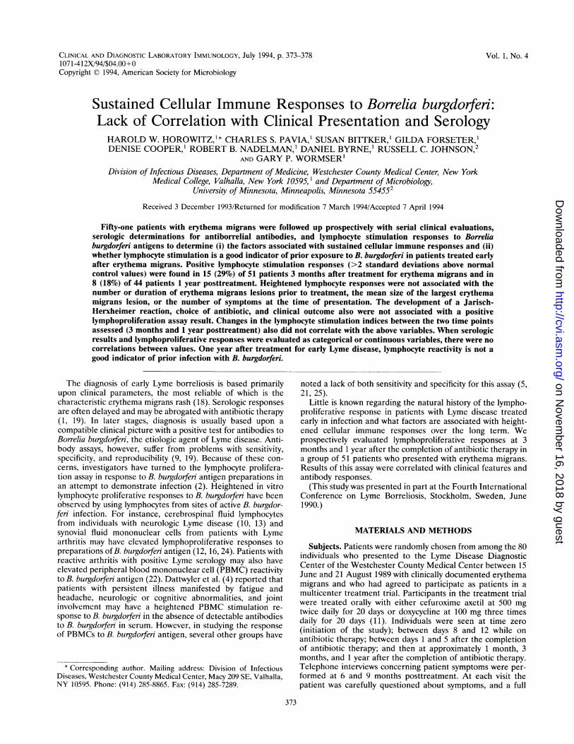
Lymphoproliferative responses. The lymphoproliferative re-sponses to whole killed B. burgdorferi were greater than theresponses to sonicated B. burgdorferi (data not shown). Forpurposes of subsequent analyses, the data derived from thelymphoproliferative responses to whole killed B. burgdorferiwere used. Positive lymphocyte stimulation responses (SI, .8)to killed B. burgdorfen were found in 15 (29%) of 51 patientsat 3 months posttreatment and 8 (18%) of 44 evaluablepatients at 1 year posttreatment (P = 0.301, comparing theproportion of patients with positive lymphocyte stimulationresponses at 3 months and 1 year) (Fig. 1). Samples were notavailable for seven patients at the 1-year visit. One (5%) of 20control patients had an SI of .8. The PHA responses frompatients' PBMCs were significantly greater at 3 months than at1 year posttreatment and greater than control PHA responses(P < 0.001). Control PHA responses were similar to the PHAresponses from patients' PBMCs at 1 year (Fig. 1). There wasno direct correlation between the PHA response and thelymphoproliferative response to B. burgdorfei at either timepoint (r = 0.0953 and P = 0.429 at 3 months, and r = 0.0182and P = 0.907 at 1 year). The lymphocyte stimulation re-sponses to killed T. pallidum at both 3 months and 1 yearposttreatment and control values were less than three timesthe background response except in one patient, for whom thevalue was four times the background response (Fig. 1).
Serologic responses. The serologic responses are noted inFig. 2. Eighty-four percent of patients had a positive serologicresponse by 5 days posttreatment.
CLIN. DIAGN. LAB. IMMUNOL.
on Novem
ber 16, 2018 by guesthttp://cvi.asm
.org/D
ownloaded from

CELLULAR IMMUNE RESPONSES TO B. BURGDORFERI 375
X . 3 MONTHS (N=51)- 1 YEAR (N=44)
30 . ............... 300
CD
020........ 200
a10 100S L ii ir + i i ii Io~~~~~~~~~~~~!TP RF1 PHA
FIG. 1. Lymphoproliferative responses. PBMC SIs in response toT. pallidum antigen (TP), B. burgdorfeni antigen (RF1), and PHA in 20healthy controls and Lyme disease patients at 3 months and 1 yearposttreatment for erythema migrans. Shaded area of RF1 indicatesvalues below 2 SDs of control values; horizontal bars indicate the meanvalue, and vertical bars indicate the 95% confidence interval aroundthe mean.
Clinical associations. Of the 51 patients on whom lympho-proliferative assays were performed at 3 months, 45 (88%)were improved or cured by 1 month posttreatment, 2 (4%)were treatment failures, and 4 (8%) had recurrent disease. Thesix patients who were treatment failures or who had recurrentdisease all experienced myalgias or arthralgias and several hada stiff neck, malaise, or headaches. Both patients who weretreatment failures were retreated with antibiotics, and one ofthe four patients with recurrences was retreated. In all in-stances, the erythema migrans lesions had resolved by 1 monthand no objective findings were noted. By 1 year, four of thesesix patients still had residual symptoms, one was cured, andone could not be evaluated because of a concurrent illness.
1
+-
Days from Initial Visit
FIG. 2. Serologic responses of patients during 12-month follow-upas percentages of patients with positive IgM (dotted line) and IgG(solid line) ELISA values at each time point. Time zero is the time ofpresentation with erythema migrans, time 10 days is the 8- to 12-dayduring-treatment visit, and time 25 days is the 1- to 5-day posttreat-ment visit. Times 50, 110, and 385 represent the 1-, 3-, and 12-monthposttreatment visits, respectively.
Five of these six patients had negative lymphoproliferativeassay results at 3 months and all six patients had negative assayresults at 1 year posttreatment (Table 1). Fourteen (31%) ofthe 45 patients who were cured or improved at 1 monthposttreatment had heightened lymphocyte SIs at 3 monthsposttreatment (Table 1).For 44 (86%) patients, repeat lymphocyte stimulation assays
were performed at 1 year posttreatment. Of these, 26 (59%)were clinical successes, 15 (34%) had residual symptoms, 1(2%) was considered a clinical failure, and 2 (5%) wereunevaluable because of concurrent illnesses. The majority ofthe patients who had residual symptoms but who were notconsidered to be treatment successes at the 1-year follow-upvisit had symptoms of arthralgias, myalgias, fatigue, or stiffneck between the 9-month posttreatment telephone interviewand the 1-year visit. These symptoms were generally mild andwere not associated with objective findings at 1 year posttreat-ment. Three (19%) of the 16 patients with residual symptoms(including the 1 patient who was a clinical failure) at the 1-yearfollow-up visit had positive lymphoproliferative assay results atthat time, while 5 (19%) of 26 patients assessed as successeshad positive lymphoproliferative assay results (P = 0.649)(Table 1). The two unevaluable patients had negative lympho-proliferative responses. The single patient considered to be aclinical failure at 1 year posttreatment had headaches, fatigue,myalgias, and paresthesias. The patient improved after intra-venous therapy with ceftriaxone. This patient had lymphocyteSI values of 4.1 at 3 months and 8.0 at 1 year after treatment,as well as positive serology for B. burgdorferi.
Several possible markers for disease severity or early dissem-ination of infection were evaluated to determine whether theywere associated with an enhanced lymphocyte stimulationresponse. The duration of erythema migrans prior to treat-ment, the number of erythema migrans lesions, the mean sizeof the largest erythema migrans lesion, the number of symp-toms at the time of presentation, and the presence of post-treatment symptoms were not associated with a heightenedlymphoproliferative response at either 3 or 12 months post-treatment (Table 1). In addition, the presence of neithermusculoskeletal nor constitutional symptoms at the time ofpresentation was associated with a positive lymphocyte stimu-lation assay result (data not shown). There was no significantdifference in the proportion of patients with a positive lympho-proliferative assay result at either 3 months or 1 year posttreat-ment between patients who received cefuroxime axetil andthose who received doxycycline, although a trend was noted forgreater responses in patients who had been treated withcefuroxime axetil (Table 1).
Eleven patients developed a Jarisch-Herxheimer reactionupon the initiation of antibiotic therapy. Four (37%) hadpositive lymphocyte SIs at 3 months posttreatment, while onlyone (10%) of the 10 tested had a positive assay result at 1 yearposttreatment (Table 1). Patients who developed a Jarisch-Herxheimer reaction were not more apt to develop a positivelymphocyte stimulation response at either 3 months or 1 yearposttreatment than those who did not (P = 0.843 at 3 months,P = 0.767 at 1 year, comparing the proportion of patients witha positive lymphocyte stimulation assay result among patientswith and without a Jarisch-Herxheimer reaction at each timepoint).The leukocyte count in peripheral blood at the time of
presentation and serum creatine phosphokinase levels at thistime, likewise, were not related to a heightened lymphocyteresponse (Table 1).
Serologic associations. The lymphocyte stimulation re-sponses at 3 months and 1 year posttreatment were not
VOL. 1, 1994
on Novem
ber 16, 2018 by guesthttp://cvi.asm
.org/D
ownloaded from

376 HOROWITZ ET AL.
TABLE 1. Association of lymphocyte proliferation assay with clinical parameters
Lymphoproliferative response at 3 mo Lymphoproliferative response at 12 moClinical or laboratory
parameter Positive Negative P value Positive Negative P value(n = 15) (n =36) (n =8) (ni = 36)
Age (yr) 44 ± 3.0a 45.3 ± 2.5 0.737 39.5 ± 3.5 45.3 ± 2.3 0.260
Duration of EMb at presentation (h) 124.8 ± 27.0 99.9 ± 16.3 0.391 129 + 36.5 99.9 ± 16.7 0.317
No. of EM lesions at presentation 1.7 ± 0.4 1.8 ± 0.4 0.351 1.1 ± 0.1 1.78 ± 0.4 0.572
Area of largest EM lesion (mm) 221.4 ± 44.5 136.6 ± 19.6 0.130 209 ± 76.3 161.2 + 21.3 0.948
Total no. of symptoms at presentation 4.2 ± 0.9 4.2 ± 0.5 0.959 3.5 ± 1.3 4.4 ± 0.3 0.359
No. of new symptoms postpresentation 1.3 + 0.4 2.2 ± 0.4 0.178 1.1 + 0.3 2.1 ± 0.3 0.310
Antibiotic (no. of patients)Doxycycline 7 19 0.691c 2 21 0.088cCefuroxime axetil 8 17 6 15
Outcome (no. of patients) at:1 modCured or improved 14 31 0.657e 7 25 0.413eNoncured 1 5 1 11
1 yearfSuccess 11 25 >0.9509 5 21 >0.9509Nonsuccess 4 9 3 13
a Values are means ± standard errors of the means.b EM, erythema migrans.c Chi-square test comparing antibiotics.d At 1 month, cured is cured and improved and noncured is treatment failure and recurrence (refer to text for definitions).eThe P value is based upon Fisher's exact test comparing cured or improved with noncured patients.f At 1 year, success is treatment success and nonsuccess is the presence of residual symptoms and treatment failures (refer to text for definitions).g The P value is based upon Fisher's exact test; data for two unevaluable patients were omitted.
correlated with ELISA IgG or IgM results at any of the six timepoints when serologic assays were performed, using categoricalvalues for lymphocyte and serologic studies. Correlationsbetween serologic and lymphoproliferative responses at thetwo time points, when they were done simultaneously, aredemonstrated in Fig. 3.
Eight patients (16%) had ELISA IgG and IgM values thatwere considered negative at all time points. Two (25%) ofthese patients had a positive lymphocyte stimulation assayresult at 3 months posttreatment, while at 1 year posttreat-ment, none of the seven patients tested had a positive lympho-proliferative assay result.When lymphoproliferative and serologic responses were
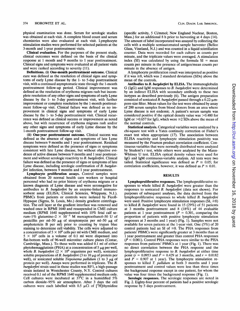
evaluated as continuous variables, no correlations were foundbetween the 3-month lymphocyte responses and any serologicresponse. Significant correlations (P c 0.01) were foundbetween the lymphocyte stimulation responses at 1 year andthe ELISA IgG values from days 8 to 12 during treatmentthrough the 1-month posttreatment visit, as well as pretreat-ment IgM ELISA values. However, a single patient with anextremely high lymphoproliferative response at 1 year and highserologic responses during the early visits appears to accountfor these statistical correlations. This patient presented withtwo erythema migrans lesions of 10 days' duration and wasconsidered a clinical cure at 1 month and 1 year posttreatment.When data for this patient were excluded from the analysis, nosignificant correlations between lymphoproliferative and sero-logic responses were found at any time point. A representativescatterplot obtained by using 1-year lymphocyte stimulationresponses and IgG optical density values obtained on days 8 to12 during treatment demonstrates the poor fit of the individual
40
30cozw
O- 2020ccwco
z10
0+LS -LS +LS -LS
IgM IgG3 MONTHS
+LS -LS +LS -LSIgM IgG
12 MONTHS
FIG. 3. Lymphoproliferative responses compared with serologicresponses at 3 and 12 months posttreatment. The number of patientswith positive serology by ELISA (solid line) and negative serology(hatched lines) among patients with positive and negative lymphopro-liferative responses (LS) at 3 months and 1 year posttreatment isindicated. Percentages are the percentages of patients with positiveserologic responses.
CLIN. DIAGN. LAB. IMMUNOL.
on Novem
ber 16, 2018 by guesthttp://cvi.asm
.org/D
ownloaded from

CELLULAR IMMUNE RESPONSES TO B. BURGDORFERI 377
2.0 than those with an initial positive ELISA IgG result to haveuz-11 persistent negative lymphoproliferative responses (P = 0.014).
D 1.5 D D /> ) DISCUSSION
co * - ** / We prospectively studied the proliferative responses ofPBMCs after antibiotic treatment of patients with early-stage(D 1.0 Lyme disease manifested by erythema migrans in order to
* * determine the factors associated with persistent heightenedresponses and to assess the usefulness of this test as an
° 0.51 ejt indicator of past exposure to B. burgdorferi. Although weco) cannot be certain that all patients who were diagnosed asI * ~ having erythema migrans actually had B. burgdorferi infection,0.0 we have confirmed in subsequent culture studies of clinical
0 5 10 15 20 25 erythema migrans lesions that our clinical impression is highlyLymphocyte Stimulation Index at 1 Year accurate on the basis of culture positivity (23). Moreover, 43
(84%) of the 51 patients in the current study had positiveFIG. 4. Representative scatterplot ELISA optical density values ELISA values for B. burgdorferi. Since we did not perform
and lymphocyte stimulation responses. A scatterplot of the ELISA LgG lymphocyte stimulation assays until 3 months after antibioticvalues at the second visit with the lymphocyte SI at 1 year after tement,ywe cannot awsa compl pcueothe lympho-treatment is shown. The circled point is the outlying patient. The t
dotted lines mark the cutoff points for the categorical variables. O.D., cyte response from the time of presentation, but can onlyoptical density. comment upon the persistence of an individual's lymphopro-
liferative responses to B. burgdorferi antigen after treatment.From our analysis, it does not appear that the lymphocyte
stimulation response is a reliable indicator of previous Lymepoints to the regression lines (Fig. 4). No R2 value for disease in patients who are treated early in the course oflymphoproliferative and serologic correlations exceeded 0.135 disease. We have demonstrated that the majority of such(data not shown). patients do not develop persistent lymphoproliferative re-
Lymphoproliferative assay variability over time. Comparing sponses. Moreover, there were no specific clinical predictorsthe lymphoproliferative responses at the 3-month and 1-year for heightened lymphocyte stimulation at 3 months or 1 yeartest dates, 27 (61%) patients had negative lymphocyte stimu- after the completion of therapy.lation responses at both time points, 9 (21%) were initially When lymphocyte stimulation and serologic values werepositive and became negative, 3 (7%) were negative and analyzed as categorical variables, there were no correlationsbecame positive, and 5 (11%) were positive at both time points between the two, except that patients initially seronegative for(Fig. 5). No significant association was found between these IgG were 2.3 times more likely than those initially seropositivelymphocyte stimulation changes and the presence of multiple for IgG to have a persistently negative lymphocyte SI at theerythema migrans lesions at the time of presentation, the two time points studied (P = 0.014). Furthermore, it is likelyduration of erythema migrans, the size of the largest erythema that the significant correlations between lymphoproliferativemigrans lesion, constitutional or musculoskeletal symptoms at and serologic responses, when analyzed as continuous vari-the time of presentation, or clinical outcome. Patients with an ables, are artifacts resulting from a single aberrant patient.initial negative ELISA IgG result were 2.3 times more likely Marked divergence between serologic and cellular responses
has been reported previously (8).Although there were few treatment failures in the present
study, the one late treatment failure demonstrated a positivelymphocyte proliferation assay result at 1 year posttreatment.There was, however, no greater likelihood of a positive lym-
x 25 phocyte stimulation test result in patients with residuasypC: \toms at 1 year than in those who were asymptomatic (Table 1).c The lack of sensitivity of the lymphocyte stimulation assay.2 20 has been noted by others, even during the chronic stages ofCoa1 \ /disease (5, 21). Furthermore, the sensitivity and specificity ofE 15 - the assay depend upon the methodology used in performinga) the assay (20) and the cutoff used in determining positivity. In
studies in which the cutoff for lymphocyte proliferation posi-s . .........- ...........................................tivity has been high, the test has shown a lack of sensitivity (5,8*221). When the chosen cutoff is lower, the test tends to lack
>% 5 specificity (21, 25). Limiting dilution assays for determinationof the frequency of B. blgdorferi-responsive T cells in the
0 peripheral blood of patients with Lyme disease may be a more
3 Months 1 Year sensitive indicator of previous infection with B. burgdorferi thanthe use of an assay that measures total PBMC responses to B.
Time Post-Treatment burgdorferi antigen (3, 10, 12). However, the use of limiting
FIG. 5. Lymphoproliferative index changes. Changes in individual dilution assays is too labor-intensive to be readily adapted topatients' lymphocyte SIs between 3 months and 1 year posttreatment the clinical setting.for patients with erythema migrans are shown. The horizontal bar The lymphocyte stimulation assay has most frequently beenindicates the 2-SD cutoff for positivity. evaluated in patients with late-stage Lyme disease (4, 10, 12,
VOL. 1, 1994
on Novem
ber 16, 2018 by guesthttp://cvi.asm
.org/D
ownloaded from

378 HOROWITZ ET AL.
13, 16, 22, 24). In the few studies which included patientsidentified as having erythema migrans, it was frequently notnoted when in relationship to the erythema migrans rash orantibiotic usage the lymphocyte stimulation tests were per-formed. Moreover, patients in previous studies rarely havebeen evaluated for long periods of time following treatment ofthe erythema migrans in order to determine the persistence oflymphocyte reactivity.
Sigal et al. (16) reported the lymphocyte stimulation re-sponses of 27 patients with early-stage Lyme disease who weresubsequently treated with antibiotics. However, 2 to 3 weeksafter a 10- to 20-day course of antibiotics, the patients' meanresponse value was approximately three times greater than thatof the initial response value. These patients were not followedup beyond the 2- to 3-week posttreatment period, making itdifficult to compare the data for those patients directly withthose for our patients. Krause and colleagues (7) serially testedtwo patients with early disseminated disease by lymphocytestimulation assays. The investigators determined that bothpatients initially had markedly elevated lymphocyte stimula-tion responses which fell fourfold, yet still remained abovenormal after successful treatment (7). The follow-up periodwas 2 months posttherapy, which was considerably shorter thanthe follow-up period used in the present study.
It is still unclear how the cumulative data regarding PBMClymphoproliferation assays should be interpreted with respectto the diagnosis of Lyme disease, particularly in patientssuspected of having late-stage Lyme disease. The results of thelymphocyte stimulation test are probably best interpreted uponthe basis of the strength of the clinical suspicion of Lymedisease. Although no clear picture has emerged regarding therelative role of cell-mediated immunity to B. burgdorferi both inkilling the organism and in protecting the patient from rein-fection, it appears that early treatment results in a reduction inthe antigen load, thereby leading to waning of lymphocyteactivation as well as serologic responses. The significance ofthe divergence of these responses in the long-term immunity toB. burgdorferi has not been explored. If the immune responseto B. burgdorferi is similar to that to syphilis, another spiro-chetal disease, both arms of the immune system will beimportant for protection (15). The role of sustained cellularimmunity for protection against Lyme disease requires furtherstudy, particularly in efforts to develop a vaccine which pro-vides lasting immunity to B. burgdorferi.
ACKNOWLEDGMENTS
We thank Barbara Moreland and Eleanor Bramesco for help inmanuscript preparation and Jeffery Collins for critical review of themanuscript.
This work was supported in part by a research protocol sponsored byGlaxo Incorporated, Research Triangle Park, N.C.
REFERENCES1. Berardi, V. P., K. E. Weeks, and A. C. Steere. 1988. Serodiagnosis
of early Lyme disease: analysis of IgM and IgG antibody responsesby using an antibody-capture enzyme immunoassay. J. Infect. Dis.158:754-760.
2. Dattwyler, R. J., J. A. Thomas, J. L. Benach, and M. G. Golightly.1986. Cellular immune response in Lyme disease: the response tomitogens, live Borrelia burgdorferi, NK cell function and lympho-cyte subsets. Zentralbl. Bakteriol. Parasitenkd. Infektionskr. Hyg.Abt. 1 Orig. A263:151-159.
3. Dattwyler, R. J., D. J. Volkman, J. J. Halperin, B. J. Luft, J.Thomas, and M. G. Golightly. 1988. Specific immune responses to
Borrelia burgdorfeni: characterization of T cell and B cell responsesto Borrelia burgdorferi. Ann. N. Y. Acad. Sci. 539:93-102.
4. Dattwyler, R. J., D. J. Volkman, B. J. Luft, J. J. Halperin, J.Thomas, and M. G. Golightly. 1988. Seronegative Lyme disease:dissociation of specific T- and B-lymphocyte responses to Borreliaburgdorferi. N. Engl. J. Med. 319:1441-1446.
5. Dressler, F., N. H. Yoshinari, and A. C. Steere. 1991. The T-cellproliferative assay in the diagnosis of Lyme disease. Ann. Intern.Med. 115:533-539.
6. Gill, J. S., and R. C. Johnson. 1992. Immunologic methods for thediagnosis of infections by Borrelia burgdorferi (Lyme disease), p.452-458. In N. R. Rose, E. C. de Macario, J. L. Fahey, H.Friedman, and G. M. Penn (ed.), Manual of clinical laboratoryimmunology. American Society for Microbiology, Washington,D.C.
7. Krause, A., V. Brade, C. Schoerner, W. Solbach, J. R. Kalden, andG. R. Bunnester. 1991. T cell proliferation induced by Borreliaburgdorfen in patients with Lyme borreliosis. Arthritis Rheum.34:393-402.
8. Krause, A., G. R. Burmester, A. Rensing, et al. 1992. Cellularimmune reactivity to recombinant OspA and flagellin from Borre-lia burgdorferi in patients with Lyme borreliosis. J. Clin. Invest.90:1077-1084.
9. Luger, S. W., and E. Krauss. 1990. Serologic tests for Lymedisease, interlaboratory variability. Arch. Intern. Med. 150:761-763.
10. Martin, R., J. Ortlauf, V. Stricht-Groh, U. Bogdahn, S. F. Gold-mann, and H. G. Mertens. 1988. Borrelia burgdorferi-specific andautoreactive T-cell lines from cerebrospinal fluid in Lyme radicu-lomyelitis. Ann. Neurol. 24:509-516.
11. Nadelman, R. B., S. W. Luger, E. Frank, M. Wisniewski, J. J.Collins, and G. P. Wormser. 1992. Comparison of cefuroximeaxetil and doxycycline in the treatment of early Lyme disease. Ann.Intern. Med. 117:273-280.
12. Neumann, A., M. Schlesier, H. Schneider, A. Vogt, and H. H.Peter. 1989. Frequencies of Borrelia burgdorferi-reactive T lympho-cytes in Lyme arthritis. Rheumatol. Int. 9:237-241.
13. Pachner, A. P., A. C. Steere, L. H. Sigal, and C. J. Johnson. 1985.Antigen-specific proliferation of CSF lymphocytes in Lyme dis-ease. Neurology 35:1642-1644.
14. Pavia, C. S., S. Bittker, and D. Cooper. 1991. Immune response tothe Lyme spirochete affected by ethanol consumption. Immuno-pharmacology 22:165-174.
15. Pavia, C. S., and C. J. Niederbuhl. 1985. Adoptive transfer ofanti-syphilis immunity with lymphocytes from Treponema palli-dum-infected guinea pigs. J. Immunol. 135:2829-2834.
16. Sigal, L. H., A. C. Steere, D. H. Freeman, and J. M. Dwyer. 1986.Proliferative responses of mononuclear cells in Lyme disease.Arthritis Rheum. 29:761-769.
17. Statistical Package for the Social Sciences. 1988. Computerprogram, MS-DOS version. SPSS Inc., Chicago.
18. Steere, A. C. 1989. Lyme disease. N. Engl. J. Med. 321:587-595.19. Stiernstedt, G., R. Dattwyler, P. H. Duray, et al. 1991. Diagnostic
tests .in Lyme borreliosis. Scand. J. Infect. Dis. Suppl. 77:136-144.20. Volkman, D. J. 1993. Cellular immune assays, p. 121-126. In P. K.
Coyle (ed.), Lyme disease. Mosby Year Book, St. Louis.21. Wallach, F. R., and H. W. Murray. 1992. Lymphocyte proliferation
assay in Lyme disease. J. Infect. Dis. 166:938-939.22. Weyand, C. M., and J. J. Goronzy. 1989. Immune responses to
Borrelia burgdorferi in patients with reactive arthritis. ArthritisRheum. 32:1057-1064.
23. Wormser, G. P., G. Forseter, D. Cooper, et al. 1992. Use of a noveltechnique of cutaneous lavage for diagnosis of Lyme diseaseassociated with erythema migrans. JAMA 268:1311-1313.
24. Yoshinari, N. H., B. N. Reinhardt, and A. C. Steere. 1991. T cellresponses to polypeptide fractions of Borrelia burgdorferi in pa-tients with Lyme arthritis. Arthritis Rheum. 34:707-713.
25. Zoschke, D. C., A. A. Skemp, and D. L. Defosse. 1991. Lympho-proliferative responses to Borrelia burgdorferi in Lyme disease.Ann. Intern. Med. 114:285-289.
CLIN. DIAGN. LAB. IMMUNOL.
on Novem
ber 16, 2018 by guesthttp://cvi.asm
.org/D
ownloaded from