laboratory diagnostics of malaria - unizg.hr
TRANSCRIPT

Laboratory diagnostics of malaria
Schloter, Eva Margareta Johanna
Master's thesis / Diplomski rad
2019
Degree Grantor / Ustanova koja je dodijelila akademski / stručni stupanj: University of Zagreb, School of Medicine / Sveučilište u Zagrebu, Medicinski fakultet
Permanent link / Trajna poveznica: https://urn.nsk.hr/urn:nbn:hr:105:925635
Rights / Prava: In copyright
Download date / Datum preuzimanja: 2022-02-06
Repository / Repozitorij:
Dr Med - University of Zagreb School of Medicine Digital Repository

1
UNIVERSITY OF ZAGREB
SCHOOL OF MEDICINE
Eva Margareta Johanna Schloter
Laboratory Diagnostics of Malaria
GRADUATE THESIS
Zagreb, 2019.

2
„This graduate thesis was made at Department of Microbiology and Parasitology, Croatian Institute
for Public Health, mentored by doc. dr. sc. Mario Sviben dr. med. and was submitted for evaluation
2018/2019

3
Content
LIST OF ABREVIATIONS 4
ABSTRACT 5
SAŽETAK 6
INTRODUCTION 7
EPIDEMIOLOGY 8
PATHOPHYSIOLOGY 9
CLINICAL PRESENTATION 12 DIFFERENTIAL DIAGNOSES 13
MALARIA DIAGNOSTICS 15
MICROSCOPY 15 LED FLUORESCENCE MICROSCOPY 19 FLUORESCENT IN SITU HYBRIDISATION ASSAY (FISH) 20 RAPID DIAGNOSTIC TESTS (RDTS) 21 POLYMERASE CHAIN REACTION (PCR) 23 LOOP MEDIATED ISOTHERMAL AMPLIFICATION (LAMP) 23 HEMOZOIN DETECTION 24 SEROLOGY 25
CONCLUSION 25
ACKNOWLEDGMENTS 27
REFERENCES 28
4
List of Abreviations WHO- World Health Organisation
NGO – Non-government Organisations
PfEMP-1 - Plasmodium falciparum erythrocyte membrane protein – 1
PCR – polymerase chain reaction
LED-FM - light emitting diode fluorescence microscopy
FISH – fluorescent in situ hybridisation
qRT-PCR – quantitative real time polymerase chain reaction
RDT – rapid diagnostic test
LAMP – loop mediated isothermal amplification
IFA – immunofluorescence antibody testing
EIA – enzyme immunoassay
ELISA – enzyme-linked immunosorbent assay

5
Abstract Malaria is a febrile illness, which is common in tropic and sub tropic countries. In more moderate
climates, it can be seen in travellers or immigrants coming from endemic regions. Five Plasmodium
species causes malaria in humans. The disease is spread by Anopheles mosquitos.
Historically, malaria also occurred in Europe, for example in Croatia. Pesticide spraying and others
epidemiological preventative methods eliminated malaria in Europe. (1)
Diagnosis of malaria is based on detection of Plasmodium parasites in the blood and patient history.
Today’s gold standard remains light microscopy of blood smear, mostly with Giemsa stain, by trained
and experienced personnel. In endemic countries, it is more difficult to use technical equipment due
to scarce resources but staff is more familiar with malaria and Plasmodium microscopy while in non-
endemic areas sophisticated technical equipment is easily available but staff does not have enough
experience in Plasmodium microscopy.
Besides light microscopy, today rapid antigen diagnostic tests and polymerase chain reaction became
the mainstay of Plasmodium detection. Methods such as fluorescence microscopy antigen detection
and isothermal amplification are under investigation to improve detection and reduce costs to make
these methods available in endemic countries with scarce resources.

6
Sažetak
Malarija je febrilna bolest, najčešda u tropskim i subtropskim krajevima svijeta. Može se nadi i u
drugim dijelovima svijeta, no tada se viđa u putnika ili u imigranata koji dolaze iz endemskih područja
u kojima je infekcija prisutna. Malariju kod ljudi uzrokuje pet vrsta parazita roda Plasmodium, a kojeg
prenosi komarac Anopheles.
Malarije je bilo i u Europi, ujedno i u Hrvatskoj, no upotrebom pesticida i drugim epidemiološkim
mjerama ista je iskorijenjena. (1)
Dijagnostika malarije bazira se na nalazu uzročnika u krvi, a značajna je jako i epideimološka
anamneza uzeta od pacijenta.
I danas je kao zlatni standard u dijagnostici malarije svjetlosna mikroskopija bojenog mikroskopskog
preparata krvi prilikom čega se najčešde koristi bojanje po Giemsi. Za isto je iznimno važno educirano
laboratorijsko osoblje. Zbog najviše ekonomskih razloga, osoblje u mikroskopskoj dijagnostici
malarije često je puno bolje osposobljeno u zemljama gdje je malarija endemska, nego u krajevima
neendemskim za ovu bolest.
Uz mikroskopsku dijagnostiku malarije brzi test detekcije antigena i molekularna detekcija polimeraza
lančanom reakcijom (PCR) sve više postaju dio rutinske dijagnostike ove infekcije. Metode
fluorescentne mikroskopije antigena i izotemalne amplifikacije također se često koriste. Sa širom
upotrebom istih, njihova cijena bi trebala postati niža te na taj način te metode i dostupnije u
zemljama sa limitiranim financijskim sredstvima.

7
Introduction Malaria is a global health burden with 216 Million clinical cases in 2016 and 445.000 deaths. (2)
Mostly affected are Africa with 90% of cases and Southeast Asia with 7%. In 15 countries of Sub-
Saharan Africa, 80% of malaria cases are reported. (2)
In agricultural societies, the economic burden of malaria disease is estimated to be around 60%
decrease in productivity compared to healthy individuals. With every episode, a patient is unable to
work for about 5-20 days. (3)
In 2015 WHO has set up the “Global technical strategy for malaria 2016–2030” and “Action and
investment to defeat malaria 2016-2030” programs, which define goals in improving control and
eradication of disease, to be achieved by the year 2030. In comparison to achievements in malaria
control by 2015, the current plan is to reduce mortality and global incidence by 90% and free a total
of 35 countries worldwide from malaria while preventing re-establishment of the disease.
Aiming to control and eliminate malaria, WHO establishes and funds vector control by insecticide
spraying as well as research to find a vaccine and to detect and treat the disease.
As of 2017, almost 40 countries were already cleared from malaria and case incidence is decreasing
since 2010 but currently stagnating. (2)
Challenges that are encountered in the eradication of malaria are climate change, parasite resistance
to pyrethroid insecticides, mutations in PfHRP-2 gene, social and political instability in endemic
countries and lack of funding.
Financial support for malaria eradication is given by WHO and NGOs, it increased since 2000. In 2016
$2,6 billion were spent for elimination and control of the disease. Funding for research was $572
million in 2015.

8
Epidemiology Malaria is caused by Plasmodium species parasites, which belong to the sporozoite family.
Plasmodium falciparum is found in Africa, especially Sub- Saharan Africa, Asia, Oceania and Latin
America.
P. falciparum and P. malariae are found in all endemic regions, P. vivax is found in all endemic
regions except Sub- Saharan Africa. P. ovale and P. knowlesi have regional distribution. P. ovale is
found in Africa, Oceania and Asia, P. knowlesi is transmitted from macaque monkeys and found in
Borneo, Malaysia, Philippines, Singapore, Thailand and Myanmar (3).
Fig.1 Map(4)
Anopheles species mosquitos, especially A. gambiae and A. funestus, transmit the parasite.
Anopheles mosquitos are most active during the wet season when humidity is high and there are
open water sources for it to breed.
Through transfusion and organ donation from asymptomatic carriers and needle sharing,
Plasmodium parasites may be transmitted from one person to another without the help of the
mosquito vector.
9
Plasmodium infection spreads with immigration and travel of diseased people and the vector may be
spread with international trade.
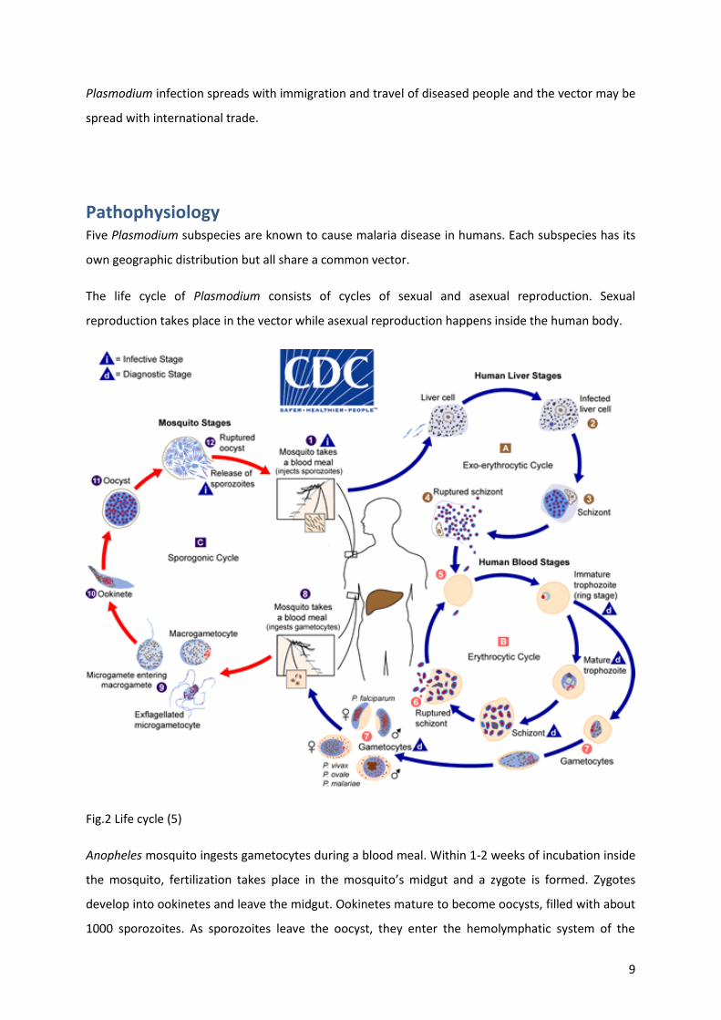
Pathophysiology Five Plasmodium subspecies are known to cause malaria disease in humans. Each subspecies has its
own geographic distribution but all share a common vector.
The life cycle of Plasmodium consists of cycles of sexual and asexual reproduction. Sexual
reproduction takes place in the vector while asexual reproduction happens inside the human body.
Fig.2 Life cycle (5)
Anopheles mosquito ingests gametocytes during a blood meal. Within 1-2 weeks of incubation inside
the mosquito, fertilization takes place in the mosquito’s midgut and a zygote is formed. Zygotes
develop into ookinetes and leave the midgut. Ookinetes mature to become oocysts, filled with about
1000 sporozoites. As sporozoites leave the oocyst, they enter the hemolymphatic system of the

10
mosquito and travel to the salivary glands, from where they will be injected together with saliva into
the human skin with the next blood meal.
Inside the human body, Plasmodium sporozoites travel to the liver and enter hepatocytes with the
help of CD81 tetraspamin and SR B1 receptor. SR B1 activates L-FABP and provides HDL- cholesterol
to hepatocytes and promotes parasite growth. Hepatic incubation time is 1-2 weeks, during which
the infected person is asymptomatic. Each infected hepatocyte can produce between 10.000 and
30.000 merozoites.
With the help of proteases, merozoites leave the hepatocytes and enter the bloodstream.
Erythrocyte entry of merozoites is mediated by apical organelles of the merozoites and erythrocyte
membrane proteins. A dense junction is formed and the merozoite drives itself into the erythrocyte
with help of an actin-myosin motor. Merozoites remain enveloped in a piece of the erythrocyte
membrane, which they transform into a parasitophorous vacuole to keep their surrounding
microenvironment stable. Proteins produced by the parasite will be transported across this
membrane.
Inside the erythrocyte, merozoites form rings and within 48-72 hours develop first into trophozoites,
then schizonts.
Schizonts leave the erythrocyte with the help of proteases, destroying the erythrocyte and causing
anemia.
Trophozoites develop into gametocytes or infect more erythrocytes.
Plasmodium falciparum is the most common species due to its worldwide distribution and with 80-
90% endemicity (6). Incubation time in the human is 8-10 days.
The parasite infects all stages of erythrocyte development, which causes high parasite mass with
severe symptoms of disease and severe anemia. Schizont release may synchronize to give fever
cycles of 36-48 hours periodicity or may remain unsynchronized and present with continuous high
fever.
Infected erythrocytes form knobs and express Plasmodium falciparum erythrocyte membrane
protein – 1 (PfEMP-1) on their membrane for cytoadherence. PfEMP-1 is a product of var gene,
which has about 60 variants. In every generation, only one gene variant is expressed and others are
silenced. The rate of gene switch is 2-18% per cell per generation.

11
PfEMP-1 is a resetting ligand, which eases the infection of neighboring cells by clumping.
Different variants of PfEMP-1 may be associated with different levels of severity and presentation of
disease. Variants binding CD36 on platelets are promoting platelet-mediated erythrocyte adherence,
on endothelium and monocytes, they promote sequestration and host immune response. Within the
brain, PfEMP-1 variants bind to ICAM-1, which promotes sequestration in microvasculature by
attachment and rolling on vascular membranes similar to activated leukocytes. Pathologic
examinations of brains in cerebral malaria found ICAM-1 to be increased.
In the placenta, PfEMP-1 variants bind CS-A of the syncytiotrophoblast and cause malaria of
pregnancy.
Additionally, variants of PfEMP-1 can bind to CR1 and blood group A antigen.
Plasmodium malariae appears in tropical regions of Africa and the Southwest Pacific and has an
incubation time of 18-40 days. Infected people may appear asymptomatic for many years after,
which makes them a carrier. Blood donors therefore must be screened for P. malariae carrier state
to avoid transmission through blood transfusions.
The fever cycles have a quartan periodicity of 72 hours.
P. malariae mostly infects older erythrocyte stages which makes it prone to destruction by the
spleen. Usually parasitemia is below 2% and therefore more difficult to detect.
It causes the highest incidence of febrile illness in children less than 10 years of age.(7) In young
children, P. malariae may cause immune complex-mediated glomerulonephritis with nephrotic
syndrome.
Plasmodium vivax is endemic in tropics and subtropics except for West Africa and even seen in
temperate climates. In West Africa, a mutation of Duffy antigen prevents its spread. (8)
With an endemicity of 50-80%, it is the second most common cause of malaria.
Incubation time in the human is 10-17 days; fever pattern is tertian with 44-48 hours periodicity.
P. vivax mostly infects younger erythrocyte stages such as reticulocytes. Due to their lower number
in peripheral blood, less parasite mass is produced and parasitemias are often as low as 1%.

12
Additionally P.vivax increases erythrocyte flexibility to escape clearance by the spleen. Anemia can
be moderate to severe.
P. vivax may sequester in pulmonary microvasculature and cause lung injury. Occasionally, nephrotic
syndrome and splenic rupture may be seen.
Relapses of disease are possible.
P. ovale presents similarly to P. vivax but besides being endemic in tropics and sub-tropics it is also
seen in Saharan Africa and southeast Asia.
Its incubation period is 10-17 days and fever cycles have a 48 hours periodicity. Similar to P. vivax it
infects reticulocytes and parasitemias are low, usually less than 2% or below 500 parasites per
microliter of blood.
By increasing the use of PCR techniques, two variants of P. ovale, P. ovale curtisi and P. ovale
wallikeri, were detected which do not cross breed. (6)
P. ovale causes the highest incidence of febrile illness in children 0-7 years of age. (7)
P. knowlesi is originally a zoonotic parasite which is transmitted from macaque monkeys in
Southeast Asia. Incubation time is 9-12 days. Severe illness is commonly seen due to 24 hours fever
cycles with severe anemia and hyperparasitemia. The parasite accumulates in brain, heart and kidney
tissues and therefore may cause life-threatening complications such as hepato-renal dysfunction and
refractory hypertension.
Pediatric cases occur mostly in school-age children. (7)
Clinical presentation
Patients with malaria disease usually present with paroxysms of chills, rigor, fever, sweating, fatigue
and sleepiness, Splenomegaly is seen occasionally. Their fever usually peaks at around 40°C. (8)
To differentiate between the Plasmodium species and exclude other illnesses such as Babesia
infection, a good travel history must be taken. The patient must be asked for the time of travel,
13
duration of stay, type of vacation and accommodation and food safety. Additionally, the patient
should be asked about his use of chemoprophylaxis and mosquito repellants.
Basic laboratory blood tests will show some degree of anemia of normochromic and normocytic type
with decreased hematocrit and hemoglobin. Leukocyte counts may be normal, increased or
decreased; Platelet counts may be normal or decreased.
Due to the anaerobic metabolism of patient and parasite, LDH and Lactate will be increased causing
acidosis. Hypoglycemia may be present, in children it is caused by a failure of gluconeogenesis
whereas in adults it may be due to parasite-induced hyperinsulinemia or medications.
Schizont rupture releases glucosyl phosphatidylinositol compounds, which stimulate monocytes to
produce TNF-alpha. TNF-alpha will cause a decrease in erythropoietin production and therefore
aggravates anemia by decreasing bone marrow response to anemia. (8)
Complications with malaria disease can be coma due to metabolic disturbances or as a presentation
of cerebral malaria; pulmonary edema due to increased capillary permeability or iatrogenic fluid
overload potentially leading to ARDS. Renal impairment is due to accumulation of LDH and worsens
cerebral malaria to the point of convulsions; it aggravates pulmonary symptoms due to fluid
overload.
During Pregnancy, malaria presents with febrile illness and anemia in the mother. It is worst in first
pregnancies and first exposures to malaria. It causes prematurity, intrauterine growth retardation,
low birth weight and will contribute to infant and maternal mortality as well as maternal morbidity.
Variants of PfEMP-1, derived from var2csa gene, interact with chondroitin sulfate A (CSA) of the
syncytiotrophoblast and parasites sequester and accumulate in intervillous space. Pregnancy
outcomes depend on FLT-1 expression; if its expression in maternal tissues is high, fetal loss is most
likely, if it is mostly expressed in fetal tissues, low birth weight is more likely. If FLT-1 is expressed in
the fetus, a greater inflammation of the placenta is seen and IFN-gamma is increased. (8)(7)
Differential diagnoses
Differential diagnoses for malaria can be any febrile illness in travellers from tropical and subtropical
regions.
Dengue fever, katayama fever of acute schistosomiasis, leptospirosis, african tick fever, yellow fever
and babesiosis should be considered to be differential diagnoses.

14
Characteristic for dengue fever would be the severe myalgias and a characteristic maculopapular
rash with petechiae and generalized lymphadenopathy.
For katayama fever, the patient should be asked for water exposure in Shistosoma endemic areas
about 4-8 weeks prior to appearance of symptoms. The rash is puritic at the site of entrance to the
skin with generalized urticaria. In blood tests, eosinophilia can be seen.
Leptospirosis is acquired from soil or fresh water exposure. To differentiate leptospirosis from
malaria one needs to pay attention to conjunctival suffusion or rash, symptoms of Weil’s disease
such as hepato-renal dysfunction with hemorrhagic symptoms and extremely high
hyperbilirubinemia.
African tick fever is transmitted by tick bites during safaris or hunting in southern Africa. Typically
eschars at the site of the tick bite are found along with lymphadenitis.
Yellow fever has a shorter incubation time than malaria and is differentiated from malaria by
conjunctival suffusion or bradycardia with jaundice. (8)
Babesiosis cannot only be acquired in tropical regions but also in temperate climate. It is transmitted
by a tick bite. B. divergens is most common in Europe, whereas B. microti and B. duncani are found
in the USA. The course of disease is most severe in immunocompromized, splenectomized or elderly
patients. If the disease does not pass asymptomatically it may resemble malaria paroxysms and
therefore might be difficult to clinically differentiate for an inexperienced physician. Blood smears to
detect babesiosis are prepared and read in a similar fashion as for malaria detection. Since Babesia is
in the same class and phylum as Plasmodium, their trophozoite stage might look similar to
Plasmodium for someone who is not so experienced in malaria microscopy. Despite being rarely
seen on blood films, tetrads or “Maltese crosses” are characteristic for babesiosis. If in doubt,
serology but also PCR may be a good method to distinguish between Babesia and Plasmodium
infection.(6)(9)

15
Malaria Diagnostics
Microscopy
Gold Standard for malaria diagnostics today is light microscopy of thick and thin blood smear.
WHO has defined Guidelines for Microscopy in research settings to make results more reproducible.
All slides should be examined under 100x plan acromat oil immersion objective lens with an ocular
lens of 10x magnification and a Field Number of 18 or 20 to have 10- 15 leukocytes per high power
field on average, if the leukocyte count is 8000/microliter. (10) Usually thick and thin smears of one
patient are placed on the same glass slides.
Fig. 3 Image glas slide
Thick smears are prepared from fresh blood or blood that was anti-coagulated with EDTA and are
good for screening for parasites. They are prepared by placing 1-2 drops equal to 6 microliters of
blood on a glass plate, then blood is spread out to a circle of 12 mm in diameter. The sample must be
dried either in a drying chamber at ambient temperature or on a warm plate with no more than 37°C
to avoid hemolysis. The thick film must be stained without previous fixation.
Good thick smears should be evenly spread and should be free of dirt or dust. There should be 10-15
leukocytes per field.
Thin smears can be made from fresh blood or EDTA blood, they are used to detect the Plasmodium
species and differentiate malaria from infection with other pathogens. A drop of 2-3 microliters of
blood is placed on a clean glass slide and spread out with a spreader slide. The end of the smear
should be feathered out because erythrocytes should not overlap in this area. After drying the slide,
the thin smear is fixed with methanol; a contamination of the thick film area with methanol must be
avoided. After drying the slide again, staining solutions such as Giemsa stain may be applied to both
thick and thin smear parts of the slide
To decide whether to examine the thick or the thin films for parasitemia estimations, the number of
asexual forms per leukocytes must be counted. If the number of asexual parasites is less than or
equal to twice the leukocyte number, thick films should be chosen. If it exceeds twice the leukocyte
number, the thin film should be chosen for examination. From the amount of asexual parasites, the
expected parasitemia can be assumed. Due to their higher concentration of erythrocytes, thick films
are more suitable for medium to low parasitemias with less than 16.00 parasites per microliter.
Above 16.000 parasites per microliter, thin films are more accurate since erythrocytes are more

16
spread out and do not overlap, which helps with counting. Sexual and asexual parasite forms must be
reported when examining thick and thin films.
During thick smear examination, the slide must first be examined for presence of gametocytes and
mixed species infections.
To examine the slide for asexual stages 3 methods can be used; the high power field method, the
leukocyte method and Earle- Perez method. In the high power field method, the number of parasites
per high power field is reported. Counting starts from the first field with a good view. At least 200
fields should be examined with ocular lenses with field number (FN) of 18 or 20. For 200 examined
high power fields, the volume of examined blood can be calculated from the ocular field number, the
volume of blood and the diameter of the thick film.
For the leukocyte method, the number of parasites per leukocytes is reported. The slides should be
examined until at least 1500 leukocytes are detected. If there is less than 1 parasite per 1500
leukocytes or the smear seems to be negative, it needs to be further examined until 2500 leukocytes
were detected.
Earle- Perez method is used, if the actual leukocyte count of the patient is not known. The slide is
prepared differently to the previously mentioned methods. 5 microliters of blood are spread out to a
rectangle of 6x15mm and an ocular with 6x6 grid is used. Slides are read in lines across the length of
the rectangle and parasite numbers are reported per band.
On thin smear examination for parasite density calculation, the number of erythrocytes and parasites
is counted. Both parasitized and normal erythrocytes are counted until 2000 erythrocytes were
detected. Erythrocytes containing more than one parasite or multinucleated forms are counted as
one infected cell. There is no need to differentiate between the species at this point.
Based on the examination of slides and depending on the methods used, the following calculations
can be made:
For thick films:
(
)
( )
For thin films:

17
From parasite density, parasitemia can be calculated:
( )
Since parasitemia in smears may lag behind clinical symptoms, inconclusive or negative tests should
be repeated after 12 hours.
Thick smear technique can be used to monitor parasitemia and therefore therapy success.
Warning signs of severe disease are the presence of trophozoites and schizonts of P. falciparum in
peripheral blood. An intracytoplasmatic pigment is a poor prognostic sign.
Microscopic characteristics of Plasmodium falciparum are normal-sized and shaped erythrocytes
filled with thick pink dots called "Maurer's clefts". They are comma-shaped and derived from the
parasitic vacuole. (11)
Usually only early trophozoites and gametocytes are seen in peripheral blood.
Early trophozoites present as delicate rings inside the erythrocyte, which look like headphones due
to their chromatin dots. In erythrocytes with multiple trophozoites, they might present as a signet
ring shape. They are usually located towards the periphery, which is called acollée position. Their size
is approximately 1/3 of erythrocyte diameter.
Mature trophozoites are more compact and thick rings but their detection in peripheral blood is a
poor prognostic sign.
Schizonts of P.falciparum carry about 8-12 merozoites, their appearance in peripheral blood is a poor
prognostic sign as well.
Gametocytes are crescent or banana- shaped and usually deform the erythrocyte.
Fig. 4 Bench card P.falciparum
P. malariae infected erythrocytes may be smaller in size but maintain a normal shape.
If specimens are overstained, black “Ziemann’s dots” may be seen as intracytoplasmic inclusions.

18
All stages of parasite development are seen in peripheral blood. Early trophozoites look like rings
with a “birds eye” dot of chromatin in the center. They are smaller than 1/3 of the erythrocyte
diameter.
Mature trophozoites have compact cytoplasm, are round to oval and may have a basket like look.
Sometimes they have band form. A dark brown pigment may be present.
Schizonts contain 6-12 merozoites, which are radially arranged and look like a daisy flower or
rosette.(12)
Gametocytes are round to oval and fill the entire erythrocyte.
Fig. 5 Bench card P. malariae
P.vivax infection may cause erythrocytes to enlarge and stick to neighboring erythrocytes.
Intracytoplasmatic pink-red granules are seen and called "Schüffner's dots". They may be absent in
early trophozoites.(13)
All stages of parasite development are seen, sometimes there may be multiple parasites per
erythrocyte.
Early trophozoites look like rings and are more than 1/3 of erythrocyte diameter in size. Their
chromatin dots are larger than in P. falciparum.
Mature trophozoites appear amoeboid and may contain yellow to golden brown pigment.
There are 12-24 merozoites in every schizont.
Gametocytes are round to oval and fill the entire erythrocyte.
Fig. 6 Bench card P.vivax
P. ovale infected erythrocytes have a fimbriated surface and are enlarged in size. Schüffner’s dots
are darker and there may be James’ dots. Both may be absent in early trophozoites.(14)
All stages of parasite development are found in peripheral blood, occasionally with multiple parasites
per erythrocyte.
Early trophozoites look similar to P. vivax.

19
Mature trophozoites look more compact and less amoeboid than P.vivax, their pigment is dark
brown.
Schizonts contain 6-14 merozoites. Gametocytes are round to oval and fill the erythrocyte.
Fig. 7 Bench card P. ovale
P. knowlesi infected erythrocytes have normal size and shape, all stages of parasite development are
seen in peripheral blood but infection with multiple parasites per erythrocyte are rather common.
Early trophozoites resemble P. falciparum while mature stages and gametocytes look more like P.
malariae. Compared to P. falciparum, early trophozoites are rarely seen in appliqué or acollée
position.
Mature trophozoites commonly appear as band forms and are only slightly amoeboid. Similar to P.
malariae, they may contain clumps of golden brown pigment, which is a poor prognostic sign.
Schizonts carry 10-16 merozoites.
Gametocytes are round and fill the erythrocyte.
All microscopic methods have in common that they are highly dependent on the examiner with his
experience and training. In the research setting, staff requires frequent training and practice with
positive samples and specimen are commonly read by at least 2 independent examiners.
Besides light microscopy as the gold standard for malaria diagnostics, there is a search for
alternatives which may be cheaper or more easily accessible and could be performed by less
experienced personnel.
LED fluorescence microscopy LED fluorescence microscopy (LED-FM) and Fluorescent in Situ Hybridisation (FISH) techniques are
under investigation to improve and help with the examination of samples in clinical settings.
LED- FM uses LED lights that excite fluorescent staining agents. Their use is similar to light
microscopy and it is possible to calculate parasitemia, less than 100 parasites per microliter are the
detection limit.

20
Advantages of the LED lights are a brighter view, longer lasting microscopes and due to low energy
expenditure, they can be run on battery. LED fluorescence microscopes might be even cheaper in use
and screening for parasites in experienced hands can be done faster than with Giemsa stain.
As a staining agent for fixed slides, Acridine orange can be used, which stains both DNA and RNA.
DNA will have apple green fluorescence while RNA looks red-orange.
R. Hathiwala et al. (15) did a cross-sectional study at a tertiary hospital in Mumbai, comparing LED
fluorescence microscopy with routine methods such as Light microscopy and RDTs. They collected
300 EDTA blood samples of patients with suspected malaria. With light microscopy (Leishman Stain)
111 samples were detected positive, 87 positive samples with LED-FM and 107 with rapid diagnostic
tests (RDTs). Only 73 samples were detected positive by all 3 testing methods. 47.75% of positive
samples had parasitemia under 1%.
LED- FM proved to have a moderate sensitivity (71%) but good specificity (96%). Comparing their
results with other studies on LED-FM they concluded that sensitivity decreases with lower parasite
densities and smaller parasite size. Their values for specificity and sensitivity varied from results in
other studies, which also tested LED-FM. It was assumed that examiners reading the slides were not
experienced enough with the detection under LED-FM and it was assumed that it matters for the
results, that slides were prepared only from the same sample but not exactly the same for light
microscopy and LED-FM.
Fluorescent in situ Hybridisation assay (FISH)
Another fluorescent staining method is Fluorescent in situ Hybridisation assay (FISH)(16). It uses thin
smears and fluorescent microscopes and combines it with rRNA oligonucleotide probes that are
specific for Plasmodium parasites. The method is capable of detecting species-specific segments of
rRNA. FISH is capable of detecting live Plasmodium parasites.
In a study with 500 voluntary participants, conducted in Kenya by Joseph Osoga et al. (16) between
November 2013 and May 2014, P-genus FISH and Giemsa stain were compared to quantitative real-
time PCR (qRT-PCR) as reference method.
Giemsa stains and the slide examination were performed according to WHO recommendation.
P-genus FISH was applied to specially prepared thin films. Samples were examined with a green filter
at an excitation of 560nm and emission of 630nm, which made the Plasmodium probes stained with
Texas red dye shine red. Two independent readers examined 300 High power fields under the

21
microscope. In this setting, the detection limit of Plasmodium was 1-2 parasites/ 300 high power
fields (100x objective). Compared to qRT-PCR, the FISH method was inferior in detecting positive
malaria cases, whereas it performed better in detecting negative cases. Sensitivity was 29.3% and
specificity was 75.8% in comparison to qRT-PCR. Positive predictive value was 56.0% and negative
predictive value was 50.5%.
Compared to qRT-PCR light microscopy with Giemsa stain had a sensitivity of 59.8% and specificity of
93%, positive predictive value was 89.8% and negative predictive value was 68.%.
The authors of the study concluded that the FISH method proved to be inferior to both methods but
this might have been due to insufficient training and experience with the method.
Rapid diagnostic tests (RDTs)
Rapid Diagnostic Tests (RDTs) are immunochromatographic essays designed in the mid-1990s to help
with rapid and early detection of Plasmodium infection in the absence of equipment or trained
personnel. Especially in rural areas, it can significantly shorten the time needed for malaria diagnosis
in the clinical setting.
According to WHO a good test for clinical use must fulfill ASSURED criteria, which means it must be
affordable, sensitive, specific, user- friendly, robust, rapid, equipment-free and deliverable to those
in need (17). For RDTs that means they must be produced to have a long shelf life and withstand hot
climate, they should be so simple that even untrained personnel is able to use and read them
correctly. WHO requires RDTs to detect parasite concentrations as low as 100 parasites per
microliter, which is equivalent to 0,002% parasitemia. Tests must have a minimum sensitivity of 95%
and a minimum specificity of 90%.
Additionally, “Global Malaria Programme” of the WHO runs an RDT testing programme together with
the United States Center of Disease Control and Prevention (CDC) to regularly evaluate quality and
usability of RDTs and gives out recommendations for their use in malaria control programs. RDTs
must pass seven rounds of standardized tests. At submission by the manufacturers, panel detection
scores are measured using 20 P. falciparum cultures of low and high parasite densities, 35 P. vivax
cultures of low and high densities and 100 patient derived samples each for P. falciparum and P.
vivax. Panel detection rates should be 75% at 200 parasites per microliter for both P. falciparum and
P.vivax. False positive rates should be less than 10% and invalid rate should be less than 5%.

22
Furthermore, RDTs are tested for thermal stability during storage. For this test, RDTs are stored for
60 days at room temperature, at 35°C and at 45°C. There should be no change of detection rate at
200 parasites per microliter.
In the final round of testing, the packaging, labeling of contents, instructions and additional items in
the kit are evaluated based on their user-friendliness. (18)
In the field setting, staff and non-medical users of the test kits must be familiar with performing and
reading the tests because it may influence the test results.
Currently, there are tests detecting PfHRP-2, LDH and aldolase. More tests are under investigation
and not commonly used in clinical practice. Only PfHRP-2 based tests are available worldwide.
Histidine-rich protein-2 (HRP-2) is found only in Plasmodium falciparum. It can be detected in both
the gametocyte and erythrocyte stages. Due to the correlation between the biomass of parasites and
protein concentration, the test can be used to predict the potential for progression to severe P.
falciparum Malaria.
Sensitivity is 96% and specificity 99% on average. HRP-2 based tests are the most widely available
RDTs.
Limitations of HRP-2 based tests are the long half-life of HRP-2 in the blood (3,0-4,7 days) and that
tests will still have positive results for 28 days after successful therapy. Recently mutations in HRP-2
genes were detected, which alter the protein in a way that it can no longer be detected by the test.
(19)
Another problem is the detection limit of 100-1000 parasites per microliter.
Cross-reactions with Rheuma Factor and false positive results with Schistosoma infection are known.
The “Prozone effect” will happen when parasitemia is too high and antigen is in excess.(6)(8)
Lactate dehydrogenase (LDH) based tests may be specific for Plasmodium falciparum or can be made
to detect pan-plasmodium LDH.

23
Since Plasmodium parasites can only perform anaerobic metabolism all subspecies and stages of
development will produce LDH. Concentration and test signals are proportional to parasitemia.
Tests based on LDH detection may detect mixed-species infections and can differentiate between P.
falciparum and P.vivax. LDH has no known antigenic variations and does not cause Prozone effect.
Since pLDH disappears soon after successful treatment, the test can be used for monitoring of
therapy.
Sensitivity for pLDH based tests is 80%, specificity is 98%. Lower sensitivities may appear in P.vivax, P.
ovale and P.malariae infections as well as in low parasitemias of less than 100-1000 parasites per
microliter.
Aldolase is found in the apical part of merozoites. Its concentration is parasite density dependent.
RDTs based on Aldolase detection are more temperature sensitive and have low detection rates at
low parasitemias.
Polymerase chain reaction (PCR)
Polymerase chain reaction (PCR) can be used to detect Plasmodium DNA but its most common target
is small subunit of 18S rRNA and can be used as conventional, quantitative or real-time method. It is
used in reference or public health laboratories and research facilities to differentiate between
Plasmodium species and to detect markers of parasite resistance to drugs. WHO provides a
quantitated Plasmodium standard to compare sensitivity of the testing method.
In clinical setting use of PCR could be to detect very low parasitemias, identify mixed species
infections, confirmation of inconclusive results from other tests and differentiation from Babesia and
other pathogens. The average detection limit of PCR based methods is 10 parasites per microliter of
blood. (6) So far FDA does not approve PCR as a routine diagnostic test for Plasmodium.
Quantitative PCR could replace manual parasite counts but results are not correlated with
conventional counting in light microscopy since extracellular forms and gametocytes would be
included and all parasites in multiply infected cells would be counted. (6)
Loop mediated isothermal amplification (LAMP) Loop-mediated isothermal amplification (LAMP) is a method of DNA amplification similar to
polymerase chain reaction (PCR) but is performed at a constant temperature, which makes it simpler
and less expensive than conventional PCR.

24
With the help of Calcium oxide based heating systems, it could be even electricity-free in the
future.(6)
In a study from Bernhard Nocht Institute Hamburg, Germany by H. Frickman, et al.(20) the
commercially available platform “Meridian illumigene Malaria platform” was compared to routine
PCR for its use as point of care test and to Giemsa stain thick and thin films, as they would be
routinely performed. 1000 patient blood samples with suspected or confirmed malaria were
examined. 238 samples were tested positive for Plasmodium parasites with microscopy or PCR. Out
of 238 positive samples, 235 samples were correctly identified. This means LAMP showed a
sensitivity of 98,7%, a specificity of 99,6% and a high negative predictive value of 99,6%, which makes
it a good screening tool. Besides that, it is able to detect parasite DNA in the blood for up to 3 weeks
after the last positive test.
In comparison to PCR, the procedure showed to be faster and cheaper; in comparison to light
microscopy positive results were obtained at earlier stages of the disease. Unfortunately LAMP was
not able to differentiate between species nor could it detect the level of parasitemia in the samples.
Hemozoin detection
Hemozoin is a product of Plasmodium parasites, produced by heme detoxification and is released
with merozoites. It’s amount in blood increases with the growth of Plasmodium parasites. There are
several approaches concerning this compound. RDTs with monoclonal antibodies can detect the
heme-detoxification protein, which catalyzes the heme detoxification to hemozoin.
Hemozoin can be detected by polarization and dark-field microscopic techniques, where it presents
as optically birefringent, paramagnetic crystal. Other detection methods are flow-cytometry and
magneto-optical technique. Limitations to the technique are the variable amounts of hemozoin
produced by wild type Plasmodium and the number of immature forms of Plasmodium, which do not
yet produce hemozoin. Both are possible causes of false negative test results.
For false positive results, causes may be that gametocytes produce most hemozoin compared to
other stages and that it remains in macrophages and leukocytes for up to 2 weeks after therapy. In
endemic regions, hemozoin may also be produced by adult Schistosoma worms, which may give false
positive results or in co-infections increase the amount of detectible hemozoin.
A drawback of the method is that it cannot quantify the parasite density, nor is it able to identify the
Plasmodium species.

25
Research on hemozoin crystal detection is so far tested in rodents with rodent-specific Plasmodium
strains, therefore it is questionable whether the results can be directly transferred to human
Plasmodium infections.(21)
Serology
During acute malaria disease in previously unexposed patients, IgG antimalarial antibodies usually
develop about a week after onset of parasitemia and persist up to one month or lifelong. (22) In the
mean time malaria can already be diagnosed by light microscopy with Giemsa stain or PCR, therefore
serology is not a method of choice in diagnosis of acute malaria.
Additionally, people in endemic regions have positive antibody titers due to previous or chronic
malaria disease.
In previously non-exposed populations, serology may confirm a past malaria exposure, help in
screening for chronic malaria or be used in blood donor screening.
In cases of rare tropical splenomegaly syndrome, serology is helpful in detecting previous malaria
disease. (23)
Malaria serology can be used in epidemiologic settings to measure public exposure to malaria and
measure transmission intensity in endemic areas. (24)
Cross reactivity with antinuclear antibodies (ANA), rheuma factor (RF) and Babesia microti antibodies
are a potential cause of false positive results in malaria serology testing.
Today, malaria serology is not commonly used for diagnosis of acute disease in clinical setting, it is
only available in specialized centers and for blood donor screening.(6)
Conclusion Confirmation of malaria by specific laboratory test remains difficult and requires continuous training
of laboratory staff.
Each of the above mentioned methods have their advantages and drawbacks and therefore cannot
replace each other but can be complementary to each other.
For clinical use, a detailed patient and travel history is essential as the first screening tool and may
guide doctors to find the right differential diagnosis for febrile illness. Laboratory diagnostic methods
can serve as a confirmation and may help to choose the right medication.
26
Today RDTs and microscopy are usually used in combination. RDTs significantly shorten the time
between patient presentation and beginning of initial empiric treatment while microscopy can be
performed in a specialized laboratory.
There is a long experience with light microscopy and especially Giemsa stain and the method unites
the most crucial elements of diagnostics. It is capable of distinguishing between Plasmodium
infection as the cause of fever and other blood parasites such as Babesia or Shistosoma. Additionally
it is possible to detect parasites and perform species differentiation, as well as parasitemia
estimation.
To improve laboratory diagnostics in rural areas, telemedicine attempts are made. Light microscopy
and hemozoin detection by polarized light microscopy in combination with Apps for image
recognition, seem to be the most promising techniques in this field. Since mobile internet is widely
spread in developing countries, smartphone based automated systems have a great chance to
improve Plasmodium detection in endemic regions. (25)

27
Acknowledgments
I would like to show my gratitude to my mentor, doc. dr. sc. Mario Sviben, at the Department of
Microbiology and Parasitology, Croatian institute of Public Health for this thesis paper. I am very
grateful for his assistance and support, and his overall effort towards the success of this essay.
I would like to thank Prof. Dr. med. August Stich at Missio Klinik Würzburg, Department of Tropical
Medicine who taught me during my Clinical Rotation and inspired me to choose malaria as the topic
for my thesis.

28
References 1. Mülder K. Malaria: Robert Koch auf Brioni. Ärzteblatt, Deutsches [Internet]. 2001;98(48);A-
3219 / B-2724 / C – 2531. Available from: https://www.aerzteblatt.de/archiv/29677/Malaria-
Robert-Koch-auf-Brioni
2. WHO, Organisation WH. World Malaria Report [Internet]. 2017 [cited 2019 Feb 11]. Available
from: https://www.medbox.org/world-malaria-report-2017/preview?q=malaria+diagnostics
3. Fairhurst RM, Wellems TE. No Title. In: Principles and Practice of Infectious Diseases, Updated
Edition [Internet]. 8th ed. Saunders, Elsevier; 2015. p. 3070-3090.e9. Available from:
http://dx.doi.org/10.1016/B978-0-443-06839-3.00275-7
4. geodistribution2 [Internet]. [cited 2019 Jun 14]. Available from:
https://www.cdc.gov/malaria/about/distribution.html
5. malaria_LifeCycle [Internet]. [cited 2019 Jun 14]. Available from:
https://www.cdc.gov/dpdx/malaria/index.html
6. Pfaller MA, Richter SS, Funke G, Jorgensen JH, Landry ML, Carroll KC, et al., editors. Manual of
Clinical Microbiology, 11th Edition [Internet]. American Society of Microbiology; 2015 [cited
2019 Jan 17]. Available from:
http://www.asmscience.org/content/book/10.1128/9781555817381
7. Ashley EA, Pyae Phyo A, Woodrow CJ. Malaria. Lancet. 2018;391(10130):1608–21.
8. FAIRHURST RM, Wellems TE. Plasmodium Species (Malaria). In: Mandell, Douglas, and
Bennett’s Principles and Practice of Infectious Diseases *Internet+. Eighth Edi. Elsevier; 2010.
p. 3437–62. Available from: https://www.elsevier.com/books-and-journals/deleted-doi
9. DPDx Babesiosis [Internet]. October 30th. 2017 [cited 2019 Mar 31]. Available from:
https://www.cdc.gov/dpdx/babesiosis/index.html
10. Research Malaria Microscopy Standards Working Group. Microscopy for the detection,
identification and quantification of malaria parasites on stained thick and thin blood films in
research settings | medbox.org [Internet]. Geneva: World Health Organization. 2015 [cited
2019 Feb 11]. Available from: https://www.medbox.org/microscopy-for-the-detection-
identification-and-quantification-of-malaria-parasites-on-stained-thick-and-thin-blood-films-
in-research-settings/preview?q=malaria+diagnostics
11. Laboratory diagnosis of malaria Plasmodium falciparum | medbox.org [Internet]. [cited 2019
Feb 11]. Available from: https://www.medbox.org/laboratory-diagnosis-of-malaria-
plasmodium-falciparum/preview?q=malaria+diagnostics
12. Laboratory diagnosis of malaria Plasmodium malariae | medbox.org [Internet]. [cited 2019
Feb 11]. Available from: https://www.medbox.org/laboratory-diagnosis-of-malaria-

29
plasmodium-malariae/preview?q=malaria+diagnostics
13. Laboratory diagnosis of malaria Plasmodium vivax | medbox.org [Internet]. [cited 2019 Feb
11]. Available from: https://www.medbox.org/laboratory-diagnosis-of-malaria-plasmodium-
vivax/preview?q=malaria+diagnostics
14. Laboratory diagnosis of malaria Plasmodium ovale | medbox.org [Internet]. [cited 2019 Feb
11]. Available from: https://www.medbox.org/laboratory-diagnosis-of-malaria-plasmodium-
ovale/preview?q=malaria+diagnostics
15. Hathiwala R, Mehta PR, Nataraj G, Hathiwala S. LED fluorescence microscopy: Novel method
for malaria diagnosis compared with routine methods. J Infect Public Health [Internet].
2017;10(6):824–8. Available from: https://doi.org/10.1016/j.jiph.2017.01.001
16. Osoga J, Waitumbi J, Guyah B, Sande J, Arima C, Ayaya M, et al. Comparative evaluation of
fluorescent in situ hybridization and Giemsa microscopy with quantitative real-time PCR
technique in detecting malaria parasites in a holoendemic region of Kenya. Malar J [Internet].
2017 Dec 24 [cited 2019 Feb 3];16(1):297. Available from:
http://malariajournal.biomedcentral.com/articles/10.1186/s12936-017-1943-4
17. Weise E. The complex art of making technology simple. USA Today [Internet]. 1997;9(8):467.
Available from: http://dx.doi.org/10.1016/S1473-3099(09)70190-2
18. Recommended selection criteria for procurement of malaria rapid diagnostic tests |
medbox.org [Internet]. [cited 2019 Feb 11]. Available from:
https://www.medbox.org/recommended-selection-criteria-for-procurement-of-malaria-
rapid-diagnostic-tests/preview?q=malaria+diagnostics
19. Mouatcho JC, Goldring JPD. Malaria rapid diagnostic tests: challenges and prospects. J Med
Microbiol [Internet]. 2013 Oct 1 [cited 2019 Feb 3];62(Pt_10):1491–505. Available from:
http://jmm.microbiologyresearch.org/content/journal/jmm/10.1099/jmm.0.052506-0
20. Frickmann H, Hinz R, Rojak S, Bonow I, Ruben S, Wegner C, et al. Evaluation of automated
loop-mediated amplification (LAMP) for routine malaria detection in blood samples of
German travelers – A cross-sectional study. Travel Med Infect Dis [Internet]. 2018;24(May
2018):25–30. Available from: https://doi.org/10.1016/j.tmaid.2018.05.006
21. Hänscheid T, Carvalho T, Grobusch MP. Hemozoin Detection for Human Malaria Diagnosis
Investigated in Rodent Models: How Similar Is Similar? Trends Parasitol [Internet].
2016;32(2):94–6. Available from: http://dx.doi.org/10.1016/j.pt.2015.11.012
22. She RC, Rawlins ML, Mohl R, Ascp MT, Perkins SL, Hill HR, et al. Comparison of
Immunofluorescence Antibody Testing and Two Enzyme Immunoassays in the Serologic
Diagnosis of Malaria. 2007;14(2):105–11.
23. Daly R, Chiodini PL. D i a g no s i s o f Tro p i c a l D i s e a s e s in Travelers Tropical diseases

30
Travel Laboratory tests. Infect Dis Clin NA [Internet]. 2019;26(3):803–18. Available from:
http://dx.doi.org/10.1016/j.idc.2012.06.001
24. Corran P, Coleman P, Riley E, Drakeley C. Serology : a robust indicator of malaria transmission
intensity ? 2019;23(12).
25. Poostchi M, Silamut K, Maude RJ, Jaeger S, Thoma G. Image analysis and machine learning for
detecting malaria. Vol. 194, Translational Research. 2018.

31
Biography
Eva Margareta Johanna Schloter was born 6th June 1988 in Cologne, Germany. In 2012, she decided
to move to Zagreb to complete her studies at the University of Zagreb School of Medicine.
During her 4th year of study, Eva completed a one-month internship at the Department of
Tropical Medicine at Missio Klinik in Würzburg, Germany. During her 5th year of study Eva
completed her internship program at the Department of Surgery at Dreifaltigkeits
Krankenhaus Wesseling and at the Family Medicine practice of Dr.med. Roger Schaffran in
Wesseling- Urfeld. She would like to specialize in Surgery and move back to Germany after
Graduation.