kobe university repository : kernel · 2.5 small interfering rna (sirna)-mediated gene silencing...
TRANSCRIPT

Kobe University Repository : Kernel
タイトルTit le
Ras inhibitors display an ant i-metastat ic effect by downregulat ion oflysyl oxidase through inhibit ion of the Ras-PI3K-Akt-HIF-1α pathway
著者Author(s)
Yoshikawa, Yoko / Takano, Osamu / Kato, Ichiro / Takahashi, Yoshihisa/ Shima, Fumi / Kataoka, Tohru
掲載誌・巻号・ページCitat ion Cancer Letters,410:82-91
刊行日Issue date 2017-12-01
資源タイプResource Type Journal Art icle / 学術雑誌論文
版区分Resource Version author
権利Rights
©2017 Elsevier. This manuscript version is made available under theCC-BY-NC-ND 4.0 license ht tp://creat ivecommons.org/licenses/by-nc-nd/4.0/
DOI 10.1016/j.canlet .2017.09.017
JaLCDOI
URL http://www.lib.kobe-u.ac.jp/handle_kernel/90004440
PDF issue: 2021-04-25
1
Ras Inhibitors Display an Anti-Metastatic Effect by Downregulation of Lysyl Oxidase
through Inhibition of the Ras-PI3K-Akt-HIF-1α Pathway
Yoko Yoshikawa1, Osamu Takano1, Ichiro Kato1, Yoshihisa Takahashi1, Fumi Shima2*, and Tohru
Kataoka1*
Affiliations
1Division of Molecular Biology, Department of Biochemistry and Molecular Biology, Kobe
University Graduate School of Medicine, Kobe 650-0017, Japan.
2Drug Discovery Science, Division of Advanced Medical Science, Department of Science,
Technology and Innovation, Kobe University Graduate School of Science, Technology and
Innovation, Kobe 650-0017, Japan.
*Corresponding authors
Tohru Kataoka, MD, PhD
Division of Molecular Biology, Department of Biochemistry and Molecular Biology, Kobe University
Graduate School of Medicine, 7-5-1 Kusunoki-cho, Chuo-ku, Kobe 650-0017, Japan.
Tel: +81-78-382-5380, Fax: +81-78-382-5399, E-mail: [email protected]
Fumi Shima, MD, PhD
Drug Discovery Science, Division of Advanced Medical Science, Department of Science, Technology
and Innovation, Kobe University Graduate School of Science, Technology and Innovation, 7-5-1
Kusunoki-cho, Chuo-ku, Kobe 650-0017, Japan.
Tel: +81-78-382-6033, Fax: +81-78-382-6034, E-mail: [email protected]

2
Abstract (184/185 words)
Metastasis stands as the major obstacle for the survival from cancers. Nonetheless most existing anti-
cancer drugs inhibit only cell proliferation, and discovery of agents having both anti-proliferative and
anti-metastatic properties would be more beneficial. We previously reported the discovery of small-
molecule Ras inhibitors, represented by Kobe0065, that displayed anti-proliferative activity on
xenografts of human colorectal cancer (CRC) cell line SW480 carrying the K-rasG12Vgene. Here we
show that treatment of cancer cells carrying the activated ras genes with Kobe0065 or an siRNA
targeting Ras downregulates the expression of lysyl oxidase (LOX), which has been implicated in
metastasis. LOX expression is enhanced by co-expression of RasG12V through activation of
phosphatidylinositol 3-kinase (PI3K)/Akt and concomitant accumulation of hypoxia-inducible factor
(HIF)-1α. Furthermore, Kobe0065 effectively inhibits not only migration and invasion of cancer cells
carrying the activated ras genes but also lung metastasis of human CRC cell line SW620 carrying the
K-rasG12V gene. Collectively, these results indicate that Kobe0065 prevents metastasis through inhibition
of the Ras-PI3K-Akt-HIF-1α-LOX signaling and suggest that Ras inhibitors in general might exhibit
both anti-proliferative and anti-metastatic properties toward cancer cells carrying the activated ras genes.
Keywords: metastasis; Ras; Ras inhibitors; LOX; PI3K; HIF-1α
Abbreviations: CRC, colorectal cancer; LOX, lysyl oxidase; PI3K, phosphatidylinositol 3-kinase; HIF,
hypoxia-inducible factor; GEF, guanine nucleotide exchange factor; GAP, GTPase-activating protein;
ECM, extracellular matrix; mTOR, mammalian target of rapamycin; β-APN, 3-aminopropionitrile
fumarate salt; DMSO, dimethyl sulfoxide; GAPDH, glyceraldehyde-3-phosphate dehydrogenase; FBS,
fetal bovine serum; DMEM, Dulbecco’s modified Eagle’s medium; 2D, two-dimensional; 3D, three-
dimensional; siRNA, small interfering RNA; H&E, hematoxylin & eosin; qRT-PCR, quantitative

3
reverse transcription-polymerase chain reaction; EMT, epithelial-mesenchymal transition.

4
1. Introduction
The ras oncogene products (H-Ras, K-Ras and N-Ras) function as binary switches by cycling between
GTP-bound (Ras•GTP) active and GDP-bound (Ras•GDP) inactive forms and play pivotal roles in
controlling cell proliferation and differentiation [1]. Interconversion between the two forms is catalyzed
by guanine nucleotide exchange factors (GEFs) or GTPase-activating proteins (GAPs) [2]. Ras•GTP
interacts directly with its downstream effectors including Raf kinases and phosphatidylinositol 3-kinases
(PI3Ks) for exerting its diverse growth-stimulating functions [1, 2]. Since mutational activation of Ras
is observed in 15~20% of human cancers, specifically in 60~90% of pancreatic and 30~50% of
colorectal carcinomas (CRCs) [1, 3, 4], agents directly inhibiting Ras are presumed to become promising
anti-cancer drugs. We succeeded in the discovery of small-molecule Ras inhibitors, Kobe0065-family
compounds, that blocked the interactions of Ras•GTP with its effectors and displayed anti-proliferative
activity toward xenografts of human CRC cell line SW480 carrying the K-rasG12V [5].
Various factors are involved in tumor progression and malignant transformation. In particular,
invasion and metastasis of cancer cells represent one of the major causes of poor survival, the inhibition
of which is one of the targets for anti-cancer drug development [6-8]. It has been reported that lysyl
oxidase (LOX) is closely related to tumor progression and metastasis under hypoxia [9, 10]. LOX, a
member of the multigene family consisting of at least five constituents, is a secreted amine oxidase that
modifies primary tumor microenvironment by crosslinking collagens and elastins in the extracellular
matrix (ECM) [11, 12]. LOX is initially synthesized as an inactive 46-kDa preproenzyme, prepro-LOX,
and the 21-amino acid signal peptide is cleaved during biosynthesis [13]. The resulting propeptide is
glycosylated to yield 50-kDa pro-LOX and subsequently secreted into the ECM. Thereafter, pro-LOX
is proteolytically cleaved by procollagen C-proteinase, yielding the 32-kDa active mature enzyme,
mature-LOX [13]. LOX was reported to be involved in the metastasis of CRC cells such as SW480 and
its patient-matched metastatic clone, SW620 [14, 15]. The expression of LOX is enhanced by the
5
activation of PI3K/Akt [16-18], downstream molecules of Ras, and targeted by hypoxia-inducible factor
(HIF)-1, a transcription factor composed of α and β subunits [9, 19]. The stability of the α subunit is
regulated by cellular oxygen levels while the β subunit is constitutively expressed [19]. Activation of
PI3K/Akt enhances the production of HIF-1α via phosphorylation and activation of mammalian target
of rapamycin (mTOR) [20, 21] . In addition, Ras were implicated in regulation of HIF-1α activation
although the underlying mechanism was unclear [22-25].
In this study, we investigate the role of Ras signaling in not only the regulation of LOX expression
and HIF-1α activation but also the metastatic properties of cancer cells by using the Kobe0065-family
compounds [5] and find that the compounds prevent metastasis through inhibition of the Ras-PI3K-Akt-
HIF-1α-LOX signaling. These results suggest that Ras inhibitors in general may exhibit an anti-
metastatic activity toward cancer cells carrying the activated ras mutations adding to an anti-
proliferative activity.
2. Materials and Methods
2.1 Reagents
Kobe0065 and sorafenib were obtained as described before [5]. LY294002, 3-aminopropionitrile
fumarate salt (β-APN) and Chrysin were obtained from Millipore (Billerica, MA, USA), SIGMA-
Aldrich (St. Lois, MO, USA) and Santa Cruz Biotech. (Santa Cruz, CA, USA), respectively. For in vitro
experiments, all the compounds were dissolved in dimethyl sulfoxide (DMSO). For in vivo experiments,
the compounds were suspended in Cremophore EL:ethanol:water = 1:1:6. Anti-LOX antibodies were
purchased from Abnova (Taipei, Taiwan) and Santa Cruz Biotech. Antibodies against glyceraldehyde-
3-phosphate dehydrogenase (GAPDH), H-Ras (C-20), K-Ras (F-234) and HIF-1α (Η−206) were
purchased from Santa Cruz Biotech. Antibodies against phosphorylated Akt and total Akt were
purchased from Cell Signaling (Danvers, MA, USA). Horseradish peroxidase-conjugated secondary

6
antibodies against rabbit and mouse immunoglobulin Gs were purchased from GE Healthcare
(Buckinghamshire, UK).
2.2 Cell lines
Mouse fibroblast NIH3T3 carrying the wild-type ras, human metastatic CRC SW620 carrying the K-
rasG12V, human pancreatic cancers Panc-1 carrying the K-rasG12D and BxPC-3 carrying the wild-type ras
and human breast cancer MCF-7 carrying the wild-type ras and the constitutively activated pik3caE545K
were obtained from the European Collection of Cell Cultures. NIH3T3 cells stably expressing H-RasG12V
or c-Raf-1S259A, Y340D, Y341D were described before [5]. SW620 cells were maintained in L-15 medium
(GIBCO, Grand island, NY, USA) containing 10% fetal bovine serum (FBS) (Nichirei Biosciences Inc.,
Tokyo, Japan). The other cell lines were maintained in Dulbecco’s modified Eagle’s medium (DMEM)
(Nakalai Tesque, Kyoto, Japan) containing 10% FBS.
2.3 Cell culture and compound treatment
For two-dimensional (2D) cell culture, 1 × 10⁷ cells seeded on a 10 cm-dish were treated with 20 µM
Kobe0065, 50 µM LY294002 or DMSO in the presence of 10% FBS for 24 h at 37 °C. Thereafter, the
FBS concentration was reduced to 2% and the cells were further incubated for 3 h. For three-dimensional
(3D) cell culture, 1 × 10⁵ cells were suspended in 40 µl of medium containing 20 µM Kobe0065, 50 µM
LY294002, 300 µM β-APN, 30 µM Chrysin or DMSO in the presence of 10% FBS, seeded on each
well of PrimeSurface® 96-well plates (Sumitomo Bakelite Co., Ltd., Tokyo, Japan) and cultured for 24
h at 37 °C. Thereafter, 160 µl of FBS-free medium containing the equal concentration of the compound
was added to each well to adjust the final FBS concentration to 2 % and further incubated for 3 h.
2.4 Animal models and drug administration

7
Female athymic nude (nu/nu) mice (6-8 weeks old) were obtained from CLEA Japan, Inc. All the
animals were maintained at the animal facilities of Kobe University Graduate School of Medicine. The
use and care of the animals were reviewed and approved by the Institutional Animal Care and Use
Committee of Kobe University. For an experimental lung metastasis model, nude mice were orally
administered with the compounds for 5 days. Subsequently, 1 × 10⁷ SW620 cells suspended in 200 µl
L-15 medium were injected via tail vein, and the oral administration was continued for further 8 weeks.
2.5 Small interfering RNA (siRNA)-mediated gene silencing
Cells were transfected with 3 sets of the Stealth™ siRNAs against human LOX, human K-Ras or non-
targeting control (Invitrogen, Carlsbad, CA, USA) by using Lipofectamine RNAiMAX (Invitrogen)
according to the manufacturer’s instructions. After transfection, the cells were cultured for 24 h at 37
°C and subjected to further experiments. The Stealth™ siRNAs were listed in supplementary Table 1.
2.6 Wound healing migration assay
After the siRNA treatment, 5 × 10⁶ cells, which reached confluency in 12-well plates, were treated
with 20 µM Kobe0065, 300 µM β-APN or DMSO in the presence of 5% FBS and a “wound” was
inflicted on the cells in each well by scratching with a 200 µl-pipet tip. In 24 h for SW620 and Panc-1
and 48 h for BxPC-3, the cells migrated into the wounded area were observed under a microscope.
2.7 Anchorage-dependent proliferation assay
After the siRNA treatment, the cells were seeded on 96-well plates (6,000 cells for SW620 and 3,000
cells for Panc-1 and BxPC-3 per well) and treated with 20 µM Kobe0065 or DMSO in the presence of
5% FBS for 72 h. Cell viability was determined by the addition of Cell Counting Reagent (Nakalai
Tesuque) according to the manufacturer’s instruction.
8
2.8 Invasion assay
After serum-deprivation for 24 h, 5 × 10⁴ cells suspended in FBS-free DMEM containing Kobe0065
or DMSO were seeded in triplicate on BD BioCoat™ Matrigel™ invasion chambers (BD Biosciences,
MA, USA), transferred onto wells containing 10% FBS as a chemo-attractant, and incubated for 24 h at
37 °C. Thereafter, the chambers were wiped out of matrigel, and the membranes (8-µm pore size) were
stained with hematoxylin for counting the number of the invaded cells.
2.9 RNA isolation and quantitative reverse transcription-polymerase chain reaction (qRT-PCR)
Total cellular RNAs from cultured cells and tissues were isolated by using Trizol (Invitrogen) and
RNeasy Mini Kit (Qiagen), respectively. Total RNAs from paraffin-embedded tissue sections were
isolated by using High Pure FFPET RNA Isolation Kit (Roche, Mannheim, Germany). All the
procedures were carried out according to the manufacturers’ instructions. QRT-PCR was carried out by
using SYBR Premix Ex Taq II Kit (Takara Bio, Kyoto, Japan) with Thermal Cycler Dice Real Time
System (Takara Bio). Relative mRNA levels were determined by the comparative Ct method, followed
by normalization with the GAPDH or β-actin mRNA levels. The primers are listed in supplementary
Table 2.
2.10 Western blot
After treatment with siRNA or compounds, cells were solubilized, separated by SDS-PAGE and
subjected to immunoblotting as described previously [26].
2.11 Immunoprecipitation of secreted-LOX in culture medium
NIH3T3 cells stably expressing H-RasG12V were treated with 20 µM Kobe0065 or DMSO in 3D

9
culture. Thereafter, the culture medium was subjected to immunoprecipitation by an anti-LOX antibody
(Santa Cruz Biotech.) and protein A-Sepharose resin at 4 °C overnight. After washing the resin twice
with PBS, proteins bound to the resin were separated by SDS-PAGE and subjected to the detection of
LOX by using another anti-LOX antibody (Abnova).
2.12 Evaluation of lung metastasis
Dissected lungs were fixed in 4% paraformaldehyde, embedded in paraffin and subjected to
hematoxylin & eosin (H&E) staining. For histological evaluation of lung micro-metastasis, the number
of micro-metastatic nodules in three non-continuous lung sections from each mouse was counted under
a light microscope. Immunohistochemical detection of LOX in the paraffin-embedded lung sections was
performed as described in [5].
2.13 Statistics
The values were presented as the mean ± SEM. Statistical significance for groups of three or more was
determined by one way analysis of variance (ANOVA). Differences were considered to be statistically
significant when the p value was less than 0.05. *, p<0.05; **, p<0.01; and ***, p<0.001.
3. Results
3.1 LOX expression is upregulated by Ras activation and downregulated by treatment with an
siRNA against Ras or Kobe0065.
We first examined the effect of Kobe0065, the most potent inhibitor among the Kobe0065-family
compounds [5], on LOX expression in three human cancer cell lines, SW620 carrying the K-rasG12V,
Panc-1 carrying the K-rasG12D and BxPC-3 carrying the wild-type ras in 2D culture. These cell lines
showed high expression of 46-kDa prepro-LOX, which was effectively downregulated by treatment with

10
20 µM Kobe0065 in SW620 and Panc-1 but not BxPC-3 (Fig. 1A). We next examined the effect of the
siRNA-mediated K-Ras knockdown on LOX expression. Treatment with K-Ras siRNA or Kobe0065
inhibited the LOX expression in SW620 and Panc-1 cells but not BxPC-3 cells at both the mRNA and
protein levels, while LOX siRNA treatment inhibited the LOX expression in all the cell lines (Fig. 1B
and C). We also observed a similar reduction in the LOX expression by K-Ras siRNA and Kobe0065 in
these cell lines grown in 3D culture (Fig. 1D, E). Also, treatment with the other Kobe0065-family
compounds, Kobe2601 and Kobe 2602 [5], inhibited the LOX expression in SW620 grown in 3D culture
(Fig. S1). Next, we examined the effect of the overexpression of H-RasG12V on LOX expression in
NIH3T3 cells. When grown in 2D culture, NIH3T3 cells stably expressing H-RasG12V failed to show any
changes in the LOX mRNA level compared to the parental cells (Fig. 1F). However, when grown in 3D
culture, an increase in the LOX mRNA level was observed depending on the H-RasG12V expression,
which was reduced to the basal level by Kobe0065 treatment. Western blot analysis showed that the
cellular level of prepro-LOX was elevated in H-RasG12V-expressing cells grown in 3D culture but not
2D culture, which was also reduced to the basal level by Kobe0065 treatment (Fig. 1G). Likewise, the
level of 32-kDa secreted mature-LOX was reduced by Kobe0065 treatment in the 3D culture medium
of H-RasG12V-expressing cells (Fig. 1H). These findings suggested that LOX expression was upregulated
by Ras activation.
3.2 Ras regulates LOX expression through activation of the PI3K-Akt pathway
To clarify the signaling pathway responsible for the Ras-dependent LOX expression, we examined
the role of PI3K/Akt because it had been implicated in upregulation of LOX expression [16-18]. In
SW620 cells grown in 3D culture, the LOX mRNA level was reduced by the treatment with a PI3K
inhibitor, LY294002, to the level attained by the treatment with Kobe0065 or K-Ras siRNA (Fig. 2A).
Western blot analysis showed that these treatments also inhibited the prepro-LOX expression and the

11
Akt phosphorylation (Fig. 2B). Likewise, SW620 cells grown in 2D culture showed a similar property
(Fig. S2). In contrast, Kobe0065 treatment failed to inhibit the prepro-LOX expression and the Akt
phosphorylation in MCF-7 cells carrying the wild-type ras and the constitutively activated pik3caE545K,
suggesting that Ras drive the PI3K/Akt activation to upregulate the LOX expression (Fig. 2C). Likewise,
NIH3T3 cells stably expressing H-RasG12V grown in 3D culture showed that the H-RasG12V-dependent
increase in the LOX mRNA and prepro-LOX levels and the Akt phosphorylation was abrogated by the
treatment with either Kobe0065 or LY294002 (Fig. 2D and E). We next examined the possible role of
another Ras effector molecule, c-Raf-1, in LOX expression. Expression of a constitutively activated
mutant of c-Raf-1, c-Raf-1S259A, Y340D, Y341D, in NIH3T3 cells failed to elevate the LOX mRNA expression
(Fig 2F). Moreover, treatment with sorafenib, an inhibitor of multiple protein kinases including Raf,
failed to reduce the LOX expression (Fig. S1). Furthermore, we examined the effect of other small
GTPases: M-Ras, Rap2A, RhoA and RalA, on LOX expression because Kobe0065 had been suggested
to interact with them [5]. The overexpression of M-RasQ71L, Rap2AG12V, RhoAG14V or wild-type RalA
had no effect on the LOX mRNA levels (Fig. S3). These results, taken together, indicated that the Ras-
PI3K-Akt signaling pathway was mainly responsible for the LOX upregulation.
3.3 Ras regulates LOX expression through PI3K/Akt-mediated HIF-1α accumulation
During normoxia, HIF-1α is hydroxylated by prolyl hydroxylases, which enables it to interact with
ubiquitin ligases, leading to the ubiquitylation and subsequent degradation in proteasomes [21]. Hypoxia
prevents the ubiquitylation of HIF-1α and leads to the accumulation in the cytoplasm, thereby causing
its nuclear translocation to enhance the expression of the target genes such as LOX [27]. Recently, it had
been reported that activation of the PI3K-Akt signaling enhances the stabilization of HIF-1α [16, 17].
This led us to examine the role of HIF-1α in LOX upregulation via the Ras-PI3K-Akt pathway. In
SW620 cells grown in 3D culture, the siRNA-mediated K-Ras knockdown as well as the treatment with
12
Kobe0065 or LY294002 reduced the amount of HIF-1α at the protein level but not the mRNA level and
the phosphorylation level of Akt (Fig. 3A and B). Also, the treatments with Kobe0065 and K-Ras siRNA
reduced the HIF-1α accumulation and the Akt phosphorylation in Panc-1 cells but not in BxPC-3 cells
(Fig. S4). Likewise, in NIH3T3 cells stably expressing H-RasG12V grown in 3D culture, H-RasG12V
expression increased the HIF-1α protein level, which was abrogated by the treatment with Kobe0065 or
LY294002, while the HIF-1α mRNA level remained unaffected (Fig. 3C and D). β-APN, an irreversible
inhibitor of the LOX catalytic activity, failed to show any effects on either HIF-1α or prepro-LOX levels
(Fig. 3B and D), while Chrysin [28], an inhibitor of HIF-1α expression, reduced both the HIF-1α and
prepro-LOX levels without affecting the HIF-1α mRNA levels (Fig. 3E and F), indicating that LOX
functions downstream of HIF-1α. Moreover, treatment with β-APN or Chrysin failed to affect the Akt
phosphorylation (Fig. 3B, D, and F). Taken together, these results indicated that the Ras-PI3K-Akt
signaling caused the HIF-1α accumulation, thereby inducing the LOX expression.
3.4 Effects of Kobe0065 treatment on migration and invasion of cancer cells.
We next examined the effect of Kobe0065 on migration and invasion of cancer cell lines, which
were relevant to their metastatic ability. Treatment with LOX siRNA or β-APN inhibited the migration
of SW620, Panc-1 and BxPC-3 as shown by the wound healing migration assay (Fig. 4A), indicating
that LOX played a crucial role in regulation of the cell migration. In contrast, treatment with K-Ras
siRNA or Kobe0065 markedly attenuated the migration of SW620 and Panc-1 cells but not BxPC-3
cells. On the other hand, these treatments failed to inhibit the anchorage-dependent proliferation under
the same culture condition containing 5% FBS as that used in the migration assay (Fig. 4B), indicating
that their migration inhibitory effect was not ascribable to the anti-proliferative activity. The failure to
inhibit proliferation at high FBS concentrations was consistent with our previous observation that
Kobe0065 exhibited an anti-proliferative activity only at low FBS concentrations [5] and presumably

13
accounted for by activation of Ras effectors such as Raf kinases via Ras-independent pathways such as
that involving protein kinase C, etc. On the other hand, Kobe0065 treatment exhibited a dose-dependent
inhibition of the cellular invasion of SW620 and Panc-1 cells but not BxPC-3 cells as tested by the
Matrigel™ invasion assay (Fig. 5A and B). These results collectively suggested the crucial role of Ras
in controlling the migration and invasion of cancer cells carrying the activated ras genes.
3.5 Kobe0065 prevents lung metastasis of human CRC cells carrying the activated ras genes.
The results described above prompted us to evaluate the anti-metastatic activity of Kobe0065 in the
mouse experimental model of lung metastasis using injection of SW620 cells via tail vein. Histological
analyses of the lung tissues showed that only 3 mice out of 4 and 3 mice out of 6 developed metastatic
tumor nodules after 8-weeks administration with 80 and 160 mg/kg Kobe0065, respectively, whereas
all of 7 control mice developed metastatic tumors in their lungs (Fig. 6A and B). Also, the numbers of
the metastatic nodules per lung were substantially diminished by Kobe0065 treatment (Fig. 6C), whereas
administration of 80 mg/ml sorafenib failed to inhibit the lung metastasis (Fig. 6A-C). Moreover, the
LOX mRNA levels in the lung sections were substantially reduced by treatment with Kobe0065 in a
dose-dependent manner but not with sorafenib (Fig. 6D). Immunohistochemical analysis showed
elevated expression of LOX in the metastatic nodules, which was inhibited by administration of
Kobe0065 but not sorafenib (Fig. 6E).
4. Discussion
In this study, we presented a series of experimental evidence showing that Ras activation upregulated
LOX expression through activation of the PI3K-Akt-HIF-1α pathway and that treatment with a Ras
inhibitor, exemplified by Kobe0065, effectively suppressed not only migration and invasion but also
lung metastasis of human cancer cell lines carrying the activated ras genes via downregulation of the
14
LOX expression. We did not observe any effects of Kobe0065 treatment on the mRNA levels of the 4
other LOX family members by microarray analysis (data not shown). Our result that the activation of
the PI3K-Akt signaling enhanced the accumulation of HIF-1α at the protein level but not the mRNA
level (Fig. 3) is consistent with the reported mechanism where the activated Akt enhances the production
of HIF-1α via phosphorylation and activation of mTOR, which phosphorylates and activates S6 kinase
thereby facilitating the translation of the HIF-1α mRNA [20, 21]. Although activation of ERK
downstream of the Ras-Raf signaling was reported to upregulate HIF-1α expression leading to the
induction of vascular endothelial growth factor [29], our result that neither constitutively activated c-
Raf (Fig. 2F) nor sorafenib (Fig. S1) affected LOX expression excluded the possibility of the
involvement of ERK. Our results also showed that the enhanced expression of LOX downstream of the
PI3K-Akt-HIF-1α signaling was necessary for migration and invasion of cancer cells carrying the
activated ras genes. It is well recognized that the PI3K-Akt signaling plays critical roles in metastasis
of cancer cells by promoting cellular events such as invasion, migration and epithelial-mesenchymal
transition (EMT) via activation of the transcription factors, Twist and Snail, and inhibition of GSK-3β
[30-32]. Our results implied that LOX also plays a critical role in metastasis working in parallel with
these known PI3K-Akt target proteins. We did not observe any inhibitory effects of Kobe0065 or K-Ras
siRNA on LOX expression (Fig. 1), Akt activation (Fig. 2C), HIF-1α accumulation (Fig. S4), migration
(Fig. 4) and invasion (Fig. 5) in cells carrying the wild-type ras genes. This may be accounted for lower
dependence of PI3K activation on Ras in these cells because PI3K isoforms are also activated by other
regulators including heterotrimeric G proteins [33].
We observed that NIH3T3 cells stably expressing H-RasG12V failed to show elevated LOX expression
in 2D culture (Fig. 1F and G) while cancer cell lines expressing K-RasG12V or K-RasG12D showed it in
both 2D and 3D culture. This might be accounted for by the failure of NIH3T3 cells to reconstitute the
surrounding ECM, which constitutes a 3D structure and is known to be hardly reproduced in cells grown

15
in 2D culture [11, 34-36]. In addition, expression of an ECM enzyme such as LOX was shown to be
enhanced by culturing cells in 3D condition [37], which could be partly accounted for by the fact that a
gradient of oxygen tension is created in 3D culture, forming a relatively hypoxic milieu in the central
area of the 3D cellular bulk [38]. This may partly account for the difficulty in detecting LOX expression
in 2D culture compared to 3D culture. Ras activation was reported to drive tumor progression with
mitochondrial dysfunction thereby recapitulating hypoxic metabolism [39-41], which may further
enhance HIF-1α accumulation and LOX expression synergistically with the activation of the PI3K-Akt
signaling.
In this paper, we showed that Ras activation plays a crucial role in metastasis of cancer cells carrying
the activated ras genes by enhancing LOX expression via activation of the PI3K-Akt-HIF-1α pathway.
Furthermore, we showed that the Ras inhibitor Kobe0065 exhibited an anti-metastatic activity toward
cancer cells carrying the activated ras genes. Our results suggest that Ras inhibitors in general may have
both anti-proliferative and anti-metastatic properties, which would be more beneficial for the treatment
of cancer patients than existing anti-cancer drugs inhibiting only cell proliferation. (total 3484/3500
words)
Acknowledgements
This work was supported by grants from the Princes Takamatsu Cancer Research Fund (grant number
13-24510), the National Institute of Biomedical Innovation (NIBIO) (grant number 06-3), the Ministry
of Health, Labor and Welfare and Japan Agency for Medical Research and Development (AMED) (grant
number 15ak0101006h0005), and NIBIO and AMED (grant number 15nk0101401h0002); and by
MEXT/JSPS Kakenhi (grant number 26293026 and 26293065). We thank Dr. Masahiro Neya for the
synthesis of the compounds used in this study.

16
Author contributions
TK supervised the study; YY, FS and TK conceived and designed experiments; YY, OT, IK and YT
performed experiments; YY, OT and IK analyzed data; YY, FS and TK wrote the manuscript.
Conflict of interest
The authors declare no conflict of interest.
17
References
[1] Karnoub AE, Weinberg RA. Ras oncogenes: split personalities. Nat Rev Mol Cell Biol 2008; 9: 517-
531.
[2] Vetter IR, Wittinghofer A. The guanine nucleotide-binding switch in three dimensions. Science 2001;
294: 1299-1304.
[3] Almoguera C, Shibata D, Forrester, K, Martin J, Arnheim N, Perucho M. Most human carcinomas
of the exsocrine pancreas contain mutant c-K-ras genes. Cell 1988; 53: 549-554.
[4] Forrester K, Almoguera C, Han K, Grizzle WE, Perucho M. Detection of high incidence of K-ras
oncogenes during human colon tumorigenesis. Nature 1987; 327: 298-303.
[5] Shima F, Yoshikawa Y, Ye M, Araki M, Matsumoto S, Liao J, et al. In silico discovery of small-
molecule Ras inhibitors that display antitumor activity by blocking the Ras-effector interaction. Proc
Natl Acad Sci U S A 2013; 110: 8182-8187.
[6] Fidler IJ. The pathogenesis of cancer metastasis: the 'seed and soil' hypothesis revisited. Nat Rev
Cancer. 2003; 3(6): 453-8.
[7] Gupta GP, Massagué J. Cancer metastasis: building a framework. Cell 2006; 1 27(4): 679-95.
[8] Mack GS, Marshall A. Lost in migration. Nat Biotechnol 2010; 28: 214-229.
[9] Erler JT, Bennewith KL, Nicolau M, Dornhofer N, Kong C, Le QT, et al. Lysyl oxidase is essential
for hypoxia-induced metastasis. Nature 2006; 440: 1222-1226.
[10] Sion AM, Figg WD. Lysyl oxidase (LOX) and hypoxia-induced metastases. Cancer Biol Ther 2006;
5(8): 909-11.
[11]Erler JT, Weaver VM. Three dimensional context regulation of metastasis. Clin Exp Metastasis
2009; 26: 35-49.
[12] Baker HE, Cox TR, and Erler JT. The rationale for targeting the LOX family in cancer. Nat Rev

18
Cancer 2012; 12(8): 540-552
[13] Kagan HM, Li W. Lysyl oxidase: properties, specificity, and biological roles inside and outside of
the cell. J Cell Biochem 2003; 88(4): 660-72.
[14] Baker AM, Cox TR, Bird D, Lang G, Murray GI, Sun XF, et al. The role of lysyl oxidase in SRC-
dependent proliferation and metastasis of colorectal cancer. J Natl Cancer Inst 2011; 103: 407-424.
[15] Cox TR, Erler JT. Lysyl oxidase in colorectal cancer. Am J Physiol Gastrointest Liver Physiol 2013;
305: G659-G666.
[16] Xiao Q, and Ge G. Lysyl oxidase extracellular matrix remodeling and cancer metastasis. Cancer
Microenviron 2012; 5: 261-273.
[17] Tammali R, Saxena A, Srivastava SK, Ramana KV. Aldose reductase inhibition prevents hypoxia-
induced increase in hypoxia-inducible factor-1α (HIF-1α) and vascular endothelial growth factor
(VEGF) by regulating 26 S proteasome-mediated protein degradation in human colon cancer cells. J
Biol Chem 2011; 286: 24089-24100.
[18] Voloshenyuk TG, Landesman ES, Khoutorova E, Hart AD, Gardner JD. Induction of cardiac
fibroblast lysyl oxidase by TGF-β1 requires PI3K/Akt, Smad3, and MAPK signaling. Cytokine 2011;
55: 90-97.
[19] Salceda S, Caro J. Hypoxia-inducible factor 1alpha (HIF-1alpha) protein is rapidly degraded by the
ubiquitin-proteasome system under normoxic conditions. Its stabilization by hypoxia depends on redox-
induced changes. J Biol Chem. 1997; 272(36): 22642-7.
[20] Alam H, Maizels ET, Park Y, Ghaey S, Feiger ZJ, Chandel NS, Hunzicker-Dunn M. Follicle-
stimulating hormone activation of hypoxia-inducible factor-1 by the phosphatidylinositol 3-
kinase/AKT/Ras homolog enriched in brain (Rheb)/mammalian target of rapamycin (mTOR) pathway
is necessary for induction of select protein markers of follicular differentiation. J Biol Chem. 2004;
279(19): 19431-40.

19
[21] Land SC, Tee AR. Hypoxia-inducible factor 1α is regulated by the mammalian target of rapamycin
(mTOR) via an mTOR signaling motif. J Biol Chem. 2007; 282(28): 20534-43.
[22] Shi GX, Andres DA, Cai W. Ras family small GTPase-mediated neuroprotective signaling in stroke.
Cent Nerv Syst Agents Med Chem 2011; 11(2): 114-37.
[23] Chan DA, Sutphin PD, Denko NC, and Giaccia AJ. Role of prolyl hydroxylation in oncogenically
stabilized hypoxia-inducible factor-1α. J Biol Chem 2002; 277: 40112–40117.
[24] Blancher C, Moore JW, Robertson N, Harris AL. Effects of ras and von Hippel-Lindau (VHL) gene
mutations on hypoxia-inducible factor (HIF)-1α, HIF-2α, and vascular endothelial growth factor
expression and their regulation by the phosphatidylinositol 3′-kinase/Akt signaling pathway. Cancer Res
2001; 61(19): 7349–7355.
[25] Kikuchi H, Pino MS, Zeng M, Shirasawa S, Chung DC. Oncogenic KRAS and BRAF differentially
regulate hypoxia-inducible factor-1α and -2α in colon cancer. Cancer Res 2009; 69(21): 8499–8506.
[26] Yoshikawa Y, Satoh T, Tamura T, Wei P, Bilasy SE, Edamatsu H., et al. The M-Ras-RA-GEF-2-
Rap1 pathway mediates tumor necrosis factor-α-dependent regulation of integrin activation in
splenocytes. Mol Biol Cell 2007; 18: 2949-2959.
[27] Denko NC, Fontana LA, Hudson KM, Sutphin PD, Raychaudhuri S, Altman R, et al. Investigating
hypoxic tumor physiology through gene expression patterns. Oncogene 2003; 22: 5907-5914.
[28] Fu Beibei, Xue Jing, Li Zhaodong, Shi Xianglin, Jiang Bing-Hua, and Fang Jing. Chrysin inhibits
expression of hypoxia-inducible factor-1α through reducing hypoxia-inducible factor-1α stability and
inhibiting its protein synthesis. Mol Cancer Ther 2007; 6(1): 220-226.
[29] Li L, Xiong Y, Qu Y, Mao M, Mu W, Wang H, Mu D. The requirement of extracellular signal-
related protein kinase pathway in the activation of hypoxia inducible factor 1α in the developing rat
brain after hypoxia-ischemia. Acta Neuropathol 2008; 115(3): 297–303.
[30] Crespo S, Kind M, Arcaro A. The role of the PI3K/AKT/mTOR pathway in brain tumor metastasis.

20
J Cancer Metastasis Treat 2016; 2: 80-9.
[31] Xue G, Restuccia DF, Lan Q, Hynx D, Dirnhofer S, Hess D, Rüegg C, Hemmings BA. Akt/PKB-
mediated phosphorylation of Twist1 promotes tumor metastasis via mediating cross-talk between
PI3K/Akt and TGF-β signaling axes. Cancer Discov 2012; 2: 248-59.
[32] Ruvolo PP. GSK-3 as a novel prognostic indicator in leukemia. Adv Biol Regul. 2017; pii: S2212-
4926(17)30086-6.
[33] Liu P, Cheng H, Roberts TM, Zhao JJ. Targeting the phosphoinositide 3-kinase pathway in cancer.
Nat Rev Drug Discov 2009; 8: 627-644.
[34] Cox TR, Bord D, Baker AM, Baker HE, Ho MW, Lang G, et al. Lox-mediated collagen crosslinking
is responsible for fibrosis-enhanced metastasis. Cancer Res 2012; 73: 1721-1732.
[35] Levental KR, Yu H, Kass L, Lakins JN, Egeblad M, Erler JT, et al. Matrix crosslinking forces tumor
progression by enhancing integrin signaling. Cell 2009; 139: 891-906.
[36] Baker AM, Bird D, Lang G, Cox TR, Erler JT. Lysyl oxidase exzymatic function increases stiffness
to drive colorectal cancer progression through FAK. Oncogene 2013; 32: 1863-1868.
[37] Ghosh S, Spagnoli GC, Martin I, Ploegert S, Demougin P, Heberer M, et al. Three-dimensional
culture of melanoma cells profoundly affects gene expression profile: a high density oligonucleotide
array study. J Cell Physiol 2005; 204(2): 522-31.
[38] Gill BJ and West JL. Modeling the tumor extracellular matrix: Tissue engineering tools repurposed
towards new frontiers in cancer biology. J Biomech 2014; 47(9): 1969-78.
[39] Ohsawa S, Sato Y, Enomoto M, Nakamura M, Betsumiya A, Igaki T. Mitochondrial defect drives
non-autonomous tumour progression through Hippo signaling in Drosophila. Nature 2012; 490(7421):
547-551.
[40] Kamphorst JJ, Cross JR, Fan J, Stanchina E, Mathew R, White EP. Hypoxic and Ras-transformed
cells support growth by scavenging unsaturated fatty acids from lysophospholipids. Proc Natl Acad Sci

21
USA 2013; 110: 8882-8887.
[41] Masgras I, Ciscato F, Brunati AM, Tibaldi E, Indraccolo S, and Curtarello M, et al. Absence of
Neurofibromin Induces an Oncogenic Metabolic Switch via Mitochondrial ERK-Mediated
Phosphorylation of the Chaperone TRAP1. Cell Rep. 2017 Jan 17; 18(3): 659-672.

22
Legends to Figures
Figure 1. Crucial role of Ras in LOX expression. A. Lysates of the indicated cells in 2D culture,
treated with Kobe0065 (65) or the vehicle DMSO (D), were subjected to the detection of prepro-LOX
and GAPDH by western blotting with the anti-LOX and anti-GAPDH antibodies, respectively. B.
Twenty-four hours after transfection of LOX siRNA (L), K-Ras siRNA (K) or the non-targeting control
RNA (ctrl), the indicated cells in 2D culture were treated with Kobe0065 (65) or DMSO (D) as described
in Materials and Methods. Thereafter, total RNAs were prepared and subjected to the measurements of
the relative LOX and K-ras mRNA levels by qRT-PCR using the GAPDH mRNA levels as an internal
control. Three independent experiments, each conducted in triplicate, yielded essentially equivalent
results. C. Lysates of the cells, treated as in B, were subjected to the detection of prepro-LOX and
GAPDH as described in A. D. Twenty-four hours after transfection of LOX siRNA (L), K-Ras siRNA
(K) or the control RNA (ctrl), the indicated cells in 3D culture were treated with Kobe0065 (65) or
DMSO (D) and subjected to the measurements of the relative LOX and K-ras mRNA levels as described
in B. Three independent experiments, each conducted in triplicate, yielded essentially equivalent results.
E. Lysates of the cell, treated as in D, were subjected to the detection of prepro-LOX and GAPDH by
western blotting as described in A. F. NIH3T3 cells (NIH) and those stably expressing H-RasG12V
(NIH/H) in 2D (left panel) or 3D (right panel) culture were treated with Kobe0065 (65) or DMSO (D)
and subjected to the measurements of the relative LOX and K-ras mRNA levels as described in B. Four
independent experiments, each conducted in triplicate, yielded essentially equivalent results. G. Lysates
of the cells, treated as in F, were subjected to the detection of prepro-LOX and GAPDH by western
blotting as described in A. H. NIH3T3 cells stably expressing H-RasG12V (NIH/H) in 3D culture were
treated with Kobe0065 (65) or DMSO (D) as described in B. The culture media were subjected to
immunoprecipitation with the anti-LOX antibody and Protein A-Sepharose at 4 °C overnight, and
secreted mature-LOX in the immunoprecipitates was detected by western blotting with the anti-LOX

23
antibody. On the other hand, lysates of the cells were subjected to detection of GAPDH by the anti-
GAPDH antibody.
Numbers below the lanes in A, C, E, G and H indicate the values of prepro-LOX/GAPDH relative to
those of the DMSO-treated cells. Numbers below the lanes in F indicate the values of prepro-
LOX/GAPDH relative to those of the DMSO-treated cells in 2D culture. Statistical analysis was
performed as described in Materials and Methods.
Figure 2. Crucial role of the PI3K-Akt signaling in Ras-mediated LOX expression. A. Twenty-four
hours after transfection of K-Ras siRNA (K) or the non-targeting control (ctrl), SW620 cells in 3D
culture were treated with Kobe0065 (65), LY294002 (LY) or DMSO (D) as described in Material &
Methods. Thereafter, total RNAs were prepared and subjected to the measurements of the relative LOX
and K-ras mRNA levels by qRT-PCR using the GAPDH mRNA levels as an internal control. Three
independent experiments, each conducted in triplicate, yielded essentially equivalent results. B. Lysates
of the cells, treated as in A, were subjected to the detection of prepro-LOX, phosphorylated Akt (p), total
Akt (t) and GAPDH by western blotting with the respective antibodies. C. SW620 and MCF-7 cells in
3D culture were treated with Kobe0065 (65) or DMSO (D) and subjected to the detection of prepro-
LOX, phosphorylated Akt (p), total Akt (t) and GAPDH by western blotting as described in B. D.
NIH3T3 cells (NIH) and those stably expressing H-RasG12V (NIH/H) in 3D culture were treated with
Kobe0065 (65), LY294002 (LY) or DMSO (D) and subjected to the measurements of the relative LOX
and K-ras mRNA levels as described in A. Three independent experiments, each conducted in triplicate,
yielded essentially equivalent results. E. Lysates of the cells, treated as in D, were subjected to the
detection of prepro-LOX, phosphorylated Akt (p), total Akt (t) and GAPDH by western blotting as
described in B. F. NIH3T3 cells (N) and those stably expressing H-RasG12V (N/H) or c-RafS259A, Y340D,
Y341D (N/R) in 3D culture were treated with Kobe0065 (65) or DMSO (D) and subjected to the
measurements of the relative LOX mRNA levels by qRT-PCR using the β-actin mRNA levels as an
24
internal control as described in A. Three independent experiments, each conducted in duplicate, yielded
essentially equivalent results.
Numbers below the lanes in B, C and E indicate the values of prepro-LOX/GAPDH and
phosphorylated Akt/total Akt relative to those of the DMSO-treated cells. Numbers below the lanes in
D and F indicate the values of the LOX/GAPDH and the LOX/β-actin mRNAs, respectively, relative to
those of the DMSO-treated NIH3T3 cells. Statistical analysis was performed as described in Materials
and Methods.
Figure 3. Crucial role of HIF-1α protein accumulation in Ras-PI3K-Akt-mediated LOX
expression. A. Twenty-four hours after transfection of K-Ras siRNA (K) or the non-targeting control
(ctrl), SW620 cells in 3D culture were treated with Kobe0065 (65), LY294002 (LY), β-APN (β) or
DMSO (D) as described in Material & Methods. Thereafter, total RNAs were prepared and subjected to
the measurements of the relative mRNA levels of LOX, K-ras and HIF-1α by qRT-PCR using the
GAPDH mRNA levels as an internal control. Three independent experiments, each conducted in
triplicate, yielded essentially equivalent results. B. Lysates of the cells, treated as in A, were subjected
to the detection of HIF-1α, prepro-LOX, phosphorylated Akt (p), total Akt (t) and GAPDH by western
blotting with the respective antibodies. C. NIH3T3 cells (NIH) and those stably expressing H-RasG12V
(NIH/H) in 3D culture were treated with Kobe0065 (65), LY294002 (LY), β-APN (β) or DMSO (D) and
subjected to the measurements of the relative HIF-1α mRNA levels as described in A. Three independent
experiments, each conducted in triplicate, yielded essentially equivalent results. D. Lysates of the cells,
treated as in C, were subjected to the detection of HIF-1α, prepro-LOX, phosphorylated Akt (p), total
Akt (t) and GAPDH by western blotting as described in B. E. SW620 and NIH3T3 cells stably
expressing H-RasG12V (NIH/H) in 3D cell culture were treated with Kobe0065 (65), Chrysin (C) or
DMSO (D) and subjected to the measurements of the relative HIF-1α mRNA levels as described in A.
Three independent experiments, each conducted in triplicate, yielded essentially equivalent results. F.

25
Lysates of the cells, treated as in E, were subjected to the detection of HIF-1α, prepro-LOX,
phosphorylated Akt (p), total Akt (t) and GAPDH by western blotting as described in B.
Numbers below the lanes in B, D and F indicate the values of HIF-1α, prepro-LOX/GAPDH and
phosphorylated Akt/total Akt relative to those of the DMSO-treated cells. Statistical analysis was
performed as described in Materials and Methods.
Figure 4. Inhibition of migration of cancer cells carrying the active ras mutations by Kobe0065. A.
Twenty-four hours after transfection of K-Ras siRNA (K), LOX siRNA (L) or the non-targeting control
(ctrl), the indicated cells were treated with Kobe0065 (65), LY294002 (LY), β-APN (β) or DMSO (D)
and subjected to the wound healing migration assay as described in Material & Methods. Shown are
photographs of the cells migrated into the wounded areas 24 h for SW620 and Panc-1 and 48 h for
BxPC-3 after the wounds were generated. B. Twenty-four hours after the transfection of various siRNAs
as described in A, the cells were subjected to the anchorage-dependent proliferation assay in the presence
of Kobe0065 (65) or DMSO (D) for 72 h as described in Materials and Methods. All the experiments,
each consisting of 8 wells, were performed 3 times.
Figure 5. Inhibition of invasion of cancer cells carrying the active ras mutations by Kobe0065. A.
The Matrigel™ cell invasion assay was carried out on SW620, Panc-1 and BxPC-3 cells in the presence
of the indicated concentrations of Kobe0065 or DMSO as described in Materials and Methods. Shown
are photographs of the hematoxylin-stain of the cells invaded into the membranes after incubation for
24 h. B. Relative number of the invading cells. The numbers of the invaded cells in 3 chambers for each
point were counted and normalized to those of the DMSO-treated cells. Statistical analysis was
performed as described in Materials and Methods.
Figure 6. Inhibition of lung metastasis of SW620 cells by oral administration of Kobe0065. Nude
mice were orally administered with the indicated doses of Kobe0065 or sorafenib for 5 consecutive days.
Subsequently, 1 × 10⁷ SW620 cells were injected via tail vein and the oral administration was continued

26
for further 8 weeks (5 days/week) as described in Materials and Methods. Thereafter, the numbers of
micro-metastatic nodules in the dissected lungs were counted under a microscope on the H&E-stained
sections as described in Materials and Methods. A. The numbers and ratios of mice carrying the lung
metastatic nodules. B. Representative photographs of the frontal sections of the lungs subjected to H&E-
staining. Arrows indicate metastatic tumor nodules. C. The numbers of the metastatic nodules per lung.
D. Total RNAs were prepared from the lung sections described in B and subjected to the measurement
of the relative LOX mRNA levels by qRT-PCR using the GAPDH mRNA levels as an internal control
as described in Materials and Methods. Three independent experiments, each conducted in triplicate,
yielded essentially equivalent results. Statistical analysis was performed as described in Materials and
Methods. E. The lung sections described in B were subjected to immunohistochemical detection of LOX
by the anti-LOX antibody (Santa Cruz Biotech.) and the horseradish peroxidase-conjugated secondary
antibody against rabbit immunoglobulin G. Shown are the immunostained images of the boxed regions
in B, where immunoreactive signals were shown by a brown color.

AFig. 1
0
0.2
0.4
0.6
0.8
1
1.2
1.4
D 65 L K D 65 L K D 65 L K
SW620 Panc-1 BxPC3
Rela
tive
mRN
A ex
pres
sion LOX KRas
D
C
G
prepro-LOX
GAPDHprepro-LOX
/GAPDH1 0.43 0.46 0.42 1 0.41 0.23 0.15 1 0.95 0.24 0.89
F
0
0.2
0.4
0.6
0.8
1
1.2
1.4
D 65 L K D 65 L K D 65 L K
SW620 Panc-1 BxPC3
Rela
tive
mRN
A ex
pres
sion LOX KRas
0
0.5
1
1.5
2
2.5
D 65 D 65 D 65 D 65
prepro-LOX
GAPDH
H-Ras
D 65 D 65 D 65 D 65
2D culture 3D culture
1 1.10 1.12 0.99 1.29 1.19 2.81 1.23
NIH NIH/H NIH NIH/H
2D culture 3D culture
NIH NIH/H NIH NIH/H
Rela
tive
LOX
mRN
A ex
pres
sion *****
H D 65mature-LOX
in cultured mediumGAPDH
in cell lysates
NIH/H
prepro-LOX/GAPDH
SW620 Panc-1 BxPC-3
BSW620 Panc-1 BxPC-3
D 65 D 65 D 65
prepro-LOX/GAPDH
GAPDH
prepro-LOX
1 0.35 1 0.47 1 0.96
prepro-LOX
GAPDHprepro-LOX
/GAPDH
E
1 0.50 0.53 0.41 1 0.46 0.26 0.46 1 0.81 0.15 0.85
comp D 65 D D D 65 D D D 65 D D
SW620 Panc-1 BxPC-3
mature-LOX/GAPDH 1 0.25LOX/GAPDH 1 0.95 0.90 1.05 1.10 1.17 1.95 0.77
siRNA ctrl ctrl L K ctrl ctrl L K ctrl ctrl L K
comp D 65 D D D 65 D D D 65 D DsiRNA ctrl ctrl L K ctrl ctrl L K ctrl ctrl L K
comp D 65 D D D 65 D D D 65 D DsiRNA ctrl ctrl L K ctrl ctrl L K ctrl ctrl L K
siRNA ctrl ctrl L K ctrl ctrl L K ctrl ctrl L Kcomp D 65 D D D 65 D D D 65 D D
comp
comp
comp
***
**
**
****
* **
***
***
***
***
**
*
** ****
* ***
***
*** **
*
K-RasLOX
K-RasLOX
BxPC-3
BxPC-3
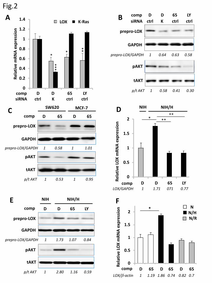
Fig.2
0
0.2
0.4
0.6
0.8
1
1.2
1.4
D K 65 LY
Rela
tive
mRN
A ex
pres
sion
LOX KRasA B
GAPDH
tAKT
prepro-LOX
pAKT
prepro-LOX/GAPDH 1 0.64 0.63 0.58
p/t AKT 1 0.58 0.41 0.30
GAPDH
tAKT
prepro-LOX
pAKT
D 65 D 65
SW620 MCF-7C
prepro-LOX/GAPDH 1 0.58 1 1.01
p/t AKT 1 0.53 1 0.95
D
E F
p/t AKT 1 2.80 1.16 0.59
0
0.5
1
1.5
2
N-D
a-D
a-65
a-LY
a-B
Rela
tive
LOX
mRN
A ex
pres
sion
D D 65 LY
NIH
*
NIH/H
****
GAPDH
tAKT
prepro-LOX
pAKT
D D 65 LY
NIH NIH/H
prepro-LOX/GAPDH 1 1.73 1.07 0.84
Rela
tive
LOX
mRN
A ex
pres
sion
0
0.5
1
1.5
2
2.5
N N/H N/RD 65 D 65 D 65
* □ N■ N/H
N/R
F
LOX/GAPDH 1 1.71 071 0.77
LOX/β-actin 1 1.19 1.86 0.74 0.82 0.7
siRNA ctrl K ctrl ctrlcomp D D 65 LY
comp D D 65 LYsiRNA ctrl K ctrl ctrl
comp
comp
comp
comp
** **
***
K-RasLOX
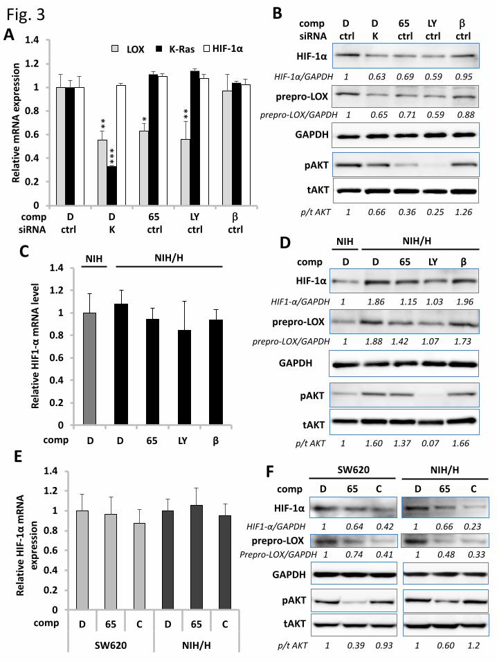
Fig. 3A
0
0.2
0.4
0.6
0.8
1
1.2
1.4
D K 65 LY β
Rela
tive
mRN
A ex
pres
sion
LOX KRas HIF-1α
GAPDH
HIF-1α
HIF-1α/GAPDH 1 0.63 0.69 0.59 0.95
B
0
0.2
0.4
0.6
0.8
1
1.2
1.4
N-D a-D a-65 a-LY a-B
C
Rela
tive
HIF1
-αm
RNA
leve
l HIF-1α
D D 65 LY β
D D 65 LY β
NIH NIH/H
GAPDH
DNIH NIH/H
HIF1-α/GAPDH 1 1.86 1.15 1.03 1.96
comp
F
0
0.2
0.4
0.6
0.8
1
1.2
1.4
D 65 C D 65 C
SW620 NIH/H
Rela
tive
HIF-
1α m
RNA
expr
essi
on
E
HIF-1α
D 65 C
NIH/H
GAPDH
HIF1-α/GAPDH 1 0.64 0.42 1 0.66 0.23
tAKT
pAKT
prepro-LOX
D 65 C
SW620
Prepro-LOX/GAPDH 1 0.74 0.41 1 0.48 0.33
p/t AKT 1 0.39 0.93 1 0.60 1.2
comp
prepro-LOX/GAPDH 1 0.65 0.71 0.59 0.88
prepro-LOX
tAKT
pAKT
p/t AKT 1 0.66 0.36 0.25 1.26
prepro-LOX
p/t AKT 1 1.60 1.37 0.07 1.66
tAKT
pAKT
prepro-LOX/GAPDH 1 1.88 1.42 1.07 1.73
comp D D 65 LY βsiRNA ctrl K ctrl ctrl ctrl
comp D D 65 LY βsiRNA ctrl K ctrl ctrl ctrl
comp
comp
***
**
* **
K-RasLOX
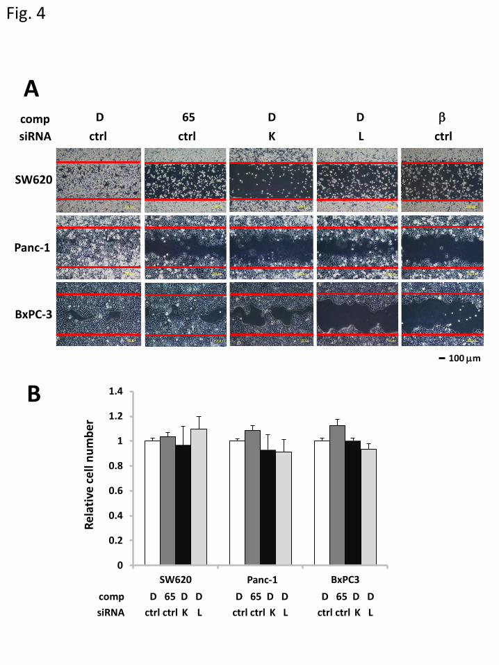
Fig. 4
Rela
tive
cell
num
ber
Panc-1
BxPC-3
SW620
B100 µm
0
0.2
0.4
0.6
0.8
1
1.2
1.4
SW620 Panc1 BxPC3
DMSO
Kobe0065
siKRas
siLOX
Panc-1
AD 65
K LβD D
ctrl ctrl ctrlsiRNA comp
comp D 65 D D D 65 D D D 65 D DsiRNA ctrl ctrl K L ctrl ctrl K L ctrl ctrl K L

Fig. 5
0
50
100
0 uM 2 uM 10 uM
DMSO Kobe0065-2 µM Kobe0065-10 µM
100 µm
Panc-1
BxPC-3
SW620
0
50
100
0 uM 2 uM 10 uM
Rela
tive
num
ber
of th
e in
vadi
ng c
ells
(%)
****** ***
***
0
50
100
0 uM 2 uM 10 uM
Kobe0065 (µM)0 2 10
A
BSW620 Panc-1 BxPC-3
0 2 10 0 2 10 Kobe0065 (µM) Kobe0065 (µM)

B
0
10
20
30
40
50
The
num
ber o
f met
asta
tic
nodu
les p
er lu
ng
Kobe0065-160 mg/kg sorafenib-80 mg/kg
vehicle Kobe0065-80 mg/kgFig. 6A
C
***
Treatment number of mice with metastasis
vehicle 7/7 (100%)
Kobe006580 mg/kg 3/4 ( 75%)
Kobe0065160 mg/kg 3/6 ( 50%)
sorafenib80 mg/kg 5/5(100%)
0
0.2
0.4
0.6
0.8
1
1.2Re
lativ
e LO
X m
RNA
leve
l
E
******
2 mmD
vehicle Kobe0065-80 mg/kg Kobe0065-160 mg/kg sorafenib-80 mg/kg
50 µm

GAPDH
prepro-LOX
prepro-LOX/GAPDH
D 65 2601 2602 sora
1 0.43 0.63 0.55 0.88
Yoshikawa Y, et al. Supplementary Fig. 1
Effects of Kobe0065-family compounds on LOX expression. SW620 cells grown in 3D culture were treated with 20 µMKobe0065 (65), 20 µM Kobe2601 (2601), 20 µM Kobe2602 (2602), 2 µM sorafenib (sora) or DMSO (D) as described in Materials and Methods and subjected to the detection of prepro-LOX and GAPDH by western blotting with the anti-LOX and anti-GAPDH antibodies, respectively. Numbers below the lanes indicate the values of prepro-LOX/GAPDH relative to those of the DMSO-treated cells.

GAPDH
tAKT
prepro-LOX
pAKT
D K 65 LY
prepro-LOX/GAPDH 1 0.68 0.63 0.51
p/t AKT 1 0.61 0.39 0.250
0.2
0.4
0.6
0.8
1
1.2
D K 65 LY
Rela
tive
mRN
A ex
pres
sion
LOX KRas
Yoshikawa Y, et al. Supplementary Fig. 2
Crucial role of the PI3K-Akt signaling in Ras-mediated LOX expression in 2D-cultured SW620. A. SW620 cells in 2D culture were analyzed for the effects of the transfection of K-Ras siRNA (K) or the non-targeting control (ctrl) and of the treatments with Kobe0065 (65), LY294002 (LY) or DMSO (D) on the relative LOX and K-ras mRNA levels as described in Fig. 2A legend. Three independent experiments, each conducted in triplicate, yielded essentially equivalent results. B.Lysates of the cells, treated as described in A, were subjected to the detection of prepro-LOX, phosphorylated Akt (p), total Akt (t) and GAPDH by western blotting with the respective antibodies. Numbers below the lanes indicate the values of prepro-LOX/GAPDH and phosphorylated Akt/total Akt relative to those of the DMSO-treated cells.
***
*****
***
compsiRNA
D D 65 LYctrl K ctrl ctrl
K-RasLOX
A B

Yoshikawa Y, et al. Supplementary Fig. 3
0
0.5
1
1.5
2
2.5
Rela
tive
LOX
mRN
A le
vel
*
Effects of various small GTPases on LOX expression. NIH3T3 cells were transfected with pEF-BOS-HA vector (V), pEF-BOS-HA-K-RasG12V (K-RasG12V), pEF-BOS-HA-M-RasQ71L (M-RasQ71L), pEF-BOS-HA-Rap2AG12V (Rap2AG12V), pEF-BOS-HA-RhoAG14V (RhoAG14V) or pCMV2-flag-RalA (RalA) by using Lipofectoamine 2000 (Invitrogen) according to the manufacturer’s instructions. Three days after transfection, total RNAs were prepared and subjected to the measurements of the relative LOX mRNA levels by qRT-PCR using the GAPDH mRNA levels as an internal control. Three independent experiments, each conducted in triplicate, yielded essentially equivalent results. Statistical analysis was performed as described in Materials and Methods.
V K-RasG12V M-RasQ71L Rap2AG12V RhoAG14V RalA
LOX/GAPDH 1 1.85 0.91 1.00 1.05 0.94

Yoshikawa Y, et al. Supplementary Fig. 4
D D 65 D D 65
siRNA ctrl K ctrl ctrl K ctrl
Panc-1 BxPC-3
comp
HIF1-α/GAPDH 1 0.79 0.69 1 1.02 0.92
GAPDH
HIF1-α
Role of Ras in accumulation of HIF-1α and Akt phosphorylation in Panc-1 and BxPC-3. Twenty-four hours after transfection of K-Ras siRNA (K) or the non-targeting control (ctrl), Panc-1 and BxPC-3 cells in 3D culture were treated with Kobe0065 (65) or DMSO (D) as described in Material & Methods. Thereafter, lysates of the cells were subjected to the detection of HIF-1a, GAPDH, phosphorylated Akt (p) and total Akt (t) by western blotting with the respective antibodies. Numbers below the lanes indicate the values of HIF-1α/GAPDH and phosphorylated Akt/total Akt relative to those of the DMSO-treated cells.
tAKT
pAKT
p/t AKT 1 0.73 0.65 1 0.96 1.02

Supplementary Table 1. The nucleotide sequences of the Stealth siRNAs
Name Species Primer Sequence
K-Ras human sense 5’-UGUGGACGAAUAUGAUCCAACAAUA-3’
antisense 5’-UAUUGUUGGAUCAUAUUCGUCCACA-3’
sense 5’-AUAACUUCUUGCUAAGUCCUGAGCC-3’
antisense 5’-GGCUCAGGACUUAGCAAGAAGUUAU-3’
sense 5’-CAAGACAGAGAGUGGAGGAUGCUUU-3’
antisense 5’-AAAGCAUCCUCCACUCUCUGUCUUG-3’
LOX human sense 5’-AUCAUAAUCUCUGACAUCUGCCCUG-3’
antisense 5’-CAGGGCAGAUCUCAGCACUUAUGAU-3’
sense 5’-UGGGUAAGAAAUCUGAUGUCCCUUG-3’
antisense 5’-CAAGGGACAUCAGAUUUCUUACCCA-3’
sense 5’-GGAUUGAGUCCUGGCUGUUAUGAUA-3’
antisense 5’-UAUCAUAACAGCCAGGACUCAAUCC-3’

Supplementary Table 2. Primers used for qRT-PCRName Species Primer Sequence
LOX mouse forward 5’-TGCCAGTGGATTGATATTACAGATGT -3’
reverse 5’-AGCGAATGTCACAGCGTACAA -3’
LOX human forward 5’-CCAGAGGAGAGTGGCT-3’
reverse 5’-CCAGGTAGCTGGGGTT-3’
HIF-1α mouse forward 5’-CCCAGATTCAGGATCAGACAC -3’
reverse 5’-TTGGATATAGGGAGCTAAC ATC-3’
HIF-1α human forward 5’-CCCAGATTCAGGATCAGACAC -3’
reverse 5’-TTGGATATAGGGAGCTAAC ATC-3’
GAPDH mouse forward 5’-GAGATTGTTGCCATCAACGAC -3’
reverse 5’-TTTGATGTTAGGGGGTCTCG -3’
GAPDH human forward 5’-CATCAATGGAAATCCCATCAC -3’
reverse 5’-GCAGAGATGATGACCCTTTTG -3’
K-Ras human forward 5’-TGTGGTAGTTGGAGCTGGTG-3’
reverse 5’-CCCAGTCCTCATGTACTGGTC -3’
β-actin mouse forward 5’-ATGAAGATCAAGATCATTGCTCCTC -3’
reverse 5’-ACATCTGCTGGAAGGTGGACAG -3’