knockout esc/ipsc medium kit - thermo fisher...
TRANSCRIPT

For support visit www.lifetechnologies.com/support or contact [email protected].
www.lifetechnologies.com Intended Use: For research use only. Caution: Not intended for any animal or human therapeutic or diagnostic use.
Related Products Product Cat. No.
GIBCO® Mouse Embryonic Fibroblasts (Irradiated) S1520-100
DMEM (1X), liquid (high glucose) with GlutaMAX™-I 10569-010
Fetal Bovine Serum, ES Cell-Qualified (FBS) 16141-079
MEM Non-Essential Amino Acids Solution (100X) 11140-050
Attachment Factor S-006-100
2-Mercaptoethanol (1,000X), liquid 21985-023
D-PBS without Ca2+ and Mg2+ (1X), liquid 14190-250
Dispase 17105-041
Explanation of Symbols and Warnings The symbols present on the product label are explained below:
See insert
Biohazard See website
Protect from light Storage condition Expir. date
Lot/batch
# GIBCO cat. #
Research Use Only
Sterilized by filtration
Product Qualification and SDS The Certificate of Analysis provides detailed quality control and product qualification information for each product. Certificates of Analysis are available on our website. Go to www.lifetechnologies.com/support and search for the Certificate of Analysis by product lot number, which is printed on the box. Safety Data Sheets (SDSs) are available at www.lifetechnologies.com/sds.
Product Qualification and Purchaser Notification Limited Warranty Life Technologies Corporation is committed to providing our customers with high-quality goods and services. Our goal is to ensure that every customer is 100% satisfied with our products and our service. If you should have any questions or concerns about a Life Technologies product or service, contact our Technical Support Representatives. All Life Technologies products are warranted to perform according to specifications stated on the certificate of analysis. The Company will replace, free of charge, any product that does not meet those specifications. This warranty limits the Company’s liability to only the price of the product. No warranty is granted for products beyond their listed expiration date. No warranty is applicable unless all product components are stored in accordance with instructions. The Company reserves the right to select the method(s) used to analyze a product unless the Company agrees to a specified method in writing prior to acceptance of the order. Life Technologies makes every effort to ensure the accuracy of its publications, but realizes that the occasional typographical or other error is inevitable. Therefore the Company makes no warranty of any kind regarding the contents of any publications or documentation. If you discover an error in any of our publications, report it to our Technical Support Representatives. Life Technologies Corporation shall have no responsibility or liability for any special, incidental, indirect or consequential loss or damage whatsoever. The above limited warranty is sole and exclusive. No other warranty is made, whether expressed or implied, including any warranty of merchantability or fitness for a particular purpose.
Limited Use Label License: Research Use Only The purchase of this product conveys to the purchaser the limited, non-transferable right to use the purchased amount of the product only to perform internal research for the sole benefit of the purchaser. No right to resell this product or any of its components is conveyed expressly, by implication, or by estoppel. This product is for internal research purposes only and is not for use in commercial applications of any kind, including, without limitation, quality control and commercial services such as reporting the results of purchaser’s activities for a fee or other form of consideration. For information on obtaining additional rights, contact [email protected] or Out Licensing, Life Technologies, 5791 Van Allen Way, Carlsbad, California 92008.
Limited Use Label License 288: Stem Cell Intellectual Property Disclaimer Products associated with this label license are not provided with a license to any patents not owned by or licensed to Life Technologies Corporation and related to stem cells. Users of Life Technologies Corporation’s products subject to this label license should determine for themselves whether they have all of the appropriate licenses in place. Further, no warranty is provided that the use of these products will not infringe third party intellectual property related to stem cells.
Limited Use Label License No: 297 Media for Stem Cell Culture The purchase of this product conveys to the buyer the non-transferable license to use the purchased amount of the product and components of the product in internal research conducted by the buyer (whether the buyer is an academic or for-profit entity), subject to certain restrictions as follows: The buyer cannot sell or otherwise transfer (a) this product (b) its components or (c) materials made using this product or its components to a third party or otherwise use this product or its components or materials made using this product or its components for activities in the Excluded Fields. The Excluded Fields include: a) the implantation or production for implantation of primate Pluripotent Stem Cells (pPSC's) or cells or materials derived from pPSCs into an animal by, or in collaboration with, a for-profit entity. b) the manufacture of therapeutic products, diagnostic products, or components of either; c) the manufacture of a product for sale or for use in commercial services or the use of a product in commercial services, and/or d) the production of materials for use in human clinical trials. Through its use of this product, the purchaser names Geron Corporation as a third party beneficiary of this license for the sole purpose of enforcing such license. Parties wishing to use this product for applications within the Excluded Fields will require and must request a direct license from Geron. Such a license request should be sent to Geron Corporation, Attn: Legal Dept., 230 Constitution Drive, Menlo Park, California, 94025. If the purchaser is not willing to accept the limitations of this limited use statement, Invitrogen is willing to accept return of the product with a full refund. For additional information on purchasing a license to this product for purposes other than research, please contact [email protected] or Out Licensing, Life Technologies, 5791 Van Allen Way, Carlsbad, California 92008.
2011 Life Technologies Corporation. All rights reserved. The trademarks mentioned herein are the property of Life Technologies Corporation or their representative owners.
KnockOut™ ESC/iPSC Medium Kit Cat. nos. A14129-01, A14130-01 Pub. Part no. A14131 MAN0004576 Rev. Date 27 June 2011
Description KnockOut™ ESC/iPSC Medium Kit contains all the necessary components for preparing complete pluripotent stem cell (PSC) growth medium for routine maintenance and derivation of human and non-human primate embryonic stem cells (ESCs) and induced pluripotent stem cells (iPSCs). The complete PSC growth medium has three components: basal medium (DMEM/F-12), KnockOut™ Serum Replacement (SR), and basic recombinant human fibroblast growth factor (bFGF). Instructions for culturing PSCs on feeder cells using the KnockOut™ ESC/iPSC Medium Kit are provided below.
Component Part No. Amount Storage
A14129-01 A14130-01
KnockOut™ Serum Replacement 10828-010 10828-028
100 mL –
– 500 mL
–5 to –20°C, protect from light
DMEM/F-12 with GlutaMAX™-I (1X), liquid 10565-018 500 mL 5 × 500 mL 2 to 8°C
FGF-basic (AA 1-155) Recombinant Human (bFGF) PHG0264 10 µg 2 × 10 µg 2 to 8°C
Shelf Life of Medium Components • DMEM/F-12: 12 months • bFGF: 5 years • KnockOut™ SR: 18 months • Complete growth medium: 2 weeks at 2 to 8°C, protected
from light
Precautions • Do not heat-inactivate KnockOut™ SR. • KnockOut™ SR cannot be used as a replacement for FBS in
the plating of feeder cells. While the formulation contains sufficient factors to allow plating of ESCs and iPSCs, fibroblasts have an increased need for undefined attachment factors and will not adequately attach in this formulation. However, once plated, the feeder cell layer will remain attached to the plates when placed into media containing KnockOut™ SR.
• KnockOut™ SR does not contain trypsin inhibitors. Therefore, trypsin must be removed or inactivated when culturing ESCs in KnockOut™ SR containing medium.
Storage and Handling Basic FGF (bFGF) • Store lyophilized human bFGF at 2 to 8°C, preferably
desiccated. • Aliquot and store reconstituted human bFGF solution at
≤–20°C (not in a frost-free freezer) for up to 3 months. Keep freeze-thaw cycles to a minimum.
• Once bFGF aliquot is thawed, store at 2 to 8°C and use within 7 days.
KnockOut™ SR • Store KnockOut™ SR at –5 to –20°C, protect from light. • To thaw KnockOut™ SR, place at 2 to 8°C overnight. If the
bottle is not completely thawed by the next day, place the bottle in a 37°C water bath and continue thawing until the ice is gone and the supplement is clear. Gently swirl the bottle occasionally while in the water bath. Alternatively, KnockOut™ SR can be thawed in a 37°C water bath, as long as the bottle is swirled frequently throughout the thawing process.
• Occasionally some flocculent material may be observed while thawing, but this material will go into solution with gentle swirling at 37°C. Minimize dwell time in waterbath.
• Store thawed KnockOut™ SR at 2 to 8°C in the dark for up to 4 weeks, or aliquot into working volumes and store at –5 to –20°C. Thaw aliquots as needed. Avoid additional freeze-thaw cycles.
Additional Materials Required In addition to the reagents provided with KnockOut™ ESC/iPSC Medium Kit, you will need the following materials (see page 4 for ordering information). • GIBCO® Mouse Embryonic Fibroblasts (MEF) (Irradiated) • DMEM (1X) with GlutaMAX™-I, liquid • Fetal Bovine Serum, ES Cell-Qualified (FBS) • MEM Non-Essential Amino Acids Solution (100X) (NEAA) • Attachment Factor • 2-Mercaptoethanol (1,000X), liquid • D-PBS without Ca2+ and Mg2+ (1X), liquid • Dispase

2 3
Preparing Solutions Basic FGF Solution (10 µg/mL) 1. To prepare 1 mL of 10-µg/mL bFGF solution, aseptically mix
the following components: Basic FGF 10 µg D-PBS without Ca2+ and Mg2+ 1 mL KnockOut™ SR 20 µL
2. Aliquot and store the bFGF solution at –20°C for up to 3 months. Once thawed, store the bFGF at 2 to 8°C and use within 7 days.
Dispase Solution (2 mg/mL) 1. To prepare 50 mL of 2-mg/mL dispase solution, aseptically
mix the following components: Dispase 10 mg DMEM/F-12 50 mL
2. Sterilize the dispase solution through 0.22 µm filter. The solution can be stored at 2 to 8°C for up to 14 days.
Preparing Media Pluripotent Stem Cell (PSC) Medium (100 mL) 1. To prepare 100 mL of complete PSC medium, aseptically mix
the following components: DMEM/F-12 79 mL KnockOut™ SR 20 mL NEAA 1 mL 2-Mercaptoethanol 100 µL
2. Add bFGF (final concentration 4 ng/ml) fresh prior to use (i.e., 0.4 µL of reconstituted bFGF per mL of medium). Complete PSC medium can be stored at 2 to 8°C for up to 14 days.
Complete MEF Medium (500 mL) 1. To prepare 500 mL of complete MEF medium, aseptically
mix the following components: DMEM 448.5 mL FBS, ESC-Qualified 50 mL NEAA 1 mL 2-Mercaptoethanol 500 µL
2. Complete MEF medium can be stored at 2 to 8°C for up to 1 month.
Preparing MEF Culture Vessels Coating Culture Vessels with Attachment Factor 1. Add the appropriate volume of Attachment Factor (AF)
solution into a culture vessel (see Table 1). Table 1 Volume of Attachment Factor for MEF feeder culture
4-chamber 6-well plate 35-mm dish 60-mm dish
0.3 mL 1 mL 1 mL 3 mL
100-mm dish T-25 flask T-75 flask T-175 flask
9 mL 3 mL 9 mL 18 mL
2. Incubate the vessel for 1 hour at room temperature. Immediately before plating MEFs, remove the AF solution from the culture vessel by aspiration. Coated culture vessels may be used immediately after incubation or stored at room temperature for up to 24 hours.
Plating MEFs 1. One day before initiating or passaging PSC culture, plate
Gibco® Mouse Embryonic Fibroblasts (Irradiated) at a density of 30,000/cm2 on a AF-coated culture vessel in MEF medium (see Table 2). Table 2 Recommended media volume for plating MEFs and PSCs
4-chamber 6-well plate 35-mm dish 60-mm dish
0.3 mL 2 mL 2 mL 5 mL
100-mm dish T-25 flask T-75 flask T-175 flask
10 mL 5 mL 12 mL 35 mL
2. Replace the MEF medium with complete PSC medium 3–4 hours before plating the PSCs. Note: MEF culture vessels can be used the day after plating MEFs and should be used within 3–4 days.
Thawing and Plating Human PSCs 1. Warm up PSC medium in a 37°C water bath. 3–4 hours
before plating PSCs, aspirate the MEF medium from the MEF culture vessel and replace it with pre-warmed PSC medium to the dish.
Note: Refer to Table 2 for the recommended volume of PSC medium for plating PSCs.
2. Immerse the vial containing the human PSCs in a 37°C water bath without submerging the cap. Swirl the vial gently. When only an ice crystal remains, remove the vial from the water bath. Spray the outside of the vial with 70% ethanol and place it in a cell culture hood.
3. Transfer the cells gently into a sterile 50-mL conical tube using a sterile serological pipette.
4. Slowly add 10 mL of pre-warmed PSC medium drop-wise to cells in the 50-mL conical tube. While adding the medium, gently move the tube back and forth to mix the cells. Adding the medium slowly helps the cells to avoid osmotic shock.
5. Centrifuge the cells at 200 × g for 2 minutes.
6. Aspirate the supernatant and re-suspend the cell pellet in 2 mL of PSC medium by gently pippeting them up and down using a sterile serological pipette.
7. Label the MEF culture vessel with the passage number of the PSCs from the vial, the date and your initials, and slowly add the PSC suspension into the dish.
Note: For initial recovery, we recommend seeding the cells at 6 × 10⁴ to 1 × 10⁵ cells/cm2.
8. Move the dish gently in several figure eight motions to disperse the PSC colonies across the surface of the dish. Place the dish into a 37°C incubator with a humidified atmosphere of 5% CO2.
9. Replace the spent medium daily. If feeding cells in more than one culture vessel, use a different pipette for each vessel to reduce risk of contamination.
10. Observe the PSCs daily and passage the cultures whenever the colonies are too big or crowded. The ratio of splitting depends on the total number of PSC colonies in the culture vessels. (Approximately 1:3 for PSCs at the first passage after recovery).
Guidelines for Passaging Human PSC Cultures • In general, split cultures when the first of the followings
occurs: (a) MEF feeder layer is two weeks old; (b) PSC colonies are becoming too dense or too large; (c) Increased differentiation occurs.
• Enzymatic passaging of PSCs will vary from cell line to cell line. Some hESC cell lines or hiPSCs may not react in the same manner to enzymatic passaging, and consequently the type of enzyme utilized, concentration of enzyme and exposure time must be empirically determined for the particular cell line to be passaged.
• A typical passage schedule for PSC is as follows: Day 0: Seed MEFs on Attachment Factor-coated tissue culture vessels. Day 1: Seed PSCs on the attached MEF feeder layer. Day 2–4: Change the medium and monitor the culture vessels for distribution and morphology of attached colonies. Day 3–4: Change the medium and monitor morphology daily. Day 5: Passage PSCs onto fresh MEF culture dishes.
Enzymatic Passaging of Human PSC Cultures 1. One or two days before splitting the PSC cultures, prepare
fresh MEF culture vessels.
2. Aspirate the spent PSC medium from the confluent PSC culture dish and add the appropriate amount of Dispase solution(2 mg/mL) to the culture dish containing the PSCs (see Table 3). Table 3 Recommended Dispase (2 mg/mL) volume
4-chamber 6-well plate 35-mm dish 60-mm dish
0.3 mL 1 mL 1 mL 2 mL
100-mm dish T-25 flask T-75 flask T-175 flask
5 mL 2 mL 5 mL 10 mL
3. Incubate the PSC culture for 5–15 minutes in a 37°C incubator with a humidified atmosphere of 5% CO2.
4. Stop the incubation when the edges of the colonies start to pull away from the plate and gently dislodge the colonies using a sterile serological pipette by gently pipetting colonies across the surface of the plate 5 to 8 times. This breaks up the colonies into smaller clumps.
5. Transfer the dispase solution/colony suspension to a 15-mL conical tube. Add a volume of pre-warmed PSC medium equal to the Dispase solution (see Table 3) to the original dish and pipette the medium across the surface of the dish to dislodge any remaining colonies. Transfer this suspension to the 15-mL conical tube and pipette up and down another 2–3 times to re-suspend colonies.
Note: Take care to not introduce bubbles or shear the PSC colonies too much. You may want to break up the colonies into small clusters of 50–500 cells for normal passaging.
6. Centrifuge the cells at 200 × g for 2 minutes at room temperature.
7. Aspirate and discard the supernatant, and then gently tap the tube to loosen the cell pellet from the bottom of the tube.
8. Add 2–5 mL of pre-warmed PSC medium to the cells and re-suspend the colonies by gently pipetting them up and down. Note: Avoid making a single cell suspension.
9. Seed the cluster of cells onto a dish plated with MEFs. Generally, PSCs are passaged at a 1:3 or 1:4 split ratio. The final volume of the medium depends on the size of the culture vessel used (see Table 2).
10. Gently swirl the plates in several figure eight motions to evenly spread out the cell clusters and place them in a 37°C incubator with a humidified atmosphere of 5% CO2.
Daily Monitoring of Passaged Human PSC Cultures • It is important to add fresh medium every day. • It is critical to observe the distribution of attached colonies
24 hours post-passage. • Colonies sometimes can all be clustered towards the middle,
close to each other or on top of each other. An even distribution of colonies is critical to maintain cells undifferentiated during their expansion. It is also important to monitor the attachment and morphology of the colonies. In all dishes, there will always be some areas with unattached cells or cells that look differentiated. However, the majority of the colonies in the dish must be well separated with a pristine morphology.
• Differentiated cells at the edges of each colony can be mechanically removed with a 27-gauge needle before passaging.
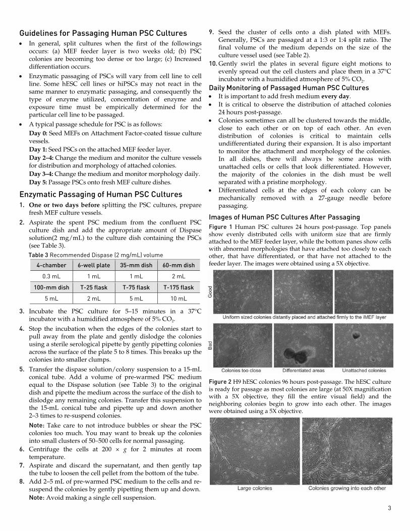
Images of Human PSC Cultures After Passaging Figure 1 Human PSC cultures 24 hours post-passage. Top panels show evenly distributed cells with uniform size that are firmly attached to the MEF feeder layer, while the bottom panes show cells with abnormal morphologies that have attached too closely to each other, that have differentiated, or that have not attached to the feeder layer. The images were obtained using a 5X objective.
Figure 2 H9 hESC colonies 96 hours post-passage. The hESC culture is ready for passage as most colonies are large (at 50X magnification with a 5X objective, they fill the entire visual field) and the neighboring colonies begin to grow into each other. The images were obtained using a 5X objective.
2 3
Preparing Solutions Basic FGF Solution (10 µg/mL) 1. To prepare 1 mL of 10-µg/mL bFGF solution, aseptically mix
the following components: Basic FGF 10 µg D-PBS without Ca2+ and Mg2+ 1 mL KnockOut™ SR 20 µL
2. Aliquot and store the bFGF solution at –20°C for up to 3 months. Once thawed, store the bFGF at 2 to 8°C and use within 7 days.
Dispase Solution (2 mg/mL) 1. To prepare 50 mL of 2-mg/mL dispase solution, aseptically
mix the following components: Dispase 10 mg DMEM/F-12 50 mL
2. Sterilize the dispase solution through 0.22 µm filter. The solution can be stored at 2 to 8°C for up to 14 days.
Preparing Media Pluripotent Stem Cell (PSC) Medium (100 mL) 1. To prepare 100 mL of complete PSC medium, aseptically mix
the following components: DMEM/F-12 79 mL KnockOut™ SR 20 mL NEAA 1 mL 2-Mercaptoethanol 100 µL
2. Add bFGF (final concentration 4 ng/ml) fresh prior to use (i.e., 0.4 µL of reconstituted bFGF per mL of medium). Complete PSC medium can be stored at 2 to 8°C for up to 14 days.
Complete MEF Medium (500 mL) 1. To prepare 500 mL of complete MEF medium, aseptically
mix the following components: DMEM 448.5 mL FBS, ESC-Qualified 50 mL NEAA 1 mL 2-Mercaptoethanol 500 µL
2. Complete MEF medium can be stored at 2 to 8°C for up to 1 month.
Preparing MEF Culture Vessels Coating Culture Vessels with Attachment Factor 1. Add the appropriate volume of Attachment Factor (AF)
solution into a culture vessel (see Table 1). Table 1 Volume of Attachment Factor for MEF feeder culture
4-chamber 6-well plate 35-mm dish 60-mm dish
0.3 mL 1 mL 1 mL 3 mL
100-mm dish T-25 flask T-75 flask T-175 flask
9 mL 3 mL 9 mL 18 mL
2. Incubate the vessel for 1 hour at room temperature. Immediately before plating MEFs, remove the AF solution from the culture vessel by aspiration. Coated culture vessels may be used immediately after incubation or stored at room temperature for up to 24 hours.
Plating MEFs 1. One day before initiating or passaging PSC culture, plate
Gibco® Mouse Embryonic Fibroblasts (Irradiated) at a density of 30,000/cm2 on a AF-coated culture vessel in MEF medium (see Table 2). Table 2 Recommended media volume for plating MEFs and PSCs
4-chamber 6-well plate 35-mm dish 60-mm dish
0.3 mL 2 mL 2 mL 5 mL
100-mm dish T-25 flask T-75 flask T-175 flask
10 mL 5 mL 12 mL 35 mL
2. Replace the MEF medium with complete PSC medium 3–4 hours before plating the PSCs. Note: MEF culture vessels can be used the day after plating MEFs and should be used within 3–4 days.
Thawing and Plating Human PSCs 1. Warm up PSC medium in a 37°C water bath. 3–4 hours
before plating PSCs, aspirate the MEF medium from the MEF culture vessel and replace it with pre-warmed PSC medium to the dish.
Note: Refer to Table 2 for the recommended volume of PSC medium for plating PSCs.
2. Immerse the vial containing the human PSCs in a 37°C water bath without submerging the cap. Swirl the vial gently. When only an ice crystal remains, remove the vial from the water bath. Spray the outside of the vial with 70% ethanol and place it in a cell culture hood.
3. Transfer the cells gently into a sterile 50-mL conical tube using a sterile serological pipette.
4. Slowly add 10 mL of pre-warmed PSC medium drop-wise to cells in the 50-mL conical tube. While adding the medium, gently move the tube back and forth to mix the cells. Adding the medium slowly helps the cells to avoid osmotic shock.
5. Centrifuge the cells at 200 × g for 2 minutes.
6. Aspirate the supernatant and re-suspend the cell pellet in 2 mL of PSC medium by gently pippeting them up and down using a sterile serological pipette.
7. Label the MEF culture vessel with the passage number of the PSCs from the vial, the date and your initials, and slowly add the PSC suspension into the dish.
Note: For initial recovery, we recommend seeding the cells at 6 × 10⁴ to 1 × 10⁵ cells/cm2.
8. Move the dish gently in several figure eight motions to disperse the PSC colonies across the surface of the dish. Place the dish into a 37°C incubator with a humidified atmosphere of 5% CO2.
9. Replace the spent medium daily. If feeding cells in more than one culture vessel, use a different pipette for each vessel to reduce risk of contamination.
10. Observe the PSCs daily and passage the cultures whenever the colonies are too big or crowded. The ratio of splitting depends on the total number of PSC colonies in the culture vessels. (Approximately 1:3 for PSCs at the first passage after recovery).
Guidelines for Passaging Human PSC Cultures • In general, split cultures when the first of the followings
occurs: (a) MEF feeder layer is two weeks old; (b) PSC colonies are becoming too dense or too large; (c) Increased differentiation occurs.
• Enzymatic passaging of PSCs will vary from cell line to cell line. Some hESC cell lines or hiPSCs may not react in the same manner to enzymatic passaging, and consequently the type of enzyme utilized, concentration of enzyme and exposure time must be empirically determined for the particular cell line to be passaged.
• A typical passage schedule for PSC is as follows: Day 0: Seed MEFs on Attachment Factor-coated tissue culture vessels. Day 1: Seed PSCs on the attached MEF feeder layer. Day 2–4: Change the medium and monitor the culture vessels for distribution and morphology of attached colonies. Day 3–4: Change the medium and monitor morphology daily. Day 5: Passage PSCs onto fresh MEF culture dishes.
Enzymatic Passaging of Human PSC Cultures 1. One or two days before splitting the PSC cultures, prepare
fresh MEF culture vessels.
2. Aspirate the spent PSC medium from the confluent PSC culture dish and add the appropriate amount of Dispase solution(2 mg/mL) to the culture dish containing the PSCs (see Table 3). Table 3 Recommended Dispase (2 mg/mL) volume
4-chamber 6-well plate 35-mm dish 60-mm dish
0.3 mL 1 mL 1 mL 2 mL
100-mm dish T-25 flask T-75 flask T-175 flask
5 mL 2 mL 5 mL 10 mL
3. Incubate the PSC culture for 5–15 minutes in a 37°C incubator with a humidified atmosphere of 5% CO2.
4. Stop the incubation when the edges of the colonies start to pull away from the plate and gently dislodge the colonies using a sterile serological pipette by gently pipetting colonies across the surface of the plate 5 to 8 times. This breaks up the colonies into smaller clumps.
5. Transfer the dispase solution/colony suspension to a 15-mL conical tube. Add a volume of pre-warmed PSC medium equal to the Dispase solution (see Table 3) to the original dish and pipette the medium across the surface of the dish to dislodge any remaining colonies. Transfer this suspension to the 15-mL conical tube and pipette up and down another 2–3 times to re-suspend colonies.
Note: Take care to not introduce bubbles or shear the PSC colonies too much. You may want to break up the colonies into small clusters of 50–500 cells for normal passaging.
6. Centrifuge the cells at 200 × g for 2 minutes at room temperature.
7. Aspirate and discard the supernatant, and then gently tap the tube to loosen the cell pellet from the bottom of the tube.
8. Add 2–5 mL of pre-warmed PSC medium to the cells and re-suspend the colonies by gently pipetting them up and down. Note: Avoid making a single cell suspension.
9. Seed the cluster of cells onto a dish plated with MEFs. Generally, PSCs are passaged at a 1:3 or 1:4 split ratio. The final volume of the medium depends on the size of the culture vessel used (see Table 2).
10. Gently swirl the plates in several figure eight motions to evenly spread out the cell clusters and place them in a 37°C incubator with a humidified atmosphere of 5% CO2.
Daily Monitoring of Passaged Human PSC Cultures • It is important to add fresh medium every day. • It is critical to observe the distribution of attached colonies
24 hours post-passage. • Colonies sometimes can all be clustered towards the middle,
close to each other or on top of each other. An even distribution of colonies is critical to maintain cells undifferentiated during their expansion. It is also important to monitor the attachment and morphology of the colonies. In all dishes, there will always be some areas with unattached cells or cells that look differentiated. However, the majority of the colonies in the dish must be well separated with a pristine morphology.
• Differentiated cells at the edges of each colony can be mechanically removed with a 27-gauge needle before passaging.
Images of Human PSC Cultures After Passaging Figure 1 Human PSC cultures 24 hours post-passage. Top panels show evenly distributed cells with uniform size that are firmly attached to the MEF feeder layer, while the bottom panes show cells with abnormal morphologies that have attached too closely to each other, that have differentiated, or that have not attached to the feeder layer. The images were obtained using a 5X objective.
Figure 2 H9 hESC colonies 96 hours post-passage. The hESC culture is ready for passage as most colonies are large (at 50X magnification with a 5X objective, they fill the entire visual field) and the neighboring colonies begin to grow into each other. The images were obtained using a 5X objective.

For support visit www.lifetechnologies.com/support or contact [email protected].
www.lifetechnologies.com Intended Use: For research use only. Caution: Not intended for any animal or human therapeutic or diagnostic use.
Related Products Product Cat. No.
GIBCO® Mouse Embryonic Fibroblasts (Irradiated) S1520-100
DMEM (1X), liquid (high glucose) with GlutaMAX™-I 10569-010
Fetal Bovine Serum, ES Cell-Qualified (FBS) 16141-079
MEM Non-Essential Amino Acids Solution (100X) 11140-050
Attachment Factor S-006-100
2-Mercaptoethanol (1,000X), liquid 21985-023
D-PBS without Ca2+ and Mg2+ (1X), liquid 14190-250
Dispase 17105-041
Explanation of Symbols and Warnings The symbols present on the product label are explained below:
See insert
Biohazard See website
Protect from light Storage condition Expir. date
Lot/batch
# GIBCO cat. #
Research Use Only
Sterilized by filtration
Product Qualification and SDS The Certificate of Analysis provides detailed quality control and product qualification information for each product. Certificates of Analysis are available on our website. Go to www.lifetechnologies.com/support and search for the Certificate of Analysis by product lot number, which is printed on the box. Safety Data Sheets (SDSs) are available at www.lifetechnologies.com/sds.
Product Qualification and Purchaser Notification Limited Warranty Life Technologies Corporation is committed to providing our customers with high-quality goods and services. Our goal is to ensure that every customer is 100% satisfied with our products and our service. If you should have any questions or concerns about a Life Technologies product or service, contact our Technical Support Representatives. All Life Technologies products are warranted to perform according to specifications stated on the certificate of analysis. The Company will replace, free of charge, any product that does not meet those specifications. This warranty limits the Company’s liability to only the price of the product. No warranty is granted for products beyond their listed expiration date. No warranty is applicable unless all product components are stored in accordance with instructions. The Company reserves the right to select the method(s) used to analyze a product unless the Company agrees to a specified method in writing prior to acceptance of the order. Life Technologies makes every effort to ensure the accuracy of its publications, but realizes that the occasional typographical or other error is inevitable. Therefore the Company makes no warranty of any kind regarding the contents of any publications or documentation. If you discover an error in any of our publications, report it to our Technical Support Representatives. Life Technologies Corporation shall have no responsibility or liability for any special, incidental, indirect or consequential loss or damage whatsoever. The above limited warranty is sole and exclusive. No other warranty is made, whether expressed or implied, including any warranty of merchantability or fitness for a particular purpose.
Limited Use Label License: Research Use Only The purchase of this product conveys to the purchaser the limited, non-transferable right to use the purchased amount of the product only to perform internal research for the sole benefit of the purchaser. No right to resell this product or any of its components is conveyed expressly, by implication, or by estoppel. This product is for internal research purposes only and is not for use in commercial applications of any kind, including, without limitation, quality control and commercial services such as reporting the results of purchaser’s activities for a fee or other form of consideration. For information on obtaining additional rights, contact [email protected] or Out Licensing, Life Technologies, 5791 Van Allen Way, Carlsbad, California 92008.
Limited Use Label License 288: Stem Cell Intellectual Property Disclaimer Products associated with this label license are not provided with a license to any patents not owned by or licensed to Life Technologies Corporation and related to stem cells. Users of Life Technologies Corporation’s products subject to this label license should determine for themselves whether they have all of the appropriate licenses in place. Further, no warranty is provided that the use of these products will not infringe third party intellectual property related to stem cells.
Limited Use Label License No: 297 Media for Stem Cell Culture The purchase of this product conveys to the buyer the non-transferable license to use the purchased amount of the product and components of the product in internal research conducted by the buyer (whether the buyer is an academic or for-profit entity), subject to certain restrictions as follows: The buyer cannot sell or otherwise transfer (a) this product (b) its components or (c) materials made using this product or its components to a third party or otherwise use this product or its components or materials made using this product or its components for activities in the Excluded Fields. The Excluded Fields include: a) the implantation or production for implantation of primate Pluripotent Stem Cells (pPSC's) or cells or materials derived from pPSCs into an animal by, or in collaboration with, a for-profit entity. b) the manufacture of therapeutic products, diagnostic products, or components of either; c) the manufacture of a product for sale or for use in commercial services or the use of a product in commercial services, and/or d) the production of materials for use in human clinical trials. Through its use of this product, the purchaser names Geron Corporation as a third party beneficiary of this license for the sole purpose of enforcing such license. Parties wishing to use this product for applications within the Excluded Fields will require and must request a direct license from Geron. Such a license request should be sent to Geron Corporation, Attn: Legal Dept., 230 Constitution Drive, Menlo Park, California, 94025. If the purchaser is not willing to accept the limitations of this limited use statement, Invitrogen is willing to accept return of the product with a full refund. For additional information on purchasing a license to this product for purposes other than research, please contact [email protected] or Out Licensing, Life Technologies, 5791 Van Allen Way, Carlsbad, California 92008.
2011 Life Technologies Corporation. All rights reserved. The trademarks mentioned herein are the property of Life Technologies Corporation or their representative owners.
KnockOut™ ESC/iPSC Medium Kit Cat. nos. A14129-01, A14130-01 Pub. Part no. A14131 MAN0004576 Rev. Date 27 June 2011
Description KnockOut™ ESC/iPSC Medium Kit contains all the necessary components for preparing complete pluripotent stem cell (PSC) growth medium for routine maintenance and derivation of human and non-human primate embryonic stem cells (ESCs) and induced pluripotent stem cells (iPSCs). The complete PSC growth medium has three components: basal medium (DMEM/F-12), KnockOut™ Serum Replacement (SR), and basic recombinant human fibroblast growth factor (bFGF). Instructions for culturing PSCs on feeder cells using the KnockOut™ ESC/iPSC Medium Kit are provided below.
Component Part No. Amount Storage
A14129-01 A14130-01
KnockOut™ Serum Replacement 10828-010 10828-028
100 mL –
– 500 mL
–5 to –20°C, protect from light
DMEM/F-12 with GlutaMAX™-I (1X), liquid 10565-018 500 mL 5 × 500 mL 2 to 8°C
FGF-basic (AA 1-155) Recombinant Human (bFGF) PHG0264 10 µg 2 × 10 µg 2 to 8°C
Shelf Life of Medium Components • DMEM/F-12: 12 months • bFGF: 5 years • KnockOut™ SR: 18 months • Complete growth medium: 2 weeks at 2 to 8°C, protected
from light
Precautions • Do not heat-inactivate KnockOut™ SR. • KnockOut™ SR cannot be used as a replacement for FBS in
the plating of feeder cells. While the formulation contains sufficient factors to allow plating of ESCs and iPSCs, fibroblasts have an increased need for undefined attachment factors and will not adequately attach in this formulation. However, once plated, the feeder cell layer will remain attached to the plates when placed into media containing KnockOut™ SR.
• KnockOut™ SR does not contain trypsin inhibitors. Therefore, trypsin must be removed or inactivated when culturing ESCs in KnockOut™ SR containing medium.
Storage and Handling Basic FGF (bFGF) • Store lyophilized human bFGF at 2 to 8°C, preferably
desiccated. • Aliquot and store reconstituted human bFGF solution at
≤–20°C (not in a frost-free freezer) for up to 3 months. Keep freeze-thaw cycles to a minimum.
• Once bFGF aliquot is thawed, store at 2 to 8°C and use within 7 days.
KnockOut™ SR • Store KnockOut™ SR at –5 to –20°C, protect from light. • To thaw KnockOut™ SR, place at 2 to 8°C overnight. If the
bottle is not completely thawed by the next day, place the bottle in a 37°C water bath and continue thawing until the ice is gone and the supplement is clear. Gently swirl the bottle occasionally while in the water bath. Alternatively, KnockOut™ SR can be thawed in a 37°C water bath, as long as the bottle is swirled frequently throughout the thawing process.
• Occasionally some flocculent material may be observed while thawing, but this material will go into solution with gentle swirling at 37°C. Minimize dwell time in waterbath.
• Store thawed KnockOut™ SR at 2 to 8°C in the dark for up to 4 weeks, or aliquot into working volumes and store at –5 to –20°C. Thaw aliquots as needed. Avoid additional freeze-thaw cycles.
Additional Materials Required In addition to the reagents provided with KnockOut™ ESC/iPSC Medium Kit, you will need the following materials (see page 4 for ordering information). • GIBCO® Mouse Embryonic Fibroblasts (MEF) (Irradiated) • DMEM (1X) with GlutaMAX™-I, liquid • Fetal Bovine Serum, ES Cell-Qualified (FBS) • MEM Non-Essential Amino Acids Solution (100X) (NEAA) • Attachment Factor • 2-Mercaptoethanol (1,000X), liquid • D-PBS without Ca2+ and Mg2+ (1X), liquid • Dispase