kinetic and modelling studies of nad+ and poly(ethylene ... ÷ in horse liver alcohol dehydrogenase...
TRANSCRIPT

Kinetic and modelling studies of NAD+ and poly(ethyleneglycol)-bound NAD+ in horse liver alcohol dehydrogenaseVanhommerig, S.A.M.; Sluyterman, L.A.A.E.; Meijer, E.M.
Published in:Biochimica et Biophysica Acta, Protein Structure and Molecular Enzymology
DOI:10.1016/0167-4838(96)00026-X
Published: 01/01/1996
Document VersionPublisher’s PDF, also known as Version of Record (includes final page, issue and volume numbers)
Please check the document version of this publication:
• A submitted manuscript is the author's version of the article upon submission and before peer-review. There can be important differencesbetween the submitted version and the official published version of record. People interested in the research are advised to contact theauthor for the final version of the publication, or visit the DOI to the publisher's website.• The final author version and the galley proof are versions of the publication after peer review.• The final published version features the final layout of the paper including the volume, issue and page numbers.
Link to publication
Citation for published version (APA):Vanhommerig, S. A. M., Sluyterman, L. A. A. E., & Meijer, E. M. (1996). Kinetic and modelling studies of NAD+and poly(ethylene glycol)-bound NAD+ in horse liver alcohol dehydrogenase. Biochimica et Biophysica Acta,Protein Structure and Molecular Enzymology, 1295(2), 125-138. DOI: 10.1016/0167-4838(96)00026-X
General rightsCopyright and moral rights for the publications made accessible in the public portal are retained by the authors and/or other copyright ownersand it is a condition of accessing publications that users recognise and abide by the legal requirements associated with these rights.
• Users may download and print one copy of any publication from the public portal for the purpose of private study or research. • You may not further distribute the material or use it for any profit-making activity or commercial gain • You may freely distribute the URL identifying the publication in the public portal ?
Take down policyIf you believe that this document breaches copyright please contact us providing details, and we will remove access to the work immediatelyand investigate your claim.
Download date: 19. Jun. 2018

ELSEVIER Biochimica et Biophysica Acta 1295 (1996) 125 - 138
BB Biochi~ic~a et Biophysica A~ta
Kinetic and modelling studies of NAD ÷ and poly( ethylene glycol)-bound NAD ÷ in horse liver alcohol dehydrogenase
Sylvia A.M. Vanhommerig *, Lamoraal A.iE. Sluyterman, Emmo M. Meijer Laboratory of Organic Chemistry, Eindhoven University of Technology. P.O. Box 513, 5600 MB Eindhoven, The Netherlands
Received 24 August 1995; revised 14 February 1996; accepted 15 February 1996
Abstract
Poly(ethylene glycol)-bound nicotinamide adenine dinucleotide (PEG-NAD +) has been successfully employed in the continuous production of L-amino acids fi'om the corresponding o~-keto acids by stereospecific reductive amination. Like many other dehydrogenases also horse liver alcohol dehydrogenase (HLADH) appears to be active with PEG-NAD ÷ as coenzyme, although the turnover number is three to four times lower. The possibilities were considered that the PEG-tail of a PEG-NAD + bound to one active site of the HLADH dimer prevents the binding of another PEG-NAD + to the second site, or that the PEG-tail causes destabilization of the active dimer. Both could be ruled out by kinetic studies. Neither can the observed lower intrinsic reactivity of PEG-NAD + account for the diminished activity of the enzyme. Molecular dynamics studies, on the other hand, show that the pulling action of the polymer chain shifts the NAD position in the active site in the outside direction, causing small but significant changes in the enzyme/coenzyme interactions of a sufficient extent to explain the experimental results.
Keywords: Poly(ethylene glycol)-bound NAD +; Alcohol dehydrogenase; Kinetic modelling; Reductive amination, stereospecific; Molecular dynamics and mechanics; (Horse liver)
1. Introduct ion
A number of synthetically useful enzymatic reactions require cofactors such as nicotinamide adenine dinucleo- tide (NAD÷). These cofactors are too expensive to be used as stoichiometric reagents in commercial applications. Re- generation of the cofactors from their reaction by-products is thus required to make the process economical. Many procedures have been suggested to achieve efficient coen- zyme recycling, including enzymatic and non-enzymatic methods [1]. One of the systems investigated, the enzyme
Abbreviations: NAD +, nicotirtamide adenine dinucleotide (oxidized form); NADH, nicotinamide adenine dinucleotide (reduced form); PEG- NAD ÷, poly(ethylene glycol)-bound nicotinamide adenine dinucleotide (oxidized form); PEG-NADH, reduced form of PEG-NAD+; NMN +, nicotinamide mononucleotide; MM, molecular mechanics; MD, molecu- lar dynamics; AMBER, assisted model building with energy refinement; PEG, poly(ethylene glycol); HLADH, horse liver alcohol dehydrogenase (EC 1.1.1.1); BSA, bovine serum albumin; DMSO, dimethyl sulfoxide; rms, root mean square; EtO-, ethanolate; iPrO-, isopropanolate; IPA, isopropanol.
* Corresponding author. Fax: + 31 40 2451036.
0167-4838/96/$15.00 © 1996 Eh;evier Science B.V. All rights reserved PII S01 6 7 - 4 8 3 8 ( 9 6 ) 0 0 0 2 6 - X
membrane reactor concept developed by Wandrey and co-workers [2,3], shows commercially attractive features. For example, continuous production of L-amino acids from the corresponding a-keto acids by stereospecific reductive amination has been achieved with little cofactor consump- tion in a membrane reactor in which NAD ÷ is covalently linked to poly(ethylene glycol) (PEG-NAD +, Fig. 1) [4].
Many other dehydrogenases accept the PEG-NAD + as coenzyme. So far, glucose dehydrogenase is the only enzyme known that is not active with PEG-NAD + [5]. Until now, the observed activities of PEG-NAD ÷ with dehydrogenases are not yet understood at the molecular level.
The N A D + / N A D H dependent horse liver alcohol de- hydrogenase (HLADH), which stereoselectively catalyses the interconversions of alcohols to their corresponding aldehyde/ketones and vice versa, seems an ideal candidate for studies aiming at the elucidation of coenzyme/apo-en- zyme interactions. Many kinetic data and results from X-ray crystallographic studies on binary and ternary com- plexes are available for HLADH [6-13], making this enzyme one of the best characterized dehydrogenases.

126 S.A.M. Vanhommerig et al. / Biochimica et Biophysica Acta 1295 (1996) 125-138
O HN--CI42-CH2-NH---CO
I I I R ~ P - - P - - R N/I- - PEG-- Sl~eer--NAD +
11 Lv~ NAD + spacer
Fig. 1. Structure of polyethylene g lycol bound N A D +. R = ribose, P =
phosphate , PEG = polyethylene glycol = (CH2-CH 2-O)n.
Several groups have performed modelling studies on this enzyme system, trying to obtain more details about the catalytic and structural features of HLADH [ 14-16].
In the past we have used a molecular mechanics ap- proach to study in detail the interactions of the coenzyme NAD + and a number of analogues in the active site of HLADH, in order to understand the essential factors in- volved in the productive binding between coenzyme and apo-enzyme [17-20]. In this paper we present the results of detailed kinetic studies on HLADH with PEG-NAD ÷ as coenzyme, and an extension of our modelling studies including molecular mechanics (MM) and dynamics (MD), to rationalize the kinetic data obtained.
2. M a t e r i a l s a n d m e t h o d s
2.1. Materials
HLADH, NMN ÷ and [3-NAD ÷ were purchased from Sigma. PEG-NADH (M r = 20000) was supplied by Prof. Dr. C. Wandrey and Dr. U. Kragl from the Institute of Biotechnology in Jtilich, prepared according to Ref. [21]. The coenzyme NAD ÷ was lyophilised from distilled wa- ter. HLADH from Sigma (specific activity 1-2 U /mg) was used because its ethanol content, less than 0.15%, proved to be low enough not to disturb activity determina- tions using isopropanol as substrate, making extensive dialysis or gel filtration of the enzyme [22] superfluous. The PEG-NADH was oxidized to PEG-NAD ÷ as de- scribed by Biickmann et al. [21]. The PEG-NAD + was purified using a bacterial filter, with a pore size of 0.2 Ixm. The coenzyme concentrations were determined spectropho- tometricaUy using the following absorption coefficients: 18000 M -l cm -1 a t }krnax = 260 nm for NAD +, 21 600 M -1 cm -~ a t h m a x = 267.5 nm for PEG-NAD + and 6220 M -1 cm -~ at 340 nm in case of the reduced forms. Sodium dithionite was purchased from Janssen Pharmaceu- tical (purity =80%; e = 7 0 0 0 M -I cm-1), potassium cyanide from Merck and Koshland reagent from Aldrich (purity 98%; e = 18000 M -1 cm- 1 at k = 410 nm and pH 10).
2.2. Kinetic measurements
All kinetic experiments were carried out at 25°C _ 0. I°C in 47 mM phosphate buffer, 0.25 mM EDTA, F- -0 .1 .
The pH was adjusted to 8.0 with KOH. A Perkin-Elmer UV/Vis spectrophotometer Lambda 3B was used. Steady-state kinetics were carried out with coenzyme satu- ration (0.55 mM NAD ÷ or 0.59 mM PEG-NAD +) and a substrate concentration range from 1-10 mM in the case of ethanol and 10-100 mM in the case of isopropanol (see Appendix A for the equation used to determine the initial rates); the NAD + and PEG-NAD ÷ concentrations were varied between 5-55 ixM at a constant ethanol concentra- tion of 10 raM. The change of absorption at 340 nm was measured. The enzyme concentrations ranged from 0.03- 0.52 IxM during the steady-state measurements. BSA was added to the enzyme stock solution to stabilize the HLADH. The interference of poly(ethylene glycol) in the steady-state kinetics was checked using 1 mM PEG concentration (2%, w/v) , 10 mM ethanol and NAD + or PEG-NAD + (varied between 5-500 IxM) as coenzyme.
2.3. Formation of adducts
The rates of formation of sulphinate adducts were deter- mined at 20.1°C in a Hi-Tech Scientific stopped-flow spectrophotometer SF-51. Equal amounts of an anaerobic 0.20 mM sodium dithionite solution in 50 mM Tris-HC! buffer (pH 7.8) and an anaerobic 1.32 mM coenzyme solution in phosphate buffer (pH 8.2) were mixed, under an argon flush. The total volume was 150 txl. The forma- tion of the sulphinate adduct was followed spectrophoto- metrically by measuring the change in absorption at 353 nm (e,ddu~ = 3200 M -1 cm-1). All the solutions used were made anaerobic by repeatedly flushing with purified argon [23].
The formation of the cyanide adduct was carried out at 25°C in a 0.01 M KCN solution. The coenzyme concentra- tion was 0.17 mM in a total volume of 2.5 ml. The reactions were followed spectrophotometrically by measur- ing the increase in absorption at 325 n m (~-adduct = 6300 M i cm -1) [24]. A Perkin-Elmer UV/Vis spectro- photometer Lambda 3B was used.
2.4. Molecular mechanics and dynamics calculations
Energy calculations, total energy-minimizations and molecular dynamics simulations were performed with the molecular mechanical simulation package AMBER 4.0 [25] on an Power Challenge SGI computer. The potential en- ergy function used by AMBER is of the form:
V= E k r ( r - r e ) 2÷ E ko(O-Oe) 2 bonds angles
dihedrals
( A ÷ E r12
i <j t t
l + cos[n -
÷ - ÷ E ° rij E H bonds

S.A.M. Vanhommerig et aL / Biochimica et Biophysica Acta 1295 (1996) 125-138 1 2 7
The crystallographic structure of the ternary complex of H L A D H / N A D H / D M S O [13] was used as a starting structure and obtained from the Protein Data Bank (PDB reference number 6ADH). The cysteine residues 46, 103, 111 and 174 were defined as negatively charged cysteines, containing thiolate groups. All histidine residues were taken as HID except the His-51, which was taken HIP (diprotonated histidine). Ethanolate or isopropanolate was substituted at the position of DMSO. Nine water molecules were added manually, according to their subsequent pres- ence in both the binary complex of HLADH (PDB file 5ADH) [12] and the X-ray structure of HLADH with NAD + and pentafluorobenzyl alcohol [6]. Then water was added in a range of 2.9 n;n around the catalytic zinc ion, applying a minimum dislance of 0.26 nm between the oxygen atom of a water molecule and the heavy atoms of the enzyme complex, resulting in 1368 water residues in the shell around the enzyme. All water molecules were treated as TIP3P residues [26]. Since the enzyme is too large to be minimized completely during the calculations, the belly option was used so that only the amino acid residues within a radius of 1.5 nm around NAD--, the alcoholate, the catalytic zinc atom, NAD + and all water molecules were allowed to move freely (6825 atoms in the belly); but first the water molecules and the substrate were optimized, keeping the rest: fixed.
The three-dimensional starting structure of PEG-NAD + was constructed from the X-ray NAD + geometry to which the PEG-residues were linked using the program QUANTA/CHARMM 3.3.2 [27]. Only 10 PEG residues plus the spacer group (see Fig. 1) were coupled to the exocyclic adenine-NH 2 group. After the MD heating simulation the
PEG-tail is positioned for the main part of the time on the surface of the water shell.
All energy minimizations (MM-calculations, using 750 steps of steepest descent algorithm, followed by the conju- gate gradient algorithm) were performed until the rms gradient value of the energy was less than 0.1 kcal ~, ~, using a dielectric constant e of I and treating all CH, CH 2 and CH 3 groups of the amino acids as united atoms. The non-bonded pair list had a cutoff of 12 A and was updated every 25 time steps. The water cap was restrained at the 29
o
A boundarYobY a harmonic potential with a force constant of 0.5 kcal A-~ (positional constraints). Atomic charges of the nicotinamide moiety, the negatively charged cysteines, iPrO- and of EtO- were derived from 6 - 3 1 G * / / 3 - 2 1 G ab initio calculations (Appendix A Section A.2). The zinc ions carried a charge of 1.45, according to the calculations by Tapia et al. [28]. Most harmonic force constants were obtained directly from the literature or extrapolated (see below, parametrization and Table 1) [29,30].
The molecular dynamics (MD) simulations up to 90 ps were carried out using a time step of 2.0 fs and the SHAKE procedure to constrain all the bond lengths. The following protocol was used: a heating phase of 5 ps to a final temperature of 300 K followed by 35 ps of equilibration and finally an observation period of 50 ps (250 conforma- tions for analysis). Analysis of atomic trajectories was achieved with the CARNAL program [31].
2.5. Parametrization
In order to maintain the tetracoordinated structure of the zinc ion, an explicit Zn -O bond and explicit angles of
T a b l e I
N e w l y a d d e d AMBER f o r c e f i e l d p a r a m e t e r s
Non-bonded parameters for zinc
Z n e = 0 . 1 0 k c a l - m o l - i r * = 1 . 4 0 ,~
Angle parameters for NAD + C A - C - - O
C A - C - - N
C A - C A - N *
C A - N ' - - C A
C A - N " - C T
Torsion parameters for NAD + C A - C A - C - O
C # - C A - C - O "
C A - C A - C - N
C # - C A - C - N "
0 = 1 1 9 . 8 °
0 = 1 1 5 . 7 °
0 = 1 1 9 . 5 °
0 = 1 2 1 . 2 °
0 = 1 2 0 . 6 °
3 , = 1 8 0 °
3, = 0 o
3, = 1 8 0 °
3' = 1 8 0 °
3, = 1 8 0 °
3' = 1 8 0 °
~ / = 1 8 0 °
3 ' = 0 °
k o = 8 0 k c a l . m o l - I . r a d - 2
k 0 = 7 0 k c a l • m o l - i . r a d - 2
k 0 = 7 0 k c a l . m o l - I . r a d - 2
k 0 = 7 0 k c a l . m o l - t - r a d 2
k 0 = 7 0 k c a l - t o o l - t . r a d - 2
V2/2 = 0 . 4 5 k c a l . m o l -
V I / 2 = 0 . 0 2 k c a l m o l -
! / _ , / 2 = 0 . 4 5 k c a l m o l
V t / 2 = 0 . 0 2 k c a l t o o l -
V2/2 = 0 . 4 5 k c a l m o l -
V I / 2 = 0 . 0 2 k c a l m o l
V2/2 = 0 . 4 5 k c a l m o l -
V I / 2 = 0 . 0 2 k c a l m o l -
Restraints to maintain tetracoordhTation around catalytic zinc"
B o n d Z n - O r = 2 . 1 4
A n g l e S - Z n - S 0 = 1 3 2 . 6 °
A n g l e N - Z n - S 0 = 1 0 7 . 8 °
A n g l e N - Z n - O 0 = 9 5 . 0 °
k r = 1 0 0 k c a l . m o l - I . A - 2
k 0 = 5 0 k c a l - m o l - t . r a d - 2
k0 = 5 0 k c a l - m o l - J • r a d - 2
k 0 = 5 0 k c a l • m o l - I . r a d - 2
a C # i s t h e s a m e a t o m t y p e a s C A a n d o n l y u s e d t o d e s c r i b e t h e t o r s i o n o f t h e c a r b o x a m i d e s i d e c h a i n .
128 S.A.M. Vanhommerig et al. / Biochimica et Biophysica Acta 1295 (1996) 125-138
N - Z n - S , S - Z n - S and N - Z n - O were inserted between the zinc ion and its ligands (cf. Table 1). The k r of the Z n - O bond is perhaps a little too low compared with the suggested value in such cases by Ref. [32]. Although ~ e y
o
suggest a req = 1.80 A, we believe that req = 2.14 A is justified, since the observed Zn-O distance in several complexes of HLADH with water or an alcohol are all around this value of 2.14 A. The parameters describing the dihedral barrier for the carboxamide orientation of the side chain of the nicotinamide moiety with 6-31G * atom charges, were estimated as follows: V~/2 = 0.08 kJ mol-1 (0.02 kcal mo1-1) and V2/2 = 1.88 kJ tool-1 (0.45 kcal mo1-1) [20]. This resulted in a rotational barrier of 12.6 kJ mol-~ around the C 3 - - C 7 bond of the nicotinamide moi- ety of the free coenzyme.
3. Results and discussion
3.1. Kinetic results
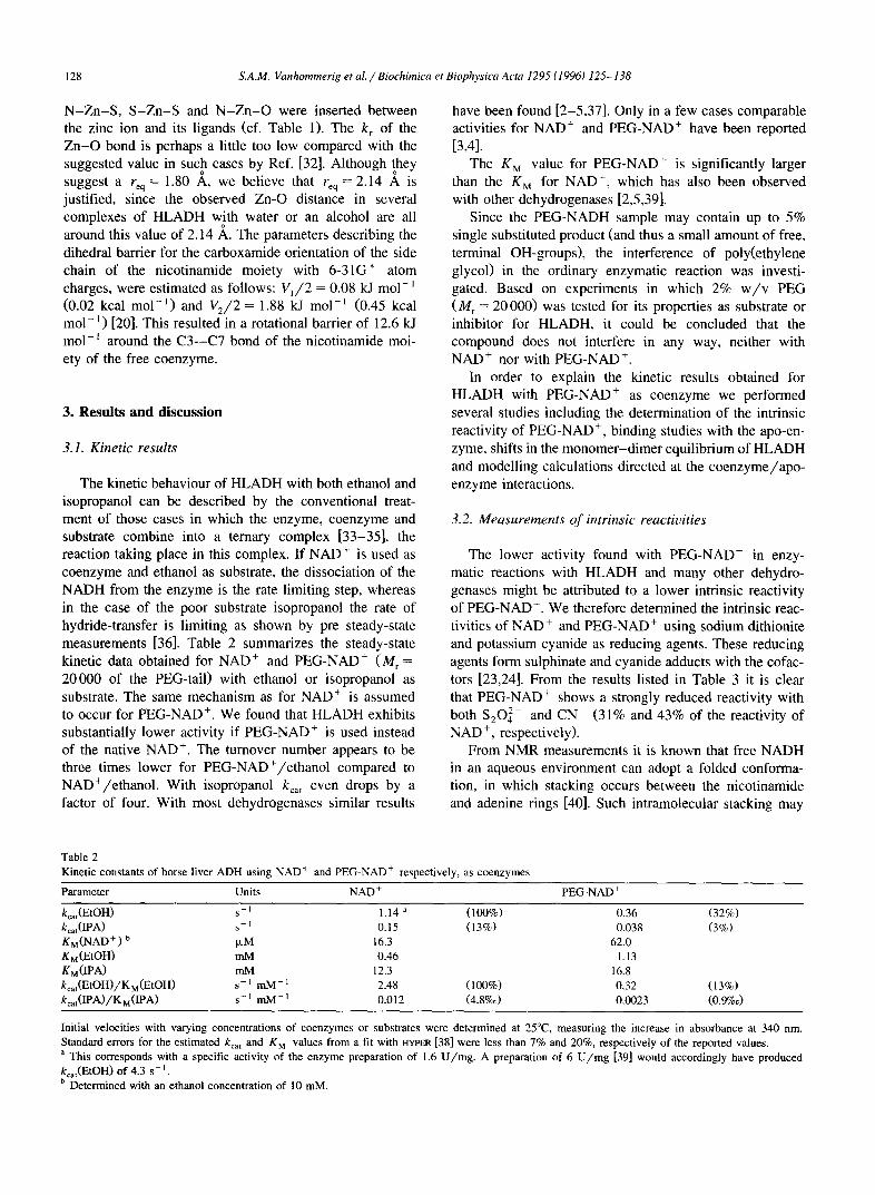
The kinetic behaviour of HLADH with both ethanol and isopropanol can be described by the conventional treat- ment of those cases in which the enzyme, coenzyme and substrate combine into a ternary complex [33-35], the reaction taking place in this complex. If NAD + is used as coenzyme and ethanol as substrate, the dissociation of the NADH from the enzyme is the rate limiting step, whereas in the case of the poor substrate isopropanol the rate of hydride-transfer is limiting as shown by pre steady-state measurements [36]. Table 2 summarizes the steady-state kinetic data obtained for NAD + and PEG-NAD + ( M r = 20000 of the PEG-tail) with ethanol or isopropanol as substrate. The same mechanism as for NAD ÷ is assumed to occur for PEG-NAD +. We found that HLADH exhibits substantially lower activity if PEG-NAD ÷ is used instead of the native NAD ÷. The turnover number appears to be three times lower for PEG-NAD+/e thano l compared to NAD+/e thano l . With isopropanol kca t e v e n drops by a factor of four. With most dehydrogenases similar results
have been found [2-5,37]. Only in a few cases comparable activities for NAD + and PEG-NAD + have been reported [3,41.
The K M value for PEG-NAD ÷ is significantly larger than the K M for NAD ÷, which has also been observed with other dehydrogenases [2,5,39].
Since the PEG-NADH sample may contain up to 5% single substituted product (and thus a small amount of free, terminal OH-groups), the interference of poly(ethylene glycol) in the ordinary enzymatic reaction was investi- gated. Based on experiments in which 2% w / v PEG ( M r = 20000) was tested for its properties as substrate or inhibitor for HLADH, it could be concluded that the compound does not interfere in any way, neither with NAD ÷ nor with PEG-NAD+.
In order to explain the kinetic results obtained for HLADH with PEG-NAD ÷ as coenzyme we performed several studies including the determination of the intrinsic reactivity of PEG-NAD ÷, binding studies with the apo-en- zyme, shifts in the monomer -d imer equilibrium of HLADH and modell ing calculations directed at the c o e n z y m e / a p o - enzyme interactions.
3.2. Measurements of intrinsic reactivities
The lower activity found with PEG-NAD + in enzy- matic reactions with HLADH and many other dehydro- genases might be attributed to a lower intrinsic reactivity of PEG-NAD ÷. We therefore determined the intrinsic reac- tivities of NAD ÷ and PEG-NAD + using sodium dithionite and potassium cyanide as reducing agents. These reducing agents form sulphinate and cyanide adducts with the cofac- tors [23,24]. From the results listed in Table 3 it is clear that PEG-NAD ÷ shows a strongly reduced reactivity with both $2042- and CN (31% and 43% of the reactivity of NAD ÷, respectively).
From NMR measurements it is known that free NADH in an aqueous environment can adopt a folded conforma- tion, in which stacking occurs between the nicotinamide and adenine rings [40]. Such intramolecular stacking may
Table 2 Kinetic constants of horse liver ADH using NAD + and PEG-NAD + respectively, as coenzymes
Parameter Units NAD + PEG-NAD +
kcat(EtOH) s-~ 1.14 a (100%) 0.36 (32%) kcat(IPA) s- r 0.15 (13%) 0.038 (3%) KM(NAD ÷ ) b tzM 16.3 62.0 KM(EtOH) mM 0.46 1.13 KM(IPA) rnM 12.3 16.8 kcat(EtOH)/K M (EtOH) s- 1 mM- 1 2.48 (100%) 0.32 (13%) kcat(IPA)/KM(IPA) s-1 mM-t 0.012 (4.8%e) 0.0023 (0.9%c)
Initial velocities with varying concentrations of coenzymes or substrates were determined at 25°C, measuring the increase in absorbance at 340 nm. Standard errors for the estimated kca t and K M values from a fit with HYPER [38] were less than 7% and 20%, respectively of the reported values. a This corresponds with a specific activity of the enzyme preparation of 1.6 U/rag. A preparation of 6 U/mg [39] would accordingly have produced kcat(EtOH) of 4.3 s- J. b Determined with an ethanol concentration of l0 mlVl.

S.A.M. Vanhommerig et al. / Biochimica et Biophysica Acta 1295 (1996) 125-138 129
Table 3 Determination of the intrinsic reactivities for various coenzymes. The formation of the sulfinate and cyanide adducts were followed spectropho- tometrically at 353 nm and 325 nm respectively
Coenzyme Relative Relative k(SO~ ), % k(CN- ), %
NAD + 100 a 100 a PEG-NAD + 31 43 NMN + 43 59
a The absolute rates at 0.1 mM dithionite and 10 mM KCN were 0.02 s -~ and 0.0012 Abs. units- s- i, respectively.
very well enhance the reactivity of NAD ÷, as is nicely illustrated by the results obtained for the nicotinamide mononucleotide (NMN+), lacking the adenoside part of N A D - (see Table 3). It is conceivable that the poly(ethyl- ene glycol) tail attached to the adenine of PEG-NAD ÷ prevents the stacking phenomenon in PEG-NAD +, result- ing in the lower intrinsic reactivity observed in solutions in the absence of enzyme '. It can therefore be argued that the reactivities of NAD ÷ and PEG-NAD +, once bound in the active site of the apo-enzyme, do not differ that much; in contrast with the conformational behaviour of free NAD +, the coenzyme binds in an extended conformation to HLADH as can be seen in the X-ray structure [13,41].
3.3. B ind ing s tud ies
The HLADH dimer consists of two subunits, both of which contain one coenz,.¢me binding site and one sub- strate binding site. Normaltly, two NAD + residues bind to the HLADH dimer in the c:oenzyme binding sites, plus two alcohol molecules in the substrate binding pockets. How- ever, either by interactions of two PEG-chains, or after the binding of one PEG-NAD ÷ molecule to a HLADH dimer, steric hindrance from the poly(ethylene glycol) tail, dan- gling outside the enzyme, with the second site might occur. From experimental data and a mathematical descrip- tion concerning PEG-propert ies by Kambe et al. [42,43], it was calculated that the hydrodynamic radius R h of poly(ethylene glycol) with a M r of 20 000 is 78 ~,. As the linear distance between the entrances of the coenzyme
o pockets in the dimer is 38 A, binding of a second PEG- NAD + might be preventeci by steric hindrance exerted by the tail of the first bound PEG-NAD ÷. Therefore, the number of coenzyme binding sites available in HLADH after binding of one PEG-NAD ÷ molecule were checked.
There are three possibili t ies altogether after the binding of the first PEG-NAD ÷:
(1) A second PEG-NAD ÷ is able to bind. (2) Only NAD ÷ is able to bind in the other coenzyme
binding site, but not a second PEG-NAD +. (3) The second coenzyme binding site can neither bind
PEG-NAD + nor NAD+. Distinction could be made between options 1 / 3 and 2
by measuring the competit ion between NAD ÷ and PEG- NAD ÷ (See Appendix A for theoretical background). This competit ion was measured by determining the activity at constant saturating PEG-NAD ÷ concentration and various subsaturating NAD + concentrations. Fig. 2 shows the theoretical relative rates of coenzyme conversion for the options 1 / 3 and 2. If option 2 is operative and all dimers contain only one PEG-NAD + at saturating concentration, each dimer still has one coenzyme site available for bind- ing NAD +. Adding NAD ÷ should therefore cause a strong increase in rate, the more so as NAD ÷ is more active than PEG-NAD ÷. If on the other hand options 1 or 3 are operative, the increase in rate will be limited, being due only to a partial, competit ive replacement of PEG-NAD ÷ by more reactive NAD ÷.
Actually, the experimental results, in which both NAD ÷ and PEG-NAD ÷ are exposed simultaneously to HLADH (see Fig. 2), are in perfect agreement with options 1 and 3, excluding option 2.
Even if the polymer chain is confined within the vol- ume of its hydrodynamic radius, (vide supra), its concen- tration can be calculated to be only 1.8% w / v , i.e., this is the highest possible concentration of the polymer of the tail of PEG-NAD ÷ bound at the first site that could occur at the second binding site, low enough not to prevent passage of ordinary NAD ÷. As already mentioned in the section dealing with the kinetic results, 2% w / v PEG in the bulk solution indeed does not affect the reaction rate. Therefore option 3 does not occur either, retaining option
1.
4
1" 3
ie,]ative rate
2
l
option 2
~ d 3
I I I I
0 25 50 75 100
i Unfortunately we were unable to carry out I H-NMR experiments on PEG-NAD + or PEG-NADH. Due to the limited solubility of the polymer bound NAD +, the H4 signals could not be measured and no further information about the occurrence of the folded conformation of PEG- NAD + could be obtained.
Fig. 2. Competition experiments between NAD + and PEG-NAD+; calcu- lated (lines, calculated using the equations in Appendix A) and experi- mental relative rates (11), using a constant, high concentration of PEG- NAD + (0.51 raM).

130 S.A.M. Vanhommerig et al. / Biochimica et Biophysica Acta 1295 (1996) 125-138
The remote possibility might be considered that, despite of HLADH being no allosteric enzyme, the nonoptimal binding of the NAD part of PEG-NAD + is transmitted to the other site, causing a similar lower affinity for NAD there. If such were the case, the observed rates would have been intermediate between the two curves in Fig. 2. This possibility can therefore be excluded too.
In summary, the conclusion can be drawn that the poly(ethylene glycol) tail of PEG-NAD ÷ does not interfere with the coenzyme binding to both sites of HLADH.
3.4. Check of the HLADH monomer-dimer equilibrium
It is known from immobilization experiments that HLADH is active only as a dimer [44]. The low activity with PEG-NAD ÷ might therefore be caused by an un- favourable shift in the monomer-dimer equilibrium, pro- ducing a partial dissociation of the dimer under the assay conditions. If such a equilibrium disturbance occurs, a decrease of the enzyme concentration at a constant PEG- NAD + concentration would result in a larger amount of inactive HLADH monomer, implying a non-linear enzyme activity vs. enzyme concentration relationship. This possi- bility was checked by measuring the activity as a function of enzyme concentration in the relevant range of 0.03-0.15 ~M, using ethanol as substrate. Since both coenzymes (PEG-NAD ÷ and NAD +) proved to exhibit a linear rela- tionship (results not shown), an equilibrium shift in the presence of PEG-NAD + in the enzyme concentration range concerned, can be ruled out.
All factors investigated thus far are unable to explain the three to four times lower activity of PEG-NAD ÷. The change in kcat(ethanol) suggests that the interaction be- tween enzyme/coenzyme has changed. We therefore ex- amined the possibility that the PEG-tail affects the interac- tions of the NAD ÷ with the enzyme pocket by carrying out modelling calculations.
3.5. Modelling calculations
In the past, a molecular mechanics approach has been used to study in detail the interactions of the coenzyme NAD ÷ and a number of analogues in the active site of HLADH, in order to understand the essential factors in- volved in the productive binding between coenzyme and apo-enzyme [17-19,45]. It seemed worthwhile, however, to reconsider their results, utilizing recent X-ray data that show up more water molecules. In the following paragraph we present the results of improved modelling studies, including molecular mechanics (MM) and molecular dy- namics (MD), to rationalize the kinetic data obtained.
Several X-ray crystallographic structures of binary and ternary complexes have been determined of HLADH [6-13] which can be used as starting structures for MD-studies aiming at the elucidation of coenzyme/apo-enzyme inter- actions. The simulations with NADH, NAD ÷ and PEG-
NAD/EtO PEG-NAD/EtO
0 45 90 0 45 90
-29000 -28000
-29000 -29000
-30000 -30000
NAD/iPrO PEG-NAD/iPrO
0 45 90 0 45 90 "27000 k . . . . . ' -27000 ~,k,a,., ,a" .
- oool - OOOl-
Fig. 3. The total potential (in kcal mol - i ) energy versus time (in ps). On the left for the N A D + / R O - complexes; on the right for the PEG- N A D + / R O - complexes.
NAD + positioned in the ternary complexes of HLADH/DMSO/coenzyme, HLADH/EtO- /coenzyme and HLADH/iPrO-/coenzyme, using the ternary crystal complex 6ADH [13], will now be discussed.
3.6. Course of the simulations
Variations of the total potential energy of the MD-al- koxide complexes as a function of time are reported in Fig. 3. First the system is being warmed up (300 K) and the energy increases rapidly. Then in the next 45 ps the energy decreases and reaches an equilibrium after _+60 ps and fluctuates with small deviations around a mean value during the rest of the simulation. Analyses based on the 40-90 ps period respectively the 50-90 ps period, did not lead to different results. Therefore, we used the data obtained from the 40-90 ps analysis period.
3.7. Overall structure
The minimized (PEG-)NAD + structures have a rms deviation of the backbone atoms of the X-ray structures of 0.6 ,~ for all complexes compared with the X-ray structure. In contrast with the MM geometries, the MD simulations of NAD ÷ and PEG-NAD ÷ complexes (collected from 40-90 ps simulations) show some differences compared with the original X-ray structure, although the overall 3-dimensional structure is maintained (see Fig. 4). It is worth mentioning that between the subunits of the X-ray structure of HLADH/NADH/DMSO rms differences have been observed for the main and side chain atoms of 0.7 and 1.0 ~,, respectively [13]. In the recently determined ternary crystal structure with pentafluoro benzylalcohol a rms difference of 0.24 A between the two subunits has been observed [6].
In Fig. 5 the average rms deviation of the backbone atoms during the 40-90 ps MD simulation are shown

S.A.M. Vanhommerig et al. / Biochimica et Biophysica Acta 1295 (1996) 125-138 131
! Fig. 4. MD-averaged structures of the NAD+/EtO - ( - - - - - - ) and PEG-NAD+/EtO - ( ) complex and the crystal structure [13] (---). The left side is fixed during the calculations.
(only the atoms included in the belly option are included in the rms deviation). In both cases a plateau has been reached: the plateau for the NAD+/EtO - complex is higher than the one for PEG-NAD+/EtO - (1.66 A versus 1.28 *). Thus the MD-averaged NAD ÷ complex happens to be further away from 1:he original X-ray structure. A similar situation exists for the iPrO- complexes (see Fig. 5). The oxygen atoms of the nine water molecules explic- itly added in the active site show rms deviations from 1.0 to 4.3 A. Similar rms dev!iations as reported above, have been communicated by other groups performing MD simu- lations on proteins [46].
NAD/EtO PEG-NAD/EtO
2.00 2.00 -
1.50
1.00 125
N AID/RrO
1.50
1.00 250
2.00 2.00
i t
125 250
PEG-NAD/iPrO
1.50
1.00 i i
125 250
Conformation number
1.50
1.00 125 250
Fig. 5. The rms deviations (in A.) of the backbone atoms for the 250 conformations over 40-90 ps simui/ation period. On the left the values for the NAD ÷ complexes; on the right the ones for the PEG-NAD- com- plexes.
!
J
Fig. 6. Stick plot of the MD averaged structures of NAD + (---) and PEG-NAD + ( - - ) from the iPrO- complexes.
3.8. Change in the coenzyme position
The primary effect of the PEG-tail is quite evident in Fig. 6, which shows the positions of NAD + and PEG- NAD +, in case iPrO- is substrate. It is apparent that the polymer tail pulls the adenine moiety in the outside direc- tion by about 2 A, dragging along with it the other parts of the coenzyme. A similar results was obtained with ethanol as substrate. This primary effect will of course induce small but significant conformational changes in the coen- zyme itself and in the surrounding peptide chain. Some of these will next be treated in detail.
The outer layer of the enzyme complex is not optimally simulated during the calculations, due to the limited num- ber of water molecules around the surface of the enzyme. The polymer tail used in the calculations is relative small (only 10 ethylene glycol residues versus over 400 in the kinetic experiments) and the 'pulling effect' can be ex- pected to be larger when a longer polymer tail is applied. Since a larger water shell around the enzyme surface (and thus around the PEG tail) would diminish this pulling effect, we assume that both errors will sufficiently com- pensate each other to provide a qualitatively correct pic- ture.
3.9. Changes in the coenzyme conformation
In addition to the shift in position of PEG-NAD + shown in Fig. 6, MD results exhibit changes for some of the torsional angles (see Table 4). Most pronounced are those of more than 100 ° in the case of PEG-NAD+/EtO - for the torsional angles ~N, CA and aA (Fig. 7), all around

132 S.A.M. Vanhommerig et al. / Biochimica et Biophysica Acta 1295 (1996) 125-138
Table 4
Overview of torsion angles a (in degrees) for N A D +, P E G - N A D ÷ as derived f rom model l ing calculat ions (MM and MD), and the X-ray structure
( N A D H / D M S O / H L A D H ) [13]
Coenzyme 0 XN ~N 13s a N ~N CA a A [3A ~/A Xa
N A D H (X-ray) 34 258 39 214 59 207 85 106 147 281 264
MM-results N A D + / i P r O - 13 250 59 181 72 208 79 62 158 296 250
P E G - N A D + / i P r O - 13 251 59 183 69 209 71 61 146 309 254
N A D + / E t O - 13 250 58 181 71 206 80 61 157 294 250
P E G - N A D ÷ / E t O - 13 252 59 184 68 211 69 63 146 309 255
MD-results N A D + / i P r O - 37 242 72 176 76 196 77 43 163 298 251
n P E G - N A D + / i P r O - 18 251 54 195 68 198 68 66 145 309 254
N A D + / E t O - 3 230 76 177 62 198 76 43 150 306 252
P E G - N A D ÷ / E t O - 10 248 39 182 84 318 299 159 122 293 263
The M D t ime-averaged values are taken f rom 250 conformat ions over 4 0 - 9 0 ps.
a Accord ing to IUPAC nomencla ture [47].
the phosphate groups. It is remarkable that almost equal differences are observed between the two MD calculations of NADH/DMSO, the one with the limited belly and the one with the free subunit. This similarity suggests that in the present, approximately calculated, structure there are roughly two conforrnational states, of lower values of ~ N, ~A and (x A, and of higher ones, differing little in their energy minima. Even the removal of a methyl group, i.e., proceeding from P E G - N A D ÷ / i P r O - to PEG- NAD+/EtO - , is sufficient to tip the scales.
Another aspect of possible interest is the decrease of 0 from 37 ° to 18 ° on adding the PEG-tail to NAD+/iPrO - (Table 4). This will be discussed below.
Furthermore, we took a closer look at a few other relevant (torsional) angles. The rotation around the glyco- sidic bond (torsion XN: C2-N1-C1'-O4') indicates the ex- tent of flexibility of the nicotinamide ring in the active site, and its positioning toward the ribose group. The average MD-values (Table 4) for ×N of 230 ° (NAD+/EtO - ) , 248 ° (PEG-NAD+/EtO - ) 242 ° (NAD+/iPrO - ) and 251 ° (PEG-NAD+/iPrO-) , correspond to an anti conformation of the ribose and nicotinamide rings with all complexes simulated, similar to the conformation observed in the X-ray structure [13].
The puckering of the ribose moieties is valued by the magnitude of the torsion angles v2(N) and v2(A) of the nicotinamide and adenine ribose respectively. The MM and MD structures all show v2(N) values similar to the X-ray value of - 3 7 ° (Table 5). The v2(A) values are
N° 7~P N o-
°
HO OH
Fig. 7. Nomencla ture of the coenzyme torsion angles and the a toms referred to in the text [12,46].
Table 5
Subs t ra te-coenzyme dis tance and the pucker ing values o f the nicot inamide r ing and ribose r ings
X- ray Minimized structure
N A D H N A D + PEG_NAD + N A D + PEG_NAD ÷ N A D +
IPRO - IRPO - ETO - E T O - IPRO -
MD-ave raged structure
P E G - N A D + N A D + P E G - N A D +
IPRO - E T O - E T O -
C4 ( N A D ) - C I ( R O - ) (,~) 4.18 ~ 3.37 3.38 3.42 3.44 3.46 3.57 3.56 3.57
c( c (degrees) 0 - 4 - 4 - 3 - 4 4 7 7 10
(x N (degrees) 0 3 3 3 3 - 2 1 2 2
v2(N) (degrees) - 37 - 38 - 38 - 38 - 38 - 32 - 34 - 33 - 36 v2(A) (degrees) - 37 - 24 - 21 - 24 - 22 - 20 - 15 - 20 - 21
The M D t ime-averaged values are taken f rom 250 conformat ions over 4 0 - 9 0 ps. The X-ray data are taken f rom the ternary complex with N A D H / D M S O / H L A D H [13].
a The C4 ( N A D ) - S (DMSO) dis tance is used for the crystal structure.

S.A.M. Vanhommerig et al. / Biochimica et Biophysica Acta 1295 (1996) 125-138 133
Table 6
RMS deviations of the MD-averaged structures from the crystal structure and RMS deviations between the two MD-averaged structures of the NAD+and PEG-NAD + complex
vs. Crystal structure MD PEG-NAD ÷ vs. MD NAD +
NAD ÷ PEG-NAD ÷ NAD + PEG-NAD ÷ EtO- iPrO- iPrO- iPrO- EtO - EtO- complex complex
Backbone atoms
1.45 1.27 1.66 1.28
Ca residues near coenzyme
46 0.98 0.91 1.79 0.98 0.85 0.46 47 0.98 0.57 1.92 0.63 1.30 0.41 48 0.65 0.26 1.89 0.40 1.73 0.40 51 0.47 0.20 1.56 0.41 1.55 0.32 67 0.13 0.47 0.11 0.13 0.20 0.52 174 1.06 0.90 1.17 0.84 0.86 0.84 178 1.64 1.06 1.28 1.07 0.40 0.95 202 0.78 0.81 0.98 0.68 1.10 1.46 203 0.83 0.84 0.89 0.68 0.71 1.15 292 0.33 0.33 0.35 0.43 0.63 0.43 317 0.51 0.46 0.69 0.56 0.13 0.30 319 1.91 1.86 1.31 1.55 0.25 0.57 369 0.47 1.56 0.69 1.05 0.52 1.37
Ca residues near enzyme surface 227 2.46 1.41 2.47 1.02 2.17 1.29 248 3.83 1.36 6.39 2.28 4.42 3.36 297 2.77 3.57 3.16 0.80 2.48 1.69
smaller in the simulated complexes than in the X-ray structure.
The deformation angles ot c and et S (defined as C2-C6- C3-C4 and C3-C5-C6-N1. respectively) reflect the extent of bending of the nicotinamide ring towards a boat confor- mation (Table 5). This puckering of the nicotinamide ring places one of the C4-hydrogens of NADH in a pseudoaxial position, which could favour the hydride-transfer rate [48]. In Table 5 the average values for ot c and et N for NAD ÷ are listed. Plots of these calculated o~ c angles versus the
calculated ot N angles are shown in Fig. 8. Only minor differences can be observed between the complexes.
3.10. Changes of the polypeptide chain
Due to some changes in the coenzyme position and geometry, the surrounding peptide chains also exhibits small conformational changes. Deviations of a few amino acids are listed in Table 6. Some Ceo's of the coenzyme neighbouring amino acids are displaced by 1-2 A. Near
Table 7 Hydrogen-bonding distances (in .&) and electrostatic interactions around the phosphate groups of the coenzyme for the time-averaged MD structures and crystal structure
X-ray NAD ÷ iPrO- PEG-NAD ÷ iPrO- NAD + EtO- PEG-NAD + EtO-
2.95 1.72 1.82 1.83 O 1PN (NAD)-HN 12 (Arg-369) O1P N (NAD)-H (WAT-757) O IP N (NAD)-H (WAT) O1P N (NAD)-HN (Val-203) 1.99 O2P N (NAD)-HN (Gly-202) 3.19 O2P N (NAD)-HN (Val-203) 3.21 O2P N (NAD)-HN (204) 2.14 O1P A (NAD)-HN11 (Arg-47) 2.91 1.68 O1P A (NAD)-H (WAT) O 1PA (NAD)-H (WAT) 1.88 O2P A (NAD)-HN11 (Arg-47) O2P A (NAD)-H (WAT) 2.39 O2P A (NAD)-H (WAT) 2.11 O2P A (NAD)-HN (Gly-202) 2.32 O2P a (NAD)-HNZ1 (Lys-228) 3.29 1.97
1.74 1.69
2.02
2.16 1.90 1.98 1.70
1.83 2.07 1.83
1.65 1.75 2.21
1.83 2.14
1.64 1.86
1.88 2.19
'WAT' denotes a water molecule.

134 S.A.M. Vanhommerig et al. / Biochimica et Biophysica Acta 1295 (1996) 125-138
NAD/iPrO PEC-NAD/iPrO
20.00 -- .~'~$je~_:"~: ":" 10.00
'~ 0.00 , 1,-z..t.l"~ .-
-10.00 ".'-. -20,00
-15,00 0.00 15,00 30.00 alfa(C)
20.0010.00 I ",.~.~_~-" " " . ..- ;-.';,;~. m ~
0.00 --"..~-~-~ { ' . -10.00 .. ~ .-.'.
-20.00 -15,00 0.00 15.00 30.00
alfa(C)
NAD/EtO PEG-NAD/EtO
10.00 10.00 . . . , d r " - - ~" ,2
0.00 " ~ 0,00 r.. .. ~ ~,,.7..a.
-~ " -I0.00 -,o.oo ..
-20.00 -20,00 l -15.00 0,00 15.00 30.00 -15.00 0.00 15.00 30.00 alfalCl aJfa(C)
Fig. 8. The variations in of ca c versus aN (in degrees) for the four MD simulations during the 40-90 ps sampling phase.
the enzyme surface sometimes large deviations are ob- served.
The average rms deviations between the MD-averaged structures of the surrounding peptides of the NAD + and the PEG-NAD + complexes are 0.81 A and 0.93 ,~, respec- tively (Table 6). Though small, they are significant, be- cause a second N A D + / i P r O - calculation showed a r m s of only 0.32 deviation from the first one.
3.11. Changes in the in terac t ions be t ween enzyme and
c o e n z y m e
These changes can best be described in terms of H- bonding patterns. The most pronounced differences occur around the phosphate groups. Near these phosphate groups strong electrostatic forces play an important role in the interaction between coenzyme and enzyme. The changes in interactions are evident from the hydrogen-bonds listed in Table 7. The encased H-bonds are maintained during the simulations. The N A D + / i P r O complex still has three H-bonds as found in the X-ray structure; with PEG- N A D + / i P r O - only 1 and with N A D + / E t O - and PEG-
Table 8 The total interaction energies for (PEG-)NAD + derived from their corre- sponding MM calculations, and relative kca t values of coenzyme/sub- strate combinations when enzyme/coenzyme dissociation is rate-limiting
EtO- complexes iPrO-complexes
NAD PEG-NAD NAD PEG-NAD
Ei,t~ r total ~ -318 -377 -319 -378 Ei,te r alcohol b - 12 - 12 - 12 - 12 Einte r enzyme c - 306 - 365 - 307 - 366 Relative kca t 1 0.32
a Total interaction energy of the coenzyme. b Einter between the coenzyme and the alkoxide. c Einter between the coenzyme and the enzyme.
N A D + / E t O 2 H-bonds are similar to those found in the X-ray structure. Furthermore three complexes have H- bonds between O2P A and two water molecules (which are also present in the refined crystal structure [41]) and only the P E G - N A D + / E t O - lacks these interactions. This is due to a new electrostatic interaction of the phosphate group with the Arg-47 side chain in the latter complex. In the N A D + / E t O - complex the HNI 1 of Arg-47 interacts with the O IP(A) oxygen (Table 7); in the case of PEG- N A D + / E t O this is replaced by O2P(A). A similar change from OIP(N) to O2P(N) occurs in the hydrogen-bond with HN (203) in the E tO- complexes.
Although there is little change in the angles around the phosphate residues with iPrO- as substrate (see Section 3.10, some of the interactions do change: hydrogen-bonds of OIP(N) with HN12 (Arg-369) and HN (203) (Table 7) are replaced by those with water, and those of O2P(A) with HN (202) and HNZI (Lys-228) are removed.
All of these changes are likely to affect the binding of the coenzyme and the rates of the concomitant conforma- tional changes, that have to take place during coenzyme association to and dissociation from the apo-enzyme, con- tributing to the observed decrease in kc~ t of ethanol oxida- tion.
It is generally accepted that the actual dissociation of the coenzyme from HLADH is preceded by a conforma- tional change of the enzyme that widens the coenzyme cleft sufficiently to allow the coenzyme to pass outside [12]. As the conformational change is a relatively slow process, the overall dissociation rate of the coenzyme, 4.3 s -~ (see footnote 'a ' of Table 2) is considerably lower than the associa t ion/dissocia t ion rates of ligands and en- zymes in general. By this mechanism the various dissocia- tion rates can be understood in terms of the interaction energies between enzyme and coenzyme (Table 8). In the first row the total energies are given. In order to have solely the interaction energies of enzyme and coenzyme, the interaction energies between coenzyme and substrate are substracted, yielding the third row. It is evident that in both the E tO- and iP rO- cases the energies of the native NAD + are consistently lower than those of PEG-NAD +. The trend (also detected with other NAD + analogues, not mentioned in this paper) is obvious enough; the stronger P E G - N A D + / e n z y m e interactions retard the widening of
319~~ ~/~174
Fig. 9. Stereo pair of the active site of the crystal structure, showing the hydrogen-bonds of the carboxamide side chain with the enzyme.

S.A.M. Vanhommerig et al. / Biochimica et Biophysica Acta 1295 (1996) 125-138
Table 9 Hydrogen-bonding pattern of the carboxamide side chain of the coenzyme with neighbouring amino acids
135
X..ray Minimized structure MD-averaged structure
NAD + PEG-NAD + NAD + PEG-NAD + NAD + PEG-NAD + NAD + PEG-NAD + IPRO - IPRO- ETO- ETO- IPRO- IPRO- ETO- ETO-
07 (NAD)-HN (Phe-319) 1.90 1,86 1.86 1.85 1,85 2.02 1.89 1.93 1,98 HNA (NAD)-O (Ala-317) 1 .!)6 1.99 1.97 1.99 1.97 1.90 1.91 1.87 1,93 HNB (NAD)-O (Val-292) 1.'73 1.87 1.89 1.87 1.88 2.51 1.89 1.95 1,92
X-ray distances are taken from the crystal conformation [13]; MD distances are averaged for 250 conformations over 40-90 ps. (Distances in ,&).
the cleft and therefore the dissociation, explaining the kinetic results with ethanol, where the enzyme-coenzyme dissociation rates are rate-limiting.
In the H-bonding interactions between the carboxamide side chain of the nicotinamide moiety and the enzyme, three amino acids are inwglved, namely Phe-319, Ala-317 and Val-292. They are shown in Fig. 9 and Table 9. H-bonds are assigned if the donor (D)-acceptor (A) dis- tance is, at least temporary, smaller than 3.5 A and the D-H-A angle value ranges between 90 ° and 180 °. An H- -A bond of _< 2.5 A will be considered as being stable [49]
Fig. 10 shows the fluctuations in the three hydrogen- bonds in these complexes. During the MD simulations all three H-bonds are largely kept intact both in the NAD + and in the PEG-NAD + complexes, with the exception of
the NAD+/iPrO - Val-292 bond, which is not quite stable (see also the hydride-transfer paragraph).
3.12. The hydride-transfer
Among the factors affecting the rate of hydride-transfer are the distance to be tided over and the out-of-plane orientation of the carboxamide side chain. As far as the first factor is concerned, the data in Table 5 are relevant. The average distances between the C4 of NAD + or PEG- NAD + and the Cl atoms of EtO- and iPrO- turn out to be almost equal to the 3.40 ,~ found by semiempirical calculations (results not explicitly shown here). The angles of approach N1-C4-C1 are also reasonably close; 107 ° versus l l4 ° for ethanol, and 104 ° versus 89 ° for iso- propanol for MD and quantum chemical calculations re-
N A D / i P r O
3.00 I J u , ~ . J . , , ..,
0 125 250
PEG-NAD/iPrO
2.00
1.00 0 125 250
NAD/EtO
3.00[ . . . . . . . . L
0 125 250
] ? E G - N A . D / E t O
3"00 tl.I . . . . x . . . . ~1.
0 125 250
O T - a ] ~ (319)
3 .00 | 4.00 L ~ , . .
2.00 ~R~'INI~m~' qm'll 1.00 ~ 1.00
0 125 250 0 125 250
3'001, .I . 3 ' 0 0 ~ 4 ~ 1 1 ~ a 1 ~ + 2.00 ~ 2.00
1.00 ~ 1.00 0 125 250 0 125 250
3"°°t~ , II J 4.001 J 2 . o o ~ 3 ' ° ° i +,. It
2.00 ~ I,., 1.00 ~ 1.00
0 125 250 0 125 250
3"001 . . . . . . . . . 3'OOL .. A . . . . . . .
0 125 250 0 125 250
ItNA-O (317) E[NB-O (292)
Fig. 10. Hydrogen-bonding distance fluctuations (in ,~) between the carboxamide side chain and the HLADH residues Phe-319 (left), Ala-317 (middle) and Val-292 (right).

136 S.A.M. Vanhommerig et al. / Biochimica et Biophysica Acta 1295 (1996) 125-138
H O
N
A-side
B-side N
I Fig. 11. The A- and B-side of the nicotinamide ring.
spectively. The PEG-tails do not change the MD values. Apparently all of these data do not provide an explanation for a reduced rate of hydrogen-transfer.
All dehydrogenases show high stereoselectivity towards the hydrogen to be transferred towards C4 of the nicotin- amide ring. In A-specific enzymes like HLADH the pro-R hydrogen atom HaA is transferred from NADH towards the substrate [50] (see Fig. 11). The H A and H~ stereospeci- ficity can be explained by the one-sided shielding by the apo-enzyme [51]. The possible relationship between the rate and the direction of hydride transfer and the syn
orientation of the carbonyl dipole, has been extensively discussed [52-54]. In the X-ray structure of the ternary complex of HLADH/DMSO/NADH the carbonyl group of the side chain is rotated 34 ° out of the plane of the pyridinium ring [13].
As far as the out-of-plane orientation is concerned, the out-of-plane values 0 (O7-C7-C3-C4, Fig. 7) of NAD ÷ and PEG-NAD ÷ complexes during the simulation period are shown in Fig. 12. For the NAD÷/iPrO - complex a time-weighted average value of 32 ° (Table 4) was calcu- lated, i.e., a positioning of the carboxamide side chain on the A-side of the nicotinamide plane, which is in nice agreement with the observed X-ray value of 34 ° and the 30 ° calculated by semiempirical studies [55]. On the other hand, the fluctuations in 0 in this case (left top of Fig. 12) are larger than those of the other complexes. These fluctua-
N A D f ~ r O PEG-NAD/iPrO
80 4
4O
vr ' ' " ~ " I 0
0 -40 ~
-40 0 125 250
NAD/EtO PEC~NAI)/EtO
-80 ~ 0 125 250 0 125 250
Fig. 12, Variation of 0 (in degrees) of (PEG-)NAD + in HLADH for the 250 conformations during the 4 0 - 9 0 ps sampling phase.
tions are accompanied by lengthening of the H-bond with Val-292 (right top of Fig. 10).
Apparently the positioning of the carboxamide side chain is quite flexible.
The iPrO- complex with the PEG-NAD + coenzyme has a smaller average 0 than the complex with NAD +. Referring to the earlier mentioned relationship between the hydride-transfer rate and the orientation of the carbonyl dipole, this seems to be in agreement with the observed, reduced rate of the hydride-transfer in the case of PEG- NAD + [48,52,53].
Not satisfying yet is the average value of 0 of only 3 °, found for NAD÷/EtO -. It deviates not only from the X-ray value (which might be explained by the fact that ethanol does not resemble DMSO, present in the crystal structure, as well as isopropanol), but also from the results of the quantum calculations. However, the changes we try to explain are very subtle ones. It may be worthwhile to re-examine this detail by repeating the MD calculation in future with an improved file.
A decrease in rate of hydrogen-transfer when ethanol is the substrate may very well have occurred, owing to the changed interactions between enzyme and coenzyme, par- ticularly in this case. It cannot be detected, however, as the dissociation of the coenzyme is the rate-limiting step.
4. Conclusions
HLADH appears to be three to four times less active with PEG-NAD + than native NAD +. The following expla- nations for the kinetic behaviour of PEG-NAD + can be ruled out as discussed in the corresponding paragraphs:
A lower intrinsic reactivity • Interference of singly substituted poly(ethylene glycol)
in the enzymatic reaction Steric hindrance exerted by the poly(ethylene glycol) tail
• Destabilization of the HLADH dimer. The reported MD calculations indicate that the PEG-tail
of enzyme-bound PEG-NAD ÷ causes shifts in the posi- tioning of the coenzyme and of the surrounding peptide chains, owing to the pulling action of the polymer chain. Such changes are evident with both substrates, ethanolate and isopropanolate. Even previous, less accurate calcula- tions, using DMSO as substrate substitute and including fewer water molecules, exhibited similar changes (not shown). Owing to the small differences in energies, needed for these detected conformational changes, the details of the changes may turn out to be different with future improved calculations, but these are unlikely to affect the main conclusion that there are changes of a sufficient extent to cause a three- to fourfold decrease of activity with PEG-NAD ÷.

S.A.M. Vanhommerig et aL / Biochimica et Biophysica Acta 1295 (1996) 125-138 137
Acknowledgements
PEG-NADH was kindly supplied by Prof. Dr. C. Wan- drey and Dr. U. Kragl from the Institute of Biotechnology, Nuclear Research Centre, Jiilich, Germany. We thank Dr. H. Haaker from the Agriculture University Wageningen for his aid in carrying out the stopped-flow experiments and Dr. P.D.J. Grootenhuis from Organon for his discus- sions regarding the modell ing calculations.
Appendix A. Competit ion between two coenzymes C 1
and C z at fixed substrate concentration
A.1. System I: normal competition
Conservation equation:
e = E + ECI + EC 2 (1)
e denotes the total concentration of subunits. The dissocia- tion constants K ' of the coenzymes at the chosen substrate concentration are:
E - C 1 E . C 2 ! t K 1 = - - and K 2 = - - - - (2a ,2b)
EC t EC 2
Insertion into Eq. (1) yields
( Ca e = E 1 + K--- 7 + = E - A (3)
The reaction rate is given by
v = k', EC, + k~ e c 2 (4)
in which k' 1 and k~ denote the reaction rates with the chosen substrate concentration at infinite coenzyme con- centration. Insertion of Eqs. (2) and (3) into Eq. (4) yieldsEq. (5) applies to both options 1 and 3. In the latter case only half of e is aw~ilable to PEG-NAD ÷ but this is then compensated by k ' (F 'EG-NAD +) being twice as high (see below, system II).
e( G' v = A k',-~l +k'2 C2'] (5)
K2 ]
A.2. System II: semi competition
If C l is the coenzyme that is capable of binding to only one of the two subunits (option 2), it means that only half of the subunits can bind both C~ and C 2, according to Eq. (5), and the other half only C 2. This yields:
v=--~--0"5e( - - - - ~ C 1 , C 2 ) 0.5ek' 2 k'l K----( + k 2 - ~ + ----------v- (6)
1 + K2 C2
As the experiments were carried out with 10 mM ethanol as substrate, the values K'j = 62.0 I~M and K~ = 16.3 IxM
could be taken directly from Table 2, whereas k'~ and k~ were calculated from their corresponding values of kca t and K M, providing the ratio k ' l /k ' 2 = 0.30. If system II is operative, the observed kca t of PEG-NAD ÷ is due to only one of the two subunits and the actual kca t would be double the one observed. For the calculation of system II the ratio k ' l /k ' 2 = 0.60 is therefore to be used.
References
[1] Wang, S.S. and King, C.K. (1979) Adv. Biochem. Eng. 12, 119. [2] Wichmann, R., Wandrey, C., Biickmann, A.F. and Kula, M.-R.
(1981) Biotechnol. Bioeng. 23, 2789-2802. [3] Wandrey, C. and Wichmann, R. (1985) Biotechnol. Ser. 5, 177-208. [4] Wandrey, C. (1987) in Proceedings 4th European Congress on
Biotechnology (Neijssel, O.M., Van der Meer, R.R. and Luyben, K.Ch.A.M., eds.), Vol. 4, pp. 171-188, Elsevier, Amsterdam.
[5] Kula, M.-R. and Wandrey, C. (1987) Methods Enzymol. 136, 9-21. [6] Ramaswamy, S., Eklund, H. and Plapp, B.V. (1994) Biochemistry
33, 5230-5237. [7] Eklund, H., NordstriSm, B., Zeppezauer, E., SSderlund, G., Ohlsson,
I., Boiwe, T., S6derberg, B.-O., Tapia, O. and Br~ind6n, C.-I. (1976) J. Mol. Biol. 102, 27-59.
[8] Eklund, H., Samama, J.-P. and Wall6n, L. (1982) Biochemistry 21, 4858-4866.
[9] Cedergren-Zeppezauer, E., Samama, J.-P. and Eklund, H. (1982) Biochemistry 21, 4895-4908.
[10] Plapp, B.V., Eklund, H., Jones, T.A. and Br~ind~n, C.-I. (1983) J. Biol. Chem. 258, 5537-5547.
[11] Cedergren-Zeppezauer, E. (1983) Biochemistry 22, 5761-5772. [12] Eklund, H., Samama, J.-P., Wall6n, L., Br'~ind6n, C.-I. ,~keson ~,.
and Jones, T.A. (1981) J. Mol. Biol. 146, 561-587. [13] Eklund, H., Samama, J.-P. and Jones, T.A., (1984) Biochemistry 23,
5982-5996. [14] Tapia, O. and Eklund, H. (1986) Enzyme 36, 101-114. [15] Colonna-Cesari, F., Pearlman, D., Karplus, M., Eklund, H., Br~indcSn,
C.-I. and Tapia, O. (1986) J. Biol. Chem. 261, 15273-15280. [16] Horajes, E., Eklund, H. and Bf~ind6n, C.-I. (1987) J. Mol. Biol. 197,
685-694. [17] De Kok, P.M.T., Beijer, N.A., Buck, H.M., Sluyterman, L.A./E. and
Meijer, E.M. (1988) Recl. Trav. Chim. Pays-Bas 107, 355-361. [18] De Kok, P.M.T., Beijer, N.A., Buck, H.M., Sluyterman, L.A./E. and
Meijer, E.M. (1988) Eur. J. Biochem. 175, 581-585. [19] Beijer, N.A., Buck, H.M., Sluyterman, L.A./E. and Meijer, E.M.
(1990) Biochim. Biophys. Acta 1039, 227-233; Beijer, N.A., Buck, H.M., Sluyterman, L.A./E. and Meijer, E.M. (1990) Ann. N.Y. Ac. Sc. 613, 494-500.
[20] Vanhommerig, S.A.M., Meier, R.J., Sluyterman, L.A./E. and Meijer, E.M. (1994) J. Mol. Struct. (Theochem) 304, 53-59.
[21] Btickmann, A.F., Kula, M.-R., Wichmann, R. and Wandrey, C. (1981) J. Appl. Biochem. 3, 301-315.
[22] Sekhar, V.C. and Plapp, B.V. (1990) Biochemistry 29, 4289. [23] Blankenhorn, G. and Moore, E.G. (1980) J. Am. Chem. Soc. 102,
1092-1098. [24] Colowick, S.P., Kaplan, N.O. and Ciotti, M.M. (1951) J. Biol.
Chem. 191,447-459. [25] Pearlman, D.A., Case, D.A., Caldwell, J.C., Seibel, C.L., Singh,
U.C., Weiner, P. and Kollman, P.A. (1991) AMBER 4.0, University of California, San Francisco, CA.
[26] Jorgensen, W.L., Chandrasekar, J., Madura, J.D., Impey, R.W. and Klein, M.L. (1983) J. Chem. Phys. 79, 926-935.
[27] QUANTA/CHARMM 3.3.2 is a molecular modelling software package of Molecular Simulations Inc., Waltham, MA.

138 S.A.M. Vanhommerig et al. /Biochimica et Biophysica Acta 1295 (1996) 125-138
[28] Tapia, O., Cardenas, R., Andres, J., Krechl, J., Campillo, M. and Colonna-Cesari, F. (1991) Int. J. Quant. Chem. 39, 767-786.
[29] Weiner, S.J., Kollman, P.A., Case, D.A., Singh, U.C., Ghio, C., Alagona, G., Profeta, S. and Weiner, P.K. (1984) J. Am. Chem. Soc. 106, 765-784.
[30] Weiner, S.J., Kollman, P.A., Nguyen, D.T. and Case, D.A. (1986) J. Comp. Chem. 7, 230-252.
[31] CARNAL (beta release) is a coordinate analysis program by, W.S. Ross, Dept. of Pharmaceutical Chemistry, UCSF, San Francisco, CA.
[32] Merz, K.M., Jr. (199l) J. Am. Chem. Soc. 113, 4060. [33] Wratten, C.C. and Cleland, W.W. (1963) Biochemistry 2, 935-941. [34] Rudolph, F.B. and Fromm, H.J. (1970) Biochemistry 9, 4660-4665. [35] Dalziel, K. and Dickinson, F.M. (1966) Biochem. J. 100, 34-46. [36] Brooks, R.L. and Shore, J.D. (1971) Biochemistry 10, 3855-3858. [37] Vasic-Racki Dj., Jonas, M., Wandrey, C., Hummel, W. and Kula,
M.-R. (1989)Appl. Microbiol. Biotechnol. 31,215-222. [38] Cleland, W.W. (1979) Methods Enzymol. 63, 103. [39] Stone et al. (1993) J. Biol Chem. 268, 892-899. [40] Oppenheimer, N.J., Arnold, L.J. Jr. and Kaplan, N.O. (1978) Bio-
chemistry, 17, 2613-2619. [41] A1-Karadaghi, S., Cedergren-Zeppezauer, E.S. and H~Svmoller, S.
(1994) Acta Cryst. D50, 793-807. [42] Kambe, Y. and Honda, C. (1983) Polymer Communications, 24,
208-210. [43] Kambe, Y. and Honda, C. (1984) Polymer Communications, 25,
154-157.
[44] Mosbach, K. and Andersson, L. (1977) in Pyridine Nucleotide-de- pendent Dehydrogenases, (Sund, H. ed), Walter de Gruyter & co, Berlin, Germany, p. 173-179.
[45] Beijer, N.A., Buck, H.M., Sluyterman, L.A.tE. and Meijer, E.M. (1990) Ann. N.Y. Ac. Sc. 613, 494-500.
[46] Rognan, D., Zimmermann, N., Jung, G. and Folkers, G. (1992) Eur. J. Biochem. 208, 101-113. And references mentioned within this article.
[47] IUPAC-IUB Nomenclature (1983) Eur. J. Biochem. 131, 9-15. [48] Almarsson, O. and Bruice, T.C. (1993) J. Am. Chem. Soc. 115,
2124-2138. [49] D-A < 3.5 ,~ and H-A < 2.5 A is normally used also in QUANTA. [50] Vennesland, B. and Westheimer, F.H. (1954) in The mechanism of
enzyme action, (McElroy, W.D. and Glass, B. eds), John Hopkins Press, Baltimore, USA.
[51] Eklund, H. and Br~ind6n, C.-I. (1987) in Coenzymes and Cofactors 2A, Pyridine nucleotide coenzymes, (Dolphin, D. ed), John Wiley & Sons, New York, USA, p. 118-120.
[52] Dutler, H. (1977) in Pyridine Nucleotide-dependent Dehydrogenases (Sund, H., ed.), Walter de Gruyter, Berlin, pp. 339-350.
[53] Donkersloot, M.C.A. and Buck, H.M. (1981) J. Am. Chem. Soc. 103, 6554-6558.
[54] Almarsson, O. and Bruice, T.C. (1993) J. Am. Chem. Soc. 115, 2125-2138.
[55] Vanhommerig, S.A.M., Meier, R.J., Sluyterman, L.A./E. and Meijer, E.M. (1995) J. Mol. Struct. (Theochem), in press.