keys€to€families€of€beetles€in€america€north€of€mexicoacademic.uprm.edu/~franz/weevilcourseresources/ivie2002... ·...
TRANSCRIPT

816 · Key to Families
These keys are specifically designed for North Americantaxa and may lead to incorrect identifications of manytaxa from outside this region. They are aimed at the suc-
cessful family placement of all beetles in North America north ofMexico, and as such will not always be simple to use. A key to themost common 50% of species in North America would be shortand simple to use. However, after an initial learning period, mostcoleopterists recognize those groups on sight, and never againkey them out. It is the odd, the rare and the exceptional that makea complex key necessary, and it is in its ability to correctly placethose taxa that a key is eventually judged. Although these keysbuild on many previous successful efforts, especially those ofCrowson (1955), Arnett (1973) and Borror et al. (1989), in manyways I have taken a new approach that owes more to Lawrenceand Britton (1994) and Lawrence et al. (1999).
Nearly 60% of the families in American Beetles have had theirmembership redefined or ranks changed since the last compre-hensive treatment of North American beetles (Arnett 1973). Over35% of the families included in the last credible linear key toNorth American beetle families (Borror et al. 1989) have beenredefined. Several taxa have even been moved since the mostrecent key, the Herculean computerized key to world taxa byLawrence et al. (1999). So, this is the first attempt at a conven-tional family key to these particular taxa. Instead of simply modi-fying existing keys by adding couplets to further divide the oldendpoints, I have started from the base and rebuilt the key struc-ture around the new family concepts. The add-on approach wouldhave resulted in keys totaling nearly 400 couplets that would havegreatly obscured family concepts instead of the <200 couplet keysprovided here. The success of this approach will be determinedafter testing by you, the users.
An effort has been made to key out a specific family in asingle couplet, but when this was in conflict with making reliableidentifications, it was overridden by practicality. This interface ofgoal and practicality sometimes causes considerable use of �and,��or,� �if...then,� and �usually.� The keys are deliberately not phy-logenetic, but when possible and practical I used shared derivedfeatures to facilitate the one-family, one-couplet goal. I hope theuser will supply the patience needed to deal with the magnificentcomplexity of beetles as represented in the longer couplets pre-sented below.
Several characters that have been traditionally favored for usein North American beetle keys are not emphasized here � espe-cially tarsal formulae � and other characters that have rarely beenused previously have been placed in critical couplets. These unfa-miliar characters will undoubtedly cause some initial discomfortfor experienced users until, through practice, they become famil-iar and proficient with them. However, I hope new users will findthese characters an aid in understanding today�s definitions offamilies. I have attempted to avoid characters requiring dissection
and, where possible, overly long lists of options, but when nec-essary, I have erred on the side of directing the user to a correctidentification.
No key will work on all specimens because of abnormalitiesof development, poor preservation, previously unknown spe-cies, sexes or variation, or simple errors in characterization. Fur-thermore, with more than 30,000 species to be considered, thereare undoubtedly rare forms that escaped my notice and evenpossibly some common and easily collected species with excep-tional characters that I overlooked. While this key should workfor at least 95% of specimens collected and 90% of North Ameri-can species, the specialized collector who delves into unique habi-tats or uses specialized methods may find a higher percentage ofproblems. Even in North America there are still many taxa to bediscovered in specialized niches like deep soils, unique unsampledhabitats, or with specialized techniques like flight intercept traps,soil washing, or Berlese funnels. These taxa will undoubtedlystretch our understanding of family characterizations in the NorthAmerican fauna. Invasions by exotic species also will continue tointroduce exceptions.
A high-quality microscope (at least 40X, preferably 60X) withgood illumination is required to see many characters, especiallythose of small specimens. Specimens must be clean and properlymounted so that dorsal, ventral, and lateral surfaces are visible(see Borror et al. [1989] for a discussion of mounting). Card-mounted specimens with the venter obscured will prove mostlyfrustrating and should be remounted on points before attempt-ing the key. In many cases, characters will be easier to see in dryspecimens than in those preserved in liquid.
Dirty specimens should be cleaned before identification isattempted. First, place the specimen in hot, but not boiling, wa-ter for a minute or two, and then vibrate in an ammonia bath(household cleaning ammonia, available at grocery stores) usingan ultrasonic cleaner (inexpensive to very expensive options areavailable from entomological suppliers, jewelry stores, and archi-tectural supply houses) for 5-20 minutes works well for cleaningand degreasing. Resistant coatings of foreign material, particu-larly those encrustations secreted by the beetle itself, may requiregentle abrasion under the microscope with a pin or camel�s-hairbrush between ammonia baths. To neutralize the base in theammonia before mounting and drying for storage, the specimenshould be rinsed in several baths of clean water in the ultrasoniccleaner, then in a final wash of high percentage alcohol to help drythe specimen (this last should NOT be done in the ultrasoniccleaner because of fire hazard).
Pubescent specimens may require further work to keep setaefrom matting after this treatment. Critical-point drying is the bestmethod, but requires an expensive and complex critical-pointdrier. As an alternative for species with sparse, stiff setae, a dip in100% ETOH and gentle blowing will usually return the setae to
Keys to Families of Beetles in America North of Mexico
by Michael A. Ivie

Key to Families · 817
their normal positions. If this does not work adequately, chemi-cal drying is another option. Run the cleaned specimens througha dehydrating wash of 100% ETOH, one or two 30-minutesoaks in hexamethyldisilazane (HMDS, available from chemicalsuppliers), and then place them in a shallow dish of HMDS, andallow it to evaporate (Brown 1993). HMDS must be used withadequate ventilation, see the material safety data sheet that comeswith the chemical.
Legs and antennae should be positioned to allow for clearviewing of the total length of the structure itself, as well as coxalcavities and sternal surfaces. Critical characters of the pro- andmesocoxal cavities and thoracic sterna will prove difficult to see inspecimens with the legs retracted. This instruction is easy to make,but frustrating to follow, as the need to see a particular structureis not obvious until after the specimen is mounted. In manycases, especially for small species, when at a critical juncture in thekey, a specimen will be found to have the leg in just the wrongposition to see a particular structure. A tiny amount of Barber�sRelaxing Fluid (Peterson 1964) applied with a fine brush to theoffending joint will usually allow enough movement of the struc-ture in a dried specimen to avoid the need for relaxing.
Barber�s Relaxing Fluid (Peterson 1964) is made with thefollowing formula (parts by volume):
53 parts 95% Ethanol (ETOH)49 parts distilled water19 parts ethyl acetate (acetic ether)7 parts benzol (benzene)
This solution must be used with adequate ventilation be-cause of the presence of benzene, known to be a hazardoussubstance that may cause acute and chronic health effects, includ-ing cancer, in humans. Consult the material safety data sheet thataccompanies the benzene.
Conventions. Figures are cited from the text throughout Vol-umes 1 and 2 with the convention of �x.Y� where �x� is thefigure number and �Y� the chapter number, e.g., �4.22� is Figure4 of chapter 22. The introductory chapter is denoted �I,� i.e.,�2.I� is the second figure in the Introduction. Figures in thisFamily Key Chapter are denoted �K�, i.e., �3.K� is the thirdfigure in this chapter. A number before the family name at theend of a couplet refers to the chapter number for that family.Chapters I and 1-22 are in Volume 1, Chapters 23-131 and K arein Volume 2.
QUALIFIERS
Especially � Most strongly or often expressed.IF...THEN � In the case denoted by IF, accompanied by the
condition indicated by THEN.Rarely � Character that occurs as an exception in a group, but may
be encountered in less than ca. 2% of specimens seen by thenormal user.
Seldom � Character that occurs as an exception in a group, but maybe seen in a distinct minority of cases, expected to be in the 10%or less range.
Often � Character expected to be present in a large proportion ofthe specimens seen by the average collector or identifier, butmay be absent in any given, or even a majority of, species orspecimens encountered.
Usually � Character present in a majority (>51%) of species andspecimens of the group, but exceptions occur.
Variable � Used when a character state in the opposing coupletoccurs, along with other states of that character.
AND � When a capitalized AND is present, it means that all of thecharacteristics before and all of the characteristics after it mustoccur together.
AND/OR � meets either all the conditions before AND all of thecharacteristics after it; OR that either the characteristics beforeit or the characteristics after it apply.
OR � When a capitalized OR is present, it means that either thecharacteristics before it or the characteristics after it apply, butnot necessarily both.
MORPHOLOGICAL TERMS
Most terms used can be interpreted using the introductorydiscussion of characters on pages 2 through 9 of Volume 1, or byreference to cited figures. It is assumed that the user is eitherfamiliar with basic insect morphology or has access to a generaltextbook such as Borror et al. (1989) and an entomological glos-sary such as Nichols and Schuh (1989). More detailed beetle-specific terminology follows Lawrence and Britton (1994) and/orLawrence et al. (1999). A few specific and important terms aredefined here because the user may not have access to these latterworks. Some important usages unique to this key are also de-fined to avoid ambiguity.
Use of �Segment�. The difference between true anatomical seg-ments and apparent articulated joints in insect appendages causesconsiderable confusion. In an anatomical sense, the term �seg-ment� should only be used in insects for the homologue ofeither a body metamere or the true segments of the primitivearthropod appendage (Nichols and Schuh 1989, Chapman 1998).However, the term is often misused in reference to antennae andtarsi (Chapman 1998), and its correct use for the palpi is uncertainin some cases. Therefore, the following terms are used in thesekeys (following Lawrence et al. 1999):
Antennomere vs. Antennal Segment. There are only 3 truesegments in the beetle antenna (scape, pedicel, and flagellum)(Chapman 1998). The annulate sections of the flagellum are nottrue segments and should not be referred to as antennal seg-ments. The technically correct use of scape, pedicel, andflagellomeres is unwieldy, so I use antennomere for the visiblearticulated parts of the antenna, numbered from proximal todistal. The scape is always antennomere 1, the pedicel isantennomere 2, etc.
Palpomere vs. Palpal Segment. The joints of the maxil-lary and labial palpi may be properly referred to as segments,being anatomically homologous to appendage segments(Chapman 1998). However, it is unclear if all of the situations
818 · Key to Families
involving articulated pieces of beetle palpi may be correctly con-sidered true segments, so the term palpomere is employed here.
Tarsomere vs. Tarsal Segment. There are 2 true segmentsinvolved in what is called the beetle tarsus: the true tarsus, usuallysubdivided in beetles into 2-4 pieces, and the apical claw-bearingpretarsus (Chapman 1998). Many authors refer to each of thearticulated pieces of these 2 true segments as tarsal segments, butI prefer the term tarsomere to distinguish them from the correctuse of segment.
Clicking Mechanism. This mechanism consists of a longprosternal intercoxal process with the dorsal or dorso-apical sur-face of the apex notched to fit against a slight projection on theanterior margin of the relatively large, deep mesocoxal cavity. Insome compact species there is a plate-like, margined ventral faceto the postcoxal portion of the intercoxal process that is tightlyreceived by the deeply emarginate mesosternum. In these casesthe true apex of the prosternum is hidden in a deep cavity be-tween the mesocoxae. This later condition co-occurs withprosternal or hypomeral antennal grooves.
Connate Ventrites. These are abdominal ventrites that are fusedand immovable relative to each other. This condition can oftenbe detected as a difference in the quality of the suture betweenthose ventrites that are connate and those that are not, by theabsence of a membrane between the connate sternites (Figs. 4-5.106) vs. a distinct membrane clearly visible at the other sutures,or by a reduction in the depth of the suture itself, especiallymedially (Fig. 58.41). However, the easiest and most certain wayto tell is to view the upturned lateral portion of the ventrite thatis held against the elytron in repose. Connate ventrites will beobviously nonmovable in this view, and lack the hinged form ofthe free, movable state.
Mesocoxal Cavities Open or Closed. Closed mesocoxal cavi-ties are defined as having the meso- and metasterna in contactlaterad the mesocoxa (Fig. 12.58). Open mesocoxal cavities aredefined as having the mesepimeron and/or the mesepisternumseparating the meso- and metasterna and reaching the mesocoxa(Figs. 23.I, 13-14.58, 4.K).
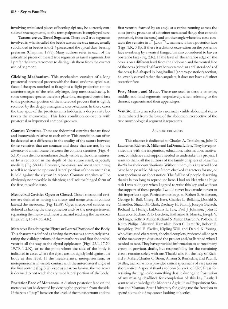
Metacoxa Reaching the Elytra or Lateral Portion of the Body.This character is defined as having the metacoxa completely sepa-rating the visible portions of the metathorax and first abdominalventrite all the way to the elytral epipleuron (Figs. 23.I, 17.70,19.70, 1-2.K), or to the point where the side of the body isindicated in cases where the elytra are not tightly held against thebody at this level. If the metasternite, metepisternum, ormetepimeron is in visible contact with the anterolateral angle ofthe first ventrite (Fig. 3.K), even as a narrow lamina, the metacoxais deemed to not reach the elytra or lateral portion of the body.
Posterior Face of Metacoxa. A distinct posterior face on themetacoxa can be detected by viewing the specimen from the side.If there is a �step� between the level of the metasternum and the
first ventrite formed by an angle at a carina running across thecoxa (or the presence of a distinct metacoxal flange that extendsposteriorly from the coxa) and another angle where the coxa con-tacts the ventrite in a ̄ |_ or ̄ \_ manner, it has a posterior face(Figs. 1.K, 3.K). If there is a distinct excavation on the posteriorface overhung by a ventral flange, it is also considered to have aposterior face (Fig. 2.K). If the level of the anterior edge of thecoxa is on a different level from the abdomen and the ventral faceof the coxa (viewed half-way between median and lateral ends ofthe coxa) is S-shaped in longitudinal (antero-posterior) section,i.e., evenly curved rather than angulate, it does not have a distinctposterior face.
Pro-, Meso-, and Meta-. These are used to denote anterior,middle, and hind segments, respectively, when referring to thethoracic segments and their appendages.
Ventrite. This term refers to a normally visible abdominal stern-ite numbered from the base of the abdomen irrespective of thetrue morphological segment it represents.
ACKNOWLEDGMENTS
This chapter is dedicated to Charles A. Triplehorn, John F.Lawrence, Richard S. Miller and LaDonna L. Ivie. They have pro-vided me with the inspiration, education, information, motiva-tion, confidence and support needed to undertake this project. Iwant to thank all the authors of the family chapters of AmericanBeetles for their contributions. Without them, this key would nothave been possible. Many of them checked characters for me, orsent specimens on short notice. The full list of people deservingthanks is too long to reproduce here. I had no idea what a hugetask I was taking on when I agreed to write this key, and withoutthe support of these people, I would never have made it even tothis imperfect stage. Particular thanks go to Robert S. Anderson,George E. Ball, Cheryl B. Barr, Charles L. Bellamy, Donald S.Chandler, Shawn M. Clark, Zachary H. Falin, J. Joseph Giersch,Richard L. Hurley, LaDonna L. Ivie, Paul J. Johnson, John F.Lawrence, Richard A. B. Leschen, Katharine A. Marske, Joseph V.McHugh, Kelly B. Miller, Richard S. Miller, Darren A. Pollock, T.Keith Philips, Alistair S. Ramsdale, Brett C. Ratcliffe, Robert E.Roughley, Paul E. Skelley, Kipling Will, and Daniel K. Young,who discussed characters, checked couplets, reviewed all or partof the manuscript, discussed the project and/or listened when Ineeded to rant. They have provided information to correct manyerrors in previous drafts, but responsibility for the remainingerrors remains solely with me. Thanks also for the help of Rich-ard S. Miller, Charles O�Brien, Alistair S. Ramsdale, and Paul E.Skelley, each of whom provided critical specimens of rare taxa onshort notice. A special thanks to John Sulzycki of CRC Press forresisting the urge to do something drastic during the frustrationof my missing deadlines for completion of this key. Lastly, Iwant to acknowledge the Montana Agricultural Experiment Sta-tion and Montana State University for giving me the freedom tospend so much of my career looking at beetles.

Key to Families · 819
I. KEY TO THE SUBORDERS OF NORTH AMERICAN COLEOPTERA
1. Notopleural sutures present (Figs. 19.I, 11.6, 2.8,4-5.10, 3.12); OR, abdomen with only 3ventrites; body form hemispherical, minutebeetles (length <1.3 mm) (Fig. 1.3); OR, small(length <2.6 mm), soft-bodied beetles (Fig. 1.2)with wings rolled in a spiral �cigar� manner (i.e.,not folded) .................................................... 2
� Notopleural sutures absent; abdomen with 4 ormore ventrites; wings folded or not, not rolled....................................... Polyphaga (Key D)
2(1). Hind coxae immovably fused to metasternum,completely dividing first ventrite (Figs. 33.I,11.6, 2.8, 4-5.10, 3.12, 13-17.12) ............................................................ Adephaga (Key C)
� Hind coxae free, first visible abdominal sterniteextending entirely across venter behind them...................................................................... 3
3(2). Minute beetles, length 2 mm or less in length(Figs. 1.3, 1.4); antenna with short club of 1 to 3antennomeres (Figs. 1.3, 1.4); wing folded inrepose ......................... Myxophaga (Key B)
� Minute to moderately large beetles, length 1.5 -25 mm; antennae either filiform (Fig. 1-2.1), orsub-moniliform and gradually widening from 4thsegment (Fig. 1.2); hind wings in repose spi-rally rolled in a spiral �cigar� manner ............................................... Archostemata (Key A)
A. KEY TO THE FAMILIES OF NORTH AMERICAN ARCHOSTEMATA
1. Notopleural sutures present; elytra reticulate,long, covering pygidium (Figs. 1-2.1); body cov-ered in scales; antennae filiform to subserrate(Figs. 1-2.1), length >4 mm ......... 1. Cupedidae
� Notopleural sutures absent, elytra smooth, short,leaving at least pygidium exposed (Fig. 1.2);body without scales; antennae sub-moniliformand gradually widened from 4th segment (Fig.1.2); length <2.6 mm ........... 2. Micromalthidae
B. KEY TO THE FAMILIES OF NORTH AMERICAN MYXOPHAGA
1. Body hemispherical (Fig. 1.3); elytra covering allterga; abdomen with 3 ventrites; antenna with11 antennomeres, 9-11forming club ........................................................... 3. Microsporidae
� Body more elongate-oval and depressed dor-soventrally (Fig. 1.4); elytra short, 3-4 tergitesexposed; abdomen with 6 or 7 ventrites; an-tenna with 9 antennomeres, antennomere 9forming narrow club .......... 4. Hydroscaphidae
C. KEY TO THE FAMILIES OF NORTH AMERICAN ADEPHAGA
1. Metacoxa greatly enlarged, a ventral plate con-cealing trochanter and basal half of femur, cov-ering most of 3 basal ventrites (Fig. 2.8) ............................................................. 8. Haliplidae
� Metacoxa greatly enlarged or not (Figs. 11.6), IFmetacoxa greatly enlarged, THEN all ventritesvisible laterally, coxa not concealing tro-chanter, basal half of femur or first 3 ventrites(Figs. 4-5.10, 3.12, 17.12) ............................. 2
2(1). Metacoxa not reaching elytron laterally,metepimeron and first ventrite in contact lat-erad of metacoxa and mesad of elytral margin(Figs. 6.6, 12.6); antenna usually at least partlypubescent (in addition to scattered long sen-sory setae); procoxal cavities usually closedbehind; IF metacoxa reaches elytron andprocoxal cavities open (one tiny species,length 2 mm or less), THEN second ventrite(first behind metacoxa) 3 times as long asmetacoxa at insertion of leg and last maxillarypalpomere distinctly narrower than penultimate(aciculate) ..................................................... 3
� Metacoxa reaching elytron laterally, junction ofmetepimeron and first ventrite not visible withelytron in place; antenna not pubescent, withonly scattered long sensory setae; procoxalcavities open behind; second ventrite less than3 times as long as metacoxa; last maxillarypalpomere not distinctly narrower thanpenultimate ................................................... 4
3(2). Mentum expanded, fused laterally to head cap-sule, covering ventral mouthparts completelywhen mandibles closed, mentum extending an-teriorly beyond other mouthparts to form cut-ting edge; outer angle of protibia with largeinwardly curved uncus (Fig. 1.5); body cylin-drical; antenna moniliform; head, pronotum, andelytra with deep canaliculate grooves (Fig. 1.5)................................................. 5. Rhysodidae
� Mentum not fused laterally to head capsule orextending beyond other mouthparts, maxillaand labium with at least palpi visible (Figs. 5.I,4.6, 45-48.6); outer angle of protibia withstraight or outwardly curved teeth or spines(Figs. 10.6, 13.6, 33.6, 38.6, 43.6); headpronotum and elytra without deep canaliculategrooves; body form and antennae variable ....................................................... 6. Carabidae
4(2). Protibia with antenna cleaner on inner apical angle(cf. Fig. 13.6); head with supraorbital setae (cf.Fig. 52.6) ............................ 9.Trachypachidae
� Protibia without antenna cleaner on inner apicalangle; head lacking supraorbital setae ........ 5
5(4). Pedicel of antenna greatly enlarged, offset frommain line of antenna, flagellum very short andcompact, not extended beyond hind margin ofhead; mid and hind legs very short; eyes usu-ally divided into 2 isolated parts on each side,rarely with only a very narrow canthus extend-ing between upper and lower portions ............................................................... 7. Gyrinidae
� Pedicel of antenna normal, antenna extended be-yond hind margin of head; mid and hind legsnot especially short; eyes not divided ........ 6
6(5). Metafemur and metatibia narrow and subcylin-drical in cross section; metatarsus shorter thanmetatibia and not tapered distally (Fig. 1.11);body not streamlined, outline of thorax andelytron discontinuous, base of pronotum dis-tinctly narrower than elytra (Fig. 1.11); length11-16 mm ............................. 11. Amphizoidae

820 · Key to Families
� Metafemur and metatibia more or less distinctlycompressed, especially so in larger species(length 6 mm or greater); metatarsus usually aslong or longer than metatibia (Fig. 3.12), dis-tinctly tapering distally (Figs. 3-4.12); bodystreamlined, outline of pronotum and elytronusually conjointly rounded (Figs. 1.10, 4-5.10,32-47.12); length 1-40 mm ............................ 7
7(6). EITHER scutellum not visible; protarsus with 5 dis-tinct tarsomeres; eyes distinct and length 1.0-1.6 mm; OR scutellum not visible; protarsus with5 distinct tarsomeres; length 1.9-5 mm; outermargin of protibia evenly curved and bearing adistinct comb of stout parallel and contiguoussetae (Fig. 3.10); AND inner apical angle withlarge, curved protibial spur (Fig. 3.10) ............................................................... 10. Noteridae
� Scutellum visible or not; protibia less evenlyrounded on outer apical angle (Fig. 2.12), outermargin lacking setal comb (Figs. 2-4.12); innerangle without large inner protibia spur (Fig.4.12); length 1.5-40 mm; IF less than 2 mm, THENprotarsus either pseudotetramerous (Figs. 7.12,39.12) or eyes absent or greatly reduced andindistinct .................................. 12. Dytiscidae
D. KEY TO THE FAMILIES OF NORTH AMERICAN POLYPHAGA(by Michael A. Ivie, couplets 3-13 by Mary Liz Jameson
and Brett Ratcliffe).
[Does not include unrecognized females of the Telegeusidae. Ex-pected to be anelytrous or larviform, they will probably key out tocouplet 183.]
1. Elytra present, complete, short, or reduced to flap-like stubs on the mesothorax ....................... 2
� Elytra totally absent ..................................... 182
2(1). Antenna with strongly asymmetrical, usuallylamellate club of 3-8 antennomeres (Figs. 17.I,2.23, 3.23, 2.31, 2.34, 56-57.34, etc.); procoxaelarge, strongly transverse or conical and pro-jecting below prosternum; procoxal cavitiesclosed; trochantins concealed (except inDiphyllostomatidae); protibiae flattened withone or more teeth on outer edge; tarsi with 5distinct tarsomeres, none of which are lobedor densely pubescent .................................. 3
� Antenna not lamellate, or coxae, tibiae or tarsinot as above ............................................... 14
3(2). Antennae with 11 antennomeres ..................... 4� Antennae with fewer than 11 antennomeres ... 5
4(3). Antennal club with 4-7 elongate antennomeres(Fig. 1.28) .............................. 28. Pleocomidae
� Antennal club with 3 circular or ovalantennomeres (Fig. 2.29) ...... 29. Geotrupidae
5(3). Body capable of being rolled into contractile ball(Fig. 2.32); middle and posterior tibiae flattenedand dilated ..................... 32. Ceratocanthidae
� Body oblong, not capable of being rolled intoball; middle and posterior tibiae not significantlyflattened and dilated .................................... 6
6(5). Longer apical spur of mesotibia pectinate alongone edge (cf. Fig. 2.30) ....... 30. Ochodaeidae
� Longer apical spur of mesotibia simple, not pecti-nate (cf. Fig. 3.30) ......................................... 7
7(6). Antennomeres of antennal club not capable ofbeing tightly closed together (Figs. 1-3.23, 1.24,1.25) .............................................................. 8
� Antennomeres of antennal club capable of beingclosed together (Figs. 13.I, 2.31, 1.33, 2.34,56.34, etc.) .................................................. 10
8(7). Abdomen with 7 ventrites, first divided bymetacoxa; head strongly constricted behindeyes; protibia lacking apical spurs; trochantinexposed; mesocoxae conical and projecting;length 5-9 mm ............ 24. Diphyllostomatidae
� Abdomen with 5-6 ventrites, first not divided;head not strongly constricted behind eyes;protibia with one or 2 apical spurs; trochantinnot visible; mesocoxae not projecting; length8-60 mm ........................................................ 9
9(8). Mentum with apex deeply emarginate; mesocoxalcavities closed laterally; body distinctly flat-tened dorsally (Fig. 1.25) ......... 25. Passalidae
� Mentum with apex simple, not deeply emargin-ate; mesocoxal cavities open laterally; bodyevenly convex dorsally (Fig. 1.23) ................................................................... 23. Lucanidae
10(7). Antennal club with 3 antennomeres, first hol-lowed out to receive second (Fig. 2.31) ...................................................... 31. Hybosoridae
� Antennal club with 3-7 antennomeres, first simple,not hollowed out to receive second (e.g., Fig.2.34) ............................................................ 11
11(10). Abdomen with 5 ventrites; dorsal surface rough-ened or tuberculate, not shining (Figs. 1.26, 1-3.27) ............................................................ 12
� Abdomen with 6 ventrites (Fig. 5.34); dorsal sur-face variably sculptured, shining or not .... 13
12(11). Eyes not divided by canthus; clypeus with sidesnarrowing apically; color brown, gray, or black;metafemora and metatibia not enlarged, not cov-ering abdomen ............................ 27. Trogidae
� Eyes divided by prominent canthus; clypeus withsides subparallel to divergent anteriorly; colortestaceous to light reddish brown; metafemoraand metatibia enlarged, covering most of ab-domen ...................................... 26. Glaresidae
13(11). Elytra shortened and widely divergent at apex(except in Lichnanthe lupina), not covering py-gidium (Fig. 1.33); eighth morphological abdomi-nal segment with spiracle ..... 33. Glaphyridae
� Elytra not shortened or widely divergent at apex,pygidium exposed or not; eighth morphologi-cal abdominal segment lacking spiracle (Fig.90.34) .................................. 34. Scarabaeidae
14(2). Tarsi with 2-5 tarsomeres, not pseudotetramerouson ALL legs (i.e., third of 5 tarsomeres on hindleg not lobed and enclosing small fourth, anyother configuration possible); antennae, mouth-parts, femora, and metacoxae variable; OR tarsipseudotetramerous and metacoxa with distinct

Key to Families · 821
posterior face (at least medially) set off fromventral surface by a carina or flange; OR tarsipseudotetramerous, head not at all rostrate, andantennae strongly or weakly clubbed but notgeniculate ................................................... 15
� Tarsi pseudotetramerous on all legs, with appar-ent penultimate tarsomere lobed below, enclos-ing and nearly hiding true fourth tarsomere(Figs. 31.I, 3.120, 36-37.124, 57-59.124, etc.);often with long antennae (Fig. 1.120), rostratehead (Figs. 1.125, 1.127, 1.128, 1.129, 1.130),or enlarged hind femora (Fig. 54-56.124);metacoxa without exposed posterior face ........................................................................ 16
15(14). Palps very short, usually immovably fixed andnot visible; head rostrate, prolonged into a vari-ously developed beak (Figs. 1.125, 1.127,1.128, 1.129, 1.130) AND/OR antennae genicu-late with compact club (Fig. 18.I) ................ 16
� Palps longer, flexible, and usually evident (e.g.,Figs. 6-7.I); head usually not prolonged into abeak but if rostrate or antennae elbowed andclub compact, then palps longer and flexible.................................................................... 26
16(14,15). Antenna usually without distinct club, filiform,moniliform, serrate or pectinate (Figs. 8-12.I);head not rostrate; if antenna distinctly clubbed,club of 5 or more antennomeres and length ofhead from top to clypeal margin less than orequal to width of head just behind eyes.... 17
� Antenna distinctly clubbed with 4 or fewerantennomeres in club (Figs. 15-16.I, 18.I); OR ifantennae moniliform, head distinctly rostrate(Fig. 1.129); OR if club with 5 or moreantennomeres, length of head from vertex toclypeal margin greater than width of head justbehind eyes ................................................ 20
17(16). Antenna usually more than half length of body,often inserted on prominence, capable of be-ing reflexed backward over body (Fig. 1.120);tibiae with 2 obvious apical spurs (Fig. 5.120);first antennomere usually several times longerthat second; pygidium never sclerotized andexposed; length 3-75 mm ........................................................................ 120. Cerambycidae
� Antenna usually less than half length of body,seldom inserted on prominence, not reflexibleback over body; tibiae without or with one ortwo apical spurs; first antennomere seldommore than 2-3 times length of second; pygidiumof some species sclerotized and exposed;length usually less than 12 mm .................. 18
18(17). All tibiae with 2 distinct apical spurs AND frontwithout �X� grooves; mesonotum with or with-out stridulatory file; ligula large, membranousand bilobed; aedeagus with median struts andtegmen bilobed ........................................... 19
� At least one tibia without 2 apical spurs OR frontwith deep �X� grooves (Figs. 5-6.124);mesonotum without stridulatory file; ligula nor-mal; aedeagus without median struts ..................................................... 124. Chrysomelidae
[NOTE: The Bruchinae, treated in Chapter 121, key out here. SeeStatus of Classification and subfamily key in Chapter 124]
19(18). Head with short but distinct temple behind eye,set off from narrowed neck (Fig. 1.122); apex ofmandible bidentate; ligula with a single lobe;mesonotum with stridulatory file ........................................................... 122. Megalopodidae
� Head lacking temples, evenly narrowed from be-hind eyes to neck (Fig. 1.123); apex of man-dible unidentate or bidentate; ligula bilobed;mesonotum without stridulatory file ........................................................ 123. Orsodacnidae
20(16). Antenna geniculate (rarely appearing straight ornearly so), club compact (Figs. 1-2.131, 69-77.131); metatrochanter not cylindrical, femurattached obliquely (Fig. 3.129) ................................................................. 131. Curculionidae
� Antenna straight (very rarely geniculate), clubloose or not evident; metatrochanter variablebut if antenna geniculate, trochanter cylindri-cal and squarely attached to femur (Fig. 4.129).................................................................... 21
21(20). Labrum visible and free (Fig. 6-9.125, 2-3.126);second tarsomere not spongy beneath (Figs 2-3.125); maxillary palpi normal ..................... 22
� Labrum never free; tarsi variable; maxillary palpirigid ............................................................. 23
22(21). Antenna situated adjacent to eye or laterally nearbase of short dorsoventrally flat rostrum; apexof third antennomere reaching well beyondfront margin of eye; all tibiae lacking spurs orspurs vestigial; notosternal sutures indistinctto obsolete ........................... 126. Anthribidae
� Antenna situated distally on long cylindrical ros-trum; apex of third antennomere not or barelyreaching front margin of eye; all tibiae withspurs; notosternal sutures distinct ........................................................ 125. Nemonychidae
23(21). Antenna either moniliform and body elongate (Fig.1.129, 7.129, 9-11.129, 14.129) (Brentinae,Cyphagoginae, Trachelizinae); OR antennastraight and clubbed, body pear-shaped (Fig.2.129, 15-17.129, 31.129) and metatrochantercylindrical, squarely joined to femur (Fig. 4.129)(Apioninae, Nanophyinae); OR antenna genicu-late, body pear-shaped and metatrochanter cy-lindrical, squarely joined to femur (Fig. 4.129)(Nanophyinae) OR antenna with 9-10antennomeres and body elongate-cylindrical(Fig. 5.129) (Cyladinae, Nanophyinae) ............................................................. 129. Brentidae
� Antenna straight, not geniculate, with 11antennomeres, club distinct; metatrochantertriangular or diamond-shaped, obliquely joinedto femur (Fig. 3.129); body form variable .... 24
24(23). Gena produced anteriorly on each side, visiblein frontal view as large tooth on each side ofapex of rostrum, laterad mandible; dorsal sur-face with obvious, recumbent, scale-like se-tae; body surface lacking metallic sheen; length12 mm or more ...................... 130. Ithyceridae
� Gena not produced anteriorly; upper surface gla-brous or with fine hair-like setae; body surfaceoften with distinct metallic sheen; length vari-able, mostly less than 10 mm ..................... 25

822 · Key to Families
25(24). Antenna situated at least length of antennomere1 from eye, positioned laterally on long quad-rate rostrum (Fig. 1.128) OR very close to eyeat base of short, robust rostrum (Fig. 3.128);protibia with anterior face apically flat, simple,not distinct from rest of surface (Figs. 5-6.128);metafemur with dorsal margin slightly to mod-erately arched (Figs. 2-4.128); pygidium obliqueto vertical (Figs. 2-4.128); elytron often with ascutellary striole (Fig. 1.128); body surface of-ten with distinct metallic sheen ................................................................... 128. Attelabidae
� Antenna situated immediately in front of eye atbase of long cylindrical rostrum (Fig. 1.127);protibia with front face at apex with shallowgrooved area filled with short, fine pilosity (Fig.1.127); metafemur with dorsal margin markedlyarched, paddle-like in shape, femur almost aswide as long; pygidium nearly horizontal;elytron lacking scutellary striole; body surfacelacking metallic sheen ................ 127. Belidae
26(15). Length 1.2 mm or less; antenna long, thin, withloose to indistinct club (Figs. 1.17, 77.17);antennomeres each with a whorl of long setaeat apex; wing fringed with long setae that arelonger than width of wing (Figs. 2-4.17, 77.17),or wing absent .............................. 17. Ptiliidae
� Length variable, antenna not as above, wingsrarely with fringe longer than width of wing ...................................................................... 27
27(26). Head with paired ocelli (Figs. 89-92.22) ......... 28� Head without paired ocelli (a single median ocel-
lus may be present) .................................... 30
28(27). Anterior edge of scutellum abruptly and sharplyelevated above mesoscutum; metepisternumreaching mesocoxal cavity and contacting firstventrite to separate metacoxa from elytral edge........................................... 66. Derodontidae
� Anterior edge of scutellum not abruptly elevated,continuous with mesoscutum; metepisternumvariable ....................................................... 29
29(28). Elytra completely covering abdomen; antennashort, not reaching middle of pronotum, an-tenna with 9 antennomeres, club of 5 pubes-cent antennomeres (Figs. 3-4.16); ventral sur-face with hydrofuge pubescence (Ochthebiinae).............................................. 16. Hydraenidae
� Elytra usually exposing 1 or more abdominal terga(Fig. 6.22); antenna short to long, reaching be-yond middle of pronotum in species with longelytra (Fig. 2.22); antennal club, if present, notinvolving 5 antennomeres; underside of bodywithout hydrofuge surface (Omaliinae) ...................................................... 22. Staphylinidae
30(27). Elytra very short, leaving 3 or more abdominaltergites exposed (Figs. 1.22, 3.22, 3.102,7.102, etc.) .................................................. 31
� Elytra longer, leaving no more than 1 or 2 abdomi-nal tergites exposed .................................. 51
31(30). Metatarsus with 1 fewer tarsomere thanmesotarsus ................................................. 32
� Metatarsus and mesotarsus with same number oftarsomeres .................................................. 35
32(31). Body greatly flattened dorsoventrally, abdomenwith 5 ventrites (Inopeplinae) ...................................................................... 116. Salpingidae
� Body not greatly flattened, abdomen with 6-7ventrites ..................................................... 33
33(32). Antenna strongly serrate to pectinate, flabellate,bipectinate or biflabellate (Figs. 8-18.102) ................................................. 102. Ripiphoridae
� Antenna, at most, very weakly serrate ........... 34
34(33). Tarsal claw with long, acute process or blade aris-ing from base, usually more than half as long asclaw (Figs. 13-16.111), rarely (Hornia) reducedto hyaline spine; antenna filiform; body corpu-lent and soft .............................. 111. Meloidae
� Tarsal claw simple; antenna weakly clubbed; bodycylindrical (Fig. 31.22) (Euaesthetinae) ...................................................... 22. Staphylinidae
35(31). Eyes large, separated frontally by less than diam-eter of third antennomere; wings well devel-oped, folded longitudinally at rest; maxillarypalp complex; antenna with antennomeres 9-11 less than half the width of antennomeres 3-5 (Atractocerus) .................... 71. Lymexylidae
� Eyes separated by more than diameter of thirdantennomere; wings, if well developed, usu-ally folded transversely; maxillary palpi simple;antenna not as above ................................. 36
36(35). Scutellary striole present; 2 basal ventrites con-nate, suture not diminished medially; antennaeof males pectinate to flabellate or plumose; ser-rate in females (Xenorhipidina) ...................................................................... 41. Buprestidae
� Scutellary striole absent; ventrites all free or 4ventrites connate; antennae variable ........ 37
37(36). Antenna with distinct club (Figs. 13.I, 15-18.I) ....................................................................... 38
� Antenna not clubbed (Figs. 8-12.I, 14.I) ......... 43[NOTE: The myxophagan family Hydroscaphidae will key out here ifan easily made mistake is made in the suborder key (above). Thesetiny (length 1.0-1.2 mm) beetles can be recognized by the elongate,narrow last antennomere (Fig. 1.4) which does not fit either the�distinct club� or �not clubbed� choice, as well as by the presence ofnotopleural sutures.]
38(37). Mesotarsus with 2, 3 or 4 tarsomeres ............................................................ 22. Staphylinidae
� Mesotarsus with 5 tarsomeres ....................... 39
39(38). Antenna with 4 apical antennomeres expandedinto asymmetrical club, first antennomere shin-ing, other 3 tomentose (Fig. 1.21); elytra usu-ally some combination of black and orange butoccasionally all black; fifth tergite with pair oflongitudinal carinae topped by stridulatory files;12 mm or greater in length, usually more than15 mm (Nicrophorinae) ................21. Silphidae
[Thanatophilus (Silphidae) may key here for individuals with an ex-tended abdomen; it lacks the stridulatory files of the fifth tergite andis 8-14 mm in length, but otherwise fits here because of antennalconfiguration.]� Antenna not as above; fifth tergite without stridu-
latory files; color variable; length 13 mm or less,usually less than 10 mm ............................. 40

Key to Families · 823
40(39). Antenna with 3 antennomeres; pronotum with an-tennal pockets anterolaterally above lateral mar-gins; dorsoventrally flattened, louse-like para-sites of beaver (Fig. 23.19) (Platypsyllus) .......................................................... 19. Leiodidae
� Antenna with 9-11 antennomeres; pronotum with-out antennal pockets .................................. 41
41(40). Procoxal cavities open .......... 22. Staphylinidae� Procoxal cavities closed ............................... 42
42(41). Lateral margins of pronotum complete; 5 ventrites................................................. 77. Nitidulidae
� Lateral margins of prontum incomplete; 6 ventrites(Cylidrella) ........................... 72. Trogossitidae
43(37). Mesotarsus with 4 or fewer tarsomeres ......................................................... 22. Staphylinidae
� Mesotarsus with 5 tarsomeres ....................... 44
44(43). Antenna with 12 antennomeres; antennabiserrate, bipectinate or biramose (Fig. 1.61) .............................................. 61. Phengodidae
� Antenna with fewer than 12 antennomeres; an-tennal type variable .................................... 45
45(44). Last maxillary and labial palpomere long, nearlyas long as, or longer than, antenna (Fig. 1.60)............................................ 60. Telegeusidae
� Last maxillary and labial palpomeres much shorterthan antenna ............................................... 46
46(45). Head covered above by pronotum (Fig. 9.62); of-ten with luminous organs on abdomen (Fig.19.62) ...................................... 62. Lampyridae
� Head visible from above; never with luminousorgans ......................................................... 47
47(46). Anterior edge of scutellum abruptly elevated, withdistinct step to mesoscutum (female Anorus)................................................. 38. Dascillidae
� Anterior edge of scutellum in same plane asmesoscutum ............................................... 48
48(47). Pronotum with lateral eversible vesicles (Fig. 2.74)(Malachiinae) .............................. 74. Melyridae
� Pronotum without eversible vesicles ............ 49
49(48). Mesosternum medially excavated, forming a cav-ity to receive extended prosternal process;Southwestern USA (female Cebrioninae) .......................................................... 58. Elateridae
� Mesosternum not excavated to receive extendedprosternal process; widespread ................. 50
50(49). Elytra individually rounded, not meeting apicallyat suture (Fig. 4.64); mandible long and narrow(Figs. 25-26.64) ...................... 64. Cantharidae
� Elytra truncate, meeting at suture apically (Figs.5-40.22); mandible often short and broad .................................................. 22. Staphylinidae
[NOTE: The archostematan family Micromalthidae will key out tothis couplet if an easily made mistake is made in the suborder key.They are exceptional for non-polyphagans in lacking notopleural su-tures. At this couplet they will match neither choice because of theindividually rounded elytra character of the cantharids, and the shortmandible of the staphylinids. The combination of a concealed tro-chantin and posteriorly emarginate scutellum will further distinguish
this family at this point. The rolled wing exhibited by the micromalthidsis unique to the Archostemata. See Chapter 2.]
51(30). Apices of penultimate 2 or 3 antennomeres eachcompletely ringed with microsetose groove (pe-riarticular gutters) (Fig. 9.18) (must be vieweddistally, difficult to see in very small specimensor in those with very compact antennal club);antenna with distinct to indistinct loose club;prothorax with sharp lateral margins; 5 or 6ventrites; protrochantin exposed or hidden, ifhidden and antenna with 11 antennomeres,antennomere 8 smaller than 7 or 9 ............. 52
� Antennae usually lacking periarticular gutters onantennal club; other characters variable; if com-plete periarticular gutters present, protrochan-tin hidden, antenna with 11 antennomeres ANDantennomere 8 not smaller than 7 and 9 .... 53
52(51). Metatibial spurs subequal in length (Figs. 16-18.19); small (1-6 mm), round to elongate oval,shining, granulate or transversely strigulatebeetles; elytra glabrous or pubescent, striateor not; prothorax as broad as elytra (Figs. 1-5.19); procoxae strongly projecting and con-stricted by procoxal cavity; often capable ofretracting into a ball-shape by curling head andprothorax under body; antenna distinctlyclubbed, often with 11 antennomeres, 5 ofwhich are involved in club and antennomere 8smaller than 7 or 9. Some genera with 10 or 11antennomeres and with distinct club of 3 or 4antennomeres (Fig. 12.19); these latter with flat-tened, externally flanged hind femora, apicalportion of which are excavate to receive tibiae;tarsal formula highly variable, 3-3-3, 4-4-4, 5-4-4, 5-5-4 or 5-5-5; one genus (Colon) with 11antennomeres and somewhat graduallyclubbed antenna that lacks small eighthantennomere (Fig. 7.19) has elytra pubescent,with characteristic shape and sutural stria (Fig.2.19) (see also, couplet 112) ..... 19. Leiodidae
[NOTE: Three very aberrant and ecologically restricted genera thatlack distinctly clubbed antennae belong here. Glacicavicolla is restrictedto ice caves in Idaho and Wyoming and characterized by elongatehead, pronotum and elytra, each separately constricted; cuticle trans-lucent, shining; eyes absent, and with elongate, slender legs and an-tennae. Two genera of Platypsyllinae are associated with mammalnests or mammals and are characterized by oval, strongly dorsoven-trally flattened body (Fig. 5.19), recumbent pubescence, an occipitalcrest overlapping anterior margin of pronotum (Fig. 5.19) and eyesabsent or barely indicated.]
� Metatibial spurs distinctly unequal; moderatelysized (4-14 mm), somewhat flattened shiningbeetles; elytra striate and glabrous; pronotumsomewhat narrowed relative to elytra (Figs. 2-3.18); procoxae strongly projecting or trans-verse; body not retractile; antenna long, clubloose and indistinct, eighth antennomere neversmaller than 7 and 9; femora simple; tarsi 5-5-5................................................... 18. Agyrtidae
53(51). Mesotarsus with 3 apparent tarsomeres, eitherclearly with 3 tarsomeres, or second tarsomerestrongly lobed and hiding small penultimate

824 · Key to Families
(third) tarsomere (Figs. 5.92, 40.93, 43-44.93).................................................................... 54
� Mesotarsus with 4 or 5 distinct tarsomeres ORfirst tarsomere distinctly lobed, engulfing verysmall second and small third of four, appearingto have 2 or 3 tarsomeres .......................... 62
54(53). Mesotarsus pseudotrimerous, with secondtarsomere strongly lobed, hiding smallpenultimate (third) tarsomere (Figs. 5. 92, 40.93,43-44.93) ..................................................... 55
� Mesotarsus truly with 3 tarsomeres, secondtarsomere not greatly lobed ....................... 57
55(54). Procoxal cavities closed (except in Holopsis);head small, usually covered by hood-likepronotum (Figs. 1-9.94); if head exposed fromabove (Figs. 10-11.94), procoxal cavitiesclosed; mostly tiny beetles less than 2 mm ............................................... 94. Corylophidae
� Head visible from above in front of pronotum;procoxal cavities open; size variable, up to11 mm .......................................................... 56
56(55). Frontoclypeal suture distinctly impressed; allventrites free; first ventrite without postcoxallines; pronotum often with sublateral lines (Figs.6-8.92) ............................... 92. Endomychidae
� Frontoclypeal suture absent; 2 basal ventritesconnate, first ventrite with postcoxal lines(Figs. 49-55.93); pronotum lacking sublaterallines .................................... 93. Coccinellidae
57(54). Eyes absent (Fig. 8.90) (Anommatus) ............................................................. 90. Bothrideridae
� Eyes present .................................................. 58
58(57). Head gradually narrowed behind eyes, withoutdistinct temples or neck; procoxal cavitiesopen; oval or elongate oval with base ofpronotum subequal to elytral base ............ 59
� Head sharply narrowed behind eyes or temples,with distinct neck; procoxal cavities open orclosed, elongate or elongate oval, with base ofpronotum distinctly narrower than elytra ... 60
59(58). Antennal scape normal, shorter than club; funiclelonger than entire club; posterior edge of lastventrite crenulate (Ostomopsis) ...................................................................91. Cerylonidae
[NOTE: The myxophagan family Microsporidae will key out here ifan easily made mistake is made in the suborder key (above). They willmatch the antennal characters, but lack the crenulation on the lastventrite. These tiny (length 0.5-1.2 mm) beetles can be easily recog-nized, having only 3 ventrites (5 in Ostomopsis). See Chapter 3.]� Antennal scape large, subequal to length of club;
funicle with 3 antennomeres, shorter than firstantennomere of club (Micropsephodes) ................................................... 92. Endomychidae
60(58). Abdomen with six ventrites, head narrowed im-mediately behind eyes (Fig. 10.22), lackingtemples; procoxal cavities open; lateral marginof pronotum coarsely dentate; trochantin ex-posed; mesocoxal cavities open (Dasycerus)............................................ 22. Staphylinidae
� Abdomen with five ventrites; head behind eyeswith distinct temples; procoxal cavities openor closed; lateral margin of pronotum simple tofinely dentate or absent; trochantin concealed;mesocoxal cavities variable ....................... 61
61(60). Abdomen very short, half length of metasternum;pronotum not margined laterally; mesocoxalcavities unstudied in North American species;scutellum not visible; elytron at base with pitat end of impressed groove (Fig. 1.65); 2 rarespecies known from Florida ........................................................................ 65. Jacobsoniidae
� Abdomen longer than metasternum (exceptAkalyptoischion, California); lateral margin ofpronotum absent to finely dentate; mesocoxalcavities closed; scutellum small but visible;elytra usually striate; common and widespread................................................. 95. Latridiidae
62(53). Antenna with 9 antennomeres, last 5 involved inclub (Figs. 3-4.16); 6 or 7 ventrites; tinyintercoxal sclerite between metacoxae; maxil-lary palp long relative to antenna (Fig. 4.16);ventral surface with hydrofuge pubescence;3.0 mm or less ....................... 16. Hydraenidae
� Antenna not as above; other characters not incombination above ..................................... 63
63(62). Antenna with 7-9 antennomeres, antennomeres7-9 usually forming loose, tomentose club (Figs.25-26.13), antennomere 6 often forming a cu-pule at base of club (Figs. 8.13, 10.13); maxil-lary palp often as long or longer than antenna(Figs. 1.13, 16.13, 19.13), always more than 1/2antennal length (Figs. 27.13, 38.13); metacoxawith ventro-posterior carina setting off convexposterior face (Fig. 1.K) that rotates against an-terior excavation of first ventrite; planes ofventral surface of metacoxa and first ventritediscontinuous; metatrochanter inserted onventral (not posterior) surface of metacoxa(Figs. 40.13, 42.13), femur held against ventralface of coxa, not against posterior face of coxaor flat to abdominal surface when fully retracted............................................ 13. Hydrophilidae
� Antenna variable but not as above; maxillary palpusually much shorter than antenna; metacoxaconfigured differently ................................ 64
64(63). Metacoxa with distinct posterior face (at leastmedially) set off from ventral surface by carinaor flange (Fig. 3.K), posterior face often exca-vated (Fig. 2.K); ventral surface of metacoxanot co-planar with first ventrite; metafemur in-serted on posterior face of metacoxa and fe-mur held posterior to coxa when retracted (Figs.2-3.K); procoxal cavities open; meso and meta-tarsi with equal number of tarsomeres ....... 65
� Metacoxa without distinct posterior face;metatrochanter often inserted on ventral sur-face or on small medial projection of coxa,never received in coxal excavation and rest-ing ventrad of metacoxa in retracted position;ventral surface of metacoxa more-or-less con-tinuous with first ventrite OR metatarsus withone fewer tarsomere than mesotarsus;procoxal cavities open or closed ............ 106

Key to Families · 825
65(64). Abdomen with 7-8 ventrites, metatarsus with 5tarsomeres .................................................. 66
� Abdomen with 6 or fewer ventrites, metatarsuswith 5 or 4 tarsomeres ................................ 71
66(65). Head with median ocellus (male Thylodrias) .................................................... 68. Dermestidae
� Head without median ocellus ........................ 67
67(66). Antenna with 12 antennomeres, biramose (Fig.2.61) (male Zarhipis) ............. 61. Phengodidae
� Antenna with 11 antennomeres, simple touniramose or biramose ............................... 68
68(67). Mesothoracic coxae distinctly separated; elytraoften reticulate (Fig. 1.59, 5.59), at least feeblycostate; femur and/or tibia compressed;pronotum with distinct longitudinal median ca-rina (Fig. 1.59), groove (Fig. 4.59) or cell (Fig.5.59), occasionally restricted to base or disc...................................................... 59. Lycidae
� Mesocoxae contiguous or nearly so; elytra notreticulate; femur and tibia seldom compressed;pronotum rarely with distinct longitudinal me-dian carina, groove or cell .......................... 69
69(68). Pronotum extended forward, covering head indorsal view (Figs. 1.62, 8-9.62 18.62, 20.62, 23-36.62); 1 or more ventrites often with luminousorgans (most obvious in males) (Fig. 19.62); sepa-ration of antennal insertions equal to or lessthan diameter of antennal fossa (Fig. 22.62) .................................................. 62. Lampyridae
� Head exposed in dorsal view when extended,OR if covered by pronotum, antennae sepa-rated by nearly twice diameter of antennalfossa; abdomen lacking luminous organs .. 70
70(69). Labrum not distinct, membranous and often hid-den beneath clypeus (Figs. 8-9.64); abdomenwith paired glandular openings on lateral edgeof tergites (Fig. 24.64); tarsomere 4 with bifidventral lobe (Figs. 27-28.64) .. 64. Cantharidae
� Labrum distinct and sclerotized; abdomen lack-ing paired glandular openings on tergites;tarsomeres 3 and 4 with bifid ventral lobes .................................................... 63. Omethidae
71(65). Posterior angles of prothorax acute, embracingelytral humeri (Figs. 1-2.56,1.57, 1-3.58); meta-tarsus with 5 tarsomeres; 3 or more ventritesconnate; prothorax dorsoventrally mobile rela-tive to mesothorax; intercoxal process ofprosternum long, notched dorsally, receivedin deep mesocoxal cavity as a clicking mecha-nism; IF clicking mechanism cannot be seenbecause visible portion of intercoxal processis flat ventrally and received tightly in deeplyemarginate mesosternum, THEN sternopleuralsuture or hypomeron grooved to receive an-tennae ........................................................ 72
� Posterior angles of prothorax not acute and em-bracing elytral humeri, or rarely somewhatacute and weakly embracing humeri (Fig. 1.54,etc.); metatarsus with 5 or 4 tarsomeres;ventrites variable; prosternal process variable,but if large and received in deeply emarginatemesosternum, apex of prosternal process notnotched dorsally nor capable of clicking; if largeprosternal process received tightly in deepmesocoxal cavity AND underside of prothoraxgrooved to receive antennae, then metatarsuswith 4 tarsomeres ....................................... 74
72(71). Labrum not externally visible; abdomen with 5connate ventrites .................. 35. Eucnemidae
FIGURE 1.K. Tropisternus sp. (13. Hydrophilidae) metacoxa, obliqueposterior view. C � posterior face of metacoxa. F � base of metacoxa(remainder removed).
FIGURE 2.K. Elateridae metacoxa, oblique posterior view.
FIGURE 3.K. Helichus immsi Hinton (44. Dryopidae) metacoxa,oblique posterior view.
826 · Key to Families
� Labrum free and visible; abdomen with 3, 4 or 5connate ventrites ....................................... 73
73(72). Antenna indistinctly to distinctly clubbed (Fig.1.57), apex received in margined cavity on pos-terolateral portion of hypomeron, just anteriorto retracted foreleg; metasternum with or with-out oblique margined groove for mesotarsus;prosternum with click mechanism hidden byplate-like ventral surface of postcoxalintercoxal process which fits tightly againstexposed portion of mesosternal cavity; elytrastrongly striate and covered with silky,subrecumbent setae; abdomen with 5 connateventrites; length 1-5 mm......... 57. Throscidae
� Antenna variable (filiform, serrate, pectinate, etc.),but not clubbed; antennal groove, if present,at or near sternopleural suture; metasternumwithout margined groove for mesotarsus; IFclick mechanism hidden as above, THEN elytranot strongly striate and setae suberect; abdo-men with 3 or 4 connate ventrites; length 1-60mm ............................................ 58. Elateridae
74(71). Mesocoxal cavities closed laterally, the meso-sternum and metasternum meeting lateradmesocoxa OR antenna elongate, anten-nomeres 3�8 with long rami, 9-11 flattened,elongate-serrate (Fig. 27.70); pronotum oftenhood-like, covering head from above (Figs. 1.69,4.69, 11-13.69) ............................................ 75
� Mesocoxal cavities open laterally, the mesoster-num and metasternum separated lateradmesocoxa by the mesepimera or mesepimeraand mesepisternum; antennae not as above;pronotum variable ....................................... 76
75(74). Metatrochanter cylindrical, short to long,squarely attached to femur, distinctly separat-ing coxa and tibia (Fig. 29.I) ...... 70. Anobiidae
� Metatrochanter short, triangular, obliquely at-tached to femur so that femur and coxa areadjacent to narrowly separated, on one side(Figs. 5-7.69) .......................... 69. Bostrichidae
76(74). Anterior margin of scutellum with abrupt, carinateelevation that fits against posterior margin ofpronotum, or scutellum absent or not visible.................................................................... 77
� Anterior margin of scutellum not abruptly el-evated, fitting under overlapping posteriormargin of pronotum .................................. 101
77(76). Procoxae strongly and distinctly projecting ven-trad of prosternum, 1/3 or more of dorsoven-tral length ventrad of intercoxal process (Figs.2-3.49, 3.67), procoxae usually conical or trans-versely conical ........................................... 78
� Procoxae not or weakly projecting ventrad ofprosternum; if procoxae conical, then lying lon-gitudinally and not or weakly projecting ven-trally ventrad of intercoxal process ........... 85
78(77). Tarsi with 4 distinct tarsomeres; metacoxal platesgreatly expanded, hiding most of first ventrite;hind wing, when developed, often fringed withlong setae; length 0.7-2 mm ..... 36. Clambidae
� Tarsi with 5 distinct tarsomeres; metacoxal platesdistinct but not hinding most of first ventrite;wing not fringed; size variable ................... 79
79(78). Antenna with distinct, simple club of 3antennomeres (Figs. 1.14, 1.67, 4.68, 7.68) ....................................................................... 80
� Antenna variously constructed, but without asimple club of 3 compact antennomeres ... 82
80(79). Elytra truncate; pygidium sclerotized and com-pletely or nearly completely exposed (Fig. 1.14).............................................. 14. Sphaeritidae
� Elytra complete; pygidium not sclerotized, com-pletely covered or with only small portion ex-posed .......................................................... 81
81(80). Upper surface of body glabrous; body contrac-tile; protibia held anterior to profemur and cov-ering antenna in hypomeral cavity when con-tracted (Fig. 3.67) (Orphilinae) .................................................................. 67. Nosodendridae
� Upper surface of body variously pubescent, se-tose or scaled (Fig. 1.68); body not stronglycontractile; protibia held posterior to profemurand antennal club not covered by leg whencontracted (Fig. 4.68) ............ 68. Dermestidae
82(79). Base of pronotum crenulate; scutellum usuallymedially notched on anterior margin; antennalinsertions not elevated; mandibles moderateand evenly curved; labrum large, sclerotizedand dorsal to mandibles .... 49. Ptilodactylidae
� Base of pronotum simple; anterior margin ofscutellum not notched; dorsal margin of anten-nal insertions elevated and protuberent; man-dibles large, abruptly curved mesad at nearlyright angle; labrum either short andmembraneous or extending between and be-low mandibles ............................................. 83
83(82). Empodium not obvious, hidden between basesof claws or absent; base of pronotum nearlystraight (Fig. 1.38) .................... 38. Dascillidae
� Empodium large, 1/3 length of claw, obviouslyplurisetose; base of pronotum stronglytrisinuate around scutellum (Fig. 1.39, 1.52) ...................................................................... 84
84(83). Tarsomeres 1-4 with large, membranous, dividedlobes; antenna lamellate (males) or increasinglyserrate apically (females) ...... 39. Rhipiceridae
� Tarsi simple, without ventral lobes; antennae ser-rate to pectinate ................... 52. Callirhipidae
85(77). Head with single median ocellus (Fig. 4.68) .................................................... 68. Dermestidae
� Head without ocellus ..................................... 86
86(85). Antenna short, not reaching middle of pronotum,scape and pedicel (antennomeres 1-2) rela-tively large, together 1/3 or more of totallength; antennomeres 3 to last transverse; bodycovered in dense tomentum ...................... 87
� Antenna short to long, scape and pedicel(antennomeres 1-2) not 1/3 of total length;antennomeres 3 to last variable; body vestiturevariable ....................................................... 89

Key to Families · 827
87(86). Head distinctly prognathous, mandibles stronglyprojecting forward (Fig. 1.47); profemur widenedmedially and armed externally with strongspines (Fig. 1-2.47); mesotarsus with 4tarsomeres ......................... 47. Heteroceridae
� Head distinctly hypognathous, mandibles eitherdirected ventrad or hidden (Figs. 1.44, 1.45);profemur simple, neither widened medially norarmed with large spines; mesotarsus with 5tarsomeres .................................................. 88
88(87). Metasternite with postcoxal lines delimiting re-tractile position of mesotibia; antenna hiddenin subocular groove and cavity between headand pronotum; body oval (Fig. 1.45) .............................................................. 45. Lutrochidae
� Metasternite without postcoxal lines; suboculargroove absent or very weakly developed, an-tenna not hidden in pronotum; body nearlyparallel-sided (Fig. 1.44) ............ 44. Dryopidae
89(86). Scape and pedicel received in deeply excavatepro- and mesosterna between pro- andmesocoxae (Fig. 2.50); pedicel longer thanscape, scape and pedicel together more than2/3 length of serrate flagellum (Fig. 2-3.50);body strongly contractile, all legs received incavities (Fig. 2.50); mesotarsus with 5tarsomeres, with long lobe on third tarsomere,fourth small and sometimes difficult to see(pseudotetramerous) .......... 50. Chelonariidae
� Antennae not received in excavations betweenpro- and mesocoxae; antennae not as above;mesotarsus usually not pseudotetramerous ...................................................................... 90
90(89). Head with subgenal ridges that fit againstprocoxae when head deflexed .. 37. Scirtidae
� Head without subgenal ridges, genae not in con-tact with procoxae ..................................... 91
91(90). Two basal ventrites connate, either with suturebetween them partially obliterated medially ORif suture between ventrites 1 and 2 not medi-ally indistinct, sternopleural sutures at leastmoderately grooved to receive antennae ......................................................................... 92
� Ventrites all free, OR 3 or 5 ventrites connate;ventrital and sternopleural sutures variable ...................................................................... 94
92(91). Suture between 2 basal sternites distinct medi-ally; mesotarsus with small, bisetose empodium;antenna filiform to distinctly clubbed; bodystrongly convex ........................ 42. Byrrhidae
� Suture between 2 basal sternites weak to absentmedially (Figs. 9.41, 58.41, etc.); mesotarsuslacking visible empodium; antenna usually ser-rate, pectinate or flabellate (Figs. 7-8.41, 28.41,46-48.41); body weakly dorsoventrally flattened.................................................................... 93
93(92). Fourth tarsomere with long lobe beneath, com-pletely divided into 2 parts; metepisternumbroad, approximately twice as long as wide ............................................ 40. Schizopodidae
� Fourth tarsomere with variable entire, undividedlobe beneath (Figs. 4-6.41, 54-55.41);metepisternum narrow, at least 3 times as longas wide (Figs. 9.41, 11.49, 18-19.41, etc.) oralmost completely concealed under elytra (Fig.15.41) ..................................... 41. Buprestidae
94(91). Legs retractile, rotated forward in repose, withtibia held anterior to femur; profemur with flangeon posterior face covering tibial excavation,protibia grooved to receive tarsus; usually withmargined excavations on propleuron,mesosternite, and ventrites to receive legs ...................................................................... 95
� If legs retractile, protibia held posterior to or ven-tral to femur; profemoral flange, if present, lo-cated on anterior face ................................ 96
95(94). Mentum strongly sclerotized, expanded, cover-ing labium and maxillae (Fig. 2.67); head notdeflexible; antenna covered by prolegs inbroad sternopleural pocket (Fig. 2.67); ventrites1 and 2 excavate for metathoracic leg;mesotibia with marginal spines; length 4-9 mm.......................................... 67. Nosodendridae
� Mentum normal, head usually retractable intopronotum to anterior margin of eyes (one ex-ception) (Fig. 1.46); antennae received in inter-nal pronotal cavities beneath head, externalanterior pronotal cavities or partly insternopleural grooves and partly under legsagainst hypomeron; excavation for metatho-racic leg, if present, limited to ventrite 1; mar-gin of mesotiba not spinose; length 1-2 mm ................................................. 46. Limnichidae
96(94). Elytra with thumb-like process on inner lateral sur-face near subapical curve, locking into ventrite5 ..................................... 53. Artematopodidae
[NOTE: Elytra must be separated from side of abdomen to see thischaracter.]� Elytra without such a locking device ............ 97
97(96). Posterior angles of pronotum with short discalcarinae (Fig. 1.54); procoxal cavity with narrowlateral extension at pleurosternal suture ............................................. 54. Brachypsectridae
� Posterior angles of prontum without short discalcarinae; procoxal cavity broad at pleurosternalsuture ......................................................... 98
98(97). Propleuron extended mesad behind procoxa forapproximately half length of trochantin; length10-15 mm .................................................... 99
� Margin of propleuron curved laterad posteriorly,not extended mesad posterior to procoxa;length 1-8 mm ........................................... 100
99(98). Posterior margin of pronotum crenulate;mesotibial spines subequal in size, smooth;antenna compressed serrate (Fig. 1.51);tarsomeres simple; empodium large and setose.............................................. 51. Eulichadidae
� Posterior margin of pronotum simple; mesotibialspines unequal in size, finely serrate; antennacylindrical-serrate (Fig. 1.38); tarsomeres 1-4with large, divided membranous lobes;empodium absent .................... 38. Dascillidae

828 · Key to Families
100(98). Posterior edge of pronotum simple; last tarsomeremuch longer than others, usually half or moretotal length of tarsus .................... 43. Elmidae
� Posterior edge of pronotum crenulate; lasttarsomere subequal in length to first .......................................................... 48. Psephenidae
101(76). Head with subgenal ridges that fit againstprocoxae when head deflexed; prosternum infront of coxa narrow, shorter than intercoxalprocess ....................................... 37. Scirtidae
� Head without subgenal ridges, genae usually notin contact with procoxae; prosternum in frontof coxae nearly as long as or longer thanintercoxal process ................................... 102
102(101). Metacoxal plates large, plate-like, longer medi-ally than metasternite, hiding most ofmetafemur, even when fully extended (Figs. 8-9.35) ....................................... 35. Eucinetidae
� Metacoxal plates narrow, forming either a parallelplate or simple carina; metafemur fully visible.................................................................. 103
103(102). Length of body 4 or more times maximum width(Fig. 1-2.71); male maxillary palp complex,multilobate ............................ 71. Lymexylidae
� Length of body 2.5 or less times maximum width;maxillary palp not branched ..................... 104
104(103). Prosternal intercoxal process complete, reach-ing behind procoxa to level of mesosternum;posterior portion of hypomeron not extendingbehind procoxa; elytral epipleuron with an in-ternally carinate edge complete to suture; headwith face narrowed; clypeal margin straight; 3basal ventrites connate ....... 48. Psephenidae
� Prosternal intercoxal process incomplete, notreaching beyond midpoint of procoxa; poste-rior portion of hypomeron variable behindprocoxa; elytral epipleuron narrowed beforereaching suture (complete in one genus); headwith face not greatly narrowed; clypeal marginemarginate (Figs. 5-6.21, 11.21); all ventritesfree ........................................................... 105
105(104). Elytra with 9 or 10 punctate striae (Figs. 1-4.18);posterior portion of hypomeron extending upto half the distance to mesal edge of procoxa;length 7-14 mm .......................... 18. Agyrtidae
� Elytra without punctate striae, otherwise variable,irregularly punctate (Fig. 1.21), with complexlow sculpture (Fig. 2.21) or up to 3 carinatecostae (Fig. 3.21); posterior portion ofhypomeron not extending behind procoxa orextending only a short distance mesad of lat-eral edge of procoxa (Figs. 9-10.21); length 7-45 mm .......................................... 21. Silphidae
106(64). Hind coxae widely separated by broad, truncateintercoxal process of first ventrite .......... 107
� Intercoxal process of first ventrite absent, acuteor rounded ................................................ 109
107(106). Mesocoxal cavities open laterally, closure in-volving mesepisterna (Georissinae) ........................................................... 13. Hydrophilidae
� Mesocoxal cavities open or not; if open, closuresolely involving mesepimeron ................. 108
108(107). Antenna geniculate, club usually of 3antennomeres; elytra short and truncate, ex-posing 2 non-flexing terga; body compact ...................................................... 15. Histeridae
� Antenna not obviously geniculate, clubbed ornot; elytra rarely exposing 2 terga, IF 2 tergaexposed, THEN exposed abdominal segmentsflexible, body not oval or body cylindrical andcompact .................................................... 109
109(106,108). Procoxae with exposed trochantin ......... 110� Trochantin concealed or absent ................. 128
110(109). Metacoxa extending laterally to reach elytralepipleuron or side of body, no visible contactbetween metathorax and first ventrite .... 111
� Metacoxa not reaching elytron, first ventrite andmetathorax visibly in contact laterad coxa ..................................................................... 116
111(110). Hind tarsus with 5 tarsomeres ...................... 112� Hind tarsus with 4 tarsomeres ...................... 157
112(111). Head with temples and occipital ridge distinct,occipital ridge closely fitting against pronotum,constricted behind to a distinct neck (difficultto see when head is retracted with ridge andtemples against pronotum); elytra with strongcharacteristic sutural stria, no other striae evi-dent (Fig. 2.19); 11 antennomeres, gradual clubof 3-4 antennomeres (Fig. 7.19); 4 (females) or 5(males) ventrites (Colon, see couplet 52) .......................................................... 19. Leiodidae
� Head without ridge and constricted neck that fitsagainst pronotum; elytra striate or not, but notas above; antenna variable; at least 5 ventrites.................................................................. 113
113(112). Prosternal process between coxae distinctly el-evated above level of prosternum, apexstrongly curved dorsally, reaching level ofpostcoxal extensions of hypomeron; cervicalsclerites absent; antenna not clubbed; elytraglabrous or subglabrous; length 8-20 mm ............................................... 120. Cerambycidae
� Prosternal process not elevated between coxaenor with apex strongly curved dorsad; cervi-cal sclerites present; antenna clubbed or not;elytra densely to sparsely setose, subglabrousor glabrous; length 1-24 mm ..................... 114
114(113). Procoxae not projecting distinctly belowintercoxal process, large and transverse; an-tenna distinctly clubbed; prothorax with sharplateral margins; IF procoxae slightly projecting,THEN antenna distinctly clubbed and tarsi notlobed beneath; not bright red .................................................................... 72. Trogossitidae
� Procoxae projecting distinctly below intercoxalprocess, conical or transverse (Figs. 21.73, 116-117.73); antennae variable; margins of protho-rax variable; IF procoxae are only slightly pro-jecting, THEN antennae feebly clubbed (Fig. 6.73), tarsi lobed beneath (Fig. 8.73) AND colorbright red .................................................. 115

Key to Families · 829
115(114). Tarsi not lobed beneath; procoxal cavity stronglytransverse; labrum subtruncate to convex,rounded or acute (Figs. 18-19.74); eye not emar-ginate (Figs. 18-19.74); antenna rarely with dis-tinct apical club, and if so, club of 5 or moreantennomeres; elytra usually confusedly punc-tate; pronotum and abdomen sometimes witheversible glands (Fig. 2.74) ....... 74. Melyridae
� Tarsi with lobes on multiple tarsomeres (Figs. 8-9.73, 50.73, 87.73); procoxal cavity circular(Figs. 21.73, 54.73), elongate or slightly trans-verse (Fig. 13.73); labrum subtruncate to con-cave or deeply emarginate (Fig. 77.73); eyeoften emarginate (Figs. 19-20.73, 77.73, 114-115.73); antenna usually apically clubbed, clubof 1 or more antennomeres (Figs. 5-7.73, 30-37.73, 42-43.73, 88-97.73, 104-112.73); elytraoften punctate-striate (Fig. 1.73); pronotum andabdomen never with eversible glands ................................................................ 73. Cleridae
116(110). Elytra short, completely exposing 1 or more ter-gites (Figs. 1.76, 5-6.77, 1.78, 2.79) ......... 117
� Elytra covering all of abdomen or exposing apexof 1 tergite ................................................ 120
117(116). Procoxal cavities broadly open (by more than halfwidth of coxa); labium with 2 palpomeres; ab-dominal intercoxal process truncate; pygidiumand last ventrite longer than preceding 4 com-bined (Fig. 1.78) ..................... 78. Smicripidae
� Procoxal cavities closed or narrowly open (byless than half width of coxa); labium with 3palpomeres or non-articulated; abdominalintercoxal process acute to broadly roundedor absent; pygidium variable .................... 118
118(117). Labial palps non-articulated; prosternal processelevated between procoxae and stronglycurved dorsally behind ....76. Brachypteridae
� Labium with 3 palpomeres; prosternal process flator elevated between procoxae, but notstrongly curved dorsally behind .............. 119
119(118). Antenna with 10 antennomeres, club of only 1antennomere; elytra more than twice as longas wide (Rhizophaginae) ..... 79. Monotomidae
� Antenna with 10 or 11 antennomeres, club of 3 ormore antennomeres; elytra less that twice aslong as wide ............................. 77. Nitidulidae
120(116). Mesotarsus with 4 tarsomeres; tarsal lobes, ifpresent, small, not obscuring penultimatetarsomere .................................................. 121
� Mesotarsus with 5 tarsomeres, fourth possiblyobscured by enlarged lobe of third(pseudotetramerous) ................................ 123
121(120). Lateral margin of pronotum crenulate (Fig. 4.98),antennal insertions concealed from above(Sphindocis) ..................................... 98. Ciidae
� Lateral margins of pronotum smooth or minutelydenticulate, antennal insertions visible fromabove ........................................................ 122
122(121). Body nearly spherical, capable of being rolledinto a ball; mandibles resting against metaster-num in retracted position (Cybocephalus) ....................................................... 77. Nitidulidae
� Body flattened-cylindrical, not at all spherical (Fig.1.96) (Mycetophaginae) ......................................................................... 96. Mycetophagidae
123(120). Antenna with 10 antennomeres, one involved inclub (Fig. 1.79) (Rhizophaginae) ................................................................. 79. Monotomidae
� Antenna with 10 or 11 antennomeres, if clubbed,club of 2 or more antennomeres .............. 124
124(123). Body extremely flattened; elytra nearly parallel-sided, disc almost perfectly flat betweenrounded lateral carinae running from humeri tonear apex, setting off vertical sides and gut-tered epipleural margin (Fig. 1.82); either large(>10 mm) and red with expanded temples (Fig.1.82) or small (<5 mm) and dull brown withouttemples ..................................... 82. Cucujidae
� Body not so distinctly flattened; elytra distinctlytransversely arched, not fitting other combina-tions above ............................................... 125
125(124). Dorsal face of mandible with tubercle that fitsinto cavity on clypeus, setose cavity at base,hidden when mandibles are closed(mycangium) (Fig. 3.75); elytra with scutellarystriole (Figs. 8-11.80, 1.75); antenna with 2 or 3antennomeres forming club (Figs. 1.75, 4-5.75,8-11.75); body oval to cylindrical (Figs. 1.75, 8-11.75) ...................................... 75. Sphindidae
� Mandible without dorsal mycangium; elytra with-out scutellary striole; antenna and body shapevariable ..................................................... 126
126(125). Antenna with a distinct club AND meso- and meta-tarsi with equal numbers of tarsomeres ... 127
� Metatarsus with one fewer tarsomere thanmesotarsus; antenna distinctly clubbed or not.................................................................. 157
127(126). Pygidium at least partially exposed, strongly scle-rotized, punctate, distinctly different from othertergites (Figs. 1.77, 7-8.77); tibiae usually spi-nose or denticulate on external margin .......................................................... 77. Nitidulidae
� Pygidium not exposed, not strongly sclerotized,similar to other tergites (Fig. 1.88); tibiae smoothon external margin ..................... 88. Byturidae
128(109). Antennal insertions concealed from above by lat-eral expansion of frons (Figs. 7-10.106, 53-54.106, 65-67.106, etc.); AND 3 basal ventritesconnate (Figs. 4-5.106), fourth and fifth mov-able (Figs. 138-139.106); AND procoxal cavi-ties closed by the mesad extension of the pos-terior portion of the hypomeron; AND procoxalprocess not expanded laterally at apex to closeprocoxal cavities (Figs. 131-132.106, 135-137.106); antenna usually with 11 anten-nomeres (rarely with 9 or 10 antennomeres) .......................................... 106. Tenebrionidae
� Without this combination of characters ....... 129
129(128). Abdomen with first 4 ventrites connate ...... 130� Abdomen with fewer than 4 ventrites connate .
.................................................................. 131

830 · Key to Families
130(129). Antenna serrate or pectinate (Fig. 1.55); antennalinsertions exposed from above (Fig. 1.55);metacoxa laterally reaching epipleuron;intercoxal process of prosternum with long,notched, apical projection, received in deepmesosternal cavity to form clicking mechanism;last ventrite without submarginal groove; men-tum without setose pit ........ 55. Cerophytidae
� Antenna moniliform, clavate or capitate (Figs.3.104, 5.104, 1-2.105, 8.105); antennal inser-tions concealed from above (Fig. 5.104);metacoxa not reaching elytron, first ventriteand metepimeron in contact laterad coxa andmesad epipleuron; prosternal process broad,widened apically (Figs. 2.104, 4.104, 5-7.105);last ventrite usually with submarginal groove;males often with median setose pit on mentum.............................................. 105. Zopheridae
[NOTE: The Monommatini, treated in Chapter 104, key out here. SeeStatus of Classification and the key in Chapter 105]
131(129). Metatarsus with 5 tarsomeres, first reduced, of-ten difficult to see; metatarsomere 1 either hid-den in apical excavation of metatibia OR, IFmetatarsal insertion fully exposed, THEN firstmetatarsomere less than 1/4 length of secondand obliquely attached under second (may onlybe visible from below in oblique distal angle);elytra covering pygidium; antenna with distinctclub of 2-4 capitate to elongate-looseantennomeres (Figs. 14-16.69, 21-22.69, 31-33.69); AND one of the following combinations:1) head somewhat to distinctly hypognathous;pronotum hood-like, projecting anteriorly (orventrally in some fully hypognathous species)beyond anterolateral angles above head (Figs.1.69, 2.69, 4.69,11-13.69,17-20.69) (Bostrich-inae, Dinoderinae, Endecatominae); 2) headprognathous (Figs. 27-30.69); intercoxal pro-cess of first ventrite truncate, metacoxaewidely separated, metacoxa reaching elytralaterally, separating metathorax and firstventrite (Lyctinae); OR 3) head prognathous(Fig. 3.69); procoxae transversly cylindrical,projecting at sides, proleg attached and di-rected laterally, femur and trochanter large(Polycaoninae) ...................... 69. Bostrichidae
� First tarsomere not so reduced; other charactersvariable; if first metatarsomere is reduced rela-tive to second, pronotum not hood-like, headnot hypognathous AND/OR metatarsus of 4tarsomeres, pygidium exposed, first ventritenot widely truncate between metacoxae;procoxae not transversely cylindrical and pro-jecting at sides ......................................... 132
132(131). Mesotarsus with 4 distinct tarsomeres ........ 133� Mesotarsus with 5 tarsomeres, or tarsi
pseudotetramerous .................................. 144
133(132). Mesocoxal cavities closed laterally ............ 134� Mesocoxal cavities open laterally ............... 140
134(133). Antennal insertions concealed from above ......................................................................... 135
� Antennal insertions exposed from above .... 136
135(134). Eyes usually present; IF eyes absent, THEN elytrawith flat tubercles (Fig. 11.103) ..................................................................... 103. Colydiidae
� Eyes absent; elytra smooth (Aglenus) ............................................................. 116. Salpingidae
136(134). Genae with pair of anteriorly directed horns ex-tending beyond labium, visible from above ............................................... 107. Prostomidae
� Genae lacking gular horns ........................... 137
137(136). Abdomen with 6 ventrites; pronotum usually large,hood-like, covering or nearly covering head(Figs. 1-9.94); pygidium usually exposed (Figs.6-7.94, 10-11.94); epipleuron incomplete;frontoclypeal suture absent; length less than2 mm .................................... 94. Corylophidae
� Abdomen with 5 or 6 ventrites; pronotum neverhood-like, head visible from above; pygidium,epipleuron and frontoclypeal suture variable;IF with 6 ventrites, THEN length 4 mm or greaterand frontoclypeal suture present ............ 138
138(137). Antenna longer, reaching to or beyond middle ofpronotum, club loose; pronotum usually withpair of sublateral discal carinae or grooves, run-ning from base laterad of basal pits (Fig. 1.92);body usually round to ovoid .................................................................... 92. Endomychidae
� Antenna shorter, not reaching beyond middle ofpronotum, club compact; IF pronotum withdiscal carinae or grooves, THEN usually a me-dian groove or pit and body elongate ...... 139
139(138). Posterior margin of last ventrite crenulate ORbody distinctly oval, length no more than twicemaximum width; antenna with 8, 9, or 10antennomeres; hind trochanter obliquely at-tached to femur, but distinctly separating coxafrom femur .............................. 91. Cerylonidae
� Posterior margin of last ventrite never crenulate,antenna with 10-11 antennomeres; body elon-gate, at least 2.75 times maximum width; hindtrochanter offset so that femur and coxa are incontact or nearly so ........... 90. Bothrideridae
140(133). Metacoxae separated by more than 1/2 trans-verse coxal diameter ................................ 141
� Metacoxae separated by less than 1/2 transversecoxal diameter .......................................... 142
141(140). Procoxal cavities narrrowly closed; pro- andmesocoxae strongly transverse; mandibletucked into cavity when closed, not visiblefrom side; antenna with 9 antennomeres, last 5forming club (Fig. 3.94); pronotum not groovedor carinate on disc (Fig. 3.94); small beetles,less than 2 mm in length (Orthoperus) ....................................................... 94. Corylophidae
� Procoxal cavities narrowly to widely open; pro-and mesocoxae circular to slightly transverse;mandible visible from side; antenna with 8-11antennomeres, if clubbed, club of 1, 2 or 3antennomeres (Figs. 1-3.92); pronotum usuallywith submarginal grooves or carinae, especiallybasally (Figs. 6-8.92); size 1-10 mm; IF less than2 mm and lacking grooves or carinae onpronotum (Eidoreus), THEN antenna with 10-11

Key to Families · 831
antennomeres, 1 or 2 of which form a distinctclub ................................... 92. Endomychidae
142(140). Intercoxal process of first ventrite absent, no partof ventrite extending between coxae to con-tact metaventrite; first ventrite lacking mar-gined metacoxal cavitites; metacoxae conicaland projecting; body soft; small triangular partof morphological abdominal sternite 2 usuallyvisible laterad of metacoxa (i.e., ventrite 1 small,divided); often colorful, with red, yellow ormetallic blue/green markings (Fig. 10.69);length 5-12 mm (Psoinae) ...... 69. Bostrichidae
� Intercoxal process of first ventrite complete; firstventrite with margined coxal cavities;metacoxae transverse; body fully sclerotized;ventrite 1 closing anterolateral angle betweenmetacoxa and abdomen, not divided bymetacoxae; never metallic; length 0.5-6.5 mm.................................................................. 143
143(142). Body elongate-oval and somewhat cylindrical;pronotum usually very convex in transversesection, edges often directed ventrally (Figs.1.98, 6.98, 10.98, 20.98, 36-42.98); pronotumwithout basal pits or impressions; head orpronotum of male often with horns or tubercles(Figs. 13-14.98, 33.98, 36.98, 39-40.98, 42.98,43-45.98); antenna with 8-10 antennomeres andclub of 2-3 antennomeres; males often with pu-bescent median fova on first ventrites; headwithout distinct temples or neck. One speciesfrom California has relatively flat pronotum withcrenulate margins directed laterally (Fig. 4.98),antenna with 11 antennomeres and 3antennomeres in club, but 2 basal sternites areconnate .......................................... 98. Ciidae
� Body oval to elongate oval, usually somewhatdorsoventally depressed; pronotum usuallyweakly convex transversely, edges directedlaterally; pronotum with 2 basal pits or impres-sions laterad scutellum (Fig. 1.96) (sometimesin posterior marginal groove and difficult to dis-cern); head and pronotum without horns or tu-bercles; antenna with 11 antennomeres, last 2-5 forming club; all ventrites free, without me-dian fovea. One genus (length less than 2 mm)somewhat cylindrical, with very convexpronotum in transverse section, with headabruptly constricted behind short temples toform distinct neck .......... 96. Mycetophagidae
144(132). Abdomen with six ventrites AND metatarsi withfive tarsomeres; terminal maxillary palpomere(4) shorter and narrower than penultimate (Figs.2-8.20); shape rather characteristic (Figs. 25-46); length 0.6-2-7 mm ...... 20. Scydmaenidae
� Abdomen with four or five ventrites; tarsi vari-able; terminal maxillary palpomere (3 or 4) aswide or wider AND/OR as long or longer thanpenultimate; size variable ........................ 145
145(144). Pregular area on each side with a laterally facingsurface bearing setose pit or cavity near endof distinct antennal groove; first ventrite withpostcoxal lines ....................... 89. Biphyllidae
� Pregular area without laterally facing setose pit;antennal grooves and postcoxal lines variable.................................................................. 146
146(145). First ventrite much longer than second (measuredbehind coxa); elytra without punctate or im-pressed striae (traces of striae occasionally vis-ible through cuticle, but not expressed on thesurface); epipleuron distinct in basal half, notreaching apex (usually narrowed at level ofthird ventrite); genae carinate and projectingventrally between eye and mentum; apex ofelytra with double suture or �subapical gap�caused by wide flange of elytral coupling sys-tem; elytra complete, exposing at most tip oflast tergite ....................... 85. Cryptophagidae
[NOTE: Two genera of tiny (<1.3 mm) cryptophagids (Amydropa,Baja California, and Hypocoprus, Rocky Mountain region) lack thesubgenal carinae. Hypocoprus has the first 2 ventrites subequal and thepygidium exposed while Amydropa lacks the double suture on theelytra. The other characters fit these 2 rare genera. Ambydropa hasgreatly reduced eyes (10 facets or fewer) and Hypocoprus has distincttemples.]� Not fitting this combination of characters, EITHER
with the first ventrite short, elytra striate,epipleuron complete to apex, gena flat betweeneye and mentum, OR elytra not covering mostof pygidium ............................................... 147
147(146). Metatrochanter transversely or obliquely at-tached to femur, distinctly separating femurfrom coxa (Figs. 28-29.I) ........................... 148
� Metatrochanter obliquely attached to femur, off-set so that femur abuts coxa (Fig. 26-27.I) ....................................................................... 157
148(147). Antennal insertions approximate or separated byless than 1/2 width of head behind eyes ANDpronotum without lateral carinae; metatarsuswith 5 tarsomeres; metatrochanter elongate,cylindrical (Fig. 29.I, 69.70, 74.70) (Ptininae) .................................................... 70. Anobiidae
� Without combination of narrowly separated an-tennal insertions and no lateral carina onpronotum; other characters variable ........ 149
149(148). Pronotum with sublateral lines or grooves thatextend from base anterad midpoint, often toanterior margin (Figs. 1-11.83); head usually withsublateral lines from median margin of eye topronotum; lateral margins of pronotum smoothor wavy or with few obtuse angles (Figs.1-11.83), not acutely denticulate or serrate; headnot sharply constricted to a distinct neck; bodyoval to elongate, subcylindrical to stronglydorsoventrally flattened (Figs. 1-11.83) ................................................. 83. Laemophloeidae
� Pronotum usually without sublateral lines that ex-tend from base anterad midpoint; head variable;IF pronotum with sublateral lines that extendfrom base to or beyond midpoint, THEN lateralmargins of pronotum sharply denticulate, ante-rior angles acutely projecting AND/OR headsharply constricted behind small temples (Figs.17.80, 19.80, 22.80); body variable ......... 150
150(149). Mesocoxal cavities open laterally ............... 151� Mesocoxal cavities closed laterally ............ 153

832 · Key to Families
151(150). Antenna with 10 antennomeres, distinctlyclubbed; elytra shortened, exposing all of py-gidium (Fig. 2.79); head abruptly constricted toform neck; 1-4 mm............... 79. Monotomidae
� Not fitting one or more of above characters ....................................................................... 152
152(151). Body elongate, flattened (Figs. 1.80, 16-25.80);meso- and metatarsi with same number oftarsomeres; head usually with distinct templesbefore abrubtly constricted neck (Figs. 1.80,4.80, 16-25.80); procoxae either closed behind(Fig. 3.80) or, if open (Brontinae, Fig. 2.80), elytratransversely flat or slightly concave betweenslightly to distinctly raised interstria betweenstria 6 and 7; elytron with scutellary striole;base of mandible with dorsal setose pit(mycangium) hidden beneath clypeus whenclosed; antenna filiform, with scape more than3 times length of pedicel ........ 80. Silvanidae
� Metatarsus with one tarsomere fewer thanmesotarsus; other characters variable ..... 157
153(150). Body shining, oval and strongly convex;pronotum tightly embracing elytra (Fig. 1.84),pronotum laterobasally with a vaguely trans-parent, thin flange which slides over a smootharea on base of humeral angle of elytron, thisarea on elytron delimited posteriorly by a thincarina; pronotum and elytra with wide propleuraand epipleura, lateral margins sharp, explanate,strongly directed ventrally so that lateral mar-gins are far below level of procoxa and mesadepipleural margin, dorsal surface forming aninverted "U" in transverse section; tarsal clawtoothed or appendiculate ...... 84. Phalacridae
� Body usually not so evenly oval, pronotum notcoadapted to pronotum in the above manner,without described flanged basal angles ofpronotum or associated elytral area; lateralmargins of pronotum and elytra laterad, ratherthan ventrad, to procoxa and eplipleura; tarsalclaws toothed only in groups with pronotumnarrowed behind ...................................... 154
154(153). Meso- and metatarsi with same number oftarsomeres; face often with beaded lateral mar-gins (Figs. 1.81, 3-4.86, 9-10.87) .............. 155
� Mesotarsus with one more tarsomere than meta-tarsus; face without beaded lateral margins .................................................................... 157
155(154). Gular sutures confluent; genae expanded anteri-orly, plate-like, concealing maxillae (Fig. 4.81)............................................. 81. Passandridae
� Gular sutures separate or absent; genae not soexpanded ................................................. 156
156(155). Procoxal cavities usually open behind (Fig. 8.86);terminal maxillary palpomere narrow, elongate;IF procoxal cavities closed behind (Fig. 7.86),THEN closure by mesad extension of hypomer-on, length less than 3 mm and pronotum some-what narrowed near base (Cryptophilus) ....................................................... 86. Languriidae
� Procoxal cavities closed behind by laterad ex-pansion of the prosternal process (Figs. 5-6.87);terminal maxillary palpomere often securiform(Figs. 5-6.87), or narrow and elongate (Fig. 4.87);length 3-22 mm ......................... 87. Erotylidae
157(111,126,147,152,154).Last visible segment of abdomenforming a terminal spine (Fig. 1.101); bodywedge-shaped, humpbacked; head retractedto hypognathous position (Fig. 1.101); metatibiaand metatarsus usually with oblique or trans-verse, comb-like serrate ridges subapically onlateral faces (Figs. 2-7.101) .. 101. Mordellidae
� Abdomen not prolonged into a terminal spine;body otherwise variable; metatibia and meta-tarsus without comb-like serrate ridges asabove, IF similar combs are present, THEN theyare apical .................................................. 158
158(157). Tarsal claw with a ventral blade or elongate lobebeneath (Figs. 13-16.111) (reduced to a largefused tooth ending about 2/3 length of upperblade in Phodaga [Fig. 15.111] SW-USA); headsharply or gradually constricted behind eyesto distinct neck ........................................ 159
� Tarsal claw without ventral blade or elongate lobebeneath, if claw toothed or apendiculate, notas in Fig. 15.111; head constricted or not ....................................................................... 160
159(158). Ventral appendage of tarsal claw usually lobe-like, membranous, occasionally blade-like andsclerotized; elytra usually meeting along su-ture to very near apex, which may be narrowlyseparately rounded (Fig. 1.110); lateral marginof pronotum absent, complete (Fig. 2.110), orindicated only at base (Fig. 3.110); mesocoxalcavities usually narrowly separated, occasion-ally contiguous; maxillae not forming suckingtube; antenna without club or with vague todistinct club of 3 antennomeres; hind wing withwell-developed radial cell; if pronotal margincompletely absent, antenna with at least vagueindication of club in last 3 antennomeres andmesocoxal cavities narrowly separated; ifelytra broadly separately rounded, pronotumwith lateral carina at base (Fig. 3.110) ................................................. 110. Stenotrachelidae
� Ventral appendage of tarsal claw blade-like andsclerotized (Figs. 13-16.111); elytra usually di-verging along suture before apex, broadly sepa-rately rounded (Fig. 1.111); pronotum lackingmarginal carina laterally; antenna without clubof 3 antennomeres; mesocoxal cavities con-tiguous; maxillae usually normal; radial cell ab-sent in hind wing; if elytra meeting on suture tovery near apex, maxillae modified into suckingtube that extends beyond mandibular apices.................................................. 111. Meloidae
160(158). Base of pronotum with marginal groove (Fig. 2.117,6.117) that extends laterally onto hypomeron,ending in a pit near posterior margin of coxa(Fig. 3.117); pronotum narrowed posteriorly, notmargined laterally (Figs. 2.115, 1-3.117); headsharply narrowed behind distinct temples toform narrow neck (Figs. 2.115, 1-3.117, 18-20.117); elytra sparsely to densely setose ..................................................................... 161

Key to Families · 833
� Basal groove of pronotum, if present, not endingin pit on hypomeron; pronotum margined later-ally or not; elytra with or without setae .... 162
161(160). Antenna with last 3 antennomeres forming elon-gate apical club, club more than 1/2 total lengthof antenna (Fig. 2.115) (Anisotria) ............................................................. 115. Pyrochroidae
[Note: Anisotria was moved to the Anthicidae by Lawrence et al.1999.]� Antenna not clubbed, with weak, short club, with
last 3 antennomeres shorter than 1/2 totallength of antenna (Fig. 1.117) OR with only lastantennomere long (subequal to antennomeres7-10) ....................................... 117. Anthicidae
162(160). Mesocoxal cavities closed laterally ............ 163� Mesocoxal cavities open laterally ............... 165
163(162). Basal 3 ventrites connate; antenna with 11antennomeres, submoniliform/triangular, fili-form, serrate to subflabellate (Figs. 1-7.112, 9-10.112); cervical sclerites present .............................................................. 112. Mycteridae
� Two or no ventrites connate; antenna with 10 to11 antennomeres, moniliform to capitate (Figs.1-5.116, 12-13.116); cervical sclerites absent.................................................................. 164
164(163). Prothorax with pleurosternal suture ending in alarge setose pit at antero-lateral margin ofprocoxal cavity; 2 basal ventrites connate; 11antennomeres; 1.5-3.8 mm; deserts of westernUSA from Idaho to Mexican border (Cononotus)........................................... 115. Pyrochroidae
� Prothorax with or without pleurosternal suture,lacking large setose pit on anterior margin ofprocoxal cavity; all ventrites usually free, or 2basal ventrites connate (Aegialites); 10-11antennomeres; length 1.5-7 mm; widespread inforests and Pacific beaches (Aegialites); if indeserts, antenna with 10 antennomeres(Dacoderus) .......................... 116. Salpingidae
165(162). Body deep, mildly to distinctly wedge-shaped(Figs. 1-2.102, 4-6.102); antenna serrate, pecti-nate or flabellate, often bipectinate orbiflabellate (Figs. 8-18.102); vertex often in-flated and narrowed above eyes in frontal view;vertex usually extending dorsally above planeof pronotum in lateral view, vertex andpronotum at least coplanar; tarsi toothed, bifidor pectinate; maxillary lobes sometimes stylet-like, extending beyond tips of mandibles .................................................. 102. Ripiphoridae
� Body usually not deep and wedge-shaped, IFbody deep and wedge-shaped, THEN antennasimple and head coplanar with or slipping un-der front margin of pronotum; tarsi variable;maxillary lobes not stylet-like ................... 166
166(165). Pronotum lacking lateral carina .................... 167� Pronotum with complete or incomplete lateral
carina ........................................................ 171
167(166). Metacoxa extending laterally to elytron or sideof body, completely separating metepisternumand first ventrite ....................................... 168
� Metacoxa not reaching elytron or side of body,metepisternum and first ventrite in contact lat-erad metacoxa .......................................... 170
168(167). Tarsi appearing 4-4-3 (actually 5-5-4,pseudotetramerous/pseudotrimerous); eyescoarsely faceted, appearing hairy, interfacetalsetae as coarse, long and dense as those onfront and sides of head adjacent to eyes; 1-4mm ............................................ 118. Aderidae
� Tarsi distinctly 5-5-4; eyes with or withoutinterfacetal setae, IF interfacetal setae present,THEN setae not as coarse, long or obvious ason front and sides of head adjacent to the eyes;4-21 mm .................................................... 169
169(168). Head prognathous, not abruptly constricted to anarrow neck, lacking distinct temples (Fig.1.109); anterior portion of prosternum as longor longer than prosternal process; first 2ventrites connate ............. 109. Oedemeridae
� Head distinctly declined, abruptly constricted toform narrow neck behind distinct temples; an-terior portion of prosternum shorter thanprosternal process (Eurygeniinae) ................................................................ 117. Anthicidae
170(167). Elytra distinctly setose; eye emarginate anteri-orly; penultimate tarsomere with large lobebeneath (Figs. 1.115, 3.115) ...................................................................... 115. Pyrochroidae
� Elytra glabrous; eye not emarginate; penultimatetarsomere simple (Fig. 1.114) (Pytho andPriognathus) .............................. 114. Pythidae
171(166). Metacoxa extending laterally to elytra or side ofbody, completely separating metepisternumand first ventrite; mesotibial spurs serrate, pec-tinate or pubescent .................................. 172
� Metacoxa not reaching elytra or side of body,metathorax and first ventrite at least narrowlyclosing metacoxal cavity laterally; mesotibialspurs variable ........................................... 174
172(171). Head vertically narrowed behind eyes to formnarrow neck, head not received into protho-rax, either bulging beyond pronotal margin, orfitting closely against pronotal margin so thathead in lateral view has a posterior carina orcrest meeting anterior margin of pronotum (Fig.5.K) ........................................ 119. Scraptiidae
� Head gradually narrowed behind eyes, fitting intopronotum in a telescoping manner ........... 173
173(172). Tarsus without lobes on penultimate tarsomere;sutural stria deeply impressed near apex ofelytra, distinctly more so than in basal half; 2basal ventrites connate; metatibia longer thanfirst metatarsomere; prosternal intercoxal pro-cess level with ventral surface of non-project-ing procoxae; EITHER intercoxal process long,parallel-sided, reaching behind procoxae ORprosternal process incomplete, narrowedapically between coxae; length 7-13 mm .................................................. 108. Synchroidae
� Tarsus with penultimate tarsomere lobed beneath(Figs. 1.100, 21-22.100) OR metatibia shorterthan first metatarsomere; IF sutural stria deeplyimpressed near apex, THEN also impressed on

834 · Key to Families
basal half (Fig. 1.100); 2 basal ventrites con-nate or all free; prosternal process incomplete,not reaching behind procoxae, narrowedapically, resting below ventral surface of usu-ally projecting procoxae; length 2-20 mm ............................................... 100. Melandryidae
174(171). Procoxal cavities closed behind; first 2 ventritesconnate; body strongly rounded (Fig. 1.97) ....................................... 97. Archeocrypticidae
� Procoxal cavities open behind; ventrites connateor free; body form variable, often elongate ..................................................................... 175
175(174). Elytra with sutural and epipleural margins ele-vated; strongly elevated carina running fromhumeral angle to near apex resulting in dis-tinctly concave elytral disc; pronotum withmedian longitudinal elevated carina on basal1/3, deep transverse grooves with pits at eachend on either side of carina (Ischalia) ........................................................... 117. Anthicidae
� Elytra and pronotum without strongly elevatedcarinae, elytral disc convex ..................... 176
176(175). Prosternal intercoxal process incomplete or ab-sent, not separating procoxae ................. 177
� Prosternal intercoxal process complete, fullyseparating procoxae ................................ 178
177(176). Antenna filiform; terminal labial and maxillarypalpomeres expanded apically; prosternumshorter than diameter of procoxae; mesotibialspurs pubescent or serrate; tarsi lobed onpenultimate tarsomeres (Fig. 22.100)(Osphyinae) ....................... 100. Melandryidae
� Antenna with long, serrate club of last 3antennomeres (Figs. 7-8.114); terminal labialand maxillary palpomeres cylindrical;prosternum as long as procoxal diameter;mesotibial spurs smooth; tarsi not lobed(Trimitomerus) .......................... 114. Pythidae
178(176). Tarsi simple, lacking lobes below ................ 179� At least some tarsomeres distinctly lobed below
.................................................................. 181
179(178). Median longitudinal line (discrimen) of metaster-num short, extended from hind margin less than1/2 total length of ventrite; mesocoxa normal,convex and punctate anterior to trochanteralinsertion .................................................... 180
� Median longitudinal line (discrimen) ofmetaventrite longer, extended from hind mar-gin more than 1/2 total length of ventrite (Fig.4.K); mesocoxa with unique, polished, ventralface anterior to trochanteral insertion (Fig. 4.K)(this polished area is rubbed by flat opposingsurface of trochanter and base of femur whenleg rotated is forward) .......... 99. Tetratomidae
180(179). Antenna short, not reaching middle of pronotum;apical 3 antennomeres forming a distinct, ratherabrupt club ................................. 113. Boridae
� Antenna longer, reaching base of elytron; apicalantennomeres somewhat wider than basalantennomeres, not forming abrupt club(Sphalma) .................................. 114. Pythidae
181(178). Antenna filiform; setae on elytra very short andindistinct, shorter than diameter of punctures;elytra uniform in color; California and Nevada(Tydessa) ............................ 115. Pyrochroidae
� Antenna strongly serrate; setae on elytra con-spicuous, several times longer than diameter
FIGURE 4.K. Eustrophinus arizonensis (Horn) (99. Tetratomidae) meso-and metacoxa, ventral view. C � polished ventral face of mesocoxa. T� mesotrochanter, femur removed.
FIGURE 5.K. Anaspis atrata Champion (119. Scraptiidae), lateralview (modified from Hatch 1965).

Key to Families · 835
of punctures; elytra reddish with dark markings,a macula around scutellum and transverse bandat apical 1/3, usually joined by line along su-ture; south Texas ....................................................................... Polypria cruxrufa Chevrolat
[NOTE: Family placement incertae sedis, see end of Chapter 100.]
182(1). One tarsal claw; eye reduced to a single omma-tidium ........................................................ 183
� Two tarsal claws; compound eye normal, reducedor with single ommatidium ........................ 184
183(182). Gonopore present (females) ... 61. Phengodidae� Gonopore not present ........................................
.............................. Larvae (not further keyed)
184(182). Head with median ocellus (female Thylodrius) ................................................. 68. Dermestidae
� Head without ocellus ................................... 185
185(184). Head prognathous; pronotum expanded anteri-orly, extending over head in retracted posi-tion (Phausis, Microphotus) or head retractileinto tubular prothorax (Pterotus); distinctly toslightly dorso-ventrally flattened; antenna with9 or fewer antennomeres; some, possibly all,bioluminescent; widespread (females) ........................................................... 62. Lampyridae
� Head hypognathous, not retractile into protho-rax; body globular-cylindrical; antenna with 11antennomeres; not bioluminescent; Florida ornear ports of entry (female Ripidiinae, NorthAmerican females unknown) ....................................................................... 102. Ripiphoridae
LITERATURE CITED
ARNETT, R. H., Jr. 1973. Beetles of the United States (a Manualfor Identification). American Entomological Institute. AnnArbor, MI. 1112 pp.
BORROR, D. J., C. A. TRIPLEHORN and N. F. JOHNSON.1989. An introduction to the study of insects (sixth ed.).Saunders College Publishing. Philadelphia, PA. 875 pp.
BROWN, B. V. 1993. A further chemical alternative to critical-point-drying for preparing small (or large) flies. Fly Times 11:10.
CHAPMAN, R. A. 1998. The Insects. Structure and Function, 4thed. Cambridge University Press. Cambridge, MA. 770 pp.
CROWSON, R. A. 1955. The Natural Classification of the Familiesof Coleoptera. Nathaniel Lloyd. London. 187 pp.
HATCH, M. H. 1965. The Beetles of the Pacific Northwest. PartIV: Macrodactyles, Palpicornes, and Heteromera. Universityof Washington Publications in Biology 16(4):1-268.
LAWRENCE, J. F. and E. B. BRITTON. 1994. AustralianBeetles. Melbourne University Press. Carlton, Victoria. x +192 pp., 16 pls.
LAWRENCE, J. F., A. M. HASTINGS, M. J. DALLWITZ, T.A. PAINE and E. J. ZURCHER. 1999. Beetles of theWorld: A Key and Information System for Families andSubfamilies. CD-ROM, Version 1.0 for MS-Windows.CSIRO Publishing. Melbourne.
NICHOLS, S. W. and R. T. SCHUH 1989. The Torre-BuenoGlossary of Entomology. New York Entomological Societyand American Museum of Natural History, New York. 840pp.
PETERSON, A. 1964. Entomological Techniques, How to Workwith Insects (tenth ed.). A. Peterson. Columbus, OH. 435 pp.