keratometry walter huang, od yuanpei university department of optometry
TRANSCRIPT
KeratometryKeratometry
Walter Huang, ODWalter Huang, OD
Yuanpei UniversityYuanpei University
Department of OptometryDepartment of Optometry
KeratometryKeratometry
DefinitionDefinition ““Kerato” = corneaKerato” = cornea ““metry” = measurement ofmetry” = measurement of
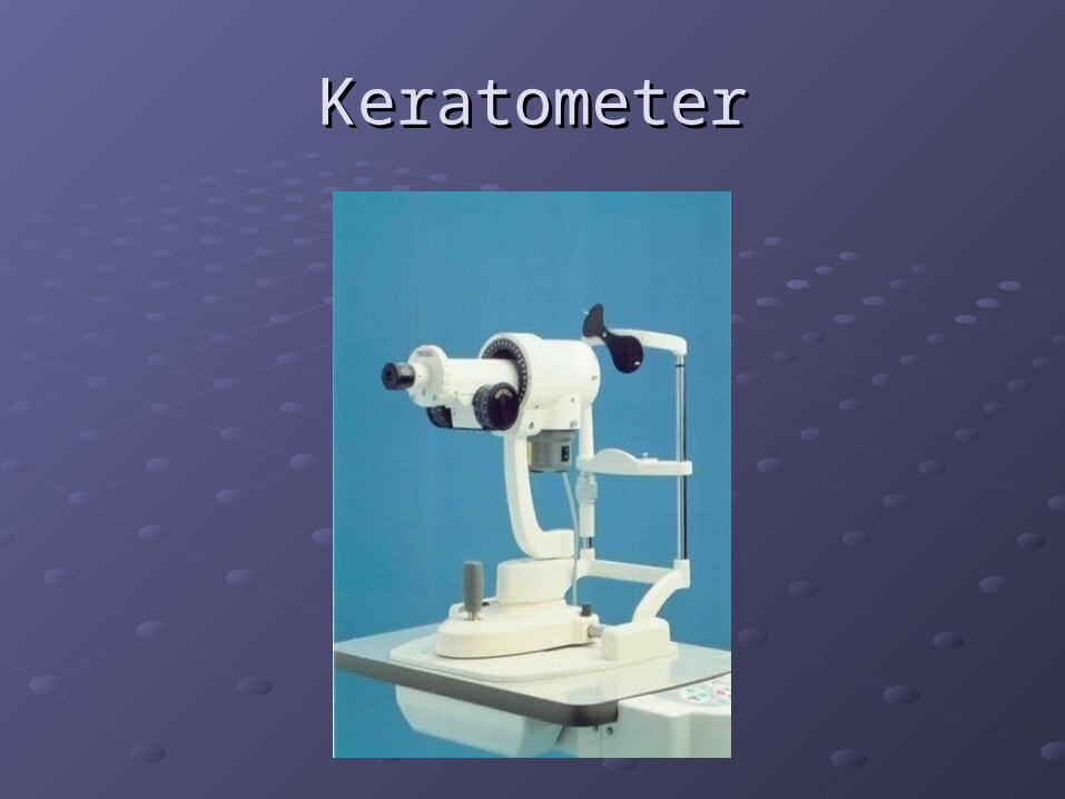
KeratometerKeratometer
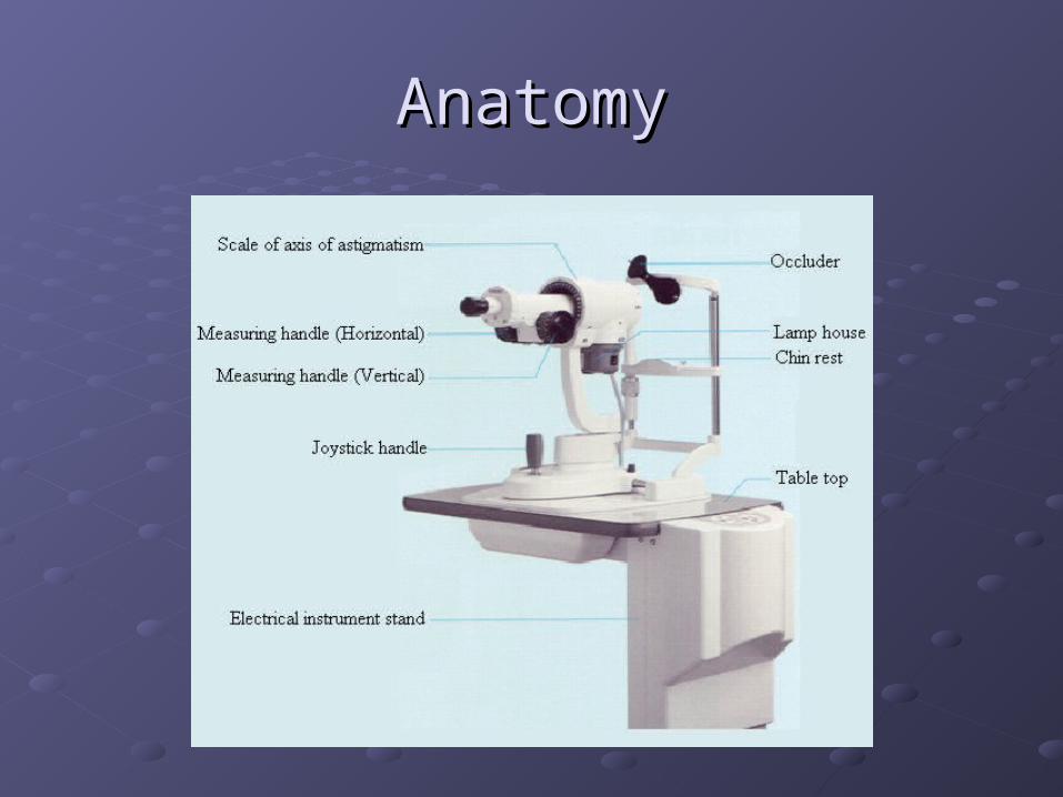
AnatomyAnatomy
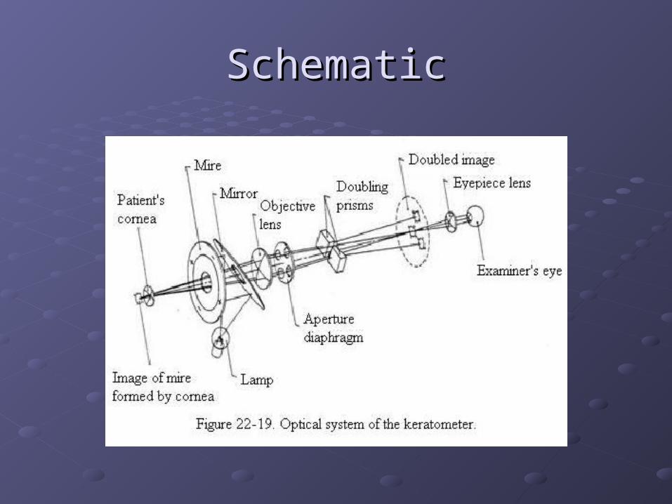
SchematicSchematic
KeratometerKeratometer
It is used to determine the curvature (i.e., It is used to determine the curvature (i.e., steepness or flatness), refracting power, steepness or flatness), refracting power, and toricity (i.e., astigmatism) of the and toricity (i.e., astigmatism) of the anterior central cornea in each of the two anterior central cornea in each of the two principal meridiansprincipal meridians
The keratometer is used to measure the The keratometer is used to measure the front surface corneal radiifront surface corneal radii
It is used to assess the integrity of the It is used to assess the integrity of the cornea and/or tear filmcornea and/or tear film
““K” Reading UsesK” Reading Uses
Fitting and evaluating contact lensesFitting and evaluating contact lenses
Evaluating the corneal topography for any Evaluating the corneal topography for any distortions or irregularitiesdistortions or irregularities Patients with irregular corneas are often not Patients with irregular corneas are often not
correctable to 20/20 visioncorrectable to 20/20 vision
Determining whether the cornea or axial Determining whether the cornea or axial length is the cause of refractive errorlength is the cause of refractive error
Evaluating the corneal healing process Evaluating the corneal healing process after surgery or injury affecting the corneaafter surgery or injury affecting the cornea
How Does It Work?How Does It Work?
The cornea is both a convex refracting The cornea is both a convex refracting surface and a convex mirrorsurface and a convex mirror
An object of known size is reflected from a An object of known size is reflected from a known distance to the corneal surfaceknown distance to the corneal surface
The size of the reflected image is measured The size of the reflected image is measured with a telescopewith a telescope
The refracting power of the cornea is The refracting power of the cornea is calculated based on an assumed index of calculated based on an assumed index of refraction (1.3375)refraction (1.3375)
CalibrationCalibration
Should be done regularly to ensure the Should be done regularly to ensure the accuracy of “K” readingsaccuracy of “K” readings
Mount a 5/8 inch steel ball bearing at the Mount a 5/8 inch steel ball bearing at the position close to that normally of the position close to that normally of the patient’s eyepatient’s eye
The steel ball has a known radius of The steel ball has a known radius of curvature, which upon proper calibration of curvature, which upon proper calibration of the keratometer , can be correctly readthe keratometer , can be correctly read
PreparationPreparation
Focus the eyepiece of the keratometer for Focus the eyepiece of the keratometer for the examiner’s eyethe examiner’s eye Turn on the powerTurn on the power Set the adjustable eyepiece as far counter-Set the adjustable eyepiece as far counter-
clockwise as possibleclockwise as possible Place a white sheet of paper in front of the Place a white sheet of paper in front of the
instrument’s objective lens to retroilluminate the instrument’s objective lens to retroilluminate the reticle (i.e., cross hairs)reticle (i.e., cross hairs)
Turn the eyepiece clockwise until the reticle is Turn the eyepiece clockwise until the reticle is first seen in sharp focusfirst seen in sharp focus
PreparationPreparation
Adjust the instrument for the patientAdjust the instrument for the patient Adjust the height of the patient’s chair and the Adjust the height of the patient’s chair and the
instrument to a comfortable position for both instrument to a comfortable position for both the patient and the examinerthe patient and the examiner
Unlock the instrument controlsUnlock the instrument controls Instruct the patient to place his chin in the Instruct the patient to place his chin in the
chin rest and his forehead against the chin rest and his forehead against the forehead rest and adjust for the patientforehead rest and adjust for the patient
PreparationPreparation
Align the instrument for the patientAlign the instrument for the patient Raise or lower the chin rest until the patient’s Raise or lower the chin rest until the patient’s
outer canthus is aligned with the hash mark outer canthus is aligned with the hash mark on the upright support of the instrumenton the upright support of the instrument
From outside the instrument, roughly align the From outside the instrument, roughly align the barrel with the patient’s right eye by raising or barrel with the patient’s right eye by raising or lowering the instrument and by moving it to lowering the instrument and by moving it to the left or right until a reflection of the mire is the left or right until a reflection of the mire is seen on the patient’s corneaseen on the patient’s cornea

ProcedureProcedure
Instruct the patientInstruct the patient Keep eyes open wide and blink normallyKeep eyes open wide and blink normally Try not to move the head or speakTry not to move the head or speak Look at the reflection of own eye in the Look at the reflection of own eye in the
keratometer barrelkeratometer barrel
ProcedureProcedure
Look into the keratometer and refine the Look into the keratometer and refine the alignment of the image of the mires (three alignment of the image of the mires (three circles) on the patient’s corneacircles) on the patient’s cornea
Focus the mires and adjust the instrument Focus the mires and adjust the instrument so that the reticle is centered in the lower so that the reticle is centered in the lower right hand circleright hand circle
Lock the instrument in placeLock the instrument in place
ProcedureProcedure
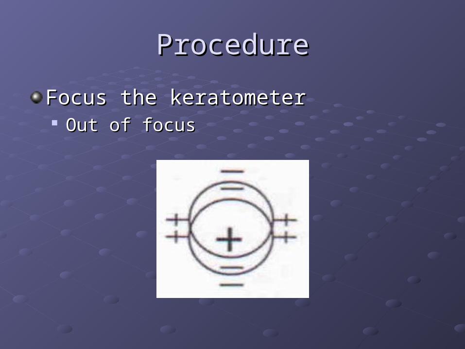
Focus the keratometerFocus the keratometer Out of focusOut of focus
ProcedureProcedure
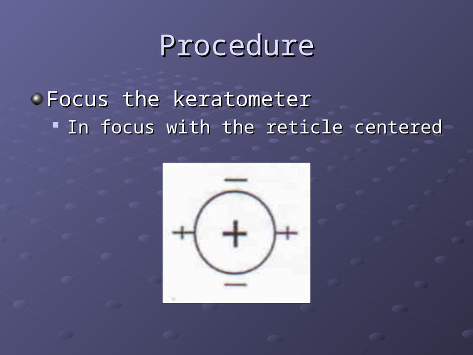
Focus the keratometerFocus the keratometer In focus with the reticle centeredIn focus with the reticle centered
ProcedureProcedure
Adjust the horizontal and the vertical Adjust the horizontal and the vertical power wheels until the mires are in close power wheels until the mires are in close appositionapposition
To locate the two principal meridians of To locate the two principal meridians of the patient’s cornea, rotate the telescope the patient’s cornea, rotate the telescope until the two horizontal plus signs of the until the two horizontal plus signs of the mires are perfectly continuous with one mires are perfectly continuous with one anotheranother
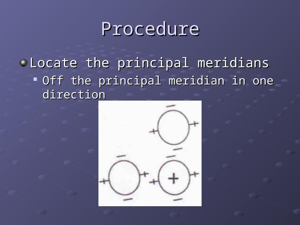
ProcedureProcedure
Locate the principal meridiansLocate the principal meridians Off the principal meridian in one directionOff the principal meridian in one direction
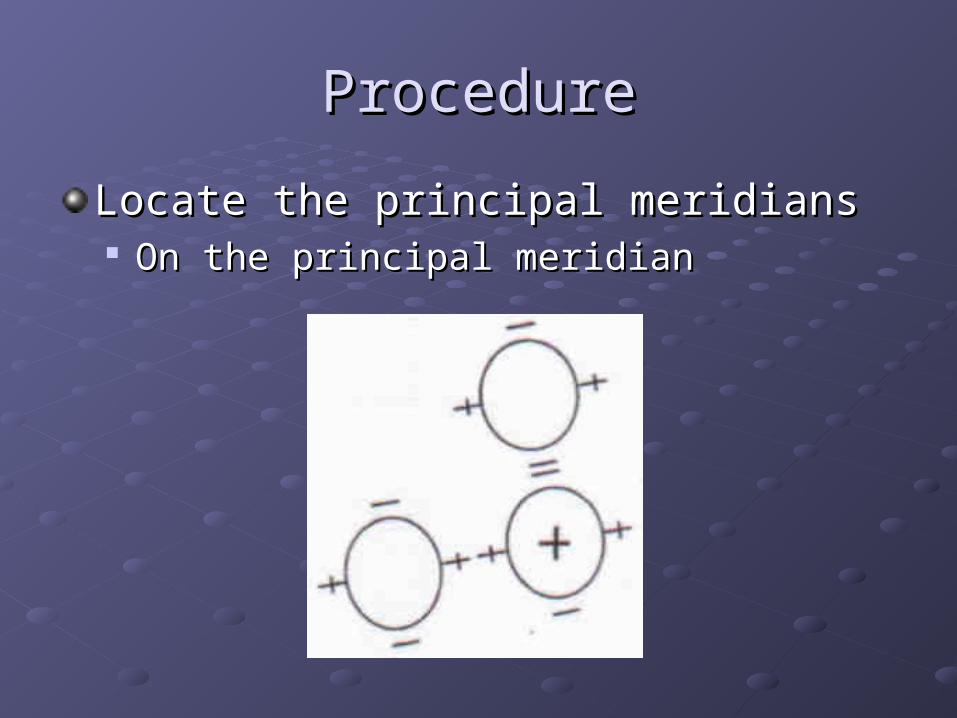
ProcedureProcedure
Locate the principal meridiansLocate the principal meridians On the principal meridianOn the principal meridian
ProcedureProcedure
Adjust the horizontal power wheel until the Adjust the horizontal power wheel until the plus signs of the mires overlap into one plus signs of the mires overlap into one image image The primary meridian is closest to 180 The primary meridian is closest to 180
degreesdegrees
ProcedureProcedure
Adjust the vertical power wheel until the Adjust the vertical power wheel until the minus signs of the mires overlap into one minus signs of the mires overlap into one image image The secondary meridian is 90 degrees from The secondary meridian is 90 degrees from
the primary meridianthe primary meridian
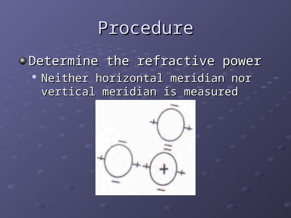
ProcedureProcedure
Determine the refractive powerDetermine the refractive power Neither horizontal meridian nor vertical Neither horizontal meridian nor vertical
meridian is measuredmeridian is measured
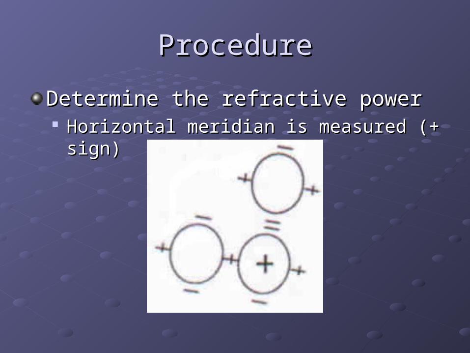
ProcedureProcedure
Determine the refractive powerDetermine the refractive power Horizontal meridian is measured (+ sign)Horizontal meridian is measured (+ sign)
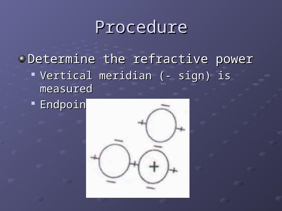
ProcedureProcedure
Determine the refractive powerDetermine the refractive power Vertical meridian (- sign) is measuredVertical meridian (- sign) is measured Endpoint is reachedEndpoint is reached
ProcedureProcedure
Throughout the procedure, adjust the Throughout the procedure, adjust the focus and recenter the reticle as neededfocus and recenter the reticle as needed
RecordingRecording
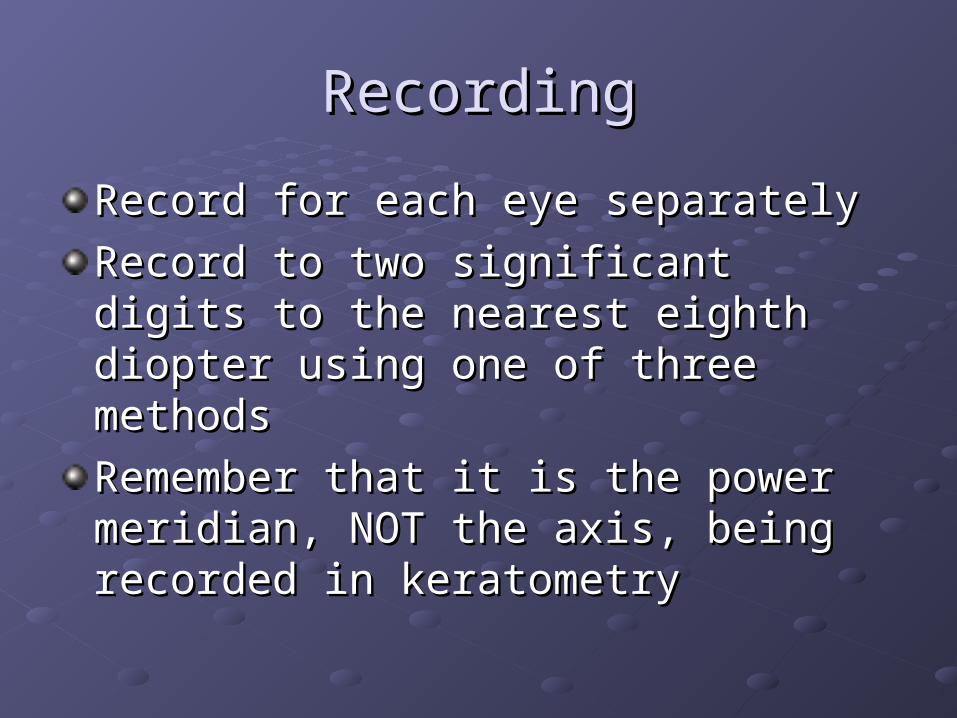
Record for each eye separatelyRecord for each eye separately
Record to two significant digits to the Record to two significant digits to the nearest eighth diopter using one of three nearest eighth diopter using one of three methodsmethods
Remember that it is the power meridian, Remember that it is the power meridian, NOT the axis, being recorded in NOT the axis, being recorded in keratometrykeratometry
RecordingRecording
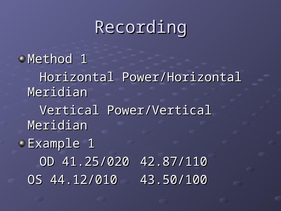
Method 1Method 1
Horizontal Power/Horizontal MeridianHorizontal Power/Horizontal Meridian
Vertical Power/Vertical Meridian Vertical Power/Vertical Meridian
Example 1Example 1
OD 41.25/020OD 41.25/020 42.87/110 42.87/110
OS 44.12/010OS 44.12/010 43.50/10043.50/100
RecordingRecording
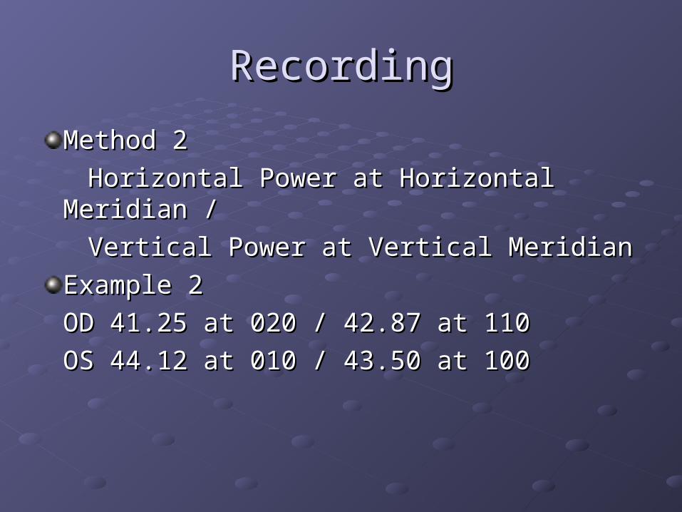
Method 2 Method 2
Horizontal Power at Horizontal Meridian /Horizontal Power at Horizontal Meridian /
Vertical Power at Vertical Meridian Vertical Power at Vertical Meridian
Example 2Example 2
OD 41.25 at 020 / 42.87 at 110OD 41.25 at 020 / 42.87 at 110
OS 44.12 at 010 / 43.50 at 100OS 44.12 at 010 / 43.50 at 100
RecordingRecording
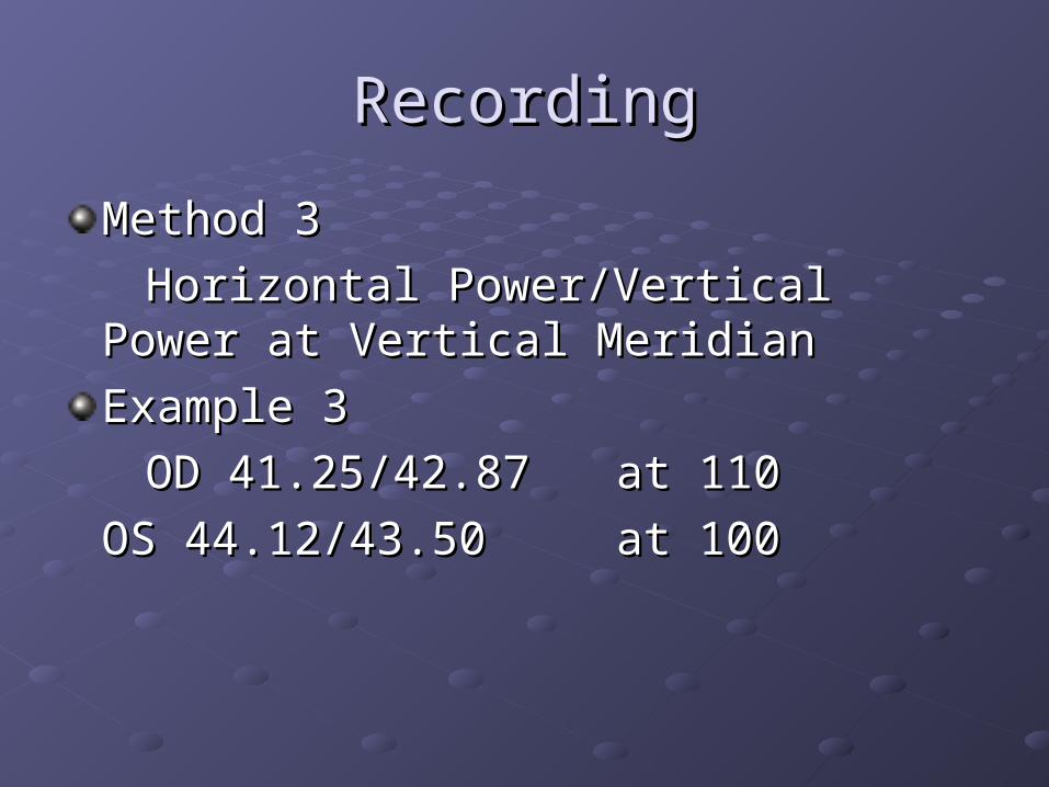
Method 3Method 3
Horizontal Power/Vertical Power at Horizontal Power/Vertical Power at Vertical MeridianVertical Meridian
Example 3Example 3
OD 41.25/42.87OD 41.25/42.87 at 110at 110
OS 44.12/43.50 OS 44.12/43.50 at 100at 100
RecordingRecording
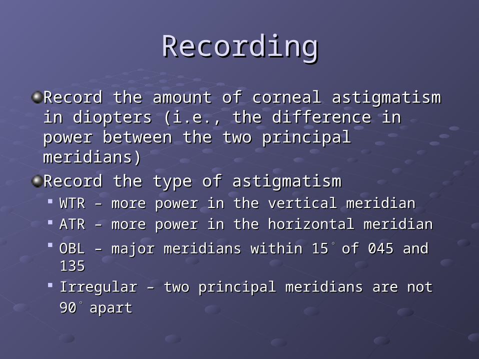
Record the amount of corneal astigmatism in Record the amount of corneal astigmatism in diopters (i.e., the difference in power between diopters (i.e., the difference in power between the two principal meridians)the two principal meridians)
Record the type of astigmatismRecord the type of astigmatism WTR – more power in the vertical meridianWTR – more power in the vertical meridian ATR – more power in the horizontal meridianATR – more power in the horizontal meridian OBL – major meridians within 15OBL – major meridians within 15
。。of 045 and 135of 045 and 135
Irregular – two principal meridians are not Irregular – two principal meridians are not
9090。。
apartapart
RecordingRecording
Record the conditions of the miresRecord the conditions of the mires Mires clear and regularMires clear and regular Mires irregular and distortedMires irregular and distorted
InterpretationInterpretation
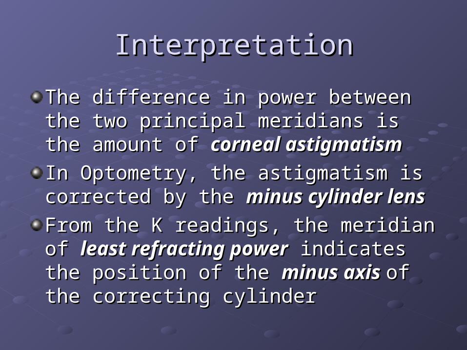
The difference in power between the two The difference in power between the two principal meridians is the amount of principal meridians is the amount of corneal astigmatismcorneal astigmatism
In Optometry, the astigmatism is corrected In Optometry, the astigmatism is corrected by the by the minus cylinder lensminus cylinder lens
From the K readings, the meridian of From the K readings, the meridian of least least refracting powerrefracting power indicates the position of indicates the position of the the minus axis minus axis of the correcting cylinderof the correcting cylinder
InterpretationInterpretation
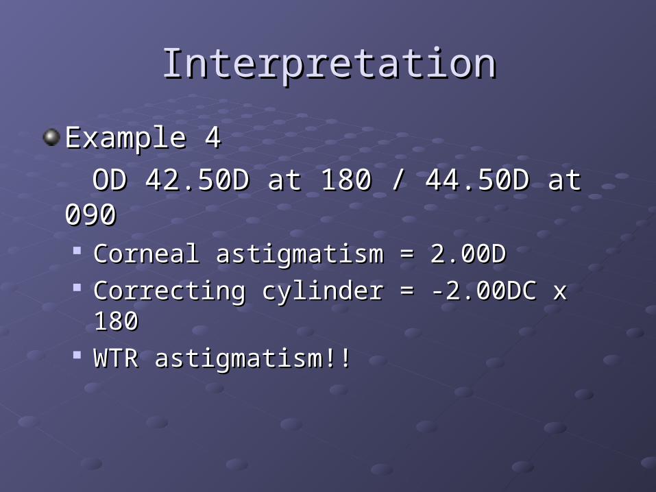
Example 4Example 4
OD 42.50D at 180 / 44.50D at 090OD 42.50D at 180 / 44.50D at 090 Corneal astigmatism = 2.00DCorneal astigmatism = 2.00D Correcting cylinder = -2.00DC x 180Correcting cylinder = -2.00DC x 180 WTR astigmatism!!WTR astigmatism!!
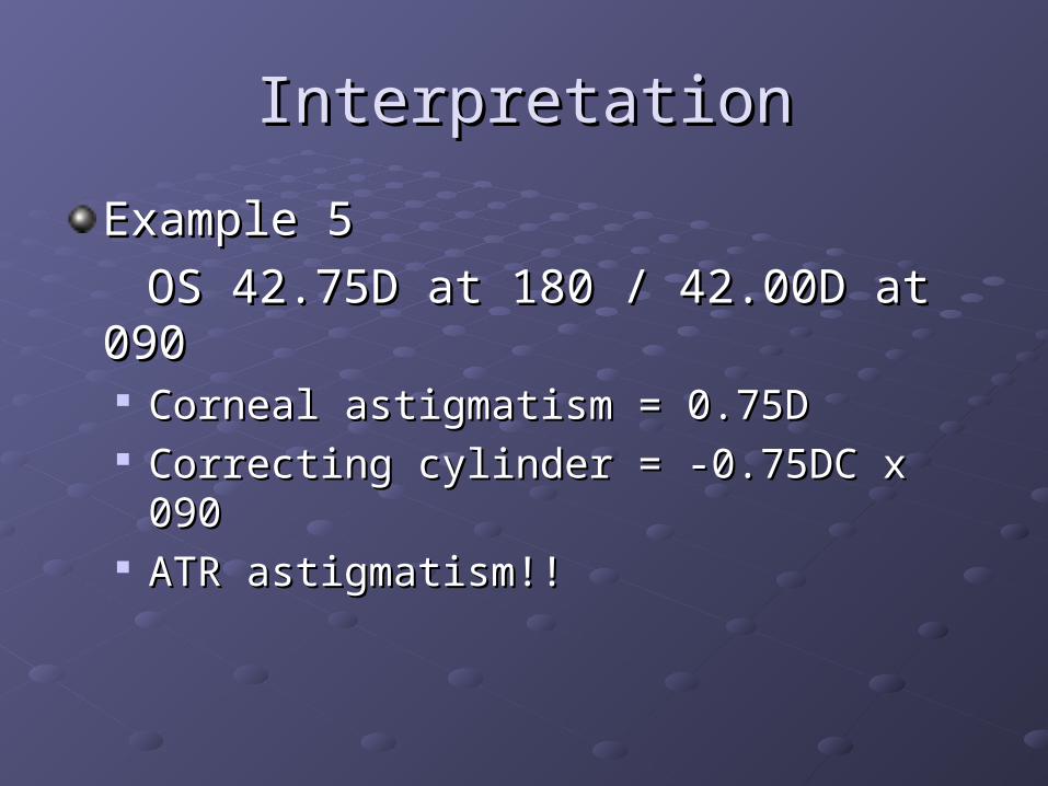
InterpretationInterpretation
Example 5Example 5
OS 42.75D at 180 / 42.00D at 090OS 42.75D at 180 / 42.00D at 090 Corneal astigmatism = 0.75DCorneal astigmatism = 0.75D Correcting cylinder = -0.75DC x 090Correcting cylinder = -0.75DC x 090 ATR astigmatism!!ATR astigmatism!!
Expected FindingsExpected Findings
Average K readings are 43.00D to 44.00DAverage K readings are 43.00D to 44.00D
The two principal meridians are expected The two principal meridians are expected
to be 90to be 90。。
apartapart