kent academic repository et al 2016.pdf · for any further enquiries regarding the licence status...
TRANSCRIPT
Kent Academic RepositoryFull text document (pdf)
Copyright & reuse
Content in the Kent Academic Repository is made available for research purposes. Unless otherwise stated all
content is protected by copyright and in the absence of an open licence (eg Creative Commons), permissions
for further reuse of content should be sought from the publisher, author or other copyright holder.
Versions of research
The version in the Kent Academic Repository may differ from the final published version.
Users are advised to check http://kar.kent.ac.uk for the status of the paper. Users should always cite the
published version of record.
Enquiries
For any further enquiries regarding the licence status of this document, please contact:
If you believe this document infringes copyright then please contact the KAR admin team with the take-down
information provided at http://kar.kent.ac.uk/contact.html
Citation for published version
Bastow, Emma L and Peswani, Amber Rose and Tarrant, Daniel S J and Pentland, Daniel R andChen, Xi and Morgan, Alan and Staniforth, Gemma L. and Tullet, Jennifer M.A. and Rowe, MichelleL. and Howard, Mark J. and Tuite, Mick F. and Gourlay, Campbell W. (2016) New links betweenSOD1 and metabolic dysfunction from a yeast model of amyotrophic lateral sclerosis. Journal
DOI
https://doi.org/10.1242/jcs.190298
Link to record in KAR
http://kar.kent.ac.uk/58393/
Document Version
Publisher pdf

RESEARCH ARTICLE
New links between SOD1 and metabolic dysfunction from a yeast
model of amyotrophic lateral sclerosisEmma L. Bastow1, Amber R. Peswani1, Daniel S. J. Tarrant1, Daniel R. Pentland1, Xi Chen2, Alan Morgan2,
Gemma L. Staniforth1, Jennifer M. Tullet1, Michelle L. Rowe1, Mark J. Howard1, Mick F. Tuite1,* and
Campbell W. Gourlay1,*
ABSTRACT
A number of genes have been linked to familial forms of the fatal
motor neuron disease amyotrophic lateral sclerosis (ALS). Over 150
mutations within the gene encoding superoxide dismutase 1 (SOD1)
have been implicated in ALS, but why such mutations lead to ALS-
associated cellular dysfunction is unclear. In this study, we identify
how ALS-linked SOD1 mutations lead to changes in the cellular
health of the yeast Saccharomyces cerevisiae. We find that it is not
the accumulation of aggregates but the loss of Sod1 protein stability
that drives cellular dysfunction. The toxic effect of Sod1 instability
does not correlate with a loss of mitochondrial function or increased
production of reactive oxygen species, but instead prevents
acidification of the vacuole, perturbs metabolic regulation and
promotes senescence. Central to the toxic gain-of-function seen
with the SOD1 mutants examined was an inability to regulate amino
acid biosynthesis. We also report that leucine supplementation
results in an improvement in motor function in a Caenorhabditis
elegans model of ALS. Our data suggest that metabolic dysfunction
plays an important role in Sod1-mediated toxicity in both the yeast and
worm models of ALS.
KEY WORDS: ALS, SOD1, Metabolism, Vacuole, Yeast
INTRODUCTION
Amyotrophic lateral sclerosis (ALS), also known as Lou Gehrig’s
disease, is a motor neuron disease characterised by progressive
muscle wasting. Muscle weakening in ALS patients is caused
specifically by the degeneration of motor neurons in the brain and
spinal cord (upper and lower motor neurons, respectively) (Waragai,
1997). The majority of ALS cases are sporadic; however, 20% of
cases are familial (fALS) and are inherited in an autosomal
dominant fashion (Siddique and Hentati, 1995-1996). Mutations in
a number of genes have been associated with both sporadic ALS and
fALS (Chen et al., 2013), leading to the proposal that ALS is a
multifactorial syndrome that includes motor system degeneration. In
line with the complex nature of this disease, these genes implicate a
range of cellular functions including RNA processing (TDP43 and
FUS3) (Arai et al., 2006; Kwiatkowski et al., 2009), cytoskeletal
organisation (Dynactin, also known as DCTN1, and Profilin, also
known as PFN1) and vesicle trafficking (VABP) (Nishimura et al.,
2004). To date, a single drug, the glutamate antagonist riluzole, has
been approved for ALS treatment and is reported to lead to a modest
extension of median survival of ALS patients (Georgoulopoulou
et al., 2013). One of the major challenges is to determine at which
stage each of the identified cellular processes contributes to disease
progression, and to what extent they represent a target for improved
therapeutic intervention.
Up to 20% of fALS cases are associated with a mutation in the
SOD1 gene (Rosen et al., 1993), which encodes superoxide
dismutase 1 (Sod1). The Sod1 enzyme is a homodimer that
requires copper and zinc binding in order to adopt a stable
conformation to facilitate conversion of superoxide anions to
hydrogen peroxide and oxygen. Sod1 is primarily cytosolic, but a
small proportion is also found within the mitochondrial
intermembrane space (Sturtz et al., 2001). The activity of Sod1
relies on Ccs1, a metallo-chaperone that interacts with immature
Sod1 to facilitate insertion of a copper ion into the Sod1 active site
(Sturtz et al., 2001). When the interaction between immature Sod1
and Ccs1 is disrupted, improper formation of a disulphide bond
within Sod1 results in a misfolded form that is prone to aggregation.
Human and yeast forms of Sod1 function similarly, yet an important
difference between them is that Sod1 activation in yeast is
dependent upon Ccs1, but human Sod1 can be activated by other
mechanisms (Sturtz et al., 2001).
The aggregation of unstable Sod1 was originally thought to
trigger fALS as a result of concurrent loss in Sod1 enzymatic
activity (Deng et al., 2006); however, mice lacking the SOD1 gene
do not develop ALS (Reaume et al., 1996) and therefore a toxic gain-
of-function is thought to underlie ALS-associated pathology. Over
150 mutations in the SOD1 gene have been linked to ALS and these
are distributed through all five exons. The most common SOD1
mutation found within the ALS population in the USA is an alanine-
to-valine substitution at codon 4 (producing Sod1A4V protein). The
A4V mutation is located in the first β-sheet of the protein, at the
dimer interface, and affects stability and enzymatic activity
(Cudkowicz et al., 1997; Rosen et al., 1994). Reduced stability
associated with ALS-linked mutations in Sod1 is generally thought
to underpin the toxic gain-of-function. However, it is unclear how
each mutation relates to the cellular dysfunction that drives disease
progression. Interestingly, the overexpression of native SOD1 also
accelerates disease progression in ALS mouse models (Xu et al.,
2015). In addition, the overexpression of SOD1 alone can lead to
ALS symptoms in mice, providing evidence that the native Sod1
protein can participate in disease (Graffmo et al., 2013).
Exactly how SOD1 mutations trigger or contribute to ALS
pathology is unknown; however, the aggregation of misfolded orReceived 4 April 2016; Accepted 16 September 2016
1Kent Fungal Group, School of Biosciences, University of Kent, Canterbury, KentCT2 7NJ, UK. 2Institute of Translational Medicine, Department of Cellular andMolecular Physiology, University of Liverpool, Liverpool L69 3BX, UK.
*Authors for correspondence ([email protected]; [email protected])
A.M., 0000-0002-0346-1289; J.M.T., 0000-0002-2037-526X; M.F.T., 0000-0002-5214-540X; C.W.G., 0000-0002-2373-6788
This is an Open Access article distributed under the terms of the Creative Commons AttributionLicense (http://creativecommons.org/licenses/by/3.0), which permits unrestricted use,distribution and reproduction in any medium provided that the original work is properly attributed.
4118
© 2016. Published by The Company of Biologists Ltd | Journal of Cell Science (2016) 129, 4118-4129 doi:10.1242/jcs.190298
JournalofCellScience

unstable Sod1 protein is a widely reported hallmark of this disease.
Aggregation of Sod1 has been observed in a variety of cell and
animal models of ALS as well as within cells and cerebrospinal fluid
of ALS patients (Kaur et al., 2016). Sod1 aggregates are reported to
accumulate at the outer surface of the mitochondria and within the
mitochondrial intermembrane space. The resultant respiratory
dysfunction and accumulation of reactive oxygen species (ROS)
associated with aggregate accumulation is thought to be an
important factor in disease progression (Vehviläinen et al., 2014).
In line with mitochondria being a potential therapeutic target, the
expression of mitochondrial targeted catalase was sufficient to
protect motor neurons from Sod1G93A–mediated toxicity in mice
(Pehar et al., 2014). However, the neuroprotective effect of catalase
was not accompanied by an increase in lifespan, highlighting the
complex nature of this disease. The expression of mutant isoforms
of Sod1 can also damage a number of other important cellular
processes and compartments, including the ER stress response (Soo
et al., 2015), glutamate excitotoxicity (Canton et al., 1998), calcium
homeostasis (Kawamata et al., 2014), proteasome exhaustion
(Kepp, 2015), metal ion regulation (Hilton et al., 2015),
inflammation (Van Dyke et al., 2016) and the regulation of
autophagy (Mis et al., 2016). It has also become clear that the
disease extends beyond motor neurons and affects other cell types
such as astrocytes, microglia, oligodendrocytes and muscle cells,
whose functions could also play a role in ALS pathology (Kaur
et al., 2016). Interestingly, recent studies also suggest that toxic
Sod1 protein products exhibit prion-like properties and can spread
between cells (Münch et al., 2011; Ayers et al., 2016).
Here, we describe a newmodel of ALS that we have developed in
the yeast Saccharomyces cerevisiae. We have incorporated several
ALS-linked mutations into the endogenous yeast SOD1 gene. Our
data show that the resulting mutant Sod1 isoforms are unstable and
have toxic effects upon the cell. In contrast to other systems studied
to date, these mutations do not lead to the formation of Sod1 protein
aggregates. In addition, the toxic effects of unstable yeast Sod1
proteins do not appear to cause mitochondrial dysfunction or result
in oxidative stress. Instead, our data suggest that toxicity is
associated with an inability to control central metabolic processes,
most probably linked to severe disruption of the vacuolar
compartment. The overall metabolic dysfunction associated with
mutant Sod1 isoforms does not result in yeast cell death, but instead
drives cells into a state of senescence. Furthermore, in a
Caenorhabditis elegans ALS model system, in which SOD1
overexpression results in motor neuron dysfunction, media
supplementation with L-leucine rescues motor neuron
degeneration. Our findings provide new evidence that soluble,
non-aggregating forms of Sod1 might a play role in the cell
dysfunction underlying ALS via the disruption of metabolic
homeostasis.
RESULTS
Expression of yeastSOD1ALSmutant alleles is toxic in yeast
A number of amino acid residues whose substitutions are associated
with fALS are conserved between yeast and humans. To construct a
yeast model of ALS we introduced mutations into the yeast SOD1
gene that led to amino acid substitutions equivalent to A4V, G37R,
H48Q, G93A and S134N (Fig. 1A). Each of these mutations are
linked with fALS in humans and their expression in mice also gives
rise to ALS symptoms (Chen et al., 2013) (Fig. 1A). In yeast Sod1,
these mutations correspond to Sod1A3V, Sod1G36R, Sod1H47Q,
Sod1G92A and Sod1S133N, respectively, and are evenly dispersed
over the mature Sod1 protein (Fig. 1A).
AHsSod1 MATKAVCVLKGDGPVQGIINFEQKESNGPVKVWGSIKG-LTEGLHGFHVHEFGDNTAGCT 59
ScSod1 -MVQAVAVLKGDAGVSGVVKFEQASESEPTTVSYEIAGNSPNAERGFHIHEFGDATNGCV 59
.:**.*****. *.*:::*** ... *..* .* * :. :***:***** * **.
HsSod1 SAGPHFNPLSRKHGGPKDEERHVGDLGNVTADKDGVADVSIEDSVISLSGDHCIIGRTLV 119
ScSod1 SAGPHFNPFKKTHGAPTDEVRHVGDMGNVKTDENGVAKGSFKDSLIKLIGPTSVVGRSVV 119
********:.:.**.*.** *****:***.:*::***. *::**:*.* * .::**::*
HsSod1 VHEKADDLGKGGNEESTKTGNAGSRLACGVIGIAQ 154
ScSod1 IHAGQDDLGKGDTEESLKTGNAGPRPACGVIGLTN 154
:* ****** .*** ****** * ******:::
D E
CB
Sod1
Pgk
Sod2
Sod1
Sod2
Sod1
Sod1
Pgk
Fig. 1. YeastSOD1ALSmutant alleles.
(A) Amino acid residues commonly
substituted within ALS patients and that
were mutated in this study are conserved
between yeast and human Sod1 (left).
Their distribution is indicated upon the
crystal structure of a human Sod1 dimer
(PDB ID: 1HL4) (right). (B–E) Wild-type
or cells deleted for SOD1 and re-
expressing SOD1 or sod1 mutants as
indicated were assayed for protein level
by western blotting during logarithmic (B)
and stationary phase (D) of growth or for
enzymatic activity during logarithmic (C)
or stationary phase (E) of growth.
4119
RESEARCH ARTICLE Journal of Cell Science (2016) 129, 4118-4129 doi:10.1242/jcs.190298
JournalofCellScience

To determine the effects of the yeast SOD1 mutations (called
sod1 mutants hereafter) on cell growth and metabolism, wild-type
SOD1 and the mutants were constitutively expressed from a
multicopy plasmid in both wild-type and sod1-null (Δsod1)
backgrounds, and both protein level and enzymatic activity were
assessed. During logarithmic and stationary phases of growth, the
overexpression of native SOD1 could be observed in both wild-type
(Fig. S1) and Δsod1 strain backgrounds (Fig. 1B,D). However, the
level of all five Sod1 mutant isoforms did not exceed, and in most
cases was significantly lower, than that of un-mutated Sod1 when
expressed in a Δsod1 background. This is most probably because of
inherent instability within the mutant proteins (Fig. 1B,D). By
contrast, the introduction of mutant sod1 expression plasmids into a
wild-type background did result in their overexpression (Fig. S1),
indicating that mutant isoforms of Sod1 become stabilised when co-
expressed with native Sod1. In line with the production of an
unstable product, no Sod1 enzymatic activity could be detected
within Δsod1 logarithmic phase cells expressing mutant Sod1
isoforms (Fig. 1C); however, low levels of enzymatic activity could
be detected in stationary phase cells expressing sod1A3V, sod1G36R
and sod1G92A (Fig. 1E and Fig. S2). Levels of Sod2 remained
relatively stable in all strains examined (Fig. 1C,E).
The expression of sod1A3V, sod1G36R, sod1H47Q, sod1G92A or
sod1S133N had no effect on wild-type cell growth (Fig. 2A) or
viability (data not shown). However, expression of the same mutant
alleles in a Δsod1 strain led to an elongated lag phase, slower growth
during logarithmic phase and reduction in final culture cell number
(Fig. 2B). Toxic effects associated with the expression of mutant
forms of Sod1 also led to a significant loss of culture viability
(Fig. 2C). These data suggest a correlation between Sod1 stability
and toxicity within our yeast ALS model system. This correlation is
also in accordance with our observation that the overexpression of
mutant Sod1 isoforms that are stabilised in a wild-type background
are not toxic to yeast cells.
Toxicity associated with Sod1 instability does not correlate
with increased ROS levels or mitochondrial dysfunction
SOD1 mutations and their toxic effects have been linked to
mitochondrial dysfunction (Vehviläinen et al., 2014). To examine
whether this was also the case in our yeast ALS model we
determined the respiratory profile of strains expressing sod1A3V,
sod1G36R, sod1H47Q, sod1G92A or sod1S133N using high resolution
respirometry (Fig. 3A). Using this technique, which measures
mitochondrial oxygen consumption, the addition of drugs that target
specific sites of the electron transport chain (ETC) allowed us to
examine mitochondrial function in detail. Initially, routine
respiration was analysed in a Δsod1 strain expressing SOD1,
sod1A3V, sod1G36R, sod1H47Q, sod1G92A or sod1S133N during the post
diauxic shift phase of growth (Fig. 3A). Surprisingly, respiration
was significantly increased in all mutant strains relative to wild type
(Fig. 3A) despite their low culture viability (Fig. 2C). The increase
in respiration observed was consistent with an increase in the
activity of the entire ETC, as shown by significant increases in the
LEAK respiration rate, which indicates inner mitochondrial
integrity and the level of control that ATPase function has on
electron transport, and maximum ETC rate (ETS, induced by
addition of the uncoupling agent FCCP) (Fig. 3A). The LEAK and
ETS oxygen flux in a Δsod1 strain were significantly decreased
compared with wild-type control but re-expression of SOD1
returned these to the wild-type control levels (Fig. 3A). The
increase in respiration rates observed suggests that the loss of cell
viability observed in cells expressing mutant Sod1 is not a result of
cell death. To confirm this, Δsod1 cells expressing SOD1, sod1A3V
or sod1G92A were grown for 24 h and then plated onto either
minimal media lacking leucine to select for plasmid retention (as
done previously; see Fig. 2C) or onto rich YPDmedia (Fig. S3). We
observed that Δsod1 cells expressing either sod1A3V or sod1G92A
exhibited a dramatic increase in colony forming units when plated
onto rich media instead of minimal selective media (Fig. S3). This
finding suggests that the expression of unstable sod1 isoforms leads
to loss of viability caused by senescence that is linked to defects in
nutritional sensing or resource availability, which can be rescued by
growth in rich media.
Changes in mitochondrial function and a decrease in viability are
often associated with ROS production. We therefore assessed the
production of both the superoxide anion and hydrogen peroxide
during logarithmic and post-diauxic shift phases of growth using the
ROS sensor dyes DHE and H2DFC-DA, respectively. Significant
A
C
BWild type controlΔsod1 control Δsod1 + SOD1
Δsod1 + sod1A3V
Δsod1 + sod1G36R
Δsod1 + sod1H47Q
Δsod1 + sod1G92A
Δsod1 + sod1S133N
4 6 8 10 12 14 16 18 20 22 24 26 2820
0.4
0.8
1.2
1.6
2.0
Time (h)
% V
iab
ilit
y
***
Wild type control
Wild type + SOD1
Wild type + sod1A3V
Wild type + sod1G36R
Wild type + sod1H47Q
Wild type + sod1G92A
Wild type + sod1S133N
0
0.4
0.8
1.2
1.6
2.0
Ab
sorb
an
ce (
OD
60
0)
Time (h)
50 10 15 20 25
Ab
sorb
an
ce (
OD
60
0)
Fig. 2. Effect of SOD1mutation on cell growth and viability. (A,B)Wild-type
(WT) or cells deleted for SOD1 containing either an empty plasmid (control)
or re-expressing SOD1 or sod1 mutants as indicated were assessed for
growth using an automatic plate reader. (C) Viability was assessed after 24 h of
growth in selective medium and assessed using a colony forming unit assay,
which calculates the percentage of cells able to form a colony within each
population. The probability of a significant difference in viability compared with
wild type was calculated for each strain; ***P<0.001. All experiments were
carried out in biological triplicate and error bars represent standard deviation.
4120
RESEARCH ARTICLE Journal of Cell Science (2016) 129, 4118-4129 doi:10.1242/jcs.190298
JournalofCellScience
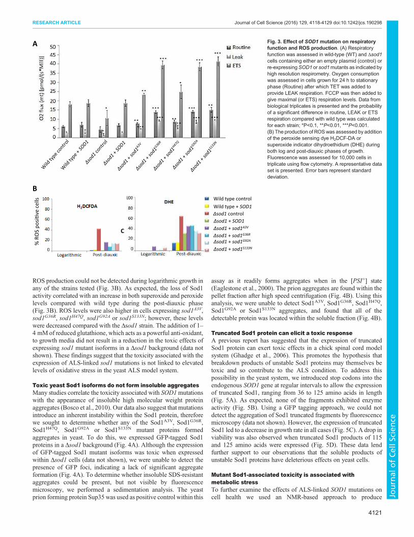
ROS production could not be detected during logarithmic growth in
any of the strains tested (Fig. 3B). As expected, the loss of Sod1
activity correlated with an increase in both superoxide and peroxide
levels compared with wild type during the post-diauxic phase
(Fig. 3B). ROS levels were also higher in cells expressing sod1A3V,
sod1G36R, sod1H47Q, sod1G92A or sod1S133N; however, these levels
were decreased compared with the Δsod1 strain. The addition of 1–
4 mM of reduced glutathione, which acts as a powerful anti-oxidant,
to growth media did not result in a reduction in the toxic effects of
expressing sod1 mutant isoforms in a Δsod1 background (data not
shown). These findings suggest that the toxicity associated with the
expression of ALS-linked sod1 mutations is not linked to elevated
levels of oxidative stress in the yeast ALS model system.
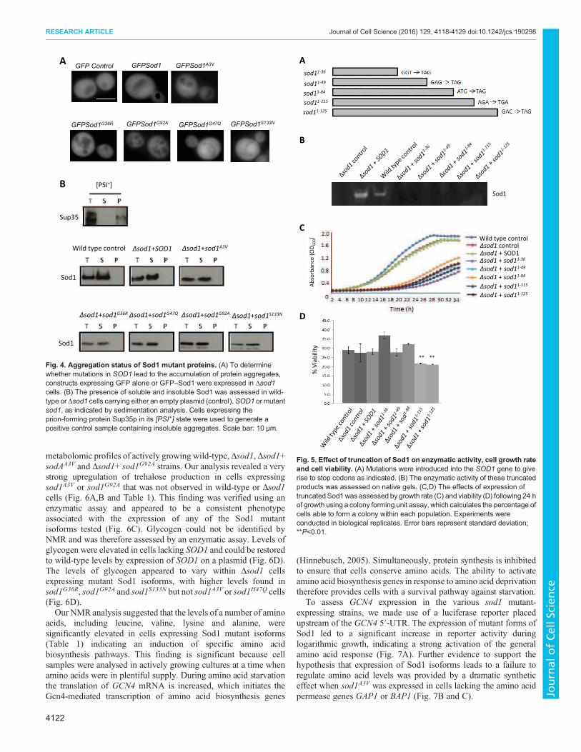
Toxic yeast Sod1 isoforms do not form insoluble aggregates
Many studies correlate the toxicity associated with SOD1mutations
with the appearance of insoluble high molecular weight protein
aggregates (Bosco et al., 2010). Our data also suggest that mutations
introduce an inherent instability within the Sod1 protein, therefore
we sought to determine whether any of the Sod1A3V, Sod1G36R,
Sod1H47Q, Sod1G92A or Sod1S133N mutant proteins formed
aggregates in yeast. To do this, we expressed GFP-tagged Sod1
proteins in a Δsod1 background (Fig. 4A). Although the expression
of GFP-tagged Sod1 mutant isoforms was toxic when expressed
within Δsod1 cells (data not shown), we were unable to detect the
presence of GFP foci, indicating a lack of significant aggregate
formation (Fig. 4A). To determine whether insoluble SDS-resistant
aggregates could be present, but not visible by fluorescence
microscopy, we performed a sedimentation analysis. The yeast
prion forming protein Sup35 was used as positive control within this
assay as it readily forms aggregates when in the [PSI+] state
(Eaglestone et al., 2000). The prion aggregates are found within the
pellet fraction after high speed centrifugation (Fig. 4B). Using this
analysis, we were unable to detect Sod1A3V, Sod1G36R, Sod1H47Q,
Sod1G92A or Sod1S133N aggregates, and found that all of the
detectable protein was located within the soluble fraction (Fig. 4B).
Truncated Sod1 protein can elicit a toxic response
A previous report has suggested that the expression of truncated
Sod1 protein can exert toxic effects in a chick spinal cord model
system (Ghadge et al., 2006). This promotes the hypothesis that
breakdown products of unstable Sod1 proteins may themselves be
toxic and so contribute to the ALS condition. To address this
possibility in the yeast system, we introduced stop codons into the
endogenous SOD1 gene at regular intervals to allow the expression
of truncated Sod1, ranging from 36 to 125 amino acids in length
(Fig. 5A). As expected, none of the fragments exhibited enzyme
activity (Fig. 5B). Using a GFP tagging approach, we could not
detect the aggregation of Sod1 truncated fragments by fluorescence
microscopy (data not shown). However, the expression of truncated
Sod1 led to a decrease in growth rate in all cases (Fig. 5C). A drop in
viability was also observed when truncated Sod1 products of 115
and 125 amino acids were expressed (Fig. 5D). These data lend
further support to our observations that the soluble products of
unstable Sod1 proteins have deleterious effects on yeast cells.
Mutant Sod1-associated toxicity is associated with
metabolic stress
To further examine the effects of ALS-linked SOD1 mutations on
cell health we used an NMR-based approach to produce
B
A
C
% R
OS
po
si�
ve
ce
lls Wild type control
Δsod1 control
Δsod1 + SOD1
Δsod1 + sod1A3V
Δsod1 + sod1G36R
Δsod1 + sod1G92A
Δsod1 + sod1S133N
Wild type + SOD1
Fig. 3. Effect of SOD1 mutation on respiratory
function and ROS production. (A) Respiratory
function was assessed in wild-type (WT) and Δsod1
cells containing either an empty plasmid (control) or
re-expressingSOD1 or sod1mutants as indicated by
high resolution respirometry. Oxygen consumption
was assessed in cells grown for 24 h to stationary
phase (Routine) after which TET was added to
provide LEAK respiration. FCCP was then added to
give maximal (or ETS) respiration levels. Data from
biological triplicates is presented and the probability
of a significant difference in routine, LEAK or ETS
respiration compared with wild type was calculated
for each strain; *P<0.1, **P<0.01, ***P<0.001.
(B) The production of ROSwas assessed by addition
of the peroxide sensing dye H2DCF-DA or
superoxide indicator dihydroethidium (DHE) during
both log and post-diauxic phases of growth.
Fluorescence was assessed for 10,000 cells in
triplicate using flow cytometry. A representative data
set is presented. Error bars represent standard
deviation.
4121
RESEARCH ARTICLE Journal of Cell Science (2016) 129, 4118-4129 doi:10.1242/jcs.190298
JournalofCellScience
metabolomic profiles of actively growing wild-type, Δsod1, Δsod1+
sodAA3V and Δsod1+ sod1G92A strains. Our analysis revealed a very
strong upregulation of trehalose production in cells expressing
sod1A3V or sod1G92A that was not observed in wild-type or Δsod1
cells (Fig. 6A,B and Table 1). This finding was verified using an
enzymatic assay and appeared to be a consistent phenotype
associated with the expression of any of the Sod1 mutant
isoforms tested (Fig. 6C). Glycogen could not be identified by
NMR and was therefore assessed by an enzymatic assay. Levels of
glycogen were elevated in cells lacking SOD1 and could be restored
to wild-type levels by expression of SOD1 on a plasmid (Fig. 6D).
The levels of glycogen appeared to vary within Δsod1 cells
expressing mutant Sod1 isoforms, with higher levels found in
sod1G36R, sod1G92A and sod1S133N but not sod1A3V or sod1H47Q cells
(Fig. 6D).
Our NMR analysis suggested that the levels of a number of amino
acids, including leucine, valine, lysine and alanine, were
significantly elevated in cells expressing Sod1 mutant isoforms
(Table 1) indicating an induction of specific amino acid
biosynthesis pathways. This finding is significant because cell
samples were analysed in actively growing cultures at a time when
amino acids were in plentiful supply. During amino acid starvation
the translation of GCN4 mRNA is increased, which initiates the
Gcn4-mediated transcription of amino acid biosynthesis genes
(Hinnebusch, 2005). Simultaneously, protein synthesis is inhibited
to ensure that cells conserve amino acids. The ability to activate
amino acid biosynthesis genes in response to amino acid deprivation
therefore provides cells with a survival pathway against starvation.
To assess GCN4 expression in the various sod1 mutant-
expressing strains, we made use of a luciferase reporter placed
upstream of the GCN4 5′-UTR. The expression of mutant forms of
Sod1 led to a significant increase in reporter activity during
logarithmic growth, indicating a strong activation of the general
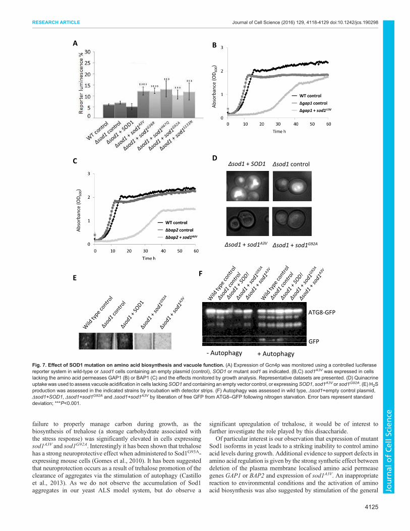
amino acid response (Fig. 7A). Further evidence to support the
hypothesis that expression of Sod1 isoforms leads to a failure to
regulate amino acid levels was provided by a dramatic synthetic
effect when sod1A3V was expressed in cells lacking the amino acid
permease genes GAP1 or BAP1 (Fig. 7B and C).
AGFPSod1A3VGFPSod1GFP Control
GFPSod1G36R GFPSod1G92AGFPSod1G47Q GFPSod1S133N
B
Wild type control Δsod1+SOD1 Δsod1+sod1A3V
Δsod1+sod1G36R Δsod1+sod1G92AΔsod1+sod1S133NΔsod1+sod1G47Q
Sod1
Sod1
Sup35
[PSI+]
Fig. 4. Aggregation status of Sod1 mutant proteins. (A) To determine
whether mutations in SOD1 lead to the accumulation of protein aggregates,
constructs expressing GFP alone or GFP–Sod1 were expressed in Δsod1
cells. (B) The presence of soluble and insoluble Sod1 was assessed in wild-
type or Δsod1 cells carrying either an empty plasmid (control), SOD1 or mutant
sod1, as indicated by sedimentation analysis. Cells expressing the
prion-forming protein Sup35p in its [PSI+] state were used to generate a
positive control sample containing insoluble aggregates. Scale bar: 10 µm.
A
B
Sod1
% V
iab
ilit
y
Wild type controlΔsod1 control
Δsod1 + sod11-36
Δsod1 + sod11-49
Δsod1 + sod11-84
Δsod1 + sod11-115
Δsod1 + sod11-125
Δsod1 + SOD1
sod11-36
sod11-49
sod11-115
sod11-125
sod11-84
Ab
sorb
an
ce (
OD
60
0)
** **
C
D
Fig. 5. Effect of truncation of Sod1 on enzymatic activity, cell growth rate
and cell viability. (A) Mutations were introduced into the SOD1 gene to give
rise to stop codons as indicated. (B) The enzymatic activity of these truncated
products was assessed on native gels. (C,D) The effects of expression of
truncatedSod1was assessed by growth rate (C) and viability (D) following 24 h
of growth using a colony forming unit assay, which calculates the percentage of
cells able to form a colony within each population. Experiments were
conducted in biological replicates. Error bars represent standard deviation;
**P<0.01.
4122
RESEARCH ARTICLE Journal of Cell Science (2016) 129, 4118-4129 doi:10.1242/jcs.190298
JournalofCellScience

The yeast vacuole, the functional equivalent of the mammalian
lysosome, is the main store of free amino acids in cells and is also
crucial for the regulation of amino acid levels during times of stress.
As a result, defects in vacuole function are associated with failure to
regulate amino acid levels. To determine whether expression of
mutant sod1 affects vacuole function, we examined both
acidification and morphology of the organelle. A clear defect in
the uptake of the dye quinacrine, which is used to assess vacuolar
pH (Preston et al., 1989), was observed in cells expressing mutant
isoforms of Sod1 (Fig. 7D). A small increase in the number of cells
exhibiting multiple vacuoles was also observed for cells lacking
SOD1 and for cells expressing mutant Sod1 isoforms (data not
shown). Cells with defective vacuolar ATPase function, which is
responsible for acidification of the vacuole, have recently been
shown to exhibit decreased H2S production (Winter et al., 2014). A
clear decrease in H2S production was observed in Δsod1 strains
expressing mutant isoforms of Sod1 compared with controls
(Fig. 7E).
Vacuole function is central to the autophagic process, which has
been implicated in ALS disease pathology (Sasaki, 2011). To
determine whether Sod1 mutation also leads to defects in autophagy
we expressed a GFP-tagged version of autophagy-related protein 8
(Atg8) in cells expressing sod1G92A or sod1A3V. The hydrolysis of
GFP from Atg8 upon autophagy is an established marker for
autophagy in response to nitrogen starvation (Huang et al., 2014)
and this event can be visualised by western blotting using an anti-
GFP antibody. Upon induction of autophagy, free GFP could be
detected in wild-type, Δsod1 and Δsod1+SOD1 cells but not in
B
D
0
10
20
30
40
50
μg
gly
cog
en
pe
r 1
06
cells **
**
****
A
**
*
C25
*
*
Wild type + control
Δsod1 + control
Δsod1 + sod1A4V
Δsod1 + sod1G93A
2468 0 (ppm)
Fig. 6. Association of SOD1 mutation with
metabolic stress. (A,B) Metabolites were
extracted and subjected to NMR analyses. The
representative 1DH+ spectra displaymetabolite
peaks that highlight the identified trehalose
peak, which is enlarged in (B). (C) To confirm
the finding, an enzymatic trehalose assay was
performed in biological triplicate and the fold
change is compared with wild type.
(D) Glycogen levels were assessed in triplicate.
Error bars represent standard deviation;
**P<0.01.
4123
RESEARCH ARTICLE Journal of Cell Science (2016) 129, 4118-4129 doi:10.1242/jcs.190298
JournalofCellScience

Δsod1 strains expressing either sod1G92A or sod1A3V, indicating a
defect in autophagy (Fig. 7F). Confirming that the process of
autophagy is impaired in cells expressing unstable sod1 isoforms,
we found a strong synthetic interaction when sod1G92A and sod1A3V
were expressed in cells lacking the mitophagy regulator ATG32
(Fig. S4).
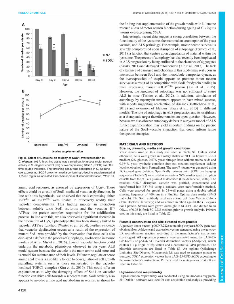
Addition of L-leucine leads to a decrease in the toxicity of
SOD1 overexpression in motor neurons of C. elegans
Our data with the yeast ALS model suggest that increased levels of
unstable Sod1 can lead to toxicity associated with defects in the
regulation of metabolism during growth. The overexpression of
native SOD1 leads to ALS in mouse models (Graffmo et al., 2013),
suggesting that elevated levels of Sod1 also lead to a toxic gain-of-
function within cells. We therefore used an established thrashing
assay to assess whether the overexpression of native SOD1 in the
motor neurons of C. elegans led to motor defects. As nematode
worms age they lose motor function and their thrashing activity
declines over time (Fig. 8A). The overexpression of SOD1 in motor
neurons decreased thrashing activity relative to controls and
reproducibly accelerated age-related motor dysfunction (Fig. 8A).
Our evidence suggests that amino acid deprivation and the
activation of amino acid biosynthesis are important in the toxic
effects of mutant Sod1 expression in yeast. Current research
suggests that leucine is used to sense amino acid levels and elicit an
appropriate response via control of the TORC1 signalling pathway
(Ham et al. 2014). Interestingly, we observed that the addition of
L-leucine to medium upon which C. elegans was growing and
feeding gave a dose-dependent and reproducible increase in motor
activity in worms overexpressing SOD1 (Fig. 8B). These data further
suggest a link between the toxic gain-of-function associated with Sod1
and the regulation of metabolism in a multicellular ALS model.
DISCUSSION
Mutations in SOD1 that are linked to ALS can reduce the stability
of the Sod1 protein or homodimer, which in turn promotes
aggregation. Such aggregates, which are widely reported within
both ALS patient samples and within cell- and animal-based models
of the disease, have been suggested to cause dysfunction in a
number of cellular compartments (Chen et al., 2013). The
overexpression of native SOD1 has also been shown to accelerate
disease progression in mouse models of ALS (Gajowiak et al.,
2015). In addition, SOD1 overexpression alone can lead to ALS-like
symptoms in mouse models and has been associated with sporadic
cases of ALS. Native SOD1 overexpression has also been shown to
lead to mitochondrial damage (Bosco et al., 2010; Guareschi et al.,
2012). Although the aggregation of Sod1 is strongly linked to
cellular dysfunction, it remains possible that some of its toxic effects
arise from unstable but soluble forms of the protein. The data
presented in our study adds weight to this hypothesis because we
have identified toxic properties of soluble Sod1 proteins in a newly
developed yeast model of ALS.
Although our data support a correlation between Sod1 stability
and toxicity, as suggested using other model systems, such toxicity
does not correlate with the formation of large aggregates and
appears to result from unstable but soluble forms of the protein. It is
likely that mutations that destabilise the Sod1 protein lead to
structurally altered soluble forms that have deleterious effects on the
cell. Within this hypothesis it is also possible that native Sod1
molecules that are not properly folded or incorporated into stable
dimers also exhibit toxic effects. This proposal is supported by the
fact that overexpression of native Sod1 can lead to disease in mouse
models (Gajowiak et al., 2015).
Overexpression of SOD1 did not lead to measurable effects on
cell growth, mitochondrial function or viability in our yeast model
system. One possibility is that fragments of the Sod1 protein,
generated as a result of proteolysis or instability, represent a toxic
species – as suggested in a chick spinal cord model of ALS (Ghadge
et al., 2006). This could also be the case in the yeast system, as
truncated forms of Sod1 in this study led to defects in growth and
decreased viability. Despite observing growth defects as a result of
expression of Sod1 truncations of varying sizes, an effect on
viability was only observed when the two longest Sod1 (1–115 and
1–125) truncated proteins were expressed. The most severe effects
on viability were observed when full-length mutant Sod1 isoforms
were expressed, indicating that unstable full-length Sod1 forms are
more deleterious to the cell than shorter Sod1 fragments.
Another interesting observation from our data was that
overexpression of mutant forms of Sod1 were not toxic in a wild-
type background. This suggests that that the presence of native Sod1
is sufficient to stabilise mutant Sod1, which in turn leads to reduced
toxicity. Toxic effects of mutant Sod1 could, however, be observed
in cells with perturbed mitophagy (via deletion of ATG32) or amino
acid uptake (via loss of GAP2 or BAP2), despite the presence of
endogenous Sod1. This suggests that mutant Sod1 can exert a
dominant effect in the yeast system, but that this effect is strongly
influenced by genetic background and cell health.
Although the toxic effects of unstable Sod1 expression in cells
lacking Sod1 led to a significant loss in viability, this did not appear
to be a result of cell death. Instead, our data suggest that mutant
Sod1 expression leads to an inability to regulate central metabolic
processes, culminating in a senescent phenotype. This manifests in a
Table 1. Table of metabolite concentrations, as determined by NMR
Metabolite Wild-type control (µM) Δsod1 control (µM) Δsod1+sod1A3V (µM) Δsod1+sod1G92A (µM)
Isopropylmalic acid 77.9±3.7 133.7±21.2*** 62.3±12.1** 69.2±5.3***
Isoleucine 17.9±2.6 17.6±2.7 21.6±4.0* 24.6±1.8***
Leucine 26.9±3.8 26.4±4.1 32.4±6.0* 36.9±2.7***
Valine 27.9±1.4 37.5±4.6*** 40.6±8.4*** 46.0±3.0***
Lysine 0.44±0.02 0.47±0.02* 0.55±0.11* 0.55±0.03***
Alanine 153.9±6.2 187.5±19.9** 227.7±47.4** 246.4±16.7***
Threonine 96.8±6.1 107.7±2.9** 110.8±13.0* 104.4±9.3*
Glutamate 0.49±0.02 0.49±0.04 0.48±0.10 0.52±0.02
Aspartate 76.2±3.31 71.5±8.4 82.4±17.1 82.4±6.2
Acetic acid 263.0±26.9 219.6+13.9** 188.4±39.0** 227±12.75**
NADH 69.4±2.8 72.2±2.6* 74.4±5.5* 76.5±3.7**
Trehalose 2.21±0.3 1.9±0.31 15.5±3.2*** 22.2±1.8***
Calculations were made from six biological replicates and the standard deviation is presented; ***P<0.001, **P<0.01, *P<0.1.
4124
RESEARCH ARTICLE Journal of Cell Science (2016) 129, 4118-4129 doi:10.1242/jcs.190298
JournalofCellScience
failure to properly manage carbon during growth, as the
biosynthesis of trehalose (a storage carbohydrate associated with
the stress response) was significantly elevated in cells expressing
sod1A3V and sod1G92A. Interestingly it has been shown that trehalose
has a strong neuroprotective effect when administered to Sod1G93A-
expressing mouse cells (Gomes et al., 2010). It has been suggested
that neuroprotection occurs as a result of trehalose promotion of the
clearance of aggregates via the stimulation of autophagy (Castillo
et al., 2013). As we do not observe the accumulation of Sod1
aggregates in our yeast ALS model system, but do observe a
significant upregulation of trehalose, it would be of interest to
further investigate the role played by this disaccharide.
Of particular interest is our observation that expression of mutant
Sod1 isoforms in yeast leads to a striking inability to control amino
acid levels during growth. Additional evidence to support defects in
amino acid regulation is given by the strong synthetic effect between
deletion of the plasma membrane localised amino acid permease
genes GAP1 or BAP2 and expression of sod1A3V. An inappropriate
reaction to environmental conditions and the activation of amino
acid biosynthesis was also suggested by stimulation of the general
A B
C D
Δsod1 + sod1A3V
Δsod1 control
Δsod1 + sod1G92A
EF
Δsod1 + SOD1
ATG8-GFP
GFP
+ Autophagy- Autophagy
Ab
sorb
an
ce (
OD
60
0)
Ab
sorb
an
ce (
OD
60
0)
Fig. 7. Effect of SOD1 mutation on amino acid biosynthesis and vacuole function. (A) Expression of Gcn4p was monitored using a controlled luciferase
reporter system in wild-type or Δsod1 cells containing an empty plasmid (control), SOD1 or mutant sod1 as indicated. (B,C) sod1A3V was expressed in cells
lacking the amino acid permeases GAP1 (B) or BAP1 (C) and the effects monitored by growth analysis. Representative datasets are presented. (D) Quinacrine
uptakewas used to assess vacuole acidification in cells lackingSOD1 and containing an empty vector control, or expressingSOD1, sod1A3V or sod1G92A. (E) H2S
production was assessed in the indicated strains by incubation with detector strips. (F) Autophagy was assessed in wild type, Δsod1+empty control plasmid,
Δsod1+SOD1, Δsod1+sod1G92A and Δsod1+sod1A3V by liberation of free GFP from ATG8–GFP following nitrogen starvation. Error bars represent standard
deviation; ***P<0.001.
4125
RESEARCH ARTICLE Journal of Cell Science (2016) 129, 4118-4129 doi:10.1242/jcs.190298
JournalofCellScience
amino acid response, as assessed by expression of Gcn4. These
effects could be a result of Sod1-mediated vacuolar dysfunction. In
line with this hypothesis, we observed that cells expressing either
sod1A3V or sod1G92A were unable to effectively acidify their
vacuolar compartments. This finding implies an interaction
between soluble toxic Sod1 isoforms and the vacuolar H+/
ATPase, the protein complex responsible for the acidification
process. In line with this, we also observed a significant decrease in
the production of H2S, a phenotype that has been strongly linked to
vacuolar ATPase function (Winter et al., 2014). Further evidence
that vacuolar dysfunction occurs as a result of the expression of
mutant Sod1 was provided by the observation that these cells also
displayed a defect in the process of autophagy, as observed in mouse
models of ALS (Mis et al., 2016). Loss of vacuolar function could
underpin the metabolic phenotypes observed in our yeast ALS
model system because the vacuole is the main amino acid store and
is crucial for maintenance of their levels. Failure to regulate or sense
amino acid levels is also likely to lead to de-regulation of cell growth
signalling systems such as those orchestrated by the vacuole-
localised TORC1 complex (Kira et al., 2016). This could offer an
explanation as to why the damaging effects of Sod1 on vacuolar
function can drive cells towards a senescent state. Sod1 toxicity also
appears to involve amino acid metabolism in worms, as shown by
the finding that supplementation of the growth mediawith L-leucine
rescued a loss of motor neuron function during ageing of C. elegans
worms overexpressing SOD1.
Interestingly, recent data suggest a strong correlation between the
functionality of the lysosome, the mammalian counterpart of the yeast
vacuole, and ALS pathology. For example, motor neuron survival is
severely compromised upon disruption of autophagy (Ferrucci et al.,
2011), a function that centres upon degradation of material within the
lysosome. The process of autophagy has also recently been implicated
in ALS progression by being attributed to the clearance of aggregates
(Sasaki, 2011) and damaged mitochondria (Xie et al., 2015). The lack
of clearance of damaged mitochondria in this model may rest upon an
interaction between Sod1 and the microtubule transporter dynein, as
the overexpression of snapin appears to promote motor neuron
survival as a result of its competition with Sod1 for dynein binding in
mice expressing human SOD1G93A protein (Xie et al., 2015).
However, the knockout of autophagy was not sufficient to cause
ALS in mice (Tashiro et al., 2012). In addition, stimulation of
autophagy by rapamycin treatment appears to have mixed success,
with reports suggesting acceleration of disease (Bhattacharya et al.,
2012) and extension of lifespan (Staats et al., 2013) in different
models. The role of autophagy in ALS progression and its usefulness
as a therapeutic target therefore remains an open question. However,
becausewe also observe autophagy defects in our yeast model of ALS
further experimentation may yield important findings on the precise
nature of the Sod1–vacuole interaction that could inform future
therapeutic strategies.
MATERIALS AND METHODS
Strains, plasmids, media and growth conditions
Yeast strains used in this study are listed in Table S1. Unless stated
otherwise, cells were grown in a rotary shaker at 30°C in liquid SC-LEU
medium (2% glucose, 0.67% yeast nitrogen base without amino acids and
0.164% yeast synthetic complete drop-out medium supplement lacking
leucine, obtained from Formedium). The Δsod1mutant was generated using
PCR-based gene deletion. Specifically, primers with SOD1 overhanging
sequences (Table S2) were used to generate a HIS3 marker gene disruption
cassette from the pUG27 plasmid as described (Gueldener et al., 2002). The
resultant SOD1 disruption cassette was purified, concentrated and
transformed into BY4741 using a standard yeast transformation method.
Cells were assayed for growth in 24-well plates using a double orbital
shaking frequency of 400 rpm in a FluoStar Optima plate reader (BMG
Labtech). The Sod1 antibody used was a kind gift from Valeria Culotta
(John Hopkins University) and was raised in rabbit against the C. elegans
Sod1 protein. Strains were grown overnight in SC-LEU and diluted to an
OD600 of 0.05 in fresh SC-LEU medium prior to growth analysis. Primers
used in this study are listed in Table S2.
Plasmid construction and site-directed mutagenesis
A gateway donor vector (pDONR222) containing the yeast SOD1 gene was
obtained from Addgene and expression vectors generated using the gateway
LR recombination reaction according to the manufacturer’s instructions
(Invitrogen). All expression plasmids were generated using the pAG425-
GPD-ccdB or pAG425-GFP-ccdB destination vectors (Addgene), which
contain a 2 µ origin of replication and a constitutive GPD promoter. The
plasmids constructed are listed in Table S3. An Agilent Quikchange
Lightning Site-Directed Mutagenesis kit was used to generate mutant or
truncated SOD1 expression vectors from pAG425-GPD-SOD1 according to
the manufacturer’s instructions. Primers used for mutagenesis of SOD1 are
listed in Table S2.
High-resolution respirometry
High-resolution respirometry was conducted using an Oroboros oxygraph-
2k; Datlab 4 software was used for data acquisition and analysis, providing
Leucine supplementa�on
untreated 1mg/ml 2mg/ml 5mg/ml
SOD1
N2
**
** **
** **
**
**
**
A
B
Fig. 8. Effect of L-leucine on toxicity of SOD1 overexpression in
C. elegans. (A) A thrashing assay was carried out to assess motor neuron
activity in C. elegans control (N2) or overexpressing SOD1 (SOD1) over the
time course indicated. The thrashing assay was conducted in C. elegans
overexpressing SOD1 grown on media containing L-leucine supplemented at
1, 2 or 5 mg/ml as indicated. Error bars represent standard deviation; **P<0.01.
4126
RESEARCH ARTICLE Journal of Cell Science (2016) 129, 4118-4129 doi:10.1242/jcs.190298
JournalofCellScience

values for oxygen concentration and respiration (oxygen flux). The exact
cell concentration was calculated using a haemocytometer to facilitate an
accurate oxygen flux/cell reading. Routine endogenous respiration was
assessed before addition of the ATP synthase inhibitor triethyltin bromide
(TET) (Sigma-Aldrich) to a concentration of 0.2 mM to yield the LEAK
respiration. Following this, the uncoupling agent (FCCP) (Fluka) was added
to a final concentration of 12 µM to give the maximal respiration, or ETS.
Last, the complex III inhibitor antimycin A (Sigma-Aldrich) was added to a
final concentration of 2 µM to provide the non-mitochondrial respiration,
which was subtracted from routine, LEAK and ETS values to yield
mitochondria-specific respiration profiles.
Viability and ROS analysis
Viability was determined by plating 600 cells from an overnight culture and
analysis of the colony forming units that grew at 30°C in 3–4 days. ROS
accumulation was assessed using the indicator dyes H2DCFDA or DHE, as
previously described (Leadsham et al., 2009).
Sedimentation analysis of protein extracts
Proteins were prepared by extraction in native cell lysis buffer [10 mMNaPO4
pH7.8, 5 mMEDTA, 0.1% (v/v) TritonX-100, 50 mMNaCl, 500 μMPMSF]
plus protease inhibitor cocktail (Roche 11836170001) at 4°C by glass bead
lysis. The protein concentration in each extract was then adjusted to
300 μg/ml. For sedimentation analysis, a 50 μl aliquot of each protein lysate
was transferred to a 7×21 mm polycarbonate tube (Beckman Coulter 343775)
that was compatible with a TLA100 rotor. The protein lysates were spun in a
Beckman Coulter OptimaMAXUltracentrifuge at 80,000 rpm for 2 h at 4°C.
A 30 μl sample of the supernatant was extracted, taking care not to disturb the
pellet. This fraction contained soluble (S) proteins. The remaining 20 μl of
supernatant was carefully removed and discarded. The pellet (P) was re-
suspended in 50 μl of lysis buffer and contained insoluble proteins. Thewhole
cell extract was used as a control for the total protein (T). The sedimentation
proteins were then analysed using SDS–PAGE and western blot analysis.
Trehalose and glycogen level assessment
Strains were grown overnight in SC-LEU medium and re-inoculated to an
OD600 of 0.1 in fresh SC-LEU medium. Cultures were then grown to an
OD600 of 0.5 before assessment. For measurement of trehalose levels, 20
OD units (40 ml) of cells were harvested at 1000 g for 5 min at 4°C, washed
in 1 ml of ice cold distilled H2O and pelleted at 16,000 g for 15 s. Cells were
then used to determine trehalose or glycogen content. Cells were then
incubated for 60 min at 95°C in 250 μl of 0.25 M Na2CO3, returned to
pH 5.2 with 0.15 ml 1 M acetic acid plus 600 μl of 0.2 M sodium acetate.
Trehalase (0.05 U/ml) was added to half of the sample and incubated at
37°C overnight. The solutions were then spun at 5000 g for 3 min and
subjected to glucose assay (Sigma GAGO-20) with 10 times less reagent,
using method 1 in the manual. Trehalose concentrations were calculated by
subtracting the concentration of glucose in the solution without trehalase
treatment from the concentration of glucose in the solution with trehalase
treatment. For glycogen level assessment, cells were lysed using glass beads
in a buffer containing 25 mM citrate pH 4.2 and 2.5 g/l sodium fluoride at
4°C. The homogenate was cleared by centrifugation at 14,000 g for 5 min
and the supernatant used to assess glycogen using the EnzyChrom glycogen
assay kit (Bioassay Systems) according to the manufacturer’s instructions.
Hydrogen sulphide assay
Overnight cultures were grown in selective medium with lead acetate strips
(Sigma 06728) attached to the lids of each culture vessel. The lids were
fastened tightly to ensure that H2Swas retained in the tube; a dark precipitate
indicates the production of H2S.
NMR metabolomics
Nuclear magnetic resonance (NMR) spectroscopy was used to analyse
metabolite extracts. All experiments were carried out at 298 K on a Bruker
Avance 3 600 MHz spectrometer, equipped with a QCI-F cryoprobe.
Datasets were acquired with 64,000 points and a proton window size of
16 ppm. Spectra were referenced against an internal standard of DSS. The
excitation sculpting method was used to suppress the water peak using
pulsed field gradients.
Bruker TopSpin™ and AMIX data analysis software were used to analyse
the NMRspectroscopy spectrum for each sample. TheMadisonMetabolomics
ConsortiumDatabase was also used for the identification and quantification of
metabolites using NMR spectroscopy (www.mmcd.nmrfam.wisc.edu/).
Luciferase assays to measure GCN4-luciferase translation
The reporter construct was based on plasmid pTH650 (Chu et al., 2011),
which expresses Renilla and Firefly luciferases as independent transcripts
from a constitutive bidirectional promoter. This plasmid had the GCN4 5′-
UTR from position −1 to −650 relative to the GCN4 gene inserted into the
BamHI site of this vector, fusing the GCN4 5′-UTR to the Firefly luciferase
ORF. To assay luciferase activity, cells were grown in 96-well plates and
processed as described (Chu et al., 2014).
ATG8–GFP autophagy assay
Strains containing a low copy plasmid expressing ATG8–GFP (a kind gift
from Professor Frank Madeo, University of Graz) were grown in synthetic
drop-out medium containing 2% glucose for 24 h to stationary phase.
Following this, 2×10⁷ cells were transferred to the same medium, lacking
ammonium sulphate to induce autophagy via nitrogen starvation, and
incubated at 30°C with shaking for 6 h. Total protein was extracted from
cells pre- and post-induction of autophagy and subjected to western blotting
using a mouse monoclonal anti-GFP antibody (Sigma 11814460001,
1:1000 dilution) and detected using an anti-mouse IgG conjugated to
horseradish peroxidase (Sigma, 1:5000) with a syngene G box Chemi XX6.
Superoxide dismutase assay
Cells were grown for 24 h to diauxic shift in YPD medium. 2×10⁷ cells were
lysed using glass beads in 0.5 ml lysis buffer [10 mMNaPO4, pH 7.8, 5 mM
EDTA, 0.1% Triton X-100, 50 mM NaCl, 0.5 mM phenylmethylsulfonyl
fluoride (PMSF) and Complete EDTA-free Protease Inhibitor cocktail
(Roche)]. The assay was carried out as previously described (Wu et al., 2009).
Quinacrine staining to assess vacuolar acidification
Some 2×10⁷ cells were harvested and washed three times in selective media
containing 2% glucose buffered to a pH of 7.5 with 50 mM Na2PO4 buffer.
Cell pellets were re-suspended in 100 µl selective media containing 2%
glucose (pH 7.5). Quinacrine was added to a final concentration of 200 µM.
Cells were incubated at 30°C for 10 min and then placed on ice. Cells were
then washed three times in 500 µl of ice cold wash buffer (50 mM Na2PO4
pH 7.5, 2% glucose) before visualisation.
Nematode culture
C. elegans were grown under standard conditions on nematode growth
media [NGM; 2% (w/v) agar, 0.3% (w/v) NaCl, 0.25% (w/v) peptone,
1 mM CaCl2, 5 μg/ml cholesterol, 25 mM KH2PO4, 1 mM MgSO4] agar
plates. Escherichia coli OP50 was used as a food source. The Psnb-1::
WTSOD-1 strain expressing human SOD1 under control of a pan-neuronal
promoter was a generous gift from Dr Jiou Wang (Johns Hopkins
University, Baltimore, USA). The wild-type reference strain was Bristol
N2 obtained from the Caenorhabditis Genetics Center (CGC, University of
Minnesota, USA). All strains were cultured and assays performed at 20°C.
Different amounts of L-leucine (L8000; Sigma) were added to the NGM
before autoclaving. Freshly poured plates were stored in the dark at 4°C until
1–2 days before use and then moved to room temperature and seeded with
E. coli OP50. Animals were grown on the supplemented plates either from
birth or L4 stage (day 0 of adulthood). About 10–20 gravid adults of test
strains were cultured to lay eggs for 6 h and then removed to set the eggs in
synchrony. Plates were inverted and transferred to a container in which
descendants were then grown at 20°C. Motility was measured on day 4 of
adulthood.
Nematode thrashing assay
Developmentally synchronised worms were transferred to 50 μl drops of
Dent’s Ringer solution (10 mM HEPES, 140 mM NaCl, 6 mM KCl, 3 mM
4127
RESEARCH ARTICLE Journal of Cell Science (2016) 129, 4118-4129 doi:10.1242/jcs.190298
JournalofCellScience

CaCl2, 1 mMMgCl2, pH 7.4) containing 0.1% (w/v) bovine serum albumin
to observe their locomotory ability. Thrashing assays were conducted at
20°C. The number of thrashes was counted for 1 min after 10 min of
equilibration. A thrash was counted when both the head and tail bent more
than 45° away from the anterior–posterior axis and back again. Animals with
moving heads and stick-like bodies were scored as partially paralysed but
were also analysed for head movements to the left or right of the central body
axis (lateral head movement) and for attempts at forward motion (forward
advancement). Individuals were categorised as immobilised following 10 s
of inactivity. About 10–20 worms per strain were used in each thrashing
assay.
Competing interests
The authors declare no competing or financial interests.
Author contributions
E.L.B., A.R.P., G.L.S., D.S.J.T., D.R.P., X.C. and C.W.G. performed experiments
that contributed to the manuscript. The project was conceived, directed and
supervised by C.W.G. and M.F.T. Themanuscript was written and edited by C.W.G.,
M.F.T., E.L.B. and G.L.S. A.M. and J.M.T. provided supervision and technical
expertise forC. elegans experiments conducted within this study. M.L.R. and M.J.H.
provided technical and analytical support for all NMR experiments conducted in
the study.
Funding
This work was supported by a Biotechnology and Biological Sciences Research
Council (BBSRC) doctoral training award to E.L. Bastow; by a Career Development
Fellowship from the Medical Research Council, UK [grant number 78573 to C.W.G.];
by a BBSRC project grant [grant number BB/J000191/1 to M.F.T.]; and by a Support
Fund from the School of Biosciences, University of Kent, UK. Deposited in PMC for
immediate release.
Supplementary information
Supplementary information available online at
http://jcs.biologists.org/lookup/doi/10.1242/jcs.190298.supplemental
ReferencesArai, T., Hasegawa, M., Akiyama, H., Ikeda, K., Nonaka, T., Mori, H., Mann, D.,
Tsuchiya, K., Yoshida, M., Hashizume, Y. et al. (2006). TDP-43 is a component
of ubiquitin-positive tau-negative inclusions in frontotemporal lobar degeneration
and amyotrophic lateral sclerosis. Biochem. Biophys. Res. Commun. 351,
602-611.
Ayers, J. I., Fromholt, S. E., O’Neal, V. M., Diamond, J. H. and Borchelt, D. R.
(2016). Prion-like propagation of mutant SOD1 misfolding and motor neuron
disease spread along neuroanatomical pathways. Acta Neuropathol. 131,
103-114.
Bhattacharya, A., Bokov, A., Muller, F. L., Jernigan, A. L., Maslin, K., Diaz, V.,
Richardson, A. and Van Remmen, H. (2012). Dietary restriction but not
rapamycin extends disease onset and survival of the H46R/H48Q mouse model
of ALS. Neurobiol. Aging 33, 1829-1832.
Bosco, D. A., Morfini, G., Karabacak, N. M., Song, Y., Gros-Louis, F., Pasinelli,
P., Goolsby, H., Fontaine, B. A., Lemay, N., McKenna-Yasek, D. et al. (2010).
Wild-type and mutant SOD1 share an aberrant conformation and a common
pathogenic pathway in ALS. Nat. Neurosci. 13, 1396-1403.
Canton, T., Pratt, J., Stutzmann, J.-M., Imperato, A. and Boireau, A. (1998).
Glutamate uptake is decreased tardively in the spinal cord of FALS mice.
Neuroreport 9, 775-778.
Castillo, K., Nassif, M., Valenzuela, V., Rojas, F., Matus, S., Mercado, G., Court,
F. A., van Zundert, B. and Hetz, C. (2013). Trehalose delays the progression of
amyotrophic lateral sclerosis by enhancing autophagy in motoneurons.
Autophagy 9, 1308-1320.
Chen, S., Sayana, P., Zhang, X. and Le, W. (2013). Genetics of amyotrophic lateral
sclerosis: an update. Mol. Neurodegener. 8, 28.
Chu, D., Barnes, D. J. and von der Haar, T. (2011). The role of tRNA and ribosome
competition in coupling the expression of different mRNAs in Saccharomyces
cerevisiae. Nucleic Acids Res. 39, 6705-6714.
Chu, D., Kazana, E., Bellanger, N., Singh, T., Tuite, M. F. and von der Haar, T.
(2014). Translation elongation can control translation initiation on eukaryotic
mRNAs. EMBO J. 33, 21-34.
Cudkowicz, M. E., McKenna-Yasek, D., Sapp, P. E., Chin,W., Geller, B., Hayden,
D. L., Schoenfeld, D. A., Hosler, B. A., Horvitz, H. R. and Brown, R. H. (1997).
Epidemiology of mutations in superoxide dismutase in amyotrophic lateral
sclerosis. Ann. Neurol. 41, 210-221.
Deng, H.-X., Shi, Y., Furukawa, Y., Zhai, H., Fu, R., Liu, E., Gorrie, G. H., Khan,
M. S., Hung, W.-Y., Bigio, E. H. et al. (2006). Conversion to the amyotrophic
lateral sclerosis phenotype is associated with intermolecular linked insoluble
aggregates of SOD1 in mitochondria. Proc. Natl. Acad. Sci. USA 103, 7142-7147.
Eaglestone, S. S., Ruddock, L. W., Cox, B. S. and Tuite, M. F. (2000). Guanidine
hydrochloride blocks a critical step in the propagation of the prion-like determinant
[PSI(+)] of Saccharomyces cerevisiae. Proc. Natl. Acad. Sci. USA 97, 240-244.
Ferrucci, M., Fulceri, F., Toti, L., Soldani, P., Siciliano, G., Paparelli, A. and
Fornai, F. (2011). Protein clearing pathways in ALS. Arch. Ital. Biol. 149, 121-149.
Gajowiak, A., Stys, A., Starzynski, R. R., Bednarz, A., Lenartowicz, M., Staron,
R. and Lipinski, P. (2015). Mice Overexpressing both non-mutated human SOD1
and mutated SOD1(G93A) genes: a competent experimental model for studying
iron metabolism in amyotrophic lateral sclerosis. Front. Mol. Neurosci. 8, 82.
Georgoulopoulou, E., Fini, N., Vinceti, M., Monelli, M., Vacondio, P., Bianconi,
G., Sola, P., Nichelli, P. and Mandrioli, J. (2013). The impact of clinical factors,
riluzole and therapeutic interventions on ALS survival: a population based study in
Modena, Italy. Amyotroph. Lateral Scler. Frontotemporal Degener. 14, 338-345.
Ghadge, G. D., Wang, L., Sharma, K., Monti, A. L., Bindokas, V., Stevens, F. J.
and Roos, R. P. (2006). Truncated wild-type SOD1 and FALS-linked mutant
SOD1 cause neural cell death in the chick embryo spinal cord. Neurobiol. Dis. 21,
194-205.
Gomes, C., Escrevente, C. and Costa, J. (2010). Mutant superoxide dismutase 1
overexpression in NSC-34 cells: effect of trehalose on aggregation, TDP-43
localization and levels of co-expressed glycoproteins. Neurosci. Lett. 475,
145-149.
Graffmo, K. S., Forsberg, K., Bergh, J., Birve, A., Zetterstrom, P., Andersen,
P. M., Marklund, S. L. and Brannstrom, T. (2013). Expression of wild-type
human superoxide dismutase-1 in mice causes amyotrophic lateral sclerosis.
Hum. Mol. Genet. 22, 51-60.
Guareschi, S., Cova, E., Cereda, C., Ceroni, M., Donetti, E., Bosco, D. A., Trotti,
D. and Pasinelli, P. (2012). An over-oxidized form of superoxide dismutase found
in sporadic amyotrophic lateral sclerosis with bulbar onset shares a toxic
mechanism with mutant SOD1. Proc. Natl. Acad. Sci. USA 109, 5074-5079.
Gueldener, U., Heinisch, J., Koehler, G. J., Voss, D. and Hegemann, J. H.
(2002). A second set of loxP marker cassettes for Cre-mediated multiple gene
knockouts in budding yeast. Nucleic Acids Res. 30, e23.
Ham, D. J., Caldow, M. K., Lynch, G. S. and Koopman, R. (2014). Leucine as a
treatment for muscle wasting: a critical review. Clin. Nutr. 33, 937-945.
Hilton, J. B., White, A. R. and Crouch, P. J. (2015). Metal-deficient SOD1 in
amyotrophic lateral sclerosis. J. Mol. Med. 93, 481-487.
Hinnebusch, A. G. (2005). Translational regulation of GCN4 and the general amino
acid control of yeast. Annu. Rev. Microbiol. 59, 407-450.
Huang, W.-P., Shintani, T. and Xie, Z. (2014). Assays for autophagy I: the Cvt
pathway and nonselective autophagy. Methods Mol. Biol. 1163, 153-164.
Kaur, S. J., McKeown, S. R. and Rashid, S. (2016). Mutant SOD1 mediated
pathogenesis of amyotrophic lateral sclerosis. Gene 577, 109-118.
Kawamata, H., Ng, S. K., Diaz, N., Burstein, S., Morel, L., Osgood, A., Sider, B.,
Higashimori, H., Haydon, P. G., Manfredi, G. et al. (2014). Abnormal
intracellular calcium signaling and SNARE-dependent exocytosis contributes to
SOD1G93A astrocyte-mediated toxicity in amyotrophic lateral sclerosis.
J. Neurosci. 34, 2331-2348.
Kepp, K. P. (2015). Genotype-property patient-phenotype relations suggest that
proteome exhaustion can cause amyotrophic lateral sclerosis. PLoS ONE 10,
e0118649.
Kira, S., Kumano, Y., Ukai, H., Takeda, E., Matsuura, A. and Noda, T. (2016).
Dynamic relocation of the TORC1-Gtr1/2-Ego1/2/3 complex is regulated by Gtr1
and Gtr2. Mol. Biol. Cell 27, 382-396.
Kwiatkowski, T. J., Bosco, D. A., LeClerc, A. L., Tamrazian, E., Vanderburg,
C. R., Russ, C., Davis, A., Gilchrist, J., Kasarskis, E. J., Munsat, T. et al.
(2009). Mutations in the FUS/TLS gene on chromosome 16 cause familial
amyotrophic lateral sclerosis. Science 323, 1205-1208.
Leadsham, J. E., Miller, K., Ayscough, K. R., Colombo, S., Martegani, E.,
Sudbery, P. and Gourlay, C. W. (2009). Whi2p links nutritional sensing to actin-
dependent Ras-cAMP-PKA regulation and apoptosis in yeast. J. Cell Sci. 122,
706-715.
Mis, M. S. C., Brajkovic, S., Frattini, E., Di Fonzo, A. and Corti, S. (2016).
Autophagy in motor neuron disease: key pathogenetic mechanisms and
therapeutic targets. Mol. Cell. Neurosci. 72, 84-90.
Munch, C., O’Brien, J. and Bertolotti, A. (2011). Prion-like propagation of mutant
superoxide dismutase-1 misfolding in neuronal cells. Proc. Natl. Acad. Sci. USA
108, 3548-3553.
Nishimura, A. L., Mitne-Neto, M., Silva, H. C. A., Richieri-Costa, A., Middleton,
S., Cascio, D., Kok, F., Oliveira, J. R. M., Gillingwater, T., Webb, J. et al. (2004).
A mutation in the vesicle-trafficking protein VAPB causes late-onset spinal
muscular atrophy and amyotrophic lateral sclerosis. Am. J. Hum. Genet. 75,
822-831.
Pehar, M., Beeson, G., Beeson, C. C., Johnson, J. A. and Vargas, M. R. (2014).
Mitochondria-targeted catalase reverts the neurotoxicity of hSOD1G93A
astrocytes without extending the survival of ALS-linked mutant hSOD1 mice.
PLoS ONE 9, e103438.
4128
RESEARCH ARTICLE Journal of Cell Science (2016) 129, 4118-4129 doi:10.1242/jcs.190298
JournalofCellScience
Preston, R. A., Murphy, R. F. and Jones, E. W. (1989). Assay of vacuolar pH in
yeast and identification of acidification-defective mutants. Proc. Natl. Acad. Sci.
USA 86, 7027-7031.
Reaume, A. G., Elliott, J. L., Hoffman, E. K., Kowall, N.W., Ferrante, R. J., Siwek,
D. F., Wilcox, H. M., Flood, D. G., Beal, M. F., Brown, R. H. et al. (1996). Motor
neurons in Cu/Zn superoxide dismutase-deficient mice develop normally but
exhibit enhanced cell death after axonal injury. Nat. Genet. 13, 43-47.
Rosen, D. R., Siddique, T., Patterson, D., Figlewicz, D. A., Sapp, P., Hentati, A.,
Donaldson, D., Goto, J., O’Regan, J. P. and Deng, H.-X. (1993). Mutations in
Cu/Zn superoxide dismutase gene are associated with familial amyotrophic lateral
sclerosis. Nature 362, 59-62.
Rosen, D. R., Bowling, A. C., Patterson, D., Usdin, T. B., Sapp, P., Mezey, E.,
McKenna-Yasek, D., O’Regan, J., Rahmani, Z. and Ferrante, R. J. (1994). A
frequent ala 4 to val superoxide dismutase-1 mutation is associated with a rapidly
progressive familial amyotrophic lateral sclerosis. Hum. Mol. Genet. 3, 981-987.
Sasaki, S. (2011). Autophagy in spinal cord motor neurons in sporadic amyotrophic
lateral sclerosis. J. Neuropathol. Exp. Neurol. 70, 349-359.
Siddique, T. and Hentati, A. (1995-1996). Familial amyotrophic lateral sclerosis.
Clin. Neurosci. 3, 338-347.
Soo, K. Y., Halloran, M., Sundaramoorthy, V., Parakh, S., Toth, R. P., Southam,
K. A., McLean, C. A., Lock, P., King, A., Farg, M. A. et al. (2015). Rab1-
dependent ER-Golgi transport dysfunction is a common pathogenicmechanism in
SOD1, TDP-43 and FUS-associated ALS. Acta Neuropathol. 130, 679-697.
Staats, K. A., Hernandez, S., Schonefeldt, S., Bento-Abreu, A., Dooley, J., Van
Damme, P., Liston, A., Robberecht, W. and Van Den Bosch, L. (2013).
Rapamycin increases survival in ALS mice lacking mature lymphocytes. Mol.
Neurodegener. 8, 31.
Sturtz, L. A., Diekert, K., Jensen, L. T., Lill, R. and Culotta, V. C. (2001). A fraction
of yeast Cu,Zn-superoxide dismutase and its metallochaperone, CCS, localize to
the intermembrane space of mitochondria. A physiological role for SOD1 in
guarding against mitochondrial oxidative damage. J. Biol. Chem. 276,
38084-38089.
Tashiro, Y., Urushitani, M., Inoue, H., Koike, M., Uchiyama, Y., Komatsu, M.,
Tanaka, K., Yamazaki, M., Abe, M., Misawa, H. et al. (2012). Motor neuron-
specific disruption of proteasomes, but not autophagy, replicates amyotrophic
lateral sclerosis. J. Biol. Chem. 287, 42984-42994.
Van Dyke, J. M., Smit-Oistad, I. M., Macrander, C., Krakora, D., Meyer, M. G. and
Suzuki, M. (2016). Macrophage-mediated inflammation and glial response in the
skeletal muscle of a rat model of familial amyotrophic lateral sclerosis (ALS). Exp.
Neurol. 277, 275-282.
Vehvilainen, P., Koistinaho, J. and Gundars, G. (2014). Mechanisms of mutant
SOD1 induced mitochondrial toxicity in amyotrophic lateral sclerosis. Front. Cell.
Neurosci. 8, 126.
Waragai, M. (1997). MRI and clinical features in amyotrophic lateral sclerosis.
Neuroradiology 39, 847-851.
Winter, G., Cordente, A. G. and Curtin, C. (2014). Formation of hydrogen sulfide
from cysteine in Saccharomyces cerevisiae BY4742: genome wide screen
reveals a central role of the vacuole. PLoS ONE 9, e113869.
Wu, C.-Y., Steffen, J. and Eide, D. J. (2009). Cytosolic superoxide dismutase
(SOD1) is critical for tolerating the oxidative stress of zinc deficiency in yeast.
PLoS ONE 4, e7061.
Xie, Y., Zhou, B., Lin, M.-Y. and Sheng, Z.-H. (2015). Progressive endolysosomal
deficits impair autophagic clearance beginning at early asymptomatic stages in
fALS mice. Autophagy 11, 1934-1936.
Xu, G., Ayers, J. I., Roberts, B. L., Brown, H., Fromholt, S., Green, C. and
Borchelt, D. R. (2015). Direct and indirect mechanisms for wild-type SOD1 to
enhance the toxicity of mutant SOD1 in bigenic transgenicmice.Hum.Mol. Genet.
24, 1019-1035.
4129
RESEARCH ARTICLE Journal of Cell Science (2016) 129, 4118-4129 doi:10.1242/jcs.190298
JournalofCellScience