kap-1, a novel corepressor for the highly conserved...
TRANSCRIPT

KAP-1, a novel corepressor for the highly conserved KRAB repression domain
Josh R. Friedman, 1 W i l l i am J. Fredericks, 1 David E. Jensen, 1 David W. Speicher, I Xiao-Pei Huang, ~ Eric G. Ne i l son , 2 and Frank J. Rauscher III 1'3
J The Wistar Institute, Philadelphia, Pennsylvania 19104 USA; 2The University of Pennsylvania School of Medicine, Philadelphia, Pennsylvania 19104 USA
The KRAB repression domain is one of the most widely distributed transcriptional effector domains yet identified, but its mechanism of repression is unknown. We have cloned a corepressor, KAP-1, which associates with the KRAB domain but not with KRAB mutants that have lost repression activity. KAP-1 can enhance KRAB-mediated repression and is a repressor when directly tethered to DNA. KAP-1 contains a RING finger, B boxes, and a PHD finger; the RING.B1-B2 structure is required for KRAB binding and corepression. We propose that KAP-1 may be a universal eorepressor for the large family of KRAB domain-containing transcription factors.
[Key Words: Corepressor; RING finger; KRAB domain; zinc-linger]
Received April 23, 1996; revised version accepted July 9~ 1996.
Transcriptional regulation of gcnc expression is medi- ated primarily by DNA sequence-specific transcription factors, which are generally composed of a DNA-binding domain and one or more separable effector domains that may activate or repress transcriptional initiation (for re- view, see Tjian and Maniatis 1994; Zawcl and Reinberg 1995). Activation domains may function by directly in- teracting with components of the basal transcriptional machinery, thereby nucleating, stabilizing, and/or facil- itating the assembly and initiation of the RNA pol II transcription complex. Alternatively, activation do- mains may function through a novel class of intermedi- ary molecules variously termed adaptors or coactivators (for review, see Guarente 1995}. These intermediary mol- ecules must be tethered to the DNA template via pro- tein-protein interactions with the effector domain of the DNA-bound transcription factor, thus serving to bridge the activation domain to the ultimate downstream tar- get.
In contrast to our knowledge of activators, less is known regarding the mechanisms utilized by repression domains. Not surprisingly, repression domains may con- tact components of the basal transcription machinery (Fondell et al. 1993, 1996; Baniahmad et al. 1995; Sauer et al. 1995; Um et al. 1995} or molecules with properties suggestive of corepressor function. Emerging models of repressor-eoreprcssor interaction include hairy-groucho in Drosophila (Paroush et al. 1994; Fisher et al. 1996), SSN6/TUP1-MCM1/MATot2 in yeast (Keleher et al. 1992; Cooper et al. 1994; Tzamarias and Struhl 1994}, MAD/MAX-roSIN-3 in mammalian cells (Ayer et al.
aCorresponding author.
1995; Schrciber-Agus et al. 1995), and N-CoR or SMRT interaction with the nuclear hormone receptor family iChen and Evans 1995; Horlein ctal. 19951. A common theme among these examples is that mutations in the DNA-bound repression domain that abolish interaction with the corepressor molecule also eliminate the repres- sion function.
The KRAB [K___mppel-associated box} is a repression do- main [Fig. 1) that is encoded by many transcription fac- tors. It was originally identified in humans as a con- served amino acid sequence motif at the amino termini of proteins that contain multiple TFIIIA/Krfippel-class Cys2-Hisz (C2H2} zinc fingers in their carboxyl termini (Bellefroid et al. 1991]. The KRAB domain has now been identified in frog, rodent, and human zinc-finger proteins fAltaba et al. 1987; Thiesen et al. 1991; Constantinou- Deltas et al. 1992; Witzgalt et al. 1994; Vissing et al. 1995}. Between 300 and 700 human genes encode C2H~ zinc-finger proteins (Klug and Schwabe 1995), and one- third of these contain KRAB domains [Bellefroid et al. 19911. To date -65 unique KRAB domain sequences have been isolated. The KRAB domain homology (Fig. 1) consists of -75 amino acids, is divided into A and B boxes {on the basis of common intron-exon boundaries), and is predicted to fold into two charged amphipathic helices (Bellefroid et al. 1991}.
The KRAB domain is a potent, DNA binding-depen- dent repression domain (Margolin et al. 1994; Witzgall et al. 1994; Pengue et al. 1995; Vissing et al. 1995). The minimal KRAB repression module is -45 amino acids, and substitutions for conserved residues within this do- main abolish repression. To date, 10 KRAB domains have been demonstrated to be potent repressors, suggest- ing that this is a common property of independently en-
GENES & DEVELOPMENT 10:2067-2078 © 1996 by Cold Spring Harbor Laboratory Press ISSN 0890-9369/96 $5.00 2067
Cold Spring Harbor Laboratory Press on August 29, 2018 - Published by genesdev.cshlp.orgDownloaded from

Friedman et al.
Figure 1. Comparison of the KRAB domains from various zinc-finger proteins. The amino acid sequences of 16 KRAB domains (retrieved from sequence data bases and referenced in the text) are aligned. Each gene encodes the indicated KRAB domain at its amino terminus; the super- ZNF7 script numeral at the left of each sequence indi- ZNFg0 cates the position of the amino acid with respect zNrs5
ZNFI41 to the initiator methionine. The zinc fingers en- ZNF45 coded by each gene are not shown. The KRAB NK10
ZNFI0 domain consensus sequence is presented at the ZNFIV7
top and consensus residues (indicating >90% ZNF91
identity) are in bold throughout. Capital, lightface KID-1 ZNFI57
letters in the consensus indicate the next most ZNFI40
frequent residue in that position. The substitu- ZNF133 KRK-I tion mutations DV--* AA and MLE--~ KKK in EEK-I
the KOX1 KRAB domain dramatically reduce re- pression activity (Margolin et al. 1994). A dot in- dicates a gap introduced to facilitate alignment.
Consensus:
KOXl
Mutations:
A Box B Box
helix helix
VTF DVAV F EEW LD QR LYR VMLENY LVSL G KPDL LEQ W L I D Q V VAFV EV P
2EVVTFGDVAVHFSREEWQCLDPGQRALYREVMLENHSSVAGLAGFLVFKPELISRLEQGEEPW 2GPLEFRDVAIEFSLEEWHCLDTAQQNLYRDVMLENYRHLVFL.GIVVTKPDLITCLEQGKKPF 2GPLTFRDVAIEFSLKEWQCLDTAQRNLYRNVMLENYRNLVFL.GITVSKPDLITCLEQGKEAW 2ELLTFRDVAIEFSPEEWKCLDPDQQNLYRDVMLENYRNLVSL.GVAISNPDLVTCLEQRKEPY 6EAVTFKDVAVVFSEEELQLLDLAQRKLYRDVMLENFRNVVSV.GHQST.PDGLPQLEREEKLW 12ESVTFKDVAVNFTQEEWHHVGPAQRSLYRDVMLENYNHLVSL.GYQVSKPEVIFKLEQGEEPW 12TLVTFKDVFVDFTREEWKLLDTAQQI~ENYKNLVSL.GYQLTKPDVILRLEKGEEPW 12NSVTFQEVAVDFSQEEWGLLDPAQKNLYKDVMLE.FRNLASV.GYQLCRHSLISKVDQEQLKT 12GLLTFRDVAIEFSPEEWQCLDTAQQNLYR/qVMLENYKNLAFL.GIALSKPDLITYLEQGKEPW 12VSVTFEDVAVLFTRDEWKKLDLSQRSLYREVMLENYSNLASMAGFLFTKPKVISLLQQGEDPW 24GSVSFEDVAVDFTRQEWHRLDPAQRTMHKDVMLETYSNLASV.GLCVAKPEMIFKLERGEELW 4GSVTFRDVAIDFSQEEWKWLQPAQRDLYRCVMLENYGHLVSL.GLSISKPDVVSLLEQGE.PW
IMAFRDVAVDFTQDEWRLLSPAQRTLYREVMLENYSNLVSL.GISFSKPELITQLEQGKETW 26CSVSFEDVAVDFTWEEWQALDAAQRTLCRDVMLETYSSLVSL.GHCMAKPELIFNLEQGLGPR 2EAVTFGDVAVAFSREEWQCLDSGQRALYKEVMLENHSSVAGLAGFLVFKPELISRLEQGQEPW
12TLVTFKDVFVDFTREEWKLLDTAQQIVYRNVMLENYKNLVSL.GYQLTKPDVILRLEKGEEPW $$ $$$ AA KKK
coded KRAB domains. Thus, the KRAB-zinc-finger pro- tein (KRAB-ZFP) family represents a large, mechanist i- cally unexplored class of transcriptional repressor molecules.
KRAB-ZFPs are l ikely to play important regulatory roles during development. Expression of the KRAB-ZFP KID1 is temporal ly and spatially restricted to the devel- oping mouse kidney (Witzgall et al. 1993). A cluster of >40 KRAB--ZFP-encoding genes has been identified on h u m a n chromosome 19p12-p13.1, and several members of this cluster are differentially expressed during T-cell ontogeny and activation (Bellefroid et al. 1993). Expres- sion of the KRAB--ZFPs HPF4, HTF10, and HTF34 is down-regulated during myeloid differentiation (Belle- froid et al. 1991). Finally, a number of KRAB-ZFPs are candidate genes for h u m a n diseases on the basis of chro- mosomal location (Tommerup et al. 1993; Crew et al. 1995).
The mechan i sm of repression by the KRAB domain is not known. In this study, we have identified KRAB-as- sociated protein-1 (KAP-1), a novel protein that binds to the KRAB domain and functions as a transcriptional corepressor. KAP-1 is thus a potential mediator of repres- sion for the large class of KRAB domain-containing tran- scription factors.
R e s u l t s
Evidence for a titratable cellular factor required for KRAB domain-mediated repression
The KRAB domain of KOX1 (Fig. 1) is a potent repressor of transcription when fused to the GAL4 DNA-binding domain, and repression by GAL4--KRAB requires DNA binding (Margolin et al. 1994). To determine if a titrat- able cofactor is involved in repression, we attempted to squelch GAL4-KRAB-mediated repression by cotrans- fection of a non-DNA-bound KRAB domain expression plasmid (Fig. 2A) (unless otherwise noted, throughout
this study the KRAB domain from the KOX1 protein was utilized). Typically, squelching refers to the inhibi t ion of transcriptional activation that is observed in the pres- ence of excess non-DNA-bound activator domain; this is thought to reflect competi t ion for a l imi t ing cofactor re- quired for activation by the DNA-bound transcription factor (Gill and Ptashne 1988). By analogy, squelching of repression by the KRAB domain would thus indicate that the KRAB domain functions through a ti tratable core- pressor. As reported previously (Margolin et al. 1994), the GAL4-KRAB fusion protein strongly represses transcrip- tion from a reporter plasmid containing five GAL4 UAS binding sites (Fig. 2B, lanes 2,5). However, cotransfection of GAL4-KRAB domain wi th a plasmid encoding the KRAB domain alone inhibi ted repression >50-fold in a dose-dependent manner (lanes 3,4,6,7). Transfection of the KRAB domain plasmid alone had lit t le effect on the reporter plasmid (lanes 9,10).
The amino terminus of the WT 1 protein lacks a KRAB domain yet exhibits a potent transcriptional repression function when fused to GAL4 (Madden et al. 1991). To determine whether the KRAB repression domain and the WT1 repression domain funct ion via s imilar titratable cofactors, we at tempted to squelch GAL4-WTI-medi- ated repression wi th the KRAB-encoding plasmid (Fig. 2B). Transcriptional repression mediated by GAL4--WT1 was unaffected by increasing concentrations of KRAB (lanes 12,13), suggesting that these two repression do- mains do not share a common target detectable by this squelching assay.
Purification of a l O0-kD KRAB domain-binding protein
We sought to identify cellular factors that bind to KRAB domains using affinity chromatography. We constructed glutathione-S-transferase fusion genes (Fig. 3) encoding the wild-type (GST-KRAB) and a mutan t KRAB domain (GST-KRAB, DV--~ AA) that lacks repression activity.
2068 GENES & DEVELOPMENT
Cold Spring Harbor Laboratory Press on August 29, 2018 - Published by genesdev.cshlp.orgDownloaded from

KAP-1, a corepressor for the KRAB domain
Expression Plasmids:
KOX1,KRAB 9O GAL4-KRAB GAL4 A B
1 90 ] [ A] I E R - . ~ KRAB
WT1 1 298 GAL4-WT1 I GAL4 ~ ~ i~~f 4 ~
Reporter Ptasmid:
5X GAL4 UAS
GAL-TK-CAT "( TK PROMOTER I CAT I SV40polyA
GaL4-K~B ~g): 0.2s----~ 0.s-----~ - " I - I-
KRAB: - 3 15 3 1 S - 3 15 - 3 5
%Activity: 100 12 3S 36 0,4 17 27 98 78 113 8 11 3
1 z 3 4 s 6 7 e 9 To 11 I z ~3
Figure 2. Squelching of KRAB domain-mediated transcrip- tional repression. (A) Schematic representation of plasmids used in transfection assays. The GAL4(1-147) DNA-binding domain was fused to the indicated regions of the KOX1 KRAB domain and the WT1 repression domain to generate GAL4-KRAB and GAL4-WT1. For squelching, the KRAB domain of KOX1 (KRAB) was expressed without a DNA-binding domain. The reporter plasmid (5xGAL4-TKCAT) consists of the CAT gene downstream of the thymidine kinase promoter and five artifi- cial GAL4 upstream activating sequences (UAS). (B) The indi- cated amounts (in micrograms) of the expression plasmids were transfected with the reporter plasmid into NIH-3T3 cells, and CAT activity in transfected cell extracts was measured and quantified as described in Materials and methods. The amount of activity in the absence of both GAL4-KRAB and GAL4-WT1 was assigned a level of 100% activity; other levels are expressed relative to this value.
These proteins were soluble and highly expressed in E. coli (Fig. 3A). Sepharose beads bound to either wild-type or mutan t proteins were incubated wi th a nuclear ex- tract from [3SS]methionine-labeled cells (Fig. 3B). The beads were washed wi th stepwise increases in salt con- centrations, and the proteins that remained bound after each wash were analyzed by SDS-PAGE (Fig. 3B). A 100- kD protein (pl00) was retained by the wild-type GST- KRAB resin but did not bind to the GST- KRAB(DV--~ AA) mutan t resin or GST alone. The 100- kD protein was also retained on the GST-KRAB resin in a single-step purification procedure after extensive wash- ing in buffer containing 0.5 M NaC1 and 0.1% Triton X-100 (Fig. 3C). GST-fusion proteins encoding KRAB do- mains from four other proteins, ZNF133, ZNF140, KRK- 1, and EEK-1 (Fig. 3C), bound to pl00, suggesting that this may be a general property of KRAB domains, p l00 failed to bind to two KRAB-domain substi tut ion muta- tions (DV--~ AA, Fig. 3B, and MLE--~ KKK; data not
shown) that greatly d imin ish repression activity (Mar- golin et al. 1994).
A KRAB-binding protein of identical mobi l i ty was also detected in extracts from Rh30, COS1, and RD cells, suggesting that p l00 is conserved in mouse, monkey, and h u m a n (data not shown). Moreover, when a 32p_ labeled recombinant KRAB protein was used as a probe in a Far Western blot assay, a 100-kD protein was de- tected that was not observed when an identical blot was probed with 3~P-labeled KRAB (DV ~ AA) protein (data not shown). These results suggest that the KRAB-pl00 interaction is direct. The correlation between loss of re- pression and loss of binding to p l00 observed with the KRAB mutants indicates that p l00 m a y play a critical role in KRAB-mediated repression.
Cloning of KAP- 1
A preparative-scale purification of KRAB-binding pro- teins from calf-thymus extract yielded a 100-kD protein that bound to wild-type GST-KRAB but not mutan t GST-KRAB(DV--~ AA). The purified protein was sub- jected to microsequence analysis, and five peptide se- quences were obtained. The amino acid sequence of one of the peptides was used to design degenerate oligonu- cleotide primers that were used in coupled reverse tran- scr ipt ion-polymerase chain reaction (RT-PCR). A 70 bp PCR product was found to encode the original peptide sequence and was used as a probe to isolate a 3.1-kb eDNA clone from a h u m a n testis cDNA library.
The nucleotide sequence of this clone revealed a 300- bp untranslated region (UTR) followed by a 2.5-kb open reading frame, a 300-bp 3'-UTR, and a poly(A ÷ ) tail. The predicted init iator meth ion ine codon is wi th in a favor- able sequence context for translation ini t ia t ion (Kozak 1992) and is immedia te ly preceded by an in-frame stop codon. The longest open reading frame encodes a poly- peptide of 835 amino acids (Fig. 4A), wi th a calculated mass of 89 kD and a pI of 7.6. In addition, all five peptide sequences obtained in the original purification are en- coded by the predicted open-reading frame. A BLAST search (Altschul et al. 1990) wi th the protein and nucle- otide sequences indicated that we had isolated a novel gene which we named KAP-1 (_KRAB-associated pro- tein-1).
Analysis of the predicted amino acid sequence of KAP-1 revealed several regions s imilar to previously identified protein motifs (Figs. 4A, B). At the amino ter- minus a region rich in alanine is followed by a RING finger (which conforms to the c o n s e n s u s C3HC41 and B1 and B2 boxes, each of which are regions of conserved cysteine and his t idine spacing often found immedia te ly carboxy-terminal to the RING finger (for review, see Freemont 1993). A region of predicted coiled-coil struc- ture (Lupas et al. 1991) is then followed by another cys- te ine/his t idine-r ich structure recently identified as a PHD finger (Aasland et al. 1995). The extreme carboxyl terminus displays significant s imilar i ty to the bromo- domain (Haynes et al. 1992). The overall architecture of KAP-1 is s imilar to the TIF1 protein (LeDouarin et al.
GENES & DEVELOPMENT 2069
Cold Spring Harbor Laboratory Press on August 29, 2018 - Published by genesdev.cshlp.orgDownloaded from

Friedman et al.
Figure 3. Identification and purification of the KRAB-binding protein pl00. (A) The GST-KRAB and GST-KRAB(DV --~ AA) fusion proteins (indi- cated by arrows) were expressed and purified as described in Materials and methods. Each lane represents 20 ~xl of 50:50 slurry separated by SDS- PAGE and visualized by Coomassie blue stain- ing. {B) Glutathione-Sepharose beads containing the proteins illustrated in Fig. A were incubated in batch with an ['~SS]methionine-labeled NIH- 3T3 nuclear extract and serially washed. Proteins that remained bound after each step were ana- lyzed by SDS-PAGE and fluorography. The arrow indicates pl00, a protein which bound to GST-
A ~ B GST- > G S T - K R A B
.~ <~ G S T K R A B (DV-*AA) a~ I I I - - I I - - I ~ o
NaCI +~ c:, +~ o +~
(kOa) ~
9731 ~ ~ (kDa) 69 97
69
45 45 ~=,,=i :<1-
21 .'
C ~. < > m
< <
m m
...........
"~'~p 100---I~
N N
~ (kDa) 97
KRAB (lanes 3-6) but not GST (lanes 1-3) or GST-KRAB(DV--* AA) (lanes 7-9). The positions of the molecular weight markers are given. (C) The pl00 KRAB-binding protein can be purified in a single step and binds to other KRAB domains. (Left) pl00 (arrow) was purified as described in A except that six washes at 4°C in a buffer containing 0.5 M NaC1/0.1% Triton-X100 were performed prior to analysis of protein by SDS-PAGE and fluorography. (Right) The KRAB domains derived from the indicated genes were purified as GST-fusion proteins and used to detect binding of pl00 (arrowl.
1995; Miki et al. 1991), which interacts with the AF-2 region of nuclear hormone receptors (Fig. 4C).
The KAP-1 cDNA encodes a lO0-kD KRAB-binding protein
To verify that the KAP-1 cDNA encodes the 100-kD KRAB-binding protein, we performed coupled in vitro transcription and translation (IVT). A 100-kD protein (Fig. 5A, lane 6) was produced that was immunoprecipi- tated (lane 5) by antisera raised against recombinant KAP-1 (amino acids 423-589). The KAP-1 antisera im- munoprecipi ta ted a protein of identical mobi l i ty (lane 2) from 3SS-labeled COS1 nuclear extracts. Transfection of an expression vector containing the KAP-1 cDNA into COS1 followed by immunoprecipi ta t ion led to an ap- proximately fivefold increase in the amount of this pro- tein (lane 3). We conclude that the KAP-1 cDNA encodes a full-length protein.
To confirm that KAP-1 is identical to the protein we originally purified, we performed a serial GST-KRAB pu- r i f ica t ion/ immunoprec ip i ta t ion experiment using nu- clear extract from KAP-l-transfected COS1 cells (Fig. 5B). To allow us to dist inguish endogenous KAP-1 from the product of the transfected KAP-1 cDNA, we placed a seven-residue epitope tag (which is recognized by anti- myc monoclonal antibodies) at the carboxyl terminus of KAP- 1. Following transfection, the p 100 KRAB-binding protein was purified from 35S-labeled COS1 nuclear ex- tracts by use of a GST-KRAB resin (Fig. 5B, lane 4). The protein was then eluted from the resin with glutathione and immunoprecipi ta ted wi th either anti-KAP-1 serum or myc-tag monoclonal antibody. The 100-kD protein purified on the GST-KRAB resin comigrates with im- munoprecipi ta ted KAP-1 (lanes 2 and 3) and is precipi- tated by the KAP-1 and myc-tag antisera (lanes 6,7), but not by p r e i m m u n e serum (lane 5). Identical results were obtained wi th endogenous KAP-1 protein from COS1 cells in a sequential GST-KRAB Western blot assay (data not shown). These results strongly suggest that the
KAP-1 cDNA encodes the p l00 KRAB domain-binding protein first identified in nuclear extracts.
KAP-1 forms a ternary complex wi th a D N A - b o u n d KRAB domain in vitro
If KAP-1 plays a role in KRAB-mediated repression, it should be able to form a stable complex wi th a DNA- bound KRAB domain. To detect such ternary complexes, we used the electrophoretic mobi l i ty shift assay (EMSA). In these experiments the KRAB domain was fused to the DNA-binding domains of the h u m a n PAX3 protein. The PAX3-KRAB protein was produced by IVT (Fig. 6A) and DNA binding of the PAX3-KRAB protein was detected via EMSA with a 32p-labeled e5 PAX3 recognition se- quence (Goulding et al. 1991). When the PAX3-KRAB protein was preincubated wi th increasing amounts of COS 1 nuclear extract, a new complex wi th reduced mo- bility was observed (Fig. 6A, lanes 3-6). Incubation of wild-type PAX3 IVT with nuclear extract failed to gen- erate the new complex (data not shown). Moreover, the PAX3-KRAB(DV--~ AA) protein failed to generate this slowly migrating complex (lanes 8-12) following incuba- tion wi th nuclear extract. Furthermore, purified, recom- binant wild-type KRAB protein but not mutan t KRAB(DV ~ AA) protein was an effective competitor of complex formation (Fig. 6B).
These data suggest that the new gel shift complex is composed of DNA-bound PAX3-KRAB protein com- plexed to a KRAB domain-binding protein present in the nuclear extract. We identified this factor as KAP-1 using an extract from COS1 cells transfected wi th myc-tagged KAP-1 (Fig. 7A); the lower mobi l i ty complex was super- shifted and/or disrupted by antisera specific for PAX3, KRAB, KAP-1, and the myc epitope (lanes 4-7), but not by p re immune serum (lane 3). Together, these results strongly suggest that we have reconsti tuted the DNA- bound KRAB-KAP-1 complex in vitro.
To isolate KRAB-KAP- 1 complexes from a cellular mi- lieu, we transfected COS1 cells wi th the PAX3-KRAB
2070 GENES & DEVELOPMENT
Cold Spring Harbor Laboratory Press on August 29, 2018 - Published by genesdev.cshlp.orgDownloaded from

KAP-1, a corepressor for the KRAB domain
KAP-1
IMAASAAAASAAAASAASGSPGPGEG 26SAGGEKRSTA~SAAASASASAAASS
RING M ~o~ ~ s s ~ D ~ ~ a a a D a v v v D ~ P v ~ o ~ c ~F inger
1 2 6 S K D I V E N Y F M R D S G S K A A T D A Q D A N
176T@VEA Q R V K Y T K D ~ T V R S T G P A K S A
: 3 6 T L T @ R D @ Q L N A N K D H Q Y Q F L E D R S E B~ 251EPAQVLASLVKRLGDKHATLQKST~c 276EVRSSIRQVSDVQKRVRMDVKMAI oiled- 301QIMKELNKRGRVLVNDAQKVTEGQ 326ERLERQHWTMTKIQKHQGHILRFA~ COil 351WALESDNNTALLLSKKLIYFQLHR 376LKMIVDPVEPHGEMKFOWDLNAWTK 401GAEAFGKIVAERPGTNSTGPAP~AP 426 RAPGPLSKQGSGS$_QPMEVQEGYG 4%IFGSGDDPYSSAEPHVSGVKRSRSGE 476GEVSGLMRKVPRVSLERLDLDLTAD 5,SISQPPVFKVFPGSTTEDYNLIVIERG 526AAAA TQQPGTAPAGTPGAP~LAGM 551AIVKEEETEAAIgApPTATE$~TK 576PVLMALAEGPGAE~PRLASPSGS~S 6OlSGLEVVAPEGTSAPGGGPGTLDDSA
651 @ . . . . . Q D V . . . . . . @ S L @ . . . . . ~ F i n g e r 676KEEDGSLSLDGADSTGVVAKLSPA 701QRKCERVLLALFCHEPCRPLHQLA~SrOmO_ 726DSTFSLDQPGGTLDLTLIRARLQE~d 751L PPYSSPQEFAQDVGEMFKQ NK omain 7i6TEDKADVQSIIGLQRFFETRMNEAF 901GDTKFSAVLVEPPPMSLPGAGLSSQ ~26ELSGGPGDGP.
B RING Fin~er Aliqrm~ent ra~-~ ~ ~ ~ R ~ R ~ ~ ~ S ~ - - - , ~ S S ~ - - ~ - - - ~ G ~ V V ~ ~ C TIFf 48 LLDq~A~HQNIQS-R~C~SF~R~(20)PPAPAPAPAPGSPAGGPSPFATQVGVIR~SQEC
~ B 1 W ~ q c ~ E ~ - - - K c l ~ F p ~ c ~ s ~ O . . . . . . . . . . . . . . . . . . . . . . . . . . F~FFIQa~w RADI8 24 TLLR~HI~KDFL---K~F~JSL~IRTHLNNQPN . . . . . . . . . . . . . . . . . . . . . . . ~L~FEFR Consensus: L C C P LLPCLH CS CL CP C
PHD Finuer Alicvnment
~ - ~ 628 ~ - l Q ~ q - ~ # ~ o ~ , . ~ 4 i ~ = , ~ ~ TIF-1 8~0 ~C~OO~'+C~r~PKV'I~IPI~T~F~SO'I~IFtT~ RBP2 296 ~FC~RG ( 5 )~LL~DG~DSY~TF~LI~I~P~4PD%~KGE~PK~ Consensus: C VC G LL C C H C P L D P EW C C
Bromodomain Alianment
~P-1 712 ~.EPCRPL~~~---~--~=R~,~Q,~---~Q~Q~V~Q~ TIFI 881 I~LYCHEMSLAFQDPVPLTVP~YYKIIKNPM~STZKKRLQEDYCM-~TKPEDFVADFRLIFQNCAE~EP P300 1075 ~RQPVDPQLLGIP ....... ~YFDIVKSPM~STIKRKLDTGQ---~QEPWQYVDDIWLMFNNAWLY~RK TAF250 1405~HTPVNAKVVK ......... ~YYKIITRPM~QTLRENVRKRL---~PSREEFREHLELIVKNSATY~GP Consensus: F PV DY I PMD Y D L N YN
1 61 260 [Ala ~ ~ = =RING B1 B2 I I I I I | I I
j32%1 52°,° I I I I
! I I ! I !
I I = , RING B1 B2, I ;~l. k\\\"t m m 1 52 254
Coiled-Coil
25%
Coiled-Coil
416 625 835 ! S e r I Ala/Pro/Gly ~ ~ I K A P - 1
PHD Bromo-like = I I I I I I I I I I I I
[ 19% j 28% i %ident i ty ; \
\
\ PHD Bromo I Gin/Pro I Ser [ Glu/Asp I "~1 ~ J TIF1
390 796 1017
Figure 4. Amino-acid sequence of KAP-1. (A) The predicted amino acid sequence of the protein encoded by the KAP-1 open reading frame. Conserved cysteine and histidine residues of the RING, B1, B2, and PHD fingers are circled. The five peptides originally sequenced are underlined. The relative locations of the RING, B1, B2, coiled-coil, and bromodomains are indicated. The brackets indicate the segment of KAP-1 expressed in Escherichia coli and utilized for antibody production. (B) Alignment of the KAP-1 RING finger, PHD finger, and bromodomain-like regions with the corresponding sequences of related proteins. Residues in aligned proteins that are identical to those of KAP-1 are in bold; those residues present in all four aligned proteins are boxed. For the RING finger and PHD finger alignments, the consensus represents residues conserved in at least three of the four proteins. The bromodomain consensus is taken from Eckner et al. (1994). Dashes indicate gaps inserted for alignment. Accession numbers (NCBI sequence ID): TIF1, 998813; PML, 239750; RAD18, 131780; MI-2, 761718; RBP2, 138858; p300, 627657; TAF250, 115942. ( C) Comparison of KAP-1 and TIF1. The areas of homology to previously identified motifs are denoted by shaded boxes; regions characterized only by a richness in particular amino acids are indicated. The middle line summarizes the percent identity between KAP-1 and TIF1 by region.
expression plasmid. Nuclear extracts from transfected cells were then tested by EMSA with the s2P-labeled e5 D N A binding site (Fig. 7B). The predominant gel shift complex comigrated wi th the complex reconstituted in vitro, and it was supershifted or disrupted by the PAX3, KRAB, and KAP-1 antisera (Fig. 7B). These results sug- gest that a stable KRAB-KAP-1 complex is extractable from transfected cells; it remains possible, however, that association occurs after extraction from the nucleus.
The RING-B1-B2 region of KAP-1 is required for binding to the KRAB domain
To identify the region of KAP-1 that is required for the interaction wi th the KRAB domain, we constructed an amino-terminal deletion of KAP-1 [KAP-l(239-835)],
which lacks the RING-B1-B2 domains (Fig. 8A), but in- cludes the complete predicted coiled-coil domain and the remainder of the protein. As shown in Figure 8C, this KAP-1 (239-835) protein was properly expressed in trans- fected COS1 cells as detected by immunoprecipitat ion with anti-KAP sera from the nuclear extract (lane 1), but it failed to bind to GST-KRAB resin (lane 2). Note that full- length endogenous KAP-1 is properly bound (lane 2). This suggests that the amino terminus of KAP-1 is the site of KRAB binding.
KAP-1 has the functional properties of a corepressor
One of the defining properties of coactivator/repressor molecules is their ability to inf luence transcription only when brought to specific target genes by a DNA-bound
GENES & DEVELOPMENT 2071
Cold Spring Harbor Laboratory Press on August 29, 2018 - Published by genesdev.cshlp.orgDownloaded from

Friedman et al.
A B
'7, Q. < "7 Protein o. , <~ Source:
Source of ~ ~ v Protein: O O 12" Primary
O ~ --> Reagent:
Secondary ~ -~ ~ - - ' Reagent:
COS1 -KAP-1
NA GST-KRAB
_EVE _~=E 6 -',
am i i ~ (kDa) - - (kDa)
' ~ : KAP-1 KAP-1 ~ - - " ~ g7 g~
1 2 3 4 5 6 1 2 3 4 5 6 7
Figure 5. KAP-1 is the p l00 KRAB-binding protein initially purified. (A} KAP-1 was immunoprecipitated from [aSS]methio- nine-labeled extracts of COS1 cells transfected with empty ex- pression plasmid (lanes 1,2) or plasmid including the KAP-I cDNA (lane 31 with preimmune (lane 1 ) or KAP- 1 antisera (lanes 2,3). The KAP-1 cDNA was transcribed and translated in vitro (KAP-1 IVT, lane 6)and immunoprecipitated with pre-immune and anti-KAP-1 sera (lanes 4,5). The anti-KAP-1 sera were gen- erated as described in Materials and methods. (B) COS1 cells were transfected with a plasmid encoding KAP-1 fused to a car- boxy-terminal myc-tag epitope. [3SS]methionine-labeled nuclear extracts were either immunoprecipitated with the indicated an- tisera (lanes 1-3) or used in direct purification on GST-KRAB-- Sepharose beads (Pharmacia) (lane 41. For each of lanes 5-7, a GST-KRAB-1 purification twice the scale of that of lane 4 was performed. GST-KRAB-l-binding proteins were then eluted with glutathione and immunoprecipitated with the indicated antisera. For this experiment, the GST-KRAB fusion protein contained the KRAB domain from KRK-1 (Fig. 1). Molecular weight markers are indicated.
transcription factor. This model provided us with two predictions that were tested wi th regard to KAP-1. First, we expected an increase in the nuclear concentration of KAP-1 to increase the rate of formation of the DNA- bound KRAB--KAP-1 complex and, if that step is rate- l imiting, to thereby increase the efficiency of KRAB-me- diated repression. This prediction was confirmed by the experiment depicted in Figure 8B, in which transfection of increasing amounts of KAP-1 expression vector en- hanced repression by PAX3-KRAB up to fivefold, but had a m i n i m a l effect in the absence of cotransfected PAX3-KRAB. It is noteworthy that the KAP-l(239-835) protein, which fails to interact wi th the KRAB domain did not enhance PAX3-KRAB-mediated repression (Fig. 8C). Together, these data suggest that KAP-1 functions as a regulator of transcription when tethered to DNA through protein-protein interactions wi th the KRAB do- main.
The second property expected of a corepressor is the abil i ty to affect transcription in the absence of its natural binding partner when directly tethered to DNA. To test this prediction, we fused KAP-1 amino acids 293-835 to the GAL4(1-147)DNA-binding domain. The amino-ter- mina l RING-B1-B2 structures were not included, thereby ensuring that any effect on transcription was not at tr ibutable to interactions wi th endogenous KRAB-con-
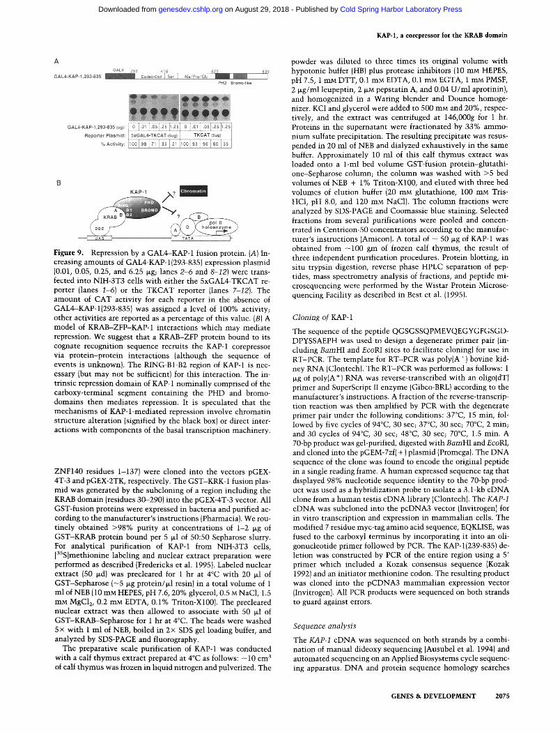
taining proteins. The GAL4-KAP-I, 293-835 fusion pro- tein significantly repressed transcription of the 5xGAL4- TKCAT reporter (Fig. 9A).
In summary, these data suggest that KAP-1 contains an intrinsic D N A binding-dependent repression function and supports the model that KAP-1 is a corepressor that mediates repression by the KRAB domain.
Discussion
We have util ized the KRAB repressor domain to identify and clone a gene encoding a novel nuclear protein (KAP- 1) that displays the hal lmarks of a corepressor. We offer a number of l ines of evidence that suggest that KAP-1 plays a key role in mediat ing KRAB domain repression: (1) KAP-1 binds to mul t ip le KRAB repression domains and the KRAB-KAP-1 interact ion can be reconstituted in vitro; (2) mutat ions in the KRAB domain that abolish repression concomitant ly abolish the interaction with KAP-1; (3)overexpression of KAP-1 enhances KRAB-me- diated repression in a manner dependent on the presence of the domain in KAP-1 that binds the KRAB domain; and (4) KAP-1 itself is a repressor when fused to a DNA- binding domain. Together, these findings are consistent with a simple model in which a DNA-bound KRAB do- main recruits the KAP-1 corepressor to the promoter via direct protein-protein interactions (Fig. 9B). Repression based on recrui tment of a corepressor may be s imilar to hairy-groucho interactions in Drosophila (Paroush et al. 1994), M A T a 2 / M C M 1 - S S N 6 / T U P 1 interactions in yeast (Keleher et al. 1992; Cooper et al. 1994; Tzamarias and Struhl 1994), p53-Elb (Yew et al. 1994), thyroid hor- mone receptors-N-CoR, or SMRT interactions (Chen and Evans 1995; Horlein et al. 1995) in m a m m a l i a n cells. It wil l be interesting to determine whether the mecha- n isms util ized by these corepressor molecules converge at a common step in the transcriptional ini t ia t ion pro- cess.
KAP-1 encodes at least four different classes of cys- te ine/his t idine-r ich motifs: The RING finger, B1 box, B2 box, and PHD finger. These structural motifs support its role as a corepressor and also suggest potential mecha- nisms of repression. The amino- terminal RING finger motif is identified by the signature C3HC 4 spacing of cysteine and his t idine residues (Freemont 1993), and our data support its proposed funct ion as a protein-protein interface (Barlow et al. 1994; Borden et al. 1995). RING- finger-containing proteins have been strongly implicated in cell growth regulation and transcription; these genes include the tumor suppressor BRCA-1 (Miki et al. 1994), the proto-oncogene PML, which is fused to the retinoic- acid receptor-a (RARa) in acute promyelocytic leukemia (Kakizuka et al. 1991), and the Drosophila protein msl-2 (Kelley et al. 1995; Zhou et al. 1995). msl-2 is particu- larly suggestive of a role for the RING finger in the reg- ulat ion of chromatin structure; it is localized to male X chromosome chromatin and is required for the hyper- transcription that is responsible for Drosophila dosage compensation.
2072 GENES & D E V E L O P M E N T
Cold Spring Harbor Laboratory Press on August 29, 2018 - Published by genesdev.cshlp.orgDownloaded from

KAP-1, a corepressor for the KRAB domain
A
COS1 Nuclear Extract : - -
PAX3-KRAB IVT: +
PAX3-KRAB(DV-O,A) IVT:
B
PAX3-KRAB IVT:
COS1 Nuclear Extract :
PAX3-KRAB: PAX3(1-381 ) i KOX1 90
PAX3-KRAB(DV-AA): PAX3(1-381 ) DV-~AA . . . .
1 V ~uxl 90
Figure 6. Detection of DNA-bound KRAB--KAP-1 complexes formed in vitro. (A) The binding of PAX3-KRAB to COS1 nuclear proteins was detected by EMSA with in vitro translated PAX3-KRAB (lanes 2-6), PAX3-KRAB(DV --o AA) (lanes 7-12), a 32P-labeled DNA probe containing the e5 PAX3 binding site, and a COS1 nuclear extract, as described in Materials and methods. The specific e5-binding complex formed in the absence of COS 1 nuclear extract is indicated by a small arrow; the more slowly migrating complex formed upon addition of the nuclear extract is indicated by a large arrow. (B) Competition of higher-order complex formation by purified GST-KRAB (0.6, 1.25, 2.5, 5, 10, and 20 I~g; lanes 2-7) or GST-KRAB(DV ~ AA) (0.6, 1.25, 2.5, 5, 10, and 20 t~g; lanes 8-13). The EMSA was performed as in A, but with a constant amount of COS1 nuclear extract. GST-fusion proteins were expressed in bacteria, purified on glutathione-Sepharose (Pharmacia), and eluted with glutathione as described in Materials and methods.
The PHD finger is a cysteine/his t idine-r ich structure that is dist inguished from the RING finger and LIM do- ma in by containing a consensus of C4HC 3 that spans 50--80 residues (Aasland et al. 1995). Many PHD-finger- containing proteins have been implicated in chromatin- mediated transcriptional modulation. These include products of the Drosophila genes trithorax and poly- comblike and the h u m a n gene HRX, which is fused to AF 10/AF 17 in the t( 11:17) translocation in acute leuke- mia (Chaplin et al. 1995). Likewise, although the bromo- domain of KAP-1 is imperfect, it is interesting to note that bromodomains are found in the adaptor proteins p300, CBP, and GCN5, as well as in the SWI2/SNF2 component of the yeast SWI/SNF transcriptional activa- tion complex of proteins (Laurent et al. 1991; Yoshimoto and Yamashi ta 1991; Georgakopoulos and Thireos 1992; Chrivia et al. 1993; Eckner et al. 1994). Thus, the overall organization of KAP-1 supports its classification as a corepressor and leads to the speculation that KAP-1 re- presses transcription by a chromatin-mediated mecha- nism.
The protein most homologous to KAP-1 is TIF1, a pu- tative coactivator for nuclear hormone receptor-medi- ated transcriptional activation (LeDouarin et al. 1995). TIF1 was cloned in a yeast two-hybrid screen for proteins
that are able to enhance transactivation by the AF-2 re- gion of the retinoid-X receptor-~/ (RXR~), and it was found to interact wi th several members of the nuclear hormone receptor family. Although KAP-1 and TIF1 share a remarkably similar organization of motifs, they are clearly not encoded by the same gene: The overall amino acid sequence homology is only 31%. The loop region between C5 and C6 of the TIF1 RING is much longer and h igh ly divergent compared wi th the analo- gous region in KAP-1. In addition, the regions between the coiled-coil domain and the PHD fingers of KAP-1 and TIF1 are only weakly similar. Finally, the amino-acid sequence of the TIF1 bromodomain conforms much more stringently to the consensus that defines the do- main than does that of KAP-1. Whether the s imilar i ty between KAP-1 and TIF1 is actually reflected in signifi- cant cross-talk between the hormone receptor and KRAB-ZFP families of transcription factors is an intrigu- ing question and remains to be tested.
In conclusion, we have isolated a corepressor, KAP-1, for the KRAB repression domain. This discovery pro- vides a new and potential ly unifying paradigm for the enormous number of KRAB-domain-containing zinc-fin- ger proteins in the h u m a n genome. A future determina- tion of the downstream targets of KAP-1 may yield ira-
GENES & DEVELOPMENT 2073
Cold Spring Harbor Laboratory Press on August 29, 2018 - Published by genesdev.cshlp.orgDownloaded from

Friedman et al.
Lein I '
Serum:
--¢>
IVT, PAX3-KRAB I
COS1, KAP-1 -myc i ¢1
P r o t e i n : COS1 , P A X 3 - K R A B r ]
Se . . . . ~ ~ ~ ~ ~
6"~ = 6-=
1 2 3 4 5 6
1 2 3 4 5 6 7
Figure 7. Identification of components of the KRAB-KAP-1 complex. (A) Use of specific antisera to identify components of the e5-binding complex. The EMSA was performed as in Fig. 6A, with the addition of the indicated antisera to the binding reac- tions. (B) Detection of PAX3--KRAB-KAP-1 complexes in nu- clear extracts. The EMSA was performed using the "S2P-labeled e5 probe and nuclear extract from COSI cells transfected with an expression plasmid encoding PAX3-KRAB. Antisera were added to the binding reactions as indicated. The arrow indicates the specific e5 binding complex containing PAX3-KRAB-KAP-1.
por tan t m e c h a n i s t i c ins ights in to t ranscr ipt ional repres- sion.
Materials and methods
Expression and reporter vectors
The plasmids encoding the GAL4(1-147) fusions to KOXI(1-90), KOXI(1-90; DV ~ AA), and to WT1(1-298)have been described (Madden et al. 1993; Margolin et al. 1994). For the squelching experiments, KOXI(1-161) was subcloned into the pCB6 + ex- pression vector. The GAL4-KAP-1 plasmid was generated from a partial KAP clone isolated from a KZAP NK cell library (Strat- agene) that includes KAP-1 nucleotides 718-2679 (amino acid residues 293-837) flanked by EcoRI sites. This EcoRI fragment was subcloned into the EcoRI site of the pM2 GAL4-fusion mammalian expression plasmid (Sadowski et al. 1992). The 5xGAL4-TKCAT and TKCAT reporter plasmids have been de- scribed by Shi et al. (1991). The PAX3--KRAB (1-381) fusion genes were constructed by use of the PAX3-FKHR plasmid de- scribed in Fredericks et al. (1995). KOXl(1-90) and the DV ~ AA mutant were subcloned into the EcoRI and XbaI sites, thereby replacing the FKHR sequences. The reporter plasmid for the PAX-KRAB fusions 2xe5TKCAT has been described (Fredericks et al. 1995).
Cell culture, transfections, and CAT assays
NIH-3T3 mouse cells, human Rh30, and COS 1 monkey kidney cells were grown and transfected as described (Margolin et al. 1994). Expression of all constructs was confirmed by immuno- precipitation with the appropriate antisera (Madden et al. 1991, 1993; Morris et al. 1991). The CMV vector expressing [5-D-ga- lactosidase, p0N260, was used in all transfections, and [~-D-ga- lactosidase activity was used to normalize transfection effi- ciency in cell extracts for the CAT assay. Cell harvesting, CAT assays, and quantitation were performed as described (Madden
1991, 1993). Each transfection experiment was performed at least three times in duplicate. The numbers derived are the averages of duplicates in a single experiment and variability was within 10%.
Generation of GST-fusion proteins and purification of KAP-1
The cDNAs encoding KOXI(1-90) and the DV---> AA mutant were subcloned into the pGEX-2TK vector (Pharmacia). The ZNF 133 and ZNF 140 GST-fusion plasmids were generated with cDNAs kindly provided by H.-J. Theisen. Regions of each cDNA including the KRAB domain (ZNF133 residues 1-198 and
KAP-1 ExDrellSlOn Plasmids:
1 ~! 239 KAP-1 |Aia ~ I I I I Co!l~.~Coit
RING B1 B2
239 [ CoikKI-Coil
PHD Bromo+.iike
416 E25 835 I Set I ~=/mo/my 1
PHD Bromo-like
10"
8 I
o - . - , , t j j+. P A X ~ ' K R A B (.g): [ - 1 . 5 3 6 13 13 [3 [ " ] " 1" ]
% Actlvl~': I 5' Z0] '6IIS I 11185 + 79157 +
I ; o - -oo+e.+ • • • • • • • • ~k°'~,7
PAX3-KRAB (.g): " 3 3 1 3 1 3 3 [ 3 ii 3
KAP-I: " ~ " [ ' 1 " 69
+ - + + ° + - %Activity: ~oo zz +2 1 80[zOJ65 4 5
I"I +3~0 PAX3 -KRA8
0 5 10 ~5 ~;g gAP-}
~<~ <~
~11"- KAP + 1
"~--" K A P+ '~. ( 239 -835 )
Figure 8. Enhancement of KRAB-mediated repression by exog- enous KAP-1. {A,B) Cells were transfected with the indicated amounts (in micrograms) of expression plasmid encoding PAX3-KRAB, and with 5 t+g (lanes 5,8), 10 lzg (lanes 6,9), or 15 I+g (lanes 7,101 of expression plasmid encoding KAP-1 or KAP- 1(239-835), along with 2.5 Izg of the reporter plasmid. The CAT assay and quantification were performed as described in Mate- rials and methods. The amount of CAT activity in the absence of PAX3-KRAB and KAP-1 was assigned a level of 100% activ- ity; other activities are reported as a percentage of this value. The degree of repression by 3 Ixg of PAX3-KRAB expression plasmid in the presence of 0, 5, 10, and 15 p+g of KAP-1 plasmid is graphically represented in the right panel. {C) Deletion of the RING-B1-B2 eliminates both binding to the KRAB domain and enhancement of KRAB-mediated repression. An expression plasmid encoding KAP-l{239-835) was transfected into COS1 cells, and transfected cell extracts were used in an anti-KAP-1 immunoprecipitation {lane 1) or purification on GST-KRAB bound to glutathione-Sepharose (lane 2). Full-length endoge- nous KAP-1 and exogenous KAP-l{239-835) are indicated by ar- rows.
2074 GENES & DEVELOPMENT
Cold Spring Harbor Laboratory Press on August 29, 2018 - Published by genesdev.cshlp.orgDownloaded from
A GAL4 2. 93 .... ~ ] ,5 62 $ ,,,,_B.~5
GAL4-KAP-1,293-835 ~ Coiled-CoiC~S_eL.L_=Al~/Pro/GIL... ~ ~ PHO Bromo-lFke
GAL4=KAP-I,293-835(ug),: .......... [L . [~ I ; i ! .... ~ ] ; ........... ~ , , , ~
Reporter Plasmicl: 5xGAL4-TKCAT (Bug} I TKCAT (~(j) I
~;-ii00i 93 ]9616o 13s
Figure 9. Repression by a GAL4-KAP-1 fusion protein. (A) In- creasing amounts of GAL4-KAP-l(293-835) expression plasmid (0.01, 0.05, 0.25, and 6.25 ~g; lanes 2-6 and 8-12) were trans- fected into NIH-3T3 cells with either the 5xGAL4-TKCAT re- porter (lanes 1-6) or the TKCAT reporter (lanes 7-12). The amount of CAT activity for each reporter in the absence of GAL4-KAP-l(293-835) was assigned a level of 100% activity; other activities are reported as a percentage of this value. (B) A model of KRAB-ZFP-KAP-1 interactions which may mediate repression. We suggest that a KRAB-ZFP protein bound to its cognate recognition sequence recruits the KAP-1 corepressor via protein-protein interactions (although the sequence of events is unknown). The RING-B1-B2 region of KAP-1 is nec- essary (but may not be sufficient) for this interaction. The in- trinsic repression domain of KAP-1 nominally comprised of the carboxy-terminal segment containing the PHD and bromo- domains then mediates repression. It is speculated that the mechanisms of KAP-l-mediated repression involve chromatin structure alteration (signified by the black box) or direct inter- actions with components of the basal transcription machinery.
ZNF140 residues 1-137) were cloned into the vectors pGEX- 4T-3 and pGEX-2TK, respectively. The GST-KRK- 1 fusion plas- mid was generated by the subcloning of a region including the KRAB domain (residues 30-290) into the pGEX-4T-3 vector. All GST-fusion proteins were expressed in bacteria and purified ac- cording to the manufacturer's instructions (Pharmacia). We rou- tinely obtained >98% purity at concentrations of 1-2 ~g of GST-KRAB protein bound per 5 ~1 of 50:50 Sepharose slurry. For analytical purification of KAP-1 from NIH-3T3 cells, [3SS]methionine labeling and nuclear extract preparation were performed as described (Fredericks et al. 1995). Labeled nuclear extract (50 p.d) was precleared for 1 hr at 4°C with 20 ~1 of GST-Sepharose (-5 p.g protein/~l resin) in a total volume of 1 ml of NEB (10 mM HEPES, pH 7.6, 20% glycerol, 0.5 M NaC1, 1.5 mM MgC12, 0.2 mM EDTA, 0.1% Triton-X100). The precleared nuclear extract was then allowed to associate with 50 ~1 of GST-KRAB--Sepharose for 1 hr at 4°C. The beads were washed 5 x with 1 ml of NEB, boiled in 2 x SDS gel loading buffer, and analyzed by SDS-PAGE and fluorography.
The preparative scale purification of KAP-1 was conducted with a calf thymus extract prepared at 4°C as follows: -10 cm 3 of calf thymus was frozen in liquid nitrogen and pulverized. The
KAP-1, a corepressor for the KRAB domain
powder was diluted to three times its original volume with hypotonic buffer (HB) plus protease inhibitors (10 mM HEPES, pH 7.5, 1 mM DTT, 0.1 mM EDTA, 0.1 mM EGTA, 1 mM PMSF, 2 ~g/ml leupeptin, 2 ~M pepstatin A, and 0.04 U/ml aprotinin), and homogenized in a Waring blender and Dounce homoge- nizer. KC1 and glycerol were added to 500 mM and 20%, respec- tively, and the extract was centrifuged at 146,000g for 1 hr. Proteins in the supernatant were fractionated by 33% ammo- nium sulfate precipitation. The resulting precipitate was resus- pended in 20 ml of NEB and dialyzed exhaustively in the same buffer. Approximately 10 ml of this calf thymus extract was loaded onto a 1-ml bed volume GST-fusion protein-glutathi- one-Sepharose column; the column was washed with >5 bed volumes of NEB + 1% Triton-X100, and eluted with three bed volumes of elution buffer (20 mM glutathione, 100 mM Tris- HC1, pH 8.0, and 120 mM NaC1). The column fractions were analyzed by SDS-PAGE and Coomassie blue staining. Selected fractions from several purifications were pooled and concen- trated in Centricon-50 concentrators according to the manufac- turer's instructions (Amicon). A total of - 50 ~g of KAP-1 was obtained from -100 gm of frozen calf thymus, the result of three independent purification procedures. Protein blotting, in situ trypsin digestion, reverse phase HPLC separation of pep- tides, mass spectrometry analysis of fractions, and peptide mi- crosequencing were performed by the Wistar Protein Microse- quencing Facility as described in Best et al. (1995).
Cloning of KAP-1
The sequence of the peptide QGSGSSQPMEVQEGYGFGSGD- DPYSSAEPH was used to design a degenerate primer pair (in- cluding BamHI and EcoRI sites to facilitate cloning) for use in RT-PCR. The template for RT-PCR was poly(A + ) bovine kid- ney RNA (Clontech). The RT-PCR was performed as follows: 1 ~g of poly(A +) RNA was reverse-transcribed with an oligo(dT) primer and SuperScript II enzyme (Gibco-BRL) according to the manufacturer's instructions. A fraction of the reverse-transcrip- tion reaction was then amplified by PCR with the degenerate primer pair under the following conditions: 37°C, 15 min, fol- lowed by five cycles of 94°C, 30 sec; 37°C, 30 sec; 70°C, 2 min; and 30 cycles of 94°C, 30 sec; 48°C, 30 sec; 70°C, 1.5 min. A 70-bp product was gel-purified, digested with BamHI and EcoRI, and cloned into the pGEM-7zf( + ) plasmid (Promega). The DNA sequence of the clone was found to encode the original peptide in a single reading frame. A human expressed sequence tag that displayed 98% nucleotide sequence identity to the 70-bp prod- uct was used as a hybridization probe to isolate a 3.1-kb cDNA clone from a human testis cDNA library (Clontech). The KAP-1 cDNA was subcloned into the pcDNA3 vector (Invitrogen) for in vitro transcription and expression in mammalian cells. The modified 7 residue myc-tag amino acid sequence, EQKLISE, was fused to the carboxyl terminus by incorporating it into an oli- gonucleotide primer followed by PCR. The KAP-l(239-835) de- letion was constructed by PCR of the entire region using a 5' primer which included a Kozak consensus sequence (Kozak 1992) and an initiator methionine codon. The resulting product was cloned into the pCDNA3 mammalian expression vector (Invitrogen). All PCR products were sequenced on both strands to guard against errors.
Sequence analysis
The KAP-1 cDNA was sequenced on both strands by a combi- nation of manual dideoxy sequencing (Ausubel et al. 1994) and automated sequencing on an Applied Biosystems cycle sequenc- ing apparatus. DNA and protein sequence homology searches
v
GENES & DEVELOPMENT 2075
Cold Spring Harbor Laboratory Press on August 29, 2018 - Published by genesdev.cshlp.orgDownloaded from

Friedman et al.
were performed by use of the BLAST network service (Altschul et al. 1990).
Antisera, immunoprecipitat ions, and immunohis tochemis try
Nucleotides 1270-1755 of the KAP-1 cDNA were amplified by PCR and subcloned into the pQE30 hexahistidine-fusion bacte- rial expression vector (Qiagen), expressed in bacteria, and puri- fied by nickel-chelate chromatography (Qiagen). Then hexahis- tidine-tagged KAP-l(423-584) was used to generate anti-KAP-1 sera in C3H mice. Soluble protein was mixed with Freund's adjuvant and injected subcutaneously. The bleeds from five mice were pooled and tested in immunoprecipitations of la- beled, in vitro transcribed/translated KAP-1. The same single bleed pool was used in all experiments. The anti-KOX1 serum was generated in rabbits against hexahistidine-KOX 1 ( 1-161 ) and anti-PAX3 serum was generated as described (Fredericks et al. 1995).
For immunoprecipitations, COS1 cells were labeled with [3SS]-methionine and harvested in ELB buffer plus protease in- hibitors (250 mM NaC1, 50 mM HEPES, pH 7.5, 0.1% NP-40, 1 mM EDTA, 0.1 mM PMSF, 2 ~g/ml leupeptin, and 11 i~g/ml aprotinin). Immunoprecipitations were performed in 1 ml of ELB plus protease inhibitors. One microliter of preimmune or anti-KAP-1 sera (or 1 ~g of myc-tag monoclonal antibody) was added and incubated for 2 hr at 4°C, followed by the addition of 100 ~1 of a 10% slurry of Protein A-Sepharose (Pharmacia) and rotation at 4°C for 30 min. The pellets were washed five times with 1 ml of ELB, boiled in 2x SDS gel loading buffer, and analyzed by SDS-PAGE and fluorography. For the sequential GST association/immunoprecipitations, proteins bound to GST-KRAB were eluted in batch in five bed volumes of elution buffer (20 mM glutathione, 100 mM Tris-HC1, pH 8.0, and 120 mM NaC1) at room temperature for 30 min. Eluted proteins were then immunoprecipitated in 1 ml ELB as described above.
Electrophoretic mobi l i ty shift assay
EMSA was performed essentially as described (Fredericks et al. 1995). The PAX3-KRAB fusion protein was transcribed/trans- lated in vitro and used in all assays unless otherwise noted. Binding reactions were assembled in 20 ~1 of binding buffer (final concentration 20 mM HEPES (pH 7.6), 50 mM NaC1, 0.2 ~g poly [d(I-C)], 0.5 mM DTT, 5 mM MgC12, 10% glycerol). In vitro translated PAX3-KRAB was incubated in the above buffer with nuclear extracts/antisera/competitor proteins for 20 min at room temperature, and then 1 ~1 of e5 probe (10 s cpm/~l)was added and the reaction was incubated for 10 min at 30°C. Amounts of antibodies used were as follows: 1 ~1 of a 1:10 dilution of anti-KAP-1 sera, 1 ~1 anti-PAX3 sera, 1 ~1 of a 1:5 dilution of anti-KOX1 sera, or 0.1 ~g myc-tag monoclonal anti- body. In experiments involving addition of competitor proteins, GST-KRAB and GST-KRAB(DV --* AA) were purified on gluta- thione-Sepharose and eluted as described above. For assays of PAX3-KRAB complexes formed in vivo, PAX3-KRAB and KAP-1 expression plasmids were transfected into COS1 cells, and nuclear extracts were prepared as described above. DNA- protein complexes were resolved on 1.5-mm 4.5% native poly- acrylamide gels by electrophoresis at 400 V for 1 hr in 45 mM Tris-borate (pH 8.3)/45 mM boric acid per 1 mM EDTA buffer. EMSA gels were dried and visualized by autoradiography.
A c k n o w l e d g m e n t s
We thank Drs. Amitabha Basu and Chin Howe for assistance with thymus extract preparation; Dr. Gerd Maul for immuno- fluorescence analyses, Dr. Laura Benjamin for assistance with
RT-PCR assays, David Reim and the Wistar Institute Micro- chemistry Core Facility for peptide sequencing, Drs. Hildegund C.J. Ertl and Zhi Quan Xiang for assistance in production of polyclonal mouse sera, and Dr. Cory Abate (Center for Advance Biotechnology and Medicine) for the gift of anti-myc-tag mono o clonal antibody. We also thank Drs. Shelley Berger, Mitch Lazar, Henrik Vissing (Novo Nordisk, Denmark), George Pren- dergast, Paul Lieberman, Giovanni Rovera, and Peter Traber for helpful discussion and Dawna Gillespie for preparation of the manuscript. J.R.F. is supported by the Medical Science Training Program, University of Pennsylvania School of Medicine. W.J.F. is supported by the Wistar Institute Cancer Training Grant CA09171. D.E.J. is supported by a Susan G. Komen Breast Can- cer Foundation Fellowship. E.G.N. is supported by DK49210 and DK45191. D.W.S. is supported by CA66671 and CA25874. F.J.R. is supported by National Institutes of Health grants CA52009, Core grant CA10815, DK49210, GM54220, DAMD 17-96-1-6141, ACS NP-954, and the Irving A. Hansen Me- morial Foundation, the Mary A. Rumsey Memorial Foundation, and the Pew Scholars Program in the Biomedical Sciences.
The publication costs of this article were defrayed in part by payment of page charges. This article must therefore be hereby marked "advertisement" in accordance with 18 USC section 1734 solely to indicate this fact.
R e f e r e n c e s
Aasland, R., T.J. Gibson, and A.F. Stewart. 1995. The PHD fin- ger: Implications for chromatin-mediated transcriptional regulation. Trends Biochem. Sci. 20: 56--59.
Altaba, A., H. Perry-O'Keefe, and D.A. Melton. 1987. Xfin: An embryonic gene encoding a multifingered protein in Xeno- pus. EMBO J. 6: 3065-3070.
Altschul, S.F., W. Gish, W. Miller, E.W. Myers, and D.J. Lipman. 1990. Basic local alignment search tool. ]. Mol. Biol. 215: 403-410.
Ascoli, C.A. and G.G. Maul. 1991. Identification of a novel nu- clear domain. ]. Cell Biol. 112: 785-795.
Ausubel, F.M., R. Brent, R.E. Kingston, D.D. Moore, J.G. Seid- man, J.A. Smith, and K. Struhl. 1994. Current protocols in molecular biology, Vol. 12. John Wiley and Sons, New York, NY.
Ayer, D.E., Q.A. Lawrence, and R.N. Eisenman. 1995. Mad-Max transcriptional repression is mediated by ternary complex formation with mammalian homologs of yeast repressor Sin3. Cell 80: 767-776.
Baniahmad, A., X. Leng, T.P. Burris, S.Y. Tsai, M.J. Tsai, and B.W. O'Malley. 1995. The tau 4 activation domain of the thyroid hormone receptor is required for release of a putative corepressor(s) necessary for transcriptional silencing. Mol. Cell. Biol. 15: 76-86.
Barlow, P.N., B. Luisi, A. Milner, M. Elliott, and R. Everett. 1994. Structure of the C3HC4 domain by 1H-nuclear mag- netic resonance spectroscopy. A new structural class of zinc- finger. I. Mol. Biol. 237: 201-211.
Bellefroid, E.J., D.A. Poncelet, P.J. Lecocq, O. Revelant, and J.A. Martial. 1991. The evolutionarily conserved KruppeLassoci- ated box domain defines a subfamily of eukaryotic multifin- gered proteins. Proc. Natl. Acad. Sci. 88: 3608-3612.
Bellefroid, E.J., J.C. Marine, T. Ried, P.J. Lecocq, M. Riviere, C. Amemiya, D.A. Poncelet, P.G. Coulie, P. de Jong, C. Szpirer, D.C. Ward, and J.A. Martial. 1993. Clustered organization of homologous KRAB zinc-finger genes with enhanced expres- sion in human T lymphoid cells. EMBO J. 12: 1363-1374.
Best, S., D.F. Reim, J. Mozdzanowski, and D.W. Speicher. 1995.
2076 GENES & DEVELOPMENT
Cold Spring Harbor Laboratory Press on August 29, 2018 - Published by genesdev.cshlp.orgDownloaded from

KAP-1, a corepressor for the KRAB domain
High sensitivity peptide sequence analysis using in situ pro- teolysis on high retention PVDF membranes and a biphasic reaction column sequencer. In Techniques in protein chem- istry V (ed. J.W. Crabb), pp. 565-574. Academic Press, San Diego, CA.
Borden, K.L., M.N. Boddy, J. Lally, N.J. O'Reilly, S. Martin, K. Howe, E. Solomon, and P.S. Freemont. 1995. The solution structure of the RING finger domain from the acute promy- elocytic leukaemia proto-oncoprotein PML. EMBO J. 14: 1532-1541.
Chaplin, T., O. Bernard, H.B. Beverloo, V. Saha, A. Hagemeijer, R. Berger, and B.D. Young. 1995. The t( 10; 11) translocation in acute myeloid leukemia {M5) consistently fuses the leu- cine zipper motif of AF10 onto the HRX gene. Blood 86: 2073-2076.
Chen, J.D. and R.M. Evans. 1995. A transcriptional co-repressor that interacts with nuclear hormone receptors. Nature 377: 454--457.
Chrivia, J.C., R.P. Kwok, N. Lamb, M. Hagiwara, M.R. Mont- rainy, and R.H. Goodman. 1993. Phosphorylated CREB binds specifically to the nuclear protein CBP. Nature 365: 855-859.
Constantinou-Deltas, C.D., J. Gilbert, R.J. Bartlett, M. Herb- streith, A.D. Roses, and J.E. Lee. 1992. The identification and characterization of KRAB-domain-containing zinc finger proteins. Genomics 12: 581-589.
Cooper, J.P., S.Y. Roth, and R.T. Simpson. 1994. The global transcriptional regulators, SSN6 and TUP1, play distinct roles in the establishment of a repressive chromatin struc- ture. Genes & Dev. 8: 1400-1410.
Crew, A.J., J. Clark, C. Fisher, S. Gill, R. Grimer, A. Chand, J. Shipley, B.A. Gusterson, and C.S. Cooper. 1995. Fusion of SYT to two genes, SSX1 and SSX2, encoding proteins with homology to the Kruppel-associated box in human synovial sarcoma. EMBO J. 14: 2333-2340.
Eckner, R., M.E. Ewen, D. Newsome, M. Gerdes, J.A. DeCaprio, J.B. Lawrence, and D.M. Livingston. 1994. Molecular cloning and functional analysis of the adenovirus E1A-associated 300-kD protein {p300) reveals a protein with properties of a transcriptional adaptor. Genes & Dev. 8: 869-884.
Fisher, A.L., S. Ohsako, and M. Caudy. 1996. The WRPW motif of the hairy-related basic helix-loop-helix repressor proteins acts as a 4-amino-acid transcription repression and protein- protein interaction domain. Mol. Cell. Biol. 16: 2670-2677.
Fondell, J.D., A.L. Roy, and R.G. Roeder. 1993. Unliganded thy- roid hormone receptor inhibits formation of a functional preinitiation complex: Implications for active repression. Genes & Dev. 7: 1400-1410.
Fondell, J.D., F. Brunel, K. Hisatake, R.G. Roeder, J.D. Fondell, A.L. Roy, and R.G. Roeder. 1996. Unliganded thyroid hor- mone receptor alpha can target TATA-binding protein for transcriptional repression. Mol. Cell. Biol. 16: 281-287.
Fredericks, W.J., N. Galili, S. Mukhopadhyay, G. Rovera, J. Ben- nicelli, F.G. Barr, and F.J. Rauscher III. 1995. The PAX3- FKHR fusion protein created by the t(2; 13) translocation in alveolar rhabdomyosarcomas is a more potent transcrip- tional activator than PAX3. Mol. Cell. Biol. 15: 1522-1535.
Freemont, P.S. 1993. The RING finger. A novel protein se- quence motif related to the zinc finger. Ann. N Y Acad. Sci. 684: 174-192.
Georgakopoulos, T. and G. Thireos. 1992. Two distinct yeast transcriptional activators require the function of the GCN5 protein to promote normal levels of transcription. EMBO ]. 11: 4145-4152.
Gill, G. and M. Ptashne. 1988. Negative effect of the transcrip- tional activator GAL4. Nature 334: 721-724.
Goulding, M.D., G. Chalepakis, U. Deutsch, J.R. Erselius, and P. Gruss. 1991. Pax-3, a novel murine DNA binding protein expressed during early neurogenesis. EMBO J. 10: 1135- 1147.
Guarente, L. 1995. Transcriptional coactivators in yeast and beyond. Trends Biochem. Sci. 20: 517-521.
Haynes, S.R., C. Dollard, F. Winston, S. Beck, J. Trowsdale, and I.B. Dawid. 1992. The bromodomain: A conserved sequence found in human, Drosophila and yeast proteins. Nucleic Ac- ids Res. 20" 2603.
Horlein, A.J., A.M. Naar, T. Heinzel, J. Torchia, B. Gloss, R. Kurokawa, A. Ryan, Y. Kamei, M. Soderstrom, C.K. Glass, and M.G. Rosenfeld. 1995. Ligand-independent repression by the thyroid hormone receptor mediated by a nuclear receptor co-repressor. Nature 377: 397-404.
Kakizuka, A., W.H. Miller Jr., K. Umesono, R.P. Warrell Jr., S.R. Frankel, V.V. Murty, E. Dmitrovsky, and R.M. Evans. 1991. Chromosomal translocation t( 15; 17) in human acute promy- elocytic leukemia fuses RAR alpha with a novel putative transcription factor, PML. Cell 66" 663-674.
Keleher, C.A., M.J. Redd, J. Schultz, M. Carlson, and A.D. Johnson. 1992. Ssn6-Tupl is a general repressor of transcrip- tion in yeast. Cell 68: 709-719.
Kelley, R.L., I. Solovyeva, L.M. Lyman, R. Richman, V. Solovyev, and M.I. Kuroda. 1995. Expression of msl-2 causes assembly of dosage compensation regulators on the X chro- mosomes and female lethality in Drosophila. Cell 81: 867- 877.
Klug, A. and J.W. Schwabe. 1995. Protein motifs 5. Zinc fingers. FASEB [. 9: 597-604.
Kozak, M. 1992. Regulation of translation in eukaryotic sys- tems. Annu. Rev. Cell Biol. 8: 197-225.
Laurent, B.C., M.A. Treitel, and M. Carlson. 1991. Functional interdependence of the yeast SNF2, SNF5, and SNF6 pro- teins in transcriptional activation. Proc. Natl. Acad. Sci. 88: 2687-2691.
LeDouarin, B., C. Zechel, J.M. Gamier, Y. Lutz, L. Tora, P. Pi- errat, D. Heery, H. Gronemeyer, P. Chambon, and R. Losson. 1995. The N-terminal part of TIF1, a putative mediator of the ligand-dependent activation function (AF-2) of nuclear receptors, is fused to B-raf in the oncogenic protein T18. EMBO I. 14: 2020-2033.
Lupas, A., M. Van Dyke, and J. Stock. 1991. Predicting coiled coils from protein sequences. Science. 252:1162-1164.
Madden, S.L., D.M. Cook, J.F. Morris, A. Gashler, V.P. Sukhatme, and F.J. Rauscher III. 1991. Transcriptional re- pression mediated by the WT1 Wilms tumor gene product. Science 253: 1550-1553.
Madden, S.L., D.M. Cook, and F.J. Rauscher III. 1993. A struc- ture-function analysis of transcriptional repression mediated by the WT1, Wilms tumor suppressor protein. Oncogene 8: 1713-1720.
Margolin, J.F., J.R. Friedman, W.K. Meyer, H. Vissing, H.J. Theisen, and F.J. Rauscher III. 1994. Kruppel-associated boxes are potent transcriptional repression domains. Proc. Natl. Acad. Sci. 91: 4509-4513.
Miki, T., T.P. Fleming, M. Crescenzi, C.J. Molloy, S.B. Blam, S.H. Reynolds, and S.A. Aaronson. 1991. Development of a highly efficient expression cDNA cloning system: Applica- tion to oncogene isolation. Proc. Natl. Acad. Sci. 88: 5167- 5171.
Miki, Y., J. Swensen, D. Shattuck-Eidens, P.A. Futreal, K. Harshman, S. Tavtigian, Q. Liu, C. Cochran, L.M. Bennett, W. Ding, R. Bell, J. Rosenthal, C. Hussey, T. Tran, M. Mc- Clure, C. Frye, T. Hattier, R. Phelps, A. Haugen-Strano, H. Katcher, K. Yakumo, Z. Gholami, D. Shaffer, S. Stone, S.
GENES & DEVELOPMENT 2077
Cold Spring Harbor Laboratory Press on August 29, 2018 - Published by genesdev.cshlp.orgDownloaded from

Friedman et al.
Bayer, C. Wray, R. Bogden, P. Dayananth, J. Ward, P. Tonin, S. Narod, P.K. Bristown, F.H. Norris, L. Helvering, P. Mor- rison, P. Rosteck, M. Lai, C. Barrett, C.Lewis, S. Neuhausen, L. Cannon-Albright, D. Goldgar, R. Wiseman, A. Kamb, and M.H. Skolnick. 1994. A strong candidate for the breast and ovarian cancer susceptibility gene BRCA1. Science 266: 66-- 71.
Morris, J.F., S.L. Madden, O.E. Tournay, D.M. Cook, V.P. Sukhatme, and F.J. Rauscher III. 1991. Characterization of the zinc finger protein encoded by the WT1 Wilms tumor locus. Oncogene 6: 2339-2348.
Paroush, Z., R.L. Finley Jr., T. Kidd, S.M. Wainwright, P.W. Ingham, R. Brent, and D. Ish-Horowicz. 1994. Groucho is required for Drosophila neurogenesis, segmentation, and sex determination and interacts directly with hairy-related bHLH proteins. Cell 79: 805-815.
Pengue, G., A. Caputo, C. Rossi, G. Barbanti-Brodano, and L. Lania. 1995. Transcriptional silencing of human immunode- ficiency virus type 1 long terminal repeat-driven gene ex- pression by the Kruppel-associated box repressor domain tar- geted to the transactivating response element. J. Virol. 69: 6577-6580.
Sadowski, I., B. Bell, P. Broad, and M. Hollis. 1992. GAL4 fusion vectors for expression in yeast or mammalian cells. Gene 118: 137-141.
Sauer, F., J.D. Fondell, Y. Ohkuma, R.G. Roeder, and H. Jackle. 1995. Control of transcription by Kruppel through interac- tions with TFIIB and TFIIE beta. Nature 375: 162-164.
Schreiber-Agus, N., L. Chin, K. Chen, R. Tortes, G. Rao, P. Guida, A.I. Skoultchi, and R.A. DePinho. 1995. An amino- terminal domain of Mxil mediates anti-Myc oncogenic ac- tivity and interacts with a homolog of the yeast transcrip- tional repressor SIN3. Cell 80: 777-786.
Shi, Y., E. Seto, L.S. Chang, and T. Shenk. 1991. Transcriptional repression by YY1, a human GLI-Kr81ppel-related protein, and relief of repression by adenovirus E1A protein. Cell 67: 377-388.
Thiesen, H.J., E. Bellefroid, O. Revelant, and J.A. Martial. 1991. Conserved KRAB protein domain identified upstream from the zinc finger region of Kox 8. Nucleic Acids Res. 19: 3996.
Tjian, R. and T. Maniatis. 1994. Transcriptional activation: A complex puzzle with few easy pieces. Cell 77: 5-8.
Tommerup, N., L. Aagaard, C.L. Lund, E. Boel, S. Baxendale, G.P. Bates, H. Lehrach, and H. Vissing. 1993. A zinc-finger gene ZNF141 mapping at p16.3/D4S90 is a candidate gene for the Wolf-Hirschhorn (4p-) syndrome. Hum. Mol. Gen. 2: 1571-1575.
Tzamarias, D. and K. Struhl. 1994. Functional dissection of the yeast Cyc8-Tupl transcriptional co-repressor complex. Na- ture 369: 758-761.
Um, M., C. Li, and J.L. Manley. 1995. The transcriptional re- pressor even-skipped interacts directly with TATA-binding protein. Mol. Cell. Biol. 15: 5007-5016.
Vissing, H., W.K. Meyer, L. Aagaard, N. Tommerup, and H.J. Thiesen. 1995. Repression of transcriptional activity by het- erologous KRAB domains present in zinc finger proteins. FEBS Lett. 369: 153--157.
Witzgall, R., E. O'Leary, R. Gessner, A.J. Ouellette, and J.V. Bonventre. 1993. Kid-1, a putative renal transcription factor: Regulation during ontogeny and in response to ischemia and toxic injury. Mol. Cell. Biol. 13: 1933-1942.
Witzgall, R., R. Volk, R.S. Yeung, and J.V. Bonventre. 1994. Genomic structure and chromosomal location of the rat gene encoding the zinc finger transcription factor kid-1. Ge- nomics 20" 203-209.
Yew, P.R., X. Liu, and A.J. Berk. 1994. Adenovirus E1B o n c o -
protein tethers a transcriptional repression domain to p53. Genes & Dev. 8: 190-202.
Yoshimoto, H. and I. Yamashita. 1991. The GAM1/SNF2 gene of Saccharomyces cerevisiae encodes a highly charged nu- clear protein required for transcription of the STA1 gene. Mol. Gen. Genet. 228: 270-280.
Zawel, L. and D. Reinberg. 1995. Common themes in assembly and function of eukaryotic transcription complexes. Annu. Rev. Biochem. 64-533-561.
Zhou, S., Y. Yang, M.J. Scott, A. Pannuti, K.C. Fehr, A. Eisen, E.V. Koonin, D.L. Fouts, R. Wrightsman, J.E. Manning, and J.C. Lucchesi. 1995. Male-specific lethal 2, a dosage compen- sation gene of Drosophila, undergoes sex-specific regulation and encodes a protein with a RING finger and a metallothio- nein-like cysteine cluster. EMBO J. 14: 2884--2895.
2078 GENES & DEVELOPMENT
Cold Spring Harbor Laboratory Press on August 29, 2018 - Published by genesdev.cshlp.orgDownloaded from

10.1101/gad.10.16.2067Access the most recent version at doi: 10:1996, Genes Dev.
J R Friedman, W J Fredericks, D E Jensen, et al. repression domain.KAP-1, a novel corepressor for the highly conserved KRAB
References
http://genesdev.cshlp.org/content/10/16/2067.full.html#ref-list-1
This article cites 60 articles, 20 of which can be accessed free at:
License
ServiceEmail Alerting
click here.right corner of the article or
Receive free email alerts when new articles cite this article - sign up in the box at the top
Copyright © Cold Spring Harbor Laboratory Press
Cold Spring Harbor Laboratory Press on August 29, 2018 - Published by genesdev.cshlp.orgDownloaded from