junior pathologist tutorial 1 bladder pathology may 2014 fileaspects’of’bladder’pathology...
TRANSCRIPT

Aspects of Bladder Pathology
Dr David Paterson Musgrove Park Hospital
Taunton 14th May 2014

Bladder Pathology
• Covering • Normal bladder • Urothelial bladder cancer • Aspects of staging • Squamous lesions • Glandular lesions • Benign mimics of malignancy
• Not covering • Cystectomies • Urine cytology • FISH/ molecular pathology • Unusual types of invasive carcinoma • Spindle cell lesions

Bladder Pathology
• Condensed from Kidney and Bladder Pathology Course” 2012 , BAUP
Talks given by Dr Ashish Candra Guy’s and St Thomas’s Dr L J McWilliam Manchester Royal infirmary Dr Murali Varma Cardiff Dr David Griffiths Cardiff Dr Jonathan Shanks The Christie Manchester

Apologies
I don’t have a collection of slides to offer as a slide set or photograph so talk will be text based Please augment this talk with reference to standard text illustrations.
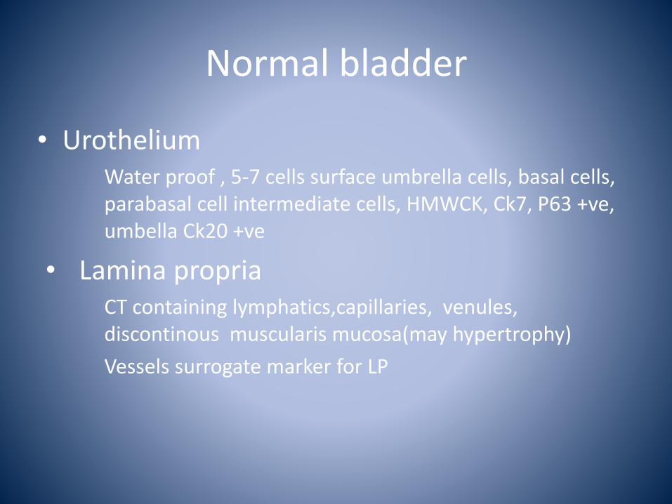
Normal bladder
• Urothelium Water proof , 5-‐7 cells surface umbrella cells, basal cells, parabasal cell intermediate cells, HMWCK, Ck7, P63 +ve, umbella Ck20 +ve
• Lamina propria CT containing lymphatics,capillaries, venules, discontinous muscularis mucosa(may hypertrophy) Vessels surrogate marker for LP

Normal Bladder
• Muscularis propria Thick bundle of smooth muscle, marks point significant change in behavior of TCC, needs to be sampled in resection/biopsies.
• Serosa Mostly fat, with age fat may extend into the muscularis and LP( don’t assume fat=perforation or deep infiltration)

Urothelial bladder cancer
• Papillary lesions • Flat lesions • Invasive lesions

Papillary lesions
• Most low grade recur but few progress • High grade tumours probable arise de novo but a few from low grade progression.
• High grade have a higher risk of progression .

Papillary carcinoma
Low grade• Chromosome 9 • Diploid • Recurrence rate high • Progression rate low • No reduction in life span
High grade• Chromosome 17 13 14 • Aneuploid • High recurrence rate • Higher progression rate • Reduced life span

Papillary Lesions
1973 WHO• Papilloma • Grade 1 carcinoma • Grade 2 carcinoma • Grade 3 carcinoma
ISUP/WHO 2004• Papilloma • PUNLMP • Low grade carcinoma • High grade carcinoma

WHO 1973
Pros• Widely used • Liked by UK pathologists • Repeatedly validated
Cons• No detailed description of
lesions • Calls low grade lesions with
little potential to progress cancer.

ISUP/WHO 2004
Pros• Based on expert consensus • Detailed descriptions • PUNLMP avoids labelling as
cancer • Correlation with cytology
better • Avoids everything going into
G2
Cons• Not as well validated • Poor reproducibility of low
grade lesions (PUNLMP vs Low grade 36%)
• Puts more cases into high grade ? Over treatment
• “Will Rogers” phenomena

Will Rogers Phenomena
Paradoxical effect of moving one element from one grade to another improves the outcome/average values of both sets. Eg Moving all the bad grade 2s into high grade will improve out come for both high grade and low grade. Eg Scottish IQ when immigrating to England

Papilloma
• Delicate non-‐fused papillae • Normal cellular organisation • Normal nuclear sizes shape and fine chromatin • No nucleoli • No mitotic figures
Recurrence rate-‐ 8% Grade progression-‐ 2% Stage progression-‐ 0% Survival-‐ 100%
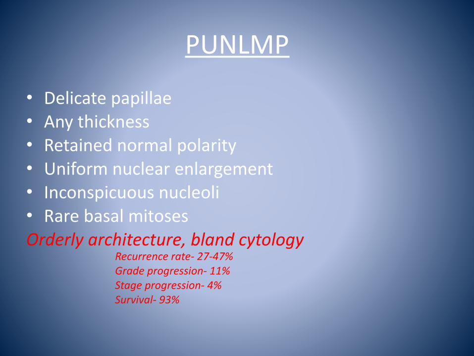
PUNLMP
• Delicate papillae • Any thickness • Retained normal polarity • Uniform nuclear enlargement • Inconspicuous nucleoli • Rare basal mitoses Orderly architecture, bland cytology
Recurrence rate-‐ 27-‐47% Grade progression-‐ 11% Stage progression-‐ 4% Survival-‐ 93%

Low grade carcinoma
• Fusion of papillae • Any thickness • Loss of polarity • Enlarged nuclei with size variation • Nucleoli relatively inconspicuous • Occasional mitoses on lower half Disturbed architecture, relatively bland cytology
Recurrence rate-‐ 58% Grade progression-‐ 7% Stage progression-‐ 12% Survival-‐ 82%
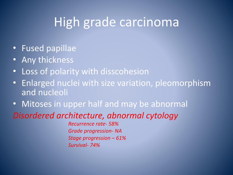
High grade carcinoma
• Fused papillae • Any thickness • Loss of polarity with disscohesion • Enlarged nuclei with size variation, pleomorphism and nucleoli
• Mitoses in upper half and may be abnormal Disordered architecture, abnormal cytology
Recurrence rate-‐ 58% Grade progression-‐ NA Stage progression – 61% Survival-‐ 74%

1973WHO/ISUP/WHO2004
• RCPath recommends use of both to enable comparison (personally I only give 1973!)
• Grading system in largely for papillary lesions, stage is more important for invasive lesions
• Management is the same for PUMLMP/low grade and G1/G2.
• High grade or G3 triggers different management

Flat lesions
• Reactive atypias vs low grade dysplasia • High grade dysplasia/CIS

Reactive atypia
• Nuclear enlargement but normal shape and even chromatin, usually associated inflammation, may show mitotic figures but not abnormal
• Seen with – Chemotherapy e.g. Mitomycin, Cyclophosphamide, BCG( granulomata)
– Raditherapy – Ketamine cystitis(increases Ki67 p53, ck20 negative, history crucial)
CIS
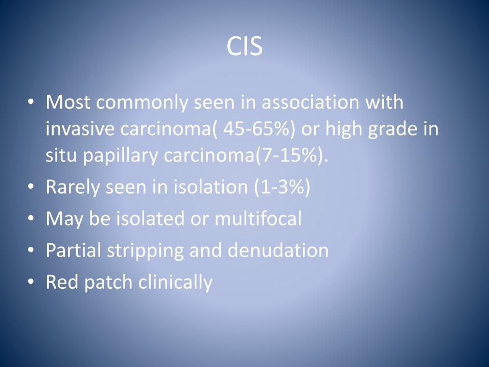
• Most commonly seen in association with invasive carcinoma( 45-‐65%) or high grade in situ papillary carcinoma(7-‐15%).
• Rarely seen in isolation (1-‐3%) • May be isolated or multifocal • Partial stripping and denudation • Red patch clinically

CK7 CK20 CD44 Ki 67 p53 Normal +ve Superficial cells Basal layer Low -‐ve
Reactive +ve Superficial cells Basal layer Variable -‐ve
Dysplasia/CIS +ve Full thickness -‐ve High may be +ve
Immunohistochemistry in flat urothelial atypia
“Not sure favour reactive atypia cant exclude low grade dysplasia” “Apparent genunie dysplasia falling short of CIS”
Both valid reports but use sparingly
Invasive lesions
• Most arise de novo from high grade lesions • Much smaller numbers arise from progression of a low grade lesion.
• Grade much less important than stage also paradox with low grade lesion e.g. nested variant behaving badly
• Presence or absence of MP must be indicated, if absent should have re-‐resection

Aspects of StagingTNM 6th &7th Edition
• pTa –confined to surface • pT1-‐ invasion of lamina propria
? pT1a to MM, pT1b deep to MM, ?depth invasion
• pT2 – into muscularis propria. pT2a inner ½, pT2b outer half-‐ cant do on biopsy
• pT3-‐ invasion of perivesical fat pT3a microscopic, pT3b macroscopic.
• pT4a – 6th-‐ prostate,uterus,vagina 7th-‐ prostatic stroma, SVs, uterus, vagina • pT4b-‐ Pelvic side wall, abdominal wall

Aspects of StagingpTa vs pT1 pitfalls
• Tangential sections • CIS extending into Von Brunn’s nests • Retraction at base pseudo microinvasion or vascular invasion.
• Avoid areas of diathermy. • Personally I err on the side of low stage ie in doubt I go for pTa.

Aspects of StagingpT1 vs pT2 Pitfalls
• Hyperplastic muscularis mucosa mimics muscularis propria.
Smoothelin-‐ positive in MP, negative in MM
• Desmoplasia in LP or scarring from previos resection mimics detrusor.
• Tumour implantation from previous TURBT • Personally take I take further levels and do Actin and Desmin If in doubt express doubt in report “suspicious of MP infiltration but not conclusive”
• We are not alone radiology now very good at assessing muscle/extravesical infiltration

Squamous lesions 1.
• Squamous epithelium in men is always metaplasia
• Non-‐keratinising squamous epithelium away from the trigone in women is metaplasia.
• Keratinising squamous is a risk factor for SCC. • Metaplasia-‐ stones, diverticula, Schistosomiasis
Squamous lesions 2
• Chondylomata-‐ associated with genital disease and urethral involvement
• Verrucous squamous hyperplasia • Benign squamous papilloma, HPV negative

Squamous lesion 3Squamous carcinoma
• Confine to pure SCC, approx 30% conventional TCC show areas of squamous differentiation.
• Often higher stage at presentation pT2 or higher.
• Clinicians often suspect as bladder filled with debris++
• No agreed grading criteria, but basaloid and verrucous carcinomas exist
• Do better with surgery
Glandular lesions 1
• Surface • Cystitis glandularis • Villous adenoma • CIS with glandular and micropapillary features • Adenocarcinoma in situ
• Lamina propria • Cystitis glandularis • Nephrogenic adenoma • Inverted papilloma with glandular features • Adenocarcinoma
• Muscularis • Urachal remnant • Mullerianosis • TCC with glandular • Adenocarcinomas

Glandular lesions 2
• Primary adenocarcinoma – Urachal ( anterior or at dome, sharp demarcation, no primary elsewhere), Non-‐urachal
• Confine to pure with true glandular spaces(glandular differentiation is detectable in approx 10% TCCs.
• Immuno to differentiate primary from metastatic • Prostate -‐ PSA PSMA • Endometrial -‐ ER PR PAX2 PAX8 positive. • Bowel Beta catenin positive • Primary bladder – CDX2 villin CK20 positve CK7 variable not very helpful.
Common benign mimics of bladder carcinoma
• Nephrongenic adenoma • Inverted papilloma

Nephrogenic adenoma 1
• 80% in bladder also in urethra, ureter and renal pelvis.
• Usually incidental at microscopy but 10%>4cm. • Associated with inflammation, surgery and calculi.
• ?adenoma or metaplasia Urothelial –CK7 CK20 uroplakin +ve Renal – CD10 RCC AMACR +ve Y chromosome in female transplants
Nephrogenic adenoma 2
• Several patterns • Polypoid • Papillary • Tubular • Hobnail
• No nuclear atypia or mitoses • Thick peritubular membranes • Associated inflammation

Nephrogenic adenoma 3
• Differential includes – Polypoid or papillary cystitis – Clear cell carcinoma – larger, females, no predisposing factors, solid pattern clear cell areas, cytological atypia may show CEA positivity.
–Microcystic TCC or signet ring adenocarcinoma-‐ more atypia
– Prostatic carcinoma-‐ PSA PSMA positive

Inverted papilloma 1
• Rare <1% urothelial neoplasms • Wide age range • Solitary often sessile may be pedunculated or polypoid
• Benign with no malignant potential and do not require cystoscopic follow up

Inverted papilloma 2
• No exophytic component • If well orientated covered by normal urothelium, peripherally pallisaded
• No or minimal cytological atypia. • If mixed exophytic and endophytic consider low grade TCC and follow up as such
• If diffuse atypia probably inverted pattern TCC(usually has conventional exophytic component )

The End
Thank You
Dr David Paterson 14/05/14