j.t.baker brand histology and cytology - deneysel intended as general references and should only be...
TRANSCRIPT

J.T.Baker® Brand
Histology and Cytology Product Information/Manual

PRODUCT SAFETY DATA SHEETS (SDS) ARE AVAILABLE ON REQUEST. PLEASE SEE PRODUCT SDS FOR COMPLETE INFORMATION REGARDING PRODUCT SAFETY AND APPROPRIATE PRECAUTIONS AND LEGAL REQUIREMENTS TO BE TAKEN WITH RESPECT TO PRODUCT HANDLING, USE AND DISPOSAL. ALL REFERENCES HEREIN WITH RESPECT TO WORDS SUCH AS "NON-HARMFUL," "NON-TOXIC," ETC. ARE INTENDED AS GENERAL REFERENCES AND SHOULD ONLY BE INTERPRETED IN THE CONTEXT OF THE COMPLETE PRODUCT SDS.
| www.avantormaterials.com | I n f o r m a t I o n a n d m a n u a l

CONTENTS
J.T.Baker® Brand Products for Histology and Cytology
Declaration of CE Conformity
J.T.Baker® Brand Product List for Histology and Cytology
J.T.Baker® Brand Product List for Histology and Cytology (2)
Fixatives Overview Formaldehyde Fixatives Bouin’s Fixative Cervix Spray Fixative Formalin Neutralizer Rapid Decalcifier
Paraffin UltraPar™ 54 – 56°C Paraffin Cleaner
Clearing Reagent UltraClear™
Histology Solvents
Stains and Dyes Overview Hematoxylin and Eosin Giemsa and May-Grünwald Leishman Papanicolaou Shorr Wright Troubleshooting Results
Mounting Media UltraKitt™ Xylene-based Mounting Media
Others and packaging Baker-Clear 12 PBS (Immuno) Private Label for OEM Partners and Packaging Options
1
2
3
4
5 6 7 8 9
10
11 11 12
13 13
16
17 18 19 23 25 27 28 30
33 3334
34 34 35 36
I N F O R M a T I O N a N D M a N U a L | www.avantormaterials.com | 2

| www.avantormaterials.com | I n f o r m a t I o n a n d m a n u a l
J.T.Baker® Brand Products for Histology and Cytology Introduction The manufacturing and purifying expertise of avantor™ Performance Materials forms the basis of a comprehensive range of chemicals, stains, and auxiliary products especially formulated and purified for use in histology and cytology applications. along with our environmentally-friendly clearing reagents, a range of solvents, alcohols, fixatives, stains, and dyes are available through our network of international distributors.
Production avantor’s J.T.Baker® brand reagents are produced using state-of-the-art production techniques in modern, certified manufacturing facilities to ensure minimal impurities. The high quality water and solvents utilized by avantor helps to improve the quality of the solutions produced. The products are bottled under controlled conditions to prevent contamination and degradation, thus assuring maximum purity and stability.
Quality Control Every reagent batch is use-tested and released in accordance with ISO-9001 regulations. a highly educated staff of specialists, working in well-equipped R&D and QC laboratories, guarantees lot-to-lot consistency and provides technical customer support.
ISO 9001 avantor’s ongoing pursuit of total quality has resulted in a NEN-ENISO 9001:2008 certified quality system for avantor’s Deventer and Gliwice facilities. This certification includes design, development, production and supply of the products.
ISO 13485
Since January 2012, avantor’s facility in Deventer is ISO 13485:2003 certified. ISO 13485 covers the design, development, production, sales and supply of high purity chemicals chemicals and kits for clinical diagnostics.
ISO 14001 Since January 2004, avantor’s facility in Deventer is also NEN-EN-ISO 14001:2004 certified. ISO 14001 is ISO’s environmental management system that describes the efforts taken to minimize harmful effects to the environment.
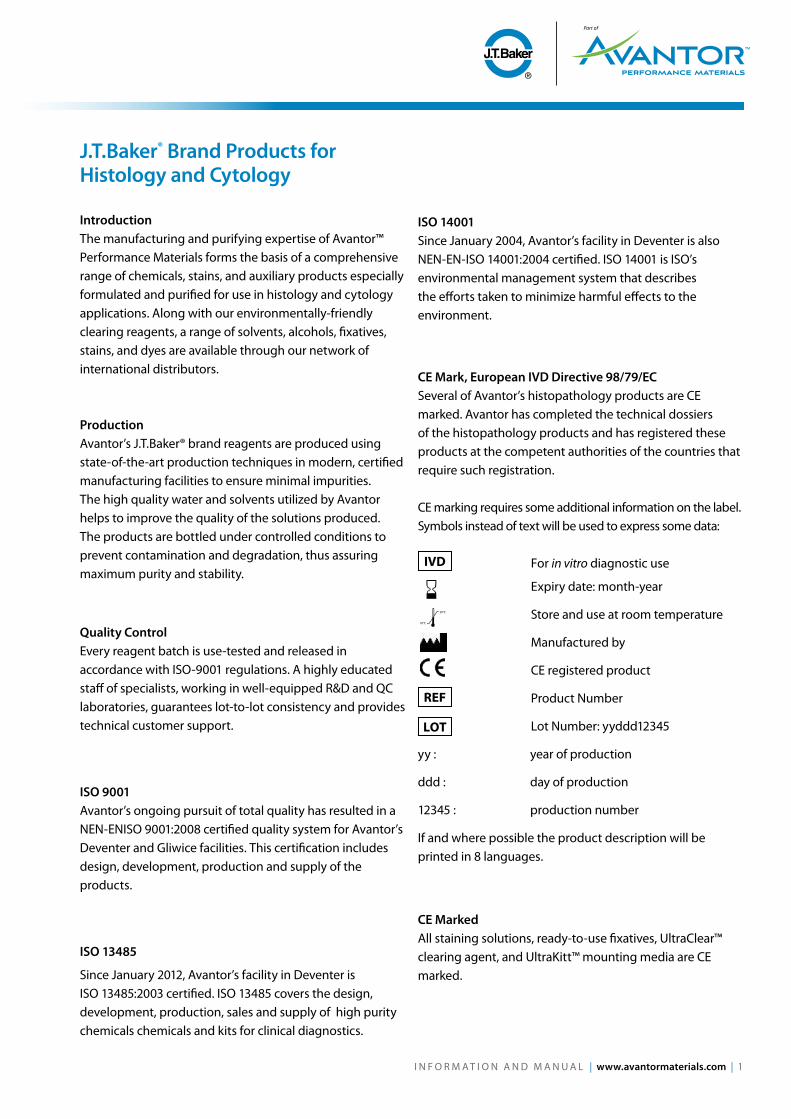
CE Mark, European IVD Directive 98/79/EC Several of avantor’s histopathology products are CE marked. avantor has completed the technical dossiers of the histopathology products and has registered these products at the competent authorities of the countries that require such registration. CE marking requires some additional information on the label. Symbols instead of text will be used to express some data: For in vitro diagnostic use
Expiry date: month-year
Store and use at room temperature
Manufactured by
CE registered product
Product Number
Lot Number: yyddd12345
yy : year of production
ddd : day of production
12345 : production number
If and where possible the product description will be printed in 8 languages.
CE Marked all staining solutions, ready-to-use fixatives, UltraClear™ clearing agent, and UltraKitt™ mounting media are CE marked.
30°C
18°C
IVD
REF
LOT
I N F O R M a T I O N a N D M a N U a L | www.avantormaterials.com | 1
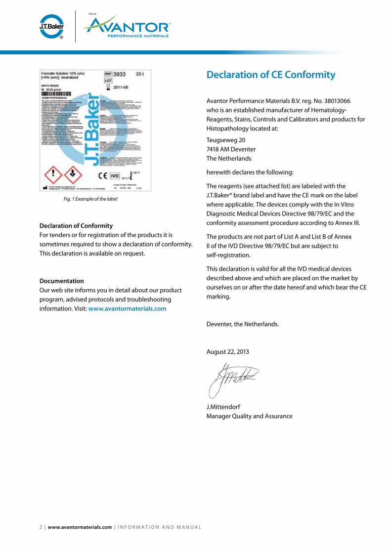
Fig. 1 Example of the label
Declaration of Conformity For tenders or for registration of the products it is sometimes required to show a declaration of conformity. This declaration is available on request.
Documentation Our web site informs you in detail about our product program, advised protocols and troubleshooting information. Visit: www.avantormaterials.com
Declaration of CE Conformity
avantor Performance Materials B.V. reg. No. 38013066 who is an established manufacturer of Hematology- Reagents, Stains, Controls and Calibrators and products for Histopathology located at:
Teugseweg 20 7418 aM Deventer The Netherlands
herewith declares the following:
The reagents (see attached list) are labeled with the J.T.Baker® brand label and have the CE mark on the label where applicable. The devices comply with the In Vitro Diagnostic Medical Devices Directive 98/79/EC and the conformity assessment procedure according to annex III.
The products are not part of List a and List B of annex II of the IVD Directive 98/79/EC but are subject to self-registration.
This declaration is valid for all the IVD medical devices described above and which are placed on the market by ourselves on or after the date hereof and which bear the CE marking.
Deventer, the Netherlands.
august 22, 2013
J.Mittendorf Manager Quality and assurance
2 | www.avantormaterials.com | I N F O R M a T I O N a N D M a N U a L

J.T.Baker® Brand Product List for Histology and Cytology
Product Product Number Content Packaging CE mark
Fixatives
Cervix Spray Fixative 3869.1200 12 x 125 ml HDPE spray bottle CE
10% v/v Buffered Formaldehyde (4% w/v) 3933.1000 1 liter HDPE bottle CE
10% v/v Buffered Formaldehyde (4% w/v) 3933.5000PC 5 liter Polycube CE
10% v/v Buffered Formaldehyde (4% w/v) 3933.9010 10 liter Polycube CE
10% v/v Buffered Formaldehyde (4% w/v) 3933.9010PE 10 liter HDPE can CE
10% v/v Buffered Formaldehyde (4% w/v) 3933.9020 20 liter Polycube CE
10% v/v Buffered Formaldehyde (4% w/v) 3933.9020PE 20 liter HDPE can CE
10% v/v Buffered Formaldehyde (4% w/v) 3933.9200 200 liter HDPE drum CE
38% g/v Buffered Formaldehyde 3859.5000 5 liter HDPE can
38% g/v Buffered Formaldehyde 3859.9010 10 liter HDPE can
Formalin Neutralizer 3934.5000 5 kg PP drum
Bouin’s Fixative 3880.1000 1 liter HDPE bottle CE
Rapid Decalcifier 3930.2500 2.5 liter HDPE bottle
Paraffin
UltraPar™ (54-56°C) 3925.5000 5 kg PP drum
Paraffin Cleaner 3451.1500 12 x 125 ml HDPE spray bottle
Clearing agents
MountingClear 3904.2500 2.5 liter Glass bottle
UltraClear™ 3905.2500PE 2.5 liter Jerrycan CE
UltraClear™ 3905.5000PE 5 liter Jerrycan CE
UltraClear™ 3905.9010PE 10 liter Jerrycan CE
Solvents
acetone 3413.5000 5 liter HDPE can
acetone 3413.9010 10 liter HDPE can
Ethanol 96 % 3405.2500PE 2.5 liter HDPE bottle
Ethanol 96 % 3405.5000 5 liter HDPE can
Ethanol 96 % 3405.9010 10 liter HDPE can
Ethanol 96 % denatured 3415.9010JC 10 liter Jerrycan
Ethanol 99.8 % 3406.2500PE 2.5 liter HDPE bottle
Ethanol 99.8 % 3406.5000 5 liter HDPE can
Ethanol 99.8 % 3406.9010 10 liter HDPE can
Ethanol 99.8 % denatured 3448.5000JC 5 liter Jerrycan
Ethanol 99.8 % denatured 3448.9010JC 10 liter Jerrycan
Isopropanol 3412.5000 5 liter HDPE can
Isopropanol 3412.9025 25 liter HDPE can
Methanol 3400.5000 5 liter HDPE can
Methanol 3400.9010 10 liter HDPE can
Toluene 3411.9010 10 liter Jerrycan
Xylene 3410.2500PE 2.5 liter Jerrycan
Xylene 3410.5000PE 5 liter Jerrycan
Xylene 3410.9010 10 liter Jerrycan
Xylene 3410.9025 25 liter Tin
HDPE = High Density Polyethylene
I N F O R M a T I O N a N D M a N U a L | www.avantormaterials.com | 3

J.T.Baker® Brand Product List for Histology and Cytology (2)
Product Product Number Content Packaging CE mark
Stains and Dyes
Eosin Y 0.5% aqueous 3446.1000PE 1 liter HDPE bottle CE
Eosin Y 0.5% aqueous 3446.2500PE 2.5 liter HDPE bottle CE
Eosin-Y alcoholic 3800.1000PE 1 liter HDPE bottle CE
Eosin-Y alcoholic 3800.2500PE 2.5 liter HDPE bottle CE
Giemsa 3856.0500 0.5 liter Glass bottle CE
Giemsa 3856.1000 1 liter Glass bottle CE
Giemsa 3856.2500 2.5 liter Glass bottle CE
Hematoxylin er (Mayer) 3870.1000 1 liter Glass bottle CE
Hematoxylin er (Mayer) 3870.2500 2.5 liter Glass bottle CE
Hematoxylin Modified (Harris, Gill II) 3873.1000 1 liter Glass bottle CE
Hematoxylin Modified (Harris, Gill II) 3873.2500 2.5 liter Glass bottle CE
Leishman 3879.1000 1 liter Glass bottle CE
May-Grünwald 3855.0500 0.5 liter Glass bottle CE
May-Grünwald 3855.1000 1 liter Glass bottle CE
May-Grünwald 3855.2500 2.5 liter Glass bottle CE
Papanicolaou 2a 3554.1000PE 1 liter HDPE bottle CE
Papanicolaou 2a 3554.2500PE 2.5 liter HDPE bottle CE
Papanicolaou 2B 3555.1000PE 1 liter HDPE bottle CE
Papanicolaou 2B 3555.2500PE 2,5 liter HDPE bottle CE
Papanicolaou 3B 3556.1000PE 1 liter HDPE bottle CE
Papanicolaou 3B 3556.2500PE 2.5 liter HDPE bottle CE
Scotch buffer 3872.5000 5 liter Polycube
Shorr 3876.1000 1 liter Glass bottle CE
Sørenson Buffer 20 x concentrated 3716 10 x 100 ml HDPE bottle
Wright 3878.1000 1 liter Glass bottle CE
Mounting media
UltraKitt™ 3921.0500 500 ml Glass bottle CE
UltraKitt™ 3921.0600 6 x 100 ml Glass bottle CE
Mounting medium High 3882.0500 500 ml PET bottle CE
Mounting medium Low 3883.0500 500 ml PET bottle CE
Cleaners, Immuno PBS and accessories
Baker-Clear 12 Lab Cleaner 7340.5000 5 liter HDPE can
Immuno PBS 20x concentrated 3409.9010 10 liter Polycube CE
Tap for Polycubes 3508 unit
aeroflow tap for use with 10 and 20 L HDPE can 4571 unit
HDPE = High Density Polyethylene
4 | www.avantormaterials.com | I N F O R M a T I O N a N D M a N U a L

Fixatives Overview
Formaldehyde Fixatives Effective and reproducible fixation requires the right concentration of buffered formaldehyde. The 40 mM phosphate buffer J.T.Baker® brand formaldehyde fixatives provide optimal performance and results in immunostaining. The J.T.Baker® brand formaldehyde fixatives are available in both ready-to-use and concentrated solution.
Bouin’s Fixative Bouin’s fixative is a picric acid-based fixative that is used for the preservation of soft or delicate tissues. Excellent preservation of nuclei and chromosomes makes Bouin’s fixative a popular choice to use with embryonic studies and skin.
Cervix Spray Fixative Cervix Spray Fixative is a specially designed, non-toxic solution used to fix prepared cell smears in such a way that Papanicolaou staining gives optimized coloration of cell structures. This product helps ensure that the cell smears are fixated to produce accurate cytology results.
Formalin Neutralizer an essential item to have on hand for histology laboratory processes, formalin neutralizer is a powdered chemical that contains a strong reducing agent to neutralize spilled or used formalin. Neutralization takes 24 hours, after which time the waste can be disposed of as non-harmful chemical waste.
Rapid Decalcifier Rapid decalcifier is a product for decalcifying hard tissue sections such as bone marrow and bone containing tissue sections. Rapid decalcifier is fast acting, and easy to control. This product also ensures tissue protection, while providing superior staining.
Fig.2 Fixatives overview
I N F O R M a T I O N a N D M a N U a L | www.avantormaterials.com | 5

Formaldehyde Fixatives
Introduction Effective and reproducible fixation requires the right concentration of buffered formaldehyde. The 40 mM phosphate buffer J.T.Baker® brand formaldehyde fixatives provide optimal performance and results in immunostaining. The J.T.Baker® brand formaldehyde fixatives are available in both ready-to-use and concentrated solution.
Features and Benefits The consistency of pH neutral embedded tissue is crucial in histopathology. Stains and dyes work and immunoglobulins react better at neutral pH. avantor offers phosphate buffered (pH 7) formaldehyde fixatives, both as ready-to-use and concentrated solutions that have to be diluted before use.
Formalin is a saturated solution of 37% formaldehyde and water. 4% (w/v) formaldehyde is the same as 10% v/v formaldehyde. Commonly used for fixation is the 10% (v/v) formaldehyde. The concentrates are available as 5 or 10 times concentrates in 1, 5, 10 and 20 liter packs.
all fixatives are intended to be used in vitro for the examination of specimens derived from the human body.
The fixatives result in improved fixation, no protein denaturation, and optimized results in immunostaining. Methanol stabilization allows the fixatives to have a shelf life of 2 years. For easy dispensing, taps are available for polycubes and HDPE cans.
Composition The formaldehyde fixatives contain water, formaldehyde, phosphate buffer, and methanol (stabilizer).
Stability and Storage For long-term storage, store 4% formaldehyde solution at room temperature (18 - 30°C). Short-term exposure to lower (to -15°C) and higher temperatures (>30°C) has been tested and did not impact the formaldehyde concentration.
Formaldehyde fixatives are stable for two years when the bottles are kept closed. Formaldehyde concentrates should be stored at room temperature (18 - 30°C). Cooling formaldehyde concentrates past room temperature may cause irreversible polymerization. Used solutions and solutions that are past their shelf-life must be disposed of, according to local disposal guidelines
Procedure 4% (w/v) formaldehyde solution is ready-to-use and doesn’t have to be diluted. 38% (w/v) formaldehyde solution should be diluted before use with distilled or deionized water. Dilute 10 fold to obtain a 3.8% w/v (10% v/v) formaldehyde solution. It is not advised to use concentrated formaldehyde directly, because it can destroy the protein and antigen structure.
Ordering InformationProduct Product Number Packaging
10% v/v Buffered Formaldehyde 3933.1000 1 liter
10% v/v Buffered Formaldehyde 3933.5000PC 5 liter
10% v/v Buffered Formaldehyde 3933.9010 10 liter
10% v/v Buffered Formaldehyde 3933.9010PE 10 liter
10% v/v Buffered Formaldehyde 3933.9020 20 liter
10% v/v Buffered Formaldehyde 3933.9020PE 20 liter
10% v/v Buffered Formaldehyde 3933.9200 200 liter
19% g/v Buffered Formaldehyde 3858.9020 20 liter
38% g/v Buffered Formaldehyde 3859.5000 5 liter
38% g/v Buffered Formaldehyde 3859.9010 10 liter
Tap for use with the 5, 10 and 20 liter cubitainers
3508
aeroflow tap for use with 10 and 20 L HDPE can
4571 Fig.3 20 liter HDPE 10% v/v Buffered Formaldehyde
6 | www.avantormaterials.com | I N F O R M a T I O N a N D M a N U a L

Bouin’s Fixative
Introduction Bouin’s fixative is a picric acid-based fixative that is used for the preservation of soft or delicate tissues. Excellent preservation of nuclei and chromosomes makes Bouin’s fixative a popular choice to use with embryonic studies and skin.
Features and Benefits Bouin’s fixative is used for preservation of soft and delicate tissues. It is used for embryonic studies and skin, due to its excellent preservation of nuclei and chromosomes. Bouin’s fixative is very compatible with the trichrome stains, due to its mordanting effect on the tissue. Bouin’s is a ready-to-use fixative for histopathology applications, specifically in vitro for the examination of specimens derived from the human body.
Composition Bouin’s fixative contains formaldehyde, acetic acid, picric acid.
Ordering InformationProduct Product Number Packaging
Bouin’s Fixative 3880.1000 1 liter
I N F O R M a T I O N a N D M a N U a L | www.avantormaterials.com | 7

Cervix Spray Fixative
Introduction Cervix Spray Fixative is a specially designed, non-toxic solution used to fix prepared cell smears in such a way that Papanicolaou staining gives optimized coloration of cell structures. This product helps ensure that the cell smears are fixated to produce accurate cytology results.
Features and Benefits Prepared cell smears should be preserved to avoid cell and tissue artifacts. Deteriorated cell material will disrupt cell morphology, therefore proper fixation of prepared cell smears is an important starting point for your diagnosis. Cervix spray fixative is used to fix prepared cell smears in such a way that staining with Papanicolaou gives optimized coloration of cell structures. Cervix spray fixative contains special glycols in an alcoholic medium, and is intended to be used in vitro for the examination of specimens derived from the human body.
Composition Cervix spray fixative contains 2-propanol, acetone and PEG.
Procedure Cervix spray fixative is ready-to-use. Spray a few times on the smear, air-dry the smear for a few seconds, and proceed according the regular laboratory instructions.
Stability and Storage Cervix spray fixative is stable for two years when the bottles are kept closed and stored at room temperature (18 - 30°C). Used solutions and solutions that are past their shelf-life must be disposed of, according to local disposal guidelines.
Ordering InformationProduct Product Number Packaging
Cervix Spray Fixative 3869.1200 12 x125 ml
Fig.4 Cervix Spray Fixative
8 | www.avantormaterials.com | I N F O R M a T I O N a N D M a N U a L

Formalin Neutralizer
Introduction an essential item to have on hand for histology laboratory processes, formalin neutralizer is a powdered chemical that contains a strong reducing agent to neutralize spilled or used formalin. Neutralization takes 24 hours, after which time the waste can be disposed of as non-harmful chemical waste.
Features and Benefits Formalin (dissolved formaldehyde) is a widely used fixative in histopathology laboratories. Formalin is considered to be an aggressive oxidizer. Discarding formalin in sewer systems is not only dangerous, but can also be harmful to our environment. an effective solution has been found in neutralizing the formaldehyde with a strong reducing agent. Formalin neutralizer is a powdered chemical capable of neutralizing formalin and it is intended to be used on spilled and used formalin.
Principle Formalin neutralizer consists of a strong reducing compound resulting in harmless waste products. Esters are formed after interaction of formaldehyde radicals. The released acid decreases the pH from 7 to 6.
Composition Formalin neutralizer contains min. 80% sodium disulfite.
Stability and Storage Formalin neutralizer is stable for two years when the bottles are kept closed and stored at room temperature (18 - 30°C). Used solutions and solutions that are past their shelf-life must be disposed of according to local disposal guidelines.
Procedure 125 grams of Formalin neutralizer is sufficient to neutralize 1 liter of 4% w/v formaldehyde (10 v/v% formaldehyde) or 1 liter of 2% w/v glutaraldehyde. 1 spoonful is approximately 125 grams. The reaction is completed after 24 hours. after the neutralization the waste can be handled as non harmful chemical waste according local environmental regulations.
Ordering InformationProduct Product Number Packaging
Formalin neutralizer 3934.5000 5 kg
Aldehyde concentration versus amount of Formalin NeutralizerFormaldehyde w/v% Glutaraldehyde or
glyoxal w/v%Neutralizer required
for 1 liter
2 1 62.5 g
4 2 125 g
6 3 187.5 g
8 4 250 g
10 5 312.5 g
Note: 4.0 w/v% is equal to 10.0 v/v%
Note: 125 gram Formalin neutralizer is 1 spoon
I N F O R M a T I O N a N D M a N U a L | www.avantormaterials.com | 9
Rapid Decalcifier
Introduction Rapid decalcifier is a product for decalcifying hard tissue sections such as bone marrow and bone containing tissue sections. Rapid decalcifier is non-hazardous, fast acting, and easy to control. This product also ensures tissue protection, while providing superior staining.
Features and Benefits Decalcifying is necessary because a high level of calcium disturbs the staining quality. Rapid decalcifier is a decalcifying solution that has the ability to decalcify the tissue completely within a short time limit. Tissues decalcified in this solution can easily be cut and routine HE techniques produce excellent stained sections. Rapid decalcifier is intended to be used for decalcifying hard tissue sections such as bone marrow and bone containing tissue sections.
Rapid decalcifier is fast-acting. The following decalcification times have been observed, and represent approximate decalcification times: bone marrow biopsies can be decalcified in as little as 15-30 minutes, needle biopsies in one hour, small bone specimens and calcified arteries in 2-4 hours, and typical surgical specimens in 2-6 hours. The speed of the processing can easily be controlled by dilution with water, while the nuclear histological detail is always preserved. Decalcifiers work even faster if they are chilled. The decalcifier is a non-toxic and non-hazardous solution, which can be disposed or in sewer systems according local environmental regulations.
Composition Rapid decalcifier is water based and contains hydrochloric acid, EDTa salts and surface active components.
Stability and Storage Rapid decalcifier is stable for two years when the bottles are kept closed and stored at room temperature (18 - 30°C). Used solutions and solutions that are past their shelf-life must be disposed of, according to local disposal guidelines.
Procedure
•Routine processes:
1. Use approximately 20-times the volume of the specimen.
2. allow to decalcify and briefly rinse with water before continuing the process.
•Use on microwave techniques: We do not recommend this technique for clinical settings. Heating decalcifiers in a microwave oven has only one major advantage: speed. In any case, care should be taken, because heat can burn and destroy tissue and may degrade the quality of the specimens you are working with.
Ordering InformationProduct Product Number Packaging
Rapid decalcifier 3930.2500 2.5 liter
10 | www.avantormaterials.com | I N F O R M a T I O N a N D M a N U a L

Paraffin
UltraPar™ 54 - 56°C Paraffin
Introduction UltraPar particle-free paraffin is intended for use in histology tissue embedding and sectioning. Unlike some other paraffin products, UltraPar paraffin does not crack or shrink during cooling, and is twice as soluble in organic solvents and UltraClear clearing agent when compared to similar products based on internal tests. Increased solubility results in improved penetration, de-paraffinization, and excellent results of stained sections.
Features and benefits UltraPar paraffin is high quality, particle free paraffin intended for tissue embedding of fixed and dehydrated specimens for microscopy. It has a melting range of 54 - 56°C and is bright and clear when melted. The small pellets are perfect for rapid melting in tissue processors and/or embedding centers.
UltraPar contains no additives and causes no damage to the blades of the microtome. It does not shrink or crack during cooling, and performs at optimal micro slicing as thin as 2, 3 or 4 μm. Sections from UltraPar paraffin embedded tissues can be used for immunohistochemistry, in-situ hybridization and PCR. With its good solubility in UltraClear clearing agent, it saves time, money, and results in less waste when compared with normal paraffin.
Composition UltraPar is purified paraffin, without the addition of DMSO and bees wax.
Stability and Storage UltraPar paraffin is stable for five years when stored at room temperature (18 - 30°C).
Procedure Carefully dehydrate the samples and remove the alcohol by treating with UltraClear clearing agent or xylene to ensure tissue saturation with paraffin and easier sectioning after embedding. Paraffin-treated specimens are embedded in suitable molds. To improve cutting, the paraffin blocks can be stored in a cool place prior to slicing. The optimum slicing temperature varies from -10 – 0 °C and sectioning can also be improved by warming the knife. Deparaffinate, rehydrate, and stain the sections according to standard histological staining protocols. after staining, dehydrate the sections in alcohol and clear in UltraClear clearing agent or xylene.
Performance Characteristics UltraPar paraffin is almost twice as soluble in UltraClear clearing agent when compared with other paraffin.
Ordering InformationProduct Product Number Packaging
UltraPar™ 3925.5000 5 kg
I N F O R M a T I O N a N D M a N U a L | www.avantormaterials.com | 11

Paraffin Cleaner
Introduction Paraffin cleaner is a special non-toxic cleaner for laboratory equipment, including processors and microtomes. J.T. Baker® brand paraffin cleaner contains mineral oils in an alcoholic medium. When used regularly, paraffin cleaner helps keep laboratory equipment clean and in good working order.
Features and Benefits Laboratory equipment requires regular cleaning to remain in good, working condition. Microtomes become polluted with paraffin residues when not cleaned properly or sufficiently. The removal of paraffin is mostly done with organic solvents, like xylene or toluene, which can produce excessive vapor in the laboratory environment. Paraffin cleaner is a specially designed cleaning solution to remove excess paraffin, with minimal odor.
Composition Paraffin cleaner consists of mineral oils in an alcoholic medium.
Stability and Storage Paraffin cleaner is stable for two years when the bottles are kept closed and stored at room temperature (18 - 30°C). Products that are past their shelf-life must be disposed of, according to local disposal guidelines.
Procedure Spray the necessary amount of paraffin cleaner on the surface that must be cleaned. Wipe clean with a tissue.
Ordering InformationProduct Product Number Packaging
Paraffin Cleaner (paraffin remover) 3451.1500 12 x125 ml
12 | www.avantormaterials.com | I N F O R M a T I O N a N D M a N U a L

Clearing Reagent
UltraClear™ Clearing Agent
Introduction UltraClear™ clearing agent is an isoparaffin-based clearing agent that can be used as an environmentally friendly xylene replacement during tissue embedding, deparaffinization, and staining processes. Not only is UltraClear clearing agent odorless, less toxic, and less flammable when compared to xylene, it is also easy to adapt with existing working protocols.
Features and Benefits Tissue that has been fixed must be embedded in paraffin to make sectioning in thin slices possible. First, the water in the tissue must be removed. This is normally done with a series of alcohols (70 – 95 – 100%). The next step is called “clearing” and consists of removal of the dehydrant, or water-removing agent, with a clearing reagent.
UltraClear clearing agent is an iso-paraffin based clearing agent for tissue embedding and deparaffination. It works with all kinds of paraffin and no methanol is needed during dehydration. Iso-paraffins are considered to be more environmentally-friendly when compared to traditional solvent containing clearing agents.
after clearing, the tissue is infiltrated with paraffin, sectioned, and put on a microscopic slide. The embedding process must be reversed in order to get the paraffin wax out of the tissue and allow water soluble dyes to penetrate the sections. The slides are deparaffinateded by running them through UltraClear clearing agent and then through alcohols and then water and finally the slides are stained. The stained slide is taken through a series of alcohols to remove the water, then through UltraClear clearing
agent to a point at which the cover glass can be mounted with UltraKitt mounting media. When UltraClear clearing agent is used in combination with Eukit/DPX or Entallan it is advised to use MountingClear as an intermediate. UltraClear clearing agent and MountingClear are intended to be used in vitro for the examination of specimens derived from the human body.
Composition UltraClear clearing agent is the brand name for a synthetically produced isoparaffinic fluid, a C11 – 12 isoparaffin. Isoparaffinic fluids are high purity, synthetic, hydrocarbon fluids made via oligomerization of C3 to C5 hydrocarbons derived from crude oil fractionation and cracking operations. Isopar fluids are purified and then fractionated into desired volatility ranges.
Stability and Storage UltraClear clearing agent is stable for five years when the bottles are kept closed and stored at room temperature (18 - 30°C). Products that are past their shelf-life must be disposed of, according to local disposal guidelines.
Procedure If xylene is replaced by UltraClear clearing agent in tissue processing, the following steps should be considered:
1. Frequent refreshment of ethanol 100%. Xylene still can dissolve small amounts of water while UltraClear clearing agent cannot. Ethanol 100% helps to complete the dehydration of the tissue.
2. Time of clearing should be increased by 30 minutes. Penetration of xylene in tissue is faster than with UltraClear clearing agent; however, UltraClear clearing agent hardens fragile tissue material much less than xylene (examples are lymph nodes and testis). This is a major advantage to using UltraClear clearing agent over xylene.
Ordering InformationProduct Product Number Packaging
UltraClear™ 3905.2500PE 2.5 liter
UltraClear™ 3905.5000PE 5 liter
UltraClear™ 3905.9010PE 10 liter
MountingClear 3904.2500 2.5 liter
I N F O R M a T I O N a N D M a N U a L | www.avantormaterials.com | 13

UltraClear clearing agent has been evaluated at two histology laboratories in the Netherlands. These evaluations resulted in recommended and optimized methods for tissue embedding on Shandon Hypercentre and the Sakura VIP 3000. Recommendations were made for deparaffination and coverslipping using both UltraClear clearing agent and UltraKitt mounting media during HE staining and PaP staining procedures. In all cases it should be noted that all recommendations are subject to individual changes and these recommendations should be used as a guideline.
Note: Lepedimic tissue should be treated with acetone prior to the routine embedding procedure. Calcium containing tissue, like bone marrow, should be treated with EDTa or acid prior to the routine embedding procedure.
A. Program on Hypercenter of ShandonReagent sequence Biopsy
Formaldehyde 10% v/v 5 min., 45°, 0, drain=2 min.
Ethanol 90% 5 min., 45°, 0, drain=2 min.
5x Ethanol 100% 5 min., 45°, 0, drain=2 min.
2x UltraClear 10 min., 45°, 0, drain=2 min.
UltraClear 15 min., 45°, 0, drain=2 min.
Paraffin 20 min., 60°, 0, drain=2 min.
Paraffin 30 min., 60°, 0, drain=2 min.
C. Method using UltraClear™ clearing agent and UltraKitt™ mounting media in a HE staining procedureReagent sequence Minutes
UltraClear 3 x 2
Ethanol 100% 2 x 1
Ethanol 96% 1
Ethanol 70% 1
Distilled Water 1
Hematoxyline 4 - 5
Flushing with tap water 1
Scotch Solution 1
Flushing with tap water 1
Eosine 1
Ethanol 96% 2 x 1
Ethanol 100% 2 x 1
UltraClear 3 x 2
Coverslipping with UltraKitt
B. Program on VIP 3000 of BayerReagent sequence Minutes Temperature
Formaldehyde 10% v/v 30 40°C
Formaldehyde 10% v/v 60 40°C
Ethanol 50% 30 40°C
Ethanol 70% 30 40°C
Ethanol 100% 30 40°C
Ethanol 100% 60 40°C
Ethanol 100% 60 40°C
Ethanol 100% 120 40°C
UltraClear 40 40°C
UltraClear 80 40°C
Paraffin 60 56°C
Paraffin 60 56°C
Paraffin 60 56°C
Paraffin 120 56°C
Note: It has been noted that the Paraffin saturation of UltraClear clearing agent strongly depends on the temperature. The relation is presented in the graph.
Reagent sequence Biopsy
Formaldehyde 10% v/v 10 min., 0, drain=2 min.
Ethanol 90% 20 min., 0, drain=2 min.
Ethanol 100% 30 min., 0, drain=2 min.
4x Ethanol 100% 60 min., 0, drain=2 min.
2x UltraClear 90 min., 0, drain=2 min.
UltraClear 120 min., 0, drain=2 min
2x Paraffin 150 min., 0, drain=2 min
Reagent sequence Biopsy
Formaldehyde 10% v/v 24h 10 min., N, drain=2min.
Ethanol 90% 20 min., 0, drain=2min.
Ethanol 100% 30 min., 0, drain=2min.
4x Ethanol 100% 60 min., 0, drain=2 min.
2x UltraClear 90 min., 0, drain=2 min.
UltraClear 120 min., 0, drain=2 min.
2x Paraffin 150 min., 0, drain=2 min.
14 | www.avantormaterials.com | I N F O R M a T I O N a N D M a N U a L

Troubleshooting Cutting problems
•Still water in tissue. Last 100% ethanol bath in dehydration process should be refreshed.
•Still ethanol in tissue. Time in UltraClear clearing agent is too short. Increase steps or time of UltraClear clearing agent.
Tissue badly stained
•Fixative not optimally used. Formaldehyde 10 v/v% buffered is the best fixative approach.
•Problems with staining solutions.
•Tissue slices too thick.
•Tissue not completely deparaffinated.
Deparaffination problem
•UltraClear clearing agent saturated with paraffin. UltraClear clearing agent should be refreshed.
•Still water in tissue. Last 100% ethanol bath in dehydration process should be refreshed.
Slide appearance is milky during coverslipping
•UltraClear clearing agent used in final bath, together with a conventional mounting medium. Use UltraKitt mounting media for the best fit.
•Final dehydration not complete. Refresh final bath of ethanol 100% more frequently.
D. Method using UltraClear clearing agent and UltraKitt mounting media in a Papanicolaou staining procedureReagent sequence Minutes
Ethanol 70% 2
Distilled Water 2 x 2
Hematoxylin ½ - 1
Distilled Water ½
Flushing with tap water 2 x 2
Ethanol 70% 2
Ethanol 96% 3 x 2
Papanicolaou 2a / 2B 1
Ethanol 96% 1
Ethanol 96% 2
Papanicolaou 3B 1½ - 2
Ethanol 96% 3 x ½
Ethanol 100% 3 x ½
UltraClear 3 x 2
Coverslipping with UltraKitt
I N F O R M a T I O N a N D M a N U a L | www.avantormaterials.com | 15

Histology Solvents
Ordering InformationProduct Product Number Packaging
acetone 3413.5000 5 liter
acetone 3413.9010 10 liter
Ethanol 96 % 3405.2500PE 2.5 liter
Ethanol 96 % 3405.5000 5 liter
Ethanol 96 % 3405.9010 10 liter
Ethanol 96 % denatured 3415.9010JC 10 liter
Ethanol 99.8 % 3406.2500PE 2.5 liter
Ethanol 99.8 % 3406.5000 5 liter
Ethanol 99.8 % 3406.9010 10 liter
Ethanol 99.8 % denatured 3448.5000JC 5 liter
Ethanol 99.8 % denatured 3448.9010JC 10 liter
Isopropanol 3412.5000 5 liter
Isopropanol 3412.9025 25 liter
Methanol 3400.5000 5 liter
Methanol 3400.9010 10 liter
Toluene 3411.9010 10 liter
Xylene 3410.2500PE 2.5 liter
Xylene 3410.5000PE 5 liter
Xylene 3410.9010 10 liter
Xylene 3410.9025 25 liter
Features and Benefits avantor offers a competitive range of conveniently packaged solvents and alcohols especially manufactured to meet the needs of histology and cytology applications. all products are of histograde quality and tested for use in histological applications. avantor’s alcohol product line consists of methanol, ethanol, ethanol denatured, and IPa, all of which are purified and denatured to be used in staining and in tissue preparation procedures.
Stability and Storage all solvents are stable for two to five years when the bottles are kept closed and stored at room temperature (18 - 30°C). Products that are past their shelf-life must be disposed of, according to local disposal guidelines.
Procedure Use regular laboratory procedures.
Fig.5 Convenient and easy-to-carry High Density PolyEthylene can (HDPE).
16 | www.avantormaterials.com | I N F O R M a T I O N a N D M a N U a L

Stains and Dyes Overview
Hematoxylin and Eosin (H&E) Stains Modern technology and temperature controlled manufacturing processes help produce Mayer, Harris and Gill-II Hematoxylin staining solutions that are ready to use, long-lasting and produce a consistent, bright staining of the nucleus. J.T.Baker® brand Eosin Y counter stains, offered in both alcoholic and aqueous solutions, are optimized when used in combination with our J.T.Baker® brand Hematoxylin stains. The J.T.Baker® brand product line also includes Scotch’s solution, which enables a stable and optimized bluing of the cell nucleus.
May-Grünwald, Giemsa, Wright and Leishman Stains May-Grünwald, Giemsa, Wright and Leishman stains are used for tissue sections, cytology smears, blood smears and bone marrow. We strive for the highest standards when designing our production methods and modifying our formulations. This ensures that J.T.Baker® brand stains result in optimized color intensity for clear results in most testing procedures.
Papanicolaou Stains Despite the technological advances in medicine over the last 100 years, the Pap smear test remains one of the most commonly used tests to detect the presence of premalignant lesions in cervix smears. The J.T. Baker® brand product line includes Papanicolaou 1 (hematoxylin solution according to Gill II/Harris), Papanicolaou 2a (Orange G6), Papanicolaou 2B, and Papanicolaou 3B (Ea 50) stains. all of our Pap staining solutions are purified and free from contamination to ensure reliable results.
Fig.6 Papanicolaou Stains
I N F O R M a T I O N a N D M a N U a L | www.avantormaterials.com | 17

Hematoxylin and Eosin
Introduction Modern technology and temperature controlled manufacturing processes help produce Mayer, Harris and Gill-II Hematoxylin staining solutions that are ready to use, long-lasting and produce a consistent, bright staining of the nucleus. J.T.Baker® brand Eosin Y counter stains, offered in both alcoholic and aqueous solutions, are optimized when used in combination with our J.T.Baker® brand Hematoxylin stains. The J.T.Baker® brand product line also includes Scotch’s solution, which enables a stable and optimized bluing of the cell nucleus.
Features and Benefits Hematoxylin needs to be oxidized to form hematein, which is used with a suitable mordant (in this case, aluminum) to stain nuclei. Hematoxylin-Eosin staining is the most widely used technique in routine and counter staining (regressive or progressive). Hematoxylin is intended to be used as a nuclear dye in the standard HE (hematoxylin and eosin) staining method for tissue section analysis. The Eosin Y solutions are especially manufactured to match our hematoxylin stains, resulting in good chromatin patterns and easy adaptable color tones. Eosin Y is the most common counterstain to aluminum hematoxylin in the HE method. Eosin Y is offered as an alcoholic solution. all solutions are intended to be used in vitro for the examination of specimens derived from the human body. These solutions are ready to use and will give reproducible results.
Principle In the first staining step, the nuclei are stained by a hematoxylin solution. The second step in Hematoxylin staining is the bluing of the tissue section. Initially, the tissue is colored purple – reddish purple. after exposure to Scotch solution and tap water, the tissue takes the blue color of a hematoxylin stained slide. Nuclei are stained blue - violet. The last step is counterstaining with eosin Y solution. Cytoplasma will show pink-orange and erythrocytes stain red.
Composition Hematoxylin (Mayer) is water based and contains hematoxylin, aluminium salt, citric acid, chloral hydrate, and iodate.
Hematoxylin (Harris, Gill II, PaP 1) is water based and contains hematoxyline, aluminum salt, organic acid, iodate and ethylene glycol.
Eosin-Y alcoholic solution is water based and contains 0.2% eosine Y, water and alcohols.
Eosin-Y 0.5% aqueous solution is alcohol based and contains eosine Y, water and alcohols.
Ordering InformationProduct Product Number Packaging
Hematoxylin (Mayer) 3870.1000 1 liter
Hematoxylin (Mayer) 3870.2500 2.5 liter
Hematoxylin Modified (Harris, Gill II, PaP1)
3873.1000 1 liter
Hematoxylin Modified (Harris, Gill II, PaP1)
3873.2500 2.5 liter
Eosin-Y alcoholic 3800.1000PE 1 liter
Eosin-Y alcoholic 3800.2500PE 2.5 liter
Eosin-Y 0.5% aqueous 3446.1000PE 1 liter
Eosin-Y 0.5% aqueous 3446.2500PE 2.5 liter
Scotch Buffer 3872.5000 5 liter
18 | www.avantormaterials.com | I N F O R M a T I O N a N D M a N U a L

Performance Characteristics Nucleus: blue / violet Cytoplasm: pink / orange Erythrocytes: red
Stability and Storage Hematoxylin and Eosin-Y 0.5% aqueous solutions are stable for two years when the bottles are kept closed and stored at room temperature (18 - 30°C). Products that are past their shelf-life must be disposed of, according to local disposal guidelines.
Eosin-Y alcoholic is stable for five years when the bottles are kept closed and stored at room temperature (18 - 30°C). Products that are past their shelf-life must be disposed of, according to local disposal guidelines.
Procedure for routine HE staining for histology sections Proceed according to the table.
Note: all reagents are ready to use.
Note: When Hematoxylin Modified (Harris, Gill II, PaP1), is used, the staining time can be reduced from 4 - 5 minutes to 1 - 2 minutes.
Note: The times as listed in the table are approximate and can be adjusted to suit personal preferences. Staining solutions will lose their staining power when heavily used and the staining times should be longer or fresh solutions should be used.
Reagent sequence Minutes
UltraClear/Xylene 3 x 2
Ethanol 100% 2 x 1
Ethanol 96% 1
Ethanol 70% 1
Destilled water 1
Hematoxylin 4 - 5
Flushing with tap water 1
Scotch Solution 1
Flushing with tap water 1
Eosin alcoholic 1
Ethanol 96% 2 x 1
Ethanol 100% 2 x 1
UltraClear/Xylene 3 x 2
Embedding with mounting medium
I N F O R M a T I O N a N D M a N U a L | www.avantormaterials.com | 19

Giemsa and May-Grünwald
Introduction May-Grünwald and Giemsa stains are used for tissue sections, cytology smears, blood smears and bone marrow. J.T.Baker® brand stains result in optimized color intensity for clear results in most testing procedures.
Features and Benefits avantor’s products have optimized formulations, resulting in balanced colors with easy adaptable tones and an avoidance of crystallization. The purple color of cell nuclei is due to molecular interaction between eosin Y and an azure B-DNa. May-Grünwald’s eosin methylene blue and Giemsa’s azure eosin methylene blue are intended to be used for staining of blood and bone marrow smears and cytological specimens, such as urine sediment or sputum. For staining of most histology specimens (mostly gastric sections), Giemsa is used. Sørensen buffer solution can be used for easy diluting.
Composition May-Grünwald is alcohol based and contains May-Grünwald’s eosin methylene blue and methanol (>85%). Giemsa is alcohol based and contains Giemsa’s azure eosin methylene blue, methanol (>50%) and glycerol.
Stability and Storage May-Grünwald and Giemsa are stable for five years when the bottles are kept closed and stored at room temperature (18 - 30°C). Products that are past their shelf-life must be disposed of, according to local disposal guidelines.
Procedures for bone marrow, cytology samples and blood smears
1. General method for bone marrow or cytology specimen or for whole blood smears:
– Prepare Sørensen buffer solution pH 7: Dilute 100 ml of the concentrated Sørensen phosphate buffer with 1.9L deionized water.
– Prepare the May-Grünwald working solution: Dilute 250 ml May-Grünwald solution with 250 ml Sorensen buffer solution pH 7.
– Prepare the Giemsa working solution: Dilute 50 ml Giemsa solution with 450 ml Sørensen buffer solution pH 7.
– Proceed according to the table below:
Reagent sequence Minutes
Undiluted May-Grünwald 3
May-Grünwald working solution 5
Move slides gently in Sørensen buffer pH 7 1
Move slides gently in Giemsa working solution 20 (blood smears) 25 (bone marrow, cytology)
Flush in tap or deionized water
Ordering InformationProduct Product Number Packaging
May-Grünwald 3855.0500 500 ml
May-Grünwald 3855.1000 1 liter
May-Grünwald 3855.2500 2.5 liter
Giemsa 3856.0500 500 ml
Giemsa 3856.1000 1 liter
Giemsa 3856.2500 2.5 liter
Sørensen buffer concentrate 3716 10 x 100 ml
20 | www.avantormaterials.com | I N F O R M a T I O N a N D M a N U a L

4. Quick staining method for whole blood smears:
– Prepare Sørensen buffer solution pH 7: Dilute 100 ml of the concentrated Sørensen phosphate buffer with 1.9L deionized water.
– Prepare the Giemsa working solution: Dilute Giemsa solution 1 to 6 up to 1 to 8 with Sørensen buffer solution pH 7.
– Proceed according to the table below:
–
–
–
Note: The times as listed in the tables are approximate and can be adjusted to suit personal preferences. Staining solutions will lose their staining power when heavily used so the staining times should be longer or fresh solutions should be used.
2. Traditional method according to Pappenheim for whole blood smears:
– Prepare Sørensen buffer solution pH 7: Dilute 100 ml of the concentrated Sørensen phosphate buffer with 1.9L deionized water.
– Prepare the Giemsa working solution: Dilute 25 ml Giemsa solution with 475 ml Sørensen buffer solution pH 7.
– Proceed according to the table below:
3. Quick staining method for whole blood smears:
– Prepare Sørensen buffer solution pH 7: Dilute 100 ml of the concentrated Sorensen phosphate buffer with 1.9L deionized water.
– Prepare the Giemsa working solution: Dilute Giemsa solution 1 to 6 up to 1 to 8 with Sørensen buffer solution pH 7.
– Proceed according to the table below:
–
–
–
–
Performance Characteristics
Reagent sequence Minutes
Undiluted May-Grünwald 3
Flush in deionized water 1
Move slides gently in Giemsa working solution 20
Flush in tap or deionized water
Reagent sequence Minutes
Undiluted May-Grünwald 2 - 3
Move slides gently in Sørensen buffer pH 7 1
Move slides gently in Giemsa working solution 4 - 5
Flush in tap or deionized water
Reagent sequence Minutes
Undiluted May-Grünwald 2 - 3
Flush in tap or deionized water 1
Giemsa working solution 4 - 5
Flush in tap or deionized water
Type Blood-cell Amount Characteristics
RBC 4 – 6 x 10¹²/liter Pink/brown discs; clearer in the middle due to their concave structure
PLT 0.2 – 0.3 x 10¹²/liter Purple colored granules; much smaller then RBC
NEUT 50 – 70 % * Transparent, pink/blue cytoplasm; 2-5 lobed bright purple nucleus
EO 2 – 4 % Typical pink-orange granulated cytoplasm; generally 2-lobed purple nucleus
LYM 20 – 40 % Transparent purple cytoplasm; one large, purple-pink nucleus
MONO 3 – 8 % Largest of the leukocytes; transparent, pink/blue cytoplasm with horseshoe-shaped pink/purple nucleus
BaSO 0.5 – 1.0 % Granulo-rich cytoplasm exhibiting dark-blue stain overruling the dark-blue nucleus stain
* From total WBC population; generally 5 – 7 x 10⁹ cells/liter
I N F O R M a T I O N a N D M a N U a L | www.avantormaterials.com | 21

Procedure for Histology samples
•Prepare the Giemsa working solution: add 20 ml Giemsa solution to 80 ml deionized water. It is important to add the Giemsa to the water and not vice versa.
•Prepare the differentiation solution: add 4 drops of Glacial acetic acid (96%) to 100 ml of deionized water. Measure the pH of the solution. It should be 3.0 – 3.2.
Proceed according to the tables below:
Performance Characteristics Nucleus: blue / violet Cytoplasm: blue Erythrocytes: pink Eosinophilic granules: orange Basophilic granules: purple
Fig.3 May-Grünwald stained blood cells (100x).
Deparaffination of tissueReagent sequence Minutes
UltraClear™/Xylene 3 x 1
Ethanol 100% 3 x 1
Ethanol 70% 1
Flush with tap water 1
Staining of tissueReagent sequence Minutes
Insert 3 times in deionized water
Giemsa working solution (use only once) 30
Differentiation fluid (differentiation to purple) dip just once
Ethanol 96% (differentiation to blue) dip just once
Isopropanol (2-propanol) dip just once
Isopropanol (2-propanol) 3 x 2
UltraClear™/Xylene (refresh each time) 3 x 2
Coverslip slides
22 | www.avantormaterials.com | I N F O R M a T I O N a N D M a N U a L

Leishman
Introduction Leishman’s stain is used for blood smears and bone marrow. J.T.Baker® brand stains result in optimized color intensity for clear results in most testing procedures.
Features and Benefits Leishman’s stain is used for staining blood and bone marrow. It is generally used to differentiate and identify leucocytes, malaria parasites, and trypanosomas (unicellar parasitic protozoa). It is based on a mixture of methylene blue and eosin. The purple color of cell nuclei is due to molecular interaction between eosin Y and an azure B-DNa complex. The intensity of the staining depends on the azure B content and on the ratio azure B/eosin Y.
Composition Leishman contains Leishman stain and methanol.
Stability and Storage Leishman staining solution is stable for five years when the bottles are kept closed and stored at room temperature (18 - 30°C). Products that are past their shelf-life must be disposed of, according to local disposal guidelines.
Procedure
•Method using a concentrated Leishman / Buffer solution:
1. Prepare Sørensen buffer solution pH 7: Dilute 100 ml of the concentrated Sørensen phosphate buffer with 1.9L deionized water. Do not use tap water.
2. Prepare a cytospin slide or smear by gently lowering the slide into a coplin jar of acetic alcohol (3% acetic acid in 95% methanol), fix for 1 minute, wash off the fixative with distilled water and drain.
3. Prepare the Leishman / Buffer solution: Dilute 50 ml Leishman solution with 150 ml Sørensen buffer solution pH 7. Mix and allow to stand for 10 minutes.
4. Proceed according to the table below:
•Method using a more diluted Leishman / Buffer solution:
1. Prepare Sørensen buffer solution pH 7: Dilute 100 ml of the concentrated Sørensen phosphate buffer with 1.9L deionized water. Do not use tap water.
2. Prepare a cytospin slide or smear by gently lowering the slide into a coplin jar of acetic alcohol (3% acetic acid in 95% methanol), fix for 1 minute, wash off the fixative with distilled water and drain.
3. Prepare the Leishman / Buffer solution: Dilute 50 ml Leishman solution with 250 ml Sørensen buffer solution pH 7. Mix and allow to stand for 10 minutes.
Ordering InformationProduct Product Number Packaging
Leishman 3879.1000 1 liter
Sørensen buffer concentrate 3716 10 x 100 ml
Reagent sequence Minutes
Undiluted Leishman 1
Leishman / Buffer solution 7
Rinse in distilled or deionized water Few sec.
Buffer 2
Rinse in distilled or deionized water Few sec.
Dry
I N F O R M a T I O N a N D M a N U a L | www.avantormaterials.com | 23

4. Proceed according to the table below:
Note: The times as listed in the tables are approximate and can be adjusted to suit personal preferences. Staining solutions will lose their staining power when heavily used and the staining times should be longer or fresh solutions should be used.
Performance Characteristics Red blood cells: red to yellowish red Neutrophils: dark purple nuclei, pale pink cytoplasm, reddish-lilac small granules Eosinophils: blue nuclei, pale pink cytoplasm, red to orange-red large granules Basophils: purple to dark blue nucleus, dark purple, almost black large granules Lymphocytes: dark purple to deep bluish purple nuclei, sky blue cytoplasm Platelets: violet to purple granules Parasites: Dark blue – black (Leishmania, malaria, etc.)
Reagent sequence Minutes
Undiluted Leishman 1
Leishman / Buffer solution 6
Buffer 1
Buffer 1
Rinse in running tap water 2
Dry
Fig.4 Leishman stain (100x).
24 | www.avantormaterials.com | I N F O R M a T I O N a N D M a N U a L

Papanicolaou
Introduction The J.T. Baker® brand product line includes Papanicolaou 1 (hematoxylin solution according to Gill II/Harris), Papanicolaou 2a (Orange G6), Papanicolaou 2B, and Papanicolaou 3B (Ea 50) stains. all of our Pap staining solutions are purified and free from contamination to ensure reliable results.
Features and Benefits Despite all of the technological advances which have occurred in medicine in the last 100 years, the Pap smear is still one of the few tests available to detect the presence of a premalignant lesions in cervix smears. The technique involves staining of nuclei with hematoxylin, followed by polychromatic cytoplasmic staining with Papanicolau 2a or Papanicolau 2B and Papanicolau 3B. The Papanicolau 2a solution stains keratinized elements. Papanicolau 3B is a cytoplasmic stain which differentiates cyanophil cells (blue-green cytoplasm) from eosinophil cells (light red cytoplasm).
avantor offers a range of Papanicolaou staining solutions with an optimized formulation in Hematoxylin and Ea 50. This results in good chromatin patterns and an excellent staining of erythrocytes, nucleoliand eosinophilic granules. The improved working procedure is fast (about one min in Pap1 is sufficient) and gives easy adaptable color tones.
Composition Papanicolaou 2a: Orange G6, tungstophosporic acid, alcohols, water. Papanicolaou 2B: Orange II, tungstophosporic acid, alcohols, water. Papanicolaou 3B: Eosine Y, Light Green F, tungstophosporic acid, acetic acid, alcohols, water.
Stability and Storage The staining solutions are stable for two years when the bottles are kept closed and stored at room temperature (18 - 30°C). Products that are past their shelf-life must be disposed of, according to local disposal guidelines.
Procedure advised method for tri-chrome staining:
Note: - When Mayer’s Hematoxylin is used staining time should be prolonged to 4-5 minutes. - Scotch Solution will standardize the bluing of cell nuclei. - The times as listed in the table are approximates and can be adjusted to suit personal preferences. - Staining solutions will lose their staining power when heavily used and the staining times should be longer or fresh solutions should be used.
Ordering InformationProduct Product Number Packaging
Papanicolaou 2a 3554.1000PE 1 liter
Papanicolaou 2a 3554.2500PE 2.5 liter
Papanicolaou 2B 3555.1000PE 1 liter
Papanicolaou 2B 3555.2500PE 2.5 liter
Papanicolaou 3B 3556.1000PE 1 liter
Papanicolaou 3B 3556.2500PE 2.5 liter
Reagent sequence Minutes
Ethanol 70% 2
Distilled Water 2 x 2
Hematoxylin ½ - 1
Distilled Water ½
Flushing with tap water 2 x 2
Ethanol 70% 2
Ethanol 96% 3 x 2
Papanicolaou 2a / 2B 1
Ethanol 96% 1
Ethanol 96% 2
Papanicolaou 3B 1½ - 2
Ethanol 96% 3 x ½
Ethanol 100% 3 x ½
UltraClear 3 x 2
Coverslipping with UltraKitt
I N F O R M a T I O N a N D M a N U a L | www.avantormaterials.com | 25

Performance Characteristics Nuclei: Blue Cytoplasm, parabasal cells: Intensive blue-green Cytoplasm, intermediate cells: Pale green-blue Cytoplasm mature superficial: Pink cells Keratin: Red Erythrocytes: Bright red
For most healthy tissue material, it is advised to use the modified Gill II Hematoxylin, since nuclei staining is bright blue/purple. Nuclei of tumor cells have different blue/purple intensity compared to nuclei of healthy cells.
The traditional Harris Hematoxylin has a strong tendency to over-stain, resulting in unclear stained nuclei. This happens when excessive dye binds to the nucleus; when this happens, no bright contrast can be observed between nuclei of tumor and normal cells and there is a substantial risk to miss important details, like the presence of bone fragments in the microscopic smear. This is not the case when our modified Hematoxylin is used.
Some tissue materials require a light blue/purple nucleus staining. In such cases, it is advisable to use Mayer’s Hematoxylin, because Mayer’s Hematoxylin stains the nuclei more moderately. With the Papanicolau 3B, the cytoplasm staining is bright and the background staining is reduced to a very low level. The cytoplasm differentiation between parabasal cells (intensive blue-green), intermediate cells (pale green-blue) and mature superficial cells (pink) is very clear. J.T.Baker® brand modified Papanicolau 3B is brighter and more specific for its purpose than Ea50’s prepared according to traditional methods.
Fig.5 Superficial and intermediate cells. Pap stain (40x).
26 | www.avantormaterials.com | I N F O R M a T I O N a N D M a N U a L

Shorr
Intoduction
Shorr staining is used in hormonal cytodiagnosis. J.T.Baker® brand stains result in optimized color intensity for clear results in most testing procedures.
Features and Benefits Hormones cause changes in the vaginal epithelium during the course of the menstrual cycle. Vaginal smears can be stained to evaluate the hormone situation. The Shorr staining is used in hormonal cytodiagnosis for the differential diagnosis of eosinophils and cyanophils. The ratio of eosinophilic to cyanophilic cells informs about the effects of the folliccular and the corpus luteum hormones.
Composition Shorr contains Orange II, Biebrich Scarlet, tungstophosporic acid, Light Green, alcohols and water.
Stability and Storage Shorr staining solution is stable for four years when the bottles are kept closed and stored at room temperature (18 - 30°C). Products that are past their shelf-life must be disposed of, according to local disposal guidelines.
Procedure
Note: The times as listed in the table are approximates and can be adjusted to suit personal preferences. Staining solutions will lose their staining power when heavily used and the staining times should be longer or fresh solutions should be used.
Performance Characteristics Eosinophilic cell plasma: red Cyanophilic cell plasma: blue-green Nuclei: brown-red
Reagent sequence Minutes
Ethanol 96% 5
Shorr 2-4
Flush in Ethanol 70% Few seconds
Flush in Ethanol 96% Few seconds
Flush in Ethanol 100% Few seconds
Flush in Ethanol 100% Few seconds
Flush in 1:1 mixture Ethanol 100%/Xylene or Ethanol 100%/UltraClear™
Few seconds
Flush in Xylene/UltraClear™ Few seconds
Embedding with mounting medium (UltraKitt mounting media)
Ordering InformationProduct Product Number Packaging
Shorr 3876.1000 1 liter
I N F O R M a T I O N a N D M a N U a L | www.avantormaterials.com | 27

Wright
Introduction Wright’s stain is used for blood smears and bone marrow. J.T.Baker® brand stains result in optimized color intensity for clear results in most testing procedures.
Features and Benefits Wright’s stain is used in histopathology for staining of blood and bone marrow smears, and provides excellent stain quality. It is generally used for differential blood picture staining and is based on eosin methylene blue. The purple color of cell nuclei is due to molecular interaction between eosin Y and an azure B-DNa complex. The intensity of the staining depends on the azure B content and on the ratio azure B/eosin Y.
The J.T.Baker® brand Wright-Giemsa formulation results in an intense differential stain for blood smears, bone marrows, mast cells, and FNa’s. Eosinates of polychrome methylene blue yield a rich color.
Composition Wright is alcohol based and contains Giemsa’s azure eosin methylene blue, Wright stain, and methanol.
Stability and Storage Wright staining solution is stable for one year when the bottles are kept closed and stored at room temperature (18 - 30°C). Products that are past their shelf-life must be disposed of, according to local disposal guidelines.
Ordering InformationProduct Product Number Packaging
Wright 3878.1000 1 liter
Sørensen buffer concentrate 3716 10 x 100 ml
Procedure
•Method using a concentrated Wright / Buffer solution:
1. Prepare Sørensen buffer solution pH 7: Dilute 100 ml of the concentrated Sørensen phosphate buffer with 1.9L deionized water. Do not use tap water.
2. Prepare a cytospin slide or smear of sample. Blood smears may be pre-fixed with methanol for 30 seconds.
3. Prepare the Wright / Buffer solution: Dilute 50 ml Wright solution with 250 ml Sørensen buffer solution pH 7. Mix and allow to stand for 10 minutes.
4. Proceed according to the table below:
•Method using a more diluted Wright / Buffer solution:
1. Prepare Sørensen buffer solution pH 7: Dilute 100 ml of the concentrated Sørensen phosphate buffer with 1.9L deionized water. Do not use tap water.
2. Prepare a cytospin slide or smear of sample. Blood smears may be pre-fixed with methanol for 30 seconds.
3. Prepare the Wright / Buffer solution: Dilute 1 ml Wright solution with 400 ml Sørensen buffer solution pH 7. Mix and allow to stand for 10 minutes.
Reagent sequence Minutes
Undiluted Wright 3
Wright / Buffer solution 6
Buffer 2
Rinse in running tap water 2
Dry
28 | www.avantormaterials.com | I N F O R M a T I O N a N D M a N U a L

4. Proceed according to the table below:
Note: The times as listed in the tables are approximates and can be adjusted to suit personal preferences. Staining solutions will lose their staining intensity when heavily used and the staining times should be longer or fresh solutions should be used.
Performance Characteristics Red blood cells: red to pink Neutrophils: dark purple nuclei, pale pink cytoplasm, reddish-lilac small granules Eosinophils: blue nuclei, pale pink cytoplasm, red to orange-red large granules Basophils: purple to dark blue nucleus, dark purple, almost black large granules Lymphocytes: dark purple to deep bluish purple nuclei, sky blue cytoplasm Platelets: violet to purple granules
Reagent sequence Minutes
Undiluted Wright 5
Wright / Buffer solution 3
Rinse with buffer 20 sec.
Dry
Fig.6 Wright stained blood cells (100x)
I N F O R M a T I O N a N D M a N U a L | www.avantormaterials.com | 29

Troubleshooting
General Staining Solutions Guideline When you start using J.T.Baker® brand stains, we advise you not to change your current usage procedure. This works well in most cases. Below you can find the advice how to change from your currently used stains to J.T.Baker® brand stains.
1. Take the stains out and replace then with J.T.Baker® brand stains.
2. Keep the staining protocol the same as with the old stain.
3. Keep all other reagents and times exactly the same as before.
It is advised to compare as followed, before making the final change to J.T.Baker® brand stains:
1. Prepare a couple of specimens in duplicate.
2. Stain one of the duplicate specimens with the existing stains and stain the other with J.T.Baker® brand stains.
3. Compare results by microscopic examination.
4. If the results are comparable, keep the existing procedure.
5. If results are not 100% satisfactory use the protocols as mentioned before to adapt the staining results.
Troubleshooting HE staining Insufficient color development
•Exhausted Hematoxylin
•Long term exposure to air or sunlight
•Too much chlorine in tap water used for flushing
Color too gray
•Over oxidation
•Over processed specimen
Color too purple
•acidic tap water
•Too little time in flushing in tap water
Color too red to brown
•Badly oxidized
•Long term exposure to air or sunlight
Troubleshooting Giemsa / May-Grünwald bone marrow, cytology samples and blood smears Insufficient color development of leucocytes
•Prepared smears not completely dried
•Glass slides used are not degreased or pre-treated
•Exhausted May-Grünwald or Giemsa working solution
Too much erythrocyte fragments
•Specimen older than 12 hours
•Blood smear dried at ambient temperature > 30°C
•Prepared smear not completely dried
Color too much blue and red
•Too long staining in May-Grünwald and Giemsa
•Too short color development in Sørensen buffer or deionized water
•ambient temperature > 30°C
•Presence of Carboxy/acetone or formaldehyde compounds
Troubleshooting Giemsa / May-Grünwald for histology samples Insufficient color development
•Glass slides used are not degreased or pre-treated
•Exhausted Giemsa working solution
Color too much blue
•Presence of Carboxy/acetone or formaldehyde compounds
•Too long staining in Giemsa
•ambient temperature > 30°C
•Slide too short in differentiation fluid
Color too much red instead of purple
•Too much glacial acetic acid in differentiation fluid
•Slide to long inserted in differentiation fluid
Poor color development of blue
•Too long inserted in Ethanol 96%
•Presence of Carboxy/acetone or formaldehyde compounds
30 | www.avantormaterials.com | I N F O R M a T I O N a N D M a N U a L

Troubleshooting Papanicolaou Red Nuclei
•Increase time in hematoxylin or decrease time in Ea50, but in this case cytoplasm should be too red as well
Nuclei are too weakly stained
•Increase staining time in hematoxylin
•Use Scotch solution for a standardized bluing of nuclei
•Reject the acid alcohol step, when used
Background Hematoxylin staining band overall color too blue
•Decrease staining time in hematoxylin
•Reject the step of ammonia bluing, when used. Bluing is too strong
Cytoplasm too pink
•Increase time in Papanicolaou 2a / 2B
Cytoplasm too orange
•Decrease time in Papanicolaou 2a / 2B
•Increase time in alcohol after Papanicolaou 2a / 2B
Cytoplasm too red
•Decrease time in Papanicolaou 3B
Cytoplasm too blue
•Increase time in Papanicolaou 3B
Cytoplasm too green
•Increase time in alcohol after Papanicolaou 3B
Troubleshooting Color Intensity The color intensity may be influenced by varying times:
Troubleshooting Shorr Nuclear staining too weakly stained
•Increase time in Shorr or decrease time in alcohol after Shorr
Nuclear staining too brown-red
•Decrease time in Shorr
•Increase time in alcohol after Shorr
Cytoplasm too orange
•Fixation with fixative not sufficient
Cytoplasm too red
•Decrease time in Shorr Cytoplasm too blue-green
•Increase time in alcohol after Shorr
Troubleshooting Leishman / Wright Red cells are blue and nuclei are deeply stained
•Prolonged staining
•Inadequate washing
•Excessively alkaline stain or stain/buffer mixture
•pH of buffer too high
Excessive redness of red cells and poor staining of nuclei
•Inadequate staining
•Excessive washing
•Excessively acid stain or stain/buffer mixture
•pH of buffer too low
Color intensity Histology sections HE StainingStep 1 Hematoxylin Step 2
EosinBlue violet Orange Remarks
5 min. 1 min. optimized
> 5 min. 1 min. ++
< 5 min. 1 min. --
5 min. < 1 min. --
5 min. > 1 min. ++
I N F O R M a T I O N a N D M a N U a L | www.avantormaterials.com | 31

Color intensity ShorrStep 1 Shorr
Step 2 Alcohol flushing
Nuclei Brown-red Red Blue-Green
2 – 4 min.
2 – 4 min. ++ ++ ++
2 – 4 min. -- -- --
Longer -- -- --
Shorter ++ ++ ++
Color intensity PapanicolaouStep 1 Hematoxylin Step 2
Papanicolaou 2A / 2BStep 3
Papanicolaou 3BViolet Blue Orange Bluish /
Greenish
½ - 1 min 1 min 1½ - 2 min
> 1 min ++
< ½ min --
> 1 min ++
< 1 min --
> 2 min ++
< 1½ min --
Color intensity Leishman / WrightStep 1 Wright Step 2 Leishman /
Wright Buffer solutionStep 3 Sorensen buffer Color intensity nucleus Color intensity
cytoplasm
See procedure
> Procedure times ++ ++
< Procedure times -- --
> Procedure times -- --
< Procedure times ++ ++
> Procedure times -- --
< Procedure times ++ ++
Color intensity Giemsa / May-Grünwald for Bone marrow, Cytology samples, Blood Smears and Histology Samples
Step May-Grünwald Step Giemsa Blue Red
+ 5 min. + 5 min. ++ ++
+ 5 min. Same time +
Same time + 5 min. +
- 5 min. - 5 min. -- --
- 5 min. Same time -
Same time - 5 min. -
32 | www.avantormaterials.com | I N F O R M a T I O N a N D M a N U a L

Mounting Media
UltraKitt™ mounting media
Introduction UltraKitt™ mounting medium is compatible with UltraClear clearing agent, xylene, toluene, IPa and benzyl alcohol. UltraKitt mounting medium is a safer and environmentally-friendly alternative for use in the laboratory environment, as it does not contain aromatic solvents like xylene or toluene.
Features and Benefits UltraKitt mounting media was designed to allow fast and effective coverslipping in histopathology or cytology laboratories, and is compatible with J.T.Baker® brand UltraClear clearing agent in all processes where xylene or toluene is used. When compared to similar alternatives, UltraKitt mounting media is safer to work with, as it is non-flammable and non-poisonous. Slides mounted with UltraKitt mounting media can also be examined within the hour, and specimens are better preserved over a long period of time.
Composition UltraKitt mounting media contains a modified acrylate resin dissolved in UltraClear.
Stability and Storage UltraKitt mounting media is stable for two years when the bottles are kept closed and stored at room temperature (18 - 30°C). Products that are past their shelf-life must be disposed of, according to local disposal guidelines.
Procedure UltraKitt mounting media can be used on coverslipping machines, which use a mounting medium to mount a cover glass on a microscopic slide. Neither UltraKitt mounting
media nor UltraClear clearing agent can be used with coverslipping machines using a tape, as the glue of the tape will only dissolve in xylene, not in UltraClear clearing agent. after the slide is taken out of the last tray with UltraClear clearing agent, drops of UltraKitt mounting media should be placed on the slide. The coverslip glass should be gently placed on the 3 drops of mounting medium.
Trouble shooting There is a cloudy result when UltraKitt mounting media is used for the first time This could occur if the UltraKitt mounting media was used in the coverslipping machine without cleaning the system first. It is known that DPX (and also Pertex) mounting medium doesn’t mix with UltraKitt mounting media and UltraClear clearing agent. For that reason the tubes can be filled with cloudy residue. To avoid this the cleaning procedure below is advised:
1. Remove DPX, Pertex from the storage bottle in the coverslipping machine.
2. Flush the system with xylene, to remove any residue of the DPX or other mounting medium.
3. Flush the system with UltraClear clearing agent and remove excess.
4. add UltraKitt mounting media and flush until system tubes are completely filled with the mounting media.
Ordering InformationProduct Product Number Packaging
UltraKitt™ 3921.0600 6 x 100 ml
UltraKitt™ 3921.0500 500 ml
Fig.7 Drops of UltraKitt™ mounting media should be placed on the slide
I N F O R M a T I O N a N D M a N U a L | www.avantormaterials.com | 33

Xylene-based Mounting Media
Features and Benefits The two new, xylene-based mounting media allow faster drying and more efficient coverslipping. These products are available in both low and high viscosity.
The xylene-based mounting media are specially designed for both automated and manual coverslipping. Mounting medium low can be used for fast and efficient automated coverslipping, while mounting medium high can be used for manual coverslipping of stained slides.
Composition Mounting medium high and low contain a modified acrylate resin dissolved in xylene.
Others and Packaging
Baker-Clear 12
Features and Benefits Regular cleaning is required for laboratories and equipment to be in good working order. Baker-Clear 12 is a specially filtered and non-sterile reagent lab cleaner for general use in cleaning tables, floors, glassware, sinks, and other laboratory surfaces. The pH of Baker-Clear 12 lab-cleaner is approximately 12 – 13 for effective destruction of microbiological contamination in all types of laboratories.
Composition Baker-Clear 12 lab cleaner is water-based and contains hydroxides, phosphates and detergents.
Stability and Storage Mounting medium high and low are stable for two years when the bottles are kept closed and stored at room temperature (18 - 30°C). Products that are past their shelf-life must be disposed of, according to local disposal guidelines.
Procedure The xylene-based mounting media can be used on coverslipping machines, which use a mounting medium to mount a cover glass on a microscopic slide. after the slide is taken out of the last tray, drops of mounting medium should be placed on the slide. The coverslip glass should be gently placed on three drops of mounting medium high.
Stability and Storage Baker-Clear 12 lab cleaner is stable for two years when the bottles are kept closed and stored at room temperature (18 - 30°C). Products that are past their shelf-life must be disposed of, according to local disposal guidelines.
Procedure Baker-Clear 12 lab-cleaner should be used diluted with water before use. Use a 2% dilution for general use, 5% for intensive use and 20% for a strong cleaning effect.
Ordering InformationProduct Product Number Packaging
Mounting medium high 3882.0500 500 ml
Mounting medium low 3883.0500 500 ml
Ordering InformationProduct Product Number Packaging
Baker-Clear 12 7340.5000 5 liter
34 | www.avantormaterials.com | I N F O R M a T I O N a N D M a N U a L

PBS (Immuno)
Features and Benefits Immuno PBS is a special phosphate buffered saline solution for use in blood-grouping and cross-matching (PBS) and immuno histo-chemistry (Immuno PBS). Two types of J.T.Baker® brand PBS are available:
1. PBS preserved with 0.05% Sodium azide for use in blood-grouping and cross-matching. This PBS is a ready-to-use solution manufactured according to the advised formulation of the ICSH.
2. Immuno PBS for use in immunohisto-chemistry staining. The immuno PBS differs from the regular PBS in ionic strength, buffer composition and preservation. The buffer composition and ionic strength of the Immuno PBS result in a much stronger antibody-antigen coupling. The result will be a much higher specificity, which contributes to a better diagnosis. also the preservative used is crucial. Especially the immunoperoxidase staining is sensitive to it. Using sodium azide results in deterioration of the peroxidase enzym. avantor uses an effective preservative, which optimizes the color development by the enzyme label. The Immuno PBS is a 20-times concentrated solution, which should be diluted with distilled or deionized water prior to use.
Composition (Immuno) PBS is water based and contains sodium phosphate buffer, sodium chloride and preservatives.
Stability and Storage PBS is stable for three years, Immuno PBS is stable for two years when the bottles are kept closed and stored at room temperature (18 - 30°C). Products that are past their shelf-life must be disposed of, according to local disposal guidelines.
Ordering InformationProduct Product Number Packaging
Immuno PBS, 20-times concentrated
3409.9010 10 liter
PBS 3059 20 liter polycube
PBS 3059.9010PC 10 liter polycube
Tap for use with the 10 and 20 liter pack sizes
3508 1 pcs
I N F O R M a T I O N a N D M a N U a L | www.avantormaterials.com | 35
Ordering Information and AssistanceCustomer Service and Technical Service India 1201-1206, Pinnacle Business Tower Shooting Range Road, Surajkund, Faridabad - 121009, India tel: +91-129-4267000, 4267100, 4267200 fax: +91-129-4267299, 4267199 e-mail: [email protected]
Netherlands Teugseweg 20, 7418 aM Deventer, the Netherlands tel: +31-570-687500 fax: +31-570-687574 e-mail: [email protected]
www.avantormaterials.com
ASK Avantor™ Our Web site features aSK avantor™, which includes live chat capabilities with customer service representatives. www.avantormaterials.com/askavantor
Lit # 9053_Brch_E_Diagnostics_Histology_cytology_manual_information_Rev03_2014_E ©2014 avantor Performance Materials, Inc. all rights reserved. Trademarks are owned by avantor Performance Materials, Inc. or its affiliates unless otherwise noted.
Worldwide Locations•China •Malaysia •NorthAmerica •India •Mexico •Poland •Korea •Netherlands •Taiwan For contact information at these locations, visit www.avantormaterials.com/WorldwideDirectory
About Avantor™ Performance Materialsavantor Performance Materials manufactures and markets high- performance chemistries and materials around the world under several respected brand names, including the J.T.Baker®, Macron Fine Chemicals™, Rankem™, BeneSphera™ and POCH™ brands.
avantor products are used in a wide range of industries. Our biomedical and life science solutions are used in pharmaceutical production, laboratory research for academic, industry and quality control, and in medical lab testing. Our electronics materials products are used in the manufacturing of semiconductors. For additional information please visit www.avantormaterials.com or follow www.twitter.com/avantor_news
Private Label for OEM Partners and Packaging options
With more than 20 years of experience working on an OEM basis with leading, global companies, avantor has a broad histology product portfolio that can also be offered under private label. Our research and development department has the capabilities to co-develop new reagents together with OEM partners. avantor’s packaging options offer another level of purity, safety, control of inventory, and quality assurance as you scale up from R&D through production.
HDPE bottle 100ml, 1, 2.5, 5 L High Density Polyethylene (HDPE)
Polycubes 5, 10 and 20 L polycube has an LDPE bag inside and a carton over pack
Glass, narrow mouth 100, 250, 500 ml, 1 and 2.5 L
PP Drum 5 kg
HDPE can 5, 10 and 20 L Jerrycan. Easy-to-carry HDPE cans
HDPE Spray bottle 100 ml