journal of usort et al j biosens bioelectron 10.4172/2155
TRANSCRIPT

Volume 4 • Issue 5 • 1000146J Biosens BioelectronISSN: 2155-6210 JBSBE, an open access journal
Research Article Open Access
Rushworth et al., J Biosens Bioelectron 2013, 4:5 DOI: 10.4172/2155-6210.1000146
*Corresponding author: Dr. Jo V Rushworth, School of Biomedical Sciences,Faculty of Biological Science, University of Leeds, UK, Tel: 00 44 113 34 37753; E-mail: [email protected]
Received November 26, 2013; Accepted December 19, 2013; Published December 26, 2013
Citation: Rushworth JV, Ahmed A, Millner PA (2013) Midland Blotting: A Rapid, Semi-Quantitative Method for Biosensor Surface Characterization. J Biosens Bioelectron 4: 146. doi: 10.4172/2155-6210.1000146
Copyright: © 2013 Rushworth JV, et al. This is an open-access article distributed under the terms of the Creative Commons Attribution License, which permits unrestricted use, distribution, and reproduction in any medium, provided the original author and source are credited.
Midland Blotting: A Rapid, Semi-Quantitative Method for Biosensor Surface CharacterizationJo V Rushworth*, Asif Ahmed and Paul A MillnerSchool of Biomedical Sciences, Faculty of Biological Science, University of Leeds, UK
AbstractBiosensor performance and readout are critically dependent upon sensor surface characteristics. It is vital that
key steps in sensor construction, such as base layer polymer/Self-Assembled Monolayer (SAM) deposition and bioreceptor tethering, are controlled tightly in order to achieve high sensitivity, specificity and reproducibility. Here, we present a rapid, semi-quantitative method by which key biosensor surface features can be characterised using chemiluminescence. This technique, which we have termed midland blotting, permits the detection of biosensor surface components through the attachment of a HorseRadish Peroxidase (HRP)-conjugated reagent to the target of interest. Upon addition of luminol-based substrate, HRP generates a reagent which emits light when it decays. The light signal is proportional to the bound HRP on the sensor surface. We show here that midland blotting allows the measurement and validation of various important surface features including: (1) availability of functional groups on the polymer or SAM layer; (2) bioreceptor tethering and (3) analyte binding. Midland blotting is rapid, cost-effective and allows for much faster optimisation of biosensor surface design. This method also provides a simple way of troubleshooting and can explain sensor performance when combined with readout data. In this report, we have focussed on midland blotting for electrochemical immunosensors as a proof of concept, but this technique is readily applicable to all biosensor systems.
Keywords: Biosensor; Midland blotting; Chemiluminescence;Bioreceptor; Method
Abbreviations: 2-ABA: 2-aminobenzylamine; 4-ATP;4-aminothiophenol; AFM: Atomic Force Microscopy; CFU: ColonyForming Units; ECL: Enhanced Chemiluminescence; EDC: 1-ethyl-3-(3-dimethylaminopropyl) Carbodiimide; EIS: ElectrochemicalImpedance Spectroscopy; EM: Electron Microscopy; HRP: HorseRadish Peroxidase; PANI: PolyAniline; Rct: Charge-transfer Resistance; S.Pyogenes-Streptococcus pyogenes; SAM: Self-Assembled Monolayer;Sulfo-SMCC: sulfosuccinimidyl-4-(N-maleimidomethyl)cyclohexane-1-carboxylate; TCEP: 3,3′,3′phosphanetriyltripropanoic acid; XPS:X-ray Photoelectron Spectroscopy
IntroductionA current challenge in biosensor fabrication is achieving high
sensitivity and specificity with low non-specific binding in biologically-relevant samples. This can often be difficult to achieve within complex biological mixtures, creating a barrier to translating lab-based technology into commercially available systems that operate well at the point-of-use. Electrochemical biosensors offer the advantages of low cost and portability and have shown potential in terms of operating in complex matrices such as blood, urine and milk [1-3]. The transducer surface is a linchpin in biosensor construction as it links the biorecognition event to signal generation [4]. Typically, the solid electrode surface is coated in either a polymer matrix or a Self-Assembled Monolayer (SAM)/ mixed Self-Assembled Monolayer (mSAM), which is usually functionalised to allow for the specific tethering of bioreceptors. Enzymes have been employed for decades as bioreceptors in amperometric or voltammetric systems due to the generation or removal of redox-active analyte [5]. More recently, Electrochemical Impedance Spectroscopy (EIS) has permitted affinity-based analyte detection without the need for redox active products [2,6,7]. In these systems, analyte binding to the functionalised transducer surface alters the resistance and capacitance of the surface, which affects the ability of solution-based electron mediators to access the surface in a manner which is proportional to analyte binding. Bioreceptors for impedimetric biosensors are highly
diverse and include antibodies, peptide aptamers, short peptides, nucleic acid and whole cells [8,9].
In order for any biosensor device to facilitate the specific detection of small amounts of analyte within a complex mixture, several properties of the biosensor surface must be well characterised and finely-tuned. The most commonly used strategies for the derivatisation of electrode surfaces are (1) conducting polymers and (2) SAMs [4]. Conducting polymers were first employed to enhance the rate of electron transfer from immobilised enzymes to the electrode surface in early enzymatic biosensors. In recent years, polymer matrices have found wider usage in providing a polymeric redox environment which can offer pendant chemical groups to which bioreceptors can be tethered. The electrochemical deposition of polymer can be targeted exclusively to the electrode surface in a highly controlled, layer-by-layer fashion. Examples of commonly used polymers include polyaniline (PANI) [10], a co-polymer of PANI and the PANI derivative 2-aminobenzylamine (PANI/2-ABA) [2], polytyraminen and polypyrroles [11,12]. SAMs are an alternative method by which electrode surfaces, particularly noble metals such as gold and platinum, can be functionalised [13]. In this case, molecules are used which possess chemi-adsorbent functional groups with high affinity for the sensor surface, such as thiol (SH) groups. Commonly used SAMs which adsorb to the sensor surface via thiol groups include 4-aminothiophenol (4-ATP) [1,7] and the phospholipid-based mixed SAM, MHDA/biotinyl-caproyl-DPPE
Journal of Biosensors & BioelectronicsJo
urna
l of B
iosens sor &Bioelectronics
ISSN: 2155-6210

Citation: Rushworth JV, Ahmed A, Millner PA (2013) Midland Blotting: A Rapid, Semi-Quantitative Method for Biosensor Surface Characterization. J Biosens Bioelectron 4: 146. doi: 10.4172/2155-6210.1000146
Page 2 of 7
Volume 4 • Issue 5 • 1000146J Biosens BioelectronISSN: 2155-6210 JBSBE, an open access journal
(16-mercaptohexadecanoic acid [MHDA]/1,2-dipalmitoyl-sn-glycero-3-phosphoethanolamine-N-caproyl biotin) [14].
Following the deposition of polymer or SAM, bioreceptors are tethered to the functionalised transducer surface by direct chemical conjugation or cross-linking by various reagents. Commonly used strategies include heterobifunctional cross-linkers, such as sulfosuccinimidyl-4-(N aleimidomethyl)cyclohexane-1-carboxylate (sulfo-SMCC), which reacts at one end with amine (NH2) groups and at the other end with thiol (SH) groups, and biotin-avidin, a very high affinity (Kd ~ 10-15 M) non-covalent interaction which is commonly used to link biotinylated moieties together through an avidin-based linker.
Polymers and SAMs offer a tuneable method of sensor surface modification and bioreceptor immobilisation. However, simple “on-sensor” methods of characterising and validating their surface chemistry - for instance, a quick and easy way to measure the availability of pendant functional groups or to confirm the successful reaction of linker moieties - are lacking. In addition, steps to confirm that the bioreceptors themselves have been oriented correctly upon the transducer surface, and are able to bind analyte specifically, are not carried out routinely. Often, impedance measurements obtained from sensors in electron mediator solutions, along with surface visualisation by Electron Microscopy (EM) or Atomic Force Microscopy (AFM), are used to confirm the addition of materials to the sensor surface. However, these imaging techniques are not quantitative and do not provide any information about the chemical reactivity or properties of the surface, in addition to being time consuming and costly. Additional surface analysis techniques such as X-ray Photoelectron Spectroscopy (XPS), ellipsometry and Surface Plasmon Resonance (SPR) can provide useful information such as surface thickness and chemical properties of a biosensor [15,16], but these are expensive, time consuming and are not necessarily available in a standard laboratory. Also, XPS and certain EM methods require high vacuum conditions which can degrade or alter surface biomolecules. A critical point is that most of these techniques must be performed on particular platforms, so-called “off-sensor” analysis, which means that the surface being analysed is not representative of the final sensor platform.
Particularly in the area of impedimetric biosensors, but of relevance to all biosensor systems, a key challenge is achieving reproducibility in biosensor performance. EIS detects very small changes in impedance (resistance and capacitance) in response to analyte binding, where minute variations in factors such as electrode surface structure hinder reproducibility. Therefore, characterising and validating the properties of the biosensor surface at each stage of construction, from the selection of appropriate electrode material to surface functionalisation and bioreceptor attachment is critical in optimising biosensor performance. Moreover, intra- and inter-batch variability of electrodes is a particular problem that impedes reliable biosensor fabrication and operation [17]. A quick and simple technique that can confirm consistent biosensor fabrication for a batch of electrodes, and pick out any anomalous electrodes, would be extremely useful.
Here, we present a novel, rapid and inexpensive “on-sensor” method of characterising fundamental properties of biosensor surfaces. This procedure, which we have termed midland blotting, is a chemiluminescence-based, semi-quantitative technique that can be used to rapidly interrogate a number of key features of sensor surfaces. The technique is based upon the commonly-used biochemical western blotting procedure which allows the visualisation and quantification of proteins based upon chemiluminescence from specifically-bound
reagents [18]. Without the need for specialist equipment, this non-destructive technique can be employed to analyse native biosensor surfaces at every stage of construction.
Figure 1A presents a schematic outline of the midland blotting technique. During biosensor construction, the electrode surface is typically functionalised with either a polymer or co-polymer, or a Self-Assembled Monolayer (SAM). The transducer layer presents functional groups to which bioreceptors can be attached, which facilitate the specific detection of bound analyte. Midland blotting allows for the detection of various biosensor surface components through the attachment of a specific Horseradish Peroxidase (HRP)-conjugated reagent against the target of interest. Upon addition of Enhanced Chemiluminescence substrate (ECL), HRP catalyses the oxidation of luminol into a reagent which emits light when it decays. The light signal is proportional to the bound HRP on the sensor surface. Although Figure 1B presents several specific examples of detectable species upon the sensor surface, midland blotting can be adapted to detect any target by employing a suitable HRP-conjugated reagent.
In this report, we demonstrate that midland blotting can be used to quantify and validate a range of different targets on the sensor surface, including; (1) different functional groups on polymer- and SAM-coated electrode surfaces; (2) the tethering of bioreceptors and (3) the binding of analyte to the biosensor surface. We also show that the quality of biosensor construction, as determined by midland blotting, can be correlated with EIS measurements and biosensor performance. Therefore, midland blotting has the potential to be employed as a simple and very useful tool at all stages of biosensor construction for the optimisation, validation, back-correlation and troubleshooting of a wide range of systems. Although we have used electrochemical impedimetric biosensors as a proof of concept here, the midland blotting technique has the potential to be used in all biosensor systems.
Figure 1: Midland blotting: a schematic overview.(A) An overview of midland blotting. The sensor is incubated in the presence of a Horseradish Peroxidase (HRP)-conjugated reagent which recognizes a target group of interest (R) upon the biosensor surface. Upon addition of a luminol-based developing reagent, HRP catalyses a reaction which generates a chemiluminescent (light) signal, which is proportional to the amount of bound HRP. (B) Target groups can include: (i) functional groups upon polymers or Self-Assembled Monolayers (SAM), such as amines (NH2) or carboxyl groups (COOH), which can be detected by biotinylation and subsequent addition of HRP-streptavidin; (ii) bioreceptors, such as antibodies, which can be detected using HRP-conjugated secondary antibodies; (iii) bound analytes, which can be detected using primary antibodies coupled with HRP-conjugated secondary antibodies.

Citation: Rushworth JV, Ahmed A, Millner PA (2013) Midland Blotting: A Rapid, Semi-Quantitative Method for Biosensor Surface Characterization. J Biosens Bioelectron 4: 146. doi: 10.4172/2155-6210.1000146
Page 3 of 7
Volume 4 • Issue 5 • 1000146J Biosens BioelectronISSN: 2155-6210 JBSBE, an open access journal
Materials and MethodsMaterials
Custom screen-printed gold electrodes (oval working electrodes, DRP-CX2220AT; circular working electrodes, CX2223AT) were supplied by DropSens S.L. (Oviedo, Spain) [2]. Custom “P4” electrodes comprising dual 1 mm diameter, sputtered gold or platinum working electrodes were fabricated by the Tyndall National Institute (Cork, Ireland). Neutravidin was purchased from Invitrogen (Paisley, UK). Horseradish peroxidase-conjugated streptavidin (HRP-streptavidin), 1-ethyl-3-(3-dimethylaminopropyl) carbodiimide (EDC) and enhanced chemiluminescence (ECL) reagent were from Thermo Fisher Scientific (Northumberland, UK). HRP-conjugated anti-rabbit and anti-sheep antibodies were from Sigma-Aldrich (Dorset, UK). Anti-digoxin antibody was prepared by Therapeutic Antibodies (Ceredigion, UK) [19]. Streptococcus pyogenes (S. pyogenes; ATCC 19615) bacteria were cultured and heat-inactivated, and cells were confirmed to be non-viable by plating, by John Wright (University of Leeds, UK). Anti-S. pyogenes polyclonal antibody was raised in a rabbit host against heat-inactivated S. pyogenes (Genescript; NJ, USA).
All other laboratory chemicals, including aniline, tyramine, 3-(4-hydroxyphenyl) propionic acid, sulfosuccinimidyl 4-[N-maleimi-domethyl]cyclohexane-1-carboxylate (sulfo-SMCC), (+)-biotin N-hy-droxysuccinimide ester (NHS-biotin) and 3,3′,3′-phosphanetriyltripro-panoic acid (TCEP), were purchased from Sigma-Aldrich (Dorset, UK) and were of analytical grade.
Methods
Electropolymerisation: Electrodes were immersed in 100% ethanol and subjected to sonication in a water bath for 5 min to remove any dielectric material upon the sensor surface prior to electropolymerisation. Electropolymerisation was carried out using GPES software on an AUTOLAB type III electrochemical workstation (Metrohm Autolab B.V.; Utrecht, Netherlands). To deposit polyaniline (PANI) and polyaniline/2-aminobenzylamine (PANI/2-ABA) co-polymer, the potential was cycled from 0.5 V through 0 V to 1 V (vs. Ag/AgCl) for 20 cycles at a scan rate of 0.05 V s-1. Solutions were either a 0:100, 50:50 or 85:15 molar ratio of 2-ABA to aniline in 1 M HCl. For polytyramine (25 mM tyramine in methanol containing 0.3 M NaOH) and poly 3-(4-hydroxyphenyl) propionic acid (25 mM 3-(4-hydroxyphenyl) propionic acid in methanol or Phosphate Buffered Saline (PBS; 10 mM sodium phosphate, 0.9% (w/v) NaCl, pH 7)) the potential was cycled twice from 0 V to 1.6 V and back to 0 V. Following electropolymerisation, electrodes were rinsed in dH2O and blow-dried gently in a stream of argon.
Self-assembled monolayer (SAM) construction: Electrodes were first cleaned by incubation in “piranha” solution (3:7 (v/v) ratio of 30% H2O2: 98% H2SO4) for 2 min before rinsing in dH2O and drying in argon. Extreme caution was taken and neoprene gloves were worn when using the highly corrosive and strongly oxidising piranha solution. To generate 4-aminothiophenol (4-ATP) SAMs, electrodes were incubated in 10 mM 4-ATP in ethanol for 16 h before washing in dH2O and drying in argon.
Antibody biotinylation: Antibodies (5 mg ml-1) were incubated with (+)-biotin N-hydroxysuccinimide ester (NHS-biotin; 0.2 mg ml-1) in PBS under gentle agitation for 1 h. Unbound NHS-biotin was removed by three rounds of centrifugation through a 30 kDa molecular weight cut-off filter (Millipore; Billerica, MA, USA) at 14, 000xg for 2.5 min each time.
Half-antibody preparation: Antibodies (~2.5 mg ml-1) were subjected to reductive cleavage in the presence of a 500-fold molar excess of TCEP (3,3′,3′-phosphanetriyltripropanoic acid) for 30 min.
Attachment of bioreceptors: Polymer-coated electrodes were first equilibrated in PBS for 30 min. For the attachment of half-antibodies, electrodes were incubated in the presence of 5 mM sulfo-SMCC for 1 h, rinsed in dH2O and blow dried in argon. Next, electrodes were incubated in the presence of half-antibodies for 1 h, which had been generated by incubation of anti-S. pyogenes antibody (2.5 mg ml-1) in the presence of TCEP (5 mM) for 30 min. For the attachment of biotinylated full antibodies, polymer-coated electrodes were incubated in the presence of NHS-biotin (1 mg ml-1), followed by Neutravidin (1 µM) and finally biotinylated antibody (1 mg ml-1), each for 30 min in PBS with washing steps in between. Finally, sensors were washed in dH2O and blow dried in argon prior to use.
Electrochemical impedance spectroscopy: Following sensor assembly, Electrochemical Impedance Spectroscopy (EIS) was employed to monitor analyte binding using FRA software on an AUTOLAB type III electrochemical workstation (Metrohm Autolab B.V.; Utrecht, The Netherlands). Fully fabricated immunosensors (comprising of half-antibodies against S. pyogenes or digoxin tethered via sulfo-SMCC to a polytyramine matrix on DropSens gold electrodes) were subjected to successive incubations in the presence of heat-inactivated S. pyogenes from 102 to 107 c.f.u. per ml, for 20 min at each concentration. After rinsing in PBS followed by dH2O, the EIS response was recorded for each concentration for specific (anti-S. pyogenes electrode) and non-specific (anti-digoxin electrode) interactions by immersing the sensors in an electron mediator solution of 2 mM K3[Fe(CN)6]/K4[Fe(CN)6] (1:1 ratio) in 10 mM PBS, pH 7. The impedance analysis was performed over a range of frequencies from 0.25 Hz to 25 kHz, using a modulation voltage of 10 mV at an applied voltage of 0 V. The difference in Rct (charge-transfer resistance) between the biosensor before and after incubation with S. pyogenes (107 c.f.u. per ml) was calculated.
Midland blotting: Briefly, electrode surfaces were incubated in the presence of HRP-conjugated reagent directed against a particular target prior to washing and signal generation using ECL reagent. To avoid evaporation of small volumes of reagents pipetted onto the electrodes, all incubations were conducted in a moist chamber comprising of a closed Petri dish containing moist tissue. (1) To detect free amine (NH2) groups, functionalised electrodes were incubated in the presence of NHS-biotin (4 mg ml-1 in PBS containing 20% (v/v) DMSO) for 30 min in order to attach biotin to the free amine groups. After three washes in dH2O followed by drying in argon, the electrodes were incubated with HRP-streptavidin (1 µg ml-1 in PBS) for 30 min. (2) To detect free carboxyl (COOH) groups, electrodes coated in poly 3-(4-hydroxyphenyl) propionic acid were incubated with 1.25 mM biotin hydrazide in the presence of 5 mM 1-ethyl-3-(3-dimethylaminopropyl) carbodiimide (EDC) for 1 h in order to attach biotin to the carboxyl groups. Electrodes were washed and incubated with HRP-streptavidin as described above. (3) To detect bound antibodies, sensors were incubated in the presence of an appropriate HRP-conjugated secondary antibody (1:1000 in PBS) for 1 h. HRP-anti rabbit antibody was used to detect rabbit anti-S. pyogenes, whereas HRP-anti sheep antibody was used to detect sheep anti-digoxin. (4) To detect bound analyte, full immunosensors (comprising of half-antibodies against S. pyogenes tethered via sulfo-SMCC to a polytyramine matrix on DropSens gold electrodes) were incubated with successive concentrations of heat-inactivated S. pyogenes and interrogated by EIS, as described in Section 2.5. Following EIS measurements, the electrodes were rinsed in dH2O
Citation: Rushworth JV, Ahmed A, Millner PA (2013) Midland Blotting: A Rapid, Semi-Quantitative Method for Biosensor Surface Characterization. J Biosens Bioelectron 4: 146. doi: 10.4172/2155-6210.1000146
Page 4 of 7
Volume 4 • Issue 5 • 1000146J Biosens BioelectronISSN: 2155-6210 JBSBE, an open access journal
Results and DiscussionMidland blotting allows detection and semi-quantitation of functional groups on polymer-coated electrodes
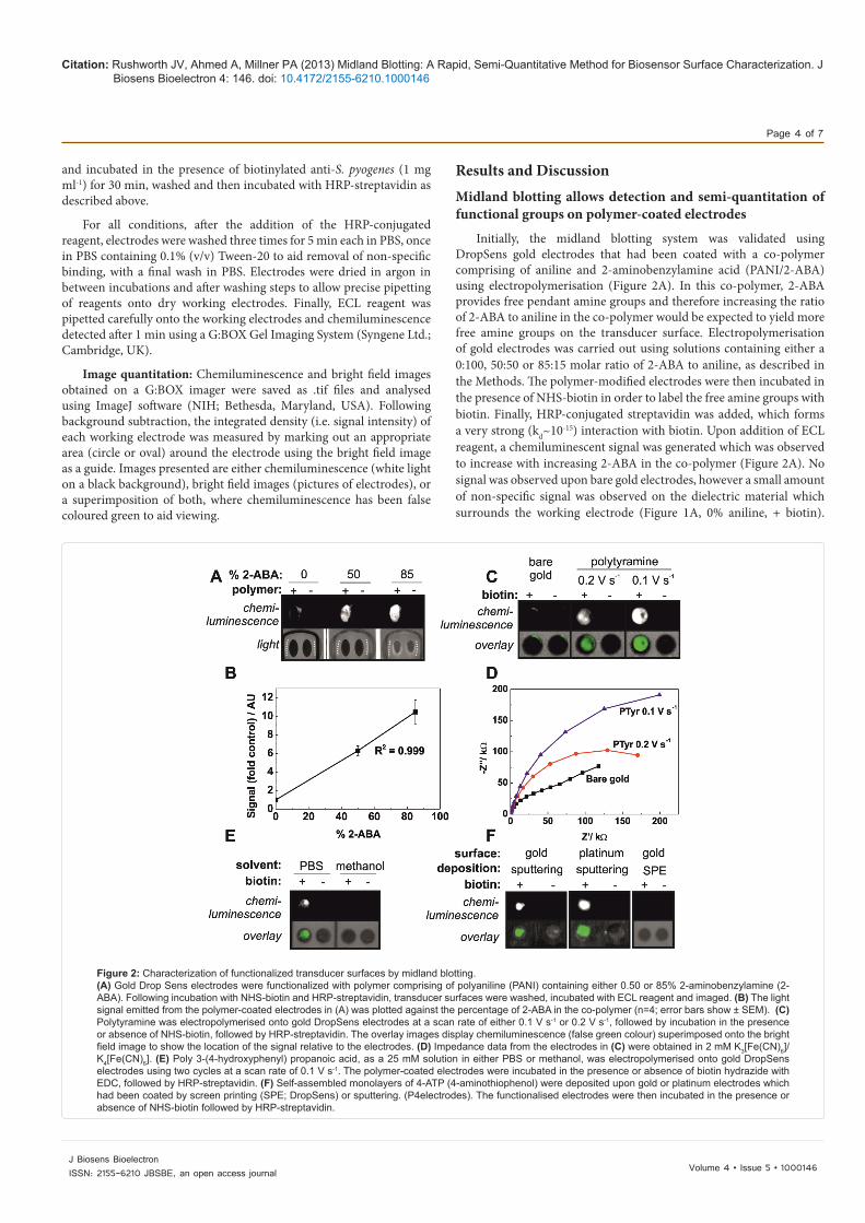
Initially, the midland blotting system was validated using DropSens gold electrodes that had been coated with a co-polymer comprising of aniline and 2-aminobenzylamine acid (PANI/2-ABA) using electropolymerisation (Figure 2A). In this co-polymer, 2-ABA provides free pendant amine groups and therefore increasing the ratio of 2-ABA to aniline in the co-polymer would be expected to yield more free amine groups on the transducer surface. Electropolymerisation of gold electrodes was carried out using solutions containing either a 0:100, 50:50 or 85:15 molar ratio of 2-ABA to aniline, as described in the Methods. The polymer-modified electrodes were then incubated in the presence of NHS-biotin in order to label the free amine groups with biotin. Finally, HRP-conjugated streptavidin was added, which forms a very strong (kd~10-15) interaction with biotin. Upon addition of ECL reagent, a chemiluminescent signal was generated which was observed to increase with increasing 2-ABA in the co-polymer (Figure 2A). No signal was observed upon bare gold electrodes, however a small amount of non-specific signal was observed on the dielectric material which surrounds the working electrode (Figure 1A, 0% aniline, + biotin).
and incubated in the presence of biotinylated anti-S. pyogenes (1 mg ml-1) for 30 min, washed and then incubated with HRP-streptavidin as described above.
For all conditions, after the addition of the HRP-conjugated reagent, electrodes were washed three times for 5 min each in PBS, once in PBS containing 0.1% (v/v) Tween-20 to aid removal of non-specific binding, with a final wash in PBS. Electrodes were dried in argon in between incubations and after washing steps to allow precise pipetting of reagents onto dry working electrodes. Finally, ECL reagent was pipetted carefully onto the working electrodes and chemiluminescence detected after 1 min using a G:BOX Gel Imaging System (Syngene Ltd.; Cambridge, UK).
Image quantitation: Chemiluminescence and bright field images obtained on a G:BOX imager were saved as .tif files and analysed using ImageJ software (NIH; Bethesda, Maryland, USA). Following background subtraction, the integrated density (i.e. signal intensity) of each working electrode was measured by marking out an appropriate area (circle or oval) around the electrode using the bright field image as a guide. Images presented are either chemiluminescence (white light on a black background), bright field images (pictures of electrodes), or a superimposition of both, where chemiluminescence has been false coloured green to aid viewing.
Figure 2: Characterization of functionalized transducer surfaces by midland blotting.(A) Gold Drop Sens electrodes were functionalized with polymer comprising of polyaniline (PANI) containing either 0.50 or 85% 2-aminobenzylamine (2-ABA). Following incubation with NHS-biotin and HRP-streptavidin, transducer surfaces were washed, incubated with ECL reagent and imaged. (B) The light signal emitted from the polymer-coated electrodes in (A) was plotted against the percentage of 2-ABA in the co-polymer (n=4; error bars show ± SEM). (C) Polytyramine was electropolymerised onto gold DropSens electrodes at a scan rate of either 0.1 V s-1 or 0.2 V s-1, followed by incubation in the presence or absence of NHS-biotin, followed by HRP-streptavidin. The overlay images display chemiluminescence (false green colour) superimposed onto the bright field image to show the location of the signal relative to the electrodes. (D) Impedance data from the electrodes in (C) were obtained in 2 mM K3[Fe(CN)6]/K4[Fe(CN)6]. (E) Poly 3-(4-hydroxyphenyl) propanoic acid, as a 25 mM solution in either PBS or methanol, was electropolymerised onto gold DropSens electrodes using two cycles at a scan rate of 0.1 V s-1. The polymer-coated electrodes were incubated in the presence or absence of biotin hydrazide with EDC, followed by HRP-streptavidin. (F) Self-assembled monolayers of 4-ATP (4-aminothiophenol) were deposited upon gold or platinum electrodes which had been coated by screen printing (SPE; DropSens) or sputtering. (P4electrodes). The functionalised electrodes were then incubated in the presence or absence of NHS-biotin followed by HRP-streptavidin.

Citation: Rushworth JV, Ahmed A, Millner PA (2013) Midland Blotting: A Rapid, Semi-Quantitative Method for Biosensor Surface Characterization. J Biosens Bioelectron 4: 146. doi: 10.4172/2155-6210.1000146
Page 5 of 7
Volume 4 • Issue 5 • 1000146J Biosens BioelectronISSN: 2155-6210 JBSBE, an open access journal
Quantitation of the light signal using ImageJ software (Figure 2B) revealed a positive correlation (R2=0.999) between chemiluminescence and the ratio of 2-ABA:aniline. These data indicate that midland blotting provides reliable, semi-quantitative information about the number of available functional groups on the transducer surface, which can give useful information about the suitability of polymer composition.
Next, we investigated whether the chemiluminescent signal obtained by midland blotting can be used to optimise electropolymerisation steps. DropSens electrodes were coated in polytyramine (presenting NH2 groups) using a scan rate of either 0.1 V s-1 or 0.2 V s-1, prior to incubation with NHS-biotin and midland blotting with HRP-streptavidin and ECL reagent (Figure 2C). Polytyramine deposition is known to be tuned by altering the electropolymerisation conditions [11]. The overlay images show that a small amount of non-specific binding was observed around the dielectric edge of the working electrodes (bare gold). The intensity of the chemiluminescent signal obtained from the polytyramine-coated electrodes was seen to be inversely proportional to the scan speed employed during electropolymerisation. In order to correlate this information with impedance data, EIS measurements of the three electrodes (bare gold, polytyramine at 0.1 V s-1 and polytyramine at 0.2 V s-1) were obtained in ferricyanide/ferrocyanide (2 mM in a 1:1 ratio in PBS) (Figure 2D). The Nyquist plots revealed that the increase in impedance upon electropolymerisation at 0.1 V s-1 was greater that at 0.2 V s-1, indicating increased deposition of polymer upon the electrode surface. Thus, the midland blotting data were corroborated by the impedance data.
Both PANI/2-ABA and polytyramine present amine groups for bioreceptor tethering. To validate the midland blotting technique using another functional group (COOH), gold DropSens electrodes were coated in poly 3-(4-hydroxyphenyl) propionic acid by electropolymerisation. The buffer/dopant solution in which monomers are dissolved is crucial to the electrodeposition of polymer due to the dependence of physico-chemical properties of the resulting polymer upon specific counter-ions [20,21]. Therefore, we tested two different solvents; PBS (10 mM sodium phosphate buffer containing 0.9% (w/v) NaCl, pH 7) and methanol. Following electropolymerisation, electrode surfaces were incubated in the presence or absence of a solution of biotin hydrazide containing EDC, in order to conjugate the carboxylic acid groups to biotin. Midland blotting was carried out using HRP-streptavidin and ECL reagent (Figure 2E).
The 3-(4-hydroxyphenyl) propionic acid dissolved in PBS generated a chemiluminescent signal, indicating the presence of carboxyl groups on the electrode surface. However, no signal was obtained when electropolymerisation was conducted in methanol. These data were also corroborated by impedance scans performed as described in Figure 2D, which revealed that the methanol-derived polymer displayed an impedance curve similar to that of bare gold, whilst the PBS-derived polymer showed increased impedance with a typical, semi-circular Nyquist plot indicative of polymer deposition (data not shown). These data indicate that midland blotting can be used to detect available carboxyl groups upon functionalised transducer surfaces, and suggest that midland blotting may be used in the optimisation of monomer solutions and counter-ions used in electropolymerisation.
Midland blotting allows detection of functional groups on SAM-coated electrodes
Besides electropolymerisation, another common method of transducer surface functionalisation is the deposition of Self-Assembled Monolayers (SAM) to which bioreceptors can be tethered. In order
to determine whether the midland blotting technique was suitable for the analysis of SAM-based transducers, SAMs were constructed using 4-aminothiophenol (4-ATP), whereby the thiol group of 4-ATP adsorbs directly to the sensor surface and the amine group is available for bioconjugation [1,7]. Gold and platinum surfaces are known to be suitable for 4-ATP SAM construction, although surface roughness is known to impede SAM assembly and packing due to the resulting poor continuity of the SAM. To this end, we prepared 4-ATP SAMs upon sputtered gold and platinum electrodes (sputtering can generate very flat surfaces) and screen-printed gold electrodes (known to be rough on the nano-scale) [22]. The SAM-coated electrodes were incubated in the presence or absence of NHS-biotin and midland blotting was carried out using HRP-streptavidin and ECL reagent (Figure 2F). A strong chemiluminescent signal was obtained for the sputtered platinum and gold-based SAMs, although the signal was higher on a platinum electrode, but no signal was observed for SAM deposition upon the screen-printed gold electrode. These data tie in with our observation that DropSens gold screen-printed electrodes are rough and heterogeneous on the micro-scale, containing pores in the gold layer, whereas the sputtered “P4” gold and platinum surfaces are flat and continuous on the nano-scale (scanning electron microscopy data, images not shown). Therefore, the rougher electrodes are not able to support the close inter-molecular packing and continuity required for stable SAM assembly. These data indicate that midland blotting could provide a simple and rapid screening tool to investigate and optimise surfaces for SAM deposition.
Midland blotting allows detection of bioreceptors at the sensor surface
The tethering of bioreceptors is a crucial step in allowing the specific and sensitive detection of analyte. It was examined, therefore, whether midland blotting could facilitate the detection of bioreceptors upon a biosensor surface. Biosensors to detect S. pyogenes bacteria were constructed by tethering biotinylated full antibodies (rabbit anti-S. pyogenes or sheep anti-digoxin) to polytyramine-functionalised gold electrodes via a biotin-Neutravidin attachment. In order to probe for the presence of antibodies, the sensors were incubated in the presence of HRP-conjugated secondary antibodies and then exposed to ECL reagent (Figure 3A). A chemiluminescent signal was observed when electrodes presenting rabbit anti-S. pyogenes antibodies were exposed to HRP-anti rabbit secondary antibody, and when electrodes presenting sheep anti-digoxin antibodies were incubated with HRP-anti sheep antibody. The control experiments (Panels 1, 3 and 4) confirmed that signal was not observed in the absence of antibody, nor when the secondary antibody applied was against a different host. These data confirm that HRP-conjugated secondary antibodies can be used for midland blotting of bioreceptors attached to biosensor surfaces. This technique could be extended easily to detect other bioreceptors, such as enzymes and peptides, by using a primary antibody against the target in combination with the appropriate HRP-conjugated secondary antibody. Equally, nucleic acids could be detected using an HRP-conjugated complementary probe sequence.
Midland blotting data align with sensor readout data to explain biosensor performance
Intra and inter-batch variability of electrodes can present a major challenge to achieving a reproducible biosensor system. Here, we explored midland blotting as a post-sensor readout tool as an attempt to explain anomalous data. A batch of biosensors against S. pyogenes was constructed by tethering half-antibodies against S. pyogenes, generated

Citation: Rushworth JV, Ahmed A, Millner PA (2013) Midland Blotting: A Rapid, Semi-Quantitative Method for Biosensor Surface Characterization. J Biosens Bioelectron 4: 146. doi: 10.4172/2155-6210.1000146
Page 6 of 7
Volume 4 • Issue 5 • 1000146J Biosens BioelectronISSN: 2155-6210 JBSBE, an open access journal
by TCEP-mediated reductive cleavage, to polytyramine-coated gold DropSens electrodes. The sensors were exposed to successive, increasing concentrations of bacteria (102-107 c.f.u. per ml) for 20 min and, after each exposure, EIS measurements obtained in ferricyanide/ferrocyanide (2 mM in a 1:1 ratio in PBS). The change in Rct between the biosensor prior to, and after, incubation with S. pyogenes (107 c.f.u. per ml) was calculated for each sensor. The data for two such sensors is presented in Figure 3B. Sensor 1 displayed a 67.3% increase in Rct following exposure to S. pyogenes, with Nyquist plot data showing increasing impedance with analyte concentration (data not shown), in line with other sensors from the same batch. In contrast, the Nyquist plot obtained for Sensor 2 revealed no increase in impedance upon exposure to S. pyogenes (data not shown) and a 29.6% decrease in Rct was observed. These data suggest that, due to aberrant sensor construction, Sensor 2 did not perform as expected.
In order to explore this further, Sensor 1 and Sensor 2 were subjected to midland blotting for the presence of bound S. pyogenes analyte. The sensors were incubated in the presence or absence of biotinylated anti-S. pyogenes full antibody, followed by HRP-streptavidin and ECL reagent (Figure 3C). Sensor 1 gave a strong chemiluminescence signal across the entire electrode, indicating the presence of bound S. pyogenes on the sensor surface. Chemiluminescence was also observed on Sensor 2, however the signal was localised to the dielectric material surrounding the electrode rather than upon the electrode itself, indicating that this was non-specific binding of the biotinylated anti-S. pyogenes antibody or the HRP-streptavidin to the dielectric material, which displayed high
non-specific affinity towards various analytes. These data indicate that S. pyogenes failed to bind to Sensor 2, which explains why the impedance data for Sensor 2 were anomalous compared with the rest of the batch.
ConclusionsMidland blotting addresses the need for a rapid, cost-effective
method of optimising key biosensor surface parameters such as the selection of electrode material, transducer surface design and deposition, bioreceptor tethering and analyte binding. The protocol for midland blotting can be completed within one or two hours, on as many samples as desired, and provides much more information than current methods of sensor characterisation such as electron microscopy or impedance curves. Furthermore, midland blotting data can be combined with sensor readout data to obtain a detailed picture of biosensor construction and performance. Although we have focussed here upon electrochemical immunosensors, the midland blotting technique is readily portable to all biosensor systems. Here, we have utilised chemiluminescence as the detection method, but other methods such as fluorescence could also be employed.
Acknowledgements
A.A. was funded by a Fully-Funded International Research Scholarship (FIRS), University of Leeds. We are grateful to John Wright for supplying heat-inactivated Streptococcus pyogenes. We thank Dr. Vas Ponnambalam for the kind gift of HRP-streptavidin and Gareth Fearnley for helpful advice on using the G:BOX imager. We also thank Dr. Tim Gibson for helpful discussions.
References
1. Billah MM, Hodges CS, Hays HC, Millner PA (2010) Directed immobilization of reduced antibody fragments onto a novel SAM on gold for myoglobin impedance immunosensing. Bioelectrochemistry 80: 49-54.
2. Caygill RL, Hodges CS, Holmes JL, Higson SP, Blair GE, et al. (2012) Novel impedimetric immunosensor for the detection and quantitation of Adenovirus using reduced antibody fragments immobilized onto a conducting copolymer surface. Biosens Bioelectron 32: 104-110.
3. Liu YT, Deng J, Xiao XL, Ding L, Yuan YL, et al. (2011) Electrochemical sensor based on a poly(para-aminobenzoic acid) film modified glassy carbon electrode for the determination of melamine in milk. Electrochim Acta 56: 4595-4602.
4. Millner PA, Hays HC, Vakurov A, Pchelintsev NA, Billah MM, et al. (2009) Nanostructured transducer surfaces for electrochemical biosensor construction-interfacing the sensing component with the electrode. Seminars in cell & developmental biology 20: 34-40.
5. Yang H (2012) Enzyme-based ultrasensitive electrochemical biosensors. Current Opinion in Chemical Biology 16: 422-428.
6. Chang BY, Park SM (2010) Electrochemical impedance spectroscopy. Annu Rev Anal Chem (Palo Alto Calif) 3: 207-229.
7. Conroy DJ, Millner PA, Stewart DI, Pollmann K (2010) Biosensing for the environment and defence: aqueous uranyl detection using bacterial surface layer proteins. Sensors (Basel) 10: 4739-4755.
8. Lindholm-Sethson B, Nystrom J, Malmsten M, Ringstad L, Nelson A (2010) Electrochemical impedance spectroscopy in label-free biosensor applications: multivariate data analysis for an objective interpretation. Anal Bioanal Chem 398: 2341-2349.
9. Millner PA, Caygill RL, Conroy DJR, Shahidan MA (2012) Impedance interrogated affinity biosensors for medical applications: novel targets and mechanistic studies. In: Biosensors for medical applications, Higson S (Ed),. Woodhead Publishing Ltd., Cambridge, UK.
10. Barton AC, Collyer SD, Davis F, Garifallou GZ, Tsekenis G, et al. (2009) Labeless AC impedimetric antibody-based sensors with pgml (-1) sensitivities for point-of-care biomedical applications. Biosens Bioelectron 24:1090-1095.
11. Pournaras AV, Koraki T, Prodromidis MI (2008) Development of an impedimetric immunosensor based on electropolymerized polytyramine films for the direct detection of Salmonella typhimurium in pure cultures of type strains and inoculated real samples. Anal Chim Acta 624: 301-307.
Figure 3: Characterization of biosensors by midland blotting.(A) Biosensors for the detection of Streptococcus pyogenes (S. pyogenes) were constructed on polytyramine-coated gold DropSens electrodes by tethering biotinylated S. pyogenes (specific) or anti-digoxin (dig; non-specific) antibodies to the polymer layer via NHS-biotin and Neutravidin. The sensors were incubated in the presence of HRP-conjugated secondary antibodies; either anti-rabbit (rb; against anti-S. pyogenes) or anti-sheep (sh; against anti-digoxin), prior to the addition of ECL and imaging. (B) Biosensors were constructed on polytyramine-coated gold DropSens electrodes by tethering anti-S. pyogenes half-antibodies to the polymer layer via sulfo-SMCC. Impedance data were recorded for two sensors from the same batch, following incubation with S. pyogenes (successive incubations from 102-107 c.f.u. per ml). The change in Rct measured upon incubation in 107 c.f.u. per ml of S. pyogenes, compared to biosensor alone, was plotted for both sensors. (C) The sensors that were interrogated electrochemically in (B) were subjected to midland blotting for the presence of S. pyogenes on the surface. Sensors were incubated in the presence or absence of biotinylated anti-S. pyogenes full antibody, followed by HRP-streptavidin prior to addition of ECL and imaging.

Citation: Rushworth JV, Ahmed A, Millner PA (2013) Midland Blotting: A Rapid, Semi-Quantitative Method for Biosensor Surface Characterization. J Biosens Bioelectron 4: 146. doi: 10.4172/2155-6210.1000146
Page 7 of 7
Volume 4 • Issue 5 • 1000146J Biosens BioelectronISSN: 2155-6210 JBSBE, an open access journal
12. Lawal AT, Adeloju SB (2013) Polypyrrole based amperometric andpotentiometric phosphate biosensors: A comparative study B. Biosens andBioelectronics 40: 377-384.
13. Gooding JJ, Darwish N (2012) The rise of self-assembled monolayers forfabricating electrochemical biosensors—an interfacial perspective. TheChemical Record 12: 92-105.
14. Billah M, Hays HCW, Millner PA (2008) Development of a myoglobin impedimetric immunosensor based on mixed self-assembled monolayer onto gold. Microchim Acta 160: 447-454.
15. Deluca JL, Hickey DP, Bamper DA, Glatzhofer DT, Johnson MB, et al. (2013) Layer-by-Layer Assembly of Ferrocene-Modified Linear Polyethylenimine Redox Polymer Films. Chemphyschem : a European journal of chemical physics and physical chemistry 14, 2149-2158.
16. Dhayal M, Ratner DM (2009) XPS and SPR analysis of glycoarray surface density. Langmuir : the ACS journal of surfaces and colloids 25:2181-2187.
17. Kadara RO, Jenkinson N, Banks CE (2009) Characterisation of commercially
available electrochemical sensing platforms. Sensor Actuat B-Chem 138: 556-562.
18. Burnette WN (1981) “Western Blotting” : Electrophoretic transfer of proteins from sodium dodecyl sulfate-polyacrylamide gels to unmodified nitrocellulose and radiographic detection with antibody and radioiodinated protein A.Analytical Biochemistry 112: 195-203.
19. Grant S, Davis F, Pritchard JA, Law KA, Higson SPJ, et al. (2003) Labeless and reversible immunosensor assay based upon an electrochemical current-transient protocol. Analytica Chimica Acta 495: 21-32.
20. Catedral MD, Tapia KG, Sarmago RV (2004) Effect of dopant ions on theelectrical conductivity and microstructure of polyaniline (emeraldine salt).Science Diliman 16: 41-46.
21. Duic L., Mandic Z (1992) Counter-ion and pH effect on the electrochemical synthesis of polyaniline. Journal of Electroanalytical Chemistry 335: 207-221.
22. García-Gonzalez R, Fernandez-Abedul MT, Pernía A, Costa-García A (2008)Electrochemical characterization of different screen-printed gold electrodes.Electrochim Acta 53: 3242-3249.