journal of the association for research in otolaryngology · signals critical to the neural control...
TRANSCRIPT

Dynamic Displacement of Normal and Detached SemicircularCanal Cupula
RICHARD D. RABBITT,1,3 KATHRYN D. BRENEMAN,1 CURTIS KING,4 ANGELA M. YAMAUCHI,1RICHARD BOYLE,2 AND STEPHEN M. HIGHSTEIN
3
1Department of Bioengineering, University of Utah, 72 South Central Campus Dr., Rm. 2646, Salt Lake City, UT 84112, USA2NASA Ames Research Center, BioVIS Center, M/S 239-11, Moffett Field, CA 94035, USA3Marine Biological Laboratory, 7 MBL Street, Woods Hole, MA 02543, USA4EI Spectra, LLC, Woodinville, WA 98077, USA
Received: 26 March 2009; Accepted: 4 May 2009; Online publication: 10 June 2009
ABSTRACT
The dynamic displacement of the semicircular canalcupula and modulation of afferent nerve dischargewere measured simultaneously in response to physio-logical stimuli in vivo. The adaptation time constant(s) of normal cupulae in response to step stimuliaveraged 36 s, corresponding to a mechanical lowercorner frequency for sinusoidal stimuli of 0.0044 Hz.For stimuli equivalent to 40–200 deg/s of angularhead velocity, the displacement gain of the centralregion of the cupula averaged 53 nm per deg/s.Afferents adapted more rapidly than the cupula,demonstrating the presence of a relaxation processthat contributes significantly to the neural represen-tation of angular head motions by the dischargepatterns of canal afferent neurons. We also investigat-ed changes in time constants of the cupula andafferents following detachment of the cupula at itsapex—mechanical detachment that occurs in re-sponse to excessive transcupular endolymph pressure.Detached cupulae exhibited sharply reduced adapta-tion time constants (300 ms–3 s, n=3) and can beexplained by endolymph flowing rapidly over theapex of the cupula. Partially detached cupulaereattached and normal afferent discharge patternswere recovered 5–7 h following detachment. Thisregeneration process may have relevance to therecovery of semicircular canal function following headtrauma.
Keywords: vestibular, inner ear micromechanics,cupula regeneration, angular motion sensation,afferent response dynamics
INTRODUCTION
The semicircular canals are responsible for sensingangular motion of the head and provide afferentsignals critical to the neural control of balance,posture, and stabilization of the visual image on theretina. Biomechanics plays a key role in this process.The morphological orientation of the three canalsunderlies the ability to sense the direction of angularmotion, and the slender geometry of the membranousduct underlies temporal integration of angular accel-eration stimuli (Rabbitt et al. 2004). Each semicircularcanal consists of toroidal loop of fluid including anenlarged ampulla where the sensory epithelium resides(Fig. 1). Sensory hair cell stereocilia project from thesurface of the crista and into the gelatinous cupulawhich spans the entire cross-section of the ampulla. Itis the diaphragm-like displacement of the cupula thatgives rise to hair bundle displacements, mechanotrans-duction, and the neural response (Hillman andMcLaren 1979; Markin and Hudspeth 1995; McLarenand Hillman 1979; Oman et al. 1979; Peterson et al.1996; Rusch and Thurm 1989). A subset of semicircu-lar canal afferents modulate their discharge rate inproportion to angular velocity of the head over a broadfrequency band of physiologically relevant head move-ments, and hence, the canals are often described as
Correspondence to: Richard D. Rabbitt & Department of Bioengineer-ing & University of Utah & 72 South Central Campus Dr., Rm. 2646,Salt Lake City, UT 84112, USA. email: [email protected]
JARO 10: 497–509 (2009)DOI: 10.1007/s10162-009-0174-yD 2009 The Author(s). This article is published with open access at Springerlink.com
497
JAROJournal of the Association for Research in Otolaryngology
angular velocity sensors (Wilson and Jones 1979). Froma mechanical perspective, angular velocity sensitivityarises because the viscous movement of endolymphwithin the slender duct causes a temporal integrationof the angular acceleration stimulus, which in turngenerates fluid displacements that reflect angularvelocity of the head (Lorenté de Nó 1927; Oman etal. 1987; Rabbitt et al. 2004; Steinhausen 1933). Thismechanical temporal integration is important becauseit provides the brain with a broadband angular headvelocity signal that feeds into movement control neuralsystems including the vestibulo-ocular reflex. Theprimary role of the cupula is to faithfully convert fluiddisplacement into displacements of the mechanosensi-
tive hair bundles. This is achieved, in part, through theelasticity of the cupula. As consequence of elasticity,the discharge modulation of velocity-sensitive canalafferent neurons is diminished and the phase is morein line with angular acceleration for angular motionstimuli below the “lower corner frequency.” Theoreticalconsiderations describe the lower corner as the frequencywhere the elastic restoring force of the cupula exactlybalances the viscous drag of the fluid in the slenderendolymphatic canal. The mechanical lower cornerfrequency therefore reflects the status and elasticity ofthe cupula as well as the viscous drag in the duct.
Here, we directly measured the adaptation timeconstant and lower corner frequency of semicircular
Apex
Apex Apex
Crista
D
E F
C
.
Crista
CanalU
U
Canal
ROI
ROI
ROI
Cupula
Afferent
Recording
Utricle
Ante
rior
Canal
Ape
x
Cupula
Beads
B
A
Horizontal Canal
Indent Stim.
Endolymph
Disp.
Late
ral
Caudal
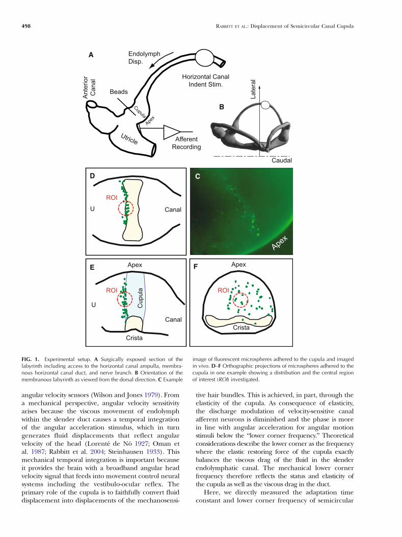
FIG. 1. Experimental setup. A Surgically exposed section of thelabyrinth including access to the horizontal canal ampulla, membra-nous horizontal canal duct, and nerve branch. B Orientation of themembranous labyrinth as viewed from the dorsal direction. C Example
image of fluorescent microspheres adhered to the cupula and imagedin vivo. D–F Orthographic projections of microspheres adhered to thecupula in one example showing a distribution and the central regionof interest (ROI) investigated.
498 RABBITT ET AL.: Displacement of Semicircular Canal Cupula

canal cupulae to physiological stimuli in the livinganimal while simultaneously recording individualafferent responses. Responses were measured bothin normal animals and in animals with damagedcupulae (detached at the apex). The oyster toadfish,Opsanus tau, was used as the experimental modelbecause of the similarity in dimensions to the humanlabyrinth and to facilitate the experimental approach.Results reveal the mechanical lower corner frequencyto align with the lowest corner frequency of thepopulation of velocity-sensitive afferent neurons. In asubset of experiments, we examined the motion ofcupula that had become detached from the apex ofthe ampulla. It has been reported previously thatdetachment at the apex acts like a relief valve andoccurs under high transcupular pressures that mayarise during trauma (Hillman 1974; Rabbitt et al.1999). The present work examined temporalresponses of detached cupulae and correspondingafferents as well as the time course of reattachmentand recovery of normal afferent responses.
METHODS
Animal preparation. Adult oyster toadfish (O. tau) wereobtained from the Marine Biological Laboratory(Woods Hole, MA, USA) and all experiments werecarried out under protocols approved at the MarineBiological Laboratory or the University of Utah. Thesurgical approach followed that described previously(Rabbitt et al. 2005). Briefly, each fish was anesthetizedwith MS222 (5 mg/L in seawater) and partially immo-bilized by an intramuscular injection of pancuroniumbromide (0.05 mg/kg; Sigma, St. Louis, MO, USA) inthe tail. The fish was secured in an acrylic tank, with twothirds of its body immersed in bubbled seawater, whilethe remainder of the body was covered with moisttissues. A craniotomy was made lateral to the dorsalcourse of the anterior canal to expose the horizontalcanal ampulla and ∼8 mm of the slender membranouslabyrinth (Fig. 1B). The orientation of the labyrinthviewed from the dorsal direction (microscope objectiveaxis) is illustrated in Figure 1A (Ghanem et al. 1998).Perilymph within the upper region of the surgicalcavity was replaced with fluorocarbon (FC-75, 3 M,Minneapolis, MN, USA) to improve optical clarity andelectrosurgical cutting outcomes. The electricalinsulating properties of fluorocarbon allowed us tocut a fistula in the horizontal canal ampulla ∼300 μmmedial to the cupula using an electrosurgicalgenerator (Valleylab, Denver, CO, USA) set to a ∼5-Wcutting waveform. A sharpened 76-μm dia. tungstenwire served as the cutting electrode. This generated∼50- to 100-μm diameter hole in the membranous wallallowing access for delivery of fluorescent microspheres
into the endolymph (Fig. 1B, beads). The microsphereswere allowed to diffuse from the fistula to the cupula. Itis important to note that fluorocarbon is immiscible withendolymph, and as a result, there was a surface tensionbarrier preventing flow of endolymph out of the fistula(Rajguru and Rabbitt 2007). Canal afferent responses tocontrolled stimuli were recorded after generation of thefistula and compared to control recordings to confirmnormal sensory transduction and neural coding,indicating that the dynamics are indeed dominated bythe long and slender canal segment.
Although great effort was taken to prevent damageto the cupula during this procedure, in a subset ofanimals, we found afferent modulation to be reduced,and in some cases, we observed a fraction of thefluorescent microspheres to diffuse over the apex ofthe cupula and into the lateral side of the ampulla.These animals defined the “dislodged cupula” group.Even in these damaged cases, the cupula remainedstructurally sound, but simply detached from theinterior surface of the membranous ampulla at itsapex. Present results address cupula responses andreattachment in modestly damaged cases where thecupula was dislodged at the apex but remainedlocated in the normal central region of the ampulla.
Mechanical stimulus. Mechanical indentation of theslender limb of the membranous duct was used as theprimary stimulus. This idea was first introduced byEwald in 1892 (Camis 1930) and later refined byDickman et al. to mimic physiological head movementsand produce nearly equivalent afferent dischargepatterns (Dickman and Correia 1989; Dickman et al.1988). We used a version of the approach detailed byRabbitt et al. (1995) where 1 μm of mechanicalindentation of the horizontal canal limb generatesmechanical cupula motion and afferent responsesnearly equivalent to ∼4 deg/s of angular head velocitystimulus in the present experimental organism. In asubset of experiments, the whole animal was rotatedusing sinusoidal angular velocity stimuli (Boyle andHighstein 1990). Cupula displacements were recordedfor mechanical indentation stimuli, but not for angularvelocity stimuli.
Neural recordings. Horizontal canal neural recordingsused glass microelectrodes (∼100 MΩ) following theapproach described previously (Boyle and Highstein1990; Rabbitt et al. 2005). Electrodes were positionedusing a micromanipulator at a location ∼500 μm fromthe horizontal canal ampulla where the nerve branch isaccessible (Fig. 1B). Extracellular potentials weremeasured using standard amplification (EXT-02F, npielectronic, Tamm, Germany). Afferent responses and
RABBITT ET AL.: Displacement of Semicircular Canal Cupula 499
mechanical stimuli were amplified, filtered at 2 kHz(LHBF-48X, npi electronic), and sampled at 5 kHz(ITC-18, HEKA Instruments, Inc., Bellmore, NY, USA).
Microspheres. Carboxylate-modified, neutrally buoyantfluorescentmicrospheres (beads) were obtained (BangsLabs, Inc. Fishers, IN, USA) and surface-modified tobind wheat germ agglutinin (WGA). Raw beads insuspension were washed twice in MES buffer with10 mg EDAC and incubated for 15 min. Followingincubation, the beads were washed and resuspended in0.1 M borate buffer (pH 8.5) with 1 mg WGA (Sigma-Aldrich) and incubated with gentle mixing for 4 h. Thebeads were then washed and resuspended in 0.1 Mborate buffer with bovine serum albumin (BSA;10 mg/mL) and mixed continuously for 15 min priorto a final wash and resuspension inMES buffer with BSA(10 mg/mL) for storage. Microspheres were placed intoadfish artificial endolymph (Ghanem et al. 2008),vortex-mixed, and loaded into a glass pipette pulled andcut to ∼50-μm tip diameter. The fluid level in thepipette was adjusted to just exceed the capillary action ofthe glass. The filled pipette was then lowered throughthe fluorocarbon until the tip contacted the endolymphthrough the fistula in the ampulla membrane. Contactwith endolymph caused the surface tension between theendolymph and the fluorocarbon to be broken andplaced the interior of the pipette in communicationwith the endolymph. Microspheres were allowed todiffuse out of the pipette and into the ampulla. Overtime, some of the beads migrated to the cupula andadhered to its surface. The microsphere loading pipettewas removed prior to collecting any data.
Fluorescent microsphere tracking. Images were collectedusing an upright microscope (Axioskop Tech, CarlZeiss, Germany) configured for epifluorescence andplaced on a vibration isolation table (TMC, Peabody,MA, USA). Long working distance air 5×, 10×, 20×objectives (Plan Apo, Mitutyo, Japan) were used toview the complete ampulla, fluorescent microbeads,and the neural recording electrode. A CCD camera(Retiga-EXi, QImaging, Surrey, BC, Canada) was usedto collect fluorescent images at a frame rate of∼100 ms and exposure time of ∼50 ms. Shuttertimes were sampled and recorded. Custom softwarewas written (Igor Pro, Wave Metrics, Lake Oswego,OR, USA) to control the stimuli (NI GPIB, NationalInstruments, Austin, TX, USA), trigger the camera,and to collect the neural data, image data, and stimulivia computer (Apple G4, Cupertino, CA, USA; ITC-18,HEKA Instruments, Inc.). Images (1392×1040) werecollected over two to ten cycles of periodicmechanical indentation stimuli with shutter times
random relative to the stimulus period. Images weretime-stamped relative to the stimulus onset triggerover multiple applications of the stimulus andsubsequently combined. Since image sampling wasrandom relative to the periodic stimulus, datacollected over several cycles provided temporalresolution of ∼50 ms with respect to the stimulustiming trigger.
Figure 1C shows an example image of fluorescentmicrospheres adhered to the cupula, imaged in theliving animal. It was straightforward to estimate thethree-dimensional locations of the beads by adjustingthe “Z”-axis focus and manually digitizing the cent-roids of the microspheres from still images. Anexample showing the distribution of fluorescentmicrospheres in one animal is provided as ortho-graphic projections in Figure 1D–F. Specific locationsand numbers of microspheres adhered to the cupulavaried substantially between individual animals. Typi-cally, ten to 50 microspheres adhered to the cupula,with five to 20 located on the central region of interest(ROI). Results reported in the present study arelimited to motion of beads in the ROI located nearthe center of the cupula (Fig. 1, dotted circular), onthe central pillar overlaying the center region of thecrista (Silver et al. 1998).
Motion of the cupula was tracked by focusing on asubset of fluorescent microspheres located in the ROIand collecting a sequence of ∼300 images whilepresenting multiple applications of the mechanicalindentation stimulus. Since the animal was alive, therewere always slight whole organ movements caused byrespiration, heartbeat, or random muscle contrac-tions. To remove these movement artifacts, we man-ually selected image registration ROIs at the apex ofthe ampulla and at the crista, being careful that theregistration ROIs did not include any microspheresadhered to the surface of the cupula. The first imagein the sequence was used as the reference configura-tion. The registration ROIs of all subsequent imageswere then aligned with the reference image using theapproach of (Thévenaz and Unser 1998), as imple-mented by Wave Metrics (Igor Pro 6, Lake Oswego,OR, USA). All images in the sequence were translatedand rotated to align the registration ROIs with thereference configuration. After registration of all images,the same approach of Thévanaz and Unser was appliedto track motion of the microspheres 20×20 pixel squareROI around each bead intensity centroid. Beads withinthe ROI were selected manually and the intensitycentroid was found using custom software (Igor Pro 6,Wave Metrics). Analysis of microspheres adhered to themembranous labyrinth (ampulla) resulted in motionsG200 nm, thus suggesting a noise floor of G200 nm—avalue that could be improved by averaging overmultiplestimulus presentations if desired. The present study
500 RABBITT ET AL.: Displacement of Semicircular Canal Cupula

used stimuli producing motions an order of magnitudeabove this noise level, and therefore, averaging was notrequired. To track cupula motion, five to ten micro-spheres within the central ROI were selected andtracked individually. The procedure resulted in micro-sphere displacements in the “x–y” coordinate frame ofthe CCD array. These data were combined to determinethe component of motion perpendicular to the surfaceof the cupula, with the surface tangent identified by thegroup of fluorescent beads.
RESULTS
Sinusoidal mechanical indentation of the canal ductproduced afferent responses consistent with previousreports in this species (Rabbitt et al. 1995). Figure 2provides example simultaneous recordings of single-unit afferent discharge (A) and cupula displacement(B) in response to ∼20-μm mechanical indentation ofthe canal duct at 0.3 Hz. As expected, cupuladisplacement was in phase with mechanical indenta-tion, and since indentation mimics head rotation(Dickman and Correia 1989), cupula displacementwould be in phase with angular velocity of the head.The magnitude was ∼4 μm in this animal whichcorresponds to a cupular gain in the central ROI of∼50 nm per deg/s of angular head velocity. Theaverage cupula gain was 53 nm per deg/s (n=9;13 nm SE; 4 deg/s rotation∼1 μm indent), consistentwith the previous report by (McLaren and Hillman1979) of ∼35 nm per deg/s. In contrast to thisprevious work, we found displacement of the cupulato be in phase with the mechanical stimulus andangular head velocity, at least at frequencies above thelower corner where many afferent neurons encodeangular head velocity (e.g., Fig. 2B, C). For indenta-tion stimuli 10–24 μm in amplitude, harmonicdistortion of the cupula displacement was less than15% and the first harmonic was nearly linear, withstimulus amplitude yielding Pearson’s R=0.86. Asubset of afferents exhibited a much higher degreeof harmonic distortion due primarily to an excitatory–inhibitory asymmetry in afferent discharge responsedynamics. Four additional example afferent responsesto the 0.3-Hz stimulus are shown in the bottom panels(Fig. 2D). Angular velocity (vel.) encoding unitsresponded with a maximum discharge rate in phasewith the peak displacement of the cupula. Theresponse gain of these units was relatively insensitiveto stimulus frequency. A subset of afferent unitsresponded with a phase advance and increased gainwith frequency—a response that encodes angularacceleration (accel.) of the head. These acceleration-sensitive units are not present in all species. In thetoadfish, convergence of both excitatory and inhibi-
tory transmitters appears to underlie a mathematicaldifferentiation of cupula displacement to arrive atacceleration-sensitive afferent discharge patterns(Holstein et al. 2004).
In order to investigate dynamics of cupula displace-ment, we used step indentation of the canal duct. Stepindentation evoked a rapid displacement of thecupula toward the utricle followed by a slow recovery(adaptation) to its resting position. Figure 3 showssimultaneous recordings of horizontal canal afferentdischarge patterns (A, D, G), and displacements offluorescent microbeads attached to the cupula (B, E,H), in response to square wave (C, F, I) mechanicalindentation of the canal duct (three example ani-mals). During excitatory step stimuli, afferents quicklyincreased their discharge rate followed by a period of
4 s
120
40
Indent
Cu
pu
laS
pk/s
4 µ
m2
0 µ
m
F091808.13
A
B
C
120
0
80
0
160
110
90
70S
pk/s
DVel. LG
Accel.Vel. HG
FIG. 2. Cupula displacement and afferent responses to 0.3-Hzsinusoidal stimuli in normal animals. Simultaneous recordings ofafferent responses (A) and cupula displacement (B) in response tosinusoidal stimulus (C) at 0.3 Hz. D Additional afferents recorded inresponse to the same stimulus showed a range of gains and relativephases of peak response. Since 0.3 Hz is above the mechanicallower corner frequency, a subset of afferents responded in phasewith, and in proportion to, cupula displacement (velocity-sensitive),while other afferents responded with a phase advance relative tocupula displacement (e.g., acceleration-sensitive).
RABBITT ET AL.: Displacement of Semicircular Canal Cupula 501

recovery back to their background discharge. Slowlyadapting afferents encode angular velocity over abroad frequency band of sinusoidal stimuli and areanalogous to the regularly discharging units in
mammals (Fernández and Goldberg 1971; Goldbergand Fernandez 1971; Rabbitt et al. 2005). The mostrapidly adapting afferents encode angular accelera-tion of the head, also over a broadband. For stepstimuli, some afferents recovered following a singleexponential, while others recovered following a fastadaptation, τf, followed by a slow adaptation, τs. Thefast component of afferent adaptation, if presentduring excitatory stimuli, was absent or greatlyreduced during inhibitory stimuli. This asymmetrywas not obvious in the motion of the cupula (B, E, H),which deflected with nearly equal time courses forboth excitatory and inhibitory stimuli for the magni-tudes of stimuli investigated here. For sinusoidalstimuli above the mechanical lower corner frequency(ω91/τ rad/s), the cupula deflected in proportion to,and in phase with, the stimulus (Fig. 2, peak cupuladisplacement B aligns with stimulus C).
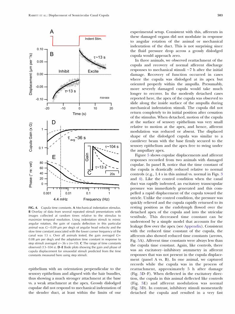
Adaptation of the cupula following step indenta-tion of the duct is more clearly demonstrated inFigure 4A where multiple stimulus cycles are overlaidand fit with exponential curves (solid curves). Thetime constant of cupula adaptation averaged t∼36 s(n=9), but varied considerably between individualanimals (13–104 s, Fig. 3C). Fluorescent microbeadswithin the central ROI of each animal moved togetherwith nearly the same time constant, thus suggestingthat the range of cupular time constants was primarilydue to interanimal variability and not spatial distribu-tion of beads. The present study did not investigatethe deflected shape of the cupula or if differences inthe time constant could be detected at differentlocations. All of the animals were of nearly the samesize, and therefore, it seems unlikely that differencesbetween mechanical time constants between animalscould be explained by morphology alone. Althoughwe cannot rule out variations in the diameter of themembranous duct as underlying interanimal variabil-ity, it seems likely that the mechanical properties ofcupulae may differ between individual animals, withrapidly adapting cases being stiffer and/or morepermeable than slowly adapting cases.
Mechanical indentation of the membranous laby-rinth is known to lead to large transcupular pressuresand, if the stimulus is excessive, can detach the cupulaat its apex (Hillman 1974; Rabbitt et al. 1999).Although we did not intend to damage the cupula,in several cases, the cupula became detached duringpreparation of the animal. Detached cupulae showeda diverse range of motions depending on the extentof damage. Afferent responses, if present, reflectedthis diversity. In the most severe detachment cases, thecupula would swing toward the utricle, leaving agaping opening for endolymph to flow over its apexfrom the canal lumen into the utricular vestibule. Thecupula always remained attached to the sensory
τ~54s
40 sec
150
0
Ind
en
tC
up
ula
Sp
k/s
4 µ
m20 µ
m
A
C
B
F011309
Ind
en
tC
upula
20
µm
160
80
10 s
Spk/s
τ~13s
F073108
D
F
E
2 µ
m
τf~7.4s
F081908
10 s
150
50
τ~34s
Ind
en
tC
up
ula
Sp
k/s
2 µ
m20 µ
m
τ~16sG
I
H
τf~2.5s
τs~10s
FIG. 3. Cupula displacement and afferent time constants in normalanimals. Simultaneous recordings of afferent responses (A, D, G) andcupulae deflections (B, E, H) in response to square wave mechanicalindentation (C, F, I) of the canal duct for three different animals.Cupula deflections in the central ROI were 3–6 μm for ductindentations of 20 μm, and cupula time constants for the units inthis figure were τ=13–54 s. Afferent response time constants wereless than cupula time constants due to adaptive processes in the haircell/afferent complex.
502 RABBITT ET AL.: Displacement of Semicircular Canal Cupula
epithelium with an orientation perpendicular to thesensory epithelium and aligned with the hair bundles,thus showing a much stronger attachment at the basevs. a weak attachment at the apex. Grossly dislodgedcupulae did not respond to mechanical indentation ofthe slender duct, at least within the limits of our
experimental setup. Consistent with this, afferents inthese damaged organs did not modulate in responseto angular rotation of the animal or mechanicalindentation of the duct. This is not surprising sincethe fluid pressure drop across a grossly dislodgedcupula would approach zero.
In three animals, we observed reattachment of thecupula and recovery of normal afferent dischargeresponses to mechanical stimuli ∼7 h after the initialdamage. Recovery of function occurred in caseswhere the cupula was dislodged at its apex butoriented properly within the ampulla. Presumably,more severely damaged cupula would take muchlonger to recover. In the modestly detached casesreported here, the apex of the cupula was observed toslide along the inside surface of the ampulla duringmechanical indentation stimuli. The cupula did notreturn completely to its initial position after cessationof the stimulus. When detached, motion of the cupulaat the surface of sensory epithelium was very smallrelative to motion at the apex, and hence, afferentmodulation was reduced or absent. The displacedshape of the dislodged cupula was similar to acantilever beam with the base firmly secured to thesensory epithelium and the apex free to swing underthe ampullary apex.
Figure 5 shows cupulae displacements and afferentresponses recorded from two animals with damagedcupulae. In panel B, notice that the time constant ofthe cupula is drastically reduced relative to normalcontrols (e.g., 1.4 s in this animal vs. normal in Figs. 3and 4). Like the control condition when the canalduct was rapidly indented, an excitatory transcupularpressure was immediately generated and this com-pelled a rapid displacement of the cupula toward theutricle. Unlike the control condition, the pressure wasquickly relieved and the cupula rapidly returned to itsresting position as the endolymph flowed over thedetached apex of the cupula and into the utricularvestibule. This decreased time constant can beunderstood by a simple model that accounts for theleakage flow over the apex (see Appendix). Consistentwith the reduced time constant of the cupula, theafferents also showed reduced time constants (arrows,Fig. 5A). Afferent time constants were always less thanthe cupula time constant. Again, like controls, therewas an excitatory–inhibitory asymmetry in afferentresponses that was not present in the cupula displace-ment (panel A vs. B). In one animal, we capturedrecords while the cupula was in the process ofreattachment, approximately 5 h after damage(Fig. 5D–F). When deflected in the excitatory direc-tion, the cupula in this animal deflected like controls(Fig. 5E) and afferent modulation was normal(Fig. 5D). In contrast, inhibitory stimuli momentarilydetached the cupula and resulted in a very fast
-0.10
-0.05
0.00
0.05
0.10
Dis
pla
ce
me
nt
(µm
pe
r °/
s)
-20 -10 0 10 20
Time (s)
F091809
Indent Stim.
τ~13 s
τ~13 s
A
B
Inhibit Excite
0.1
1
0.001 0.01 0.1 1
Frequency (Hz)
80
60
40
20
0
Phase (
° re
: vel)
Cupula
Gain
(x G
)
τavg
~36 s
4.4 mHz
D
E
τ~13 s
τ~104 s
20s
Gain
C
FIG. 4. Cupula time constants. A Mechanical indentation stimulus.B Overlay of data from several repeated stimuli presentations withimages collected at random times relative to the stimulus tomaximize temporal resolution. Using indentation stimuli to mimicangular rotation, the gain of cupula deflection in this particularanimal was G∼0.09 μm per deg/s of angular head velocity and theslow time constant associated with the lower corner frequency of thecanal was 13 s. Over all animals tested, the gain averaged G=0.08 μm per deg/s and the adaptation time constant in response tostep stimuli averaged τ∼36 s (n=10). C The range of time constantsobserved (13–104 s). D–E Bode plots showing the gain and phase ofcupula displacement for sinusoidal stimuli predicted from the timeconstants measured here using step stimuli.
RABBITT ET AL.: Displacement of Semicircular Canal Cupula 503
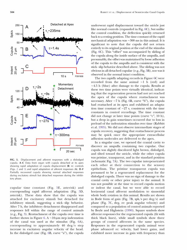
cupular time constant (Fig. 5E, asterisk) andcorresponding rapid afferent adaptation (Fig. 5D,asterisk). These data show that the cupula wasattached for excitatory stimuli but detached forinhibitory stimuli, suggesting a stick–slip behavior.After 7 h, the inhibitory detachment disappeared andresponses fell within the range of control animals(e.g., Fig. 3). Reattachment of the cupula over time isfurther shown in Figure 6. A ∼10-μm step indentationof the canal was used as the stimulus (Fig. 6A),corresponding to approximately 40 deg/s stepincrease in excitatory angular velocity of the head.In the dislodged case (Fig. 6B, curve “a”), the cupula
underwent rapid displacement toward the utricle justlike normal controls (expanded in Fig. 6C), but unlikethe control condition, the deflection quickly returnedback to a resting position. The time constant of the rapidmechanical adaptation was ∼300 ms in this animal. It issignificant to note that the cupula did not returnentirely to its original position at the end of the stimulus(Fig. 6C). This “offset” was accompanied by sliding ofthe cupula along the inside surface of the ampulla, andpresumably, the offset wasmaintained by loose adhesionof the cupula to the ampulla and is consistent with thestick–slip behavior described above. The sliding was notobvious in all detached cupulae (e.g., Fig. 5B), nor was itobserved in the normal intact condition.
The two rapidly adapting records in Figure 6C wererecorded from the same animal ∼1 h (red) and∼4.5 h (blue) after damage to the cupula. Results atthese two time points were virtually identical, indicat-ing that the regeneration process had not yet reachedthe apex of the cupula where reattachment wasnecessary. After ∼7 h (Fig. 6B, curve “b”), the cupulahad reattached at its apex and exhibited an adapta-tion time constant of ∼27 s, consistent with the timeconstants in control recordings. The time constantdid not change at later time points (curve “c”, 10 h),but a drop in gain sometimes occurred due to loss inpreload of the indentation stimulus over time (Rabbittet al. 1995). We did not observe intermediate stages ofcupula recovery, suggesting that reattachment processmay be quick once the appropriate extracellularadhesion molecules are delivered to the apex.
In a singular case, we opened the cranial cavity todiscover an ampulla containing two cupulae. Onecupula was slightly discolored light brown, dislodged,and tilted toward the utricle, while the other cupulawas pristine, transparent, and in the standard position(schematic Fig. 7A). The two cupulae interpenetratedeach other at their connection to the sensoryepithelium. The pristine transparent cupula waspresumed to be a regenerated replacement for thedislodged cupula. There was no sign of damage to thecranial cavity or other prior injury to the animal. Itwas not possible at the time to record cupula motionor indent the canal, but we were able to recordhorizontal canal afferent modulation to sinusoidalwhole body rotation in this animal. Results are shownin Bode form of gain (Fig. 7B, spk/s per deg/s) andphase (Fig. 7C, deg, re: peak angular velocity) andcompared to a population of control animals reportedby Boyle and Highstein (1990). Square symbols showafferent responses for the regenerated cupula (fit withthick black lines), while small symbols show threegroups of control afferents in the same species.Responses in the regenerated cupula animal werephase advanced re: velocity, had lower gains, andexhibited more increase in gain with frequency than
10 s
Indent
Cupula
Spk/s
2 µ
m2
0 µ
m
250
150
50
τ~1.4s
A
C
B
τf~235ms
Detached at Apex
F091908
Indent
20 sec
Cupula
120
80
40
Spk/s
4 µ
m2
0 µ
m
τ ~27s
Momentary
Detachment
τ ~300ms
τf~2s
τs~9s
D
F
E
F011409
*
*
*
*
FIG. 5. Displacement and afferent responses with a dislodgedcupula. A–C Data from organ with cupula detached at its apexshowing rapid adaptation of cupula displacement (B) re: controls(Figs. 2 and 3) and rapid adaptation of afferent responses (A). D–FPartially recovered cupula showing normal attached responsesduring excitatory stimuli but detached responses during the inhibi-tory phase.
504 RABBITT ET AL.: Displacement of Semicircular Canal Cupula

most control afferents. The gain did approach aver-age controls as the frequency exceeded 1 Hz, but thephase remained abnormally advanced. It was con-firmed by dye injection that the regenerated cupulawas attached at the apex. It was further confirmedafter the experiment that both cupulae were neu-trally buoyant in endolymph, and therefore, thedensity of the cupula could be ruled out as a factorcontributing to the change in afferent responsedynamics. The source of the change in responsedynamics is not known, but it is possible that theregeneration of the cupula was not complete andthat it was leaky or more permeable to endolymph
than normal. Based on the simple analysis in theAppendix, increased permeability could account forthe data, but it is also possible that non-mechanicalfactors were at play. The implication of completecupula regeneration raises numerous questionsabout how, and under what conditions, this avascularextracellular reconstruction is accomplished.
DISCUSSION
The average adaptation time constant of the cupulameasured in normal animals was 36 s and the averagegain was 53 nm per deg/s of angular head velocity. It
3 (sec)210-1
Offset
Initia
l D
iaphra
m
Deflectioin
Deta
ched
Movem
ent
τ~300 ms
Detached
C
Cu
pu
la D
isp
. (n
m p
er
°/s)
151050-5-10
Time (sec)
Stim
.
10
µm
40 °
/s
τ~27 s
A
B100
80
60
40
20
0
-20
Detatched
Attached
b
c
a
FIG. 6. Reattachment the cupula. A Step mechanical indentationstimulus generating a pulse of endolymph flow toward the ampullaand transcupular pressure. B Displacement of the cupula whendamaged (a) showing rapid adaptation followed by a maintainedoffset position. After ∼6 h, the cupula recovered and exhibited thenormal slow adaptation (b, c) associated with the elasticity of thecupula slowly pushing the endolymph back into the canal duct (τ∼27 s). C Expanded view for the damaged cupula showing immediatedeflection of the cupula followed by rapid adaptation as endolymphflowed over its apex. Cupula adaptation was τ∼300 ms in this case.
0.01
0.1
1
10
100
Gai
n (s
pk/s
per
°/s
)
0.12 3 4 5 6 7
12 3 4 5
Frequency (Hz)
200
150
100
50
0
Pha
se r
e: V
el (
°)
Control : A, HG, LG
Regenerate d
B
A
C
RegeneratedCupula
DegeneratingCupula
Utricle Canal Lumen
Apex
Crista
FIG. 7. Regeneration of the cupula. A Sketch showing observationin one animal of a degenerating cupula displaced toward the utricleand a second regenerated cupula spanning the cross-section of theampulla. Bode plots showing the gain (B) and the phase (C) of apopulation of single afferent units in response to sinusoidal angularrotation of the whole animal in comparison to control recordingsfrom a population of normal animals from Boyle and Highstein(1990). Afferent responses did not span the normal range and werebiased toward high phase advance (C) and low gain (B).
RABBITT ET AL.: Displacement of Semicircular Canal Cupula 505

has been shown previously that step stimuli (Rabbittet al. 2005), or more complex waveforms (Paulin et al.2004), can be used to estimate frequency domainresponses of vestibular afferents. This nearly linearbehavior allows us to use the time constant for stepstimuli measured here to predict the mechanicallower corner frequency for sinusoidal angular motionof the head to be ∼0.0044 Hz (ω=1/τ rad/s).Physically, the mechanical lower corner occurs at thefrequency where the viscous drag force of endolymphbalances the elastic restoring force of the cupula(Oman et al. 1987; Rabbitt et al. 2004; Steinhausen1933; Van Buskirk 1987). Above the lower cornerfrequency, present data confirm that the biomechan-ics of the semicircular canals serves to integrate theangular acceleration stimuli to generate cupulardisplacements that are proportional to and in phasewith angular velocity of the head (valid at least up tothe 10 Hz Nyquist frequency of the present imageacquisition approach). For angular motion stimulibelow the lower corner frequency, the gain of thecupula is attenuated and the phase is advanced re:angular velocity. This is illustrated in Figure 4 in theBode form of gain (D) and phase (E). We also usedthe mechanical time constant to estimate the elasticityof the cupula. This estimate used the viscosity ofendolymph (Steer et al. 1967) and the morphology ofthe toadfish labyrinth (Ghanem et al. 1998), com-bined with Eqs. 3, 6, and 11 (Appendix) to estimate acupula elastic shear modulus of ∼0.12 N/m2
(1.2 dyn/cm2).Across the experimental population, afferents
adapted more rapidly than the cupulae in the sameanimals, thus showing that adaptation to step stimuli(and the lower corner frequency observed for sinu-soidal rotation) does not directly reflect displacementof the cupula but also includes adaptive properties ofhair cell/afferent complexes. Although there aredifferences between species, there is no doubt thatthe hair cell/afferent complexes contribute additionalsignal processing beyond mechanical inputs thatshape afferent responses (Anastasio et al. 1985; Bairdet al. 1988; Boyle and Highstein 1990; Brichta andGoldberg 1996; Ezure et al. 1978; Fernández andGoldberg 1971; O’Leary and Honrubia 1976; Peterkaand Tomko 1984; Curthoys 1982). This may explainwhy morphologically based mechanical models ofcanal function do not describe responses of allafferents (Hullar 2006). Cupular adaptation timeconstants reported here in normal control animalswere recorded near the center of each cupula, on thesurface facing the utricular vestibule. This part of thecupula overlies the region of the sensory epitheliuminnervated by the most rapidly adapting afferents inthis species (Boyle et al. 1991; Boyle and Highstein1990), thus highlighting the difference between the
slowly adapting cupula and more rapidly adaptingafferents. The decreased mechanical adaptation timeconstants reported here for dislodged cupula werepathological and cannot explain diversity of afferentadaptation times observed within individual animalsunder healthy conditions. We did not investigateregional variability in the present study, and it remainspossible that different regions of the cupula mightadapt with different time constants. It also remainspossible that motion of the sensory hair bundlesmight not occur in perfect step with displacement ofthe cupula. These hypothetical mechanical explana-tions for differences between afferent discharge vs.cupula motion, however, seem unlikely given all thedata at hand. Rather, present results for normal intactcupulae are consistent with previous reports attributingthe diversity in afferent adaptation properties primarilyto non-mechanical factors (Highstein et al. 2005;Lysakowski and Goldberg 2003; Rabbitt et al. 2005).
Normally, the cupula is attached around its entireperimeter, occludes the complete cross-section of theampulla, and prevents endolymph from flowing past(Hillman and McLaren 1979; McLaren and Hillman1979). In a subset of experiments reported here,the cupula had become detached at its apex, thusallowing endolymph to flow in the restricted spacebetween the cupula and the apex of the ampulla.These animals did not manifest obvious vestibularsymptoms prior to the experiment, suggesting thatcupula detachment likely occurred during thesurgical procedure. The condition was consistentwith earlier reports describing cupula detachmentresulting from high transcupular pressures inducedby mechanical trauma or rapid deformation of themembranous labyrinth (Hillman 1974; Rabbitt et al.1999). When detached modestly at the apex,cupulae and afferents still responded but withabnormally reduced low-frequency gains and muchfaster adaptation time constants. This can beunderstood by endolymph leakage over the apex(e.g., Figs. 5 and 6 and Appendix). Reattachmentwas observed to occur abruptly after about 5–7 h,consistent with the hypothesis that extracellular adhe-sive molecules arrived at the damaged apex of thecupula after this period of time. The molecular cellbiology of this process is unknown, but may involve up-regulation of cupula generation by supporting cells inthe crista (Anniko and Nordemar 1982) and thesubsequent transport time from the crista to the apex.
ACKNOWLEDGMENTS
Financial support was provided by the NIDCD R01 DC06685(Rabbitt) and NASA GSRP 56000135 & NSF IGERT DGE-9987616 (Breneman).
506 RABBITT ET AL.: Displacement of Semicircular Canal Cupula

Open Access
This article is distributed under the terms of theCreative Commons Attribution Noncommercial Li-cense which permits any noncommercial use, distri-bution, and reproduction in any medium, providedthe original author(s) and source are credited.
APPENDIX: ONE-DIMENSIONAL MODEL
Present experimental results show that the cupulaadapts much more rapidly after it has been partiallydetached from the ampullary membrane at its apex.The reduced adaptation time occurs because theendolymph is allowed to flow over the apex of thecupula instead of around the entire endolymphaticduct. We can understand the basic principle using asimple one-canal model of the fluid mechanics(Oman et al. 1987; Rabbitt et al. 2004) modified toallow fluid flow through the cupula (Damiano 1999;Rabbitt 1999). In the simplest analysis, endolymphvolume displacement, Qe, is governed by a second-order equation:
md2Qe
dt2þ c
dQe
dt¼ �
��f ��P ð1Þ
where ���
(rad/s2) is the angular acceleration of theskull and ΔP is the pressure drop across the cupula.The effective mass (m) and damping (c) coefficients aris-ing from the endolymph fluid mechanics in the slenderduct are given approximately by (Rabbitt et al. 2004):
m � �‘
Adð2Þ
c � 8��‘A2d
ð3Þ
and the forcing coefficient arising from the inertialmass of the accelerating endolymph is:
f ¼ 2��R2 cos #ð Þ: ð4Þ
In these expressions, ρ is the endolymph density, μ isthe endolymph viscosity, ‘ is the length of the slenderportion of the endolymphatic duct measured along itscurved centerline, Ad=πr
2 is the cross-sectional area ofthe slender endolymphatic duct, R is the radius ofcurvature of the toroidal canal, and ϑ is the anglebetween the plane of rotation and plane of the canal. Inthe simplest case, the volume displacement of thecupula, Qc, is approximated using:
kQc ¼ �P ð5Þ
where the volumetric stiffness, k, is approximated by:
k � 8��hA2c
: ð6Þ
In this, γ is the elastic shear stiffness of thecupula, h is its thickness, and Ac is the cross-sectional area of the ampulla spanned by thecupula. We have assumed that partial detachmenthas not compromised the stiffness. We also assumethat viscous leakage around the detached cupula isgoverned by Stokes flow, so the relative volume flowbetween the endolymph and cupula is related tothe pressure drop across the cupula according to:
�P ¼ �dQe
dt� dQc
dt
� �ð7Þ
where Γ is the Stokes hydraulic resistance associatedwith the dislodged gap between the cupula and theampulla wall. Combining equations, the volumedisplacement of the cupula is approximated by:
m*d2Qc
dt2þ c*
dQc
dtþ k*Qc ¼ �
��f ð8Þ
where the new damping and stiffness coefficientsaccounting for leak past the cupula are:
c* ¼ c þ mk=� ð9Þ
and
k* ¼ k 1þ c=�ð Þ: ð10ÞWith this, the slow time constant governing adap-
tation of the cupula is:
�1fc*
k*¼ c þ mk=�
k þ ck=�ð11Þ
and the fast time constant governing onset transientsis:
�2fm*
c*¼ m
c þ mk=�: ð12Þ
We note that this model reduces to the standardmodel as the hydraulic resistance preventing endolymphleakage around the dislodged cupula becomes large, i.e.,as Γ→∞. For a step angular velocity of the head ofmagnitude �
&
(rad/s), the cupula volume displacementpredicted by this simple model follows the time course:
Qc tð Þ ¼ �&f �1�2
m �1 � �2ð Þ e�t=�1 � e�t=�2� �
: ð13Þ
This expression can also be used to estimate cupulavolume displacements in response to mechanical
RABBITT ET AL.: Displacement of Semicircular Canal Cupula 507

indentation by noting that 1-μm indentation is equiva-lent to approximately 4 deg/s (0.07 rad/s) of angularhead velocity. For the current experimental prepa-ration, O. tau, the physical and geometrical param-eters in the control condition are approximately m=1,500 g/cm4, c∼330,000 dyn s/cm5, k∼14,000 dyn/cm5,and f∼1.2 g/cm (Ghanem et al. 1998; Rabbitt et al.2004).
REFERENCES
ANASTASIO TJ, CORREIA MJ, PERACHIO AA. Spontaneous and drivenresponses of semicircular canal primary afferents in the unanes-thetized pigeon. J. Neurophysiol. 54:335–347, 1985.
ANNIKO M, NORDEMAR H. Formation of the cupula cristae ampullaris:Development in vivo and in vitro. Am. J. Otolaryngol. 3:31–40, 1982.
BAIRD RA, DESMADRYL G, FERNANDEZ C, GOLDBERG JM. The vestibularnerve of the chinchilla. II. Relation between afferent responseproperties and peripheral innervation patterns in the semicir-cular canals. J. Neurophysiol. 60:182–203, 1988.
BOYLE R, HIGHSTEIN SM. Resting discharge and response dynamics ofhorizontal semicircular canal afferents of the toadfish, Opsanustau. J. Neurosci. 10:1557–1569, 1990.
BOYLE R, CAREY JP, HIGHSTEIN SM. Morphological correlates ofresponse dynamics and efferent stimulation in horizontalsemicircular canal afferents of the toadfish, Opsanus tau. J.Neurophysiol. 66:1504–1521, 1991.
BRICHTA AM, GOLDBERG JM. Afferent and efferent responses frommorphological fiber classes in the turtle posterior crista. Ann.N. Y. Acad. Sci. 781:183–195, 1996.
CAMIS M. The Physiology of the Vestibular Apparatus. Oxford,Clarendon, 1930.
CURTHOYS IS. The response of primary horizontal semicircular canalneurons in the rat and guinea pig to angular acceleration. Exp.Brain Res 47(2):286–294, 1982.
DAMIANO ER. A poroelastic continuum model of the cupula partitionand the response dynamics of the vestibular semicircular canal.J. Biomech. Eng. 121:449–461, 1999.
DICKMAN JD,CORREIAMJ. Responses of pigeonhorizontal semicircular canalafferent fibers. I. Step, trapezoid, and low-frequency sinusoid mechan-ical and rotational stimulation. J. Neurophysiol. 62:1090–1101, 1989.
DICKMAN JD, REDER PA, CORREIA MJ. A method for controlledmechanical stimulation of single semicircular canals. J. Neurosci.Methods. 25:111–119, 1988.
EZURE K, SCHOR RH, YOSHIDA K. The response of horizontalsemicircular canal afferents to sinusoidal rotation in the cat.Exp. Brain Res. 33:27–39, 1978.
FERNÁNDEZ C, GOLDBERG JM. Physiology of peripheral neuronsinnervating the semicircular canals of the squirrel monkey. II.Response to sinusoidal stimulation and dynamics of peripheralvestibular system. J. Neurophysiol. 34:661–676, 1971.
GHANEM TA, BRENEMAN KD, BROWN HM, RABBITT RD. Ionic compo-sition of inner ear fluids in the oyster toadfish, Opsanus tau. Biol.Bull. 214:83–90, 2008.
GHANEM TA, RABBITT RD, TRESCO PA. Three-dimensional reconstruc-tion of the membranous vestibular labyrinth in the toadfish,Opsanus tau. Hear Res. 124:27–43, 1998.
GOLDBERG JM, FERNANDEZ C. Physiology of peripheral neuronsinnervating semicircular canals of the squirrel monkey. I.Resting discharge and response to constant angular accelera-tions. J. Neurophysiol 34:635–660, 1971.
HIGHSTEIN SM, RABBITT RD, HOLSTEIN GR, BOYLE RD. Determinants ofspatial and temporal coding by semicircular canal afferents. J.Neurophysiol. 93:2359–2370, 2005.
HILLMAN DE. Cupular structure and its receptor relationship. BrainBehav. Evol. 10:52–68, 1974.
HILLMAN DE, MCLAREN JW. Displacement configuration of semicir-cular canal cupulae. Neuroscience. 4:1989–2000, 1979.
HOLSTEIN GR, RABBITT RD, MARTINELLI GP, FRIEDRICH VL, JR., BOYLE
RD, HIGHSTEIN SM. Convergence of excitatory and inhibitoryhair cell transmitters shapes vestibular afferent responses. Proc.Natl. Acad. Sci. U. S. A. 101:15766–15771, 2004.
HULLAR TE. Semicircular canal geometry, afferent sensitivity, andanimal behavior. Anat. Rec. A Discov. Mol. Cell Evol. Biol.288:466–472, 2006.
LORENTÉ DE NÓ R. Contribucion al estudio matematico del organodel equilibrio. Trabajo publicado en la. 7:202–206, 1927.
LYSAKOWSKI A, GOLDBERG J. Morphophysiology of the vestibularperiphery. In: Highstein SM, Popper A, Fay R (eds) TheVestibular System. NY, Springer, 2003.
MARKIN VS, HUDSPETH AJ. Gating-spring models of mechanoelectricaltransduction by hair cells of the internal ear. Annu. Rev.Biophys. Biomol. Struct. 24:59–83, 1995.
MCLAREN JW, HILLMAN DE. Displacement of the semicircular canalcupula during sinusoidal rotation. Neuroscience 4:2001–2008,1979.
O’LEARY DP, HONRUBIAV. Analysis of afferent responses from isolatedsemicircular canal of the guitarfish using rotational accelerationwhite-noise inputs. II. Estimation of linear system parametersand gain and phase spectra. J. Neurophysiol. 39:645–659, 1976.
OMAN CM, FRISHKOPF LS, GOLDSTEIN MH. Cupula motion in thesemicircular canal of the skate (Raja erinacea). Acta Otolaryngol.Stock. 87:528–538, 1979.
OMAN CM, MARCUS EN, CURTHOYS IS. The influence of semicircularcanal morphology on endolymph flow dynamics. Acta Otolar-yngol. Stock. 103:1–13, 1987.
PAULIN MG, HOFFMAN LF, ASSAD C. Dynamics and the single spike.IEEE Trans. Neural Netw. 15:987–994, 2004.
PETERKA RJ, TOMKO DL. Differences between cats in responseproperties of horizontal semicircular canal primary afferents.Exp. Brain Res. 56:162–166, 1984.
PETERSON EH, COTTON JR, GRANT JW. Structural variation in ciliarybundles of the posterior semicircular canal. Quantitative anatomyand computational analysis. Ann. N. Y. Acad. Sci. 781:85–102, 1996.
RABBITT RD. Directional coding of three-dimensional movements bythe vestibular semicircular canals. Biol. Cybern. 80:417–431,1999.
RABBITT RD, BOYLE R, HIGHSTEIN SM. Mechanical indentation of thevestibular labyrinth and its relationship to head rotation in thetoadfish, Opsanus tau. J. Neurophysiol. 73:2237–2260, 1995.
RABBITT RD, BOYLE R, HIGHSTEIN SM. Influence of surgical pluggingon horizontal semicircular canal mechanics and afferent re-sponse dynamics. J. Neurophysiol. 82:1033–1053, 1999.
RABBITT RD, BOYLE R, HOLSTEIN GR, HIGHSTEIN SM. Hair-cell versusafferent adaptation in the semicircular canals. J. Neurophysiol.93:424–436, 2005.
RABBITT RD, DAMIANO ER, GRANT JW. Biomechanics of the semicircularcanals and otolith organs. In: Highstein SM, Popper A, Fay R (eds)The Vestibular System. NY, Spirnger, pp. 153–201, 2004.
RAJGURU SM, RABBITT RD. Afferent responses during experimentallyinduced semicircular canalithiasis. J Neurophysiol. 97:2355–2363, 2007.
RUSCH A, THURM U. Cupula displacement, hair bundle deflection,and physiological responses in the transparent semicircularcanal of young eel. Pflugers Arch. 413:533–545, 1989.
SILVER RB, REEVES AP, STEINACKER A, HIGHSTEIN SM. Examination ofthe cupula and stereocilia of the horizontal semicircular canal inthe toadfish Opsanus tau. J. Comp. Neurol. 402:48–61, 1998.
508 RABBITT ET AL.: Displacement of Semicircular Canal Cupula

STEER RW, LI YT, YOUNG LR, MEIRY JL. Physical properties of thelabyrinthine fluids and quantification of the phenomenon ofcaloric stimulation. Third Symposium on the Role of VestibularOrgans in Space Exploration. Ames: NASA, pp. 409–420, 1967.
STEINHAUSEN W. Über die beobachtungen der cupula in derbognegangsampullen des labyrinthes des libenden hecths.Pfluegers Arch. 232:500–512, 1933.
THÉVENAZ P, UNSER M. A pyramid approach to subpixel registrationbased on intensity. IEEE Trans. Image Process. 7:27–41, 1998.
VAN BUSKIRK G. Vestibular mechanics. In: Skalak R, Chien S (eds)Handbook of Bioengineering. New York, McGraw Hill, pp. 31.31–31.17, 1987.
WILSON V, JONES G. Mammalian Vestibular Physiology. New York,Plenum, 1979.
RABBITT ET AL.: Displacement of Semicircular Canal Cupula 509