journal of proteomics -...
TRANSCRIPT

Journal of Proteomics 147 (2016) 28–39
Contents lists available at ScienceDirect
Journal of Proteomics
j ourna l homepage: www.e lsev ie r .com/ locate / jp rot
Tackling probiotic and gut microbiota functionality through proteomics
Lorena Ruiz a, Claudio Hidalgo b, Aitor Blanco-Míguez c, Anália Lourenço c,d,Borja Sánchez b,⁎, Abelardo Margolles b
a Department of Nutrition, Food Science and Technology, Complutense University of Madrid, Avda. Puerta de Hierro s/n, 28040 Madrid, Spainb Department of Microbiology and Biochemistry of Dairy Products, Instituto de Productos Lácteos de Asturias (IPLA), Consejo Superior de Investigaciones Científicas (CSIC), Villaviciosa, Asturias,Spainc ESEI – Department of Computer Science, University of Vigo, Edificio Politécnico, Campus Universitario As Lagoas s/n, 32004, Ourense, Spaind CEB - Centre of Biological Engineering, University of Minho, Campus de Gualtar, 4710-057 Braga, Portugal
⁎ Corresponding author.E-mail address: [email protected] (B. Sánchez).
http://dx.doi.org/10.1016/j.jprot.2016.03.0231874-3919/© 2016 Elsevier B.V. All rights reserved.
a b s t r a c t
a r t i c l e i n f oArticle history:Received 15 January 2016Received in revised form 19 February 2016Accepted 10 March 2016Available online 18 March 2016
Probiotics are livemicroorganismswhichwhen administered in adequate amounts confer a health benefit on thehost. Many strains exert their beneficial effects after transiently colonizing the human gut, where they interactwith the rest of the intestinal microorganisms and with the host mucosa. Indeed the human gut harbours ahuge number of microorganisms also known as gut microbiota. Imbalances in the relative abundances of the in-dividual components of the gutmicrobiotamay determine the health status of the host and alterations in specificgroups have been related to different diseases and metabolic disorders.Proteomics provide a set of high-throughput methodologies for protein identification that are extremely usefulfor studying probiotic functionality and helping in the assessment of specific health-promoting activities, suchas their immunomodulatory activity, the intestinal colonization processes, and the crosstalk mechanisms withthe host. Furthermore, proteomics have been used to identify markers of technological performance and stressadaptation, which helps to predict traits such as behaviour into food matrices and ability to survive passagethrough the gastrointestinal tract. The aim of this review is to compile studies in which proteomics have beenused to assess probiotic functionality and to identify molecular players supporting their mechanisms of action.Significance: Probiotics are live microorganisms which when administered in adequate amounts confer a healthbenefit on the host. Molecular basis underlying the functional properties of probiotic bacteria responsible for thehealth promoting effects have been in the background formany years. Breakthrough of omics technologies in theprobiotic andmicrobiota fields has had a very relevant impact in the elucidation of probiotic mechanisms and inthe procedures to select thesemicroorganisms, based on solid scientific evidence. It is unquestionable that, in thenear future, the evolution of proteomic techniques will play a pivotal role in the generation of knowledge aboutthe functions of probiotics and gut commensals, still a pending issue in the field of intestinal microbiomics.
© 2016 Elsevier B.V. All rights reserved.
Keywords:ProbioticsGut microbiotaStress adaptationProteomicsMetaproteomics
Contents
1. Gut microbiota and probiotics . . . . . . . . . . . . . . . . . . . . . . . . . . . . . . . . . . . . . . . . . . . . . . . . . . . . . . . 292. Proteomic approaches . . . . . . . . . . . . . . . . . . . . . . . . . . . . . . . . . . . . . . . . . . . . . . . . . . . . . . . . . . . 29
2.1. Gel-based proteomics . . . . . . . . . . . . . . . . . . . . . . . . . . . . . . . . . . . . . . . . . . . . . . . . . . . . . . . . 302.2. Gel-free proteomics. . . . . . . . . . . . . . . . . . . . . . . . . . . . . . . . . . . . . . . . . . . . . . . . . . . . . . . . . 30
3. Adaptation of probiotics to environment. . . . . . . . . . . . . . . . . . . . . . . . . . . . . . . . . . . . . . . . . . . . . . . . . . . 313.1. Adaptation to food . . . . . . . . . . . . . . . . . . . . . . . . . . . . . . . . . . . . . . . . . . . . . . . . . . . . . . . . . 313.2. Adaptation to the intestinal environment . . . . . . . . . . . . . . . . . . . . . . . . . . . . . . . . . . . . . . . . . . . . . . . 33
4. Proteomics for the study of changes in probiotic functionality . . . . . . . . . . . . . . . . . . . . . . . . . . . . . . . . . . . . . . . . . 345. Proteomics of simple and complex microbial populations: metaproteomics . . . . . . . . . . . . . . . . . . . . . . . . . . . . . . . . . . . 346. Proteomics of sub-cellular fractions . . . . . . . . . . . . . . . . . . . . . . . . . . . . . . . . . . . . . . . . . . . . . . . . . . . . . 347. Bioinformatics tools . . . . . . . . . . . . . . . . . . . . . . . . . . . . . . . . . . . . . . . . . . . . . . . . . . . . . . . . . . . . 358. Conclusions and perspectives. . . . . . . . . . . . . . . . . . . . . . . . . . . . . . . . . . . . . . . . . . . . . . . . . . . . . . . . 35
29L. Ruiz et al. / Journal of Proteomics 147 (2016) 28–39
Conflict of interest . . . . . . . . . . . . . . . . . . . . . . . . . . . . . . . . . . . . . . . . . . . . . . . . . . . . . . . . . . . . . . . 36Acknowledgements . . . . . . . . . . . . . . . . . . . . . . . . . . . . . . . . . . . . . . . . . . . . . . . . . . . . . . . . . . . . . . 36References . . . . . . . . . . . . . . . . . . . . . . . . . . . . . . . . . . . . . . . . . . . . . . . . . . . . . . . . . . . . . . . . . . 36
1. Gut microbiota and probiotics
Since the beginning of the 20th century we have scientific evidencethat there are beneficial microbes consumed in food that exert healthyeffects. Already in 1907, Elie Metchnikoff, the Nobel Prize in Physiologyor Medicine in 1908, published a book with the title “The Prolongationof Life: Optimistic Studies”. In this book he mentioned some observa-tions related to the consumption of bacteria responsible for dairy fer-mentation, and he highlighted the association between theconsumption of fermented dairy products in some Eastern Europeanareas and an unusually large number of centenarians [1]. Later on,fermented milks including specific lactic acid bacteria strains selectedfor specific health purposes started to be commercialized [2] and duringthe 50′s the first therapies using probioticswere published in renownedmedical journals [3].
Probiotics are traditionally associated with fermented foods, beinglactobacilli and bifidobacteria the two main bacterial groups used bythe food industry. During the last decades, probiotics have been definedin many different ways [4], but the first broad consensus definition wascoined by a joint Expert Consultation Scientific Committee working onbehalf of the FAO and the WHO [5]. The scientific panel definedprobiotics as “live microorganisms which when administered in ade-quate amounts confer a health benefit on the host” (this definitionwas recently revised by the International Scientific Association ofProbiotics and Prebiotics; [6]. In the FAO/WHO document, somein vitro tests to screen potential probiotic microorganisms were recom-mended, including adherence to mucus and/or human epithelial cellsand cell lines, antimicrobial activity against potential pathogens, abilityto reduce pathogen adhesion or displaying bile salt hydrolase activity.These screening tests became the dogma for probiotic characterization,but this phenotypic characterization does not allow going deeply intothe mechanisms underlying the functionality of probiotics, a key issueto generate solid evidence-based science to support the observed bene-ficial effects attributed to these bacteria. Mechanistic studies have alsobeen hampered by the lack of genetic tools to genetically modifylactobacilli and bifidobacteria; in the particular case of bifidobacteriagene silencing or protein production has been achieved only for a fewmodel strains [7].
Maybe the key feature of probiotic microorganisms, in addition totheir health promoting effects, is their ability to modulate the humanmicrobiota. In most of the studies, this term relates to the microbialcommunity inhabiting the human gastrointestinal tract (GIT), althougha probiotic can target the microbiota from other body locations, mainlymucosae. In the case of our gut, about 1014 microorganisms endow uswith relevant metabolic and functional attributes with their pool of ge-nomes, also known asmicrobiome [8]. Currently, it is estimated that 10million unique genes compose the human gut microbiome [9] (http://gigadb.org/dataset/100064).
The gut microbiota exerts a fundamental role in human health bypromoting intestinal homeostasis, stimulating development of the im-mune system, providing protection against pathogens, and contributingto the production ofmicronutrients and energy [10]. Therefore, it can beeasily deduced thatmicrobiota plays a pivotal role in human health, no-tably at the level of the relative compositions of their single microbialspecies [11]. Indeed modifications in its composition have been relatedto a number of metabolic disorders and diseases, notably with an auto-immune or chronic inflammatory component, including inflammatorybowel diseases, systemic lupus erythematosus (SLE), metabolic syn-drome, rheumatoid arthritis, type-1 diabetes, and obesity [12–17]. Cur-rently, the interaction between intestinal microbiota and different
organs, such as gut-liver axis and gut-brain axis, is becoming evident;thus dysbiosis in this microbial community has been associated withliver disease, mood, autism or brain development disturbances [18].
Microbiota increases in number, density and complexity from theoral cavity to the colon [19], and it contains microorganisms belongingto the three domains of life: Eucarya, Bacteria and Archea. Bacteroidetesand Firmicutes are the dominant bacterial phyla in adults, whereas themain archaea identified to date is themethanogenicMethanobrevibactersmithii [9,20]. Almost all bacteria members can be ascribed to ninephyla, with Bacteroides and Firmicutes accounting from almost 90% ofthese populations; other phyla such as Actinobacteria – in whichbifidobacteria are is included – constitute subdominant groups [9,21].
In this populated scenario, orally ingested probiotics must deal withstressful conditions characterizing the human GIT (acidic pH, bile anddigestive enzymes), starving conditions and microbial antagonism in-terrelationships. The advent of omics techniques during the last decadehas allowed overtakingmany of the inconveniences associatedwith themolecular characterization of probiotic functionality, and proteomicsplays a pivotal role in this process. Using different proteomic methods,mainly, but not exclusively, gel-based approaches, scientists have beenable to identify the molecular players involved in different stress re-sponses critical for survival during industrial processing [22] and/oralong the gastrointestinal tract transit [23,24], and to know the proteinsinvolved in important metabolic functions, such as mucin utilization[25], as well as in adhesion, immune stimulation and other host-microbial interactions [26,27].
2. Proteomic approaches
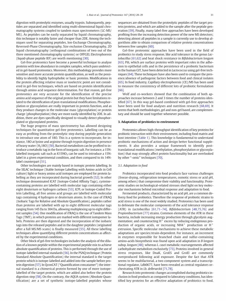
In microbiology, the classical definition of proteome can be adaptedto “the complete protein complement of a cell or subcellular fraction of amicroorganism in a defined growth phase under concrete and precisephysiological conditions” [28]. During the last decades a huge amountof genetic information has been obtained thanks to the developmentof genomics (mainly DNA sequencing technologies and platforms) andBioinformatics (algorithms,massive data storage and query and data in-tegration). However genomics is not enough to explain the complex bi-ological events that are mediated by proteins, as the presence of asimple gene says very few about its expression and the production ofa bioactive protein. Therefore, in the omics era, proteomics has becomemore interesting since they allow detecting proteins involved in themain cellular functions such as catalysis and stress responses. The pro-teomic approaches involve all the techniques used to identify and quan-tify the complete set of proteins present in a sample, cell or tissue underdefined experimental conditions. A detailed review of common tech-niques was written by Monteoliva and Albar [29] and further reviewedby Abdallah and co-workers [30]. Further reviews for the application ofproteomics to the study of probiota/microbiota functionality are alsoavailable in the scientific literature [31,32]. Setting a proteomics exper-iment involves all parameters affecting sample preparation (basicallyprotein extraction andpurification), followed by gel-based/gel-free pro-tein separation coupled to a mass spectrometer step in which the poly-peptides/proteins are finally detected through their mass-to-chargeratio (Fig. 1). The most common approach is the so-called “Bottom-upproteomics”, in which proteins are digested (usually through the actionof trypsin) and the resulting mix of peptides detected in themass spec-trometer. This contrastswith the “Top-down” proteomics, inwhichpro-teins are not digested prior separation, and which is very useful for thedetection of protein degradation products, isoforms, posttranslationalmodifications or truncated proteins [33]. In the identification step,

Fig. 1. Overview of classical setups for a gel-based (upper panel) or a gel-free (lower panel) proteomic experiment. Scissors represent the moment inwhich proteins are digested, usuallywith trypsin. *Additionally, classical SDS-PAGE fractionation can be performed prior LC-MS/MS analysis.
30 L. Ruiz et al. / Journal of Proteomics 147 (2016) 28–39
proprietary, open-source or in-house scripts/pipelines/software are de-veloped allowing protein identification. In this review we will focus onthe most useful proteomic methods and their application in theprobiotics field.
2.1. Gel-based proteomics
Traditionally, polyacrylamide gel electrophoresis has been used toset up differences between proteomes. The two dimensional polyacryl-amide gel electrophoresis (2DE) [34] allows resolving completeproteomes into individual spots corresponding in most cases to a singleprotein, obtaining a kind of protein barcode of the sample. A stainingprotocol is then applied to the gels (Coomassie, silver etc.) allowingthe visualization of the spots, which are further picked up from the geland treated with proteases to release protein-specific peptides; thesepeptides are finally identified by mass spectrometry. Other possibilityto detect proteins produced/repressed/degraded under certain experi-mental conditions is to include radioactive-labelled amino acids in thegrowth medium compared to control conditions [35]. These aminoacids will be incorporated into newly synthesised proteins, and in thisway proteins that are synthesised after for instance a stress challengewill display radioactivity and can be visualized in the gel. In addition,radiolabelling is one of the most sensitive protein detection methods,allowing detection of low-abundant proteins.
Use of 2DE is a widespread technique for massive eukaryotic andprokaryotic protein identification due to the powerful results obtainedwith relatively low cost compared to other gel-free techniques. Howev-er, 2DEpresents somedisadvantages such as reproducibility limitations,and is often insensitive to low abundant proteins such as regulators,membrane associated proteins or basic proteins. Some modificationsof the protocols have been assayed in order to improve the migrationof those specific protein subsets [36]. For instancehydrophobic proteins,mainly membrane-associated or membrane-integral proteins, are notwell solubilized in the absence of detergents, and those detergentssuch as SDS are not compatible with IEF [37]. In this sense new zwitter-ionic detergents such as sulfobetaines have been included to improvesolubilisation of hydrophobic proteins. Very alkaline proteins (pI N 7)are problematic in terms of resolution, resulting in poor 2D patterns. Amethod for improving the resolution of the alkaline proteomeof the lac-tic acid bacterium Lactobacillus hilgardii, has been optimized using a
combination of cup loading for sample loading and use of different re-ducing agents [38]. In the case of alkaline proteomes use ofwide pH gra-dients in the first dimension step, inclusion of higher concentrations ofreducing agents such as dithiothreitol in the cathode or alkylation ofthiol groups with iodoacetamide before protein loading in the IEF stepoften offers substantial improvements in the separation of these pro-teins. In the case of low-abundant proteins, use of ultrazoom gels cover-ing a wide range of isoelectric points, use of sensitive protein stains orlabels such as radiolabelling, or inclusion of prefractionation steps dur-ing sample preparation solve in part the problem of detecting these mi-nority proteins [36].
A variation of 2DE is the use offluorescent dyes to label proteins dur-ing the separation process, allowing detection of low-abundant proteinsand sample multiplexing. This technique is denominated 2D-differencein gel electrophoresis (DIGE) that was firstly described by Ünlü and co-workers [39]. The use of this method improves the dynamic range inprotein detection, increasing the linearity relationship between proteinconcentration and fluorescent signal. In addition, DIGE reduces thenumber of experiments required for sample comparison in different ex-perimental conditions, as many samples can be loaded in the same gelusing different fluorescent dyes.
Gel-based proteomics have been used in the field of probiotics toa) obtain systematic maps for taxonomy or protein function predictionand to b) analyse differential protein expression under different envi-ronmental situations or stress conditions. In this sense, the stress re-sponse to acid conditions or bile presence has been studied by severalauthors [23,24,40,41], as well as the different behaviour of strainswhen co-cultivated with other bacteria [42] or with different carbonsources [43]. The adhesion capability is another desired probiotic traitthat has been tested through gel-based proteomics [44]. Gel-based pro-teomics have also been used to compare bacterial polymorphisms be-tween closely related strains [45,46] and to identify protein featuresresponsible for adaptations to the gastro intestinal tract conditions[47–49].
2.2. Gel-free proteomics
Some of the limitations of 2DE have been solved by gel-free proteo-mic techniques. In gel-free proteomics, the pool of proteins present in aprotein extract is reduced to small peptides through an enzymatic
31L. Ruiz et al. / Journal of Proteomics 147 (2016) 28–39
digestion with proteolytic enzymes, usually trypsin. Subsequently, pep-tides are separated and identified using multi-dimensional liquid chro-matography systems coupled to tandem mass spectrometers (LC-MS/MS). As peptides can be easily separated by liquid chromatography,this technique is notably faster and cheaper than 2DE. Among the tech-niques used for separating peptides, Ion-Exchange Chromatography,Reversed-Phase Chromatography, Size exclusion Chromatography, 2Dliquid chromatography (orthogonal combinations of two out of thethree mentioned chromatography systems) or OFFGEL Electrophoresis(liquid-phase peptide IEF) are worth mentioning [30].
Gel-free proteomics have become a powerful technique to analyseproteinswith low abundance in complex samples,whichpass undetect-ed in gel-based proteomics. Moreover, this methodology provides moresensitive andmore accurate protein quantification, as well as the possi-bility to identify highly hydrophobic or basic proteins. Modifications inthe protein affecting relative mass or isoelectric point are not consid-ered in gel-free techniques, which are based on protein identificationthrough amino acid sequence determination. For that reason, gel-freeproteomics are very accurate for the identification of the preciseamino acid sequence of the original protein but they have limitations re-lated to the identification of post-translationalmodifications. Phosphor-ylation or glycosylation are really important in protein function, and asthey produce changes in the molecular mass (glycosylation) or proteincharge (phosphorylation) they are more easily identified by 2DE. In ad-dition, there are dyes specifically designed to visually detect phosphor-ylated or glycosylated proteins.
The huge progress of mass spectrometry has allowed designingtechniques for quantitative gel-free proteomics. Labelling can be aseasy as profiting from the proteolytic step during peptide generationto introduce one atom of 18O. This is a system to incorporate, for in-stance, 18O at lysine and arginine through trypsin digestion in presenceof heavywater (H218O) [50]. Bacterialmetabolism can be profited to in-troduce ametabolic tag in the formof inorganic salt. For instance, a 15N-labelled inorganic salt such as K15NO3 can be used to introduce a 15N-label in a given experimental condition, and then compared to its 14N-label counterpart [51].
Other technologies are mainly based in isotopic protein labelling. Inthe SILAC technique (Stable Isotope Labelling with Amino acids in Cellculture) light or heavy amino acid isotopes are employed for protein la-belling as they are incorporated during bacterial growth [52]. In othertechnique denominated ICAT (Isotope-Coded Affinity Tags), cysteine-containing proteins are labelled with molecular tags containing eithereight deuterium or hydrogen carbons [53]. ICPL or Isotope-Coded Pro-tein Labelling, all free amino acid groups are labelled with moleculartags containing 4 hydrogen or deuterium atoms. In the case of iTRAQ(Isobaric Tags for Relative and Absolute Quantification), peptides ratherthan proteins are labelled with up to eight different molecular tagsranging from 145Da to 304Da, allowingmultiplexing up to eight differ-ent samples [54]. One modification of iTRAQ is the use of TandemMassTags (TMT), in which proteins aremarkedwith different isotopomer la-bels. Proteins are then digested and the incorporation of the isobarictags into the released peptides and in the daughter peptides (originatedafter a full MS/MS scans) is finally measured [55]. All these labellingtechniques allow quantifying different protein concentrations as affect-ed by the experimental conditions.
Other block of gel-free technologies includes the analysis of the dilu-tion of a knownpeptidewithin the experimental peptidemix to achieveabsolute quantification of proteins. This is performed through the use ofisotope-labelled internal standards [56]. In the PSAQ technique (ProteinStandard Absolute Quantification) the internal standard is the targetproteinwhich is isotope-labelled and added into the sample before pro-tein digestion [57]. In QconCAT (“Quantification concatamer”) the inter-nal standard is a chimerical protein formed by one of more isotope-labelled of the target protein, which are added also before the proteindigestion step [58]. On the contrary, the AQUA system (Absolute Quan-tification) are a set of synthetic isotope-labelled peptides whose
sequences are obtained from the proteolytic peptides of the target pro-tein/proteins, andwhich are added to the sample after the peptide gen-eration [59]. Finally, many label-free approaches have been developedprofiting from the increasing detection power of the newMS detectors;detecting almost all peptides in a sample is currently use to develop al-gorithms able to obtain comparison of relative protein concentrationsbetween few samples [60].
Gel-free proteomic approaches have been used in the field ofprobiotics to study stress response, like acid tolerance in the genus Lac-tobacillus [61,62] and heat shock resistance in Bifidobacterium longum[63]. Pili, which are surface proteins with important roles in the adhe-sion to epithelial cells and in the persistence of a probiotic bacteriumin the humanGIT, have been detected in Lactococcus using gel-free tech-niques [64]. These techniques have also been used to compare the pres-ence/absence of pathogenic factors between food and clinical isolates[65]. In food industry, Capillary electrophoresis (CE)/MS has been usedto measure the consistency of different lots of probiotic formulation[66].
Wolf and co-workers showed that the combination of both ap-proaches increase between 2 and 3 times the number of proteins iden-tified [67]. In this way gel-based combined with gel-free approacheshave been used for food analyses and nutrition research [68,69]. Itseems that bothmethodologies, gel and non-gel based, are complemen-tary and should be used together whenever possible.
3. Adaptation of probiotics to environment
Proteomics allows high-throughput identification of key proteins forprobiotic interactionwith their environment, including foodmatrix andhost intestine (Table 1). This knowledge is fundamental to understandprobiotic mechanisms of action and design better functional supple-ments. It also provides a unique framework to identify post-translational modifications (methylation, phosphorylation or glycosyla-tion) that may strongly affect protein functionality but are overlookedby other “-omic” techniques [70].
3.1. Adaptation to food
Probiotics incorporated into food products face various challenges(freeze-drying, refrigeration temperatures, osmotic stress or acid pH,among others) that compromise their viability and functionality. Prote-omic studies on technological-related stresses shed light on key molec-ular mechanisms behind microbial response and adaptation to food.
Fermented products, characterized by an acid pH, are a common de-livery food for probiotics. Therefore the response of probiotic strains toacid stress is one of the most widely studied. Proteomics has been usedto delineate the molecular components of the acid tolerance response(ATR) in Lactobacillus [61,71–74], Bifidobacterium [48,75,76] andPropionibacterium [77] strains. Common elements of the ATR in thesebacteria, include increasing energy production through glycolysis aug-mentation; and counteracting H+ excess by reducing endogenous pro-duction of organic acids, or increasing NH4+ production or H+
extrusion. Specific molecular mechanisms to achieve these metabolicadaptations are species/strain-dependent. For instance, an incrementin enzymes responsible for branched chain and sulfur-containingamino-acids biosynthesis was found upon acid adaptation in B longumsubsp. longum [48]; whereas L. caseimetabolic rearrangements affectedcarbohydrate metabolism exclusively [73]. Proteins involved in generalstress responses, like DnaK, GroES and GroEl are commonlyoverproduced following acid exposure. Despite the fact that ATRseems to be multifactorial, a two component system and a transcrip-tional regulator, Ldb0677, have been revealed as central regulators or-chestrating ATR in Lb. delbrueckii [71,78].
Research into proteomic changes accomplished during probiotics in-clusion in food products as compared to laboratory conditions, has iden-tified key proteins for an effective adaptation of probiotics to food.
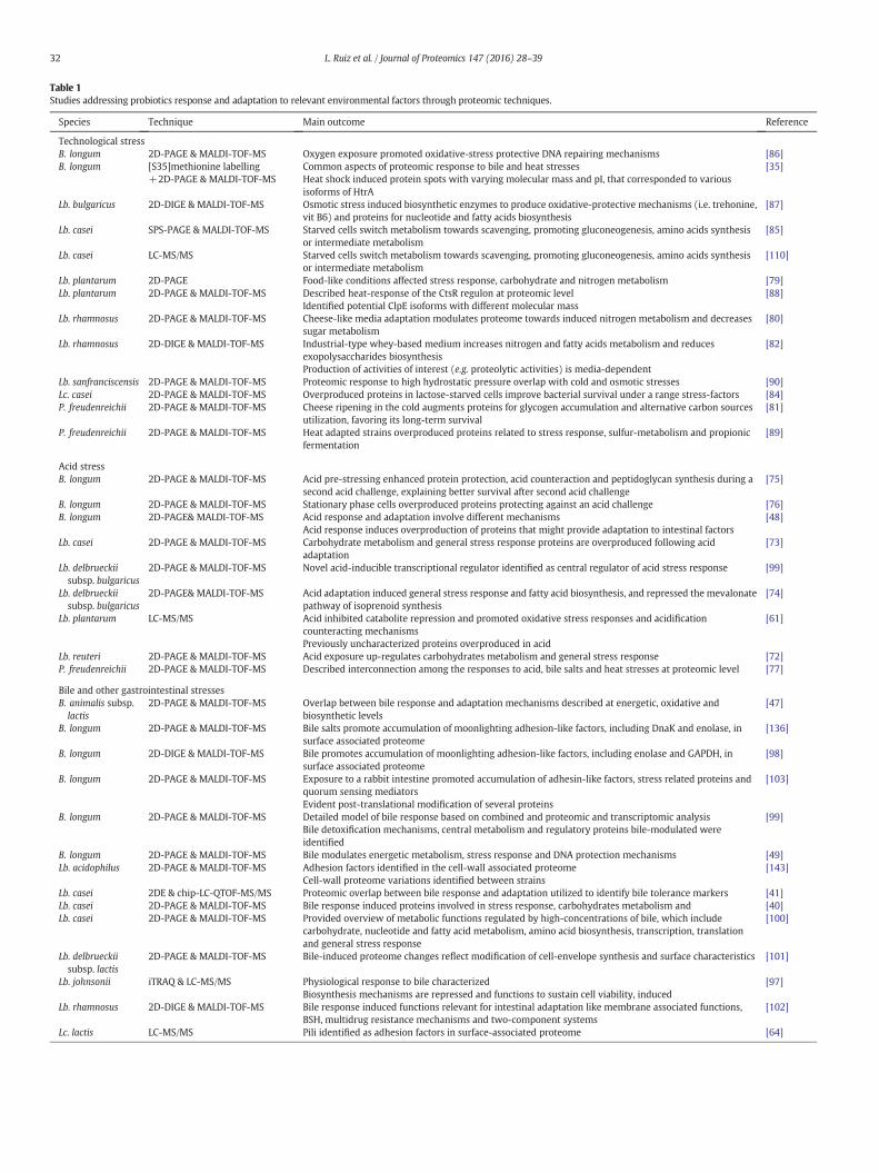
Table 1Studies addressing probiotics response and adaptation to relevant environmental factors through proteomic techniques.
Species Technique Main outcome Reference
Technological stressB. longum 2D-PAGE & MALDI-TOF-MS Oxygen exposure promoted oxidative-stress protective DNA repairing mechanisms [86]B. longum [S35]methionine labelling
+2D-PAGE & MALDI-TOF-MSCommon aspects of proteomic response to bile and heat stressesHeat shock induced protein spots with varying molecular mass and pI, that corresponded to variousisoforms of HtrA
[35]
Lb. bulgaricus 2D-DIGE & MALDI-TOF-MS Osmotic stress induced biosynthetic enzymes to produce oxidative-protective mechanisms (i.e. trehonine,vit B6) and proteins for nucleotide and fatty acids biosynthesis
[87]
Lb. casei SPS-PAGE & MALDI-TOF-MS Starved cells switch metabolism towards scavenging, promoting gluconeogenesis, amino acids synthesisor intermediate metabolism
[85]
Lb. casei LC-MS/MS Starved cells switch metabolism towards scavenging, promoting gluconeogenesis, amino acids synthesisor intermediate metabolism
[110]
Lb. plantarum 2D-PAGE Food-like conditions affected stress response, carbohydrate and nitrogen metabolism [79]Lb. plantarum 2D-PAGE & MALDI-TOF-MS Described heat-response of the CtsR regulon at proteomic level
Identified potential ClpE isoforms with different molecular mass[88]
Lb. rhamnosus 2D-PAGE & MALDI-TOF-MS Cheese-like media adaptation modulates proteome towards induced nitrogen metabolism and decreasessugar metabolism
[80]
Lb. rhamnosus 2D-DIGE & MALDI-TOF-MS Industrial-type whey-based medium increases nitrogen and fatty acids metabolism and reducesexopolysaccharides biosynthesisProduction of activities of interest (e.g. proteolytic activities) is media-dependent
[82]
Lb. sanfranciscensis 2D-PAGE & MALDI-TOF-MS Proteomic response to high hydrostatic pressure overlap with cold and osmotic stresses [90]Lc. casei 2D-PAGE & MALDI-TOF-MS Overproduced proteins in lactose-starved cells improve bacterial survival under a range stress-factors [84]P. freudenreichii 2D-PAGE & MALDI-TOF-MS Cheese ripening in the cold augments proteins for glycogen accumulation and alternative carbon sources
utilization, favoring its long-term survival[81]
P. freudenreichii 2D-PAGE & MALDI-TOF-MS Heat adapted strains overproduced proteins related to stress response, sulfur-metabolism and propionicfermentation
[89]
Acid stressB. longum 2D-PAGE & MALDI-TOF-MS Acid pre-stressing enhanced protein protection, acid counteraction and peptidoglycan synthesis during a
second acid challenge, explaining better survival after second acid challenge[75]
B. longum 2D-PAGE & MALDI-TOF-MS Stationary phase cells overproduced proteins protecting against an acid challenge [76]B. longum 2D-PAGE& MALDI-TOF-MS Acid response and adaptation involve different mechanisms
Acid response induces overproduction of proteins that might provide adaptation to intestinal factors[48]
Lb. casei 2D-PAGE & MALDI-TOF-MS Carbohydrate metabolism and general stress response proteins are overproduced following acidadaptation
[73]
Lb. delbrueckiisubsp. bulgaricus
2D-PAGE & MALDI-TOF-MS Novel acid-inducible transcriptional regulator identified as central regulator of acid stress response [99]
Lb. delbrueckiisubsp. bulgaricus
2D-PAGE& MALDI-TOF-MS Acid adaptation induced general stress response and fatty acid biosynthesis, and repressed the mevalonatepathway of isoprenoid synthesis
[74]
Lb. plantarum LC-MS/MS Acid inhibited catabolite repression and promoted oxidative stress responses and acidificationcounteracting mechanismsPreviously uncharacterized proteins overproduced in acid
[61]
Lb. reuteri 2D-PAGE & MALDI-TOF-MS Acid exposure up-regulates carbohydrates metabolism and general stress response [72]P. freudenreichii 2D-PAGE & MALDI-TOF-MS Described interconnection among the responses to acid, bile salts and heat stresses at proteomic level [77]
Bile and other gastrointestinal stressesB. animalis subsp.lactis
2D-PAGE & MALDI-TOF-MS Overlap between bile response and adaptation mechanisms described at energetic, oxidative andbiosynthetic levels
[47]
B. longum 2D-PAGE & MALDI-TOF-MS Bile salts promote accumulation of moonlighting adhesion-like factors, including DnaK and enolase, insurface associated proteome
[136]
B. longum 2D-DIGE & MALDI-TOF-MS Bile promotes accumulation of moonlighting adhesion-like factors, including enolase and GAPDH, insurface associated proteome
[98]
B. longum 2D-PAGE & MALDI-TOF-MS Exposure to a rabbit intestine promoted accumulation of adhesin-like factors, stress related proteins andquorum sensing mediatorsEvident post-translational modification of several proteins
[103]
B. longum 2D-PAGE & MALDI-TOF-MS Detailed model of bile response based on combined and proteomic and transcriptomic analysisBile detoxification mechanisms, central metabolism and regulatory proteins bile-modulated wereidentified
[99]
B. longum 2D-PAGE & MALDI-TOF-MS Bile modulates energetic metabolism, stress response and DNA protection mechanisms [49]Lb. acidophilus 2D-PAGE & MALDI-TOF-MS Adhesion factors identified in the cell-wall associated proteome
Cell-wall proteome variations identified between strains[143]
Lb. casei 2DE & chip-LC-QTOF-MS/MS Proteomic overlap between bile response and adaptation utilized to identify bile tolerance markers [41]Lb. casei 2D-PAGE & MALDI-TOF-MS Bile response induced proteins involved in stress response, carbohydrates metabolism and [40]Lb. casei 2D-PAGE & MALDI-TOF-MS Provided overview of metabolic functions regulated by high-concentrations of bile, which include
carbohydrate, nucleotide and fatty acid metabolism, amino acid biosynthesis, transcription, translationand general stress response
[100]
Lb. delbrueckiisubsp. lactis
2D-PAGE & MALDI-TOF-MS Bile-induced proteome changes reflect modification of cell-envelope synthesis and surface characteristics [101]
Lb. johnsonii iTRAQ & LC-MS/MS Physiological response to bile characterizedBiosynthesis mechanisms are repressed and functions to sustain cell viability, induced
[97]
Lb. rhamnosus 2D-DIGE & MALDI-TOF-MS Bile response induced functions relevant for intestinal adaptation like membrane associated functions,BSH, multidrug resistance mechanisms and two-component systems
[102]
Lc. lactis LC-MS/MS Pili identified as adhesion factors in surface-associated proteome [64]
32 L. Ruiz et al. / Journal of Proteomics 147 (2016) 28–39

33L. Ruiz et al. / Journal of Proteomics 147 (2016) 28–39
Siragusa et al. [79] mimicked in vitro food fermentations to reveal thatLactobacillus plantarum adjusts its metabolism to the carbohydratesavailable in the food matrix. Furthermore, reduction in enzymes con-nected with lactic acid production (i.e. glycolysis) and increase in thesynthesis of proteins involved in fatty acids biosynthesis was observedin fermented foods, supporting in vitro observations in acidified MRSbroth, which is a complex medium used to growth lactic acid bacteria[61]. Similar proteomic experiments were performed on Lb. rhamnosusand Propionibacterium freudenreichii grown under simulated industrialconditions as compared to laboratory MRS [80–82]. Their results evi-denced strong differences between the metabolic pathways activatedin MRS, where glucose and lactose are abundant and lactic acid is themajor metabolic end product, and those activated in “cheese-like”broth, where carbohydrates are scarce and fatty acids abundant, andmetabolism is directed towards and increased acetic acid production.
Indeed, carbohydrate starvation is a common food stress for probiot-ic bacteria and proteomic analysis of nutrient-deprived lactobacilli evi-denced metabolic activation of routes to metabolize alternative carbonsources. Increased production of general stress response proteins isalso observed, and would contribute to the higher tolerance ofnutrient-deprived cells to various stresses [83–85].
Proteomics has also been used to study probiotics response to otherfood associated stress factors including oxidative stress [86]; osmoticstress [87]; heat stress [35,88,89]; high pressures [90]; or the presenceof dietary components such as prebiotic ingredients [43,91–94] or bio-active dietary polyphenols like rutin [95].
3.2. Adaptation to the intestinal environment
Intestinal colonization is considered a requisite for probiotics healthpromotion [6]. Gastrointestinal passage imposes important challengesfor probiotic survival, like acidity in the stomach or the presence of
Table 2Studies addressing probiotic functionality through proteomic techniques.
Species Technique Main outcome
B. animalissubsp. lactis
2D-DIGE & MALDI-TOF-MS Xylo-oligosacharides utiliza
B. animalissubsp. lactis
LC-ESI-Q-TOF MS/MS Membrane-associated protand immunoregulatory pro
B. breve 2D-DIGE & MALDI-TOF-MS/MS Intestinal cells co-cultivatestrengthening the epithelia
B. longum 2D-DIGE & MALDI-TOF-MS B-glucans utilization modeB. longum 2D-PAGE & MALDI-TOF-MS Probiotic administration to
partially counteracting theB. longum 2D-PAGE & MALDI-TOF-MS Identified plasminogen bin
Co-culture with intestinal cadhesin-like factors
B. longum 2D-PAGE & MALDI-TOF-MS Probiotic administration paa gliadin-induced enteropa
Lb. acidophilus 2D-DIGE & MALDI-TOF-MS Proposed molecular modelLb. acidophilus 2D-DIGE & MALDI-TOF-MS Identified molecules involvLb. acidophilus 2D-PAGE & MALDI-TOF-MS In acid pH, cell wall associa
cytokine secretion patternLb. acidophilus 2D-PAGE & MALDI-TOF-MS Identified proteins overpro
involved in cholesterol redLb. acidophilus 2D-PAGE & MALDI-TOF-MS Identified elements regulat
capability of the strainLb. fermentum 2D-PAGE Co-administration of Lb. fer
the intestinal proteomeLb. plantarum LC-MS/MS Identified surface-proteomLb. plantarum 2D-PAGE & MALDI-TOF-MS/MS or MDLC
coupled to nano-ESI-MS/MSQuorum sensing regulatesbinding and pathogens exc
Lb. rhamnosus LC-MS/MS Surface-associated proteomimmuno-stimulation and p
Lb. rhamnosus 2D-PAGE & MALDI-TOF-MS Growth in mucin resulted iunknown extracellular pro
P. freudenreichii LC-MS/MS and nano-LC-MS/MS Identify adhesion and imm
bile salts and digestive proteases in the intestine. Furthermore,probiotics encounter a densely populated microbial ecosystem in thelarge intestine, where they need to compete against autochthonous mi-crobiota to colonize the intestinal environment. Proteomics underin vitro conditions mimicking GIT environments or using in vivomodelshave revealed some of themechanisms behind probiotics adaptation tothe intestinal environment.
Proteomics have helped in understanding the response and adapta-tion of probiotic bacteria to bile. Bile salts are biological detergentswhich participate in fat emulsion and absorption. They can also disruptand destabilize bacterial membranes, leading to ions leaking, proteinsmiss-folding and aggregation and cell death. Accordingly, bile exposuregenerally translates into overproduction of ribosomal and general stressresponse proteins, as a mean to promote protein recycling; as well asenergy production systems and proteins involved in membrane andcell wall biosynthesis, reflecting the need to counteract membranedamage [35,40,47,49,96–102].
Other proteins involved in the response mechanism to bile in otherprobiotic strains have been identified through proteomics. For instance,bile salt hydrolase role in bile detoxification is not clear, but it isoverproduced in some Lactobacillus exposed to bile [96,97,102], and ina B. longum strain exposed to a rabbit intestine [103] as revealed by dif-ferent proteomic techniques. From the bifidobacterial strains tested,only one B. longum strain overproduced BSH following an in vitro bilechallenge [99], but it was constitutively overproduced in a bile adaptedB. animalis strain [47].
Proteomics have also contributed to characterize prebiotic utiliza-tion pathways availing better symbiotics design. Probiotic bacteria areadapted to utilize a range of carbon sources that, otherwise, would beundigested by their hosts, favoring their establishment in the intestinalenvironment. For instance, β-glucans and xylo-oligosaccharides utiliza-tion pathways in bifidobacteria [91–93]; lactitol and cellobiose
Reference
tion model proposed from proteomic profiles [92]
eome, includes carbohydrate import mechanisms, adhesin-like factorsteins
[93]
d with B. breve overproduced cytoskeleton elements, presumablyl barrier
[118]
l proposed from proteomic profiles [91]gliadin-induced enteropathy rat model modulates jejunal proteome,gliadin-induced inflammatory changes
[106]
ding proteins in cytoplasmic proteomeells induces overproduction of proteins including several moonlighting
[133]
rtially counteracted gliadin-induced proteome changes at jejunal level inthy rat model
[106]
for lactitol metabolism [94]ed in cellobiose metabolism [43]ted GroES and GroEL augment, correlating to induction of alteredin splenocytes
[135]
duced following growth in cholesterol and elements presumablyucing capability
[107]
ed by CcpA, that might be important for the cholesterol reducing [108]
mentum and aureomycin to piglets counteracted the antibiotic effects on [113]
e changes in a sortase-mutant with increased pro-inflammatory signaling [131]adhesion factors correlating to increased biofilm formation, intestinallusion
[111]
e includes moonlighting protein with predicted functions in adhesion,athogens exclusion
[26]
n reduced production of secreted proteins and production of previouslyteins
[137]
unomodulation determinants [22]
34 L. Ruiz et al. / Journal of Proteomics 147 (2016) 28–39
utilization pathways in lactobacilli [43,94]; and human milk oligosac-charides metabolizing capabilities in infant-associated bifidobacteria[104] have been studied through proteomics.
4. Proteomics for the study of changes in probiotic functionality
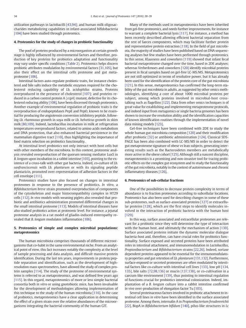
The pool of proteins produced by amicroorganism at certain growthstage is highly influenced by environmental factors and therefore, pro-duction of key proteins for probiotics adaptation and functionalitymay vary under specific conditions (Table 2). Proteomics helps discernprobiotic attributes modulation by environmental factors [31,105], butalso their effect on the intestinal cells proteome and gut meta-proteome [106].
Intestinal factors auto-regulate probiotic traits, for instance choles-terol and bile salts induce the metabolic enzymes required for the cho-lesterol reducing capability of Lb. acidophilus strains. Proteinsoverproduced in the presence of cholesterol [107]; and proteins re-duced in a carbon control protein A (ccpA) mutant, defective for its cho-lesterol reducing ability [108], have been discerned throughproteomics.Another example of environmental regulation of probiotic traits is theoverproduction of endopeptidases PepE and PepO, known to be essen-tial for producing the angiotensin conversion inhibitory peptide, follow-ing Lb. rhamnosus growth in soya milk or Lb. helveticus growth in skimmilk [80,109]. Indeed, lactobacillus incubation in milk at refrigerationtemperatures overproduced factors, related to amino acids metabolismand DNA protection, that also enhanced bacterial persistence in themammalian digestive tract [110], thus highlighting the importance offood matrix selection on probiotics functionality.
At intestinal level probiotics not only interact with host cells butwith other members of the microbiota. In this context, proteomic anal-ysis revealed overproduction of the quorum sensing mediator LuxS inB. longum upon incubation in a rabbit intestine [103], pointing to the ex-istence of a cross-talk with other gut bacteria. Indeed, co-culture of Lb.sanfranciscensis with Lb. plantarum or with its signaling peptideplantaricin, promoted over-representation of adhesion factors in thecell envelope [111].
Proteomics studies have also focused on changes in intestinalproteomes in response to the presence of probiotics. In vitro, aBifidobacterium breve strain promoted overproduction of componentsof the cytoskeleton and major histocompatibility complex in HT29cells [112]. In vivomodels with weaning piglets also revealed that pro-biotic and antibiotics administration promoted differential changes inthe small intestinal proteome [113]. Models of intestinal diseases havealso studied probiotics action at proteomic level. For instance, a jejunalproteome analysis in a rat model of gliadin-induced enteropathy re-vealed that B. longum modulates inflammation [106].
5. Proteomics of simple and complex microbial populations:metaproteomics
The human microbiota comprises thousands of different microor-ganisms that co-habit in the same environmental niche. From an analyt-ical point of view, this fact increases notably the complexity at the levelof sample processing and data analysis, and difficult massive proteinidentification. During the last ten years, improvements in protein/pep-tide separation and identification, such as the development of high-resolutionmass spectrometers, have allowed the study of complex pro-tein samples [114]. The study of the proteome of environmental sys-tems is referred to as metaproteomics, and was defined few years ago[115]. In this regard, metaproteomics of more or less simple bacterialconsortia both in vitro or using gnotobiotic mice, has been invaluablefor the development of methodologies allowing implementation ofthis technique to the study of the human microbiota [116]. In the caseof probiotics, metaproteomics have a clear application in determiningthe effect of a given strain over the relative abundances of the microor-ganisms integrating the bacterial community of the human GIT.
Many of the methods used in metaproteomics have been inheritedfrom gel-free proteomics, and needs further improvements, for instanceto warrant a complete bacterial lysis [117]. For instance, a method hasbeen recently described allowing efficient bacterial separation fromthe rest of faeces components, which may facilitate further proteinand representative protein extraction [118]. In the field of gut microbi-ota, themajority of studies have been published based onDNA sequenc-ing analyses but few studies have been performed through proteomics.In this sense, Klaassens and coworkers [119] showed that infant fecalbacterial metaproteome changed over the time, based in 2DE analyses;whereas Verberkmoes and coworkers [120] identify microbial proteinspresent in fecal samples based on gel-free LC-MS/MS. Metaproteomicsare not still optimized in terms of resolutive power, but it has alreadybeen used for the identification of the protein core of the gutmicrobiota[121]. In this sense, metaproteomics has confirmed the long-term sta-bility of the gut microbiota in adults, as suggested by other omicsmeth-odologies, identifying a core of about 1000 microbial proteins persubject, among which proteins involved in microbial-host cross-talking such as flagellins [122]. Data from other omics techniques is ofgreat value for establishing and implementingmetaproteomeprotocols,and indeed input frommetagomics or single bacteria genomes has beenshown to increase the resolution ability and the identification capacitiesof known identification routines through the implementation of noveldata mining models [123].
Gel-free techniques have been combined with 2DE to study thewhole human gut microbiota composition [120] and their modificationafter probiotic [32] or antibiotic administration [124]. Classical SDS-PAGE has been also combined with LC-MS/MS in order to detect thegutmetaproteome signature of obese vs lean subjects, generating inter-esting results such as the Bacteroidetes members are metabolicallymore active in the obese cohort [125]. Although still a nascent discipline,metaproteomics is a promising and non-invasive tool for tracing probi-otic effects on the complex gut ecosystem and to study the functionalityof the gutmicrobiota, notably in the context of autoimmune and chronicinflammatory diseases [126].
6. Proteomics of sub-cellular fractions
One of the possibilities to decrease protein complexity in terms ofabundance is to fraction proteomes according to subcellular locations.Proteomics has also been used for describing changes in some of thesesub-proteomes, such as surface-associated proteins [127] or extracellu-lar proteins [128], which are the first steps to identify molecules in-volved in the interaction of probiotic bacteria with the human host[129].
In this way, surface associated and extracellular proteomes are rele-vant for a probiotic since they will determine the type of interactionwith the human host, and ultimately the mechanism of action [130].Surface associated proteins initiate the dynamic molecular dialoguebacteria-host and, therefore, can have crucial roles for probiotics func-tionality. Surface exposed and secreted proteins have been attributedroles in intestinal attachment, and immunomodulation in Lactobacillus,Propionibacterium and Bifidobacterium strains [22,26]. Indeed, sortasedependent proteins appeared to be essential for the immunomodulato-ry properties and gut retention of Lb. plantarum [131,132]. Furthermore,surface-exposed or secreted proteomes are often modulated by intesti-nal factors like co-culture with intestinal cell lines [133], low pH [134,135], bile salts [72,98,136] or mucin [137,138], or co-cultivation in acaecum-like environment [139], thus pointing to intestinal regulationof functions crucial for probiotics intestinal colonization. Indeed, im-plantation of a B. longum culture into a rabbit intestine confirmedin vivo over-production of elongation factor Tu [103].
Several proteinaceous factors involved in probiotic attachment to in-testinal cell lines in vitro have been identified in the surface associatedproteome. Among them, internalin A in Propionibacterium freudenreichii[22], BopA in Bifidobacterium bifidum [140], pilus-like structures in Lc
35L. Ruiz et al. / Journal of Proteomics 147 (2016) 28–39
lactis [64], bifidobacteria [141] and lactobacilli [142], S-layer proteins(slpA) in Lb. acidophilus [143] or fimbriae in Lb. rhamnosus [144] are re-markable and have been identified using proteomic approaches. In thissense, proteomics has been relevant in the identification a high numberofmoonlighting proteins, proteins that are secretedwithout harbouringany signal peptide. Apart from being importantmetabolic proteins, theyhave been shown to be associated to the bacterial surface displayingplasminogen or mucin binding activities. Examples of moonlightingproteins are enzymes involved in carbohydratemetabolism like enolaseor glyceraldehyde 3-phosphate dehydrogenase (GADPH), general stressproteins like DnaK, GroES or GroEL and elongation factors such as EF-Tu,which in some cases appeared overproduced following bile exposure[133,136,137,145,146].
7. Bioinformatics tools
Today,more than ever, the study of humanmicrobiome andprobiot-ic functionality attracts considerable attention from the bioinformaticscommunity. Late in 2000s, the Human Microbiome Project (HMP)[147] and the metagenomics of the Human Intestinal Tract (MetaHit)[148] emerged as the two main international initiatives devoted to thestudy of human microbiome and the development of computationalmethods to analyse sequenced metagenomes. Now, the spectrum ofsoftware tools dedicated or supporting these analyses is quite vast.
Many efforts have been devoted to gene function prediction, notablyin probiotic research, where very specific effects are observed in concisestrains harbouring certain genetic traits. Regarding the gut microbiome,approximately 50% of the genes are not yet characterized using stan-dard annotation methods [149]. Therefore, conventional methods forputative gene characterization and functional prediction, based onalignment to homologous genes with existing annotations (e.g.BLAST), were rendered ineffective [150]. Alternative computationalmethods approached the problem by integrating standard homology-based with additional information, namely sequence features, co-expression data, protein-protein interactions, binding sites, and subcel-lular localisation data.
The annotation pipelines of RAST, MG-RAST [151] and IMG/M [152]are perhaps the most well-known comparative genomics-based auto-mated pipelines where researchers working on probiotics or gut micro-biota can easily analyse genomes/metagenomes. Databases such asPfam [150] and COG [153] enable methods encompassing comparisonswith sequence-diverse protein families or recurring sequence motifs;and, the KEGG Orthology and KEGG pathways databases [154] areoften used to predict the composition ratio of microbial gene familiesand pathways from the human microbiome project [155,156]. Toolssuch as RAPSearch [157] and PAUDA [158] propose faster alternativesthan BLAST to the alignment of environmental sequencing reads. Be-sides comparative genomics, there are structure-based approaches,functional prediction methods based on evolutionary conservationand phylogeny, and network context-based approaches (e.g. co-expression and metabolic networks) [156,159,160].
Regarding proteomics software packages two open source tools,Unipept and MetaProteomeAnalyzer, are worth mentioning. Unipeptis a web application powered by the UniProt database and an imple-mentation of a custom lowest common ancestor algorithm [161]. It al-lows tryptic peptide and metaproteome analysis or identification ofunique tryptic peptides in a given sample/experimental condition, high-ly valuable in targeted proteomics. In addition, it provides a set of inter-active data visualizations such as themetaproteome clustering tool. TheMetaProteomeAnalyzer is a software package dedicated to the integra-tion ofmetaproteomic profileswith othermetadata (genomic, function-al etc.). This tool provides with an intuitive environment for datamining, analysis and interpretation, as well as with methodologies todecrease data redundancy [162].
During the next years, further improvements in protein databasesearching, raw mass spectra filtering, data mining and graphical
representation among other computational processes, will allow deter-mining how gut metaproteome composition affects human healththrough bottom-up or top-down methodologies allowing discoveringover or sub-representation of keymetabolic pathways/features throughhigh-throughput and non-invasive techniques.
Human gutmetaproteomics is also an emerging researchfield that ischaracterized by a high level of complexity [163,164]. Conventionalhigh-throughput spectral interpretation algorithms have been devel-oped in the context of properly assigning peptide-spectral masses orpeptide sequence information (inferred from MS/MS fragmentation)to the proteins from which peptides are theoretically derived. In addi-tion, there are many ambiguous peptides that can be derived from dif-ferent proteins [165]. This, also known as the protein inferenceproblem, is an important bottleneck in shotgun proteomics which hasbeen addressed using different strategies, such as Bayesian approachesor Lasso models [166,167]. The development of cross-species proteinidentification approaches and metagenomics-based approaches waschallenged by the complexity of the gutmicrobial proteome and the dy-namic distribution of species between individuals. New approaches aimto increase the sensitivity of the peptide identification by peptide spec-trum matching and one possibility is to integrate syntheticmetaproteome information and metagenomic information [123]. Thework of Muth et al. [121] further discusses alternatives to the peptideidentification via database searching and presents de novo sequencingas a valid alternative independent from protein sequence databases.The field of human gut microbiome metaproteomics has been recentlyreviewed [168].
Genome-scale metabolic models (GEMs) are perhaps the latest toolin human gut bioinformatics [169,170]. Metagenomics studies canquantify the relative abundance of each species in a community but itdoes not enable description of the function of each individual. TheGEMs can describe the metabolism of each species and the integratedanalysis of these models may allow us to explore the interactions be-tween predominant bacteria in the gut ecosystems. For example, El-Semman and colleagues reconstructed two GEMs for Bifidobacteriumadolescentis L2–32 (the iBif452 model) and Faecalibacterium prausnitziiA2–165 (the iFap484) to support the study of the anti-inflammatoryrole that these microorganisms play in the gut ecosystem [171]. Alongthis line of research, Bayesian inference of metabolic networks hasbeen employed to reveal a metabolic system with greater prevalenceamong inflammatory bowel disease patients [172]; and the construc-tion and functional analysis of proteome interaction networks enabledthe analysis of nutrient-affected pathways in human pathologies [173].
8. Conclusions and perspectives
Traditionally, probiotics have been selected on the basis of a goodtechnological performance, mainly their suitability to survive during in-dustrial processing and storage, a robust metabolic behaviour that al-lows profitable biomass yields, and a stress resistant phenotype thatguarantee their passage through the gastrointestinal tract and subse-quent viability in the gut. In this regard, the molecular basis underlyingthe functional properties of probiotic bacteria responsible for the healthpromoting effects have been in the background formany years. Howev-er, the breakthrough of omics technologies in the probiotic andmicrobi-ota fields has had a very relevant impact in the elucidation of probioticmechanisms and in the procedures to select these microorganisms,based on solid scientific evidence. During the last decade we haveseen a tremendous eclosion of genomics and metagenomics methodol-ogies that were very useful to identify the population of microbesinhabiting our gut, and its relation to different diseases and physiologi-cal disorders. It is unquestionable that, in the near future, the evolutionof proteomic techniques will play a pivotal role in the generation ofknowledge about the functions of probiotics and gut commensals, stilla pending issue in the field of intestinal microbiomics. In addition, en-richment and curation of database content will increase the knowledge
36 L. Ruiz et al. / Journal of Proteomics 147 (2016) 28–39
about functions of proteins, which is a very important aspect for prote-omic analyses in general. This will include not only more efforts in cu-rating individual (and mostly draft) probiotic genomes, but alsodeciphering the functions of the hypothetical or putative proteins thatare distributed along all genetic entries.
Conflict of interest
The authors declare no conflict of interest in relation to thework de-scribed in this paper.
Acknowledgements
Research in our lab is funded by Grants AGL2013-44039-R andAGL2013-44761-P from the Spanish “Plan Estatal de I + D + I”. Partof the authors is also partially funded by the [15VI013] Contract-Programme from the University of Vigo and the AgrupamentoINBIOMED from DXPCTSUG-FEDER unha maneira de facer Europa(2012/273). Lorena Ruiz has received funding from the People Pro-gramme (Marie Curie Actions) of the European Union's Seventh Frame-work Programme FP7/2007-2013/under REA grant agreement n°624773. Borja Sánchez was recipient of a Ramón y Cajal postdoctoralcontract from the Spanish Ministry of Economy and Competitiveness.
References
[1] E. Metchnikoff, I.I. Metchnikoff, The Prolongation of Life: Optimistic Studies, http://books.google.com/books?hl=en&lr=&id=U8bgKGvZJV0C&pgis=11908.
[2] T. Matsuzaki, R. Yamazaki, S. Hashimoto, T. Yokokura, The effect of oral feeding ofLactobacillus casei strain Shirota on immunoglobulin E production in mice, J. DairySci. 81 (1998) 48–53, http://dx.doi.org/10.3168/jds.S0022-0302(98)75549-3.
[3] D. Gordon, J. Macrae, D. Wheater, A Lactobacillus preparation for use with antibi-otics, Lancet 269 (1957) 899–901, http://dx.doi.org/10.1016/S0140-6736(57)91222-9.
[4] J. Schrezenmeir, M. de Vrese, Probiotics, prebiotics, and synbiotics—approaching adefinition, Am. J. Clin. Nutr. 73 (2001) 361S–3364, http://dx.doi.org/10.1007/10_2008_097.
[5] Fao, Probiotics in food, Food Nutr. Pap. 85 (2001) 71, http://dx.doi.org/10.1201/9781420009613.ch16.
[6] C. Hill, F. Guarner, G. Reid, G.R. Gibson, D.J. Merenstein, B. Pot, et al., Expert consen-sus document: the International Scientific Association for probiotics and prebioticsconsensus statement on the scope and appropriate use of the term probiotic, Nat.Rev. Gastroenterol. Hepatol. 11 (2014) 9, http://dx.doi.org/10.1038/nrgastro.2014.66.
[7] Z. Sun, A. Baur, D. Zhurina, J. Yuan, C.U. Riedel, Accessing the inaccessible: molecu-lar tools for bifidobacteria, Appl. Environ. Microbiol. 78 (2012) 5035–5042, http://dx.doi.org/10.1128/AEM.00551-12.
[8] F. Bäckhed, R.E. Ley, J.L. Sonnenburg, D.A. Peterson, J.I. Gordon, Host-bacterial mu-tualism in the human intestine, Science 307 (2005) 1915–1920, http://dx.doi.org/10.1126/science.1104816.
[9] J. Qin, R. Li, J. Raes, M. Arumugam, K.S. Burgdorf, C. Manichanh, et al., A human gutmicrobial gene catalogue established by metagenomic sequencing: article: nature,Nature 464 (2010) 59–65, http://dx.doi.org/10.1038/nature08821.
[10] J.K. Nicholson, E. Holmes, J. Kinross, R. Burcelin, G. Gibson, W. Jia, et al., Host-gutmicrobiota metabolic interactions, Science 80- (336) (2012) 1262–1267, http://dx.doi.org/10.1126/science.1223813.
[11] H.J. Flint, K.P. Scott, P. Louis, S.H. Duncan, The role of the gut microbiota in nutritionand health, Nat. Rev. Gastroenterol. Hepatol. 9 (2012) 577–589, http://dx.doi.org/10.1038/nrgastro.2012.156.
[12] C. Manichanh, N. Borruel, F. Casellas, F. Guarner, The gut microbiota in IBD, Nat.Rev. Gastroenterol. Hepatol. 9 (2012) 599–608, http://dx.doi.org/10.1038/nrgastro.2012.152.
[13] A. Hevia, C. Milani, P. López, A. Cuervo, S. Arboleya, S. Duranti, et al., Intestinaldysbiosis associated with systemic lupus erythematosus, MBio. 5 (2014),e01548-14, http://dx.doi.org/10.1128/mBio.01548-14.
[14] M. Vijay-Kumar, J.D. Aitken, F.A. Carvalho, T.C. Cullender, S. Mwangi, S. Srinivasan,et al., Metabolic syndrome and altered gut microbiota in mice lacking Toll-like re-ceptor 5, Science 80- (328) (2010) 228–231, http://dx.doi.org/10.1126/science.1179721.
[15] J.U. Scher, S.B. Abramson, The microbiome and rheumatoid arthritis, Nat. Rev.Rheumatol. 7 (2011) 569–578, http://dx.doi.org/10.1038/nrrheum.2011.121.
[16] L. Wen, R.E. Ley, P.Y. Volchkov, P.B. Stranges, L. Avanesyan, A.C. Stonebraker, et al.,Innate immunity and intestinal microbiota in the development of Type 1 diabetes,Nature 455 (2008) 1109–1113, http://dx.doi.org/10.1038/nature07336.
[17] P.J. Turnbaugh, R.E. Ley, M.A. Mahowald, V. Magrini, E.R. Mardis, J.I. Gordon, Anobesity-associated gut microbiomewith increased capacity for energy harvest, Na-ture 444 (2006) 1027–1031, http://dx.doi.org/10.1038/nature05414.
[18] P.A. Smith, Brain,meet gut, Nature 526 (2015) 312–314, http://dx.doi.org/10.1038/526312a.
[19] G.P. Donaldson, S.M. Lee, S.K.Mazmanian, Gut biogeography of the bacterial micro-biota, Nat. Rev. Microbiol. 14 (2015) 20–32, http://dx.doi.org/10.1038/nrmicro3552.
[20] E.E. Hansen, C.A. Lozupone, F.E. Rey, M. Wu, J.L. Guruge, A. Narra, et al., Pan-genome of the dominant human gut-associated archaeon, Methanobrevibactersmithii, studied in twins, Proc. Natl. Acad. Sci. U. S. A. 108 (Suppl.) (2011)4599–4606, http://dx.doi.org/10.1073/pnas.1000071108.
[21] M. Arumugam, J. Raes, E. Pelletier, D. Le Paslier, T. Yamada, D.R. Mende, et al.,Enterotypes of the human gut microbiome, Nature 473 (2011) 174–180, http://dx.doi.org/10.1038/nature10187.
[22] C. Le Maréchal, V. Peton, C. Plé, C. Vroland, J. Jardin, V. Briard-Bion, et al., Surfaceproteins of Propionibacterium freudenreichii are involved in its anti-inflammatoryproperties, J. Proteome 113 (2014) 447–461, http://dx.doi.org/10.1016/j.jprot.2014.07.018.
[23] B. Sanchez, et al., Proteomics of stress response in Bifidobacterium, Front. Biosci.Volume 6905 (2008), http://dx.doi.org/10.2741/3198.
[24] B. Sánchez, L. Ruiz, M. Gueimonde, A. Margolles, Omics for the study of probioticmicroorganisms, Food Res. Int. 54 (2013) 1061–1071, http://dx.doi.org/10.1016/j.foodres.2013.01.029.
[25] F. Turroni, F. Bottacini, E. Foroni, I. Mulder, J.-H. Kim, A. Zomer, et al., Genome anal-ysis of Bifidobacterium bifidum PRL2010 reveals metabolic pathways for host-derived glycan foraging, Proc. Natl. Acad. Sci. U. S. A. 107 (2010) 19514–19519,http://dx.doi.org/10.1073/pnas.1011100107.
[26] E. Espino, K. Koskenniemi, L. Mato-Rodriguez, T.A. Nyman, J. Reunanen, J. Koponen,et al., Uncovering surface-exposed antigens of Lactobacillus rhamnosus by cell shav-ing proteomics and two-dimensional immunoblotting, J. Proteome Res. 14 (2015)1010–1024, http://dx.doi.org/10.1021/pr501041a.
[27] M.-A. von Schillde, G. Hörmannsperger, M. Weiher, C.-A. Alpert, H. Hahne, C.Bäuerl, et al., Lactocepin secreted by Lactobacillus exerts anti-inflammatory effectsby selectively degrading proinflammatory chemokines, Cell Host Microbe 11(2012) 387–396, http://dx.doi.org/10.1016/j.chom.2012.02.006.
[28] A. Gorg, G. Boguth, C. Obermaier, W. Weiss, Two-dimensional electrophoresis ofproteins in an immobilized pH 4–12 gradient, Electrophoresis 19 (1998)1516–1519, http://dx.doi.org/10.1002/elps.1150190850.
[29] L. Monteoliva, J.P. Albar, Differential proteomics: an overview of gel and non-gelbased approaches, Brief. Funct. Genomic. Proteomic. 3 (2004) 220–239, http://dx.doi.org/10.1093/bfgp/3.3.220.
[30] C. Abdallah, E. Dumas-Gaudot, J. Renaut, K. Sergeant, Gel-based and gel-free quan-titative proteomics approaches at a glance, Int. J. Plant Genomics. 1–17 (2012),http://dx.doi.org/10.1155/2012/494572.
[31] R.A. Siciliano, M.F. Mazzeo, Molecular mechanisms of probiotic action: a proteomicperspective, Curr. Opin. Microbiol. 15 (2012) 390–396, http://dx.doi.org/10.1016/j.mib.2012.03.006.
[32] J. Aires, M.-J. Butel, Proteomics, human gut microbiota and probiotics, Expert Rev.Proteomics. 8 (2011) 279–288, http://dx.doi.org/10.1586/epr.11.5.
[33] J.C. Tran, L. Zamdborg, D.R. Ahlf, J.E. Lee, A.D. Catherman, K.R. Durbin, et al., Map-ping intact protein isoforms in discovery mode using top-down proteomics, Na-ture 480 (2011) 254–258, http://dx.doi.org/10.1038/nature10575.
[34] P.H. O'Farrell, High resolution two-dimensional electrophoresis of proteins, J. Biol.Chem. 250 (1975) 4007–4021, http://dx.doi.org/10.1016/j.bbi.2008.05.010.
[35] K. Savijoki, A. Suokko, A. Palva, L. Valmu, N. Kalkkinen, P. Varmanen, Effect of heat-shock and bile salts on protein synthesis of Bifidobacterium longum revealed by[35S]methionine labelling and two-dimensional gel electrophoresis, FEMSMicrobiol.Lett. 248 (2005) 207–215, http://dx.doi.org/10.1016/j.femsle.2005.05.032.
[36] J.L. Lopez, Two-dimensional electrophoresis in proteome expression analysis, J.Chromatogr. B Anal. Technol. Biomed. Life Sci. 849 (2007) 190–202, http://dx.doi.org/10.1016/j.jchromb.2006.11.049.
[37] T. Rabilloud, Membrane proteins and proteomics: love is possible, but so difficult,Electrophoresis 30 (2009), http://dx.doi.org/10.1002/elps.200900050.
[38] C. Lamberti, E. Pessione, M.G. Giuffrida, R. Mazzoli, C. Barello, A. Conti, et al., Com-bined cup loading, bis(2-hydroxyethyl) disulfide, and protein precipitation proto-cols to improve the alkaline proteome of Lactobacillus hilgardii, Electrophoresis 28(2007) 1633–1638, http://dx.doi.org/10.1002/elps.200600496.
[39] M. Unlü, M.E. Morgan, J.S. Minden, Difference gel electrophoresis: a single gelmethod for detecting changes in protein extracts, Electrophoresis 18 (1997)2071–2077, http://dx.doi.org/10.1002/elps.1150181133.
[40] C. Alcántara, M. Zúñiga, Proteomic and transcriptomic analysis of the response tobile stress of Lactobacillus casei BL23, Microbiology 158 (2012) 1206–1218,http://dx.doi.org/10.1099/mic.0.055657-0.
[41] E. Hamon, P. Horvatovich, M. Bisch, F. Bringel, E. Marchioni, D. Aoudé-Werner,et al., Investigation of biomarkers of bile tolerance in Lactobacillus casei using com-parative proteomics, J. Proteome Res. 11 (2012) 109–118, http://dx.doi.org/10.1021/pr200828t.
[42] L. Ruiz, B. Sánchez, C.G. de los Reyes-Gavilán, M. Gueimonde, A. Margolles, Cocul-ture of Bifidobacterium longum and Bifidobacterium breve alters their protein ex-pression profiles and enzymatic activities, Int. J. Food Microbiol. 133 (2009)148–153, http://dx.doi.org/10.1016/j.ijfoodmicro.2009.05.014.
[43] G.C. van Zanten, N. Sparding, A. Majumder, S.J. Lahtinen, B. Svensson, S. Jacobsen,The differential proteome of the probiotic Lactobacillus acidophilus NCFM grownon the potential prebiotic cellobiose shows upregulation of two β-glycoside hydro-lases, Biomed Res. Int. 2015 (2015) 347216, http://dx.doi.org/10.1155/2015/347216.
[44] E. Izquierdo, P. Horvatovich, E. Marchioni, D. Aoude-Werner, Y. Sanz, S. Ennahar, 2-DE and MS analysis of key proteins in the adhesion of Lactobacillus plantarum, a

37L. Ruiz et al. / Journal of Proteomics 147 (2016) 28–39
first step toward early selection of probiotics based on bacterial biomarkers, Elec-trophoresis 30 (2009) 949–956, http://dx.doi.org/10.1002/elps.200800399.
[45] J. Aires, P. Anglade, F. Baraige, M. Zagorec, M.-C. Champomier-Vergès, M.-J. Butel,Proteomic comparison of the cytosolic proteins of three Bifidobacterium longumhuman isolates and B. longum NCC2705, BMC Microbiol. 10 (2010) 29, http://dx.doi.org/10.1186/1471-2180-10-29.
[46] C. Hidalgo-Cantabrana, B. Sánchez, D. Moine, B. Berger, C.G. de Los Reyes-Gavilán,M. Gueimonde, et al., Insights into the ropy phenotype of the exopolysaccharide-producing strain Bifidobacterium animalis subsp. lactis A1dOxR, Appl. Environ.Microbiol. 79 (2013) 3870–3874, http://dx.doi.org/10.1128/AEM.00633-13.
[47] B. Sánchez, M.-C. Champomier-Vergès, B. Stuer-Lauridsen, P. Ruas-Madiedo, P.Anglade, F. Baraige, et al., Adaptation and response of Bifidobacterium animalissubsp. lactis to bile: a proteomic and physiological approach, Appl. Environ.Microbiol. 73 (2007) 6757–6767, http://dx.doi.org/10.1128/AEM.00637-07.
[48] B. Sánchez, M.-C. Champomier-Vergès, M. del C. Collado, P. Anglade, F. Baraige, Y.Sanz, et al., Low-pH adaptation and the acid tolerance response of Bifidobacteriumlongum biotype longum, Appl. Environ. Microbiol. 73 (2007) 6450–6459, http://dx.doi.org/10.1128/AEM.00886-07.
[49] B. Sánchez, M.-C. Champomier-Vergès, P. Anglade, F. Baraige, C.G. de Los Reyes-Gavilán, A. Margolles, et al., Proteomic analysis of global changes in protein expres-sion during bile salt exposure of Bifidobacterium longum NCIMB 8809, J. Bacteriol.187 (2005) 5799–5808, http://dx.doi.org/10.1128/JB.187.16.5799-5808.2005.
[50] M. Miyagi, K.C.S. Rao, Proteolytic 18O-labeling strategies for quantitative proteo-mics, Mass Spectrom. Rev. 26 (2007) 121–136, http://dx.doi.org/10.1002/mas.
[51] J.H. Ippel, L. Pouvreau, T. Kroef, H. Gruppen, G. Versteeg, P. Van Den Putten, et al., Invivo uniform 15N-isotope labelling of plants: using the greenhouse for structuralproteomics, Proteomics 4 (2004) 226–234, http://dx.doi.org/10.1002/pmic.200300506.
[52] X. Chen, S. Wei, Y. Ji, X. Guo, F. Yang, Quantitative Proteomics Using SILAC: Princi-ples, Applications and Developments, 2015, http://dx.doi.org/10.1002/pmic.201500108.
[53] S.P. Gygi, B. Rist, S.A. Gerber, F. Turecek, M.H. Gelb, R. Aebersold, Quantitative anal-ysis of complex protein mixtures using isotope-coded affinity tags, Nat. Biotechnol.17 (1999) 994–999, http://dx.doi.org/10.1038/13690.
[54] K. Aggarwal, L.H. Choe, K.H. Lee, Shotgun proteomics using the iTRAQ isobaric tags,Brief. Funct. Genomic. Proteomic. 5 (2006) 112–120, http://dx.doi.org/10.1093/bfgp/ell018.
[55] J. Parker, N. Zhu, M. Zhu, S. Chen, Profiling thiol redox proteome using isotope tag-ging mass spectrometry, J. Vis. Exp. 2–7 (2012), http://dx.doi.org/10.3791/3766.
[56] V. Brun, C. Masselon, J. Garin, A. Dupuis, Isotope dilution strategies for absolutequantitative proteomics, J. Proteome 72 (2009) 740–749, http://dx.doi.org/10.1016/j.jprot.2009.03.007.
[57] X. Fang, W.W. Zhang, Affinity separation and enrichment methods in proteomicanalysis, J. Proteome 71 (2008) 284–303 doi: 10.1016/j.jprot.2008.06.011.
[58] J.M. Pratt, D.M. Simpson, M.K. Doherty, J. Rivers, S.J. Gaskell, R.J. Beynon,Multiplexed absolute quantification for proteomics using concatenated signaturepeptides encoded by QconCAT genes, Nat. Protoc. 1 (2006) 1029–1043, http://dx.doi.org/10.1038/nprot.2006.129.
[59] J.R. Barr, V.L. Maggio, D.G. Patterson, G.R. Cooper, L.O. Henderson, W.E.Turner, et al., Isotope dilution-mass spectrometric quantification of specificproteins: model application with apolipoprotein A-I, Clin. Chem. 42 (1996)1676–1682.
[60] A. Iliuk, J. Galan, W.A. Tao, Playing tag with quantitative proteomics, Anal. Bioanal.Chem. 393 (2009) 503–513, http://dx.doi.org/10.1007/s00216-008-2386-0.
[61] T. Heunis, S. Deane, S. Smit, L.M.T. Dicks, Proteomic profiling of the acid stress re-sponse in Lactobacillus plantarum 423, J. Proteome Res. 13 (2014) 4028–4039,http://dx.doi.org/10.1021/pr500353x.
[62] M.A. A.O. Hussain, X. Wu, N. Natt, Cytosolic proteomes of Lactobacillus rhamnosusATCC27773 cells grown in pH 5.5 and 6.5, J. Proteomics Comput. Biol. 2 (1)(2015) 7 http://www.avensonline.org/fulltextarticles/JPCB-02-0004.html(accessed January 14, 2016).
[63] E. Guillaume, B. Berger, M. Affolter, M. Kussmann, Label-free quantitative proteo-mics of two Bifidobacterium longum strains, J. Proteome 72 (2009) 771–784,http://dx.doi.org/10.1016/j.jprot.2009.03.004.
[64] M. Meyrand, A. Guillot, M. Goin, S. Furlan, J. Armalyte, S. Kulakauskas, et al., Surfaceproteome analysis of a natural isolate of Lactococcus lactis reveals the presence ofpili able to bind human intestinal epithelial cells, Mol. Cell. Proteomics 12 (2013)3935–3947, http://dx.doi.org/10.1074/mcp.M113.029066.
[65] A. Pessione, C. Lamberti, L. Cocolin, S. Campolongo, A. Grunau, S. Giubergia, et al.,Different protein expression profiles in cheese and clinical isolates of Enterococcusfaecalis revealed by proteomic analysis, Proteomics 12 (2012) 431–447, http://dx.doi.org/10.1002/pmic.201100468.
[66] G. Klein, J.P. Schanstra, J. Hoffmann, H. Mischak, J. Siwy, K. Zimmermann, Proteo-mics as a quality control tool of pharmaceutical probiotic bacterial lysate products,PLoS ONE 8 (2013), e66682, http://dx.doi.org/10.1371/journal.pone.0066682.
[67] S. Wolff, A. Otto, D. Albrecht, J.S. Zeng, K. Büttner, M. Glückmann, et al., Gel-free andgel-based proteomics in Bacillus subtilis: a comparative study, Mol. Cell. Proteomics5 (2006) 1183–1192, http://dx.doi.org/10.1074/mcp.M600069-MCP200.
[68] I.M. Carmen Piñeiro, Mónica Carrera, Benito Cañas, Xabier Lekube, Proteomics andFood Analysis: Principles, Techniques, and Applications, Handb. Food Anal. thirded., 2015 369–391. http://www.crcnetbase.com/doi/abs/10.1201/b18668-22(accessed January 14, 2016).
[69] E. Mangiapane, R. Mazzoli, A. Pessione, B. Svensson, K. Riedel, E. Pessione, Ten yearsof subproteome investigations in lactic acid bacteria: a key for food starter and pro-biotic typing, J. Proteome 127 (2015) 332–339, http://dx.doi.org/10.1016/j.jprot.2015.04.028.
[70] J. Koponen, K. Laakso, K. Koskenniemi, M. Kankainen, K. Savijoki, T.A. Nyman, et al.,Effect of acid stress on protein expression and phosphorylation in Lactobacillusrhamnosus GG, J. Proteome 75 (2012) 1357–1374, http://dx.doi.org/10.1016/j.jprot.2011.11.009.
[71] Z. Zhai, F.P. Douillard, H. An, G. Wang, X. Guo, Y. Luo, et al., Proteomic characteriza-tion of the acid tolerance response in Lactobacillus delbrueckii subsp. bulgaricusCAUH1 and functional identification of a novel acid stress-related transcriptionalregulator Ldb0677, Environ. Microbiol. 16 (2014) 1524–1537, http://dx.doi.org/10.1111/1462-2920.12280.
[72] K. Lee, H.G. Lee, K. Pi, Y.J. Choi, The effect of low pH on protein expression by theprobiotic bacterium Lactobacillus reuteri, Proteomics 8 (2008) 1624–1630, http://dx.doi.org/10.1002/pmic.200700663.
[73] C. Wu, G. He, J. Zhang, Physiological and proteomic analysis of Lactobacillus casei inresponse to acid adaptation, J. Ind. Microbiol. Biotechnol. 41 (2014) 1533–1540,http://dx.doi.org/10.1007/s10295-014-1487-3.
[74] A. Fernandez, J. Ogawa, S. Penaud, S. Boudebbouze, D. Ehrlich, M. van de Guchte,et al., Rerouting of pyruvate metabolism during acid adaptation in Lactobacillusbulgaricus, Proteomics 8 (2008) 3154–3163, http://dx.doi.org/10.1002/pmic.200700974.
[75] J. Jin, Q. Qin, H. Guo, S. Liu, S. Ge, H. Zhang, et al., Effect of pre-stressing on the acid-stress response in Bifidobacterium revealed using proteomic and physiological ap-proaches, PLoS ONE 10 (2015), e0117702, http://dx.doi.org/10.1371/journal.pone.0117702.
[76] L. Waddington, T. Cyr, M. Hefford, L.T. Hansen, M. Kalmokoff, Understanding theacid tolerance response of bifidobacteria, J. Appl. Microbiol. 108 (2010)1408–1420, http://dx.doi.org/10.1111/j.1365-2672.2009.04540.x.
[77] P. Leverrier, J.P.C. Vissers, A. Rouault, P. Boyaval, G. Jan, Mass spectrometry proteo-mic analysis of stress adaptation reveals both common and distinct response path-ways in Propionibacterium freudenreichii, Arch. Microbiol. 181 (2004) 215–230,http://dx.doi.org/10.1007/s00203-003-0646-0.
[78] Y. Cui, W. Liu, X. Qu, Z. Chen, X. Zhang, T. Liu, et al., A two component system is in-volved in acid adaptation of Lactobacillus delbrueckii subsp. bulgaricus, Microbiol.Res. 167 (2012) 253–261, http://dx.doi.org/10.1016/j.micres.2011.11.003.
[79] S. Siragusa, M. De Angelis, M. Calasso, D. Campanella, F. Minervini, R. Di Cagno,et al., Fermentation and proteome profiles of Lactobacillus plantarum strains duringgrowth under food-like conditions, J. Proteome 96 (2014) 366–380, http://dx.doi.org/10.1016/j.jprot.2013.11.003.
[80] C.G. Bove, M. De Angelis, M. Gatti, M. Calasso, E. Neviani, M. Gobbetti, Metabolicand proteomic adaptation of Lactobacillus rhamnosus strains during growthunder cheese-like environmental conditions compared to de Man, Rogosa, andSharpe medium, Proteomics 12 (2012) 3206–3218, http://dx.doi.org/10.1002/pmic.201200157.
[81] M. Dalmasso, J. Aubert, V. Briard-Bion, V. Chuat, S.-M. Deutsch, S. Even, et al., Atemporal-omic study of Propionibacterium freudenreichii CIRM-BIA1 adaptationstrategies in conditions mimicking cheese ripening in the cold, PLoS ONE 7(2012), e29083, http://dx.doi.org/10.1371/journal.pone.0029083.
[82] K. Koskenniemi, J. Koponen, M. Kankainen, K. Savijoki, S. Tynkkynen, W.M. De Vos,et al., Proteome analysis of Lactobacillus rhamnosus GG using 2-D DIGE and massspectrometry shows differential protein production in laboratory and industrial-type growth media, J. Proteome Res. 8 (2009) 4993–5007, http://dx.doi.org/10.1021/pr9003823.
[83] A. Hartke, S. Bouche, X. Gansel, P. Boutibonnes, Y. Auffray, Starvation-inducedstress resistance in Lactococcus lactis subsp. lactis IL1403, Appl. Environ. Microbiol.60 (1994) 3474.
[84] M.A. Hussain, M.I. Knight, M.L. Britz, Proteomic analysis of lactose-starved Lactoba-cillus casei during stationary growth phase, J. Appl. Microbiol. 106 (2009) 764–773,http://dx.doi.org/10.1111/j.1365-2672.2008.03961.x.
[85] A. Al-Naseri, J.P. Bowman, R. Wilson, R.E. Nilsson, M.L. Britz, Impact of lactose star-vation on the physiology of Lactobacillus casei GCRL163 in the presence or absenceof Tween 80, J. Proteome Res. 12 (2013) 5313–5322, http://dx.doi.org/10.1021/pr400661g.
[86] M. Xiao, P. Xu, J. Zhao, Z. Wang, F. Zuo, J. Zhang, et al., Oxidative stress-related re-sponses of Bifidobacterium longum subsp. longum BBMN68 at the proteomic levelafter exposure to oxygen, Microbiology 157 (2011) 1573–1588, http://dx.doi.org/10.1099/mic.0.044297-0.
[87] C. Li, P.-Z. Li, J.-W. Sun, G.-C. Huo, L.-B. Liu, Proteomic analysis of the response toNaCl stress of Lactobacillus bulgaricus, Biotechnol. Lett. 36 (2014) 2263–2269,http://dx.doi.org/10.1007/s10529-014-1601-7.
[88] P. Russo, M. de la Luz Mohedano, V. Capozzi, P.F. de Palencia, P. López, G. Spano,et al., Comparative proteomic analysis of Lactobacillus plantarum WCFS1 andΔctsR mutant strains under physiological and heat stress conditions, Int. J. Mol.Sci. 13 (2012) 10680–10696, http://dx.doi.org/10.3390/ijms130910680.
[89] R. Anastasiou, P. Leverrier, I. Krestas, A. Rouault, G. Kalantzopoulos, P. Boyaval,et al., Changes in protein synthesis during thermal adaptation of Propionibacteriumfreudenreichii subsp. shermanii, Int. J. Food Microbiol. 108 (2006) 301–314, http://dx.doi.org/10.1016/j.ijfoodmicro.2005.11.015.
[90] S. Hörmann, C. Scheyhing, J. Behr, M. Pavlovic, M. Ehrmann, R.F. Vogel, Compara-tive proteome approach to characterize the high-pressure stress response of Lacto-bacillus sanfranciscensis DSM 20451(T), Proteomics 6 (2006) 1878–1885, http://dx.doi.org/10.1002/pmic.200402086.
[91] J. Zhao, P.C.K. Cheung, Comparative proteome analysis of Bifidobacterium longumsubsp. infantis grown on Glucans from different sources and a model for their uti-lization, J. Agric. Food Chem. 61 (2013) 4360–4370, http://dx.doi.org/10.1021/jf400792j.
[92] O. Gilad, S. Jacobsen, B. Stuer-Lauridsen, M.B. Pedersen, C. Garrigues, B. Svensson,Combined transcriptome and proteome analysis of Bifidobacterium animalis
38 L. Ruiz et al. / Journal of Proteomics 147 (2016) 28–39
subsp. lactis BB-12 grown on xylo-oligosaccharides and a model of their utilization,Appl. Environ. Microbiol. 76 (2010) 7285–7291, http://dx.doi.org/10.1128/AEM.00738-10.
[93] O. Gilad, K. Hjernø, E.C. Østerlund, A. Margolles, B. Svensson, B. Stuer-Lauridsen,et al., Insights into physiological traits of Bifidobacterium animalis subsp. lactisBB-12 through membrane proteome analysis, J. Proteome 75 (2012) 1190–1200,http://dx.doi.org/10.1016/j.jprot.2011.10.031.
[94] A. Majumder, A. Sultan, R.R. Jersie-Christensen, M. Ejby, B.G. Schmidt, S.J. Lahtinen,et al., Proteome reference map of Lactobacillus acidophilus NCFM and quantitativeproteomics towards understanding the prebiotic action of lactitol, Proteomics 11(2011) 3470–3481, http://dx.doi.org/10.1002/pmic.201100115.
[95] M.F. Mazzeo, R. Lippolis, A. Sorrentino, S. Liberti, F. Fragnito, R.A. Siciliano, Lactoba-cillus acidophilus-rutin interplay investigated by proteomics, PLoS ONE 10 (2015),e0142376, http://dx.doi.org/10.1371/journal.pone.0142376.
[96] E. Hamon, P. Horvatovich, E. Izquierdo, F. Bringel, E. Marchioni, D. Aoudé-Werner,et al., Comparative proteomic analysis of Lactobacillus plantarum for the identifica-tion of key proteins in bile tolerance, BMC Microbiol. 11 (2011) 63, http://dx.doi.org/10.1186/1471-2180-11-63.
[97] J.Y. Lee, E.A.B. Pajarillo, M.J. Kim, J.P. Chae, D.K. Kang, Proteomic and transcriptionalanalysis of Lactobacillus johnsonii pf01 during bile salt exposure by itraq shotgunproteomics and quantitative RT-PCR, J. Proteome Res. 12 (2013) 432–443, http://dx.doi.org/10.1021/pr300794y.
[98] L. Ruiz, Y. Couté, B. Sánchez, C.G. De los Reyes-Gavilán, J.C. Sanchez, A. Margolles,The cell-envelope proteome of Bifidobacterium longum in an in vitro bile environ-ment, Microbiology 155 (2009) 957–967, http://dx.doi.org/10.1099/mic.0.024273-0.
[99] H. An, F.P. Douillard, G. Wang, Z. Zhai, J. Yang, S. Song et al., Integratedtranscriptomic and proteomic analysis of the bile stress response in acentenarian-originated probiotic Bifidobacterium longum BBMN68., Mol. Cell. Pro-teomics. (2014) 2558–2572. doi:http://dx.doi.org/10.1074/mcp.M114.039156.
[100] R. Wu, Z. Sun, J. Wu, H. Meng, H. Zhang, Effect of bile salts stress on protein synthe-sis of Lactobacillus casei Zhang revealed by 2-dimensional gel electrophoresis, J.Dairy Sci. 93 (2010) 3858–3868, http://dx.doi.org/10.3168/jds.2009-2967.
[101] P. Burns, B. Sánchez, G. Vinderola, P. Ruas-Madiedo, L. Ruiz, A. Margolles, et al., In-side the adaptation process of Lactobacillus delbrueckii subsp. lactis to bile, Int. J.Food Microbiol. 142 (2010) 132–141, http://dx.doi.org/10.1016/j.ijfoodmicro.2010.06.013.
[102] K. Koskenniemi, K. Laakso, J. Koponen, M. Kankainen, D. Greco, P. Auvinen, et al.,Proteomics and transcriptomics characterization of bile stress response in probiot-ic Lactobacillus rhamnosus GG, Mol. Cell. Proteomics 10 (2011), M110.002741,http://dx.doi.org/10.1074/mcp.M110.002741.
[103] J. Yuan, B. Wang, Z. Sun, X. Bo, X. Yuan, X. He, et al., Analysis of host-inducing pro-teome changes in Bifidobacterium longum NCC2705 grown in vivo, J. Proteome Res.7 (2008) 375–385, http://dx.doi.org/10.1021/pr0704940.
[104] J.-H. Kim, H.J. An, D. Garrido, J.B. German, C.B. Lebrilla, D.A.Mills, Proteomic analysisof Bifidobacterium longum subsp. infantis reveals the metabolic insight on con-sumption of prebiotics and host glycans, PLoS One 8 (2013), e57535, http://dx.doi.org/10.1371/journal.pone.0057535.
[105] M. van de Guchte, T. Chaze, G. Jan, M.Y. Mistou, Properties of probiotic bacteria ex-plored by proteomic approaches, Curr. Opin. Microbiol. 15 (2012) 381–389, http://dx.doi.org/10.1016/j.mib.2012.04.003.
[106] M. Olivares, M. Laparra, Y. Sanz, Oral administration of Bifidobacterium longumCECT 7347 modulates jejunal proteome in an in vivo gliadin-induced enteropathyanimal model, J. Proteome 77 (2012) 310–320, http://dx.doi.org/10.1016/j.jprot.2012.09.005.
[107] Y. Kim, J.Y. Whang, K.Y. Whang, S. Oh, S.H. Kim, Characterization of the cholesterol-reducing activity in a cell-free supernatant of Lactobacillus acidophilus ATCC 43121,Biosci. Biotechnol. Biochem. 72 (2008) 1483–1490, http://dx.doi.org/10.1271/bbb.70802.
[108] J. Lee, Y. Kim, H.S. Yun, J.G. Kim, S. Oh, S.H. Kim, Genetic and proteomic analysis offactors affecting serum cholesterol reduction by Lactobacillus acidophilus A4, Appl.Environ. Microbiol. 76 (2010) 4829–4835, http://dx.doi.org/10.1128/AEM.02892-09.
[109] Y.F. Chen,W.J. Zhao, R.N.Wu, Z.H. Sun,W.Y. Zhang, J.C.Wang, et al., Proteome anal-ysis of Lactobacillus helveticus H9 during growth in skim milk, J. Dairy Sci. 97(2014) 7413–7425, http://dx.doi.org/10.3168/jds.2014-8520.
[110] B. Lee, S. Tachon, R.A. Eigenheer, B.S. Phinney, M.L. Marco, Lactobacillus casei low-temperature, dairy-associated proteome promotes persistence in the mammaliandigestive tract, J. Proteome Res. 14 (2015) 3136–3147, http://dx.doi.org/10.1021/acs.jproteome.5b00387.
[111] M. Calasso, R. Di Cagno, M. De Angelis, D. Campanella, F. Minervini, M. Gobbetti, Ef-fects of the peptide pheromone plantaricin A and cocultivation with Lactobacillussanfranciscensis DPPMA174 on the exoproteome and the adhesion capacity of Lac-tobacillus plantarum DC400, Appl. Environ. Microbiol. 79 (2013) 2657–2669,http://dx.doi.org/10.1128/AEM.03625-12.
[112] B. Sánchez, I. González-Rodríguez, S. Arboleya, P. López, A. Suárez, P. Ruas-Madiedo, et al., The effects of Bifidobacterium breve on immunemediators and pro-teome of HT29 cells monolayers, Biomed Res. Int. 2015 (2015) 479140, http://dx.doi.org/10.1155/2015/479140.
[113] X. Wang, F. Yang, C. Liu, H. Zhou, G. Wu, S. Qiao, et al., Dietary supplementationwith the probiotic Lactobacillus fermentum I5007 and the antibiotic aureomycindifferentially affects the small intestinal proteomes of weanling piglets, J. Nutr.142 (2012) 7–13, http://dx.doi.org/10.3945/jn.111.147074.
[114] C.A. Kolmeder, W.M. de Vos, Metaproteomics of our microbiome — developing in-sight in function and activity in man and model systems, J. Proteome 97 (2014)3–16, http://dx.doi.org/10.1016/j.jprot.2013.05.018.
[115] A. Siggins, E. Gunnigle, F. Abram, Exploring mixed microbial community function-ing: recent advances inmetaproteomics, FEMSMicrobiol. Ecol. 80 (2012) 265–280,http://dx.doi.org/10.1111/j.1574-6941.2011.01284.x.
[116] P.E. Abraham, R.J. Giannone, W. Xiong, R.L. Hettich, Metaproteomics: extractingand mining proteome information to characterize metabolic activities in microbialcommunities, Curr. Protoc. Bioinformatics. 46 (2014) 13.26.1–13.26.14, http://dx.doi.org/10.1002/0471250953.bi1326s46.
[117] K. Chourey, J. Jansson, N. Verberkmoes, M. Shah, K.L. Chavarria, L.M. Tom, et al., Di-rect cellular lysis/protein extraction protocol for soil metaproteomics, J. ProteomeRes. 9 (2010) 6615–6622, http://dx.doi.org/10.1021/pr100787q.
[118] A. Hevia, S. Delgado, A. Margolles, B. Sánchez, sApplication of density gradient forthe isolation of the fecal microbial stool component and the potential use thereof,Sci. Rep. 5 (2015) 16807, http://dx.doi.org/10.1038/srep16807.
[119] E.S. Klaassens, W.M. De Vos, E.E. Vaughan, Metaproteomics approach to study thefunctionality of the microbiota in the human infant gastrointestinal tract, Appl. En-viron. Microbiol. 73 (2007) 1388–1392, http://dx.doi.org/10.1128/AEM.01921-06.
[120] N.C. Verberkmoes, A.L. Russell, M. Shah, A. Godzik, M. Rosenquist, J. Halfvarson,et al., Shotgun metaproteomics of the human distal gut microbiota, ISME J. 3(2009) 179–189, http://dx.doi.org/10.1038/ismej.2008.108.
[121] T. Muth, C.A. Kolmeder, J. Salojärvi, S. Keskitalo, M. Varjosalo, F.J. Verdam, et al.,Navigating through metaproteomics data - a logbook of database searching, Prote-omics n/a–n/a (2015), http://dx.doi.org/10.1002/pmic.201400560.
[122] C.A. Kolmeder, M. de Been, J. Nikkilä, I. Ritamo, J. Mättö, L. Valmu, et al., Compara-tive metaproteomics and diversity analysis of human intestinal microbiota testifiesfor its temporal stability and expression of core functions, PLoS One 7 (2012),http://dx.doi.org/10.1371/journal.pone.0029913.
[123] K. Rooijers, C. Kolmeder, C. Juste, J. Doré, M. de Been, S. Boeren, et al., An iterativeworkflow for mining the human intestinal metaproteome, BMC Genomics 12(2011) 6, http://dx.doi.org/10.1186/1471-2164-12-6.
[124] M. Ferrer, V.A.P.M. dos Santos, S.J. Ott, A. Moya, Gut microbiota disturbance duringantibiotic therapy: a multi-omic approach, Gut Microbes 5 (2013) 1591–1601,http://dx.doi.org/10.1136/gutjnl-2012-303184.
[125] C.A. Kolmeder, J. Ritari, F.J. Verdam, T. Muth, S. Keskitalo, M. Varjosalo, et al., Colon-ic metaproteomic signatures of active bacteria and the host in obesity, Proteomics15 (2015) 3544–3552, http://dx.doi.org/10.1002/pmic.201500049.
[126] J.S. Lichtman, J.L. Sonnenburg, J.E. Elias, Monitoring host responses to the gut mi-crobiota, ISME J. (2015) 1–8, http://dx.doi.org/10.1038/ismej.2015.93.
[127] L. Ruiz, B. Sánchez, P. Ruas-Madiedo, C.G. de Los Reyes-Gavilán, A. Margolles, Cellenvelope changes in Bifidobacterium animalis ssp. lactis as a response to bile,FEMS Microbiol. Lett. 274 (2007) 316–322, http://dx.doi.org/10.1111/j.1574-6968.2007.00854.x.
[128] B. Sánchez, P. Bressollier, M.C. Urdaci, Exported proteins in probiotic bacteria: ad-hesion to intestinal surfaces, host immunomodulation and molecular cross-talking with the host, FEMS Immunol. Med. Microbiol. 54 (2008) 1–17, http://dx.doi.org/10.1111/j.1574-695X.2008.00454.x.
[129] A. Pessione, G. Lo Bianco, E. Mangiapane, S. Cirrincione, E. Pessione, Characteriza-tion of potentially probiotic lactic acid bacteria isolated from olives: evaluation ofshort chain fatty acids production and analysis of the extracellular proteome,Food Res. Int. 67 (2015) 247–254, http://dx.doi.org/10.1016/j.foodres.2014.11.029.
[130] I. González-Rodríguez, L. Ruiz, M. Gueimonde, A. Margolles, B. Sánchez, Factors in-volved in the colonization and survival of bifidobacteria in the gastrointestinaltract, FEMS Microbiol. Lett. 340 (2013) 1–10, http://dx.doi.org/10.1111/1574-6968.12056.
[131] D.M. Remus, R.S. Bongers, M. Meijerink, F. Fusetti, B. Poolman, P. de Vos, et al., Im-pact of Lactobacillus plantarum sortase on target protein sorting, gastrointestinalpersistence, and host immune response modulation, J. Bacteriol. 195 (2013)502–509, http://dx.doi.org/10.1128/JB.01321-12.
[132] E.K. Call, Y.J. Goh, K. Selle, T.R. Klaenhammer, S. O'Flaherty, Sortase-deficientlactobacilli: effect on immunomodulation and gut retention, Microbiology 161(2015) 311–321, http://dx.doi.org/10.1099/mic.0.000007.
[133] X. Wei, X. Yan, X. Chen, Z. Yang, H. Li, D. Zou, et al., Proteomic analysis of the inter-action of Bifidobacterium longumNCC2705with the intestine cells Caco-2 and iden-tification of plasminogen receptors, J. Proteome 108 (2014) 89–98, http://dx.doi.org/10.1016/j.jprot.2014.04.038.
[134] J. Antikainen, V. Kuparinen, V. Kupannen, K. Lähteenmäki, T.K. Korhonen, pH-dependent association of enolase and glyceraldehyde-3-phosphate dehydroge-nase of Lactobacillus crispatus with the cell wall and lipoteichoic acids, J. Bacteriol.189 (2007) 4539–4543, http://dx.doi.org/10.1128/JB.00378-07.
[135] R. Kuwana, N. Yamamoto, Increases in GroES and GroEL from Lactobacillus acidoph-ilus L-92 in response to a decrease in medium pH, and changes in cytokine releasefrom splenocytes: transcriptome and proteome analyses, J. Biosci. Bioeng. 114(2012) 9–16, http://dx.doi.org/10.1016/j.jbiosc.2012.02.010.
[136] M. Candela, M. Centanni, J. Fiori, E. Biagi, S. Turroni, C. Orrico, et al., DnaK fromBifidobacterium animalis subsp. lactis is a surface-exposed human plasminogen re-ceptor upregulated in response to bile salts, Microbiology 156 (2010) 1609–1618doi:10.1099/mic.0.038307-0.
[137] B. Sánchez, M.C. Urdaci, A. Margolles, Extracellular proteins secreted by probioticbacteria as mediators of effects that promote mucosa-bacteria interactions, Micro-biology 156 (2010) 3232–3242, http://dx.doi.org/10.1099/mic.0.044057-0.
[138] B. Sánchez, N.N. Saad, J.M.J.-M.J.-M.M. Schmitter, P. Bressollier, M.C.M.C. Urdaci, B.Sanchez, et al., Adhesive properties, extracellular protein production, and metabo-lism in the Lactobacillus rhamnosus GG strain when grown in the presence ofmucin, J. Microbiol. Biotechnol. 20 (2010) 978–984, http://dx.doi.org/10.4014/jmb.0911.11007.
[139] B. Sanchez, L. Ruiz, A. Suarez, C.G. de los Reyes-Gavilan, A. Margolles, Humancecum content modulates production of extracellular proteins by food and
39L. Ruiz et al. / Journal of Proteomics 147 (2016) 28–39
probiotic bacteria, FEMSMicrobiol. Lett. 324 (2011) 189–194, http://dx.doi.org/10.1111/j.1574-6968.2011.02408.x.
[140] S. Guglielmetti, I. Tamagnini, D. Mora, M. Minuzzo, A. Scarafoni, S. Arioli, et al., Im-plication of an outer surface lipoprotein in adhesion of Bifidobacterium bifidum toCaco-2 cells, Appl. Environ. Microbiol. 74 (2008) 4695–4702, http://dx.doi.org/10.1128/AEM.00124-08.
[141] F. Turroni, F. Serafini, M. Mangifesta, S. Arioli, D. Mora, D. van Sinderen, et al., Ex-pression of sortase-dependent pili of Bifidobacterium bifidum PRL2010 in responseto environmental gut conditions, FEMS Microbiol. Lett. 357 (2014) 23–33, http://dx.doi.org/10.1111/1574-6968.12509.
[142] P. Tripathi, A. Beaussart, D. Alsteens, V. Dupres, I. Claes, I. Von Ossowski, et al., Ad-hesion and nanomechanics of pili from the probiotic Lactobacillus rhamnosus GG,ACS Nano 7 (2013) 3685–3697, http://dx.doi.org/10.1021/nn400705u.
[143] N. Ashida, S. Yanagihara, T. Shinoda, N. Yamamoto, Characterization of adhesivemoleculewith affinity to Caco-2 cells in Lactobacillus acidophilus by proteome anal-ysis, J. Biosci. Bioeng. 112 (2011) 333–337, http://dx.doi.org/10.1016/j.jbiosc.2011.06.001.
[144] J. Rintahaka, X. Yu, R. Kant, A. Palva, I. von Ossowski, Phenotypical analysis of theLactobacillus rhamnosus GG fimbrial spaFED operon: surface expression and func-tional characterization of recombinant SpaFED pili in Lactococcus lactis, PLoS ONE9 (2014), e113922, http://dx.doi.org/10.1371/journal.pone.0113922.
[145] V. Vastano, A. Pagano, A. Fusco, G. Merola, M. Sacco, G. Donnarumma, The Lactoba-cillus plantarum Eno A1 enolase is involved in immunostimulation of Caco-2 cellsand in biofilm development, Adv. Exp. Med. Biol. (2015), http://dx.doi.org/10.1007/5584_2015_5009.
[146] C. Castaldo, V. Vastano, R.A. Siciliano, M. Candela, M. Vici, L. Muscariello, et al., Sur-face displaced alfa-enolase of Lactobacillus plantarum is a fibronectin binding pro-tein, Microb. Cell Factories 8 (2009) 14, http://dx.doi.org/10.1186/1475-2859-8-14.
[147] Human Microbiome Project Consortium, A framework for human microbiome re-search, Nature 486 (2012) 215–221, http://dx.doi.org/10.1038/nature11209.
[148] J. Qin, Y. Li, Z. Cai, S. Li, J. Zhu, F. Zhang, et al., Ametagenome-wide association studyof gut microbiota in type 2 diabetes, Nature 490 (2012) 55–60, http://dx.doi.org/10.1038/nature11450.
[149] Human Microbiome Project Consortium, Structure, function and diversity of thehealthy human microbiome, Nature 486 (2012) 207–214, http://dx.doi.org/10.1038/nature11234.
[150] R.D. Finn, P. Coggill, R.Y. Eberhardt, S.R. Eddy, J. Mistry, A.L. Mitchell, et al., The Pfamprotein families database: towards a more sustainable future, Nucleic Acids Res. 44(2016) D279–D285, http://dx.doi.org/10.1093/nar/gkv1344.
[151] A. Wilke, J. Bischof, W. Gerlach, E. Glass, T. Harrison, K.P. Keegan, et al., The MG-RAST metagenomics database and portal in 2015, Nucleic Acids Res. 44 (2016)D590–D594, http://dx.doi.org/10.1093/nar/gkv1322.
[152] V.M. Markowitz, I.-M.A. Chen, K. Chu, E. Szeto, K. Palaniappan, M. Pillay, et al., IMG/M 4 version of the integrated metagenome comparative analysis system, NucleicAcids Res. 42 (2014) D568–D573, http://dx.doi.org/10.1093/nar/gkt919.
[153] R.L. Tatusov, N.D. Fedorova, J.D. Jackson, A.R. Jacobs, B. Kiryutin, E.V. Koonin, et al.,The COG database: an updated version includes eukaryotes, BMC Bioinformatics 4(2003) 41, http://dx.doi.org/10.1186/1471-2105-4-41.
[154] M. Kanehisa, S. Goto, Y. Sato, M. Kawashima, M. Furumichi, M. Tanabe, Data, infor-mation, knowledge and principle: back to metabolism in KEGG, Nucleic Acids Res.42 (2014), http://dx.doi.org/10.1093/nar/gkt1076.
[155] O. Manor, R. Levy, E. Borenstein, Mapping the inner workings of the microbiome:genomic- and metagenomic-based study of metabolism and metabolic interac-tions in the human microbiome, Cell Metab. 20 (2014) 742–752, http://dx.doi.org/10.1016/j.cmet.2014.07.021.
[156] R. Joice, K. Yasuda, A. Shafquat, X.C. Morgan, C. Huttenhower, Determining micro-bial products and identifying molecular targets in the human microbiome, CellMetab. 20 (2014) 731–741, http://dx.doi.org/10.1016/j.cmet.2014.10.003.
[157] Y. Ye, J.-H. Choi, H. Tang, RAPSearch: a fast protein similarity search tool for shortreads, BMC Bioinformatics. 12 (2011) 159, http://dx.doi.org/10.1186/1471-2105-12-159.
[158] D.H. Huson, C. Xie, A poor man's BLASTX-high-throughput metagenomic proteindatabase search using PAUDA, Bioinformatics 30 (2014) 38–39, http://dx.doi.org/10.1093/bioinformatics/btt254.
[159] P. Dudhagara, S. Bhavsar, C. Bhagat, A. Ghelani, R. Patel, S. Bhavsar, et al., Web re-sources for metagenomics studies, Genomics. Proteomics Bioinformatics. 13(2015) 296–303, http://dx.doi.org/10.1016/j.gpb.2015.10.003.
[160] Y. Kim, I. Koh, M. Rho, Deciphering the human microbiome using next-generationsequencing data and bioinformatics approaches, Methods 79-80 (2015) 52–59,http://dx.doi.org/10.1016/j.ymeth.2014.10.022.
[161] B. Mesuere, G. Debyser, M. Aerts, B. Devreese, P. Vandamme, P. Dawyndt, Theunipept metaproteomics analysis pipeline, Proteomics 15 (2015) 1437–1442,http://dx.doi.org/10.1002/pmic.201400361.
[162] T. Muth, A. Behne, R. Heyer, F. Kohrs, D. Benndorf, M. Hoffmann, et al., TheMetaProteomeAnalyzer: a powerful open-source software suite formetaproteomics data analysis and interpretation, J. Proteome Res. 14 (2015)1557–1565, http://dx.doi.org/10.1021/pr501246w.
[163] A. Tholey, C. Treitz, M. Kussmann, E. Bendixen, S.P. Schrimpf, M.O. Hengartner,Model organisms proteomics–from holobionts to human nutrition, Proteomics13 (2013) 2537–2541, http://dx.doi.org/10.1002/pmic.201370144.
[164] W.-L. Wang, S.-Y. Xu, Z.-G. Ren, L. Tao, J.-W. Jiang, S.-S. Zheng, Application ofmetagenomics in the human gut microbiome, World J. Gastroenterol. 21 (2015)803–814, http://dx.doi.org/10.3748/wjg.v21.i3.803.
[165] E. Qeli, C.H. Ahrens, PeptideClassifier for protein inference and targeted quantita-tive proteomics, Nat. Biotechnol. 28 (2010) 647–650, http://dx.doi.org/10.1038/nbt0710-647.
[166] Y.F. Li, R.J. Arnold, Y. Li, P. Radivojac, Q. Sheng, H. Tang, A Bayesian approach to pro-tein inference problem in shotgun proteomics, Lect. Notes Comput. Sci. (IncludingSubser. Lect. Notes Artif. Intell. Lect. Notes Bioinformatics) 2008, pp. 167–180,http://dx.doi.org/10.1007/978-3-540-78839-3_15.
[167] T. Huang, H. Gong, C. Yang, Z. He, ProteinLasso: a Lasso regression approach to pro-tein inference problem in shotgun proteomics, Comput. Biol. Chem. 43 (2013)46–54, http://dx.doi.org/10.1016/j.compbiolchem.2012.12.008.
[168] W. Xiong, P.E. Abraham, Z. Li, C. Pan, R.L. Hettich, Microbial metaproteomics forcharacterizing the range of metabolic functions and activities of human gut micro-biota, Proteomics 15 (2015) 3424–3438, http://dx.doi.org/10.1002/pmic.201400571.
[169] S. Shoaie, J. Nielsen, Elucidating the interactions between the human gut microbi-ota and its host through metabolic modeling, Front. Genet. 5 (2014) 86, http://dx.doi.org/10.3389/fgene.2014.00086.
[170] B. Ji, J. Nielsen, From next-generation sequencing to systematic modeling of the gutmicrobiome, Front. Genet. 6 (2015) 219, http://dx.doi.org/10.3389/fgene.2015.00219.
[171] I.E. El-Semman, F.H. Karlsson, S. Shoaie, I. Nookaew, T.H. Soliman, J. Nielsen,Genome-scale metabolic reconstructions of Bifidobacterium adolescentis L2-32and Faecalibacterium prausnitzii A2-165 and their interaction, BMC Syst. Biol. 8(2014) 41, http://dx.doi.org/10.1186/1752-0509-8-41.
[172] M. Shafiei, K.A. Dunn, H. Chipman, H. Gu, J.P. Bielawski, BiomeNet: a Bayesianmodel for inference of metabolic divergence among microbial communities,PLoS Comput. Biol. 10 (2014), e1003918, http://dx.doi.org/10.1371/journal.pcbi.1003918.
[173] G. Leoni, A. Rosato, G. Perozzi, C. Murgia, Zinc proteome interaction network as amodel to identify nutrient-affected pathways in human pathologies, Genes Nutr.9 (2014) 436, http://dx.doi.org/10.1007/s12263-014-0436-0.