journal of insect physiology - core.ac.uk · though triatoma infestans, ... em taxonomia de...
TRANSCRIPT

Journal of Insect Physiology 58 (2012) 178–187
Contents lists available at SciVerse ScienceDirect
Journal of Insect Physiology
journal homepage: www.elsevier .com/ locate/ j insphys
Cathepsin L of Triatoma brasiliensis (Reduviidae, Triatominae): Sequencecharacterization, expression pattern and zymography
Peter J. Waniek a,⇑, Juliana E. Pacheco Costa a, Ana M. Jansen a, Jane Costa b, Catarina A.C. Araújo a,b
a Laboratório de Biologia de Tripanosomatídeos, FIOCRUZ, Avenida Brasil, 4365 Manguinhos, Rio de Janeiro, Brazilb Laboratório de Biodiversidade Entomológica Instituto Oswaldo Cruz, FIOCRUZ, Avenida Brasil, 4365 Manguinhos, Rio de Janeiro, Brazil
a r t i c l e i n f o a b s t r a c t
Article history:Received 3 July 2011Received in revised form 6 November 2011Accepted 8 November 2011Available online 13 November 2011
Keywords:Digestive enzymesCysteine proteinasesCathepsin LTriatoma brasiliensisInsect midgutTrypanosoma cruzi
0022-1910 � 2011 Elsevier Ltd.doi:10.1016/j.jinsphys.2011.11.008
⇑ Corresponding author. Tel.: +55 21 2598 4324; faE-mail address: [email protected] (P.J. Wa
Open access under the El
Triatoma brasiliensis is considered one of the main vectors of Chagas disease commonly found in semi-aridareas of northeastern Brazil. These insects use proteases, such as carboxypeptidase B, aminopeptidasesand different cathepsins for blood digestion. In the present study, two genes encoding cathepsin L fromthe midgut of T. brasiliensis were identified and characterized. Mature T. brasiliensis cathepsin L-like pro-teinases (TBCATL-1, TBCATL-2) showed a high level of identity to the cathepsin L-like proteinases of otherinsects, with highest similarity to Rhodnius prolixus. Both cathepsin L transcripts were highly abundant inthe posterior midgut region, the main region of the blood digestion. Determination of the pH in the wholeintestine of unfed T. brasiliensis revealed alkaline conditions in the anterior midgut region (stomach) andacidic conditions in the posterior midgut region (small intestine). Gelatine in-gel zymography showed theactivity of at least four distinct proteinases in the small intestine and the cysteine proteinase inhibitorstransepoxysuccinyl-L-leucylamido-(4-guanidino)butane (E-64) and cathepsin B inhibitor and N-(L-3-trans-propylcarbamoyl-oxirane-2-carbonyl)-L-isoleucyl-L-proline (CA-074) were employed to character-ize enzymatic activity. E-64 fully inhibited cysteine proteinase activity, whereas in the samples treatedwith CA-074 residual proteinase activity was detectable. Thus, proteolytic activity could at least partiallybe ascribed to cathepsin L. Western blot analysis using specific anti cathepsin L antibodies confirmed thepresence of cathepsin L in the lumen of the small intestine of the insects.
� 2011 Elsevier Ltd. Open access under the Elsevier OA license.
1. Introduction 2007). T. brasiliensis is a native species able to colonize different
The heteroxenous flagellate Trypanosoma cruzi (Kinetoplastida,Trypanosomatidae) is the causative agent of American Trypanoso-miasis, a disease with a strong socioeconomic impact in LatinAmerica (Chagas, 1909; Dias, 2006; Garcia et al., 2007). This trop-ical parasitic infection is highly abundant in South and CentralAmerica, where 5–10 million people are infected and approxi-mately 25 million people are living in risk areas (WHO, 2002,2010; Garcia et al., 2007). Chagas disease is usually transmittedby the feces of triatomines, which contains metacyclic T. cruzi form,but transplantation of organs, blood transfusion and oral infectionare alternative transmission routes (Beard et al., 2001; CDC, 2002,2006; Dias, 2006; Coura and Borges-Pereira, 2010).
Though Triatoma infestans, formerly the major T. cruzi vector,has been eradicated from Brazil, in the northeastern semi-aridareas of the country Triatoma brasiliensis has became one of themain Chagas disease vectors. This triatomine is regularly infectedwith T. cruzi and widely distributed, occurring in six Brazilianstates (Guarneri et al., 2000; Costa et al., 2002, 2003; Vitta et al.,
x: +55 21 2560 6572.niek).
sevier OA license.
ecotopes such as households, sylvatic and peridomicilar environ-ments and re-colonizes areas previously controlled by insecticides(Costa et al., 2002, 2003). The potential of these insects to be nat-urally infected by T. cruzi and its large distribution shows theimportance for the transmission of the disease in some localitiesof Brazil.
After infecting the vector, T. cruzi must interact with the hostileenvironment of the insects’ digestive tract, in which enzymes anddigestion products are some of the factors that might modulatethe parasite distribution and its development to infective metacy-clic forms (Garcia et al., 1995, 2007, 2011; Azambuja et al., 2005;Araújo et al., 2007, 2008). In order to understand the survival of T.cruzi in the hostile environment of the midgut, an investigation ofthe enzymes involved in the digestion process of the vector, suchas cysteine proteinases (EC 3.4.22), hydrolases with a cysteine res-idue in their active site, is indicated. Cysteine proteinases of triato-mines, cathepsin B and L (Tryselius and Hultmark, 1997;Matsumoto et al., 1997; Kuipers and Jongsma, 2004) belong to thepapain superfamily and the group of C1 peptidases (Rawlings andBarrett, 1993; Johnson and Jiang, 2005).
Primarily these enzymes are lysosomal peptidases, in mammalsgenerally endopeptidases, though cathepsins C and X areexopeptidases (Turk et al., 2001). Furthermore, cathepsins are
P.J. Waniek et al. / Journal of Insect Physiology 58 (2012) 178–187 179
involved in several pathological processes, such as osteoporosis,neurological disorders, prohormone processing, auto-immune dis-eases and they also play an important role in apoptosis (Chapmanet al., 1997; Tepel et al., 2000; Leist and Jäättelä, 2001; Cimermanet al., 2001; Hou et al., 2002; Brömme et al., 2004). Insect cathep-sins are homologous to mammalian cathepsins and the majority ofthese cysteine proteinases is present in lysosomes, but can also befound in extracellular spaces. Besides their participation in thedigestion process (Matsumoto et al., 1997), cathepsins are also in-volved in intracellular protein degradation, embryogenesis andmetamorphosis of insects (Yamamoto and Takahashi, 1993; Shibaet al., 2001; Uchida et al., 2001; Liu et al., 2006).
Triatomine digestion has been studied for many years and severalproteinases have been identified and characterized by their specificenzymatic activity (Houseman, 1978; Houseman and Downe, 1980,1981, 1982; Billingsley and Downe, 1985; Borges et al., 2006). Morerecent studies have demonstrated the presence of genes encodingcathepsin B and cathepsin B and L in the midgut of Rhodnius prolixusand Triatoma infestans, respectively (Lopez-Ordoñez et al., 2001;Kollien et al., 2004). Apparently cathepsin L-like enzymes are themain cysteine proteinases, a crucial factor in Hemiptera digestion(Terra and Ferreira, 2005). But there is still a gap between the bio-chemical and molecular biological findings. Because the digestivetract of triatomines is an interface between the insect and its envi-ronment, it is essential to understand its physiology as well as theinteraction with T. cruzi at all levels. In the present study we reportthe identification of two novel genes encoding cathepsin L in themidgut of T. brasiliensis (tbcatL-1 and tbcatL-2). In addition to the re-ported cDNA sequences, the expression patterns in different regionsof the T. brasiliensis digestive tract were analyzed. Finally, we supple-mented the molecular biology results with cathepsin in-gel activityassays and immunoblotting experiments.
2. Material and methods
2.1. Reagents
Unless specifically stated, all reagents were obtained fromSigma–Aldrich, St. Louis, MS, USA.
2.2. Insect origin, maintenance and feeding
Adults and fifth instar nymphs of T. brasiliensis maintained at26 ± 1 �C and 60–70% relative humidity, were kindly provided byProf. Dr. Jurberg (Laboratório Nacional e Internacional de Referênciaem Taxonomia de Triatomíneos, FIOCRUZ, Rio de Janeiro, Brazil).The insects were reared in plastic beakers, covered with smoothgauze and fed on rabbit blood through latex membranes 2 weeksafter molting (Garcia et al., 1989; Mello et al., 1996). Only fully en-gorged insects were used for further experiments.
2.3. Tissue preparation
For sequence identification and RT-PCR, the salivary glands,anterior midgut (stomach), posterior midgut (small intestine) andfat body of always ten unfed fifth instar nymphs, fifth instarnymphs at 3, 5, 10, and 15 days after feeding (daf) and the same tis-sues from adult insects at 5 daf including the gonads were dis-sected. The respective tissues were frozen, pooled in liquidnitrogen and stored at �80 �C.
2.4. Determination of intestinal pH
The pH-values of the whole midgut and rectum of unfed fifth in-star nymphs were estimated using a universal indicator solution
(Merck, Darmstadt, Germany). Guts were entirely submerged inindicator solution and the resulting coloration of the tissue wascompared with the supplied color card.
2.5. RNA isolation, reverse transcription and amplification of firstcathepsin encoding sequence
Total RNA was isolated using the RNeasy Mini Kit (Qiagen,Hilden, Germany), following the manufacturers’ protocols. Nucleicacid concentrations were measured by a Bio Photometer(Eppendorf, Hamburg, Germany). Reverse transcription was carriedout as described previously (Araújo et al., 2006). Degenerate cathep-sin forward and reverse primers, Cat-Deg-F 50-TGYGGNWSNT-GYTGGGCNTT-30 and Cat-Def-R 50-CCCCANSWRTTYTTNAYDATCCA-30, were designed according to the highly conserved cathepsin Lregions, CGSCWSF and WLVKNSWG, respectively (Fig. 2). For thefirst strand amplification, cDNA from the small intestine at 5 dafwas used. The cycling parameters in an iCycler Thermal Cycler (Bio-Rad, Hercules, CA, USA) were carried out as described previously anddiffered only in the annealing temperatures of 51.5 �C (Araújo et al.,2006). Gene amplification products of the predicted size, approxi-mately 500 bp, were cloned into pGEM T-Easy vector (Promega,Madison, WI, USA), following the manufactures’ instructions and se-quenced at least twice from both directions (Plataforma Genômica –Sequenciamento de DNA/PDTIS-FIOCRUZ/IOC).
2.6. Rapid amplification of cDNA ends (RACE)
50- and 30-RACE procedures were carried out using commercialkits (Invitrogen, Carlsbad, CA, USA) following the manufacturer’sinstructions. Total RNA from the small intestine of fifth instarnymphs at 5 daf was used for both methods. For the 50-ends RACEamplification of the tbcatL-1 and tbcatL-2 cDNA, the GSP1 primersCat1-R 50-AGCTTTTTCATCTCCT-30 and Cat2-R 50-TGATGATTCAG-TATCTA-30 were used for the first strand synthesis. For the subse-quent PCR amplifications, the GSP2 primers Cat3-R 50-GCTTCATAGGGGTATGATGATTC-30 and Cat4-R 50-CTAACATATTGGAACGCTT-TATCC-30 with a forward abridged anchor primer were used. A sec-ond PCR was carried out using the GSP3 primers Cat5-R 50-GTCCACCTTCACAGCCATTGT-30 and Cat6-R 50-CCATATTCCTTGGAGCAGTCCATT-30 with a nested abridged universal amplification for-ward primer (Invitrogen). For the amplification of the 30-ends ofthe cDNA, the first strand was synthesized using the suppliedadapter primer. For the subsequent amplification, the suppliednested abridged universal amplification primer and the Cat1-F 50-GGTAGACTGCTCCACTAGTTAT-30 and Cat2-F 50-AATGGACTGCTCCAAGGAATAT-30 forward primers were used. The resulting productsof 600 and 550 bp, respectively, were cloned and sequenced as de-scribed above.
2.7. Sequences and identity analyses
Identity analysis of the cDNA sequences with sequences inGenBank was performed using the blastx utility, version 2.2.12(http://www.ncbi.nlm.nih.gov/). The deduced amino acid se-quences were aligned using ClustalW v. 1.83 and slight correctionswere made subsequently. Predicted signal peptide cleavage siteswere calculated using SignalP v. 3.0 (Bendtsen et al., 2004). Isoelec-tric points and molecular weights were determined with the Com-pute pI/MW tool (http://www.expasy.org/tools). Phylogeneticanalysis of mature cathepsin L amino acid sequences was carriedout by the neighbor-joining (NJ) method with pairwise deletionand amino acid p-distance correction using MEGA v. 4.0 (Tamuraet al., 2007). As outgroups the cathepsin L amino acid sequencesof the crustaceans Lepeophtheirus salmonis and Metapenaeus ensis
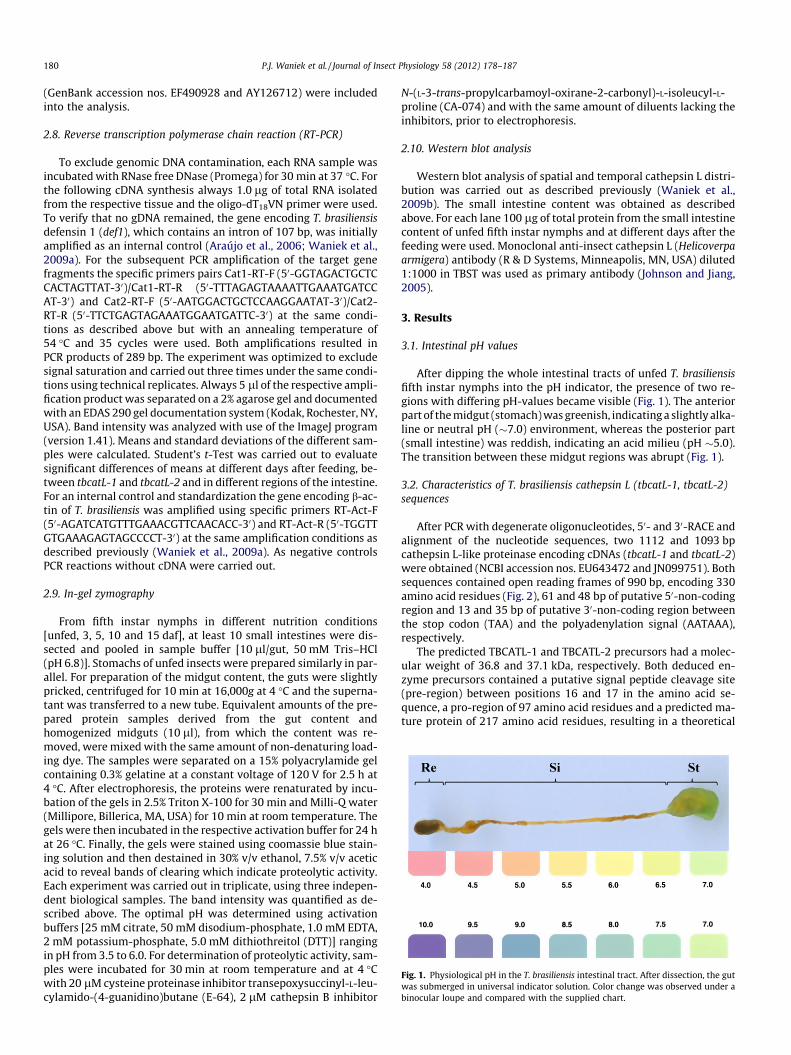
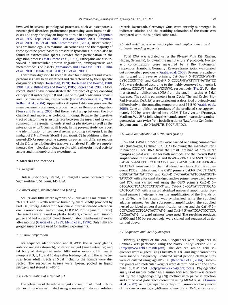
Fig. 1. Physiological pH in the T. brasiliensis intestinal tract. After dissection, the gutwas submerged in universal indicator solution. Color change was observed under abinocular loupe and compared with the supplied chart.
180 P.J. Waniek et al. / Journal of Insect Physiology 58 (2012) 178–187
(GenBank accession nos. EF490928 and AY126712) were includedinto the analysis.
2.8. Reverse transcription polymerase chain reaction (RT-PCR)
To exclude genomic DNA contamination, each RNA sample wasincubated with RNase free DNase (Promega) for 30 min at 37 �C. Forthe following cDNA synthesis always 1.0 lg of total RNA isolatedfrom the respective tissue and the oligo-dT18VN primer were used.To verify that no gDNA remained, the gene encoding T. brasiliensisdefensin 1 (def1), which contains an intron of 107 bp, was initiallyamplified as an internal control (Araújo et al., 2006; Waniek et al.,2009a). For the subsequent PCR amplification of the target genefragments the specific primers pairs Cat1-RT-F (50-GGTAGACTGCTCCACTAGTTAT-30)/Cat1-RT-R (50-TTTAGAGTAAAATTGAAATGATCCAT-30) and Cat2-RT-F (50-AATGGACTGCTCCAAGGAATAT-30)/Cat2-RT-R (50-TTCTGAGTAGAAATGGAATGATTC-30) at the same condi-tions as described above but with an annealing temperature of54 �C and 35 cycles were used. Both amplifications resulted inPCR products of 289 bp. The experiment was optimized to excludesignal saturation and carried out three times under the same condi-tions using technical replicates. Always 5 ll of the respective ampli-fication product was separated on a 2% agarose gel and documentedwith an EDAS 290 gel documentation system (Kodak, Rochester, NY,USA). Band intensity was analyzed with use of the ImageJ program(version 1.41). Means and standard deviations of the different sam-ples were calculated. Student’s t-Test was carried out to evaluatesignificant differences of means at different days after feeding, be-tween tbcatL-1 and tbcatL-2 and in different regions of the intestine.For an internal control and standardization the gene encoding b-ac-tin of T. brasiliensis was amplified using specific primers RT-Act-F(50-AGATCATGTTTGAAACGTTCAACACC-30) and RT-Act-R (50-TGGTTGTGAAAGAGTAGCCCCT-30) at the same amplification conditions asdescribed previously (Waniek et al., 2009a). As negative controlsPCR reactions without cDNA were carried out.
2.9. In-gel zymography
From fifth instar nymphs in different nutrition conditions[unfed, 3, 5, 10 and 15 daf], at least 10 small intestines were dis-sected and pooled in sample buffer [10 ll/gut, 50 mM Tris–HCl(pH 6.8)]. Stomachs of unfed insects were prepared similarly in par-allel. For preparation of the midgut content, the guts were slightlypricked, centrifuged for 10 min at 16,000g at 4 �C and the superna-tant was transferred to a new tube. Equivalent amounts of the pre-pared protein samples derived from the gut content andhomogenized midguts (10 ll), from which the content was re-moved, were mixed with the same amount of non-denaturing load-ing dye. The samples were separated on a 15% polyacrylamide gelcontaining 0.3% gelatine at a constant voltage of 120 V for 2.5 h at4 �C. After electrophoresis, the proteins were renaturated by incu-bation of the gels in 2.5% Triton X-100 for 30 min and Milli-Q water(Millipore, Billerica, MA, USA) for 10 min at room temperature. Thegels were then incubated in the respective activation buffer for 24 hat 26 �C. Finally, the gels were stained using coomassie blue stain-ing solution and then destained in 30% v/v ethanol, 7.5% v/v aceticacid to reveal bands of clearing which indicate proteolytic activity.Each experiment was carried out in triplicate, using three indepen-dent biological samples. The band intensity was quantified as de-scribed above. The optimal pH was determined using activationbuffers [25 mM citrate, 50 mM disodium-phosphate, 1.0 mM EDTA,2 mM potassium-phosphate, 5.0 mM dithiothreitol (DTT)] rangingin pH from 3.5 to 6.0. For determination of proteolytic activity, sam-ples were incubated for 30 min at room temperature and at 4 �Cwith 20 lM cysteine proteinase inhibitor transepoxysuccinyl-L-leu-cylamido-(4-guanidino)butane (E-64), 2 lM cathepsin B inhibitor
N-(L-3-trans-propylcarbamoyl-oxirane-2-carbonyl)-L-isoleucyl-L-proline (CA-074) and with the same amount of diluents lacking theinhibitors, prior to electrophoresis.
2.10. Western blot analysis
Western blot analysis of spatial and temporal cathepsin L distri-bution was carried out as described previously (Waniek et al.,2009b). The small intestine content was obtained as describedabove. For each lane 100 lg of total protein from the small intestinecontent of unfed fifth instar nymphs and at different days after thefeeding were used. Monoclonal anti-insect cathepsin L (Helicoverpaarmigera) antibody (R & D Systems, Minneapolis, MN, USA) diluted1:1000 in TBST was used as primary antibody (Johnson and Jiang,2005).
3. Results
3.1. Intestinal pH values
After dipping the whole intestinal tracts of unfed T. brasiliensisfifth instar nymphs into the pH indicator, the presence of two re-gions with differing pH-values became visible (Fig. 1). The anteriorpart of the midgut (stomach) was greenish, indicating a slightly alka-line or neutral pH (�7.0) environment, whereas the posterior part(small intestine) was reddish, indicating an acid milieu (pH �5.0).The transition between these midgut regions was abrupt (Fig. 1).
3.2. Characteristics of T. brasiliensis cathepsin L (tbcatL-1, tbcatL-2)sequences
After PCR with degenerate oligonucleotides, 50- and 30-RACE andalignment of the nucleotide sequences, two 1112 and 1093 bpcathepsin L-like proteinase encoding cDNAs (tbcatL-1 and tbcatL-2)were obtained (NCBI accession nos. EU643472 and JN099751). Bothsequences contained open reading frames of 990 bp, encoding 330amino acid residues (Fig. 2), 61 and 48 bp of putative 50-non-codingregion and 13 and 35 bp of putative 30-non-coding region betweenthe stop codon (TAA) and the polyadenylation signal (AATAAA),respectively.
The predicted TBCATL-1 and TBCATL-2 precursors had a molec-ular weight of 36.8 and 37.1 kDa, respectively. Both deduced en-zyme precursors contained a putative signal peptide cleavage site(pre-region) between positions 16 and 17 in the amino acid se-quence, a pro-region of 97 amino acid residues and a predicted ma-ture protein of 217 amino acid residues, resulting in a theoretical
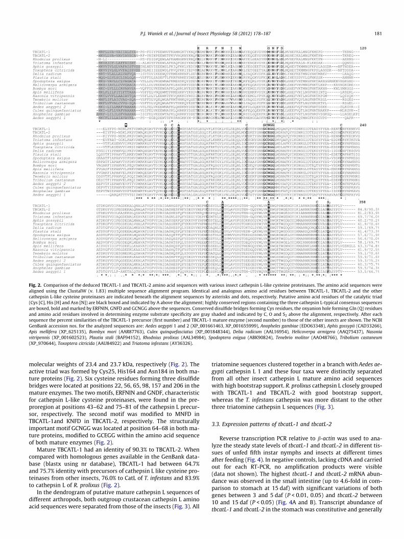
Fig. 2. Comparison of the deduced TBCATL-1 and TBCATL-2 amino acid sequences with various insect cathepsin L-like cysteine proteinases. The amino acid sequences werealigned using the ClustalW (v. 1.83) multiple sequence alignment program. Identical and analogous amino acid residues between TBCATL-1, TBCATL-2 and the othercathepsin L-like cysteine proteinases are indicated beneath the alignment sequences by asterisks and dots, respectively. Putative amino acid residues of the catalytic triad(Cys [C], His [H] and Asn [N]) are black boxed and indicated by A above the alignment; highly conserved regions containing the three cathepsin L-typical consensus sequencesare boxed, bold and marked by ERFNIN, GNFD and GCNGG above the sequences. Conserved disulfide bridges forming Cys residues, the oxyanion hole forming Gln (Q) residuesand amino acid residues involved in determining enzyme substrate specificity are gray shaded and indicated by C, O and S2 above the alignment, respectively. After eachsequence the percent similarities of the TBCATL-1 precursor (first number) and TBCATL-1 mature enzyme (second number) to those of the other insects are shown. The NCBIGenBank accession nos. for the analyzed sequences are: Aedes aegypti 1 and 2 (XP_001661463, XP_001655999), Anopheles gambiae (EDO63348), Aphis gossypii (CAD33266),Apis mellifera (XP_625135), Bombyx mori (AAR87763), Culex quinquefasciatus (XP_001848344), Delia radicum (AAL16954), Helicoverpa armigera (AAQ75437), Nasoniavitripennis (XP_001602523), Plautia stali (BAF94152), Rhodnius prolixus (AAL34984), Spodoptera exigua (ABK90824), Tenebrio molitor (AAO48766), Tribolium castaneum(XP_970644), Toxoptera citricida (AAU84922) and Triatoma infestans (AY36326).
P.J. Waniek et al. / Journal of Insect Physiology 58 (2012) 178–187 181
molecular weights of 23.4 and 23.7 kDa, respectively (Fig. 2). Theactive triad was formed by Cys25, His164 and Asn184 in both ma-ture proteins (Fig. 2). Six cysteine residues forming three disulfidebridges were located at positions 22, 56, 65, 98, 157 and 206 in themature enzymes. The two motifs, ERFNIN and GNDF, characteristicfor cathepsin L-like cysteine proteinases, were found in the pre-proregion at positions 43–62 and 75–81 of the cathepsin L precur-sor, respectively. The second motif was modified to MNFD inTBCATL-1and KNFD in TBCATL-2, respectively. The structurallyimportant motif GCNGG was located at position 64–68 in both ma-ture proteins, modified to GCEGG within the amino acid sequenceof both mature enzymes (Fig. 2).
Mature TBCATL-1 had an identity of 90.3% to TBCATL-2. Whencompared with homologous genes available in the GenBank data-base (blastx using nr database), TBCATL-1 had between 64.7%and 75.7% identity with precursors of cathepsin L like cysteine pro-teinases from other insects, 76.0% to CatL of T. infestans and 83.9%to cathepsin L of R. prolixus (Fig. 2).
In the dendrogram of putative mature cathepsin L sequences ofdifferent arthropods, both outgroup crustacean cathepsin L aminoacid sequences were separated from those of the insects (Fig. 3). All
triatomine sequences clustered together in a branch with Aedes ae-gypti cathepsin L 1 and these four taxa were distinctly separatedfrom all other insect cathepsin L mature amino acid sequenceswith high bootstrap support. R. prolixus cathepsin L closely groupedwith TBCATL-1 and TBCATL-2 with good bootstrap support,whereas the T. infestans cathepsin was more distant to the otherthree triatomine cathepsin L sequences (Fig. 3).
3.3. Expression patterns of tbcatL-1 and tbcatL-2
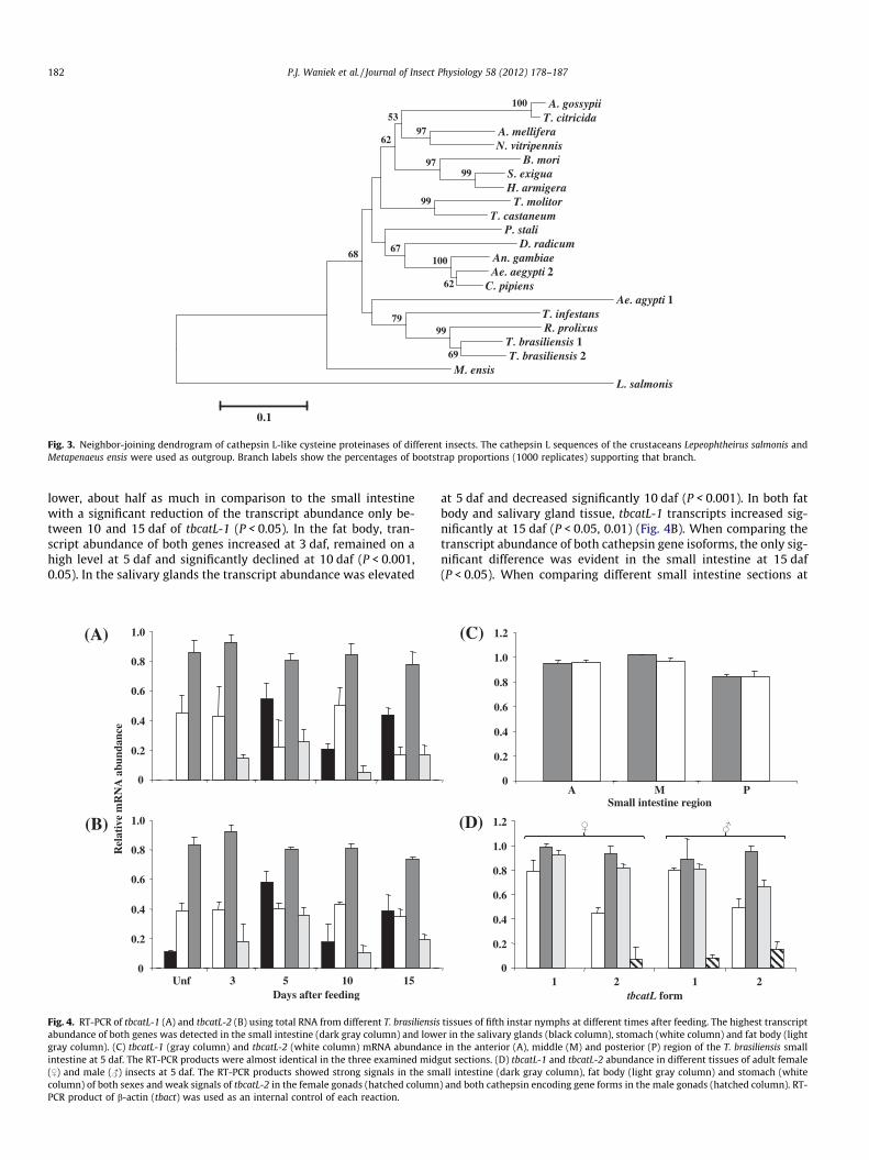
Reverse transcription PCR relative to b-actin was used to ana-lyze the steady state levels of tbcatL-1 and tbcatL-2 in different tis-sues of unfed fifth instar nymphs and insects at different timesafter feeding (Fig. 4). In negative controls, lacking cDNA and carriedout for each RT-PCR, no amplification products were visible(data not shown). The highest tbcatL-1 and tbcatL-2 mRNA abun-dance was observed in the small intestine (up to 4.6-fold in com-parison to stomach at 15 daf) with significant variations of bothgenes between 3 and 5 daf (P < 0.01, 0.05) and tbcatL-2 between10 and 15 daf (P < 0.05) (Fig. 4A and B). Transcript abundance oftbcatL-1 and tbcatL-2 in the stomach was constitutive and generally
A. gossypiiT. citricida
A. melliferaN. vitripennis
B. moriS. exiguaH. armigera
T. molitorT. castaneum
P. staliD. radicum
An. gambiaeAe. aegypti 2
C. pipiensAe. agypti 1
T. infestansR. prolixus
T. brasiliensis 1T. brasiliensis 2
M. ensisL. salmonis
100
99
69
99
62
100
99
97
97
79
67
53
62
68
0.1
Fig. 3. Neighbor-joining dendrogram of cathepsin L-like cysteine proteinases of different insects. The cathepsin L sequences of the crustaceans Lepeophtheirus salmonis andMetapenaeus ensis were used as outgroup. Branch labels show the percentages of bootstrap proportions (1000 replicates) supporting that branch.
182 P.J. Waniek et al. / Journal of Insect Physiology 58 (2012) 178–187
lower, about half as much in comparison to the small intestinewith a significant reduction of the transcript abundance only be-tween 10 and 15 daf of tbcatL-1 (P < 0.05). In the fat body, tran-script abundance of both genes increased at 3 daf, remained on ahigh level at 5 daf and significantly declined at 10 daf (P < 0.001,0.05). In the salivary glands the transcript abundance was elevated
Fig. 4. RT-PCR of tbcatL-1 (A) and tbcatL-2 (B) using total RNA from different T. brasiliensisabundance of both genes was detected in the small intestine (dark gray column) and lowgray column). (C) tbcatL-1 (gray column) and tbcatL-2 (white column) mRNA abundancintestine at 5 daf. The RT-PCR products were almost identical in the three examined mid($) and male (#) insects at 5 daf. The RT-PCR products showed strong signals in the smcolumn) of both sexes and weak signals of tbcatL-2 in the female gonads (hatched columnPCR product of b-actin (tbact) was used as an internal control of each reaction.
at 5 daf and decreased significantly 10 daf (P < 0.001). In both fatbody and salivary gland tissue, tbcatL-1 transcripts increased sig-nificantly at 15 daf (P < 0.05, 0.01) (Fig. 4B). When comparing thetranscript abundance of both cathepsin gene isoforms, the only sig-nificant difference was evident in the small intestine at 15 daf(P < 0.05). When comparing different small intestine sections at
tissues of fifth instar nymphs at different times after feeding. The highest transcripter in the salivary glands (black column), stomach (white column) and fat body (lighte in the anterior (A), middle (M) and posterior (P) region of the T. brasiliensis smallgut sections. (D) tbcatL-1 and tbcatL-2 abundance in different tissues of adult femaleall intestine (dark gray column), fat body (light gray column) and stomach (white) and both cathepsin encoding gene forms in the male gonads (hatched column). RT-

P.J. Waniek et al. / Journal of Insect Physiology 58 (2012) 178–187 183
5 daf, only slight differences in the transcript abundance of bothgenes were observed between tissues coming from anterior, mid-dle and posterior region of this midgut section (Fig. 4C).
In adult insects at 5 daf the highest tbcatL-1 and tbcatL-2 mRNAconcentrations were detected in the small intestine without signif-icant differences between genes and sexes, respectively (Fig. 4D).Slightly lower concentrations were detected for tbcatL-1 andtbcatL-2 transcripts in the female and male stomach and fat body(Fig. 4D). The tbcatL-2 transcript abundances detected in the smallintestine tissue of female and male insects, respectively, were al-ways significantly higher in comparison to that of fat body(P < 0.05, 0.01). In comparison to other tissues the abundance ofboth cathepsin L encoding mRNAs in the small intestine was ingeneral significantly higher (P < 0.05–0.0001), except when com-paring the tbcatL-1 small intestine concentrations with those ofmale stomach and fat body of both sexes. Transcript abundancesof tbcatL-1 were significantly higher than tbcatL-2 in the stomach(P < 0.01, 0.05) and fat body (P < 0.05). In female fat body bothcathepsin L encoding mRNAs were significantly more abundantthan in males (P < 0.05). TbcatL-2 transcripts were abundant inthe gonads of both sexes whereas tbcatl-1 was only detectable inthe testis, always in a significant lower level than in the other tis-sues of adult insects (Fig. 4D). When comparing the transcript con-centrations in the fat body of fifth instar nymphs at 5 daf withadult insects, in female and male bugs both tbcatL-1 and tbcatL-2were significantly more abundant (P < 0.0005, 0.005, 0.001).
3.4. Proteolytic activity of triatomine midguts
To determine the pattern of midgut proteinase activity with re-spect to pH in fifth instar nymphs of T. brasiliensis the wide-rangingproteinase substrate gelatine was used. Gelatinase activity of elec-trophoretic separated proteins led to a degradation of the gelatinematrix and appeared in colorless, non-stainable areas in the gel.Only fresh midgut content samples showed proteolytic activity,samples stored at �20 �C lost the major part of their activity andcould not be visualized by the methodology used in the presentstudy (data not shown). Both, the small intestine content (Fig. 5)and the small intestine tissue samples (data not shown) showedup to four distinct bands of proteolytic degradation, although theactivity of the gut content was always more intense. Stomach con-tent of unfed fifth instar nymphs never generated proteolytic activ-ity bands (data not shown).
Content of small intestine at 5 daf produced three broad prote-olytic activity bands corresponding to the molecular weights ofcysteine proteinases (about 28–35 kDa), showing the maximumintensity at pH 4.5. Therefore further experiments were carriedout at this pH value. Also among the other tested conditions prote-olytic degradation of gelatine became visible (Fig. 5A). Only at a pH3.5 and 4.0 an additional band of about 45 kDa was visible in T. bra-siliensis samples. In small intestine homogenates of T. infestans this45 kDa band remained visible also in all tested pH values in a sim-ilar intensity (data not shown). The other activity band detected inthe small intestine of T. infestans slightly differed in their molecularweight from those of T. brasiliensis (Fig. 5B).
Using specific proteinase inhibitors, the analysis revealed thatthe midgut activity contained cysteine like enzymes in small intes-tine samples at 5 daf (Fig. 5B). E-64 fully inhibited all proteinaseactivity bands of T. brasiliensis after 30 min incubation at roomtemperature, while in T. infestans a residual activity of the 45 kDaband remained (Fig. 5B). After incubation with the specific cathep-sin B inhibitor CA-074, in T. infestans 22.9% and in T. brasiliensis72.5% of remaining activity was detected. After incubation withE-64 at 4 �C a residual activity was visible in T. brasiliensis smallintestine samples, indicating a minor affinity of the inhibitor tothe enzyme at low temperatures (data not shown).
Cathepsin activity was detected in unfed insects and at 3, 5 and10 daf, at 15 daf no activity was observed. Proteolytic activity in-creased at 3 daf and reached its maximum at 5 daf (Fig. 5C).
3.5. Detection of T. brasiliensis cathepsin L by immunoblotting
To verify the zymography results of intestinal triatominecathepsins, the midgut content samples were separated by SDS–PAGE and analyzed by immuno blotting using specific antibodiesto Helicoverpa armigera cathepsin L. H. armigera mature cathepsinL amino acid sequence has an identity of 70.0 and 69.6% with thatof TBCATL-1 and TBCATL-2, respectively. The antibodies used in theexperiment showed affinity – even at the dilution of 1:1000 – toT. brasiliensis cathepsin L. Various band signals with a molecularweight ranging from about 30–38 kDa, similar to zymography,were detected (Fig. 6). Since the samples were separated underreducing conditions, the molecular weights differed slightly fromthose observed in in-gel zymograms.
4. Discussion
The establishment of a T. cruzi infection in the intestinal tract ofthe vector depends on many factors which modulate the parasite-vector interaction (Azambuja et al., 2005; Garcia et al., 2007). Themidgut of triatomines is the interface for development and multi-plication of parasites and exerts in its physiological and biochem-ical conditions a great influence on the T. cruzi development(Kollien and Schaub, 2000; Garcia et al., 2007, 2011). In somehematophagous insects (e.g. Pediculidae, Culicidae) the midgut isresponsible for both storage and digestion of the blood, whereasin Hemiptera these two functions occur in different midgut regions(Lehane, 2005; Waniek, 2009). Dipteran insects use serine protein-ases (trypsins and chymotrypsins) as their major luminal proteo-lytic enzymes in their digestion process, which are active atalkaline pH (Johnston et al., 1991; Chougule et al., 2005), the phy-logenetically distant hemipterans possess an rather differentdigestion, using cysteine and/or aspartic proteinases, which arehighly active in acidic conditions (Houseman, 1978; Housemanand Downe, 1980, 1981, 1982; Houseman et al., 1984; Lehane,1994; Borges et al., 2006). These peculiarities of the triatominemidgut physiology and digestion must be specifically taken intoaccount in the studies of triatomine–trypanosomatid interactions.
So far, triatomine cathepsin L encoding cDNA sequences havebeen identified and characterized in R. prolixus and T. infestans(Lopez-Ordoñez et al., 2001; Kollien et al., 2004). In their deducedamino acid sequences triatomine cathepsin L precursors are struc-turally similar, possess all characteristic motifs and are highly con-served but less as for example triatomine defensins or lysozymes(Kollien et al., 2004; Araújo et al., 2006; Waniek et al., 2009a,b).Triatomine cathepsins are synthesized as pre-proenzymes. In gen-eral signal peptides are approximately 20 amino acids long, hydro-phobic and cleaved during their passage to the endoplasmaticreticulum (von Heijne, 1983; Turk et al., 2000). Signal peptides ofinsect cathepsins L are within the usual boundaries and allTriatoma cathepsin B and L signal peptides, so far identified, arecomposed of 16 amino acid residues. Activation peptides areimportant for the proper folding of the protein and for protectionof the cell from potentially negative effects of unregulated proteo-lytic activity. Mainly at their C-terminus cathepsin L activationpeptides are more heterogenous in different species and in theirlength they exceed those of cathepsins B (Coulombe et al., 1996;Turk et al., 2000). The activation peptide length varied from 94 to110 amino acids in insect cathepsin L sequences analyzed in thepresent study. After cleavage, these peptides act as cathepsin Linhibitors, playing an important role in the activity regulation of

37.2 29.0
19.7
6.7
51.4
3.5 4.0 4.5 5.0 5.5 6.0
pH
98.2 kDa
37.2 29.0
19.7
6.7
51.4 98.2 kDa
37.2 29.0
19.7
6.7
51.4 98.2 kDa
1 2 3 4 5 6
Unf 3 5 10 15
0
20
40
60
80
100
3.5 4.0 4.5 5.0 5.5 6.0
Act
ivity
[%]
pH
0
20
40
60
80
100
Act
ivity
[%]
WI CA-074 WI CA-074
Days after feeding
(A)
(B)
(C)
0
20
40
60
80
100
120
Act
ivity
[%]
Unf 3 5 10 15Days after feeding
Fig. 5. Cysteine proteinase activity of small intestine content by gelatine in-gel zymography. (A) pH (3.0–6.0) dependence of T. brasiliensis intestinal proteolytic activity insamples at 5 daf, (B) Enzymatic inhibition of midgut proteinases: 1 and 3 without inhibitor (WI), 2 and 4 CA-074 (20 lM), 3 and 6 E-64 (2 lM), lanes 1–3 T. infestans, lanes 4–6T. brasiliensis, (C) Time course of intestinal proteolytic activities, unfed and different days after feeding. Graphs on the right side reflect the intensity of the respective band.Bands used for quantitation are marked with arrowheads.
184 P.J. Waniek et al. / Journal of Insect Physiology 58 (2012) 178–187
these digestive enzymes (Coulombe et al., 1996; Cygler and Mort,1997). Considering the ERFNIN and GCNGG motifs, important forthe globular folding of the N-terminus of the activation peptide(Coulombe et al., 1996), the T. infestans cathepsin L sequence (ERY-NIN, GCDGG) differed from that of T. brasiliensis and R. prolixus(ERFNIN, GCEGG). The GNFD motif was more variable, modifiedto KNFD in TBCATL-2 and T. infestans cathepsin L, MNFD inTBCATL-1 and KNLF in the R. prolixus cathepsin L amino acid se-quence (Lopez-Ordoñez et al., 2001; Kollien et al., 2004). The initialamino acids of the mature enzyme (Leu-Pro), the number of disul-fide bridge forming cysteine residues, the active site and S2 resi-dues were identical in all four triatomine cathepsin L sequences.Both mature T. brasiliensis cathepsin L amino acid sequences hada closer identity with cathepsin L of R. prolixus than that of T. infe-stans. Therefore the sequence of T. infestans was separated from theother three triatomine cathepsins in the dendrogram. This resultindicates the occurrence of, at least, two cathepsin L subgroupsin triatomines. T. brasiliensis and T. infestans are phylogenetically
closer than T. brasiliensis and R. prolixus, therefore TBCATL-1 andTBCATL-2 should cluster together with the amino acid sequenceof T. infestans. Since this is not the case, we can conclude thatTBCATL-1/-2 and R. prolixus cathepsin L encoding genes might beorthologous counterparts, whereas the more distant T. infestanscathepsin L belongs to a second triatomine cathepsin L group. Ifwe include different cathepsin B, cathepsin D, carboxy- and ami-no-peptidase isoforms, so far identified at DNA and protein level,the complexity of the triatomine digestive system becomes clearer.
Expression analyses by RT-PCR and northern blotting haveshown high cathepsin L transcript abundance in the posteriorintestine (small intestine) of R. prolixus whereas in the crop (stom-ach) cathepsin L mRNA was absent (Lopez-Ordoñez et al., 2001).These authors also have shown high cathepsin L transcript abun-dance in second instar nymphs, lower in unfed first instar nymphsand in fed first, third and fourth instars nymphs but absent in fifthinstars. These findings are surprising as the last nymphal stage isalso strongly dependent on blood digestion in view of nutrient
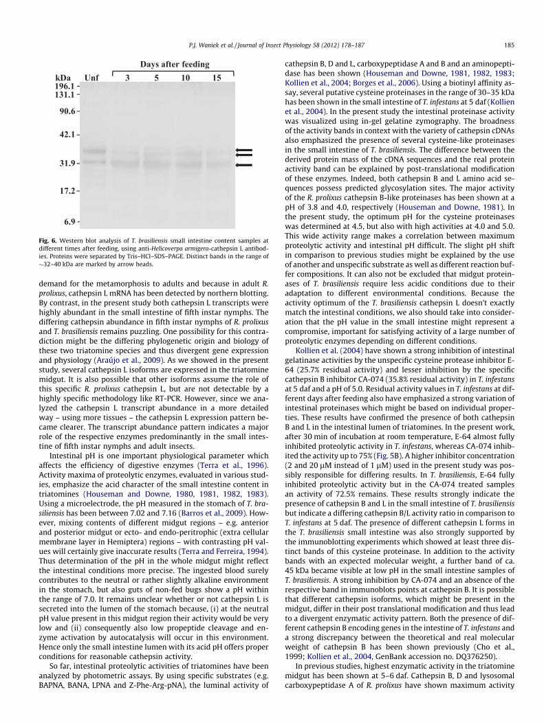
Fig. 6. Western blot analysis of T. brasiliensis small intestine content samples atdifferent times after feeding, using anti-Helicoverpa armigera-cathepsin L antibod-ies. Proteins were separated by Tris–HCl–SDS–PAGE. Distinct bands in the range of�32–40 kDa are marked by arrow heads.
P.J. Waniek et al. / Journal of Insect Physiology 58 (2012) 178–187 185
demand for the metamorphosis to adults and because in adult R.prolixus, cathepsin L mRNA has been detected by northern blotting.By contrast, in the present study both cathepsin L transcripts werehighly abundant in the small intestine of fifth instar nymphs. Thediffering cathepsin abundance in fifth instar nymphs of R. prolixusand T. brasiliensis remains puzzling. One possibility for this contra-diction might be the differing phylogenetic origin and biology ofthese two triatomine species and thus divergent gene expressionand physiology (Araújo et al., 2009). As we showed in the presentstudy, several cathepsin L isoforms are expressed in the triatominemidgut. It is also possible that other isoforms assume the role ofthis specific R. prolixus cathepsin L, but are not detectable by ahighly specific methodology like RT-PCR. However, since we ana-lyzed the cathepsin L transcript abundance in a more detailedway – using more tissues – the cathepsin L expression pattern be-came clearer. The transcript abundance pattern indicates a majorrole of the respective enzymes predominantly in the small intes-tine of fifth instar nymphs and adult insects.
Intestinal pH is one important physiological parameter whichaffects the efficiency of digestive enzymes (Terra et al., 1996).Activity maxima of proteolytic enzymes, evaluated in various stud-ies, emphasize the acid character of the small intestine content intriatomines (Houseman and Downe, 1980, 1981, 1982, 1983).Using a microelectrode, the pH measured in the stomach of T. bra-siliensis has been between 7.02 and 7.16 (Barros et al., 2009). How-ever, mixing contents of different midgut regions – e.g. anteriorand posterior midgut or ecto- and endo-peritrophic (extra cellularmembrane layer in Hemiptera) regions – with contrasting pH val-ues will certainly give inaccurate results (Terra and Ferreira, 1994).Thus determination of the pH in the whole midgut might reflectthe intestinal conditions more precise. The ingested blood surelycontributes to the neutral or rather slightly alkaline environmentin the stomach, but also guts of non-fed bugs show a pH withinthe range of 7.0. It remains unclear whether or not cathepsin L issecreted into the lumen of the stomach because, (i) at the neutralpH value present in this midgut region their activity would be verylow and (ii) consequently also low propeptide cleavage and en-zyme activation by autocatalysis will occur in this environment.Hence only the small intestine lumen with its acid pH offers properconditions for reasonable cathepsin activity.
So far, intestinal proteolytic activities of triatomines have beenanalyzed by photometric assays. By using specific substrates (e.g.BAPNA, BANA, LPNA and Z-Phe-Arg-pNA), the luminal activity of
cathepsin B, D and L, carboxypeptidase A and B and an aminopepti-dase has been shown (Houseman and Downe, 1981, 1982, 1983;Kollien et al., 2004; Borges et al., 2006). Using a biotinyl affinity as-say, several putative cysteine proteinases in the range of 30–35 kDahas been shown in the small intestine of T. infestans at 5 daf (Kollienet al., 2004). In the present study the intestinal proteinase activitywas visualized using in-gel gelatine zymography. The broadnessof the activity bands in context with the variety of cathepsin cDNAsalso emphasized the presence of several cysteine-like proteinasesin the small intestine of T. brasiliensis. The difference between thederived protein mass of the cDNA sequences and the real proteinactivity band can be explained by post-translational modificationof these enzymes. Indeed, both cathepsin B and L amino acid se-quences possess predicted glycosylation sites. The major activityof the R. prolixus cathepsin B-like proteinases has been shown at apH of 3.8 and 4.0, respectively (Houseman and Downe, 1981). Inthe present study, the optimum pH for the cysteine proteinaseswas determined at 4.5, but also with high activities at 4.0 and 5.0.This wide activity range makes a correlation between maximumproteolytic activity and intestinal pH difficult. The slight pH shiftin comparison to previous studies might be explained by the useof another and unspecific substrate as well as different reaction buf-fer compositions. It can also not be excluded that midgut protein-ases of T. brasiliensis require less acidic conditions due to theiradaptation to different environmental conditions. Because theactivity optimum of the T. brasiliensis cathepsin L doesn’t exactlymatch the intestinal conditions, we also should take into consider-ation that the pH value in the small intestine might represent acompromise, important for satisfying activity of a large number ofproteolytic enzymes depending on different conditions.
Kollien et al. (2004) have shown a strong inhibition of intestinalgelatinase activities by the unspecific cysteine protease inhibitor E-64 (25.7% residual activity) and lesser inhibition by the specificcathepsin B inhibitor CA-074 (35.8% residual activity) in T. infestansat 5 daf and a pH of 5.0. Residual activity values in T. infestans at dif-ferent days after feeding also have emphasized a strong variation ofintestinal proteinases which might be based on individual proper-ties. These results have confirmed the presence of both cathepsinB and L in the intestinal lumen of triatomines. In the present work,after 30 min of incubation at room temperature, E-64 almost fullyinhibited proteolytic activity in T. infestans, whereas CA-074 inhib-ited the activity up to 75% (Fig. 5B). A higher inhibitor concentration(2 and 20 lM instead of 1 lM) used in the present study was pos-sibly responsible for differing results. In T. brasiliensis, E-64 fullyinhibited proteolytic activity but in the CA-074 treated samplesan activity of 72.5% remains. These results strongly indicate thepresence of cathepsin B and L in the small intestine of T. brasiliensisbut indicate a differing cathepsin B/L activity ratio in comparison toT. infestans at 5 daf. The presence of different cathepsin L forms inthe T. brasiliensis small intestine was also strongly supported bythe immunoblotting experiments which showed at least three dis-tinct bands of this cysteine proteinase. In addition to the activitybands with an expected molecular weight, a further band of ca.45 kDa became visible at low pH in the small intestine samples ofT. brasiliensis. A strong inhibition by CA-074 and an absence of therespective band in immunoblots points at cathepsin B. It is possiblethat different cathepsin isoforms, which might be present in themidgut, differ in their post translational modification and thus leadto a divergent enzymatic activity pattern. Both the presence of dif-ferent cathepsin B encoding genes in the intestine of T. infestans anda strong discrepancy between the theoretical and real molecularweight of cathepsin B has been shown previously (Cho et al.,1999; Kollien et al., 2004, GenBank accession no. DQ376250).
In previous studies, highest enzymatic activity in the triatominemidgut has been shown at 5–6 daf. Cathepsin B, D and lysosomalcarboxypeptidase A of R. prolixus have shown maximum activity

186 P.J. Waniek et al. / Journal of Insect Physiology 58 (2012) 178–187
at 6 daf (Houseman and Downe, 1983). In the T. brasiliensis smallintestine, muramidase activity has reached its highest activity at5 daf (Waniek et al., 2009b). The results of the present studyshowed highest proteolytic activity at 5 daf and thus strongly cor-roborate these previous findings (Fig. 5C). It seems that 5–6 daf isthe period with the highest metabolic activity in triatomines. Alsoin the T. brasiliensis small intestine the proteolytic activity in-creased strongly at 3 daf and reached its peak at 5 daf. It is interest-ing that at 15 daf proteolytic activity was not detectable by in-gelzymography, whereas in unfed bugs a clear band was visible. Thisapparent paradox might be explained by loss of water and a subse-quent higher protein concentration in the intestinal tract of long-lasting starved (unfed) insects.
Acknowledgments
We thank Prof. O. Fernandes for technical support and Prof.V. Bongertz (FIOCRUZ, Rio de Janeiro) for the critical suggestionsand English corrections. We are also thankful to two anonymousreviewers for significant suggestions. The present work receivedfinancial support from Fundação de Amparo à Pesquisa do Estadodo Rio de Janeiro (FAPERJ – Cientista do Nosso Estado:E-26/100.456/2007), Conselho Nacional de DesenvolvimentoCientífico e Tecnológico (CNPq – Edital Universal: 472276/2006-9, PDJ: 151187/2009-6) and Fundação Oswaldo Cruz (FIOCRUZ).C.A.C.A. is a CNPq Research Fellow (158817/2010-9) and P.J.W. isa FAPERJ Research Fellow (E-26/152.913/2005).
References
Araújo, C.A.C., Waniek, P.J., Stock, P., Mayer, C., Jansen, A.M., Schaub, G.A., 2006.Sequence characterization and expression patterns of defensin and lysozymeencoding genes from the gut of the reduviid bug Triatoma brasiliensis. InsectBiochemistry and Molecular Biology 36, 547–560.
Araújo, C.A.C., Cabello, P.H., Jansen, A.M., 2007. Growth behaviour of twoTrypanosoma cruzi strains in single and mixed infections: in vitro and in theintestinal tract of the blood-sucking bug, Triatoma brasiliensis. Acta Tropica 101,225–231.
Araújo, C.A.C., Waniek, P.J., Jansen, A.M., 2008. Development of a Trypanosoma cruzi(TcI) isolate in the digestive tract of an unfamiliar vector, Triatoma brasiliensis(Hemiptera, Reduviidae). Acta Tropica 107, 195–199.
Araújo, C.A.C., Waniek, P.J., Jansen, A.M., 2009. An overview of Chagas disease andthe role of triatomines on its distribution in Brazil. Vector Borne and ZoonoticDiseases 9, 227–234.
Azambuja, P., Ratcliffe, N.A., Garcia, E.S., 2005. Towards an understanding of theinteractions of Trypanosoma cruzi and Trypanosoma rangeli within the reduviidinsect host Rhodnius prolixus. Anais da Academia Brasileira de Ciencias 77, 397–404.
Barros, V.C., Assumpção, J.G., Cadete, A.M., Santos, V.C., Cavalcante, R.R., Araújo, R.N.,Pereira, M.H., Gontijo, N.F., 2009. The role of salivary and intestinal complementsystem inhibitors in the midgut protection of triatomines and mosquitoes. PLoSONE 4, e6047.
Beard, C.B., Dotson, E.M., Pennington, P.M., Eichler, S., Cordon-Rosales, C., Durvasula,R.V., 2001. Bacterial symbiosis and paratransgenic control of vector-borneChagas disease. International Journal for Parasitology 31, 621–627.
Bendtsen, J.D., Nielsen, H., von Heijne, G., Brunak, S., 2004. Improved prediction ofsignal peptides: signalP 3.0. Journal of Molecular Biology 340, 783–795.
Billingsley, P.F., Downe, A.E.R., 1985. Cellular localisation of aminopeptidase in themidgut of Rhodnius prolixus Stål (Hemiptera: Reduviidae) during blooddigestion. Cell and Tissue Research 241, 421–428.
Borges, E.C., Machado, E.M.M., Garcia, E.S., Azambuja, P., 2006. Trypanosoma cruzi:effects of infection on cathepsin D activity in the midgut of Rhodnius prolixus.Experimental Parasitology 112, 130–133.
Brömme, D., Nallaseth, F.S., Turk, B., 2004. Production and activation of recombinantpapain-like cysteine proteases. Methods 32, 199–206.
C.D.C., 2002. Chagas Disease after Organ Transplantation – United States, 2001.MMWR 51, 210–212.
C.D.C., 2006. Chagas Disease after Organ Transplantation – Los Angeles, California,2006. MMWR 55, 798–800.
Chagas, C., 1909. Nova tripanozomiaze humana. Über eine neue Trypanosomiasisdes Menschen. Memórias do Instituto Oswaldo Cruz 1, 159–218.
Chapman, H.A., Riese, R.J., Shi, G.-P., 1997. Emerging roles for cysteine proteases inhuman biology. Annual Review of Physiology 59, 63–88.
Cho, W.-L., Tsao, S.-M., Hays, A.R., Walter, R., Chen, J.-S., Snigirevskaya, E.S., Raikhel,A.S., 1999. Mosquito cathepsin B-like protease involved in embryonicdegradation of vitellin is produced as a latent extraovarian precursor. Journalof Biological Chemistry 274, 13311–13321.
Chougule, N.P., Giri, A.P., Sainani, M.N., Gupta, V.S., 2005. Gene expression patternsof Helicoverpa armigera gut proteases. Insect Biochemistry and MolecularBiology 35, 355–367.
Cimerman, N., Brguljan, P.M., Krasovec, M., Suskovic, S., Kos, J., 2001. Circadian andconcentration profile of cathepsin S in sera from healthy subjects and asthmaticpatients. Pflügers Archive: European Journal of Physiology 442 (Suppl. 1), R204–R206.
Costa, J., Peterson, A.T., Beard, C.B., 2002. Ecologic niche modelling anddifferentiation of populations of Triatoma brasiliensis Neiva, 1911, the mostimportant Chagas‘ disease vector in northeastern Brazil (Hemiptera,Reduviidae, Triatominae). American Journal of Tropical Medicine and Hygiene67, 516–520.
Costa, J., Almeida, C.E., Dotson, E.M., Lins, A., Vinhaes, M., Silveira, A.C., Beard, C.B.,2003. The epidemiologic importance of Triatoma brasiliensis as a Chagas diseasevector in Brazil: a revision of domiciliary captures during 1993–1999. Memóriasdo Instituto Oswaldo Cruz 98, 443–449.
Coulombe, R., Grochulski, P., Sivaraman, J., Ménard, R., Mort, J.S., Cygler, M., 1996.Structure of human procathepsin L reveals the molecular basis of inhibition bythe prosegment. EMBO Journal 15, 5492–5503.
Coura, J.R., Borges-Pereira, J., 2010. Chagas disease: 100 years after its discovery. Asystemic review. Acta Tropica 115, 5–13.
Cygler, M., Mort, J.S., 1997. Proregion structure of members of the papainsuperfamily. Mode of inhibition of enzymatic activity. Biochimie 79, 645–652.
Dias, J.C.P., 2006. Notas sobre o Trypanosoma cruzi e suas características bio-ecológicas, como agente de enfermidades transmitidas por alimentos. Revistada Sociedade Brasileira de Medicina Tropical 39, 370–375.
Garcia, E.S., Gonzalez, M.S., Azambuja, P., Rembold, H., 1989. Chagas’ disease and itsinsect vector. Curring effect of azadirachtin A in the triatomine host, Rhodniusprolixus, from its parasite, Trypanosoma cruzi. In: Borovsky, D., Spielman, A.(Eds.), Host Development Mechanisms in Vector Arthropods. Florida UniversityPress, Vero Beach, FL, pp. 263–269.
Garcia, E.S., Gonzalez, M.S., Azambuja, P., Baralle, P.E., Fraidenraich, D., Torres, H.N.,Flawiá, M.M., 1995. Induction of Trypanosoma cruzi metacyclogenesis in the gutof the hematophagous insect vector, Rhodnius prolixus, by hemoglobin andpeptides carrying aD-globin sequences. Experimental Parasitology 81, 255–261.
Garcia, E.S., Ratcliffe, N.A., Whitten, M.M., Gonzalez, M.S., Azambuja, P., 2007.Exploring the role of insect host factors in the dynamics of Trypanosoma cruzi–Rhodnius prolixus interactions. Journal of Insect Physiology 53, 11–21.
Garcia, E.S., Genta, F.A., de Azambuja, P., Schaub, G.A., 2011. Interactions betweenintestinal compounds of triatomines and Trypanosoma cruzi. Trends inParasitology 26, 499–505.
Guarneri, A.A., Carvalho, M.G., Pereira, M.H., Diotaiuti, L., 2000. Potencial biológicodo Triatoma brasiliensis. Cadernos de Saúde Pública 16, 101–104.
von Heijne, G., 1983. Patterns of amino acids near signal sequence cleavage sites.European Journal of Biochemistry 133, 17–21.
Hou, W.-S., Li, W., Keyszer, G., Weber, E., Levy, R., Klein, M.J., Gravallese, E.M.,Goldring, S.R., Brömme, D., 2002. Comparison of cathepsins K and S expressionwithin the rheumatoid and osteoarthritic synovium. Arthritis and Rheumatism46, 663–674.
Houseman, J.G., 1978. A thiol-activated digestive proteinase from adults of Rhodniusprolixus Stål (Hemiptera: Reduviidae). Canadian Journal of Zoology 56, 1140–1143.
Houseman, J.G., Downe, A.E.R., 1980. Endoproteinase activity in the posteriormidgut of Rhodnius prolixus Stål (Hemiptera: Reduviidae). Insect Biochemistry10, 363–366.
Houseman, J.G., Downe, A.E.R., 1981. Exoproteinase activity in the posterior midgutof Rhodnius prolixus Stål (Hemiptera: Reduviidae). Insect Biochemistry 11, 579–582.
Houseman, J.G., Downe, A.E.R., 1982. Characterization of an acidic proteinase fromposterior midgut of Rhodnius prolixus Stål (Hemiptera: Reduviidae). InsectBiochemistry 12, 651–655.
Houseman, J.G., Downe, A.E.R., 1983. Activity cycles and the control of four digestiveproteinases in the posterior midgut of Rhodnius prolixus Stål (Hemiptera:Reduviidae). Journal of Insect Physiology 29, 141–148.
Houseman, J.G., MacNaughton, W.K., Downe, A.E.R., 1984. Cathepsin B andaminopeptidase in the posterior midgut of Euchistus euchistoides (Hemiptera:Pentatomidae). Canadian Entomologist 116, 1393–1396.
Johnson, G.D., Jiang, W., 2005. Characterization of cathepsin L secreted by Sf21insect cells. Archives of Biochemistry and Biophysics 444, 7–14.
Johnston, K.A., Lee, M.J., Gatehouse, J.A., Anstee, J.H., 1991. The partial purificationand characterization of serine protease activity in midgut of larval Helicoverpaarmigera. Insect Biochemistry 21, 389–397.
Kollien, A.H., Schaub, G.A., 2000. The development of Trypanosoma cruzi inTriatominae. Parasitology Today 16, 381–387.
Kollien, A.H., Waniek, P.J., Nisbet, A.J., Billingsley, P.F., Schaub, G.A., 2004. Activityand sequence characterization of two cysteine proteases in the digestive tract ofthe reduviid bug Triatoma infestans. Insect Molecular Biology 13, 569–579.
Kuipers, A.G.J., Jongsma, M.A., 2004. Isolation and molecular characterization ofcathepsin L-like cysteine protease cDNAs from western flower thrips(Frankliniella occidentalis). Comparative Biochemistry and Physiology B 139,65–75.
Lehane, M.J., 1994. Digestive enzymes, haemolysins and symbionts in the search forvaccines against blood-sucking insects. International Journal for Parasitology24, 27–32.
Lehane, M.J., 2005. Managing the blood meal. In: Lehane, M.J. (Ed.), The Biology ofBlood-Sucking in Insects. Cambridge University Press, Cambridge, pp. 84–115.

P.J. Waniek et al. / Journal of Insect Physiology 58 (2012) 178–187 187
Leist, M., Jäättelä, M., 2001. Triggering of apoptosis by cathepsins. Cell Death andDifferentiation 8, 324–326.
Liu, J., Shi, G.-P., Zhang, W.-Q., Zhang, G.-R., Xu, W.-H., 2006. Cathepsin L function ininsect moulting: molecular cloning and functional analysis in cotton bollworm,Helicoverpa armigera. Insect Molecular Biology 15, 823–834.
Lopez-Ordoñez, T., Rodriguez, M.H., De la Cruz Hernández-Hernández, F., 2001.Characterization of a cDNA encoding a cathepsin L-like protein of Rhodniusprolixus. Insect Molecular Biology 10, 505–511.
Matsumoto, I., Emori, Y., Abe, K., Arai, S., 1997. Characterization of a gene familyencoding cysteine proteinases of Sitophilus zeamais (maize weevil), and analysisof the protein distribution in various tissues including alimentary tract andgerm cells. Journal of Biochemistry 121, 464–476.
Mello, C.B., Azambuja, P., Garcia, E.S., Ratcliffe, N.A., 1996. Differential in vitro andin vivo behavior of three strains of Trypanosoma cruzi in the gut and hemolymphof Rhodnius prolixus. Experimental Parasitology 82, 112–121.
Rawlings, N.D., Barrett, A.J., 1993. Evolutionary families of peptidases. BiochemicalJournal 290, 205–218.
Shiba, H., Uchida, D., Kobayashi, H., Natori, M., 2001. Involvement of cathepsin B-and L-like proteinases in silk gland histolysis during metamorphosis of Bombyxmori. Archives of Biochemistry and Biophysics 390, 28–34.
Tamura, K., Dudley, J., Nei, M., Kumar, S., 2007. MEGA4: molecular evolutionarygenetics analysis (MEGA) software version 4.0. Molecular Biology and Evolution24, 1596–1599.
Tepel, C., Brömme, D., Herzog, V., Brix, C., 2000. Cathepsin K in thyroid epithelialcells: sequence, localization and possible function in extracellular proteolysis ofthyroglobulin. Journal of Cell Science 113, 4487–4498.
Terra, W.R., Ferreira, C., 1994. Insect digestive enzymes: properties,compartmentalization and function. Comparative Biochemistry andPhysiology B 109, 1–62.
Terra, W.R., Ferreira, C., 2005. Biochemistry of digestion. In: Gilbert, L.I., Iatrov, K.,Gill, S.S. (Eds.), Comprehensive Molecular Insect Science. Biochemistry andMolecular Biology. Elsevier, Oxford, pp. 171–224.
Terra, W.R., Ferreira, C., Baker, J.E., 1996. Compartmentalization of digestion. In:Lehane, M.J., Billingsley, P.F. (Eds.), Biology of the Insect Midgut. Chapman andHall, U.K, pp. 206–235.
Tryselius, Y., Hultmark, D., 1997. Cysteine proteinase 1 (CP1), a cathepsin L-likeenzyme expressed in the Drosophila melanogaster haemocyte cell line mbn-2.Insect Molecular Biology 6, 173–181.
Turk, B., Turk, D., Turk, V., 2000. Lysosomal cysteine proteases: more thanscavengers. Biochimica et Biophysica Acta 1477, 98–111.
Turk, V., Turk, B., Turk, D., 2001. Lysosomal cysteine proteases: facts andopportunities. EMBO Journal 20, 4629–4633.
Uchida, K., Ohmori, D., Ueno, T., Nishizuka, M., Eshita, Y., Fukunaga, A., Kominami, E.,2001. Preoviposition activation of cathepsin-like proteinases in degenerationovarian follicles of the mosquito Culex pipiens pallens. Developmental Biology237, 68–78.
Vitta, A.C.R., Mota, T.R.P., Diotaiuti, L., Lorenzo, M.G., 2007. The use of aggregationsignals by Triatoma brasiliensis (Heteroptera: Reduviidae). Acta Tropica 101,147–152.
Waniek, P.J., 2009. The digestive system of human lice: current advances andpotential applications. Physiological Entomology 34, 203–210.
Waniek, P.J., Castro, H.C., Sathler, P.C., Miceli, L., Jansen, A.M., Araújo, C.A.C., 2009a.Two novel defensin-encoding genes of the Chagas disease vector Triatomabrasiliensis (Reduviidae, Triatominae): gene expression and peptide-structuremodeling. Journal of Insect Physiology 55, 840–848.
Waniek, P.J., Mendonça-Lima, L., Menezes, G.B., Jansen, A.M., Araújo, C.A.C., 2009b.Recombinant expression and characterization of a lysozyme from the midgut ofTriatoma brasiliensis (Hemiptera, Reduviidae) in comparison with intestinalmuramidase activity. Physiological Entomology 34, 309–317.
WHO, 2002. Control of Chagas Disease. Second Report of the WHO expertcommittee, WHO, Geneva.
WHO, 2010. Chagas Disease (American Trypanosomiasis). <http://www.who.int/mediacentre/factsheets/fs340/en/>.
Yamamoto, Y., Takahashi, S.Y., 1993. Cysteine proteinase from Bombyx eggs: role inprogrammed degradation of yolk proteins during embryogenesis. ComparativeBiochemistry and Physiology B 106, 35–45.