journal of forensic medicine thakur et al., et al., j ... · unopened esophagus and then pulled...
TRANSCRIPT

Sudden Death- Case series of Ruptured Esophageal VaricesIshita Thakur, Rashid Nehal Khan, Abhijit Rudra
Forensic Medicine and Toxicology, Armed Forces Medical College, Pune
*Corresponding author: Ishita Thakur, Forensic Medicine and Toxicology, AFMC, Pune, India, Tel: 7263077907; E-mail: [email protected]
Received date: June 15, 2018; Accepted date: September 19, 2018; Published date: September 26, 2018
Copyright: © 2018 Thakur I, et al. This is an open-access article distributed under the terms of the Creative Commons Attribution License, which permits unrestricteduse, distribution, and reproduction in any medium, provided the original author and source are credited.
Abstract
IntroductionSudden death due to Gastroinstestinal (GI) causes amount to 10% of all sudden death. Esophageal varices is oneof the GI causes of sudden death. It develops as a complication of liver cirrhosis. The prevalence of esohgaealvarices in cirrhosis patients is 50% and mortality rate varies between 20%-35% with male female ratio of 1:9. Theone year bleeding rate of small and large varices is 5% and 15%. Mortality rate increases with increased age.
Case-SeriesSix case of sudden death were seen in the mortuary of a tertiary hospital. The six cases were males, alcoholics. Onautopsy, similar findings of cirrhotic liver and hemorrhagic mucosa of the stomach with bleeding spots on lower onethird of esophagus were observed.
DiscussionCollaterals develop with development of portal hypertension. Some sites are pre disposed to rupture like theesophageal varices. Risk factors of variceal rupture are red streaks in lumen of esophagus and deranged liverfunction tests. On gross examination, signs of deranged liver functions are noted. On internal examination,esophagus is everted attached to the stomach and varices are seen shine to through the mucosa. Liver had cirrhoticchanges and spleen was enlarged and congested in all the six cases.
ConclusionThe case series emphasizes the importance of fatal esophageal variceal hemorrhage as an important cause ofsudden death occurring outside the hospital in alcohol-addicted individuals, often in isolated scenarios.
Keywords: Esophageal varices; Cirrhosis; Liver
IntroductionWorld Health Organization (WHO) defines sudden death in its
International classification of diseases, version 10 (ICD-10) as death,non-violent and not otherwise explained, occurring in less than twentyfour hours from the onset of symptoms [1].
10% of sudden deaths are due to gastrointestinal causes [2].Esophageal varices is one of its cause. They are Porto-systemiccollaterals — i.e., vascular channels which link the portal venous andthe systemic venous circulation. It is a complication of cirrhosis. Itdevelops in sub mucosa of the distal esophagus. It is a life-threateningcomplication of portal hypertension. The mortality rate is 10%-30% ofall upper gastrointestinal bleeding [3]. The hemodynamic abnormalitymanifests as bleeding varices or ruptured varices in one third of thepatients [4].
Spontaneous stoppage of bleeding occurs in up to 40% ofindividuals [5,6] .Mortality with the first variceal bleeding is 30%-50%[7,8]. Highest number of deaths have been reported in winter thansummer [9].
Case SeriesCase 1:
A 47 year old male, known alcoholic brought to hospital inunresponsive state where he was declared dead. On postmortemexamination: faint post mortem lividity noted. On Examination ofabdominal cavity, stomach contained 700 mL of blood. Lower end ofesophagus shows thickened wall with mucus and streaks of blood.Liver had cirrhotic changes (yellowish colored with macronodules,fibrosed in appearance) and weighed 1100 g. On dissection, cut sectionwere pale and architecture was poor. Spleen was enlarged, brain andlungs were congested and oedematous. No other external/internalinjuries were present on the body.
Case 2:
A 44 year old male, known alcoholic with sudden onset ofhematemesis after which he fell unconscious and was taken to thehospital where he was declared dead. On examination, stomachcontained 1000 mL of blood in stomach Lower end of esophagusshows thickened wall with mucus and streaks of blood. Liver showedcirrhotic changes (yellowish colored with macronodules andmicronodules, fibrosed in appearance) weighing. On dissection, cutsection were pale. Spleen was enlarged, brain and lungs were congestedand oedematous and kidneys were pale. No other external/internalinjuries were present on the body.
Case 3:
A 35 year old male, known alcoholic with history of being foundunresponsive at home. The individual was unemployed and used to
Jour
nal of Forensic Medicine
ISSN: 2472-1026
Journal of Forensic Medicine Thakur et al., et al., J Forensic Med 2018, 3:2DOI: 10.4172/2472-1026.1000124
Case Series Open Access
J Forensic Med, an open access journalISSN:2472-1026
Volume 3 • Issue 2 • 1000124
stay alone at home. On examination, yellowish discoloration of thesclera, poor oral hygiene with dried blood stains at the nostrils. Onexamination, Stomach was cut along the greater curvature and itshowed 800 mL of blood with congested mucosa (Figures 1 and 2).Lower end of esophagus shows multiple streaks of blood and tortuousblood vessels. Liver weighed 1200 g, on its gross examination multiplenodules were present over the surface of the liver, yellow tan colour.On dissection of liver, pale cut sections with distorted architecturewere seen. Spleen was enlarged, brain and lungs were congested andoedematous. No other external/internal injuries were present on thebody.
Case 4:
A 41 year old male, known alcoholic with sudden onset ofheamtemesis after which individual fell unconscious and was declareddead. On examination eyes had yellowish discoloration of the sclera.Stomach was cut along the greater curvature and it showed 1000 mL ofblood with congested mucosa (Figure 3) Lower end of esophagusshows thickened wall with mucus and streaks of blood. Liver weighed1100 g, grossly multiple nodules were present over the surface of theliver, yellow tan colour (Figure 6). On dissection of liver, pale cutsections. Spleen was enlarged, brain and lungs were congested aoedematous and kidneys were pale. No other external /internal injurieswere present on the body.Liver was sent for Histopathologicalexamination (Figure 10).
Case 5:
60 year old man known alcoholic and diabetic with complaints ofhematemesis, weakness, abdominal pain since two months turningunresponsive and declared dead. Individual was admitted earlier forabdominal pain. Post mortem lividity not appreciated with obese builtand nourishment. Lower end of esophagus showed multiple bleedingpoints and tortuous blood vesssles (Figure 4). Stomach contained100mL of blood. Liver pale, with multiple macro- and micro-nodulesof varying size (Figure 5) and was sent for histopathology examination(Figure 7). Spleen. Kidneys were congested.
Case 6:
42 year old man chronic alcoholic with complaints of hematemesisand weakness since two months.individual was admitted for weaknessa month back and was given multivitamins. Post mortem lividityappreciated with average built and nourishment. Lower end ofesophagus shows multiple bleeding points and tortuous blood vessels.Peritoneal cavity contains 2 L of yellow colored clear fluid. Stomachcontained 200 mL of blood, mucosa rugae are hemorrhagic andprominent. Liver pale, with multiple nodules of varying size and wassent for histopathological examination (Figure 8). Enlarged spleen.
MethodologyPresent studys was conducted from the year 2014-2016 and we had
six cases with similar findings. In our study the age of the sampleranged from 35 years to 60 years. All the cases were that of male, withhistory of alcohol intake and deranged liver function. Mallory-Weisssyndrome was ruled out on gross examination. Esophagus is leftattached to the stomach. Stomach was opened along the greatercurvature of the stomach. A string is tied at the upper end of theunopened esophagus and then pulled through the lumen to evert theesophagus. Varices shine through the mucosa.
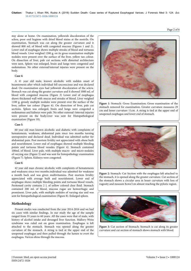
Figure 1: Stomach: Gross Examination; Gross examination of thestomach removed for examination. Greater curvature measures 29cm and lesser curvature 11cm. A string is tied at the upper end ofunopened esophagus and lower end of stomach.
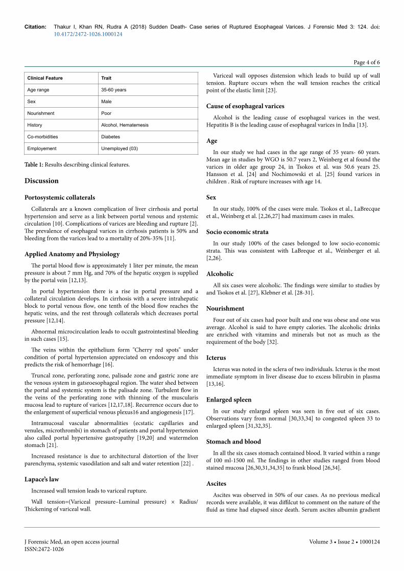
Figure 2: Stomach: Cut Section with the esophagus left attached tothe stomach, it is opened along the greater curvature. Cut section ofthe stomach shows a circular area in lesser curvature with loss ofrugosity and measure 8cmx7cm almost reaching the pyloric region.
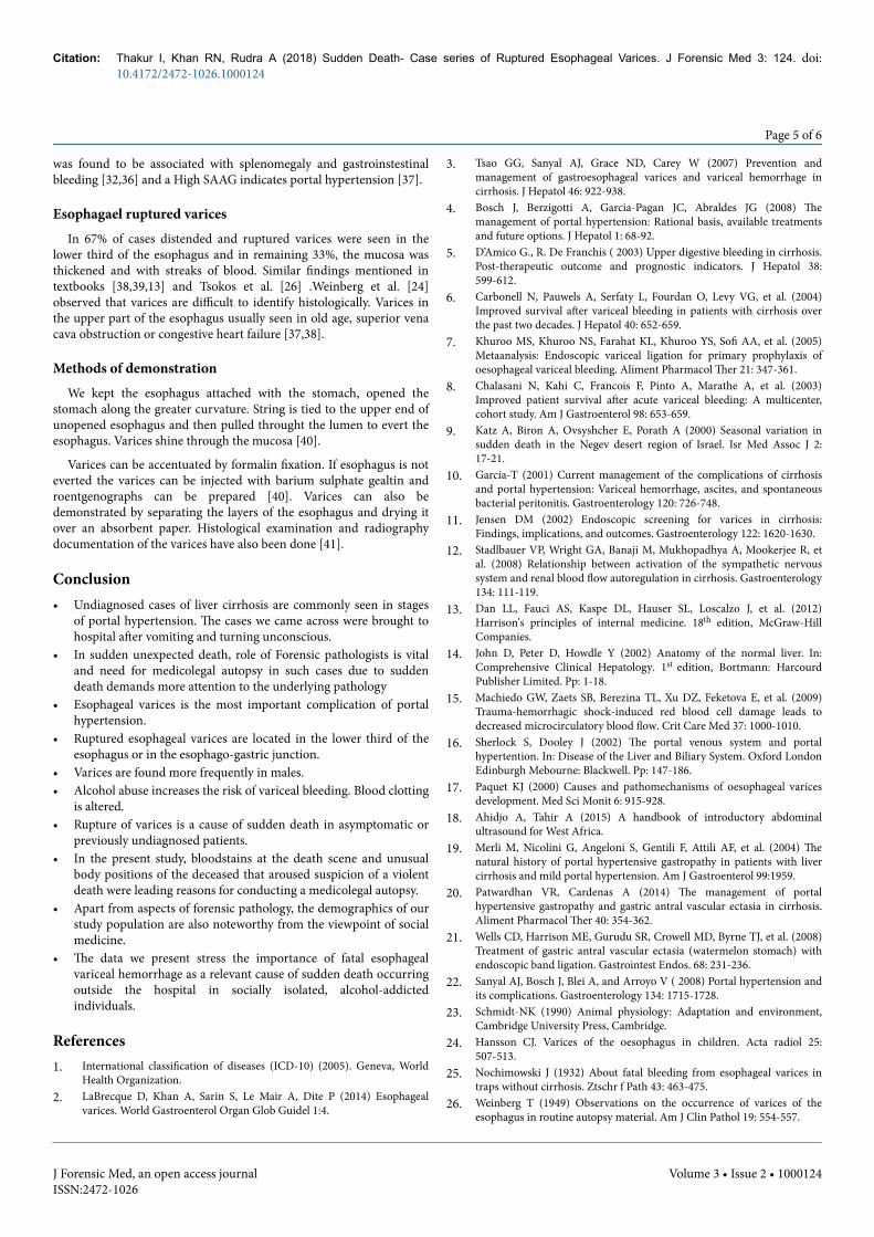
Figure 3: Cut section of Stomach; Stomach is cut along its greatercurvature and cut section of stomach shows stomach with blood.
Citation: Thakur I, Khan RN, Rudra A (2018) Sudden Death- Case series of Ruptured Esophageal Varices. J Forensic Med 3: 124. doi:10.4172/2472-1026.1000124
Page 2 of 6
J Forensic Med, an open access journalISSN:2472-1026
Volume 3 • Issue 2 • 1000124

Figure 4: Everted lumen of Esophagus; String is tied to the upperend of unopened esophagus and is then pulled through the lumento evert the esophagus. Varices are seen to shine through themucosa with multiple bleeding spots.
Figure 5: Liver on gross examination; Gross examination of the livershowing macro and micro nodules of varying size over the surfaceof the liver.
Figure 6: Liver on gross examination; Gross of the liver showingyellow tan colour with multiple nodules spread throughout thesurface of the liver.
Figure 7: Histopathology of Liver; Histopathology of liver showingperivenular and pericellular fibrosis (chicken-wire fence pattern)Central-central and/or central-portal fibrous septa Micronodularcirrhosis is seen in the histopathological examination of the liver.
Figure 8: Histopathology of Liver; Sections from the liver both rightand left lobes show regenerating parenchymal nodules seperated bydense bands of fibrosis.The liver cell plate thickness is more than 3cell layer thick. Mild periportal inflammation comprising oflymphocytes and plasma cells. Hepatocytes show evidence ofmacrovesicular steatosis.
ResultsIn present study, age range of cases were 35 years- 60 years. All the
cases were of male sex (100%), known alcoholic and belonged to lowersocio-economic strata. In all the cases there was history of vomiting ofblood and turning unconscious. Co-morbidities like diabetes andanemia were present in a two cases. Post mortem lividity was notappreciated in three of our cases and was faint in two and in one wasappreciated. Four out of six cases had poor built and one was obeseand one was average. Icterus was noted in the sclera of two individuals.Enlarged spleen was seen in five out of six cases. In all the six cases,stomach contained blood. It varied within a range of 100 ml-1500 ml.Ascites was noted in three out of six individuals in the case series. Inour study none of the six cases had any previous medical records. Infour out of six cases distended and ruptured varices were seen in thelower third of the esophagus and in two cases the mucosa washemorrhagic, congested and with streaks of blood (Table 1).
Citation: Thakur I, Khan RN, Rudra A (2018) Sudden Death- Case series of Ruptured Esophageal Varices. J Forensic Med 3: 124. doi:10.4172/2472-1026.1000124
Page 3 of 6
J Forensic Med, an open access journalISSN:2472-1026
Volume 3 • Issue 2 • 1000124
Clinical Feature Trait
Age range 35-60 years
Sex Male
Nourishment Poor
History Alcohol, Hematemesis
Co-morbidities Diabetes
Employement Unemployed (03)
Table 1: Results describing clinical features.
Discussion
Portosystemic collateralsCollaterals are a known complication of liver cirrhosis and portal
hypertension and serve as a link between portal venous and systemiccirculation [10]. Complications of varices are bleeding and rupture [2].The prevalence of esophageal varices in cirrhosis patients is 50% andbleeding from the varices lead to a mortality of 20%-35% [11].
Applied Anatomy and PhysiologyThe portal blood flow is approximately 1 liter per minute, the mean
pressure is about 7 mm Hg, and 70% of the hepatic oxygen is suppliedby the portal vein [12,13].
In portal hypertension there is a rise in portal pressure and acollateral circulation develops. In cirrhosis with a severe intrahepaticblock to portal venous flow, one tenth of the blood flow reaches thehepatic veins, and the rest through collaterals which decreases portalpressure [12,14].
Abnormal microcirculation leads to occult gastrointestinal bleedingin such cases [15].
The veins within the epithelium form "Cherry red spots" undercondition of portal hypertension appreciated on endoscopy and thispredicts the risk of hemorrhage [16].
Truncal zone, perforating zone, palisade zone and gastric zone arethe venous system in gatsroesophageal region. The water shed betweenthe portal and systemic system is the palisade zone. Turbulent flow inthe veins of the perforating zone with thinning of the muscularismucosa lead to rupture of varices [12,17,18]. Recurrence occurs due tothe enlargement of superficial venous plexus16 and angiogenesis [17].
Intramucosal vascular abnormalities (ecstatic capillaries andvenules, microthrombi) in stomach of patients and portal hypertensionalso called portal hypertensive gastropathy [19,20] and watermelonstomach [21].
Increased resistance is due to architectural distortion of the liverparenchyma, systemic vasodilation and salt and water retention [22] .
Lapace’s lawIncreased wall tension leads to variceal rupture.
Wall tension=(Variceal pressure–Luminal pressure) × Radius/Thickening of variceal wall.
Variceal wall opposes distension which leads to build up of walltension. Rupture occurs when the wall tension reaches the criticalpoint of the elastic limit [23].
Cause of esophageal varicesAlcohol is the leading cause of esophageal varices in the west.
Hepatitis B is the leading cause of esophageal varices in India [13].
AgeIn our study we had cases in the age range of 35 years- 60 years.
Mean age in studies by WGO is 50.7 years 2, Weinberg et al found thevarices in older age group 24, in Tsokos et al. was 50.6 years 25.Hansson et al. [24] and Nochimowski et al. [25] found varices inchildren . Risk of rupture increases with age 14.
SexIn our study, 100% of the cases were male. Tsokos et al., LaBrecque
et al., Weinberg et al. [2,26,27] had maximum cases in males.
Socio economic strataIn our study 100% of the cases belonged to low socio-economic
strata. This was consistent with LaBreque et al., Weinberger et al.[2,26].
AlcoholicAll six cases were alcoholic. The findings were similar to studies by
and Tsokos et al. [27], Klebner et al. [28-31].
NourishmentFour out of six cases had poor built and one was obese and one was
average. Alcohol is said to have empty calories. The alcoholic drinksare enriched with vitamins and minerals but not as much as therequirement of the body [32].
IcterusIcterus was noted in the sclera of two individuals. Icterus is the most
immediate symptom in liver disease due to excess bilirubin in plasma[13,16].
Enlarged spleenIn our study enlarged spleen was seen in five out of six cases.
Observations vary from normal [30,33,34] to congested spleen 33 toenlarged spleen [31,32,35].
Stomach and bloodIn all the six cases stomach contained blood. It varied within a range
of 100 ml-1500 ml. The findings in other studies ranged from bloodstained mucosa [26,30,31,34,35] to frank blood [26,34].
AscitesAscites was observed in 50% of our cases. As no previous medical
records were available, it was diffilcut to comment on the nature of thefluid as time had elapsed since death. Serum ascites albumin gradient
Citation: Thakur I, Khan RN, Rudra A (2018) Sudden Death- Case series of Ruptured Esophageal Varices. J Forensic Med 3: 124. doi:10.4172/2472-1026.1000124
Page 4 of 6
J Forensic Med, an open access journalISSN:2472-1026
Volume 3 • Issue 2 • 1000124
was found to be associated with splenomegaly and gastroinstestinalbleeding [32,36] and a High SAAG indicates portal hypertension [37].
Esophagael ruptured varicesIn 67% of cases distended and ruptured varices were seen in the
lower third of the esophagus and in remaining 33%, the mucosa wasthickened and with streaks of blood. Similar findings mentioned intextbooks [38,39,13] and Tsokos et al. [26] .Weinberg et al. [24]observed that varices are difficult to identify histologically. Varices inthe upper part of the esophagus usually seen in old age, superior venacava obstruction or congestive heart failure [37,38].
Methods of demonstrationWe kept the esophagus attached with the stomach, opened the
stomach along the greater curvature. String is tied to the upper end ofunopened esophagus and then pulled throught the lumen to evert theesophagus. Varices shine through the mucosa [40].
Varices can be accentuated by formalin fixation. If esophagus is noteverted the varices can be injected with barium sulphate gealtin androentgenographs can be prepared [40]. Varices can also bedemonstrated by separating the layers of the esophagus and drying itover an absorbent paper. Histological examination and radiographydocumentation of the varices have also been done [41].
Conclusion• Undiagnosed cases of liver cirrhosis are commonly seen in stages
of portal hypertension. The cases we came across were brought tohospital after vomiting and turning unconscious.
• In sudden unexpected death, role of Forensic pathologists is vitaland need for medicolegal autopsy in such cases due to suddendeath demands more attention to the underlying pathology
• Esophageal varices is the most important complication of portalhypertension.
• Ruptured esophageal varices are located in the lower third of theesophagus or in the esophago-gastric junction.
• Varices are found more frequently in males.• Alcohol abuse increases the risk of variceal bleeding. Blood clotting
is altered.• Rupture of varices is a cause of sudden death in asymptomatic or
previously undiagnosed patients.• In the present study, bloodstains at the death scene and unusual
body positions of the deceased that aroused suspicion of a violentdeath were leading reasons for conducting a medicolegal autopsy.
• Apart from aspects of forensic pathology, the demographics of ourstudy population are also noteworthy from the viewpoint of socialmedicine.
• The data we present stress the importance of fatal esophagealvariceal hemorrhage as a relevant cause of sudden death occurringoutside the hospital in socially isolated, alcohol-addictedindividuals.
References1. International classification of diseases (ICD-10) (2005). Geneva, World
Health Organization.2. LaBrecque D, Khan A, Sarin S, Le Mair A, Dite P (2014) Esophageal
varices. World Gastroenterol Organ Glob Guidel 1:4.
3. Tsao GG, Sanyal AJ, Grace ND, Carey W (2007) Prevention andmanagement of gastroesophageal varices and variceal hemorrhage incirrhosis. J Hepatol 46: 922-938.
4. Bosch J, Berzigotti A, Garcia-Pagan JC, Abraldes JG (2008) Themanagement of portal hypertension: Rational basis, available treatmentsand future options. J Hepatol 1: 68-92.
5. D’Amico G., R. De Franchis ( 2003) Upper digestive bleeding in cirrhosis.Post-therapeutic outcome and prognostic indicators. J Hepatol 38:599-612.
6. Carbonell N, Pauwels A, Serfaty L, Fourdan O, Levy VG, et al. (2004)Improved survival after variceal bleeding in patients with cirrhosis overthe past two decades. J Hepatol 40: 652-659.
7. Khuroo MS, Khuroo NS, Farahat KL, Khuroo YS, Sofi AA, et al. (2005)Metaanalysis: Endoscopic variceal ligation for primary prophylaxis ofoesophageal variceal bleeding. Aliment Pharmacol Ther 21: 347-361.
8. Chalasani N, Kahi C, Francois F, Pinto A, Marathe A, et al. (2003)Improved patient survival after acute variceal bleeding: A multicenter,cohort study. Am J Gastroenterol 98: 653-659.
9. Katz A, Biron A, Ovsyshcher E, Porath A (2000) Seasonal variation insudden death in the Negev desert region of Israel. Isr Med Assoc J 2:17-21.
10. Garcia-T (2001) Current management of the complications of cirrhosisand portal hypertension: Variceal hemorrhage, ascites, and spontaneousbacterial peritonitis. Gastroenterology 120: 726-748.
11. Jensen DM (2002) Endoscopic screening for varices in cirrhosis:Findings, implications, and outcomes. Gastroenterology 122: 1620-1630.
12. Stadlbauer VP, Wright GA, Banaji M, Mukhopadhya A, Mookerjee R, etal. (2008) Relationship between activation of the sympathetic nervoussystem and renal blood flow autoregulation in cirrhosis. Gastroenterology134: 111-119.
13. Dan LL, Fauci AS, Kaspe DL, Hauser SL, Loscalzo J, et al. (2012)Harrison's principles of internal medicine. 18th edition, McGraw-HillCompanies.
14. John D, Peter D, Howdle Y (2002) Anatomy of the normal liver. In:Comprehensive Clinical Hepatology. 1st edition, Bortmann: HarcourdPublisher Limited. Pp: 1-18.
15. Machiedo GW, Zaets SB, Berezina TL, Xu DZ, Feketova E, et al. (2009)Trauma-hemorrhagic shock-induced red blood cell damage leads todecreased microcirculatory blood flow. Crit Care Med 37: 1000-1010.
16. Sherlock S, Dooley J (2002) The portal venous system and portalhypertention. In: Disease of the Liver and Biliary System. Oxford LondonEdinburgh Mebourne: Blackwell. Pp: 147-186.
17. Paquet KJ (2000) Causes and pathomechanisms of oesophageal varicesdevelopment. Med Sci Monit 6: 915-928.
18. Ahidjo A, Tahir A (2015) A handbook of introductory abdominalultrasound for West Africa.
19. Merli M, Nicolini G, Angeloni S, Gentili F, Attili AF, et al. (2004) Thenatural history of portal hypertensive gastropathy in patients with livercirrhosis and mild portal hypertension. Am J Gastroenterol 99:1959.
20. Patwardhan VR, Cardenas A (2014) The management of portalhypertensive gastropathy and gastric antral vascular ectasia in cirrhosis.Aliment Pharmacol Ther 40: 354-362.
21. Wells CD, Harrison ME, Gurudu SR, Crowell MD, Byrne TJ, et al. (2008)Treatment of gastric antral vascular ectasia (watermelon stomach) withendoscopic band ligation. Gastrointest Endos. 68: 231-236.
22. Sanyal AJ, Bosch J, Blei A, and Arroyo V ( 2008) Portal hypertension andits complications. Gastroenterology 134: 1715-1728.
23. Schmidt-NK (1990) Animal physiology: Adaptation and environment,Cambridge University Press, Cambridge.
24. Hansson CJ. Varices of the oesophagus in children. Acta radiol 25:507-513.
25. Nochimowski J (1932) About fatal bleeding from esophageal varices intraps without cirrhosis. Ztschr f Path 43: 463-475.
26. Weinberg T (1949) Observations on the occurrence of varices of theesophagus in routine autopsy material. Am J Clin Pathol 19: 554-557.
Citation: Thakur I, Khan RN, Rudra A (2018) Sudden Death- Case series of Ruptured Esophageal Varices. J Forensic Med 3: 124. doi:10.4172/2472-1026.1000124
Page 5 of 6
J Forensic Med, an open access journalISSN:2472-1026
Volume 3 • Issue 2 • 1000124
27. Tsokos M, Türk EE (2002) Esophageal variceal hemorrhage presenting assudden death in outpatients: A study of 45 medicolegal autopsy cases.Arch Pathol Lab Med 126: 1197-200.
28. Kleber G, Sauerbruch T, Ansari H, Paumgartner G (1991) Prediction ofvariceal hemorrhage in cirrhosis: A prospective follow-up study.Gastroenterology 100: 1332-1337.
29. Lieber CS (2000) Alcohol: Its metabolism and interaction with nutrients.Annu Rev Nutr 2: 395-430.
30. Liangpunsakul S, Ulmer BJ, Chalasani N (2003) Predictors andimplications of severe hypersplenism in patients with cirrhosis. Am J MedSci 326: 111-116.
31. Takuma Y, Nouso K, Morimoto Y, Tomokuni J, Sahara A, et al. (2013)Measurement of spleen stiffness by acoustic radiation force impulseimaging identifies cirrhotic patients with esophageal varices.Gastroenterology 144: 92-101.
32. Hoefs JC (1983) Serum protein concentration and portal pressuredetermine the ascites fluid protein concentration in patients with chronicliver disease. J Lab Clin Med 102.
33. Okuda K, Kono K, Ohnishi K, Kimura K, Omata M, et al. (1984) Clinicalstudy of eighty-six cases of idiopathic portal hypertension andcomparison with cirrhosis wit splenomegaly. Gastroenterology 86:600-610.
34. Shah SH, Hayes PC, Allan PL, Nicoll J, Finlayson ND (1996)Measurement of spleen size and its relation to hypersplenism and portalhemodynamics in portal hypertension due to hepatic cirrhosis. Am JGastroenterol 91: 2580-2583.
35. Chalasani N, Imperiale TF, Ismail A, Sood G, Carey M, et al. (1999)Predictors of large esophageal varices in patients with cirrhosis. Am JGastroenterol 94: 3285-3291.
36. Garcia-Tsao G, Groszmann RJ, Fisher RL, Conn HO, Atterbury CE et al.(1985) Portal pressure, presence of gastroesophageal varices and varicealbleeding. Hepatology 5:419-424.
37. Kahsner JT (1942) Human pathology. 6th edition, Philadelphia: J. B.Lippincott Company. Pp: 563.
38. Boyd W (1944) Pathology of internal diseases. 4th edition, Philadelphia:Lea and Febiger. Pp: 322.
39. Christian HA(1942) Osier's principles and practice of medicine. 14th
editon, New york: Appleton-century Co. Inc. Pp: 38.40. Ludwig J (2002) Handbook of autopsy practice. Springer Science &
Business Media.41. Abramowsky CR, Gonzalvo AA (1975) Postmortem demonstration of
esophageal varices by a simple method. Am J Clin Pathol 64: 672-677.
Citation: Thakur I, Khan RN, Rudra A (2018) Sudden Death- Case series of Ruptured Esophageal Varices. J Forensic Med 3: 124. doi:10.4172/2472-1026.1000124
Page 6 of 6
J Forensic Med, an open access journalISSN:2472-1026
Volume 3 • Issue 2 • 1000124