journal of experimental & clinical cancer research biomed
TRANSCRIPT

BioMed Central
Journal of Experimental & Clinical Cancer Research
ss
Open AcceResearchA dosimetric comparison of different treatment plans of palliative spinal bone irradiation: analysis of dose coverage with respect to ICRU 50 reportFundagul Andic*, Sule Baz Cifci, Yasemin Ors, Umar Niang, Ahmet Dirier and Mustafa AdliAddress: Department of Radiation Oncology, Faculty of Medicine, Gaziantep University, Gaziantep, Turkey
Email: Fundagul Andic* - [email protected]; Sule Baz Cifci - [email protected]; Yasemin Ors - [email protected]; Umar Niang - [email protected]; Ahmet Dirier - [email protected]; Mustafa Adli - [email protected]
* Corresponding author
AbstractBackground: This study aimed to analyze three-dimensional (3D) dosimetric data of conventional two-dimensional (2D) palliative spinal bone irradiation using different reference points and treatment plans withrespect to the International Commission on Radiation Units and Measurements (ICRU) Report 50.
Methods: Forty-five simulation CT scans of 39 patients previously treated for thoraco-lumbar spinal bonemetastases were used. Three different treatment plans were created: (1) single posterior field plans usingthe ICRU reference points (ICRUrps); (2) single posterior field plans using the International BoneMetastasis Consensus Working Party reference points (IBMCrps); (3) two opposed anterior-posterior(AP-PA) field plans using the ICRUrps. The intended dose range for planning target volume (PTV) was 90%to 110% of the prescribed dose for AP-PA field plans. Cumulative dose-volume histograms were generatedfor each plan, and minimum, maximum and mean doses to the PTV, medulla spinalis, esophagus andintestines were analyzed.
Results: The mean percentages of minimum, maximum and mean PTV doses ± standard deviation were,respectively, 91 ± 1.3%, 108.8 ± 1.3% and 99.7 ± 1.3% in AP-PA field plans; 77.3 ± 2.6%, 122.2 ± 4.3% and99.8 ± 2.6% in ICRUrp single field plans; and 83.7 ± 3.3%, 133.9 ± 7.1% and 108.8 ± 3.3% in IBMCrp singlefield plans. Minimum doses of both single field plans were significantly lower (p < 0.001) while maximumdoses were significantly higher (p < 0.001) than AP-PA field plans. Minimum, maximum and mean doseswere higher in IBMCrp single field plans than in ICRUrp single field plans (p < 0.001). The mean medullaspinalis doses were lower in AP-PA field plans than single posterior field plans (p < 0.001). Maximum dosesfor medulla spinalis were higher than 120% of the prescribed dose in 22 of 45 (49%) IBMCrp single fieldplans. Mean esophagus and intestinal doses were higher (p < 0.001) in AP-PA field plans than single fieldplans, however, less than 95% of the prescribed dose.
Conclusion: In palliative spinal bone irradiation, 2D conventional single posterior field radiotherapy didnot accomplish the ICRU Report 50 recommendations for PTV dose distribution, while the AP-PA fieldplans did achieve the intended dose ranges with a homogenous distribution and reasonable doses to themedulla spinalis, esophagus and intestines.
Published: 7 January 2009
Journal of Experimental & Clinical Cancer Research 2009, 28:2 doi:10.1186/1756-9966-28-2
Received: 17 November 2008Accepted: 7 January 2009
This article is available from: http://www.jeccr.com/content/28/1/2
© 2009 Andic et al; licensee BioMed Central Ltd. This is an Open Access article distributed under the terms of the Creative Commons Attribution License (http://creativecommons.org/licenses/by/2.0), which permits unrestricted use, distribution, and reproduction in any medium, provided the original work is properly cited.
Page 1 of 7(page number not for citation purposes)

Journal of Experimental & Clinical Cancer Research 2009, 28:2 http://www.jeccr.com/content/28/1/2
BackgroundExternal beam radiotherapy is a well-recognized and effec-tive modality in the palliation of symptomatic bonemetastases and complication control [1]. Under- or over-dosing the target volume and dose heterogeneity may notbe major concerns, since many patients treated for pallia-tive purposes have short survival. However, long termsymptom control associated with bone involvement andnormal tissue complications becomes more vital in cancerpatients with long life-expectancy. Some breast and pros-tate cancer patients even with spinal cord compressionmay live for several years after radiotherapy.
Single posterior field or two opposed anterior-posteriorfields (AP-PA) conventional two-dimensional (2D) radio-therapy planning without dose volume information iswidely used for palliative spinal bone irradiation usingthe International Commission on Radiation Units andMeasurements reference points (ICRUrps) and the Inter-national Bone Metastasis Consensus Working Party refer-ence points (IBMCrps) [2,3].
To our knowledge, dosimetric assessment of conventional2D palliative spinal bone irradiation using three-dimen-sional (3D) dose information has not been reported. Thisstudy aimed to analyze 3D dosimetric data of palliativespinal bone irradiation using different reference pointsand treatment plans with respect to the InternationalCommission on Radiation Units and Measurements(ICRU) Report 50 [2].
MethodsCT simulationForty-five simulation CT scans of 39 patients previouslytreated for thoraco-lumbar spinal bone metastases wereused for treatment planning. CT scanning was performedwith a 6 detector helical CT (Brilliance, Philips MedicalSystems, Netherlands) and with a 5-mm slice thickness.
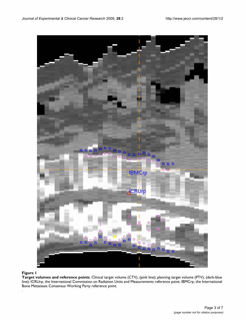
Volumes of interestTarget volumes were contoured in corresponding CT slices(Figure 1). One vertebra above and below the involvedvertebra(e) were included in the clinical target volume(CTV). However, the upper end-plate of the upper verte-bra and the lower end-plate of the lower vertebra were notincluded in the CTV, to limit the distal and proximal bor-ders of the treatment fields in the inter-vertebral space. Todetermine the planning target volume (PTV), 10 mm wasadded to CTV in lateral directions and 5 mm in anterior-posterior and superior-inferior directions. Treatmentfields were determined by adding 7–10 mm to the PTVusing multi-leaf collimators.
Portions of the esophagus located in thoracic radiother-apy fields, the intestines located in lumbar radiotherapyfields and the medulla spinalis in all fields were deline-ated as critical organs.
Treatment planningPrecise PLAN®2.11 (Elekta, Crawley, UK) treatment plan-ning system (TPS), which enables 3D conformal radio-therapy planning, was used for treatment plans. Tocalculate the dose distribution of the photon beam, theTPS uses an irregular field algorithm, for different depthsand field sizes, based on data measures in a phantom. Thealgorithm takes into account the inhomogeneity of thepatient's tissue and uses an integration scheme to evaluatethe scatter component of the dose. The dose calculationgrid is set to 2.5 mm.
Three different treatment plans were created (1) singleposterior field treatment plans using ICRUrps; (2) singleposterior field treatment plans using IBMCrps; and (3)two opposed anterior-posterior (AP-PA) field plans usingICRUrps.
The ICRUrp was defined as the center of the PTV, the IBM-Crp was defined as the mid-vertebral body point in thecentral plane, and the prescription dose was normalizedto these points (Figure 1). Dose distributions of treatmentplans in one case are shown in Figure 2.
The nominal prescribed dose was 2000 cGy in 5 fractionsusing 6-MV photons for posterior fields and 18-MV foranterior fields. In AP-PA field plans, beam weights wereused as 1 and 1.5–2 in AP and PA fields, while assuring theintended dose range of 90% to 110% of the prescribeddose for the PTV. No dose constraint was used in singleposterior field plans.
Cumulative dose-volume histograms (DVH) were gener-ated for each plan, and minimum, maximum and meandoses to the PTV, medulla spinalis, esophagus and intes-tines were calculated for both single posterior fields andAP-PA field plans.
Cumulative dose-volume histograms of treatment plansin one case for PTV and medulla spinalis are shown in Fig-ure 3.
Statistical analysisThe mean, minimum and maximum dose levels werecompared using the Paired-Samples T test for parametricdata on the PTV and medulla spinalis and the Wilcoxontest for non-parametric data on the esophagus and intes-tines. P-values of less than 0.05 were considered statisti-
Page 2 of 7(page number not for citation purposes)
Journal of Experimental & Clinical Cancer Research 2009, 28:2 http://www.jeccr.com/content/28/1/2
Page 3 of 7(page number not for citation purposes)
Target volumes and reference pointsFigure 1Target volumes and reference points. Clinical target volume (CTV), (pink line); planning target volume (PTV), (dark-blue line); ICRUrp, the International Commission on Radiation Units and Measurements reference point; IBMCrp, the International Bone Metastasis Consensus Working Party reference point.

Journal of Experimental & Clinical Cancer Research 2009, 28:2 http://www.jeccr.com/content/28/1/2
cally significant. Values are expressed as mean (range) ±standard deviation (SD).
ResultsDose ranges of the PTVs for all plans are shown in Table1. AP-PA field plans achieved the intended dose rangesand homogeneity for PTVs, unlike the single posteriorfield plans. Minimum doses of both single posterior fieldplans were significantly lower (p < 0.001) while maxi-mum doses were significantly higher (p < 0.001) than AP-PA field plans. Minimum, maximum and mean doseswere higher in IBMCrp single field plans with an increaseddose heterogeneity than in ICRUrp single field plans (p <0.001).
The mean depth of the PTV from skin surface in the cen-tral plane was 9.8 (7.4–13.5) ± 1.1 cm and the meanpatient thickness was 22.1 (14.4–29.1) ± 3.7 cm. Only intwo plans were the ICRUrps and IBMCrps located at thesame sites, which were in the mid-vertebral body. Of 45ICRUrps, 35 were located on the medulla spinalis behindthe vertebral body and 8 were located in the posterior 1/3
of the vertebral body. None of the ICRUrps were locatedin the anterior half of the vertebral body or anterior to thevertebral body.
The mean dose, expressed as percentages of the prescribeddose, to the portion of the esophagus in the thoracic radi-otherapy fields was 78.6% (70–85%) ± 4.1% in theICRUrp single field plans, 84.6% (74–92%) ± 5.5% in theIBMCrp single field plans and 94.5% (87–99%) ± 3.1% inthe AP-PA field plans. The mean dose to the intestineslocated in the lumbar radiotherapy fields was 66.2% (58–78%) ± 5.1% in the ICRUrp single field plans, 73.1% (64–88%) ± 6.2% in the IBMCrp single field plans and 90.8%(82–99%) ± 3.7% in the AP-PA fields plans. The meandoses to the esophagus and intestines were higher in theAP-PA field plans than in the single posterior field plans(p < 0.001).
Dose ranges to the medulla spinalis for all plans areshown in Table 2. The mean doses to the medulla spinaliswere lower in the AP-PA field plans than in the single pos-terior field plans (p < 0.001).In all IBMCrp single field
Dose distributions in one case for ICRUrp single field plan (A), IBMCrp single field plan (B) and two opposed anterior-poste-rior field plan (C)Figure 2Dose distributions in one case for ICRUrp single field plan (A), IBMCrp single field plan (B) and two opposed anterior-posterior field plan (C). ICRUrp, the International Commission on Radiation Units and Measurements reference point; IBMCrp, the International Bone Metastasis Consensus Working Party reference point. The isodose lines are shown as follows: 75% (blue), 80% (yellow), 90% (dark blue), 95% (red), 100 (pink), 110% (green), 115% (orange).
Page 4 of 7(page number not for citation purposes)

Journal of Experimental & Clinical Cancer Research 2009, 28:2 http://www.jeccr.com/content/28/1/2
plans, maximum doses to the medulla spinalis weregreater than 115% of the prescribed dose and in 22 of 45(49%) plans the maximum doses were greater than 120%of the prescribed dose. In only 4 ICRUrp single field plansdid the medulla spinalis receive a dose greater than 115%of the prescribed dose. In the AP-PA field plans, none of
the doses to the medulla spinalis exceeded 106% of pre-scribed dose.
DiscussionThe results of the present study showed that neither IBM-Crp nor the ICRUrp single posterior field plans accom-
Cumulative dose-volume histograms of one case for planning target volume (PTV) (dark-blue line) and medulla spinalis (red line) in single field plan using the International Commission on Radiation Units and Measurements reference point (circles), in single field plan using the International Bone Metastasis Consensus Working Party reference point (squares) and two opposed anterior-posterior field plan (triangles)Figure 3Cumulative dose-volume histograms of one case for planning target volume (PTV) (dark-blue line) and medulla spinalis (red line) in single field plan using the International Commission on Radiation Units and Meas-urements reference point (circles), in single field plan using the International Bone Metastasis Consensus Working Party reference point (squares) and two opposed anterior-posterior field plan (triangles).
Page 5 of 7(page number not for citation purposes)

Journal of Experimental & Clinical Cancer Research 2009, 28:2 http://www.jeccr.com/content/28/1/2
plished the ICRU Report 50 recommendations for dosedistribution, while the AP-PA field plans achieved theintended dose ranges and homogeneity.
The ICRU Report 50 recommends selecting a referencepoint that is clinically relevant and representative of thedose distribution throughout the PTV, where the dose canbe accurately determined and where there is no large dosegradient [2]. The point located at the center or central partof the PTV generally fulfills these requirements and is rec-ommended as the ICRU reference point (ICRUrp). Whilea homogeneous dose within 95% to 107% of the pre-scribed dose is recommended for the target volume, a var-iation of ± 10% from the prescribed dose is widely used inclinical practice and was used in the present study for AP-PA field plans [2].
Thoracic and lumbar spinal irradiation is performedeither with a single posterior field or two opposed AP-PAfields [4]. The International Bone Metastasis ConsensusWorking Party recommends dose prescriptions to themid-vertebral body for single-posterior fields and includ-ing at least one vertebral body above and below theinvolved vertebra(e) in the treatment volumes [3]. How-ever, these recommendations are not supported with dosi-metric data or treatment outcomes.
Radiotherapy planning and delivery, and dose distribu-tion may affect treatment outcome by dose coverage anddose heterogeneity in the target volume. Although severalstudies investigated optimal radiotherapy fractionation,the dose-volume effect on radiotherapy outcome, in terms
of pain relief and duration of response, has not been eval-uated [5-13]. Furthermore, higher re-treatment rates havebeen reported in single-fraction palliative radiotherapythan in multifraction radiotherapy [12-14]. The relationbetween higher re-treatment rates and physician bias, pri-mary site, pain severity and duration of symptoms hasbeen evaluated, but the relation between high re-treat-ment rates and dose coverage has not been investigated.Studies investigating the relationship between radiother-apy technique and treatment outcome would provideimportant information, particularly for patients with longlife-expectancies.
Dose heterogeneity may become vitally important inpatients with long life expectancies. Minimum target vol-ume doses as low as 70% of the prescribed dose maydiminish treatment success, while maximum target vol-ume doses reaching as high as 130% of the prescribeddose may cause serious normal-tissue side effects in suchpatients. In the present study, the mean minimum dosefor PTV in the ICRUrp single field plans was 77.3% (72–81%) ± 2.6% of the prescribed dose, and the mean maxi-mum dose for PTV in the IBMCrp single field plans was133.9% (115–147%) ± 7.1% of the prescribed dose.When the medulla spinalis doses were assed, maximumdoses were higher than 120% of the prescribed dose in 22of 45 (49%) IBMCrp single field plans but lower than106% of prescribed dose in all AP-PA field plans. Whenthe dose distribution to the esophagus and intestines wereevaluated, mean doses were higher in the AP-PA fieldplans than the single field plans, but less than 95% of theprescribed dose.
Table 1: The mean percentages of minimum, maximum and mean planning target volume (PTV) doses ± standard deviation for all plans
Mean dose (range) % ± SD
Single field-ICRUrp Single field-IBMCrp Two opposed fields
Minimums 77.3 (72–81) ± 2.6 83.7 (74–89) ± 3.3 91 (90–95) ± 1.3Maximums 122.2 (114–130) ± 4.3 133.9 (115–147) ± 7.1 108.8 (104–110) ± 1.3Means 99.8 (94–107) ± 2.6 108.8 (95–116) ± 3.3 99.7(97–102) ± 1.3
ICRUrp, the International Commission on Radiation Units and Measurements reference point; IBMCrp, the International Bone Metastasis Consensus Working Party reference point; SD, standard deviation.
Table 2: The mean percentages of minimum, maximum and mean medulla spinalis doses ± standard deviation for all plans
Mean dose (range) % ± SD
Single field-ICRUrp Single field-IBMCrp Two opposed fields
Minimums 94.2 (85–102) ± 3.0 103.4 (96–109) ± 3.3 96.2 (94–101) ± 1.5Maximums 108.8 (101–118) ± 3.6 120.1 (115–129) ± 3.5 103.2 (101–106) ± 1.4Means 102 (95–112) ± 3.1 112.7 (107–117) ± 2.3 100.3 (98–104) ± 1.3
ICRUrp, the International Commission on Radiation Units and Measurements reference point; IBMCrp, the International Bone Metastasis Consensus Working Party reference point; SD, standard deviation.
Page 6 of 7(page number not for citation purposes)

Journal of Experimental & Clinical Cancer Research 2009, 28:2 http://www.jeccr.com/content/28/1/2
Publish with BioMed Central and every scientist can read your work free of charge
"BioMed Central will be the most significant development for disseminating the results of biomedical research in our lifetime."
Sir Paul Nurse, Cancer Research UK
Your research papers will be:
available free of charge to the entire biomedical community
peer reviewed and published immediately upon acceptance
cited in PubMed and archived on PubMed Central
yours — you keep the copyright
Submit your manuscript here:http://www.biomedcentral.com/info/publishing_adv.asp
BioMedcentral
ConclusionIn palliative spinal bone irradiation, 2D conventional sin-gle posterior field radiotherapy did not accomplish theICRU Report 50 recommendations for PTV dose distribu-tion, however, two opposed AP-PA field treatment plansdid achieve the intended dose ranges with a homogenousdose distribution and reasonable doses to the medullaspinalis, esophagus and intestines.
In patients with long life-expectancies, care must be takento obtain a homogenous dose distribution throughout thetarget volume and conformal treatment plans rather thansingle field treatment plans should be considered in thesepatients.
Abbreviations3D: three-dimensional; 2D: two-dimensional; ICRU:International Commission on Radiation Units and Meas-urements; ICRUrp: the International Commission onRadiation Units and Measurements reference point; IBM-Crp: the International Bone Metastasis Consensus Work-ing Party reference point; AP-PA: two opposed anterior-posterior fields; PTV: planning target volume; CTV: clini-cal target volume; TPS: treatment planning system; DVH:dose-volume histograms; SD: standard deviation.
Competing interestsThe authors declare that they have no competing interests.
Authors' contributionsFA conceived of the study, coordinated the study, helpedacquisition of data, performed the statistical analysis anddraft the manuscript. SB has performed treatment plans,participated in acquisition of data and helped to draft themanuscript. YO has performed treatment plans, partici-pated in acquisition of data and helped to draft the man-uscript. UN has been helped acquisition of data anddrafting the manuscript. AD has been helped acquisitionand analysis of data and helped to draft the manuscript.MA have participated in the conception and design of thestudy and revising the manuscript critically for importantintellectual content. All authors read and approved thefinal manuscript.
References1. Agarawal JP, Swangsilpa T, Linden Y van der, Rades D, Jeremic B,
Hoskin PJ: The Role of External Beam Radiotherapy in theManagement of Bone Metastases. Clin Oncol (R Coll Radiol) 2006,18(10):747-760.
2. ICRU 50: Prescribing, recording, and reporting photon beamtherapy. Bethesda, MD: International Commission on RadiationUnits and Measurements Press; 1993.
3. Chow E, Wu JS, Hoskin P, Coia LR, Bentzen SM, Blitzer PH: Interna-tional consensus on palliative radiotherapy endpoints forfuture clinical trials in bone metastases. Radiother Oncol 2002,64:275-280.
4. IAEA-TECDOC-1549: Criteria for Palliation of Bone Metas-tases – Clinical Applications. 2007 [http://www-pub.iaea.org].Austria: International Atomic Energy Agency Pres
5. Rades D, Stalpers LJ, Veninga T, Schulte R, Hoskin PJ, Obralic N,Bajrovic A, Rudat V, Schwarz R, Hulshof MC, Poortmans P, Schild SE:Evaluation of five radiation schedules and prognostic factorsfor metastatic spinal cord compression. J Clin Oncol 2005,23:3366-3375.
6. Chow E, Harris K, Fan G, Tsao M, Sze WM: Palliative radiother-apy trials for bone metastases: a systematic review. J ClinOncol 2007, 25:1423-1436.
7. Sze WM, Shelley MD, Held I, Wilt TJ, Mason MD: Palliation of met-astatic bone pain: single fraction versus multifraction radio-therapy – a systematic review of randomised trials. Clin Oncol(R Coll Radiol) 2003, 15(6):345-352.
8. Wu JS, Wong R, Johnston M, Bezjak A, Whelan T: Meta-analysis ofdose-fractionation radiotherapy trials for the palliation ofpainful bone metastases. Int J Radiat Oncol Biol Phys 2003,55:594-605.
9. Maranzano E, Bellavita R, Rossi R, De Angelis V, Frattegiani A, BagnoliR, Mignogna M, Beneventi S, Lupattelli M, Ponticelli P, Biti GP, LatiniP: Short-course versus split-course radiotherapy in meta-static spinal cord compression: results of a phase III, rand-omized, multicenter trial. J Clin Oncol 2005, 23:3358-3365.
10. Jeremic B, Shibamoto Y, Acimovic L, Milicic B, Milisavljevic S, NikolicN, Aleksandrovic J, Igrutinovic I: A randomized trial of three sin-gle-dose radiation therapy regimens in the treatment ofmetastatic bone pain. Int J Radiat Oncol Biol Phys 1998, 42:161-167.
11. Hoskin PJ, Price P, Easton D, Regan J, Austin D, Palmer S, Yarnold JR:A prospective randomised trial of 4 Gy or 8 Gy single dosesin the treatment of metastatic bone pain. Radiother Oncol 1992,23:74-78.
12. Hartsell WF, Scott CB, Bruner DW, Scarantino CW, Ivker RA, RoachM 3rd, Suh JH, Demas WF, Movsas B, Petersen IA, Konski AA, Clee-land CS, Janjan NA, DeSilvio M: Randomized trial of short-versuslong-course radiotherapy for palliation of painful bonemetastases. J Natl Cancer Inst 2005, 97:798-804.
13. Steenland E, Leer JW, van Houwelingen H, Post WJ, Hout WB vanden, Kievit J, de Haes H, Martijn H, Oei B, Vonk E, Steen-Banasik Evan der, Wiggenraad RG, Hoogenhout J, Wárlám-Rodenhuis C, vanTienhoven G, Wanders R, Pomp J, van Reijn M, van Mierlo I, RuttenE: The effect of a single fraction compared to multiple frac-tions on painful bone metastases: a global analysis of theDutch Bone Metastasis Study. Radiother Oncol 1999, 52:101-109.
14. Linden YM van der, Lok JJ, Steenland E, Martijn H, van HouwelingenH, Marijnen CA, Leer JW, Dutch Bone Metastasis Study Group: Sin-gle fraction radiotherapy is efficacious: a further analysis ofthe Dutch Bone Metastasis Study controlling for the influ-ence of retreatment. Int J Radiat Oncol Biol Phys 2004, 59:528-537.
Page 7 of 7(page number not for citation purposes)