journal of electromyography and kinesiology - core.ac.uk · the gothic arch tracing resulted from 4...
TRANSCRIPT
Journal of Electromyography and Kinesiology 25 (2015) 531–539
Contents lists available at ScienceDirect
Journal of Electromyography and Kinesiology
journal homepage: www.elsevier .com/locate / je lek in
Effects of experimental insoles on body posture, mandibular kinematicsand masticatory muscles activity. A pilot study in healthy volunteers
http://dx.doi.org/10.1016/j.jelekin.2015.02.0011050-6411/� 2015 Elsevier Ltd. All rights reserved.
⇑ Corresponding author at: Department of Biomedical Sciences, ‘‘Alma MaterStudiorum’’ University of Bologna, via san Vitale, 59 40125 Bologna (BO), Italy. Tel.:+39 0512088133; fax: +39 051225208.
E-mail address: [email protected] (I. Marini).
Ida Marini a,⇑, Giulio Alessandri Bonetti a, Francesco Bortolotti b, Maria Lavinia Bartolucci b,Maria Rosaria Gatto a, Ambra Michelotti b
a Department of Biomedical Sciences, Section of Orthodontics and Gnathology, ‘‘Alma Mater Studiorum’’ University of Bologna, Italyb Department of Neurosciences, Section of Orthodontics and Gnathology, University of Naples ‘‘Federico II’’, Italy
a r t i c l e i n f o
Article history:Received 25 August 2014Received in revised form 31 January 2015Accepted 3 February 2015
Keywords:Body postureInsolesMandibular kinematicsOptoelectronic stereophotogrammetryEMG
a b s t r a c t
Background: It has been hypothesized that different plantar sensory inputs could influence the wholebody posture and dental occlusion but there is a lack of evidence on this possible association. Objectives:To investigate the effects of experimental insoles redistributing plantar pressure on body posture,mandibular kinematics and electromyographic (EMG) activity of masticatory muscles on healthy sub-jects. Methods: A pilot study was conducted on 19 healthy volunteers that wore custom-made insolesnormalizing the plantar pressure distribution for 2 weeks. Body posture parameters were measured bymeans of an optoelectronic stereophotogrammetric analysis; mandibular kinematics was analyzed bymeans of gothic arch tracings; superficial EMG activity of head and neck muscles was performed. Mea-surements were carried out 10 days before the insertion of the insoles, immediately before the insertion,the day after, 7 and 14 days after, in four different exteroceptive conditions. Results: The outcomes of thepresent study show that insoles do not modify significantly over time the parameters of body posture,SEMG activity of head and neck muscles and mandibular kinematics. Conclusions: In this pilot studythe experimental insoles did not significantly influence the body posture, the mandibular kinematicsand the activity of masticatory muscles during a 14-day follow up period.
� 2015 Elsevier Ltd. All rights reserved.
1. Introduction
Body posture is a vital non-volitional motor function based onin-born neural mechanisms that can be defined as the position orattitude of the body (Deliagina et al., 2006). Following the so-called‘‘muscle chains’’ theory, many studies proposed an influence ofdental occlusion on body posture (descending chain theory)(Valentino and Melito, 1991; Valentino et al., 1991, 2002;Tardieu et al., 2009; Perinetti et al., 2010). However, this possibleinfluence remains controversial and essentially undemonstrated(Michelotti et al., 2011; Manfredini et al., 2012; Marini et al.,2013a).
The human foot is a biomechanical structure considered as afunctional unit that performs static and dynamic functions: it sup-ports the body weight and propels the body forward in walkingand running (Ker et al., 1987; Bramble and Lieberman, 2004).
These functions involve the counterbalancing of the gravitationalload and maintenance of the body equilibrium, that is dynamicin nature (Wright et al., 2012). Since the physiology of the footseems to contribute to the postural control with great sensitivity(Wright et al., 2012), some authors proposed also the ‘‘ascendingchain’’ theory and they postulated that a disturbance at this levelor a different plantar sensory input could influence the whole bodyposture and dental occlusion (Valentino and Melito, 1991;Valentino et al., 1991, 2002; Chinappi and Getzoff, 1994, 1995,1996). Literature data provide lacking results about the ascendingchain theory since no studies tested this theory with an instrumentthat recorded the body posture in a measurable and repeatableway.
Although there is not scientific evidence regarding this theory,some chiropractors and some dental practitioners suggest to fol-low this dental-kinesiologic approach (Chinappi and Getzoff,1995, 1996; Baldini, 2010; Cuccia, 2011; Fournier et al., 2011;Baldini et al., 2012; Silvestrini-Biavati et al., 2013). In addition,the Internet and the mass media contributed to spread thesebeliefs, inducing patients to increase the requests of simultaneoustreatments for their postural and occlusal or dental disorders.
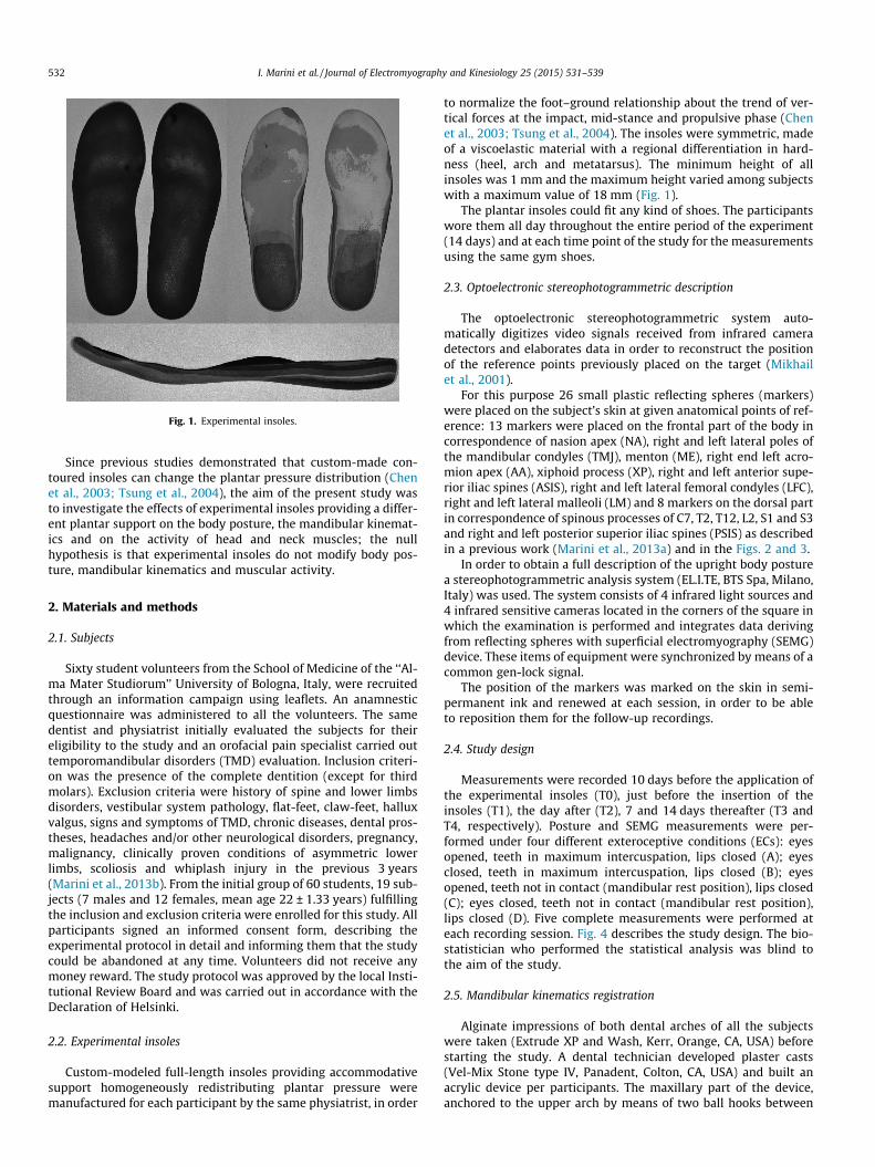
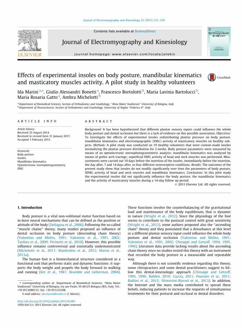
Fig. 1. Experimental insoles.
532 I. Marini et al. / Journal of Electromyography and Kinesiology 25 (2015) 531–539
Since previous studies demonstrated that custom-made con-toured insoles can change the plantar pressure distribution (Chenet al., 2003; Tsung et al., 2004), the aim of the present study wasto investigate the effects of experimental insoles providing a differ-ent plantar support on the body posture, the mandibular kinemat-ics and on the activity of head and neck muscles; the nullhypothesis is that experimental insoles do not modify body pos-ture, mandibular kinematics and muscular activity.
2. Materials and methods
2.1. Subjects
Sixty student volunteers from the School of Medicine of the ‘‘Al-ma Mater Studiorum’’ University of Bologna, Italy, were recruitedthrough an information campaign using leaflets. An anamnesticquestionnaire was administered to all the volunteers. The samedentist and physiatrist initially evaluated the subjects for theireligibility to the study and an orofacial pain specialist carried outtemporomandibular disorders (TMD) evaluation. Inclusion criteri-on was the presence of the complete dentition (except for thirdmolars). Exclusion criteria were history of spine and lower limbsdisorders, vestibular system pathology, flat-feet, claw-feet, halluxvalgus, signs and symptoms of TMD, chronic diseases, dental pros-theses, headaches and/or other neurological disorders, pregnancy,malignancy, clinically proven conditions of asymmetric lowerlimbs, scoliosis and whiplash injury in the previous 3 years(Marini et al., 2013b). From the initial group of 60 students, 19 sub-jects (7 males and 12 females, mean age 22 ± 1.33 years) fulfillingthe inclusion and exclusion criteria were enrolled for this study. Allparticipants signed an informed consent form, describing theexperimental protocol in detail and informing them that the studycould be abandoned at any time. Volunteers did not receive anymoney reward. The study protocol was approved by the local Insti-tutional Review Board and was carried out in accordance with theDeclaration of Helsinki.
2.2. Experimental insoles
Custom-modeled full-length insoles providing accommodativesupport homogeneously redistributing plantar pressure weremanufactured for each participant by the same physiatrist, in order
to normalize the foot–ground relationship about the trend of ver-tical forces at the impact, mid-stance and propulsive phase (Chenet al., 2003; Tsung et al., 2004). The insoles were symmetric, madeof a viscoelastic material with a regional differentiation in hard-ness (heel, arch and metatarsus). The minimum height of allinsoles was 1 mm and the maximum height varied among subjectswith a maximum value of 18 mm (Fig. 1).
The plantar insoles could fit any kind of shoes. The participantswore them all day throughout the entire period of the experiment(14 days) and at each time point of the study for the measurementsusing the same gym shoes.
2.3. Optoelectronic stereophotogrammetric description
The optoelectronic stereophotogrammetric system auto-matically digitizes video signals received from infrared cameradetectors and elaborates data in order to reconstruct the positionof the reference points previously placed on the target (Mikhailet al., 2001).
For this purpose 26 small plastic reflecting spheres (markers)were placed on the subject’s skin at given anatomical points of ref-erence: 13 markers were placed on the frontal part of the body incorrespondence of nasion apex (NA), right and left lateral poles ofthe mandibular condyles (TMJ), menton (ME), right end left acro-mion apex (AA), xiphoid process (XP), right and left anterior supe-rior iliac spines (ASIS), right and left lateral femoral condyles (LFC),right and left lateral malleoli (LM) and 8 markers on the dorsal partin correspondence of spinous processes of C7, T2, T12, L2, S1 and S3and right and left posterior superior iliac spines (PSIS) as describedin a previous work (Marini et al., 2013a) and in the Figs. 2 and 3.
In order to obtain a full description of the upright body posturea stereophotogrammetric analysis system (EL.I.TE, BTS Spa, Milano,Italy) was used. The system consists of 4 infrared light sources and4 infrared sensitive cameras located in the corners of the square inwhich the examination is performed and integrates data derivingfrom reflecting spheres with superficial electromyography (SEMG)device. These items of equipment were synchronized by means of acommon gen-lock signal.
The position of the markers was marked on the skin in semi-permanent ink and renewed at each session, in order to be ableto reposition them for the follow-up recordings.
2.4. Study design
Measurements were recorded 10 days before the application ofthe experimental insoles (T0), just before the insertion of theinsoles (T1), the day after (T2), 7 and 14 days thereafter (T3 andT4, respectively). Posture and SEMG measurements were per-formed under four different exteroceptive conditions (ECs): eyesopened, teeth in maximum intercuspation, lips closed (A); eyesclosed, teeth in maximum intercuspation, lips closed (B); eyesopened, teeth not in contact (mandibular rest position), lips closed(C); eyes closed, teeth not in contact (mandibular rest position),lips closed (D). Five complete measurements were performed ateach recording session. Fig. 4 describes the study design. The bio-statistician who performed the statistical analysis was blind tothe aim of the study.
2.5. Mandibular kinematics registration
Alginate impressions of both dental arches of all the subjectswere taken (Extrude XP and Wash, Kerr, Orange, CA, USA) beforestarting the study. A dental technician developed plaster casts(Vel-Mix Stone type IV, Panadent, Colton, CA, USA) and built anacrylic device per participants. The maxillary part of the device,anchored to the upper arch by means of two ball hooks between
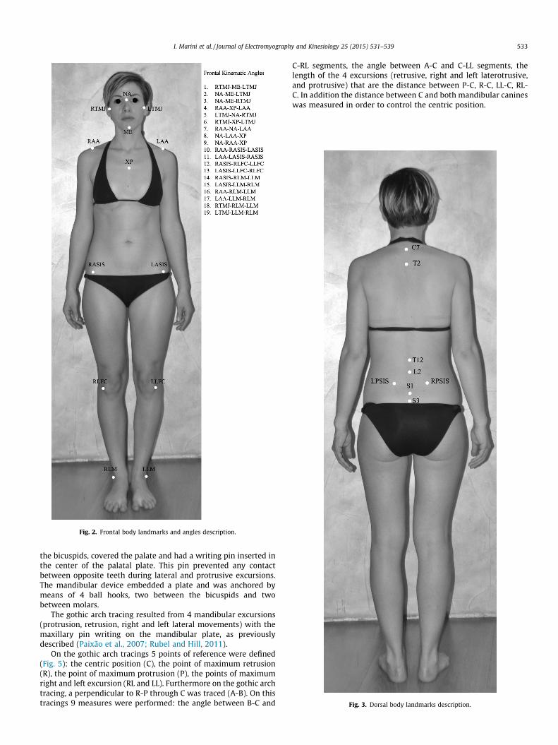
Fig. 2. Frontal body landmarks and angles description.
Fig. 3. Dorsal body landmarks description.
I. Marini et al. / Journal of Electromyography and Kinesiology 25 (2015) 531–539 533
the bicuspids, covered the palate and had a writing pin inserted inthe center of the palatal plate. This pin prevented any contactbetween opposite teeth during lateral and protrusive excursions.The mandibular device embedded a plate and was anchored bymeans of 4 ball hooks, two between the bicuspids and twobetween molars.
The gothic arch tracing resulted from 4 mandibular excursions(protrusion, retrusion, right and left lateral movements) with themaxillary pin writing on the mandibular plate, as previouslydescribed (Paixão et al., 2007; Rubel and Hill, 2011).
On the gothic arch tracings 5 points of reference were defined(Fig. 5): the centric position (C), the point of maximum retrusion(R), the point of maximum protrusion (P), the points of maximumright and left excursion (RL and LL). Furthermore on the gothic archtracing, a perpendicular to R-P through C was traced (A-B). On thistracings 9 measures were performed: the angle between B-C and
C-RL segments, the angle between A-C and C-LL segments, thelength of the 4 excursions (retrusive, right and left laterotrusive,and protrusive) that are the distance between P-C, R-C, LL-C, RL-C. In addition the distance between C and both mandibular canineswas measured in order to control the centric position.
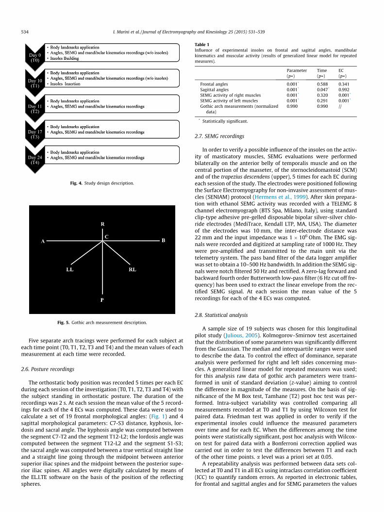
Fig. 4. Study design description.
Fig. 5. Gothic arch measurement description.
Table 1Influence of experimental insoles on frontal and sagittal angles, mandibularkinematics and muscular activity (results of generalized linear model for repeatedmeasures).
Parameter(p=)
Time(p=)
EC(p=)
Frontal angles 0.001* 0.588 0.341Sagittal angles 0.001* 0.047* 0.992SEMG activity of right muscles 0.001* 0.320 0.001*
SEMG activity of left muscles 0.001* 0.291 0.001*
Gothic arch measurements (normalizeddata)
0.990 0.990 //
* Statistically significant.
534 I. Marini et al. / Journal of Electromyography and Kinesiology 25 (2015) 531–539
Five separate arch tracings were performed for each subject ateach time point (T0, T1, T2, T3 and T4) and the mean values of eachmeasurement at each time were recorded.
2.6. Posture recordings
The orthostatic body position was recorded 5 times per each ECduring each session of the investigation (T0, T1, T2, T3 and T4) withthe subject standing in orthostatic posture. The duration of therecordings was 2 s. At each session the mean value of the 5 record-ings for each of the 4 ECs was computed. These data were used tocalculate a set of 19 frontal morphological angles: (Fig. 1) and 4sagittal morphological parameters: C7-S3 distance, kyphosis, lor-dosis and sacral angle. The kyphosis angle was computed betweenthe segment C7-T2 and the segment T12-L2; the lordosis angle wascomputed between the segment T12-L2 and the segment S1-S3;the sacral angle was computed between a true vertical straight lineand a straight line going through the midpoint between anteriorsuperior iliac spines and the midpoint between the posterior supe-rior iliac spines. All angles were digitally calculated by means ofthe EL.I.TE software on the basis of the position of the reflectingspheres.
2.7. SEMG recordings
In order to verify a possible influence of the insoles on the activ-ity of masticatory muscles, SEMG evaluations were performedbilaterally on the anterior belly of temporalis muscle and on thecentral portion of the masseter, of the sternocleidomastoid (SCM)and of the trapezius descendens (upper), 5 times for each EC duringeach session of the study. The electrodes were positioned followingthe Surface Electromyography for non-invasive assessment of mus-cles (SENIAM) protocol (Hermens et al., 1999). After skin prepara-tion with ethanol SEMG activity was recorded with a TELEMG 8channel electromyograph (BTS Spa, Milano, Italy), using standardclip-type adhesive pre-gelled disposable bipolar silver-silver chlo-ride electrodes (MediTrace, Kendall LTP, MA, USA). The diameterof the electrodes was 10 mm, the inter-electrode distance was22 mm and the input impedance was 1 � 106 Ohm. The EMG sig-nals were recorded and digitized at sampling rate of 1000 Hz. Theywere pre-amplified and transmitted to the main unit via thetelemetry system. The pass band filter of the data logger amplifierwas set to obtain a 10–500 Hz bandwidth. In addition the SEMG sig-nals were notch filtered 50 Hz and rectified. A zero-lag forward andbackward fourth order Butterworth low-pass filter (6 Hz cut off fre-quency) has been used to extract the linear envelope from the rec-tified SEMG signal. At each session the mean value of the 5recordings for each of the 4 ECs was computed.
2.8. Statistical analysis
A sample size of 19 subjects was chosen for this longitudinalpilot study (Julious, 2005). Kolmogorov–Smirnov test ascertainedthat the distribution of some parameters was significantly differentfrom the Gaussian. The median and interquartile ranges were usedto describe the data. To control the effect of dominance, separateanalysis were performed for right and left sides concerning mus-cles. A generalized linear model for repeated measures was used;for this analysis raw data of gothic arch parameters were trans-formed in unit of standard deviation (z-value) aiming to controlthe difference in magnitude of the measures. On the basis of sig-nificance of the M Box test, Tamhane (T2) post hoc test was per-formed. Intra-subject variability was controlled comparing allmeasurements recorded at T0 and T1 by using Wilcoxon test forpaired data. Friedman test was applied in order to verify if theexperimental insoles could influence the measured parametersover time and for each EC. When the differences among the timepoints were statistically significant, post hoc analysis with Wilcox-on test for paired data with a Bonferroni correction applied wascarried out in order to test the differences between T1 and eachof the other time points. a level was a priori set at 0.05.
A repeatability analysis was performed between data sets col-lected at T0 and T1 in all ECs using intraclass correlation coefficient(ICC) to quantify random errors. As reported in electronic tables,for frontal and sagittal angles and for SEMG parameters the values
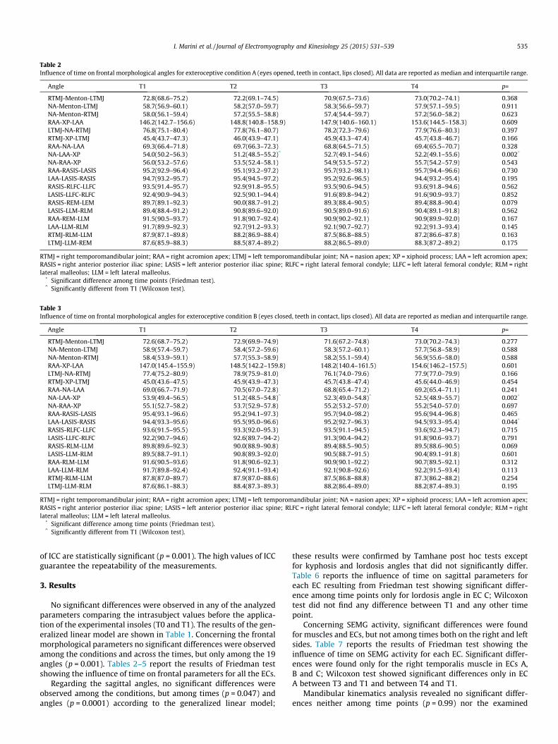
Table 2Influence of time on frontal morphological angles for exteroceptive condition A (eyes opened, teeth in contact, lips closed). All data are reported as median and interquartile range.
Angle T1 T2 T3 T4 p=
RTMJ-Menton-LTMJ 72.8(68.6–75.2) 72.2(69.1–74.5) 70.9(67.5–73.6) 73.0(70.2–74.1) 0.368NA-Menton-LTMJ 58.7(56.9–60.1) 58.2(57.0–59.7) 58.3(56.6–59.7) 57.9(57.1–59.5) 0.911NA-Menton-RTMJ 58.0(56.1–59.4) 57.2(55.5–58.8) 57.4(54.4–59.7) 57.2(56.0–58.2) 0.623RAA-XP-LAA 146.2(142.7–156.6) 148.8(140.8–158.9) 147.9(140.6–160.1) 153.6(144.5–158.3) 0.609LTMJ-NA-RTMJ 76.8(75.1–80.4) 77.8(76.1–80.7) 78.2(72.3–79.6) 77.9(76.6–80.3) 0.397RTMJ-XP-LTMJ 45.4(43.7–47.3) 46.0(43.9–47.1) 45.9(43.3–47.4) 45.7(43.8–46.7) 0.166RAA-NA-LAA 69.3(66.4–71.8) 69.7(66.3–72.3) 68.8(64.5–71.5) 69.4(65.5–70.7) 0.328NA-LAA-XP 54.0(50.2–56.3) 51.2(48.5–55.2)^ 52.7(49.1–54.6) 52.2(49.1–55.6) 0.002*
NA-RAA-XP 56.0(53.2–57.6) 53.5(52.4–58.1) 54.9(53.5–57.2) 55.7(54.2–57.9) 0.543RAA-RASIS-LASIS 95.2(92.9–96.4) 95.1(93.2–97.2) 95.7(93.2–98.1) 95.7(94.4–96.6) 0.730LAA-LASIS-RASIS 94.7(93.2–95.7) 95.4(94.5–97.2) 95.2(92.6–96.5) 94.4(93.2–95.4) 0.195RASIS-RLFC-LLFC 93.5(91.4–95.7) 92.9(91.8–95.5) 93.5(90.6–94.5) 93.6(91.8–94.6) 0.562LASIS-LLFC-RLFC 92.4(90.9–94.3) 92.5(90.1–94.4) 91.6(89.8–94.2) 91.6(90.9–93.7) 0.852RASIS-REM-LEM 89.7(89.1–92.3) 90.0(88.7–91.2) 89.3(88.4–90.5) 89.4(88.8–90.4) 0.079LASIS-LLM-RLM 89.4(88.4–91.2) 90.8(89.6–92.0) 90.5(89.0–91.6) 90.4(89.1–91.8) 0.562RAA-REM-LLM 91.5(90.5–93.7) 91.8(90.7–92.4) 90.9(90.2–92.1) 90.9(89.9–92.0) 0.167LAA-LLM-RLM 91.7(89.9–92.3) 92.7(91.2–93.3) 92.1(90.7–92.7) 92.2(91.3–93.4) 0.145RTMJ-RLM-LLM 87.9(87.1–89.8) 88.2(86.9–88.4) 87.5(86.8–88.5) 87.2(86.6–87.8) 0.163LTMJ-LLM-REM 87.6(85.9–88.3) 88.5(87.4–89.2) 88.2(86.5–89.0) 88.3(87.2–89.2) 0.175
RTMJ = right temporomandibular joint; RAA = right acromion apex; LTMJ = left temporomandibular joint; NA = nasion apex; XP = xiphoid process; LAA = left acromion apex;RASIS = right anterior posterior iliac spine; LASIS = left anterior posterior iliac spine; RLFC = right lateral femoral condyle; LLFC = left lateral femoral condyle; RLM = rightlateral malleolus; LLM = left lateral malleolus.
* Significant difference among time points (Friedman test).^ Significantly different from T1 (Wilcoxon test).
Table 3Influence of time on frontal morphological angles for exteroceptive condition B (eyes closed, teeth in contact, lips closed). All data are reported as median and interquartile range.
Angle T1 T2 T3 T4 p=
RTMJ-Menton-LTMJ 72.6(68.7–75.2) 72.9(69.9–74.9) 71.6(67.2–74.8) 73.0(70.2–74.3) 0.277NA-Menton-LTMJ 58.9(57.4–59.7) 58.4(57.2–59.6) 58.3(57.2–60.1) 57.7(56.8–58.9) 0.588NA-Menton-RTMJ 58.4(53.9–59.1) 57.7(55.3–58.9) 58.2(55.1–59.4) 56.9(55.6–58.0) 0.588RAA-XP-LAA 147.0(145.4–155.9) 148.5(142.2–159.8) 148.2(140.4–161.5) 154.6(146.2–157.5) 0.601LTMJ-NA-RTMJ 77.4(75.2–80.9) 78.9(75.9–81.0) 76.1(74.0–79.6) 77.9(77.0–79.9) 0.166RTMJ-XP-LTMJ 45.0(43.6–47.5) 45.9(43.9–47.3) 45.7(43.8–47.4) 45.6(44.0–46.9) 0.454RAA-NA-LAA 69.0(66.7–71.9) 70.5(67.0–72.8) 68.8(65.4–71.2) 69.2(65.4–71.1) 0.241NA-LAA-XP 53.9(49.4–56.5) 51.2(48.5–54.8)^ 52.3(49.0–54.8)^ 52.5(48.9–55.7) 0.002*
NA-RAA-XP 55.1(52.7–58.2) 53.7(52.9–57.8) 55.2(53.2–57.0) 55.2(54.0–57.0) 0.697RAA-RASIS-LASIS 95.4(93.1–96.6) 95.2(94.1–97.3) 95.7(94.0–98.2) 95.6(94.4–96.8) 0.465LAA-LASIS-RASIS 94.4(93.3–95.6) 95.5(95.0–96.6) 95.2(92.7–96.3) 94.5(93.3–95.4) 0.044*
RASIS-RLFC-LLFC 93.6(91.5–95.5) 93.3(92.0–95.3) 93.5(91.1–94.5) 93.6(92.3–94.7) 0.715LASIS-LLFC-RLFC 92.2(90.7–94.6) 92.6(89.7–94-2) 91.3(90.4–94.2) 91.8(90.6–93.7) 0.791RASIS-RLM-LLM 89.8(89.6–92.3) 90.0(88.9–90.8) 89.4(88.5–90.5) 89.5(88.6–90.5) 0.069LASIS-LLM-RLM 89.5(88.7–91.1) 90.8(89.3–92.0) 90.5(88.7–91.5) 90.4(89.1–91.8) 0.601RAA-RLM-LLM 91.6(90.5–93.6) 91.8(90.6–92.3) 90.9(90.1–92.2) 90.7(89.5–92.1) 0.312LAA-LLM-RLM 91.7(89.8–92.4) 92.4(91.1–93.4) 92.1(90.8–92.6) 92.2(91.5–93.4) 0.113RTMJ-RLM-LLM 87.8(87.0–89.7) 87.9(87.0–88.6) 87.5(86.8–88.8) 87.3(86.2–88.2) 0.254LTMJ-LLM-RLM 87.6(86.1–88.3) 88.4(87.3–89.3) 88.2(86.4–89.0) 88.2(87.4–89.3) 0.195
RTMJ = right temporomandibular joint; RAA = right acromion apex; LTMJ = left temporomandibular joint; NA = nasion apex; XP = xiphoid process; LAA = left acromion apex;RASIS = right anterior posterior iliac spine; LASIS = left anterior posterior iliac spine; RLFC = right lateral femoral condyle; LLFC = left lateral femoral condyle; RLM = rightlateral malleolus; LLM = left lateral malleolus.
* Significant difference among time points (Friedman test).^ Significantly different from T1 (Wilcoxon test).
I. Marini et al. / Journal of Electromyography and Kinesiology 25 (2015) 531–539 535
of ICC are statistically significant (p = 0.001). The high values of ICCguarantee the repeatability of the measurements.
3. Results
No significant differences were observed in any of the analyzedparameters comparing the intrasubject values before the applica-tion of the experimental insoles (T0 and T1). The results of the gen-eralized linear model are shown in Table 1. Concerning the frontalmorphological parameters no significant differences were observedamong the conditions and across the times, but only among the 19angles (p = 0.001). Tables 2–5 report the results of Friedman testshowing the influence of time on frontal parameters for all the ECs.
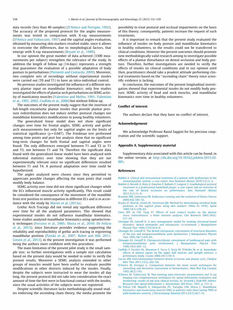
Regarding the sagittal angles, no significant differences wereobserved among the conditions, but among times (p = 0.047) andangles (p = 0.0001) according to the generalized linear model;
these results were confirmed by Tamhane post hoc tests exceptfor kyphosis and lordosis angles that did not significantly differ.Table 6 reports the influence of time on sagittal parameters foreach EC resulting from Friedman test showing significant differ-ence among time points only for lordosis angle in EC C; Wilcoxontest did not find any difference between T1 and any other timepoint.
Concerning SEMG activity, significant differences were foundfor muscles and ECs, but not among times both on the right and leftsides. Table 7 reports the results of Friedman test showing theinfluence of time on SEMG activity for each EC. Significant differ-ences were found only for the right temporalis muscle in ECs A,B and C; Wilcoxon test showed significant differences only in ECA between T3 and T1 and between T4 and T1.
Mandibular kinematics analysis revealed no significant differ-ences neither among time points (p = 0.99) nor the examined
Table 4Influence of time on frontal morphological angles for exteroceptive condition C (eyes opened, teeth not in contact, lips closed). All data are reported as median and interquartilerange.
Angle T1 T2 T3 T4 p=
RTMJ-Menton-LTMJ 72.9(68.5–76.0) 73.1(69.6–75.3) 72.4(69.2–74.0) 73.1(70.9–76.0) 0.730NA-Menton-LTMJ 58.5(56.7–59.5) 59.2(57.1–60.3) 58.9(56.6–60.7) 58.0(56.8–59.4) 0.536NA-Menton-RTMJ 57.7(54.4–59.1) 57.9(55.3–59.8) 57.7(56.6–59.7) 57.2(55.3–58.7) 0.657RAA-XP-LAA 146.7(146.0–158.3) 148.4(141.9–157.9) 147.4(139.9–159.6) 154.5(147.4–158.9) 0.373LTMJ-NA-RTMJ 79.5(75.1–81.2) 77.1(76.2–79.8) 76.9(72.6–81.1) 78.0(76.5–80.3) 0.549RTMJ-XP-LTMJ 45.6(43.8–47.0) 46.2(43.9–47.7) 45.6(44.0–47.4) 46.1(44.2–47.1) 0.247RAA-NA-LAA 69.4(66.9–72.0) 70.3(65.9–72.5) 68.8(65.5–72.0) 69.3(66.2–71.2) 0.217NA-LAA-XP 54.0(50.4–56.6) 51.9(48.5–55.5)^ 51.5(50.1–55.4) 52.2(49.7–55.8) 0.012*
NA-RAA-XP 55.5(52.2–59.0) 54.3(52.9–57.5) 55.5(54.1–57.3) 55.1(54.5–56.1) 0.730RAA-RASIS-LASIS 95.2(92.9–96.2) 95.2(93.8–97.3) 95.6(93.6–97.8) 95.5(94.7–97.0) 0.281LAA-LASIS-RASIS 94.6(93.5–95.7) 95.5(94.8–96.7) 95.1(93.1–96.3) 94.5(93.3–95.5) 0.025*
RASIS-RLFC-LLFC 93.5(91.5–95.5) 92.8(92.0–95.5) 93.4(91.0–94.5) 93.9(92.9–94.6) 0.465LASIS-LLFC-RLFC 92.1(91.1–94.3) 92.5(89.4–94.2) 91.2(90.5–94.2) 91.8(90.5–93.2) 0.992RASIS-RLM-LLM 89.8(89.6–92.2) 89.8(89.0–90.9) 89.3(88.4–90.6)^ 89.5(89.0–90.6) 0.008*
LASIS-LLM-RLM 89.5(88.4–91.1) 91.0(89.2–91.6) 90.4(89.0–91.6) 90.5(89.3–91.8) 0.643RAA-RLM-LLM 91.6(90.4–94.5) 91.8(90.6–92.2) 90.7(90.2–92.1) 90.6(89.8–92.0) 0.363LAA-LLM-RLM 91.7(90.4–92.4) 92.5(91.2–93.3) 92.1(91.0–92.6) 92.0(91.4–93.5) 0.213RTMJ-RLM-LLM 87.8(87.0–89.6) 88.1(87.0–88.7) 87.3(86.8–88.7) 87.2(86.5–88.0) 0.205LTMJ-LLM-RLM 87.7(86.3–88.4) 88.4(87.4–89.1) 88.1(86.6–89.1) 88.3(87.1–89.5) 0.500
RTMJ = right temporomandibular joint; RAA = right acromion apex; LTMJ = left temporomandibular joint; NA = nasion apex; XP = xiphoid process; LAA = left acromion apex;RASIS = right anterior posterior iliac spine; LASIS = left anterior posterior iliac spine; RLFC = right lateral femoral condyle; LLFC = left lateral femoral condyle; RLM = rightlateral malleolus; LLM = left lateral malleolus.
* Significant difference among time points (Friedman test).^ Significantly different from T1 (Wilcoxon test).
Table 5Influence of time on frontal morphological angles for exteroceptive condition D (eyes closed, teeth not in contact, lips closed). All data are reported as median and interquartilerange.
Angle T1 T2 T3 T4 p=
RTMJ-Menton-LTMJ 72.6(70.0–74.9) 72.7(69.7–74.7) 71.4(68.1–73.6) 72.7(67.8–74.2) 0.345NA-Menton-LTMJ 57.5(56.5–59.5) 57.6(56.4–59.2) 57.9(55.8–60.1) 56.7(55.5–57.9) 0.141NA-Menton-RTMJ 57.0(54.5–58.7) 57.1(55.0–59.0) 57.0(54.1–58.9) 55.9(54.5–57.5) 0.363RAA-XP-LAA 147.6(145.4–156.8) 148.1(140.3–158.6) 148.3(141.4–159.6) 155.3(145.0–157.8) 0.643LTMJ-NA-RTMJ 78.1(75.2–81.2) 78.3(76.3–80.2) 76.3(72.9–80.7) 78.1(77.2–79.8) 0.274RTMJ-XP-LTMJ 45.1(43.7–47.3) 45.5(44.0–47.6) 45.8(44.0–47.4) 45.9(44.2–46.7) 0.643RAA-NA-LAA 69.8(66.3–71.3) 70.3(65.6–72.0) 68.6(65.6–70.8) 68.8(66.1–70.8) 0.643NA-LAA-XP 54.5(49.2–56.7) 51.8(58.9–55.6)^ 52.3(50.0–55.5) 52.2(49.6–56.1) 0.010*
NA-RAA-XP 55.6(53.0–57.7) 53.8(53.2–58.1) 55.6(53.7–57.4) 55.1(54.1–57.4) 0.382RAA-RASIS-LASIS 94.8(93.1–96.3) 94.9(93.8–97.5) 95.8(93.7–98.1) 95.7(94.5–96.9) 0.363LAA-LASIS-RASIS 94.4(93.6–95.8) 95.4(94.6–97.0) 95.1(92.8–96.2) 94.4(93.6–95.1) 0.073RASIS-RLFC-LLFC 93.6(91.7–95.8) 93.1(91.9–95.6) 93.3(91.3–94.6) 93.9(93.0–94.7) 0.296LASIS-LLFC-RLFC 92.2(90.7–94.6) 92.6(89.4–94.2) 91.1(90.5–94.5) 92.0(90.8–93.8) 0.776RASIS-RLM-LLM 89.9(89.3–92.1) 72.7(69.7–74.7)^ 89.3(88.5–90.4)^ 89.4(89.0–90.7) 0.001*
LASIS-LLM-RLM 89.6(88.6–91.4) 90.8(89.6–91.8) 90.5(88.6–91.6) 90.4(89.5–91.6) 0.518RAA-RLM-LLM 91.5(90.3–93.3) 92.0(90.6–92.3) 90.8(90.3–92.0) 90.6(89.8–91.9) 0.337LAA-LLM-RLM 91.8(90.6–92.5) 92.4(91.2–93.2) 92.3(90.8–92.6) 92.3(91.5–93.4) 0.141RTMJ-RLM-LLM 87.8(87.0–89.5) 88.2(87.0–88.7) 87.5(86.9–88.6) 87.3(86.6–87.8) 0.267LTMJ-LLM-RLM 87.7(86.6–88.6) 88.3(87.6–89.2) 88.1(86.6–88.9) 88.3(87.1–89.5) 0.337
RTMJ = right temporomandibular joint; RAA = right acromion apex; LTMJ = left temporomandibular joint; NA = nasion apex; XP = xiphoid process; LAA = left acromion apex;RASIS = right anterior posterior iliac spine; LASIS = left anterior posterior iliac spine; RLFC = right lateral femoral condyle; LLFC = left lateral femoral condyle; RLM = rightlateral malleolus; LLM = left lateral malleolus.
* Significant difference among time points (Friedman test).^ Significantly different from T1 (Wilcoxon test).
536 I. Marini et al. / Journal of Electromyography and Kinesiology 25 (2015) 531–539
parameters (p = 0.99). Table 8 reports the results of Friedman testfor the influence of time on gothic arch tracings.
4. Discussion
The present study presents a new protocol for the evaluation ofthe possible effects of a different plantar sensory input on bodyposture changes. In a previous study the same device was usedin order to evaluate the effects of an experimental occlusal inter-ference on body posture (Marini et al., 2013a). Since Maeda andcoworkers showed that experimental leg length discrepanciescan affect body posture and dental occlusion (Maeda et al.,2011), in this investigation custom-made insoles were used in
order to verify the postural effects of a standardized plantar sup-port without reproducing a pathological condition performingasymmetrical disturbance.
The integrated system of optoelectronic stereophotogrammet-ric analysis and SEMG, together with the gothic arch tracing andthe study design, represent a consistent way of approaching to thisfield. The accuracy of the proposed protocol was tested using thegeneralized cross validatory splines algorithm (GCVC) that is thebest known and effective automatic algorithm for smoothingbiomechanical data. The test performed has shown that the overallaccuracy of the software proposed was superior or similar to GCVC,while the time spent for computation was higher for the latter;moreover the new software is virtually insensible to the numberof samples, allowing the computation of the derivatives for short
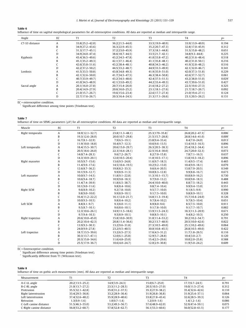
Table 6Influence of time on sagittal morphological parameters for all exteroceptive conditions. All data are reported as median and interquartile range.
Angle EC T1 T2 T3 T4 p=
C7-S3 distance A 33.8(25.2–42.0) 36.2(21.5–44.0) 33.5(19.9–40.9) 33.9(13.9–40.9) 0.194B 34.9(27.2–43.6) 36.3(22.9–45.5) 35.2(20.7–47.3) 32.8(17.0–45.9) 0.312C 31.3(17.7–45.1) 37.2(23.0–45.9) 37.3(18.1–44.8) 31.5(15.8–48.2) 0.651D 34.9(24.0–47.4) 38.6(18.7–44.5) 33.5(21.7–42.1) 34.8(9.3–44.8) 0.225
Kyphosis A 43.4(36.5–49.6) 43.2(34.5–47.8) 41.0(33.8–47.2) 40.2(31.6–48.4) 0.518B 45.1(35.2–49.3) 42.1(37.1–46.4) 41.1(35.8–48.1) 40.2(31.0–50.1) 0.216C 42.6(35.8–51.6) 43.2(38.4–48.1) 40.8(34.3–46.2) 41.9(32.8–48.2) 0.516D 42.2(37.2–50.2) 44.5(33.2–48.7) 44.9(33.3–49.9) 42.1(32.0–46.7) 0.341
Lordosis A 41.5(33.5–50.8) 43.0(34.8–49.3) 41.9(35.9–51.0) 42.0(37.3–51.8) 0.229B 42.1(32.6–50.0) 41.7(34.5–47.5) 46.3(38.4–50.8) 42.6(37.7–52.7) 0.061C 40.7(33.9–49.7) 43.2(34.5–48.6) 42.4(37.5–51.5) 43.2(38.0–51.0) 0.029*
D 41.0(34.5–48.9) 42.1(33.0–49.2) 44.2(33.4–49.3) 43.7(39.6–51.0) 0.427Sacral angle A 20.1(16.0–27.8) 20.7(15.4–26.0) 21.6(18.2–27.2) 22.5(19.6–27.3) 0.325
B 20.4(14.9–27.9) 20.6(16.0–25.2) 23.1(18.1–27.6) 21.7(18.7–26.7) 0.092C 21.0(15.7–26.7) 19.6(15.6–23.4) 22.6(17.7–27.4) 21.0(19.4–27.1) 0.124D 22.7(17.6–26.7) 20.3(16.4–24.5) 21.3(17.1–26.6) 23.1(20.5–26.2) 0.131
EC = exteroceptive condition.* Significant difference among time points (Friedman test).
Table 7Influence of time on SEMG parameters (lV) for all exteroceptive conditions. All data are reported as median and interquartile range.
Muscle EC T1 T2 T3 T4 p=
Right temporalis A 18.9(12.1–32.7) 23.8(11.3–48.1) 25.3(179–35.8)^ 26.8(20.2–47.3)^ 0.006*
B 19.3(12.6–28.0) 20.0(10.7–29.8) 21.5(15.7–28.8) 28.8(14.6–41.0) 0.009*
C 16.7(8.1–32.9) 7.9(6.5–19.1) 23.8(9.4–33.4) 8.4(7.0–24.0) 0.013*
D 11.9(10.0–18.8) 10.4(9.7–12.3) 10.6(9.6–13.5) 13.4(10.3–16.5) 0.896Left temporalis A 18.4(15.5–30.7) 20.6(15.0–29.7) 26.3(20.5–38.3) 25.4(18.2–34.4) 0.141
B 20.5(18.6–26.0) 22.3(16.6–28.1) 22.5(16.4–30.0) 24.7(20.0–32.3) 0.073C 14.0(7.4–28.3) 9.6(7.2–26.7) 8.2(7.0–15.8) 7.8(7.1–16.3) 0.637D 14.3(10.9–20.1) 12.0(10.5–20.4) 11.0(10.5–17.1) 13.8(10.3–18.2) 0.896
Right masseter A 10.5(9.7–15.6) 13.6(9.5–24.8) 11.6(9.7–18.3) 11.4(9.5–17.4) 0.465B 11.4(9.9–17.6) 14.5(10.4–19.5) 10.2(9.2–13.0) 10.4(9.9–18.1) 0.079C 12.6(8.7–36.2) 19.8(8.2–33.3) 9.6(6.4–20.5) 13.0(7.9–20.5) 0.549D 10.1(9.9–12.7) 9.9(8.9–11.3) 10.0(8.3–12.8) 9.9(8.8–16.7) 0.673
Left masseter A 10.0(9.7–14.5) 11.8(9.1–22.8) 11.3(10.1–15.7) 10.0(8.9–16.2) 0.730B 10.6(9.4–18.7) 10.9(9.8–18.3) 9.7(9.0–13.2) 10.0(9.8–18.3) 0.488C 11.4(7.4–39.9) 11.8(6.5–38.7) 24.4(10.0–40.0) 8.8(7.5–18.2) 0.443D 10.1(9.0–11.6) 9.8(8.4–10.6) 9.8(7.4–10.4) 9.9(9.4–13.0) 0.551
Right SCM A 9.6(8.9–10.2) 9.2(7.8–10.0) 9.5(7.7–10.0) 9.1(8.5–9.9) 0.990B 9.8(9.0–10.0) 9.9(8.9–10.1) 9.1(7.5–10.0) 9.8(8.9–10.1) 0.328C 16.4(11.2–22.2) 16.1(12.0–21.7) 14.8(11.3–19.4) 15.7(10.4–24.0) 0.126D 10.0(9.3–10.5) 9.8(8.4–10.2) 9.7(8.4–10.2) 9.7(8.5–10.4) 0.651
Left SCM A 8.8(8.1–9.7) 9.3(8.0–11.1) 8.9(8.0–9.6) 8.5(7.5–10.0) 0.911B 9.5(8.7–10.1) 9.6(8.6–10.1) 9.1(7.8–10.0) 9.1(7.7–10.7) 0.882C 12.3(10.6–26.1) 14.9(11.5–18.6) 13.9(11.5–24.3) 18.3(11.3–20.9) 0.671D 9.7(9.4–10.3) 9.5(8.9–10.1) 9.8(8.5–10.1) 9.4(8.2–10.3) 0.250
Right trapezius A 20.6(10.0–45.0) 15.8(10.0–30.9) 31.0(11.4–53.2) 30.2(10.2–54.7) 0.791B 20.2(10.4–42.3) 20.8(11.4–36.6) 30.2(13.7–48.9) 20.5(10.0–42.6) 0.822C 13.9(9.3–30.3) 13.2(9.6–31.8) 27.3(10.5–49.0) 25.1(10.2–49.6) 0.064D 24.0(9.9–27.8) 23.2(9.5–40.5) 30.0(10.8–45.5) 28.8(10.5–49.0) 0.422
Left trapezius A 18.7(13.5–39.6) 13.2(6.5–27.3) 17.6(4.3–31.2) 11.7(1.8–26.5) 0.116B 30.3(13.7–47.1) 12.0(6.1–25.8) 12.9(5.7–28.8) 10.4(3.0–2.7) 0.120C 20.3(15.8–34.6) 11.6(4.8–25.0) 15.4(2.5–29.6) 10.0(2.0–25.8) 0.588D 25.5(17.9–36.7) 10.6(4.0–24.7) 12.0(2.9–30.8) 11.9(5.0–24.2) 0.609
EC = Exteroceptive condition.* Significant difference among time points (Friedman test).^ Significantly different from T1 (Wilcoxon test).
Table 8Influence of time on gothic arch measurements (mm). All data are reported as median and interquartile range.
Measurement T1 T2 T3 T4 p=
A-C-LL angle 20.2(13.5–25.2) 14.9(3.9–24.3) 15.0(6.7–25.0) 17.7(6.7–24.5) 0.791B-C-RL angle 21.0(13.7–27.2) 23.5(11.2–28.5) 20.5(10.5–27.0) 19.0(11.5–27.4) 0.312Protrusion 35.5(32.1–42.8) 35.0(31.2–37.5) 35.3(27.9–38.2) 35.4(32.6–42.6) 0.110Right laterotrusion 33.4(29.5–36.8) 33.2(28.9–36.4) 31.9(26.8–36.8) 31.9(25.4–37.1) 0.064Left laterotrusion 37.4(32.6–40.2) 35.9(28.9–40.6) 33.8(27.8–43.4) 32.6(28.5–39.3) 0.126Retrusion 1.3(0.9–1.6) 1.0(0.7–1.4) 1.2(0.9–1.6) 1.4(1.2–1.6) 0.086C-Left canine distance 51.5(46.3–55.0) 53.2(49.3–56.7) 57.6(48.9–62.0) 52.9(47.4–59.1) 0.075C-Right canine distance 56.0(53.2–60.7) 57.8(52.8–62.7) 56.1(53.3–60.6) 56.0(52.8–61.5) 0.177
I. Marini et al. / Journal of Electromyography and Kinesiology 25 (2015) 531–539 537
538 I. Marini et al. / Journal of Electromyography and Kinesiology 25 (2015) 531–539
data records (less than 40 samples) (D’Amico and Ferrigno, 1992).The accuracy of the proposed protocol for the angles measure-ments was tested in comparison with X-ray measurements(D’Amico and Vallasciani, 1997) and the sagittal angles evaluation,obtained by measuring skin markers resulted stable, since it allowsto overcome the differences, due to morphological factors, thatemerge with X-ray measurements (Bryant et al., 1989).
In our opinion the great number of data achieved (3300 mea-surements per subject) strengthen the relevance of the study. Inaddition the length of follow up (14 days) represents a strengththat guarantees the evaluation of a possible adaptation of bodyposture to perturbations (Perinetti and Contardo, 2009). Moreover,two complete sets of recordings without experimental insoleswere carried out (T0 and T1) to have an intra-individual control.
No previous studies investigated the influence of a different sen-sory plantar input on mandibular kinematics; only few studiesinvestigated the effects of plantar arch perturbations on SEMG activ-ity of masticatory muscles (Valentino and Melito, 1991; Valentinoet al., 1991, 2002; Ciuffolo et al., 2006) but without follow up.
The outcomes of the present study suggest that the insertion offull-length viscoelastic plantar insoles that provide plantar pres-sure redistribution does not induce neither postural changes normandibular kinematics modifications in young healthy volunteers.
The generalized linear model does not show significantchanges over time for frontal angles, SEMG activity and gothicarch measurements but only for sagittal angles on the limits ofstatistical significance (p = 0.047). The Friedman test performedamong time points and post hoc analysis show that no significantlong-term changes for both frontal and sagittal angles werefound. The only differences emerged between T1 and T2 or T1and T3, not between T1 and T4. Therefore the significant datafound with the generalized linear model have been analyzed withinferential statistics over time showing that they are notexperimentally relevant since no significant differences resultedbetween T1 and T4. A postural adaptation over time could behypothesized.
The angles analyzed were chosen since they permitted toappreciate possible changes affecting the main joints that couldmodify body balance.
SEMG activity over time did not show significant changes whilethe ECs influenced muscle activity significantly. This result couldbe considered the consequence of the movement of the mandiblefrom rest position to intercuspation in different ECs and is in accor-dance with the study by Marini et al. (2013a).
Gothic Arch Tracings did not reveal any significant differenceover time for all the analyzed parameters. This showed thatexperimental insoles do not influence mandibular kinematics.Some studies analyzed mandibular kinematics using optoelectron-ic techniques (Ferrario et al., 2005; Sforza et al., 2010; De Felícioet al., 2013); since literature provides evidence supporting thereliability and reproducibility of gothic arch tracing in registeringmandibular position (Paixão et al., 2007; Rubel and Hill, 2011;Linsen et al., 2013), in the present investigation it was performedbeing the authors more confident with this procedure.
The main limitation of the present pilot study is the small sam-ple size; so further investigations with a sample size calculationbased on the present data would be needed in order to verify thepresent results. Moreover a SEMG analysis extended to othergroups of muscles would have been useful to evaluate possiblemodifications in other districts induced by the insoles. Finally,despite the subjects were instructed to wear the insoles all daylong, the present protocol did not take into consideration the exactamount of time the feet were in functional contact with the insoles,since the usual activities of the subjects were not registered.
Despite scientific literature lacks methodologically sound stud-ies endorsing the ascending chain theory, the media promote the
possibility to treat postural and occlusal impairments on the basisof this theory; consequently, patients increase the request of suchtreatments.
It is important to remark that the present study evaluated theinfluence of insoles on body posture and mandibular kinematicsin healthy volunteers, so the results could not be transferred toclinical conditions. However the present outcomes should promotenew methodologically valid research aiming to investigate possibleeffects of a plantar disturbance on dental occlusion and body pos-ture. Therefore, further investigations are needed to verify theeffects of insoles on clinical conditions and in our opinion untilthen, practitioners should take a prudent attitude performing clin-ical treatments based on the ‘‘ascending chain’’ theory since scien-tific evidence is lacking.
In conclusion, the outcomes of the present longitudinal investi-gation showed that experimental insoles do not modify body pos-ture, SEMG activity of head and neck muscles, and mandibularkinematics over time in healthy volunteers.
Conflict of interest
The authors declare that they have no conflict of interest.
Acknowledgement
We acknowledge Professor Raoul Saggini for his precious coop-eration and the scientific support.
Appendix A. Supplementary material
Supplementary data associated with this article can be found, inthe online version, at http://dx.doi.org/10.1016/j.jelekin.2015.02.001.
References
Baldini A. Clinical and instrumental treatment of a patient with dysfunction of thestomatognathic system: a case report. Ann Stomatol (Roma) 2010;1(2):2–5.
Baldini A, Beraldi A, Nota A, Danelon F, Ballanti F, Longoni S. Gnathological posturaltreatment in a professional basketball player: a case report and an overview ofthe role of dental occlusion on performance. Ann Stomatol (Roma)2012;3(2):51–8.
Bramble DM, Lieberman DE. Endurance running and the evolution of Homo. Nature2004;432:345–52.
Bryant JT, Reid JG, Smith BL, Stevenson JM. Method for determining vertebral bodypositions in the sagittal plane using skin markers (Phila Pa 1976). Spine1989;14(3):258–65.
Chen WP, Ju CW, Tang FT. Effects of total contact insoles on the plantarstress redistribution: a finite element analysis. Clin Biomech 2003;18(6):S17–24.
Chinappi AS, Getzoff H. A new management model for treating structural-baseddisorders: dental orthopedic and chiropractic co-treatment. J ManipulativePhysiol Ther 1994;17(9):614–9.
Chinappi AS, Getzoff H. The dental-chiropractic cotreatment of structural disordersof the jaw and temporomandibular joint dysfunction. J Manipulative PhysiolTher 1995;18(7):476–81.
Chinappi AS, Getzoff H. Chiropractic/dental cotreatment of lumbosacral pain withtemporomandibular joint involvement. J Manipulative Physiol Ther1996;19(9):607–12.
Ciuffolo F, Ferritto AL, Muratore F, Tecco S, Testa M, D’Attilio M, et al. Immediateeffects of plantar inputs on the upper half muscles and upright posture: apreliminary study. Cranio 2006;24(1):50–9.
Cuccia AM. Interrelationships between dental occlusion and plantar arch. J BodywMov Ther 2011;15(2):242–50.
D’Amico M, Ferrigno G. Comparison between the more recent techniques forsmoothing and derivative assessment in biomechanics. Med Biol Eng Comput1992;30(2):193.
D’Amico M, Vallasciani M. Non-ionising opto-electronic measurement and X-rayimaging two complementary techniques for spinal deformities evaluation andmonitoring: results of one year clinical activity. In: Sevastik J, Diab KM, editors.Research into Spinal Deformities 1. Amsterdam: IOS Press; 1997. p. 151–4.
De Felício CM, Mapelli A, Sidequersky FV, Tartaglia GM, Sforza C. Mandibularkinematics and masticatory muscles EMG in patients with short lasting TMD ofmild-moderate severity. J Electromyogr Kinesiol 2013;23(3):627–33.
I. Marini et al. / Journal of Electromyography and Kinesiology 25 (2015) 531–539 539
Deliagina TG, Orlovsky GN, Zelenin PV, Beloozerova IN. Neural bases of posturalcontrol. Physiology (Bethesda) 2006;21(3):216–25.
Ferrario VF, Sforza C, Lovecchio N, Mian F. Quantification of translational and glidingcomponents in human temporomandibular joint during mouth opening. ArchOral Biol 2005;50(5):507–15.
Fournier R, Aknin JJ, Bourgier S, Gebeile-Chauty S. Dento-facial orthopedics andosteopathy. Orthod Fr 2011;82(4):331–40.
Hermens H, Freriks B, Merletti R, Stegeman D, Blok J, Rau G, et al. EuropeanRecommendations for Surface Electromyography. Enschede: RoessinghResearch and Development; 1999.
Julious SA. Sample size of 12 per group rule of thumb for a pilot study. Pharm Stat2005;4(4):287–91.
Ker RF, Bennett MB, Bibby SR, Kester RC, Alexander RM. The spring in the arch of thehuman foot. Nature 1987;325(7000):147–9.
Linsen SS, Stark H, Klitzschmüller M. Reproducibility of condyle position andinfluence of splint therapy on different registration techniques in asymptomaticvolunteers. Cranio 2013;31(1):32–9.
Maeda N, Sakaguchi K, Mehta NR, Abdallah EF, Forgione AG, Yokoyama A. Effects ofexperimental leg length discrepancies on body posture and dental occlusion.Cranio 2011;29(3):194–203.
Manfredini D, Castroflorio T, Perinetti G, Guarda-Nardini L. Dental occlusion, bodyposture and temporomandibular disorders: where we are now and where weare heading for. J Oral Rehabil 2012;39(6):463–71.
Marini I, Gatto MR, Bartolucci ML, Bortolotti F, Alessandri Bonetti G, Michelotti A.Effects of experimental occlusal interference on body posture: anoptoelectronic stereophotogrammetric analysis. J Oral Rehabil 2013a;40(7):509–18.
Marini I, Paduano S, Bartolucci ML, Bortolotti F, Bonetti GA. The prevalence oftemporomandibular disorders in patients with late whiplash syndrome whoexperience orofacial pain: a case-control series study. J Am Dent Assoc2013b;144(5):486–90.
Michelotti A, Buonocore G, Manzo P, Pellegrino G, Farella M. Dental occlusion andposture: an overview. Progr Orthod 2011;12(1):53–8.
Mikhail E, Bethl J, McGlone C. Introduction to Modern Photogrammetry. Hoboken,NJ: John Wiley; 2001.
Paixão F, Silva WA, Silva FA, Ramos Gda G, Cruz MV. Evaluation of thereproducibility of two techniques used to determine and record centricrelation in angle’s class I patients. J Appl Oral Sci 2007;15(4):275–9.
Perinetti G, Contardo L. Posturography as a diagnostic aid in dentistry: a systematicreview. J Oral Rehabil 2009;36(12):922–36.
Perinetti G, Contardo L, Biasati A, Perdoni L, Castaldo A. Dental malocclusion andbody posture in young subjects: a multiple regression study. Clinics (São Paulo)2010;65(7):689–95.
Rubel B, Hill EE. Intraoral gothic arch tracing. NY State Dent J 2011;77(5):40–3.Sforza C, Ugolini A, Rocchetta D, Galante D, Mapelli A, Giannì AB. Mandibular
kinematics after orthognathic surgical treatment a pilot study. Br J OralMaxillofac Surg 2010;48(2):110–4.
Silvestrini-Biavati A, Migliorati M, Demarziani E, Tecco S, Silvestrini-Biavati P,Polimeni A, et al. Clinical association between teeth malocclusions, wrongposture and ocular convergence disorders: an epidemiological investigation onprimary school children. BMC Pediatr 2013;13:12.
Tardieu C, Dumitrescu M, Giraudeau A, Blanc JL, Cheynet F, Borel L. Dental occlusionand postural control in adults. Neurosci Lett 2009;450(2):221–4.
Tsung BY, Zhang M, Mak AF, Wong MW. Effectiveness of insoles on plantar pressureredistribution. J Rehabil Res Dev 2004;41(6A):767–74.
Valentino B, Melito F. Functional relationships between the muscles of masticationand the muscles of the leg. An electromyographic study. Surg Radiol Anat1991;13(1):33–7.
Valentino B, Fabozzo A, Melito F. The functional relationship between the occlusalplane and the plantar arches. An EMG study. Surg Radiol Anat1991;13(3):171–4.
Valentino B, Melito F, Aldi B, Valentino T. Correlation between interdental occlusalplane and plantar arches. An EMG study. Bull Group Int Rech Sci StomatolOdontol 2002;44(1):10–3.
Wright WG, Ivanenko YP, Gurfinkel VS. Foot anatomy specialization for posturalsensation and control. J Neurophysiol 2012;107(5):1513–21.
Ida Marini is an aggregate Professor at the Depart-ment of Biomedical Sciences, Section of Orthodonticsand Gnathology of the University of Bologna.
Giulio Alessandri Bonetti is an associate Professor ofthe School of Dentistry of the University of Bologna.He is the director of the Section of Orthodontics ofthe Department of Biomedical Sciences of the sameUniversity.
Francesco Bortolotti is a postgraduate student inOrthodontics at the University of Naples ‘‘FedericoII’’.
Maria Lavinia Bartolucci is a postgraduate studentin Orthodontics at the University of Naples ‘‘FedericoII’’.
Maria Rosaria Gatto is a professor of Biomedicalstatistics of the School of Dentistry of the Universityof Bologna.
Ambra Michelotti since 2001 is an associate pro-fessor in Clinical Gnathology at the University ofNaples ‘‘Federico II’’. Her research interests aremainly focused on the basic physiology of the jawmuscles, on the etiology, diagnosis and managementof temporomandibular disorders and on the rela-tionship between the jaw musculature andorthodontics.