journal of dental research - dental age estimation · additional services and information for...
TRANSCRIPT

http://jdr.sagepub.com
Journal of Dental Research
DOI: 10.1177/00220345650440012001 1965; 44; 243 J DENT RES
Stanley M. Garn, Arthur B. Lewis and Robert M. Blizzard Endocrine Factors in Dental Development
http://jdr.sagepub.com The online version of this article can be found at:
Published by:
http://www.sagepublications.com
On behalf of: International and American Associations for Dental Research
can be found at:Journal of Dental Research Additional services and information for
http://jdr.sagepub.com/cgi/alerts Email Alerts:
http://jdr.sagepub.com/subscriptions Subscriptions:
http://www.sagepub.com/journalsReprints.navReprints:
http://www.sagepub.com/journalsPermissions.navPermissions:
http://jdr.sagepub.com/cgi/content/refs/44/1/243 Citations
at Kings College London on July 12, 2010 http://jdr.sagepub.comDownloaded from

The clearest evidence for hormonal mediation of dental development comesfrom congenital cretins and individuals with acquired juvenile hypothy-roidism. The role of the growth hormone (GSH) is separately evident inhypopituitarism and, less clearly, in the panhypopituitary state. Childrenwith the adrenogenital syndrome and precocious puberty, if of prolongedduration, tend to be advanced in dental development, though hormonalspecificity is less clear in such conditions. In theory, the XO might be ex-pected to resemble the normal XV in the timing of dental development, butin practice the XO with multiple defects of dental development, juvenileosteoporosis, and immunological peculiarities can no longer be consideredas a simple example of ovarian agenesis.
-S. M. G.
Endocrine Factors in Dental Development
STANLEY M. GARN, ARTHUR B. LEWIS, and ROBERT M. BLIZZARDPels Research Institute, Yellow Springs, Ohio, and Johns Hopkins Hospital, Baltimore, Maryland
Surprisingly little has been written about the role of hormones in modifying or mediatingthe course of dental development in man. Specific reports in the dental literature aremeager, and accounts in endocrine journals both few and anecdotal. The more formalpapers range from Taft's 1941 report on Lina Medina (the famous five-year-old motherfrom Lima)' to the series of observations on growth disturbances made by Helmut Seckel2and the comprehensive summary provided by Prader and Perabo in ZUrich.8 Other reportsinclude Cohen and Wagner,4 Cohen and Garn,5 and the recent paper by Wagner, Cohen,and Hunt on tooth formation in sexual precocities.Y The primate literature is similarlyrestricted to work emanating from the original Yale monkey colony and published byVan Wagenen and Hurme7 and by Seipel, Van Wagenen, and Anderson.8
In retrospect, it is easy to understand why so little is known for sure about tooth for-mation, tooth movement, and alveolar growth in endocrinopathies or following surgicalor hormonal treatment. The lack of adequate reference standards for dental developmenthas been a deterrent. The familiar Logan-Kronfeld values, employed by Lawson Wilkins9and others, were based on a very small sample of abnormal cadavers and provided arestricted impression of normal dental variability.", 11 In fact, they were not radiographicstandards at all. Further, the very diversity of patients commonly seen in clinical practicehas also tended to limit systematic investigations of dental development. Ranging in ageas they do from birth through adulthood, and including non-endocrine precocities, re-tardations, and chromosomal aberrations as well as diverse endocrinopathies, it has beenpatently difficult to acquire for study a sizable series of patients of appropriate age andsingle verified diagnosis.
This study was supported in part by Grants DE-01294, GM-10146, and AM-04499 from the NationalInstitute of Dental Research, National Institutes of Health, Bethesda, Md.
243
at Kings College London on July 12, 2010 http://jdr.sagepub.comDownloaded from

244 GARN, LEWIS. AND BLIZZARD
Yet the need for information is obvious and the evidence derived from studies of dentaldevelopment in normal extremes of maturation is more than suggestive.12 13 Moreover,techniques for assessing cusp calcification, crown development, root growth, and toothmovement from radiographs are now well advanced. Accordingly, and after extensiveprevious experience in evaluating normal dental development, we undertook the analysisof dental status in various endocrinopathies, part of which is now summarized here.
Materials and MethodsStarting in 1959, we began the systematic exploration of tooth formation in a variety
of endocrine and non-endocrine developmental retardations and precocities. Cases in-cluded (a) extremes of maturational timing in children participating in the Fels Longi-tudinal Studies, (b) congenital hypothyroids and athyrotics from hospitals in Baltimore,Cincinnati, and Columbus, (c) hypopituitary and panhypopituitary dwarfs from Balti-more and Columbus, (d) constitutional and non-constitutional sexual precocities, and (e)examples of the XO chromosomal type (Turner's syndrome). Patients with parathyroidinvolvement, "primordial" and unclassified dwarfs, many juvenile hypothyroids, andtwo cases of phosphatase-deficiency disease are not included here.
In this work we found it useful to group functionally similar endocrinopathies, such ascretins of various types, and sexual precocities, whether of adrenal or gonadal origin. Thequestion in each group was whether, given somatic and sexual delay or advancement, theformation or movement of the teeth showed a deviation from normal timing and to whatextent.To this latter purpose we have made extensive use of "skeletal age," as judged from
the P-A hand radiograph, and employing the Greulich-Pyle method of assessment,'4simply because this is the most widely known developmental measure. However, thehand alone is not a sufficient indication of skeletal development, especially in endocrinop-athies and we have made use of total-body counts" in the analysis of skeletal developmentin hypopituitarism.
Methodologically, "dental-age" assessments were made from oblique-jaw or semi-oblique views, using standards for dental development derived from the Fels LongitudinalStudies and previously published in extenso'] 11,13,15
In this study no attempt was made to rate "dental age" of individual teeth closer than0.5 years for children and 0.25 years for infants, since our studies have shown that intra-tooth correlations within the same individual and intraindividual correlations over anyappreciable time period are insufficiently high to justify estimates made to small fractionsof a year."6The same strictures apply, of course, to the skeletal age determinations. Patterned
differences from child to child make overly close estimates spurious even in normal de-velopment.'7 In endocrinopathies, where regional differences may be exaggerated andwhere different methodologies do yield grossly disparate results,'8 overly precise skeletalage estimates are scarcely justifiable.The present analysis of dental development in endocrine and non-endocrine growth
advancements and retardations is primarily the comparison of developmental status (i.e.,dental and skeletal age) with chronological age. In some cases we have made reference tocompact bone formation, which appears to be a more sensitive developmental measure,particularly in nutritional deficiencies,'9 and the XO (Turner's) syndrome. However, the
J. dent. Res. Supplement to No. I
at Kings College London on July 12, 2010 http://jdr.sagepub.comDownloaded from

ENDOCRINE FACTORS IN DENTAL DEVELOPMENT 245
uniform method of approach and the comparison of dental and skeletal with chronologicalage within individuals here minimizes the extent to which the standards used may notbe wholly applicable to the patients considered.
ResultsDENTAL DEVELOPMENT IN NORMAL MATURATIONAL EXTREMES.-In the first part of the
study we reviewed dental development in extremes of normal maturation. Such extremeswere selected on the basis of (a) menarcheal timing, (b) complete hand union, (c) "boneage" (using the atlas method), and (d) tibial union, that is, complete union of the proximalepiphysis of the tibia.13 Accordingly, appropriate stages of tooth formation and movementwere chosen for comparison, primarily root completion and movement of P2 and M2.The results here, selecting the Fels children most advanced and least advanced in
somatic and sexual development, closely approximated the findings in the total group.Children most advanced somatically or sexually were, in general, most advanced dentallyand vice versa. However, the degree of relationship was modest at best, maturationalcorrelations being of the order of 0.30, somewhat higher for girls than for boys.13
The developmental relationship, as well as the degree of magnitude, is well illustratedin Figures 1 and 2, which picture the most advanced girl in the Fels Longitudinal Studies(No. 277), approximately 25 per cent advanced in menarcheal timing, 10 per cent ad-vanced dentally at that age, with a further advance following menarche. In this respectshe followed the trend previously observed by Talmers20 for second molar emergence inearly maturing girls.
With this trend shown for extremes of normal maturation taken from a study population,the correlations previously given,13 and the experimental primate study cited,7 there ispresumptive evidence for steroid mediation of later stages of movement and completionof the later-forming teeth. Particular attention may well be given to canine formationand emergence in early maturing boys. However, the relationship is not all steroid-de-pendent, as shown during the prepubertal period.DENTAL DEVELOPMENT IN DELAYED GROWTH.-In the second part of the investigation,
we reviewed a series of children who had been referred for examination because of smallsize, retarded growth, or developmental delay. Though varied as to diagnostic type (hypo-thyroidism, celiac disease, anemia, constitutional delay, non-endocrine dwarfism), theywere characterized as a whole by retarded skeletal development, as much as six years be-hind expectancy.
As shown in Table 1, these children also tended toward delayed dental development,using the Fels Standards as a comparison. Against the chance or 50:50 hypothesis, thedelay was significantly different from zero, with a x2 value of 5.5. However, in this groupas in a much larger series of growth retardations (including many hypothyroids) referredby the Children's Hospital, Cincinnati, the dental delay was far less marked than theretardation in skeletal status. This trend is clearly shown in Figures 3 and 4, where dentaland skeletal development in juvenile hypothyroidism is pictured.
In general, then, children referred for developmental delay proved retarded dentallyas well but the dental retardation was approximately one-third the magnitude of theskeletal delay.DENTAL DEVELOPMENT IN CRETINS. Since a preliminary review of dental development
in 80 hypothyroid children proved unrewarding because of variations in the age at onset
Vl.o 44, 1965
at Kings College London on July 12, 2010 http://jdr.sagepub.comDownloaded from

w0
DENTAL AGE N
NO. 277EARLY SEXUALMATURATION
voMENARCHE4 6 8 10 12 14 6
CHRONOLOGICAL AGE
[Fic. I.-Dental advancement in earl sexual maturation. This ,irl, earliest to mature in the Eels Longitu(linal Series, is characterized 1)v dental advancement increasing after menarche, as show n by her positionabove the iso(IeveIop)mental (V .r) line. In general, children who are a(lvainedl or retardledl in sexual andosseous mat uration are a(ldvafnced or retarded(lentilly lbut to a less p)ronoL1I1cedl degreee.
l1I( 2.-Oblique-jaw view of early miaturing subject No. 277 at 12.0 years of age. Note a(lvatlce( (IC-velolnlent of the premolars and )articularl) the second molar.
246
at Kings College London on July 12, 2010 http://jdr.sagepub.comDownloaded from

PT\BLE I
[)\ATA IA ) S h.EllAiSK LL )A L '.lVL)PM EVI IN GROWN!TH Ri [FAR! )AT ()X N
Ch ron o- I)en tal I)iffer- Skeletal Differ-Sitbrvtt Sex. logical Age ence Age ence
Age (Yr.) ()A (A -DA (BA) CA BADiagnosis and Source
m 0 92 0. 67 -0.25 08 ----0 84 Hxpoth}roiti: ENSmll 18.83 2 00 +0.17 1 )50 0 33 Hypothyroid: INSf 1 92 1 33 -0 59 0 83 0109 Slow growth: MRm1 2 33 t.90 0 83 1.75 1 75 Genetic short stature:
RMB3 00 2. 33 0- 67 2 .50 0 50 Celiac: INS4 ,75 4.66 -(0.08 1.75 -3 00 Anemia: ENS
ma 9 17 5 (K 0.0017 3 25 I 92 Small size: INSf irn58 5 r(o ---0.08 3 25 -2.33 Retarde(I growth: INStol 6 33 5.()08 :---1 25 2.17 -4. 16 HxpothvrohlI: ENS
9 0(( 8.60 ---( 4( 7 2.5 1. 75 Growth' failure: MI 14m] 10} 00M 10.08 +(-t08 8X( ----1 92 Growth failure: MI Rm1 10. 00 10.06 +.006 8 (8 -1(92 Growth failure: MRf 13.33 13 00 0 33 12 75 0 58 Delayed puberty: MR
13.3 50 1350 (1 00 11 5( 2. 00 Delayed puberty M R14 .O 14 50 0 00 13 08 0 42 Retarded growth: MRm13 (1( 14.0--O.030 12 .0 2.530 TDelaexd publerty: MlR1415 33 1i.n5( +0.17 13.50 l83 .XAddison's disease: INS
ma 170017 14 75 -225 9 (10 8 00 Constitutional delax:IRM1
17 .5(0 14. 50 3 .00 12 00 5O5 Constitutional delay:RM B
Flie. 3.-Retarded skleletlal anI mental development in tjuvenile Ihy1)oth\ roid subject 14. \\. (N\o. 9).WX ith a chronological age of 6.33 years, the delay in skeletal development elthe delta in dental dlevelopiment (if) is of the order of 2(0 per cent.
247
1.2.3.4.
5.6.7.8.9.
10(.II.12.13.14.15.16.17.18.
1). 1B..Is. C..\. M.,J. C.\I. S..
S. 11l..X. 1)..R. \\..XN. V..
MI. S..S. 1t1..S. K.M. S..l. Rf.N. \..I). R..
19. '1'. .\
at Kings College London on July 12, 2010 http://jdr.sagepub.comDownloaded from

FIG. 4.--Comparative dental and skeletal retardation in a congenital hypothyroid shown at one year ofage. Skeletal development (A) is equivalent to the newborn, xNhile dental development is more nearlyequivalent to the 8-month level (B), and well behind the age-norm (C).
TABLE 2
)1'\TAL A \ 1) SKi 1. 1', AL l)vuPwMuiN IN (RE INISM
(hrono- Dental )iffer- Skeletal 1)iffer-Subject Sex logical Age elite \ge elme Diagnosis and Soureu
Age (Yr.) (DA) (.'DA-A (BA CA BA
1. J. B. m 0. 25 0. 26 --0.01 Birth 0 25 Cretin: NS2. 1). H... 1f 0 25 0. 25 0 .00 Birth 0. 25 ongenital cretin: E'NS3. N. ). .. f 0.42 0 37 0 05 Birth. 0 .42 (vertreated cretin: ENS4. R. C. m 1 )08 0 7 0 .33 0. 16 0 .92 Cretin: RMB5. J. B. mI 11. 33 1 .17 0.16 0.33 1,00 Congenital cretin: INS6. 1'. E. f 1 .75 2. 75 +1 ,00 2.42 + 0.67 Geneticcretin:I NS (overtrcated)7. P). 41'. mTll 2 .00 1.50 0- 5(0 0 75 1 25 Goitrous cretin:1NS8 (3. A.. f 3 08 4. 00 92 Birth 3 .08 Cretin: FNS9. A. N. f 4.00 3.92 0-08 2.17 1 83 Congenital cretin: MR
10. R. B... m 4. 0 4. 00 0 08 3 66 0.42 Overtreated cretin: INS11. L1. H. f 4.66 4.17 0.49 2 50 2.16 Cretin:ENS12 J. M... f 5 .00 4. 17 0 .63 2.75 2. 25 .\thvrotic cretin: FNS13. N. 1). f 558 6.50 -0 .92 0. 58 5 00 Overtreated cretin: ENS14. V. H.. f 5.66 4.75 09.1 1. 5 3.91 (retin:RMB15. N. 1). f 7_50 7 17 0 33 3. 25 4 25 Congenitalcretin:INS16. 1).X.r .u )11 9.17 133 3.66 6 184 Cretin: RMIB17. N. I1. f 12.17 10 33 1 84 9. 50 2. 67 retina : FNS18. RH. m1 12 33 8.75 3_58 6 33 6.00 Congenitalgoitrouscretin:IFNS19. 3. . f 12 .33 11. 2 1.08 8.00 4 .33 Cretin: ENS20. R. H.. mi 14.33 11.00 4.33 9.50 --- 4.83 Conigenital goitrous cretin: FNS21. L. C. f1 16.08 08.( 8 1 25 - 14.83 Cretin: FNS (untreated until 16)
at Kings College London on July 12, 2010 http://jdr.sagepub.comDownloaded from

ENDOCRINE FACTORS IN DENTAL DEVELOPMENT 249
and duration and extent of treatment, we turned to a series of cretins ranging in agefrom one to sixteen years. All exhibited growth failure and the facies typical of the cretin.One subject, in fact, had been untreated until age sixteen (Table 2).
Seventeen of the cretins had delayed appearance of ossification centers, reduction inlength of the middle segment of the fifth digit, deficient compact bone, and extremelyshort stature. In some cases the ossification delays were so pronounced as to necessitate acomplete review of the clinical records. One subject, for example, had an osseous statusof less than birth when nearly three years old.
As a group the cretins were delayed dentally by as much as 8 years (or 50 per cent), asshown in Figure 5 and Table 2. The dental delay was far less pronounced than the skeletaldelay. Roughly speaking, the dental delay was a third as much as the skeletal delay, muchas in the general developmental delays described in the previous section (see also Fig. 6).
16
14
1 CRETINISMcD 12 /
4 0 1 14 16
a.-
2 :24 ' i ~ 4 1
CHRONOLOGICAL AGE
FIG. 5.-Dental and skeletal delay in cretins. In this group tooth formation and movement (crosses) tendto be delayed, in some cases markedly so, but to a far less extent than skeletal development (solid circles). Itmay be observed that the single subject above the isodevelopmental line in both skeletal and dental develop-ment is an overtreated cretin (T.E.). Subject R. H., seen separately at 12.33 and 14.33 years, showedisodevelopmental progress in both dental and skeletal age, whereas subject L. C. (untreated older cretin) wasthe most retarded both skeletally and dentally.
At least one case (T. E.) was an overtreated cretin. In this subject both dental andskeletal status were in excess of age-expectancy (Fig. 7). Under these circumstances it ispossible to say that physiologically effective thyroid hormone is essential to dental develop-ment, though less so than is true for skeletal development.DENTAL DEVELOPMENT IN HYPOPITUITARISM.-Subjects in the fourth group studied
were characterized by growth hormone deficiency in childhood, with or without otheranterior pituitary insufficiencies. Subjects for whom age-matched hand and oblique-jawfilms were lacking were excluded from the tabulations, as were patients who had beengiven extensive hormonal therapy, with the single exception of J. N. (No. 8), who hadbeen treated briefly with testosterone 8.5 years before the time of our examination.
As shown in Table 3 and Figure 8, these nine examples of hypopituitarism and pan-
Vol. 44, 1965
at Kings College London on July 12, 2010 http://jdr.sagepub.comDownloaded from

lI 6. (Crctin, nearly 5() pr cent (delayed in bone age at 4.6 ears andI approximately 13 per cent re-tarde(I in tooth forniatiol.
Fic. 7. Skeletal and dental (Ievelopmnent ii an overtreateI cretin, F'. EL. t I .75 years nf age she is advance(d both skeletally (left) and dentallx (rig//). A similar trend exias observed for the overtreated cretinN. 1). (No. 13).
I'
at Kings College London on July 12, 2010 http://jdr.sagepub.comDownloaded from

ENDOCRINE FACTORS IN DENTAL DEVELOPMENT 251
hypopituitarism were delayed in ossification timing by nearly 50 per cent on the average(47 per cent). Dental delay was also considerable, approximating 25 per cent. Thus, whileadhering to the general rule that dental delay is less marked than delay in skeletal develop-ment, these hypopituitary cases evidenced relatively greater dental delay than in simplegrowth retardations. It seems likely, therefore, that growth hormone is relatively moreessential to dental development than thyroid hormone per se. (See Refs. 3, 4, 21.) It isinteresting that in the sixteen-year-old hypopituitary reported by Schour, Brodie, andKing,2' dental age was 71 per cent of expectancy (i.e., retarded 29 per cent), while boneage was nearly 56 per cent retarded, closely approximating the average for our nine cases.
16-
14-
2 HYPOPITUITARISM0t 12-/
loO /,{DENTALz -~~~~~ %~AGE
26- / 1 0xSKELETAL
a4-ij / 42 '0 AG
,4.
B 2 4 6 8 0 12 14 16CHRONOLOGICAL AGE
FIG. 8. Comparative dental and skeletal retardation in hypopituitarism and panhypopituitarism. Here,skeletal development tends to be retarded by 50 per cent, whereas tooth formation and movement is retardedapproximately 25 per cent. The relative dental delay is more marked in hypopituitarism than in some endo-crinopathies and growth failures.
TABLE 3
DENTAL AND SKELETAL RETARDATION IN HYPOPITUITARISM
Sex
m
mm
m
m
fmm
Chrono-logicalAge(Yr.)
3.504.504.667.7511.0011.2511.3312.8017.80
DentalAge(DA)
3.003.083.808.006.009.009.508.5013.08
Differ-ence
CA-DA
-0.50- 1.42-0.80+0.25-5.00- 2.25- 1.83-4.30-4.72
SkeletalAge(Wil-kins)(BA)
1.502.003.004.004.256.417.757.50
. . .. ...
Differ-ence
CA-BA
-2.00- 2.50-1.66-3.75-6.75-4.84-3.58-5.30
SkeletalAge
(Greu-lich andPyle)
0.581.50
4.504.757.258.009.50
Diagnosis and Source
Hypopituitary: RMBPanhypopituitary: RMBHypopituitary: RMBHypopituitary: RMBPanhypopituitary: RMBPanhypopituitary: RMBHypopituitary: RMBPanhypopituitary*: RMBPanhypopituitary: FNS
* Treated with testosterone at 4.5 years.
Subject
1. J.P.2. P. H......3. S. M......4. R. B...5. R. A......6. S.S.......7. J. T.......18. J. N.*. .. .9. P. J.......
Vol. 44, 1965
at Kings College London on July 12, 2010 http://jdr.sagepub.comDownloaded from

252 (ARN, LEWIS, AND BLIZZARD
DENTAL ADVANCEMENT IN SEXUAL PRECOCITIES. In contrast to the constitutionaldelays, cretins, and hypopituitary cases just described, the group of constitutional andnon-constitutional sexual precocities was advanced in both somatic and sexual develop-ment. Depending upon the specific cause, breast enlargement, pubic-hair development,and genital enlargement were markedly early. Skeletal development was generally ad-vanced, in some cases with premature and an atypical order of union of one or moredigital epiphyses. Compact (cortical) bone formation tended to be even more advancedthan "bone age" per se, reflecting the osteotrophic nature of the steroid hormones ofadrenal and gonadal origin in these precocities.22
This group of twelve developmental precocities was further unique in the trend towardadvanced dental development (Table 4 and Fig. 9), though the average 9 per cent dental
TABLE 4
DENTAL AND SKELETAL DEVELOPMENT IN SEXUAL PRECOCITY
S c loical- Dental Differ- Skeletal Differ-Subject Sex loAge Age ence Age ence Diagnosis and Source
(ye.) (DA) CA-DA (BA) CA-BA
1. B. B... f 2.00 2. 75 +0.75 2.83 +0.83 Precocious puberty: FNS2. R. L... m 2.00 2.21 +0.21 4.00 +2.00 Virilizing adrenal hyperplasia: RMB3. K. B.. f 2.83 3.33 +0.50 7.50 +4.67 Adrenal hyperplasia (congenital):
ENS4. S. H.. m 3.66 4.08 +0.42 9.00 +5.34 Adrenogenital: FNS5. L. S.... f 4.00 4.50 +0.50 7.50 +3.50 Virilizing adrenal hyperplasia: RMB6. J. W... f 4.50 4.33 -0. 17 3.50 -1.00 Sexual precocity, not constitutional:
RMB7. R. K... f 4.66 6.00 +1.34 10.42 +5.76 Adrenogenital: FNS*8. C. T... f 7.50 6.75 -0.75 10.33 +2.83 Constitutional sexual precocity:
RMB9. C. G.. . f 8.54 9.00 +0.46 11.00 +2.46 Constitutional sexual precocity:
RMB10. L. W... f 9.17 8.50 +0.67 12.25 +3.08 Virilizing adrenal hyperplasia: RMB11. J. K... f 9.50 10.25 +0.75 18.00 +8.50 Constitutional sexual precocity:
RMB12. N.J... f 9.92 9.58 -0.34 10.75 +1.08 Constitutional sexual precocity:
RMB
* Female pseudohermaphroditism.
advancement was only a fraction of the 70 per cent of advancement evidenced in theposteroanterior hand radiograph (Fig. 10). However, it should be noted that the develop-mental precocities (unlike the cretins and hypopituitary cases) were in general studied a
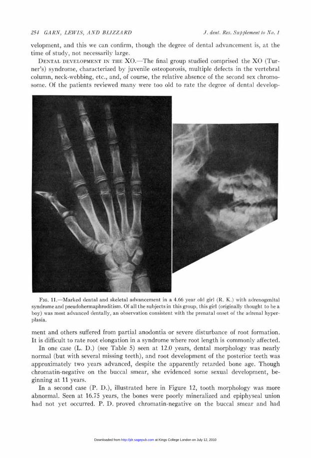
relatively short time after the onset of symptoms. It is of interest, therefore, to reviewCase R. K. (Fig. 11, and No. 7 in Table 4), who was considered to be a male at birth andwas so named. As an example of the adrenogenital syndrome with pseudohermaphroditism,of prenatal origin, she had been exposed to the growth-stimulating influence of androgenichormones of adrenal origin even prior to birth, and she proved to be dentally most ad-vanced (29 per cent), both absolutely and relatively, of any of the precocities we hadreviewed beyond two years of age.We have not attempted here to divide the adrenogenital syndrome cases into salt-
retaining or salt-losing categories. Rather, the point has been to determine whether ad-vanced skeletal development is associated in these conditions with advanced dental de-
.1. dent. Res. Supplement to No. I
at Kings College London on July 12, 2010 http://jdr.sagepub.comDownloaded from

I-
2
CL
LIJ
LiC
164/
''L~~~9E12-
2~ t 6 lb10 2 14 1'6 1iCHRONOLOGICAL AGE
8
Fmjo. 9.-Comparative s-keletal andl mentall advancement ini constitutional and( non-constitutional sexuallprecocities. Here, mentall development tends to be advanced, as show n by the position of the crosses abovethe isodevelopmental line. However, the relatively small mentall advancement (about 9 per cent) stands inmarked contrast to the acceleration in ossification timing, elilhvseal union, and compact hone develop-ment. Note in p)articular subject J. K. at 9.50 years.
FIG. 10.-Dental and skeletal development in a child with the adrenogenital syndrome. Less than 3 yearsof age, she was half a year advancedl dlentally andl more than 4.5 years advanced skeletall , Nith markedhvincreasedl thickness of compact boo e.
at Kings College London on July 12, 2010 http://jdr.sagepub.comDownloaded from
254 GI 1? N. 1 AWlS, A/JND 131/R A RD J dpV
veloprment, anti this We ciln confirm, though the degree of dental advancement is, at thetimee of study, not necessarily large.
R)ENVT.AL DEVIAIOPNIE N' IN TIE X(). The final group studied comprised the X() (Tur-ner's) syndrome, (haLra'cterized by juvenile osteoporosis, multiple defects in the vertebrallcolumn, neck-webbing, etc., and, of course, the relative aLbsence of the sec onci sex chrbrmo-somle. Of the patients reviewed mansytere too o(1d to rate the degree of (lentell develop-
Fic. 11.-Marked dental andl skeletal a(lvancement in a 4.66 year old girl (R/. K.) Wiltl adrenogenitalsvndlrome and pseudloherma)hroditismn. Of all the subjects in this groLups, this girl (originally thought to lbe ahoy) was most advanced dentally, an observation consistent with the ptrenLtal onset of the adrenal hvper-plsia.
mient and others suffered from partial anodontia or severe disturbance of root formation.It is difficultt to rate root elongation in. a sx nldrone where root length is commo11oltlV affected.
In one case (L. D.) (see Table 5) seen at 12.0 years dental morphology wats nearlynormal (but with several missing teeth), and root development of the posterior teeth was
approximately two years advanced, despite the apparently retarded bone age. Thoughchromatin-negative on the but cal smear, she evidleflced some sexual development, be-ginning at 11 years.
In a second case (P. 1).), illustrated here in Figure 12, tooth morphology was moreabnormal. Seen. at 16.75 vears, the bones were poorly mineralized and epiphvseal unionhad not yet occurred. P. D. proved chroniatin-negative on the buccal stneaLr and had
.1. deal. Res. Supplemoil lo No. I
at Kings College London on July 12, 2010 http://jdr.sagepub.comDownloaded from

ENDOCRINE FT CTORS IN DENTAL DEl'ELOPMENTI 255
abundant pubic hair but was without breast development. Estrogen output. was in themale range. Dentallx, extreme reduction of root length was obvious, as shown in Figure 12,but the developmental status was appropriate for her chronological age.
I)ental development has been reported to be advanced in the XO (see Gorlin 3 in thepresent Svmposium). Dr. Meinhard Robinow has found precocious eruption of the firstpermanent molars by 4 vears in a chromatin-negative XO seen in the Dayton Birth Defectclinicc (Fig. t3) and possible dental advancement in other cases of Turner's syndrome.
It remains to be seen, however, whether there is a division between chromatin-negativeand chromatin-positive gonadal genesis cases with respect to dental development, whether
TABLE' 5
DENTAL ANT) SKIELETAL A(m. IN TURNER'S SYNDROME
Subject
1. 1)..P. 1).......I
Karyotype
Chromatin- negative(hrornatin-negative
Chronologi-cal Age
12.00)16. 75
BoneAge
11 O*13. 75
DentalAge
14.0016-17
Interp)olated.
FIG. 12.-Comparative skeletal and dental development in an XO (Turner's syndrome). This girl is 3years retarded in epiphyseal union and further delayed in Ibone mineralization. Dentally, she is characterizedbLx short, stubby roots, lut the apparent dental development appears to be consistent rith her chronologicalage. IIn other XN cases, tooth eruption and tooth formation have been reported as advanced (see text).
Voll 44, 1.965
at Kings College London on July 12, 2010 http://jdr.sagepub.comDownloaded from

256 GI1RV, LIVIS, AND BLIZZA/ RI)
or not root developmentt and apical closure are altvanced only where there is snile sexualdevelopment, and whether dental advancement is independent of or associated withreductions in tooth number an(-d hypopltstic roots.
1DiscussiollLIt seems to be a valid generalization that dental development follows the direction of
skeletal (levelolment in the endocrine extremes its in the normal extremes. The teeth arereta(r(Ie(l in tlelayedl growth, in juvenile hypothxyroiclism, in thle athvrotic state, an(I in
l c.. 13.-Case C. T., a verified XO who reportedly erupted the first permanent molars prior to 4.0 yearsof age, here lpicture(l at 3.6 x ears. Note that eruption status of the first permanent molar is more than 2 yearsadvanced at this time, w hile the second molar an(l secondl l)remolar are up to 2 y ears aldvanced, though thecharacteristically reduced root length complicates assessment. The most adlvance(l girl in the Eels Longi-tudlinal Series di(l not attain a comparable stage (f mentall development until 6.5--7.0 years.
hypopituitarism. Thexy mayl be advanced in the constitutional and non-constitutionalsexual precocities, and they are probablyN advanced in the XC) as well.
However, the degree of advancement or retalrdlation in dentatl development, does notequal the degree of advancement or retardation in osseous development. In most pre-cocities or retardations the teeth depart from expectancy less than half as iuch (usuallyone-fourth as much) as the bones.* The exception, of course, is in hvpopituitarism, where
* For 19 growth failures and delays the teeth averaged 91 iier cent of expectancy and the skeletal development, 68 per cent. For 21 cretin records the comparable figures were 93 per cent of normal, and 38 per cent,respectively . The hypopituitary cases as mentioned here were 77 per cent of expectancy in tooth formationand 53 per cent of expectancy in skeletal development, while the figures for dental and skeletal status in the(evelolmental precocities were 109 per cent and 170 jer cent. The rank order correlations (rho) for rela-tive dental and skeletal status were 0.49, 0.14, ).201, and 01. 67 for these fotur groups (if endocrinopat hies.
J. de)II. RCS. Supploncill lo XI). -1
at Kings College London on July 12, 2010 http://jdr.sagepub.comDownloaded from

ENDOCRINE FACTORS IN DENTAL DEVELOPMENT 257
the dental delay (about 25 per cent) is roughly half as much as the skeletal delay (about50 per cent).The sexual precocities, involving osteotrophic hormones, constitute a special case.
The teeth in these children were, in general, slightly advanced (about 9 per cent), butthe endocrinopathies were not of long standing. There is a suggestion that prolonged ex-posure to auxogenic steroids of the androgenic type may bring about greater dental ad-vancement, as in Case R. K., and the data further confirm the suggestion that anabolicsteroids can and do spur tooth formation and movement.The XO syndrome is now of dubious value to the basic problem. Viewed as it once was,
simply as gonadal agenesis, one might anticipate normal dental development until afterthe first decade and dental delay thereafter. But there is no evidence for delay, and indeedwe are given clinical evidence for early eruption advancement in the XO. Further, fre-quent dental abnormalities and reduced root length make stage assessments difficult.Finally, the existence of multiple immunochemical abnormalities in the XO quite com-plicates the picture.24
In our experience this work could be sharpened by more detailed attention to thespecific stage of tooth formation involved, as well as acquisition of endocrine patientswith a more circumscribed age at onset of symptoms. As with defects of prenatal origin,the concept of stage-specificity is applicable. A degree of dental development, once attained,is not likely to be reversed by osteotrophic or somatotrophic hormonal failure. Similarly,one stage of tooth formation and development-say, crown formation may be inde-pendent of a particular trophic hormone, while another stage say, root elongation mayprove markedly hormone-dependent. However, it is impressive that, in the adrenogenitalsyndrome, crown formation and root development are both advanced simultaneously.
SummaryDental and skeletal development were compared in extremes of non-endocrine and
endocrine advancement and delay. In non-endocrine developmental delays, in congenitalhypothyroidism, and in hypopituitarism, the teeth were delayed, but to a lesser extentthan the postcranial skeleton. In hypopituitarism, the degree of dental retardation (about25 per cent) more nearly approximated the degree of skeletal retardation (50 per cent),whereas in the athyrotic, dental delay was little more than 10 per cent retarded whenskeletal development was 60 per cent retarded. In constitutional and endocrine sexualprecocities, dental advancement was noted along with skeletal advancement, but (exceptfor the adrenogenital syndrome with pseudohermaphroditism) the degree of dental ad-vancement was small. In the XO, dental development tended toward advancement, butthe inconstant advancement could not be explained on endocrine grounds alone.
The authors acknowledge with sincere thanks the invaluable assistance of Dr. Frederic N. Silverman ofthe Children's Hospital in Cincinnati, Dr. Meinhard Robinow of the Yellow Springs Clinic, and Dr. John S.Spaulding of the Johns Hopkins Hospital. Thanks are due also to Sachiyo Kakehashi for the illustrations andto Dorothy Gross for typing the manuscript.
References1. TAFT, L. L. Dental Findings in Five Year Old Peruvian Mother, N. '. J. Dent., 11:225, 1941.2. SECKEL, H. P. G. Six Examples of Precocious Sexual Development, Amer. J. Dis. Child., 79:278, 1950.3. PRADER, VON A., and PERABO, F. Kdrperwachstrum Knochen- und Zahnentwicklung bei den Endokri-
nen Erkrankungen im Kindersalter, Helv. paediat. Acta, 7:517-29, 1952.4. COHEN, M. M., and WAGNER, R. Dental Development in Pituitary Dwarfism, J. dent. Res., 27:445-58,
1948.
Vol. 44, 1965
at Kings College London on July 12, 2010 http://jdr.sagepub.comDownloaded from

258 GA RN, LEWIS, AND BLIZZARD
5. COHEN, M. M., and GARN, S. M. Factors in Occlusion, Amer. J. Orthodont., 40:671-85, 1954.6. WAGNER, R., COHEN, M. M., and HUNT, E. E., JR. Dental Development in Idiopathic Sexual Precocity
Congenital Adrenocortical Hyperplasia, and Adrenogenic Virilism, J. Pediat., 63:566-76, 1963.7. VAN WAGENEN, G., and HURME, V. 0. Effect of Testosterone Propionate on Permanent Canine Tooth
Eruption in the Monkey (Macaca mulatta), Proc. Soc. exp. Biol., 73:296, 1950.8. SEIPEL, C. M., VAN WAGENEN, G., and ANDERSON, B. G. Developmental Disturbances and Malocclu-
sion of the Teeth Produced by Androgen Treatment in the Monkey (Macaca mulatta), Amer. J. Ortho-dont., 40:37, 1954.
9. WILKINS, L. The Diagnosis and Treatment of Endocrine Disorders in Childhood and Adolescence. Spring-field, Ill.: Charles C Thomas, 1950.
10. GARN, S. M., LEWIS, A. B., and POLACHECK, D. L. Variability of Tooth Formation in Man, Science, 128:1510, 1958.
11. ---. Variability of Tooth Formation, f. dent. Res., 38:135-48, 1959.12. LEWIS, A. B., and GARN, S. M. The Relationship between Tooth Formation and Other Maturational
Factors, Angle Orthodont., 30:70-77, 1960.13. GARN, S. M., LEWIS, A. B., and KEREWSKY, R. S. Genetic, Nutritional and Maturational Correlates of
Dental Development, J. dent. Res., 44:228-42, 1965.14. GREULICH, W. W., and PYLE, S. I. Radiographic Atlas of Skeletal Development of the Hand and Wrist.
Stanford: Stanford University Press, 1950.15. GARN, S. M., and LEWIS, A. B. Phylogenetic and Intra-specific Variations in Tooth Sequence Poly-
morphism. In Dental Anthropology, ed. D. R. BROTHWELL, Pp. 53-73. New York: Macmillan Co., 1963.16. GARN, S. M., LEWIS, A. B., and BONNE, B. Third Molar Formation and Its Developmental Course, Angle
Orthodont., 32:270-79, 1962.17. GARN, S. M., and RoHMANN, C. G. Variability in the Order of Ossification of the Bony Centers of the
Hand and Wrist, Amer. J. phys. Anthrop., 18:219-28, 1960.18. GARN, S. M., SILVERMAN, F. N., and ROHMANN, C. G. A Rational Approach to the Assessment of Skele-
tal Maturation, Ann. Radiol., 7:297-307, 1964.19. GARN, S. M., ROHMANN, C. G., BEHAR, M., VITERI, F., and GUZMAN, M. A. Compact Bone Deficiency
in Protein-Calorie Malnutrition, Science, 45:1444-45, 1964.20. TALMERS, D. A. Time of Eruption of Second Permanent Molar and Relationship to Body Size and Areo-
lar Development: Preliminary Report, N.Y. State dent. J., 18:314-15, 1952.21. SCHOUR, I., BRODIE, A. G., and KING, E. Q. Dental Changes in a Hypopituitary Condition: A Case Re-
port, Angle Orthodont., 4:285-304, 1934.22. GARN, S. M., ROHMANN, C. G., and NOLAN, P., JR. Studies on the Development of Compact Bone in
Normal Individuals and in Endocrine and Nutritional Abnormalities. Part I, Progress Report, GrantAM-03816, 1963.
23. GORLIN, R. J., REDMAN, R. S., and SHAPIRO, B. L. The Effect of X-Chromosome Aneuploidy on JawGrowth, J. dent. Res., 44:269-82, 1965.
J. dent. Res. Supplement to No. I
at Kings College London on July 12, 2010 http://jdr.sagepub.comDownloaded from