journal of crystal growth - ssn institutions on the structure, growth and... · centre for crystal...
TRANSCRIPT

Studies on the structure, growth and characterization of morpholiniumperchlorate single crystals
A. Arunkumar, P. Ramasamy n
Centre for Crystal Growth, SSN college of Engineering, Kalavakkam 603110, India
a r t i c l e i n f o
Article history:Received 2 April 2013Received in revised form30 June 2013Accepted 1 October 2013Communicated by K. JacobsAvailable online 16 November 2013
Keywords:A1. SolubilityA2. Growth from solutionsB1. Organic compoundsB2. Nonlinear optic materials
a b s t r a c t
Morpholinium perchlorate, an organic nonlinear optical material was synthesized and the crystals weregrown by the slow evaporation solution growth technique. Single crystal X-ray diffraction study revealedthat the grown crystal belongs to orthorhombic systemwith space group of P212121. The structure of thecompound was also confirmed by 1H NMR studies. The Fourier transform infrared analysis was used toidentify the various functional groups present in the title compound. The UV–visible absorption spectrumwas recorded to study the optical transmittance in the range from 200 to 1100 nm. The optical band gap,reflectance, refractive index (n), extinction coefficient and electric susceptibility were calculated usingtransmittance data. The mechanical stability of the grown crystal was studied by Vickers′s micro hardnesstest. The PL spectrum of the title compound shows red emission at 648 nm.
& 2013 Elsevier B.V. All rights reserved.
1. Introduction
The technological society of optoelectronics and photonics hascreated more attention on organic optical materials. Nowadaysorganic and semiorganic NLO materials are emerging as analternative to inorganic materials because of their efficient mole-cular nonlinearity over a broad frequency range, low cost, lowrefractive index, low dielectric constant, inherent synthetic flex-ibility, moderate optical damage density, fast response with thebetter process ability and ease of fabrication into devices. Theorganic nonlinear optical materials generally have the largersecond order nonlinear optical coefficient and hence they arebeing used in many applications such as second harmonic gen-eration, sum frequency generation, THz wave generation, opticalparametric oscillators, etc., [1–5]. Researches to explore the thirdorder nonlinear optical phenomena in organic and inorganic singlecrystals have received limited attention compared to second ordernonlinear optical materials. For the past ten years, the third ordernonlinear optical materials from the organometallic, organic,inorganic and semi organic single crystalline compounds havereceived great deal of attention due to their potential applicationsin all optical switching, optical limiters, optical information storage,all optical logic gates, laser radiation protection etc., [6,7]. Crystals oforganic salts are often colorless, transparent, inexpensive to produce
and easy to grow [8]. Morpholine is colorless, oily and volatile innature having great importance in industrial purposes [9]. Molecularionic simple complex crystals like perchlorate with Morpholine (ofratio 1:1), shows nonlinear optical physical properties unique to thecrystal structure. The distinct features of molecular ionic crystal giveempathizing correlation between the crystal packing and physicalproperties. This initiated many researchers to synthesize and togrow the newly designed molecular ionic crystals. In this paper wepresent the structure of morpholinium perchlorate (MP) at roomtemperature, crystal growth and its characterization.
2. Experiment
The title material MP was synthesized by the chemical reactionof commercially available Morpholine (Merck) with Perchloric acid(SRL), taken in the stoichiometric ratio 1:1 by dissolving in themixture of (1:1) ethanol and deionized water. The chemical reactionis shown in Fig. 1. Stoichiometrically calculated amounts of thematerials were transferred into a beaker and dissolved in ethanoland deionized water which is stirred well with the help of amagnetic stirrer to make a homogeneous solution of the materialat a temperature of 50 1C for a proper chemical reaction. The whiteprecipitate was obtained after two hours. The precipitate is allowedto dry. The dried salt was collected and used for the further growthof MP. The synthesized material was purified by the repeatedrecrystallization process. The dried precipitate was dissolved usingthe same solvent. But the crystallization did not occur in this solutionas it has high viscosity and low pH value. The selection of solvent is
Contents lists available at ScienceDirect
journal homepage: www.elsevier.com/locate/jcrysgro
Journal of Crystal Growth
0022-0248/$ - see front matter & 2013 Elsevier B.V. All rights reserved.http://dx.doi.org/10.1016/j.jcrysgro.2013.10.005
n Corresponding author: Tel.: þ91 9283105760; fax: þ91 44 27475166.E-mail addresses: [email protected],
[email protected] (P. Ramasamy).
Journal of Crystal Growth 388 (2014) 124–131

important to grow good quality single crystals of considerable size.The solubility test can be performed to choose the solvent for crystalgrowth. The solubility was measured by taking excess amount of MPin the solvent and it is continuously stirred to achieve uniformconcentration over the entire volume of the solution. Solubility curvefor MP was determined by using acetone as a solvent in thetemperature range from 30 to 45 1C with the interval of 5 1C. Thestudies were carried out in a constant-temperature water bath witha cryostat facility with an accuracy of þ0.01 K. MP exhibits a positivesolubility – temperature gradient in acetone solution. The solubilityalmost increases linearly with the increase of temperature. Fig. 2depicts the solubility curve of MP. The obtained dried precipitate wasdissolved using acetone and then allowed to evaporate at roomtemperature to yield the crystalline powder salt of MP. The well-defined single crystals of MP were harvested from mother solutionafter a growth period of 45 days. The grown single crystals of MP areshown in Fig. 3
3. Characterization
As grown MP crystals have been subjected to various character-ization studies to analyze the structural, thermal, optical andmechanical properties The single crystal X-ray diffraction measure-ments were done using a Bruker AXS Kappa APEX II single crystalCCD diffractometer equipped with graphite-monochromated MoKαradiation (λ¼0.71,073 Å) at room temperature with a crystal ofdimension 0.35�0.25�0.2 mm3. Accurate unit cell parameterswere determined from the reflections of 36 frames measured inthree different crystallographic zones. The data collection, datareduction and absorption correction were performed by APEX2,SAINT-plus and SADABS program using SHELXL-97 [10]. The struc-ture was solved by direct methods procedure and the non-hydrogenatoms were subjected to anisotropic refinement by full-matrix leastsquares on F2 using SHELXL-97 program [11]. The positions of allthe hydrogen atoms were identified from difference electrondensity map, and they were constrained to ride on the correspond-ing non-hydrogen atoms. The hydrogen atom bound to carbonatoms were constrained to a distance of C–H¼0.97 Å and Uiso (H)¼1.2 Ueq (C). The final refinement converges to an R-values ofR1¼0.0485 and WR2¼0.1330. The ORTEP drawing was performedwith the ORTEP3 program [12]. The crystallographic refinementparameters are listed in Table 1. The possible hydrogen bondsobserved in the structure are listed in Table 2. The crystallineperfection of the grown single crystals was characterized by HRXRDby employing a multicrystal X-ray diffractometer developed atNPL [13]. The well-collimated and monochromated MoKα1 beamobtained from the three monochromator Si crystals set in dispersive(þ ,� ,�) configuration has been used as the exploring X-ray beam.The specimen crystal is aligned in the (þ ,� ,� ,þ) configuration.Due to dispersive configuration, though the lattice constant of themonochromator crystal(s) and the specimen are different, theunwanted dispersion broadening in the diffraction curve (DC) ofthe specimen crystal is insignificant. The specimen can be rotatedabout the vertical axis, which is perpendicular to the plane ofdiffraction, with minimum angular interval of 0.4′′. The rocking ordiffraction curves were recorded by changing the glancing angle(angle between the incident X-ray beam and the surface of thespecimen) around the Bragg diffraction peak position θB (taken aszero for the sake of convenience) starting from a suitable arbitraryglancing angle and ending at a glancing angle after the peak so thatall the meaningful scattered intensities on both sides of the peak areincluded in the diffraction curve. The DC was recorded by the so-called ω scan wherein the detector was kept at the same angularposition 2θB with wide opening for its slit. This arrangement is veryappropriate to record the short range order scattering caused by thedefects or by the scattering from local Bragg diffractions fromagglomerated point defects or due to low angle and very low anglestructural grain boundaries [14]. Before recording the diffractioncurve to remove the non-crystallized solute atoms which remainedon the surface of the crystal and the possible layers which maysometimes form on the surfaces on crystals grown by solutionmethods [15] and also to ensure the surface planarity, the specimenwas first lapped and chemically etched in a non-preferentialetchant of water and acetone mixture in 1:2 volume ratio. The FTIRspectrum of MP crystals was recorded in the range 4000–400 cm�1
NH
O
Morpholine
Cl
O
O
OH
O
perchloric acid
+CH3CH2OH + H2O
NH2+O Cl O
O
-O
O
Morpholinium perchlorate
Fig. 1. Reaction scheme of MP.
3035
40
45
50
55
60
65
70
Temperature (°C)
Con
cent
ratio
n (g
m/1
00m
l)
35 40 45
Fig. 2. Solubility diagram of MP.
Fig. 3. As grown single crystals of morpholinium perchlorate (MP).
A. Arunkumar, P. Ramasamy / Journal of Crystal Growth 388 (2014) 124–131 125
employing a JASCO FT-IR 410 spectrometer by the KBr pelletmethod to study the functional groups in sample. In the presentinvestigation, the 1H spectra of MP was recorded using acetone assolvent on a Bruker 300 MHz (Ultra shield) TM instrument at 23 1C(300 MHz for 1H NMR) to confirm the molecular structure. Opticaltransmittance was studied at room temperature using a PerkinElmerLambda 35 UV–vis spectrometer in the region 200–1100 nm.Fluorescence of the MP crystal was studied in the emission rangeof 200–750 nm using JASCO fluorimeter.
4. Results and discussions
4.1. Single crystal X- ray diffraction studies
The title compound MP crystallizes in orthorhombic spacegroup P212121. The structure of morpholinium perchlorate in thelow-temperature form (100 K) was previously reported [16] innon-standard unit cell parameter settings. The previously reportedcell parameters values are “a¼8.1515 (3) Å, b¼9.5435(4) Å,c¼28.9022 (12) Å and V¼2248.41 (16) Å3”, with three independentformula units in the unit cell. The crystal structure shows pseudoinversion centre. The structure was partially resolved in centro-symmetric space group Pnma with half anions and cations in theasymmetric form and with high R-value. But the systematic absent
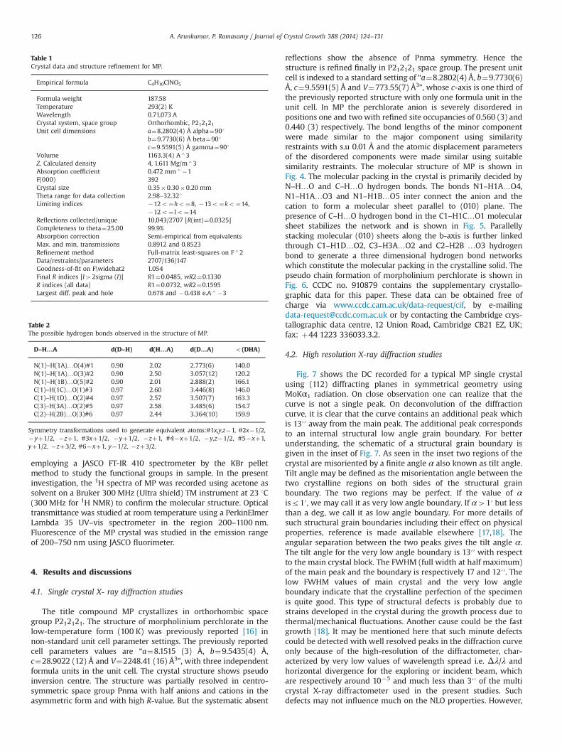
reflections show the absence of Pnma symmetry. Hence thestructure is refined finally in P212121 space group. The present unitcell is indexed to a standard setting of “a¼8.2802(4) Å, b¼9.7730(6)Å, c¼9.5591(5) Å and V¼773.55(7) Å3”, whose c-axis is one third ofthe previously reported structure with only one formula unit in theunit cell. In MP the perchlorate anion is severely disordered inpositions one and twowith refined site occupancies of 0.560 (3) and0.440 (3) respectively. The bond lengths of the minor componentwere made similar to the major component using similarityrestraints with s.u 0.01 Å and the atomic displacement parametersof the disordered components were made similar using suitablesimilarity restraints. The molecular structure of MP is shown inFig. 4. The molecular packing in the crystal is primarily decided byN–H…O and C–H…O hydrogen bonds. The bonds N1–H1A…O4,N1–H1A…O3 and N1–H1B…O5 inter connect the anion and thecation to form a molecular sheet parallel to (010) plane. Thepresence of C–H…O hydrogen bond in the C1–H1C…O1 molecularsheet stabilizes the network and is shown in Fig. 5. Parallellystacking molecular (010) sheets along the b-axis is further linkedthrough C1–H1D…O2, C3–H3A…O2 and C2–H2B …O3 hydrogenbond to generate a three dimensional hydrogen bond networkswhich constitute the molecular packing in the crystalline solid. Thepseudo chain formation of morpholinium perchlorate is shown inFig. 6. CCDC no. 910879 contains the supplementary crystallo-graphic data for this paper. These data can be obtained free ofcharge via www.ccdc.cam.ac.uk/data-request/cif, by [email protected] or by contacting the Cambridge crys-tallographic data centre, 12 Union Road, Cambridge CB21 EZ, UK;fax: þ44 1223 336033.3.2.
4.2. High resolution X-ray diffraction studies
Fig. 7 shows the DC recorded for a typical MP single crystalusing (112) diffracting planes in symmetrical geometry usingMoKα1 radiation. On close observation one can realize that thecurve is not a single peak. On deconvolution of the diffractioncurve, it is clear that the curve contains an additional peak whichis 13′′ away from the main peak. The additional peak correspondsto an internal structural low angle grain boundary. For betterunderstanding, the schematic of a structural grain boundary isgiven in the inset of Fig. 7. As seen in the inset two regions of thecrystal are misoriented by a finite angle α also known as tilt angle.Tilt angle may be defined as the misorientation angle between thetwo crystalline regions on both sides of the structural grainboundary. The two regions may be perfect. If the value of αisr1′, we may call it as very low angle boundary. If α41′ but lessthan a deg, we call it as low angle boundary. For more details ofsuch structural grain boundaries including their effect on physicalproperties, reference is made available elsewhere [17,18]. Theangular separation between the two peaks gives the tilt angle α.The tilt angle for the very low angle boundary is 13′′ with respectto the main crystal block. The FWHM (full width at half maximum)of the main peak and the boundary is respectively 17 and 12′′. Thelow FWHM values of main crystal and the very low angleboundary indicate that the crystalline perfection of the specimenis quite good. This type of structural defects is probably due tostrains developed in the crystal during the growth process due tothermal/mechanical fluctuations. Another cause could be the fastgrowth [18]. It may be mentioned here that such minute defectscould be detected with well resolved peaks in the diffraction curveonly because of the high-resolution of the diffractometer, char-acterized by very low values of wavelength spread i.e. Δλ/λ andhorizontal divergence for the exploring or incident beam, whichare respectively around 10�5 and much less than 3′′ of the multicrystal X-ray diffractometer used in the present studies. Suchdefects may not influence much on the NLO properties. However,
Table 1Crystal data and structure refinement for MP.
Empirical formula C4H10ClNO5
Formula weight 187.58Temperature 293(2) KWavelength 0.71,073 ACrystal system, space group Orthorhombic, P212121Unit cell dimensions a¼8.2802(4) Å alpha¼901
b¼9.7730(6) Å beta¼901c¼9.5591(5) Å gamma¼901
Volume 1163.3(4) A43Z, Calculated density 4, 1.611 Mg/m43Absorption coefficient 0.472 mm4�1F(000) 392Crystal size 0.35�0.30�0.20 mmTheta range for data collection 2.98–32.321Limiting indices �12o¼ho¼8, �13o¼ko¼14,
�12o¼ lo¼14Reflections collected/unique 10,043/2707 [R(int)¼0.0325]Completeness to theta¼25.00 99.9%Absorption correction Semi-empirical from equivalentsMax. and min. transmissions 0.8912 and 0.8523Refinement method Full-matrix least-squares on F42Data/restraints/parameters 2707/136/147Goodness-of-fit on F\widehat2 1.054Final R indices [I42sigma (I)] R1¼0.0485, wR2¼0.1330R indices (all data) R1¼0.0732, wR2¼0.1595Largest diff. peak and hole 0.678 and �0.438 e.A4�3
Table 2The possible hydrogen bonds observed in the structure of MP.
D–H…A d(D–H) d(H…A) d(D…A) o(DHA)
N(1)–H(1A)…O(4)#1 0.90 2.02 2.773(6) 140.0N(1)–H(1A)…O(3)#2 0.90 2.50 3.057(12) 120.2N(1)–H(1B)…O(5)#2 0.90 2.01 2.888(2) 166.1C(1)–H(1C)…O(1)#3 0.97 2.60 3.446(8) 146.0C(1)–H(1D)…O(2)#4 0.97 2.57 3.507(7) 163.3C(3)–H(3A)…O(2)#5 0.97 2.58 3.485(6) 154.7C(2)–H(2B)…O(3)#6 0.97 2.44 3.364(10) 159.9
Symmetry transformations used to generate equivalent atoms:#1x,y,z�1, #2x�1/2,�yþ1/2, �zþ1, #3xþ1/2, �yþ1/2, �zþ1, #4�xþ1/2, �y,z�1/2, #5�xþ1,yþ1/2, �zþ3/2, #6�xþ1, y�1/2, �zþ3/2.
A. Arunkumar, P. Ramasamy / Journal of Crystal Growth 388 (2014) 124–131126

a quantitative analysis of such unavoidable defects is of greatimportance, particularly in case of phase matching applications asexplained in the recent article [19].
4.3. FT-IR spectral analysis
Fig. 8 shows the FTIR spectrum of MP. The symmetric andasymmetric stretching vibrations of NH2þ cation occurred at 3415and 3282 cm�1. The CH asymmetric and symmetric vibrations ofmorpholinium ring are observed at 2945 and 2865 cm�1. Thestretching vibration of NH2þ cation occurred at 2449 cm�1. TheC–O–C stretching vibrations of morpholinium ring occurred at1105 (asymmetric) and 1035 (symmetric) cm�1. The C–N in-planeand out of plane bending vibrations occurred at 940 and525 cm�1. The stretching vibrations of chlorate counter anionoccurred at 2203, 2163 and 1144 cm�1. The C–N stretchingvibration occurred at 1315 cm�1. The NH bending vibrationoccurred at 1574 cm�1. The N–H wagging vibration occurred at691 cm�1. The CH bending vibration is observed at 895 cm�1.
Fig. 4. Molecular structure of MP.
Fig. 5. Sheet of MP. Fig. 6. Chain formation of MP.
Fig. 7. High resolution X-ray diffraction curve of MP.
A. Arunkumar, P. Ramasamy / Journal of Crystal Growth 388 (2014) 124–131 127

Hence the spectrum shows the characteristic absorptions of bothmorpholine and perchloric acid as morpholinium perchlorate.
4.4. NMR spectral analysis
The NH proton signal is observed at 2.7 ppm. The peak at3.5 ppm is assigned to CH2 protons ortho to NH group. The signalat 3.95 ppm is assigned to CH2 protons meta to NH group. The 1HNMR spectrum of MP is shown in Fig. 9.
4.5. UV–vis–NIR spectral analysis
Fig. 10 shows the UV–vis spectrum of MP crystal. The crystalhas sufficient transmission in the UV and visible region. The cut-
off wavelength of MP crystal was found to be 215 nm and theabsorption at 279 nm was due to the promotion of an electronfrom a ‘non-bonding’ (lone-pair) n orbital to an ‘anti-bonding’ πorbital designated as πn (n-πn) and no characteristic absorptionwas observed in the entire visible region. The dependence ofoptical absorption coefficient with the photon energy helps tostudy the band structure and the type of transition of the electron.The absorption coefficient (α) and the optical parameters such asrefractive index (n), reflectance (R) and extinction coefficient (K)have been determined from the transmission (T) spectrum basedon the following relation:
α¼ 2:3026t
log ð1=TÞ ð1Þ
where T is transmittance, ‘t’ is the thickness of the crystal, ‘α’ is
4000 3500 3000 2500 2000 1500 1000 500
0
20
40
60
80
100
Tran
smitt
ance
(%)
wavenumber(cm-1)
3415
3282
3211
3059
2945 28
6527
77 2449
2023
1574
1426
1455
1315
1105
940
895
870
822
691
626
594
Fig. 8. FTIR spectrum of MP.
Fig. 9. 1H NMR spectrum of MP.
200 400 600 800 1000 12000
10
20
30
40
50
60
70
80 % T % R
Wavelength (nm)
% T
rans
mitt
ance
70
75
80
85
90
95
100
% R
efle
ctan
ce
Fig. 10. Transmittance and reflectance spectrum of MP crystal.
A. Arunkumar, P. Ramasamy / Journal of Crystal Growth 388 (2014) 124–131128

related to the extinction coefficient K by
K ¼ αλ4π
ð2Þ
The reflectance (R) in terms of the absorption coefficient andrefractive index (n) can be derived from the relations
R¼expð�αtÞ7
ffiffiffiffiffiffiffiffiffiffiffiffiffiffiffiffiffiffiffiffiffiffiffiffiffiffiffiffiffiffiffiffiffiffiffiffiffiffiffiffiffiffiffiffiffiffiffiffiffiffiffiffiffiffiffiffiffiffiffiffiffiffiffiffiffiffiffiffiffiffiffiffiffiffiffiffiffiffiffiffiffiffiffiffiffiffiffiffiffiexpð�αtÞT�expð�3αtÞTþexpð�2αtÞT2
q
expð�αtÞþexpð�2αtÞT ð3Þ
n¼ �ðRþ1Þ72ffiffiffiR
p
ðR�1Þ ð4Þ
In the high photon energy region, the energy dependence ofabsorption coefficient suggests the occurrence of direct band gapof the crystal obeying the following equation for high photonenergies (hν) [20]
ðαhvÞ2 ¼ AðEg�hvÞ ð5Þ
where Eg is the optical band gap of the crystal and A is a constant.The band gap of the crystal was evaluated by plotting (αhν)2 versushv as shown in Fig. 11 and it was found to be 5 eV. The wide band gapof the MP crystals confirms the large transmittance in the visibleregion. Fig. 12 represents the variation of refractive index withrespect to wavelength (215–1100 nm) respectively. The estimatedrefractive index (n) of MP crystal from the graph is 1.41 at 600 nm.
From the optical constants, the electric susceptibility χc can becalculated according to the relation [21]
εr ¼ ε0þ4πχc
4π¼ n2�K2 ð6Þ
χc ¼ðn2�K2�ε0Þ
4πð7Þ
where ε0 is the dielectric constant in the absence of any contribu-tion from free carriers. The estimated electric susceptibility (χc) isfound to be 0.19 at 600 nm.
The real and imaginary dielectric constants, εr and εi can becalculated from the following relations [22]:
εr ¼ n2�K2 and εi ¼ 2nK ð8Þ400 600 800 1000 1200
1.4
1.5
1.6
1.7
1.8
Ref
ract
ive
Inde
x (n
)
Wavelength (nm)
Fig. 12. Plot of refractive index vs. wavelength.
1 2 3 4 5 6 7
0
1x10⁷
2x10⁷
3x10⁷
4x10⁷
5x10⁷
6x10⁷
(αhν
)2 eV
2 cm -2
photon energy (eV)
Fig. 11. Plot of (αhν)2 vs. photon energy.
0
1
2
3
4
5
6
Inte
nsity
wavelength (nm)
648 nm
450 500 550 600 650 700 750360 380 400 420 440
0
1
2
3
4
5
Inte
nsity
wavelength (nm)
Fig. 13. (a) Excitation and (b) emission spectra of MP.
A. Arunkumar, P. Ramasamy / Journal of Crystal Growth 388 (2014) 124–131 129

The real εr and imaginary εi dielectric constants at 600 nmwere found to be 2.40 and 2.5�10�5 respectively.
4.6. Fluorescence analysis
The excitation spectrum was recorded in the range of 220–350 nm and the sample was excited at 434 nm. The emissionspectrum was measured in the range of 450–750 nm. The excita-tion and emission spectrum of MP is given in Fig. 13 (a) and (b).Sharp peak was observed at 648 nm and it shows the character-istic nature of red emission. The full width at half maximum ofemission peak is 4.78 nm which shows that the quality of thecrystal is good.
4.7. Mechanical properties
Microhardness studies were conducted for the loads in therange 5–35 g. Vickers′s hardness (Hv) number increases initiallywith load up to 25 g (Fig. 14(a)) and cracks were observed beyond25 g. This type of load dependent variation of hardness is termedas reverse indentation size effect. At low loads, the indenterpenetrates only the top surface layers generating dislocations,which results in the increase of hardness in this region. The loadindependence of hardness at higher loads can be attributed to themutual interaction or rearrangement of dislocations [23]. Therelation between load and the size of indentation can be corre-lated using Meyer′s law, P¼k1d
m, where k1 is a constant and ‘m’ isthe Meyer′s index. Fig. 14 (b) shows the plot between log P and logD. From the slope of graph the work hardening coefficient (m) wascalculated and it is found to be 2.7 which indicates that MP crystalbelongs to soft material category.
4.8. Nonlinear optical studies
In order to know its second order nonlinear optical property ofMP crystal, the Kurtz Perry powder technique was used. Completeabsence of second order NLO property was observed from theresult and it is due to the pseudo inversion centre present in thecrystal packing and pseudo symmetry present in the structure ofMP which is similar to the structure of L-Histidinium sulfate [24].
5. Conclusion
Simple ionic morpholinium perchlorate crystal was synthe-sized and bulk crystals were grown using the slow evaporation
solution growth technique. The structure was solved with stan-dard unit cell parameter settings at room temperature. Thepresence of functional groups in the MP crystal was confirmedusing FTIR studies. The crystalline perfection of the grown crystalwas fairly good confirmed by high resolution X-ray diffraction. Theoptical behavior was evaluated by UV–vis–NIR and photolumines-cence analyses which substantiate the suitability of MP for opto-electronic applications. The optical band gap, refractive index andelectric susceptibility were calculated from the linear optical data.From microhardness measurements it is observed that the MPcomes under the soft materials category. Red emission wasobserved in the PL spectrum of crystal.
Acknowledgments
The authors thank SAIF, IIT Madras, Chennai for recording NMRspectrum, and single crystal data collection. The authors thankDr. G. Bhagavannarayana, NPL, New Delhi for HRXRD studies.
Appendix A. Supplementary material
Supplementary data associated with this article can be found inthe online version at http://dx.doi.org/10.1016/j.jcrysgro.2013.10.005.
References
[1] D.S. Chemla, J. Zyss (Eds.), Academic Press, New York, 1987.[2] L.R. Dalton, P.A. Sullivan, B.C. Olbricht, D.H. Bale, J. Takayesu, S. Hammond,
H Rommel, B.H. Robinson, Tutorials in Complex Photonic Media, SPIE,Bellingham, WA, 2007.
[3] R.W. Munn, C.N. Ironside, Principles and Applications of Nonlinear OpticalMaterials, Chapman & Hall, London, 1993.
[4] M. Thakur, J. Xu, A. Bhowmilk, L. Zhou, Appl. Phys. Lett. 74 (1999) 635–637.[5] T. Kaino, B. Cai, K. Takayama, Adv. Funct. Mater. 12 (2002) 599–603.[6] M. Somac, A. Somac, B.L. Davies, M.G. Humphery, M.S. Wong, Opt. Mater. 21
(2003) 485–488.[7] L.V. Natarajan, R.L. Sutherland, V.P. Tondiglia, T.J. Bunning, W.W. Adams,
J. Nonlinear Opt. Phys. Mater. 5 (1996) 89–98.[8] P.V. Dhanaraj, N.P. Rajesh, J. Kalyana Sundar, S. Natarajan, G. Vinitha, Mater.
Chem. Phys. 129 (2011) 457–463.[9] P. Szklarz, M. Owczarek, G. Bator, T. Lis, K. Gatner, R. Jakubas, J. Mol. Struct. 929
(2009) 48–57.[10] Bruker–Nonius AXS, APEX2 and SAINT, Bruker–Nonius AXS, Madison, Wisconsin,
USA, 2004.[11] G.M. Sheldrick, Acta Cryst. A64 (2008) 112–122.[12] L.J Farrugia, ORTEP-3 program for molecular drawing, J. Appl. Cryst. 30 (1997)
561–565.[13] Krishan Lal, G. Bhagavannarayana, J. Appl. Cryst. 22 (1989) 209–215.[14] G. Bhagavannarayana, S.K. Kushwaha, J. Appl. Cryst. 43 (2010) 154–162.
0 5 10 15 20 25 30 35 405
10
15
20
25
30
35H
ardn
ess
(Hv)
Load (P)0.0 0.5 1.0 1.5 2.0 2.5
1.2
1.4
1.6
1.8
2.0
Log
D
Log P
Fig. 14. (a) Load (P) vs. Hardness number (Hv) and (b) Plot of log P vs. log D of MP.
A. Arunkumar, P. Ramasamy / Journal of Crystal Growth 388 (2014) 124–131130

[15] G. Bhagavannarayana, S. Parthiban, Subbiah Meenakshisundaram, J. Appl.Cryst. 39 (2006) 784–790.
[16] Mikhail S. Grigoriev, Konstantin E. German, Alesia Ya. Maruk, Acta Cryst. E64(2008) o390–o401.
[17] G. Bhagavannarayana, R.V. Ananthamurthy, G.C. Budakoti, B. Kumar, K.S. Bartwal,J. Appl. Cryst. 38 (2005) 768–771.
[18] G. Bhagavannarayana, P. Rajesh, P. Ramasamy, J. Appl. Cryst. 43 (2010)1372–1376.
[19] G. Bhagavannarayana, B. Riscob, Mohd Shakir, Mater. Chem. Phys. 126 (2011)20–23.
[20] A. Ashour, N. El-Kadry, S.A. Mahmoud, Thin Solid Films 269 (1995) 111–120.[21] J. Tauc, R. Grigorovici, A. Vancu, Phys. Status Solidi B 15 (1966) 627–637.[22] V. Gupta, A. Mansingh, J. Appl. Phys. 80 (1996) 1063–1073.[23] J.H. Gong, Y. Li, J. Mater. Sci. 35 (2000) 209–213.[24] H.A. Petrosyan, H.A. Karapetyan, A.K. Atanesyan, A.M. Petrosyan, J. Mol. Struct.
963 (2010) 168-174.
A. Arunkumar, P. Ramasamy / Journal of Crystal Growth 388 (2014) 124–131 131