journal of cardiology cases - connecting repositories · blood pressure unexpectedly dropped to 40...
TRANSCRIPT

Journal of Cardiology Cases 13 (2016) 47–51
Case Report
Successful treatment of prolonged cardiopulmonary arrest of Kounissyndrome during coronary angioplasty
Tomomi Akita (MD), Masahito Kawata (MD, PhD)*, Ayu Sakaguchi (MD),Yukinori Kato (MD), Hideya Suehiro (MD), Hiroki Takada (MD), Takeshi Matsuura (MD),Kohei Kamemura (MD), Yasutaka Hirayama (MD), Kazumasa Adachi (MD),Akira Matsuura (MD), Susumu Sakamoto (MD)
Department of Cardiology, Akashi Medical Center, Akashi, Japan
A R T I C L E I N F O
Article history:
Received 25 February 2015
Received in revised form 18 September 2015
Accepted 5 October 2015
Keywords:
Kounis syndrome
Coronary spasm
Anaphylaxis
Percutaneous coronary intervention
Optical coherence tomography
A B S T R A C T
We experienced a case of Kounis syndrome with cardiopulmonary arrest and severe coronary spasm. A
70-year-old man with cardiac pacemaker and chronic dialysis was treated for angina pectoris of the right
coronary artery. After diagnostic coronary angiography of the right coronary artery, optical coherence
tomography was performed with contrast medium and low-molecular-weight dextran. The patient’s
blood pressure unexpectedly dropped to 40 mmHg and erythema of the breast was noted.
Electrocardiogram showed remarkable ST elevation in II, III, aVF leads. Coronary angiography showed
total occlusion of the proximal right coronary artery. Although intracoronary infusion of sodium nitrate
did not dilate the coronary artery promptly, coronary balloon angioplasty recovered the artery flow.
Since severe anaphylaxis-related shock was contemplated, methyl prednisolone and epinephrine were
administered intravenously. We could not introduce percutaneous cardiopulmonary support due to
kinking of the vein. After 1 hour of cardiopulmonary resuscitation with frequent ventricular fibrillation
and direct current shock, the sinus rhythm and blood pressure recovered. Following 2 months of
intensive care treatment for other complications, including infection, the patient was discharged from
hospital without any residual disability.
<Learning objective: An anaphylactic reaction is one of the causes of sudden deterioration of a patient’s
condition observed during interventional procedures. Kounis syndrome is a rare and not yet well known
important concept that deals with the reaction. Therefore, we report a severe case of Kounis syndrome
with cardiopulmonary arrest.
� 2015 Japanese College of Cardiology. Published by Elsevier Ltd. All rights reserved.
Contents lists available at ScienceDirect
Journal of Cardiology Cases
jo u rn al ho m epag e: ww w.els evier . c om / lo cat e/ jcc as e
Introduction
Kounis syndrome is an anaphylactic reaction that causes eithercoronary spasm (type I) or acute coronary syndrome due to plaquerupture or coronary erosion (type II) [1,2]. Usually a skin reactionand reduced blood pressure are accompanying symptoms. Wereport a case with an abrupt and prolonged cardiac arrest due tosevere coronary spasm and reduction in blood pressure by vesseldilatation of the whole body after intracoronary infusion of low-molecular-weight dextran.
* Corresponding author at: 743-33 Yagi, Ohkubo-cho, Akashi 674-0063,
Japan. Tel.: +81 78 936 1101; fax: +81 78 936 7456.
E-mail address: [email protected] (M. Kawata).
http://dx.doi.org/10.1016/j.jccase.2015.10.001
1878-5409/� 2015 Japanese College of Cardiology. Published by Elsevier Ltd. All rights
Case report
A 70-year-old man with cardiac pacemaker was admitted to ourhospital for percutaneous coronary intervention (PCI) of the rightcoronary artery (RCA).
He was admitted to the hospital 1 month before the presentadmission with dyspnea and diagnosed with complete atrioven-tricular block with a heart rate of 33/min. He had diabetes mellitusand hypertension and was administered several drugs. Cardiacechocardiography showed no wall motion abnormality withejection fraction of 83% as well as abnormal relaxation (E/A ratio0.79) and mildly elevated end-diastolic pressure (E/E0 15.8) andpulmonary systolic pressure (48 mmHg). Before DDD pacemakerimplantation, coronary angiography was performed and 75%diameter stenosis at the distal RCA was detected. No allergicreaction was observed after coronary angiography (CAG). He
reserved.

T. Akita et al. / Journal of Cardiology Cases 13 (2016) 47–5148
gradually recovered from heart failure after DDD pacemakerimplantation and was discharged from hospital 1 month prior tothe present admission. His prescription medications were azilsar-tan 20 mg, amurodipine 5 mg, glimepiride 1 mg, linagliptin 5 mg,and metformin 500 mg per day. He showed no abnormal finding onphysical examination. Blood pressure was 138/64 mmHg and theheart rate was 86/min. The electrocardiogram (ECG) showedpacemaker rhythm (A sensing and V pacing, Fig. 1A). The whiteblood cell count was 5610/mL. The red blood cell count was454 � 104/mL. Platelet count was 16.2 � 104/mL. The proportion ofeosinophils was 2.6% and was less than 2% during hospitalization.IgE level was not measured. Blood urea nitrogen level was 26.9 mg/dL and serum creatinine level was 0.77 mg/dL. Na, K, and Cl levelswere 142, 4.4 and 107 mEq/L, respectively. Blood glucose level was158 mg/dL and hemoglobin A1c was 7.9% indicating poor diabeticcontrol. Other blood data showed no abnormalities. Chestradiography showed cardiothoracic ratio of 46%. On the secondday, PCI was performed. A 6Fr Launcher SAL1.5SH (Medtronic, Inc.,Minneapolis, MN, USA) guiding catheter was engaged for the RCA.CAG of the RCA was performed once in the left anterior obliqueposition (Fig. 2A). A guide wire was introduced into the RCA andoptical coherence tomography (OCT, St Jude Medical, Inc., St Paul,MN, USA) was performed. A mixture of contrast media and low-molecular weight-dextran (1:1, 18 mL) was injected into theRCA for removal of blood. Suddenly, the patient complainedof bilateral arm pain and dyspnea. Systolic blood pressure droppedto 40 mmHg. Broad redness on the surface of his breast wasobserved. ECG showed remarkable ST elevation at II, III, aVF leadsand ST depression at V1-4 leads (Fig. 1B). His consciousnessbecame Japan coma scale 300 and breathing stopped. Subsequent-ly, cardiopulmonary resuscitation was performed. Anaphylacticshock was assumed and 125 mg methyl prednisolone was injected
Fig. 1.Electrocardiogram during percutaneous coronary intervention. (A) Before cor
Ventricular fibrillation. (D) Recovery to sinus rhythm.
intravenously and 0.5 mg epinephrine was injected intramuscu-larly followed by intubation. CAG showed total occlusion at theproximal RCA (Fig. 2B). We did not perform CAG of the leftcoronary artery because of cardiopulmonary arrest and risk ofcontrast medium allergy. Since intracoronary infusion of 2.5 mgisosorbide dinitrate did not dilate the coronary artery promptly, apercutaneous coronary balloon angioplasty was performed with a3-mm balloon catheter (Fig. 2C). After the procedure, both of theRCA coronary flow and the ST elevation at II, III, and aVF wereregained (Fig. 2D). However, cardiac arrest continued and 1 mgepinephrine was administered intravenously twice with a 4-mininterval. Repetitive ventricular fibrillation occurred and biphasicdirect current (DC) shocks with 150 J were performed (Fig. 1C). Wecould not introduce percutaneous cardiopulmonary support(PCPS) due to kinking of the left iliac vein. Moreover, we did notintroduce intra-aortic balloon pump (IABP) because of prolongedintroduction of PCPS. After 1-hour cardiopulmonary resuscitationwith frequent ventricular fibrillation and DC shocks, a sinusrhythm and systolic blood pressure of 100 mmHg was recovered(Fig. 1D). OCT showed a partial lipid-rich plaque at the spastic siteand fibrous plaques at other sites (Fig. 3). Tryptase on the day of PCIwas 39.1 mg/L and the next day was 23.8 mg/L (normal range 2.6–9 mg/L) (Fig. 4). Kounis syndrome was diagnosed. Troponin T levelwas 4.99 ng/mL (N < 0.014) on day 3. Maximal creatine kinase (CK)level was 20,230 IU/L and maximal CK-MB level was 227 IU/L onday 4. There was a large discrepancy between these values mainlybecause of vigorous chest compression and electrical defibrillation.The causal antigen was considered as either low molecular dextranor contrast media. The drug-induced lymphocyte stimulation test(DLST) was negative for either agent and for xylocaine. The patientwas complicated by bacteremia and was treated with antibiotics.After intensive care for treatment of the infection and cardiac
onary angiography. (B) Occurrence of ST change at II, III, aVF, and V1-4 leads. (C)
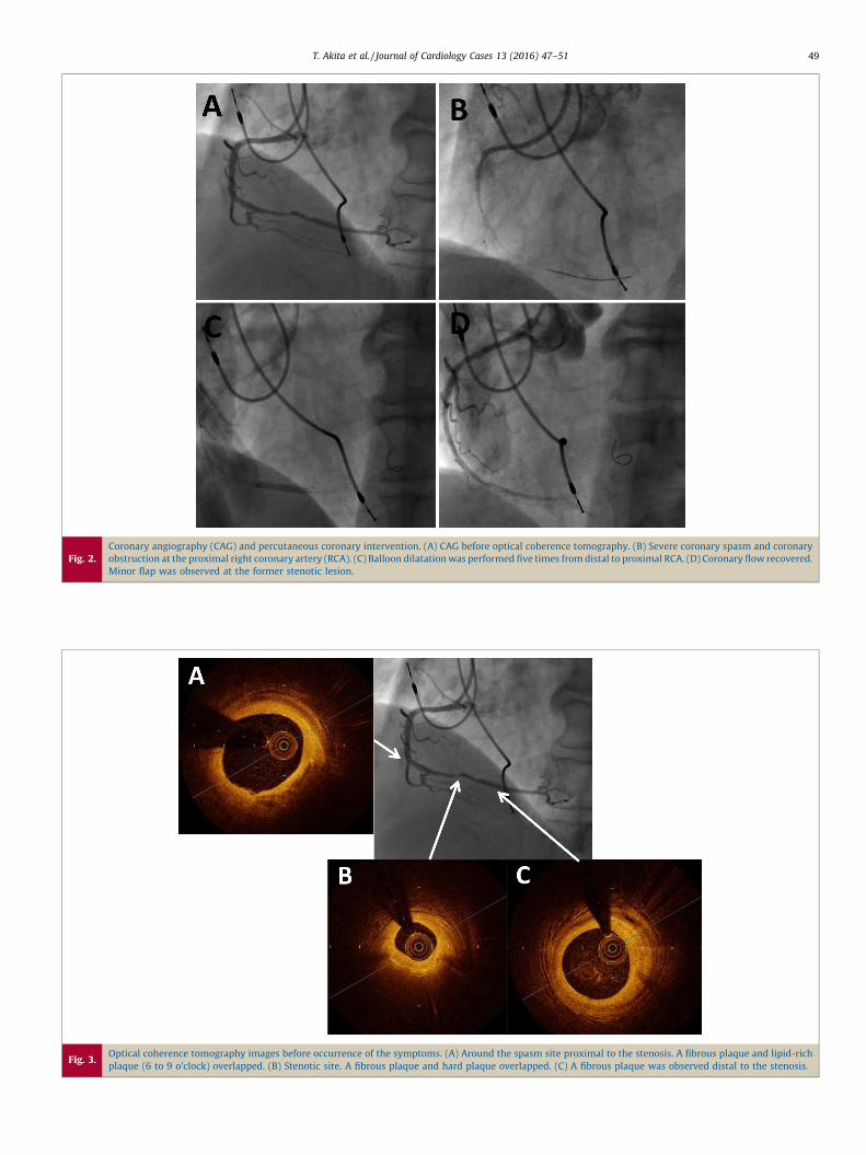
Fig. 2.Coronary angiography (CAG) and percutaneous coronary intervention. (A) CAG before optical coherence tomography. (B) Severe coronary spasm and coronary
obstruction at the proximal right coronary artery (RCA). (C) Balloon dilatation was performed five times from distal to proximal RCA. (D) Coronary flow recovered.
Minor flap was observed at the former stenotic lesion.
Fig. 3.Optical coherence tomography images before occurrence of the symptoms. (A) Around the spasm site proximal to the stenosis. A fibrous plaque and lipid-rich
plaque (6 to 9 o’clock) overlapped. (B) Stenotic site. A fibrous plaque and hard plaque overlapped. (C) A fibrous plaque was observed distal to the stenosis.
T. Akita et al. / Journal of Cardiology Cases 13 (2016) 47–51 49

Fig. 4.Progress of hospitalization. CK is indicated with a solid line. CK-MB is shown with dots. Consciousness of the patient is shown with a dotted line. CK, creatine
kinase; JCS, Japan Coma Scale; PCI, percutaneous coronary intervention; mPSL, methyl prednisolone; DIC, disseminated intravascular coagulation.
T. Akita et al. / Journal of Cardiology Cases 13 (2016) 47–5150
rehabilitation for 2 months, the patient was transferred to anotherhospital to rehabilitate the weakened muscles of the legs. Beforedischarge, ECG showed no abnormal Q and the rhythm was Asensing and V pacing. Echocardiography showed left ventriculardiffuse mild hypokinesis with an ejection fraction of 57.5%. Neitherfinding indicated previous myocardial infarction.
Discussion
The first reports of cardiovascular diseases with allergicreaction and anaphylaxis were published more than 60 years ago[3,4]. Kounis syndrome was first reported by NG Kounis in1991[1]. It consists of three types of responses: type I, coronaryspasm; type II, acute myocardial infarction due to coronaryerosion; and type III, stent thrombosis due to allergic inflamma-tion. All conditions are associated with mast-cell activation in thesetting of hypersensitivity and anaphylactic reaction. Histamineis one of the representative mediators [2]. The increase intryptase level is another marker of on-going allergic reaction[2]. Tryptase level was high in our case on the day of theoccurrence. Different drugs and agents such as latex, food, andcontrast media are reported to be the causes of this reaction[2]. It is reported that tryptase level is high in acute coronarysyndrome (ACS) cases [5] with tryptase levels reaching11.13 � 1.55 mg/L. Tryptase level in our case was 39.1 mg/L andwas much higher than that in ACS cases. Since the cut-off value oftryptase for the diagnosis of Kounis syndrome has not been clearlydetermined, elevation of tryptase levels is supportive for thediagnosis of Kounis syndrome [2]. The accurate incidence of Kounissyndrome is not known, but many clinical case reports with variousallergens have been published [2].
In our case, immediately after performing OCT, bradycardia andshock occurred with precordial erythema. CAG showed severediffuse RCA spasm with total obstruction. Catheter-induced spasmsometimes occurs near the catheter tip. In our case, spasmoccurred at the proximal RCA. Therefore, the possibility ofcatheter-induced spasm was low. OCT wire-induced rare coronaryspasm has also been reported [6]. It recovered by nitrate injection.It is difficult to completely deny mechanically stimulated coronaryspasm. However, it is impossible to explain the severe shock state.Although the OCT showed a lipid-rich plaque at the spastic site, it isdifficult to determine the causal relationship with Kounissyndrome. We could not perform left coronary angiographybecause the patient’s circulatory condition deteriorated and
required treatment. The possibility of left coronary artery spasmwas low given the finding of ST elevation of II, III, and aVF and STdepression of V1-4. However, the possibility of a mild spasm of theleft coronary artery could not be excluded. After 1 hour ofcardiopulmonary resuscitation (CPR), sinus rhythm and bloodpressure finally recovered. We could not introduce PCPS or IABP.This may be one factor for the longer CPR required. However, alarger factor contributing to the delayed recovery may be thatepinephrine and steroid treatment were not immediately active foranaphylactic shock. Repetitive ventricular fibrillation may beanother factor of delayed recovery from the shock.
As for the cause of Kounis syndrome in our case, we speculatethat the low-molecular-weight dextran may have been thecausative agent. Contrast medium and xylocaine are othercandidates, although the previous CAG and the first shot ofcontrast medium on the same day did not cause an anaphylacticreaction. Although DLST tests of these materials were negative,low-molecular-weight dextran was the most likely agent becauseit had been administered to this patient for the first time andKounis syndrome occurred immediately after its administration.There have been several reports describing how low-molecular-weight dextran (dextran 40) caused coronary spasm before Kounissyndrome was advocated [7,8]. In addition, a report has alsosuggested that Kounis syndrome occurred due to dextran 40[9]. Although it is rare that low-molecular-weight dextran couldcause Kounis syndrome, the incidence rate may increase in thefuture because of its frequent usage in OCT for PCI. Thus, we shouldbe attentive to the possible occurrence of Kounis syndrome duringPCI procedure.
Treatment of Kounis syndrome is often difficult because bothcardiac and allergic symptoms are to be treated simultaneouslyand rapidly. In patients with the type I variant, the use ofhydrocortisone 1–2 mg/kg/d intravenously and H2 antihistamines,such as diphenhydramine (1–2 mg/kg) and ranitidine (1 mg/kg),are recommended to reduce allergic symptoms. The use of calciumchannel blockers and nitrates may abolish vasospasm. However,these drugs may cause hypotension and deteriorate the circulatorycondition. Nicorandil may be another good candidate to treatKounis syndrome because it can dilate the coronary artery withoutlowering blood pressure. In our case, given the urgency of thesevere condition, coronary artery dilatation by balloon catheterwas also successful and may represent a treatment option forsevere Kounis syndrome. Epinephrine is the drug of choice foranaphylaxis. It has been described that epinephrine administration

T. Akita et al. / Journal of Cardiology Cases 13 (2016) 47–51 51
aggravates ischemia and vasospasm in Kounis syndrome. It isrecommended to administer epinephrine intramuscularly with ata dose of 0.2–0.5 mg (1:1000). After cardiopulmonary arrest, 1 mgof epinephrine intravenously is required according to the advancedlife support guidelines. It is important to be alert for the occurrenceof Kounis syndrome and to treat both allergy and coronary lesionsimmediately.
We experienced a rare case of Kounis syndrome after CAG inOCT procedures. We must be more attentive to the occurrence ofthe syndrome during OCT with low-molecular-weight dextran.
Conflict of interest
Authors have no conflict of interest.
Acknowledgments
This case was presented at the 206th Kinki Area Meeting of theJapanese Society of Internal Medicine.
References
[1] Kounis N, Zavras G. Histamine-induced coronary artery spasm: the concept ofallergic angina. Br J Clin Pract 1991;45:121–8.
[2] Kounis N. Coronary hypersensitivity disorder: the Kounis syndrome. Clin Ther2013;35:563–71.
[3] Pfister CW, Plice SG. Acute myocardial infarction during a prolonged allergicreaction to penicillin. Am Heart J 1950;40:945–7.
[4] Schultheiss E. Clinical aspects of allergic heart diseases. Dtsch Med J1964;15:15–8.
[5] Xiang M, Sun J, Lin Y, Zhang J, Chen H, Yang D, Wang J, Shi G-P. Usefulness ofserum tryptase level as an independent biomarker for coronary plaque insta-bility in a Chinese population. Atherosclerosis 2012;215:494–9.
[6] Dobarro D, Jimenez-Valero S, Moreno R. Severe coronary spasm induced by OCTwire. There are no innocuous procedures. J Invasive Cardiol 2010;22:385.
[7] Brown RI, Aldridge HE, Schwartz L, Henderson M, Brooks E, Coutanche M. Theuse of dextran-40 during percutaneous transluminal coronary angioplasty: areport of three cases of anaphylactoid reactions – one near fatal. CathetCardiovasc Diagn 1985;11:591–5.
[8] Klugmann S, Salvi A, Valente M, Zanei P, Maiolino P, Camerini F. Coronary arteryspasm after administration of dextran 40: implications concerning percutane-ous transluminal coronary angioplasty. Am Heart J 1986;111:1202–4.
[9] Xu M, Wu XS, Jiang TY, He JQ. Kounis syndrome: allergic acute coronarysyndrome. Chin Med J (Engl) 2013;126:2591–2.