journal of anatomy
TRANSCRIPT
REVIEW
Development and evolution of the vertebrate primarymouthVladimır Soukup, Ivan Horacek and Robert Cerny
Department of Zoology, Charles University in Prague, Czech Republic
Abstract
The vertebrate oral region represents a key interface between outer and inner environments, and its structural
and functional design is among the limiting factors for survival of its owners. Both formation of the respective
oral opening (primary mouth) and establishment of the food-processing apparatus (secondary mouth) require
interplay between several embryonic tissues and complex embryonic rearrangements. Although many aspects
of the secondary mouth formation, including development of the jaws, teeth or taste buds, are known in con-
siderable detail, general knowledge about primary mouth formation is regrettably low. In this paper, primary
mouth formation is reviewed from a comparative point of view in order to reveal its underestimated morpho-
genetic diversity among, and also within, particular vertebrate clades. In general, three main developmental
modes were identified. The most common is characterized by primary mouth formation via a deeply invagi-
nated ectodermal stomodeum and subsequent rupture of the bilaminar oral membrane. However, in salaman-
der, lungfish and also in some frog species, the mouth develops alternatively via stomodeal collar formation
contributed both by the ecto- and endoderm. In ray-finned fishes, on the other hand, the mouth forms via an
ectoderm wedge and later horizontal detachment of the initially compressed oral epithelia with probably a
mixed germ-layer derivation. A very intriguing situation can be seen in agnathan fishes: whereas lampreys
develop their primary mouth in a manner similar to the most common gnathostome pattern, hagfishes seem to
undergo a unique oropharyngeal morphogenesis when compared with other vertebrates. In discussing the early
formative embryonic correlates of primary mouth formation likely to be responsible for evolutionary–develop-
mental modifications of this area, we stress an essential role of four factors: first, positioning and amount of
yolk tissue; closely related to, second, endoderm formation during gastrulation, which initiates the process and
constrains possible evolutionary changes within this area; third, incipient structure of the stomodeal primor-
dium at the anterior neural plate border, where the ectoderm component of the prospective primary mouth is
formed; and fourth, the prime role of Pitx genes for establishment and later morphogenesis of oral region both
in vertebrates and non-vertebrate chordates.
Key words: collar; ectoderm; endoderm; oral membrane; primary mouth; stomodeum; wedge.
Oro-pharyngeal domain and mouth indevelopment and evolution
The design of the mouth opening, its size and functional
capacities are determining factors for survival of every
animal and, correspondingly, often key variables in the evo-
lutionary history of many clades, including vertebrates. In
many cases, minute rearrangements in developmental
mechanisms producing these structures became major
sources of substantial phylogenetic divergence. The two
crown groups of bilateralians, protostomes and deuterosto-
mes, differ exactly in this respect. The production of yolk-
rich eggs in deuterostomes (and namely in vertebrates) has
postponed oral formation into late embryonic stages and,
at the same time, provided a leeway for alternative position-
ing of the mouth and timing of its formation, which could
become a broad field for refinement and structural rear-
rangements of the developmental mechanisms involved.
The development of the oral opening in deuterostomes
proceeds via regulated interactions between cell populations
Correspondence
Dr Robert Cerny, Laboratory for the study of craniofacial evolution
and development, Department of Zoology, Charles University in
Prague, Vinicna 7, 128 44 Prague, Czech Republic.
Accepted for publication 11 June 2012
ªª 2012 The AuthorsJournal of Anatomy ªª 2012 Anatomical Society
J. Anat. (2012) doi: 10.1111/j.1469-7580.2012.01540.x
Journal of Anatomy
of the ectoderm and endoderm lineages, and is imple-
mented into the pathways producing another structure
characterizing the anterior pole of deuterostome embryo,
the pharyngeal slits (Swalla & Smith, 2008). Pharyngo-
tremia, or perforation of the anterior archenteron with
pharyngeal slits, represents, together with deuterostomy,
radial cleavage and regulative eggs, the essential charac-
teristic of deuterostome body organization (e.g. Romer &
Parsons, 1986). In terms of developmental dynamics, pha-
ryngeal slits are produced by an autonomous endoderm
regulation (Veitch et al. 1999; Piotrowski & Nusslein-
Volhard, 2000; Graham & Smith, 2001; Graham et al.
2005), which, especially in vertebrates, is supplemented by
an independent placodal patterning in the anterior ecto-
derm (Schlosser, 2005). These endoderm and ectoderm
interactions are further modified by intervention of
migrating neural crest cell population into interpouch
vacuities to produce novel skeletal designing mechanisms,
which, specifically in the preotic region, completely over-
power the initial endoderm patterning. Correspondingly,
the invasion of neural crest mesenchyme into the region
of presumptive mouth, another key innovation of verte-
brates, overwrites the primary mechanisms of the oral
opening with dozens of novel morphogenetic modules,
mostly produced by regulated interactions along the epi-
thelial–mesenchymal boundary (e.g. Stock, 2001; Fraser
et al. 2010). As a result, the mouth of vertebrates repre-
sents a very complex structure equipped with specialized
products of epithelial–mesenchymal interactions – teeth,
jaws, glands and sensory cells specifically designed in
each particular clade. In accord with previous proposals
(Dickinson & Sive, 2006), we term this set of mostly neural
crest-derived food-processing adaptations characterizing
the adult mouth of particular vertebrate organisms, the
‘secondary mouth’.
The secondary mouth can be exemplified by an oral
siphon with velar tentacles covered by tunica as in ascidians;
velum with tentacles, oral cirri and pre-velar region includ-
ing derivatives of the preoral pit as in amphioxus; upper and
lower lips with collar of tentacles and laterally placed velum
as in lampreys; or jaw-joint including jaws with lips and
teeth as in gnathostomes. Yet, the focus of this article is
none of the above-mentioned structures, but rather the
structure that precedes them both in ontogenetic and phylo-
genetic respects – the ‘primary mouth’ (in the sense of
Dickinson & Sive, 2006). The primary mouth preforms the
oral opening by the interplay of the primary embryonic
tissues (ectoderm and endoderm), and establishes the topo-
graphic setting and organizational platform for the inter-
vention of novel mechanisms producing the secondary
mouth.
During chordate embryogenesis, the prospective mouth
region is established at the discrete anterior domain at the
border zone of neural ectoderm, non-neural ectoderm and
anterior endoderm (Fig. 1, left), and its early specification
seems to include the same molecular pathways in all
groups. Aside from this, the major clades of chordates differ
significantly with respect to the final position of mouth
opening (Fig. 1, right). In urochordates, mouth develop-
ment is associated with the neuropore (Manni et al. 2005;
Veeman et al. 2010), in cephalochordates it appears on the
left side of the pharynx (e.g. Lankester & Willey, 1890;
Willey, 1891; Hatschek, 1893; Urata et al. 2007; Yasui & Kaji,
2008), while in vertebrates the prospective mouth is posi-
tioned medially and ventrally to the developing brain at
the anterior end of the pharynx.
The primary mouth is generally thought to be formed
both by an invagination of ectoderm that forms the stomo-
deum and by an anterior expansion of the foregut endo-
derm, i.e. comparable morphogenesis to that producing the
pharyngeal slits. The process often terminates with a bilami-
nar membrane separating the ectoderm stomodeum and
endoderm pharynx, the so-called stomo-pharyngeal, oro-
pharyngeal or oral membrane, whose disappearance initi-
ates the development of the secondary mouth. However,
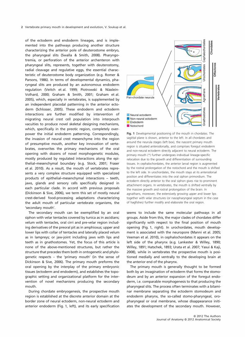
Fig. 1 Developmental positioning of the mouth in chordates. The
sagittal plane is shown, anterior to the left. In all chordates and
around the neurula stages (left box), the nascent primary mouth
region is situated anterodorsally, and comprises foregut endoderm
and non-neural ectoderm directly adjacent to neural ectoderm. The
primary mouth (*) further undergoes individual lineage-specific
relocation due to the growth and differentiation of surrounding
tissues. In cephalochordates, the anterior larval region is augmented
by the rostral prolongation of the notochord and the mouth is shifted
to the left side. In urochordates, the mouth stays at its anterodorsal
position and differentiates into the oral siphon primordium. The
ectoderm directly anterior to the oral siphon gives rise to prominent
attachment organs. In vertebrates, the mouth is shifted ventrally by
the massive growth and rostral prolongation of the brain. In
agnathans, moreover, the extensively growing upper and lower lips
together with velar structures (or nasopharyngeal septum in the case
of hagfishes) further modify and elaborate the oral region.
ªª 2012 The AuthorsJournal of Anatomy ªª 2012 Anatomical Society
Vertebrate primary mouth in development and evolution, V. Soukup et al.2
formation of the primary mouth does not always proceed
in this straightforward way, and alternative developmental
scenarios may take place. Despite the potential significance
of such information for our comprehension of the chordate
evolution, the actual forms of interplay between ectoderm
patterning the oral cavity and endoderm developmental
dynamics, their heterochronies and heterotopies at particu-
lar stages of early oral development, and ⁄or rearrange-
ments of the signalling cascades responsible for the
plethora of states characterizing particular vertebrate clades
are still largely unknown. This review is intended to uncover
the underestimated diversity in primary mouth morphogen-
esis, and to address potential formative correlates that may
take part in the evolution and development of this
neglected, though important, embryonic structure.
Modes of primary mouth formation invertebrates
In vertebrates, the morphogenesis of the primary mouth is
initiated during the course of early embryogenesis at the
time of late neurulation. It takes place at the anterior-most
cranial domain where the ectoderm and endoderm meet
directly without intervening mesoderm or mesenchyme
(Fig. 2, left panel). This domain, well-defined also by the
early expression of Pitx genes, is subsequently shifted ven-
trally by the expansion of the growing neural tube and
forms a distinct invagination. Mouth development in verte-
brates is generally understood to progress through: first, a
stage of deep stomodeal invagination abutting the under-
lying endoderm foregut lining; followed by, second, the
reduction of this epithelial contact zone to a thin, one–two-
cell-thick oral membrane; and third, perforation and rup-
ture of this membrane, which opens the primary mouth.
The pattern of primary mouth formation via stomodeum
and oral membrane is indeed widely distributed over many
groups of vertebrates, and it is best exemplified inXenopus,
which has recently become arguably the most prominent
model vertebrate for early oral organogenesis (Dickinson &
Sive, 2006, 2009). However, a significantly different pattern
of mouth formation can, for example, be observed among
urodele amphibians (salamanders), which represent the sis-
ter clade of frogs. The mouth also forms in a dissimilar man-
ner in ray-finned fishes, a clade including the majority of
vertebrate species (Fig. 2). We will review developing oral
regions of vertebrates from a comparative point of view in
order to identify shared and derived developmental
processes, and also to reveal the remarkable diversity of
vertebrate primary mouth formation.
Mouth development via definitive stomodeum andrupture of oral membrane
Xenopus as the model organism
In Xenopus, an anuran, which currently represents a model
system for studying the vertebrate primary mouth
(Dickinson & Sive, 2006, 2009), the prospective oral ecto-
derm is found in the anterior-most part of the neurula at
the border between the transverse neural fold and adjacent
epidermis (Figs 1 and 8). Individual cells of this ectoderm
region are fated to give rise not only to the stomodeum,
but also to the head epidermis, cement and hatching
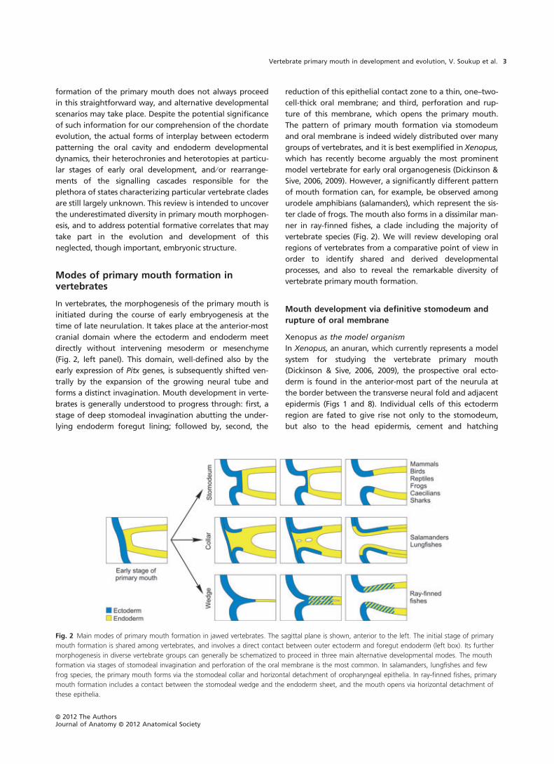
Fig. 2 Main modes of primary mouth formation in jawed vertebrates. The sagittal plane is shown, anterior to the left. The initial stage of primary
mouth formation is shared among vertebrates, and involves a direct contact between outer ectoderm and foregut endoderm (left box). Its further
morphogenesis in diverse vertebrate groups can generally be schematized to proceed in three main alternative developmental modes. The mouth
formation via stages of stomodeal invagination and perforation of the oral membrane is the most common. In salamanders, lungfishes and few
frog species, the primary mouth forms via the stomodeal collar and horizontal detachment of oropharyngeal epithelia. In ray-finned fishes, primary
mouth formation includes a contact between the stomodeal wedge and the endoderm sheet, and the mouth opens via horizontal detachment of
these epithelia.
ªª 2012 The AuthorsJournal of Anatomy ªª 2012 Anatomical Society
Vertebrate primary mouth in development and evolution, V. Soukup et al. 3

glands, and adenohypophyseal and olfactory placodes
(Eagleson et al. 1986; Drysdale & Elinson, 1991; Schlosser &
Ahrens, 2004; Dickinson & Sive, 2007). During the course of
further development, this region subdivides and the cells
become more and more restricted according to their pro-
spective fates (Pieper et al. 2011). The future stomodeal
subregion then consists of a relatively thick ectoderm
directly juxtaposed to a multi-layered endoderm foregut
with a distinct basal lamina in between (Dickinson & Sive,
2006). Dissolution of this basal lamina, which provides an
opportunity for subsequent extensive cellular rearrange-
ments, starts relatively early in this species (Dickinson & Sive,
2009). Only then do the ectoderm cells invaginate in the
form of a relatively shallow stomodeum, while the number
of both the ectoderm and endoderm cells is gradually
reduced. The reduction of the number of cells is caused by
increased programmed cell death in the inner and outer
ectoderm layers, while no apoptosis has been found in the
endoderm (Dickinson & Sive, 2006). The resulting oral mem-
brane consists of one stomodeal ectoderm layer anteriorly
and one foregut endoderm layer posteriorly. Themembrane
thins to a single-layered unit at some points by means of
intercalation of cells of both germ-layers. The eventual per-
foration of the oral membrane is initiated by a single open-
ing, which is gradually enlarged in Xenopus (Dickinson &
Sive, 2006). However, the oral membrane of another frog
species, Rana japonica, is perforated by several small open-
ings temporarily giving the membrane a net-like appear-
ance (Watanabe et al. 1984). The remnants of the former
membrane can be recognized only for a short period after
perforation, and the limits of the ectoderm and endoderm
epithelial linings are no longer discernible.
Mouth development via definitive stomodeum and
rupture of oral membrane in other jawed vertebrates
In many other vertebrate groups, such as mammals, birds,
reptiles, caecilians or chondrichthyans (sharks), the mouth
develops classically via formation of the stomodeum and
oral membrane similarly to Xenopus, with only minor dif-
ferences that apparently reflect some lineage-specific
embryonic settings (Cook & Neal, 1921; Teipel, 1932;
Waterman, 1977; Waterman & Balian, 1980; Waterman &
Schoenwolf, 1980). In these groups the initial contact of sto-
modeal ectoderm and foregut endoderm is formed below
the closing neural folds, but is subsequently transferred by
the increasingly growing head-fold so that the prospective
oral membrane is finally established between the foregut
endoderm and the ventral head ectoderm. The stomodeum
develops in this ventral ectoderm as a shallow invagination,
which becomes deeper and wider due to further growth of
the brain and also due to the increasing volume of immi-
grating mesenchyme forming the surrounding jaw struc-
tures (e.g. Waterman & Schoenwolf, 1980; Ballard et al.
1993). The stomodeum and closely abutting foregut endo-
derm collectively form the oral membrane, which finally
ruptures, but, interestingly, variable mechanisms seem to
be responsible for its rupture in different vertebrate species.
A number of apoptotic cells can be found within the epi-
thelia of the oral membrane in Xenopus, Rana and mouse
(Watanabe et al. 1984; Poelmann et al. 1985; Dickinson &
Sive, 2006). The apoptosis may cause its thinning, generat-
ing weak spots in the membrane and its subsequent perfo-
ration. While some cells of the membrane undergo
apoptosis, the remaining non-apoptotic cells intercalate
among each other, and incorporate into the epithelia of
the upper and lower jaws. On the other hand, no apoptosis
has been found during regression of the oral membrane in
chick and hamster (Waterman, 1977; Waterman &
Schoenwolf, 1980) and, moreover, the chick oral membrane
even contains some proliferating cells (Miller & Olcott,
1989). In general, the rate of proliferation in the oral mem-
brane seems to be much lower than that in the surrounding
epithelia, suggesting that the heavily proliferating ecto-
derm and endoderm epithelial linings of the upper and
lower jaws are pulling the less proliferating oral membrane
apart, finally causing its rupture. This might further indicate
that processes of cell intercalation within the membrane
and its fusion with the surrounding epithelia are the result
and not the cause of its rupture (Waterman, 1985; Miller &
Olcott, 1989).
Interestingly, similar differential proliferation rates were
also identified in the case of chick branchial membranes
(closing plates, Miller et al. 1993), i.e. derivatives of pharyn-
geal groove ectoderm and pouch endoderm, which are situ-
ated between adjacent pharyngeal arches. Correspondingly
to oral membrane, branchial membranes also represent
transient structures, and their rupture creates gill slits in the
primarily aquatic vertebrates possessing functional gills. In
chick, it was shown that branchial membranes also undergo
cell interdigitations of the ectoderm and endoderm linings
and progress to a single cellular layer that eventually rup-
tures. However, it was concluded that cellular reorganiza-
tion rather than massive degradation is the main
mechanism responsible for their rupture (Waterman, 1985).
Branchial membranes in birds and mammals, nevertheless,
perforate only temporarily and are subsequently closed
when neural crest mesenchyme cells invade the pharyngeal
arches.
Mouth development via stomodeal collar formation
Salamanders
The general pattern of mouth formation in salamanders
(urodele amphibians) differs significantly from those of the
above-mentioned vertebrates (Fig. 2). The oral area initially
consists of a double-layered ectoderm, while the inner
region of the prospective mouth is filled with a compact
mass of ‘oral endoderm’ (Fig. 3A). The stomodeum with a
well-defined lumen does not develop, and only a shallow
groove is visible externally (Takahama et al. 1988). Mouth
ªª 2012 The AuthorsJournal of Anatomy ªª 2012 Anatomical Society
Vertebrate primary mouth in development and evolution, V. Soukup et al.4
development starts when the inner (basal) layer of the for-
merly double-layered oral ectoderm undergoes involution
and migrating ectodermal cells gradually cover the oral
endoderm mass as a ‘sleeve’ forming the so-called stomo-
deal collar (Figs 2 and 3A,B; Adams, 1924; Reisinger, 1933;
Soukup et al. 2008). No basal lamina is found between the
outer ectoderm layer and the oral endoderm in the oral
area at this time, but distinct basal laminae are shared by
the inner ectoderm layer and the oral endoderm cells sepa-
rating the oral epithelia from the surrounding head mesen-
chyme (Fig. 3A,B).
Opening of the mouth occurs as a result of the formation
of small cavities that arise among the initially compact oral
endoderm mass and that fuse to finally form walls of the
future mouth. Degenerating cells were observed only infre-
quently and only in the oral ectoderm, suggesting that a
process of active remodelling rather than cell death is
responsible for opening of the mouth (Takahama et al.
1988). None of the previous researchers observed a struc-
ture reminiscent of a double-layered oral membrane
(Kingsley & Thyng, 1904; Greil, 1905; Johnston, 1910; Land-
acre, 1921; Adams, 1924, 1931; Marcus, 1930; Reisinger,
1933; Stroer, 1933; Balinsky, 1947; de Beer, 1947; Chibon,
1970), but Takahama et al. (1988) proposed that the oral
membrane might be represented by the outer layer of the
oral ectoderm together with the entire solid oral endoderm
mass. The process of horizontal cleft formation was then
compared with the rupture of a typical oral membrane. In
the Mexican axolotl, it seems that the oral membrane does
exist as a double-layered anterior structure composed of
the outer ectoderm and the oral endoderm linings (Fig. 4);
it represents only a transient structure and it is unique on
account of its very superficial position: it connects the upper
lip epithelium to the lower jaw even anteriorly to the lower
lip (Fig. 4D). Such an external position of the oral mem-
brane in salamanders may on the other hand explain the
presence of the endoderm cells within the lip epithelia and
its extent up to the outer head surface (Figs 3 and 4).
Another noteworthy feature of salamander development
is that the oral membrane does not always represent the last
connection between the upper and lower mouthparts. In
some axolotl specimens it was observed that the roof and
floor of the mouth cavity are still connected by incompletely
detached oral endoderm cells even after perforation of the
external oral membrane (Fig. 4E). These cells can eventually
take a form of epithelial bridges that, interestingly, were
also observed during mouth opening in a basal actinoptery-
gian fish (Kralovic et al. 2010), suggesting that such epithe-
lial connections possibly represent incidental structures that
form during the general process of epithelial splitting.
A B
C D
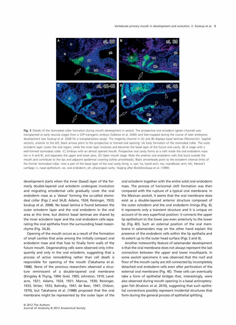
Fig. 3 Details of the stomodeal collar formation during mouth development in axolotl. The prospective oral ectoderm (green channel) was
transplanted at early neurula stages from a GFP-transgenic embryo (Sobkow et al. 2006) and fate-mapped during the course of later embryonic
development (see Soukup et al. 2008 for a transplantation assay). The magenta channel in (A) and (B) displays basal laminae (fibronectin). Sagittal
sections, anterior to the left, black arrows point to the prospective or formed oral opening. (A) Early formation of the stomodeal collar. The outer
ectoderm layer covers the oral region, while the inner layer involutes and becomes the basal layer of the future oral cavity. (B) A stage with a
well-formed stomodeal collar. (C) Embryo with an almost opened mouth. Prospective oral cavity forms as a cleft inside the oral endoderm mass
(oe in A and B), and separates the upper and lower jaws. (D) Open mouth stage. Note the anterior oral endoderm cells that burst outside the
mouth and contribute to the lips and adjacent epidermal covering (white arrowheads). Black arrowheads point to the ectoderm internal limits of
the former stomodeal collar, now a part of the basal layer of the oral cavity lining. e, eye; ha, hyoid arch; ma, mandibular arch; Mc, Meckel’s
cartilage; n, nasal epithelium; oe, oral endoderm; ph, pharyngeal cavity. Staging after Bordzilovskaya et al. (1989).
ªª 2012 The AuthorsJournal of Anatomy ªª 2012 Anatomical Society
Vertebrate primary mouth in development and evolution, V. Soukup et al. 5
The distinctive way by which mouth formation is realized
in salamanders, consequently also causes an alternative
distribution of respective epithelial linings. Generally it is
assumed that the oral cavity is lined by the ectoderm epithe-
lium anteriorly and endoderm epithelium posteriorly with a
sharp border represented by the oral membrane (e.g. Romer
& Parsons, 1986; Kardong, 1995). In salamanders, the poster-
ior part of the oral cavity is indeed lined by endoderm,
whereas the anterior mouth lining is composed of cells of
dual germ-layer origin: the ectoderm basal layer (former sto-
modeal collar) and the endoderm apical layer (former solid
oral endodermmass; Figs 3 and 4). The ectoderm–endoderm
border zone in salamanders is consequently rather complex,
comprising the previous extent of the stomodeal collar
together with the above-described oral membrane.
Mouth development via stomodeal collar in other
vertebrates
Mouth formation via developmental stages analogous to
the stomodeal collar has also been reported for lungfish
embryos, which, as in salamanders, have their oral region
plugged by a mass of yolk-laden oral endoderm cells. In
lungfishes, this foregut endoderm mass contacts the
double-layered ectoderm, which successively takes a form
of a shallow stomodeal plate (Kerr, 1902, 1910; Greil, 1913).
Based on histological evidence, it was reported that the
inner ectoderm layer disappears from the contact zone with
the oral endoderm and, by the tailbud stage, it forms a con-
tinuous sheet with cells of the basal oral endoderm (Kerr,
1902, 1910; Greil, 1913). The remaining apical ectoderm
layer covering the oral endoderm was described as dimin-
ishing shortly before the opening of the mouth, leaving the
underlying endoderm cells exposed to the external surface
(Kemp, 2002). The oropharyngeal cavity is then opened by
a horizontal cleft that forms inside the oral endoderm mass
and spreads from behind. Interestingly, the yolk-laden
endoderm cells can be found at the very tips of the mouth,
suggesting a substantial endoderm contribution to the oro-
pharyngeal cavity in lungfishes (Kemp, 2002), a situation
equivalent to that in salamanders (Soukup et al. 2008).
Comparable developmental morphogenesis of the oral
epithelia forming a stomodeal collar-like structure instead
of a clear stomodeum was also reported for the Tailed Frog
Ascaphus truei (Reiss, 1997), which belongs to the basal-
most anuran lineage. In this species with an unusual ven-
trally placed sucker mouth, the stomodeum is shallow and
ventrally placed. The outer layer of the oral ectoderm con-
tacts a ventral part of the flattened anterior mouth endo-
derm mass, while the inner ectoderm layer expands
posteriorly on the endoderm dorsal surface. Moreover,
A B
C D
E
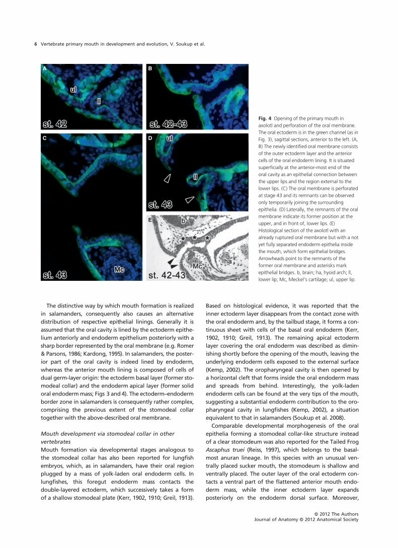
Fig. 4 Opening of the primary mouth in
axolotl and perforation of the oral membrane.
The oral ectoderm is in the green channel (as in
Fig. 3), sagittal sections, anterior to the left. (A,
B) The newly identified oral membrane consists
of the outer ectoderm layer and the anterior
cells of the oral endoderm lining. It is situated
superficially at the anterior-most end of the
oral cavity as an epithelial connection between
the upper lips and the region external to the
lower lips. (C) The oral membrane is perforated
at stage 43 and its remnants can be observed
only temporarily joining the surrounding
epithelia. (D) Laterally, the remnants of the oral
membrane indicate its former position at the
upper, and in front of, lower lips. (E)
Histological section of the axolotl with an
already ruptured oral membrane but with a not
yet fully separated endoderm epithelia inside
the mouth, which form epithelial bridges.
Arrowheads point to the remnants of the
former oral membrane and asterisks mark
epithelial bridges. b, brain; ha, hyoid arch; ll,
lower lip; Mc, Meckel’s cartilage; ul, upper lip.
ªª 2012 The AuthorsJournal of Anatomy ªª 2012 Anatomical Society
Vertebrate primary mouth in development and evolution, V. Soukup et al.6
dense ectoderm bands were also found running along the
endoderm wall from the corners of the mouth (Reiss, 1997).
Interestingly, for the Agile Frog (Rana dalmatina), oral
formation was described as developing via the stomodeal
collar-like structure with some ectoderm bands running
inside the mouth over the foregut endoderm (Reisinger,
1933). Moreover, the same author observed that at least
some ectoderm cells invade the mouth in the case of the
Midwife Toad (Alytes obstetricans) in a salamander-like
manner, aside from the fact that its oral formation other-
wise develops via a definitive stomodeum and rupture of
classic oral membrane. Similarly to the situation in lungfish-
es, all these intriguing pieces of data are derived from histo-
logical descriptions and, thus, caution should be taken
when drawing decisive conclusions.
Mouth development via ectodem wedge anddetachment of initially compressed oropharyngealepithelia
In the ray-finned fishes (Actinopterygii), mouth formation is
strongly influenced by the fact that the whole oropharyn-
geal region is mechanically constrained by the developing
brain dorsally and by the yolk sac and pericardium ventrally,
leaving rather limited space for the oropharyngeal struc-
tures. In zebrafish, a recent paradigmatic fish model system,
the earliest described stage of mouth development involves
a wedge formed by several ectoderm cells beneath the cra-
nial end of the head merged with the anterior part of uni-
cellular archenteric endoderm layer (Fig. 2; Waterman &
Kao, 1982; Warga & Nusslein-Volhard, 1999; Wallace & Pack,
2003). From this initial contact the oral ectoderm begins to
form a small stomodeum, which enlarges and deepens by
separation of the ectoderm wedge cells that, however, are
not clearly separated from the endoderm lining. The border
between alternate oral epithelial linings is hindered by the
absence of a distinct basal lamina, which probably dissolves
already at earlier stages (Senior, 1909; Waterman & Kao,
1982). The single-layered oropharyngeal epithelium further
undergoes tubulation by ventromedial folding and fusion
of its lateral edges, finally forming a squeezed tube with no
lumen (Senior, 1909; Edwards, 1929; Sucre et al. 2009). The
lumen of the oropharynx appears when roof and floor oral
epithelia start detaching from each other leaving tenuous
epithelial connections between them. In other words, small
cavities arise among oropharyngeal epithelia, merge and
enlarge to finally develop into lumen of the oropharynx.
Opening to the outer environment is therefore achieved by
the breaking down of various epithelial bridges instead of
perforation of a single and definitive oral membrane. In
zebrafish, however, some of the last epithelial connections
can be found at the posterior-most stomodeum (Waterman
& Kao, 1982), but whether these represent the ectoderm–
endoderm bordering zone and thus are comparable to an
oral membrane remains to be determined.
Importantly, early development of endoderm foregut lin-
ing is radically different in teleosts on one hand, and the
non-teleost actinopterygians on the other (withAmia repre-
senting an intermediate state; see Nelsen, 1953; Cooper &
Virta, 2007). In the first case, a single-cell-thick endoderm
lining arises during gastrulation, forming a squeezed tube
with no lumen, as described above for zebrafish. The fore-
gut lining of bichirs, sturgeons, paddlefish and gars arises,
on the other hand, from a widely hollowed archenteric
endoderm, similar to the foregut formation in amphibians
(Kerr, 1907; Detlaff et al. 1993). This feature undoubtedly
represents an ancestral condition for the ray-finned fishes,
and the accelerated development of brain together with a
massive yolk-ball is one of the characteristics of teleost line-
age. Aside from this, a general mode of the oropharyngeal
cavity formation comprising detachment and separation of
the oropharyngeal epithelia and formation and subsequent
rupture of the epithelial bridges between them was also
observed in the Senegal bichir (Polypterus senegalus;
Kralovic et al. 2010), which appears among the basal-most
actinopterygian clades. This mode of mouth development
might therefore be regarded as a blueprint for all ray-
finned fishes.
Noteworthy epithelial rearrangements have been
reported to occur during and after perforation of the
mouth and also gill slits in carp (Cyprinus carpio; Edwards,
1929), a close relative to zebrafish. In the region between
the hyoid and first branchial arch, the lateral head ecto-
derm was observed to contact the pharyngeal endoderm by
a wedge of cells in a similar way to that described for the
zebrafish oral region. However, before opening of the first
gill slit, the ectoderm wedge cells push themselves between
the apical sides of the compressed endoderm epithelial lin-
ings. The ectoderm cells were seen to populate the whole
pharyngeal cavity when the hyobranchial gill slit became
open. According to Edwards (1929), this cell behaviour
should take place in the oral region as well. The resulting
oropharyngeal epithelium should, therefore, be of double-
germ-layer origin with ectoderm squamous cells apically
and endoderm columnar cells basally.
Interestingly, a completely different situation regarding
oropharyngeal epithelial morphodynamics was reported for
another teleost fish, Pterophyllum scalare, a derived teleost
and distant relative to the carp. According to Colle-
Vandevelde (1966), the apical layer of the double-layered
ectoderm oral epithelium stays in contact with the under-
lying pharyngeal endoderm lining, together forming the
oral as well as branchial membranes. The basal ectoderm
was depicted as passing over the membranes and becoming
fused with the basal layer of pharyngeal endoderm. These
morphodynamics should consequently lead to oral linings
of double-germ-layer origin but with the apical layer
formed by endoderm and the basal layer by ectoderm, i.e.
differing from the carp (Edwards, 1929), but comparable to
salamanders (see above).
ªª 2012 The AuthorsJournal of Anatomy ªª 2012 Anatomical Society
Vertebrate primary mouth in development and evolution, V. Soukup et al. 7

Mouth development in agnathans
Agnathans (jawless fishes) have been, and still are, recog-
nized as among the most important animals for under-
standing vertebrate evolutionary history (e.g. Janvier, 1996;
Mallatt, 1996; McCauley & Kuratani, 2008). Absence of the
jaw is regarded as a primitive trait for vertebrates, and thus
these animals are expected to exhibit other primitive char-
acter states as well. Lampreys and hagfishes, modern agna-
than representatives, were a key subject of the earliest
embryology research (reviewed by Gorbman, 1997; Kuratani
et al. 2001; Ota & Kuratani, 2006; Richardson et al. 2010),
but whilst lampreys recently reached the level of model
animals in evolutionary and developmental biology (e.g.
Nikitina et al. 2009), hagfish embryonic material has not
been accessible until only very recently and still is very defi-
cient (Ota & Kuratani, 2006; Ota et al. 2007). Consequently,
hagfish embryology remains very incompletely known.
Moreover, the phylogenetic relationship between lampreys
and hagfishes is still not completely resolved. They either
form a monophyletic clade Cyclostomata that represents a
sister group to Gnathostomata (the ‘cyclostome hypothesis’
currently supported mostly by molecular data: Mallatt &
Sullivan, 1998; Kuraku et al. 1999; Delarbre et al. 2002), or,
according to the ‘craniate hypothesis’ (supported mostly by
morphological arguments: e.g. Løvtrup, 1977; Janvier, 1981,
1996; Donoghue & Sansom, 2002; Gess et al. 2006; Near,
2009), hagfishes represent a separate clade, a sister group
to vertebrates (i.e. gnathostomes plus lampreys). The lam-
prey primary mouth clearly forms via a deep stomodeum
and rupture of the oropharyngeal membrane, and bears a
resemblance to the main mode of the gnathostome devel-
opment, whereas hagfish embryos probably develop their
primary mouth in a strikingly different manner. Because of
both taxonomic and developmental uncertainties, we deal
with lampreys and hagfishes separately.
Lampreys
The development of the oral region in the ammocoete lar-
vae differs from that in gnathostomes (Fig. 1). At first, a
thickened ectoderm anterior to the forming stomodeum
develops into a nasohypophyseal plate, which gives rise to
the prospective adenohypophyseal and olfactory placodes
(Honma et al. 1990; Kuratani et al. 2001). The nasohypo-
physeal plate stays tightly connected to the forebrain and is
passively brought to the top of the head by an extensively
growing upper lip, which separates the nasohypophyseal
plate from the stomodeum and forms a prominent part of
the lamprey head (for a review, see Kuratani et al. 2001).
The lamprey nasohypophyseal plate thus develops outside
the prospective mouth whereas, in gnathostomes, the nas-
ohypophyseal complex is separated into the nasal and ade-
nohypophyseal placodes, where the latter is incorporated
into the stomodeum. The expansion of the oral ectoderm
forming the stomodeum and its independence from the
adenohypophyseal anlage probably represents the most
profound difference between the lamprey and gnathos-
tome early oral development (e.g. Romer & Parsons, 1986;
Kuratani et al. 2001).
The stomodeum in the lamprey is formed by the relatively
deeply invaginated single-layered oral epithelium, which at
the posterior blind end abuts against the single-layered
pharyngeal endoderm together forming an oral membrane
(Fig. 5). Because the stomodeum primarily forms on the
ventral side of the head, is rather deep, and the foregut
lining reaches the anterior notochord dorsally, the oral
membrane has a large extent (Fig. 1; compare agnathans
and gnathostomes). The oral membrane is finally broken
through and the resulting vertical slit is flanked by
extended velar outgrowths. The velum, which develops in a
position of former oral membrane, finally forms a special-
ized structure consisting of paired muscular flaps, and inter-
nal and external velar bars in each flap (Mallatt, 1996;
Kuratani et al. 2001).
Hagfishes
Our understanding of the development of mouth and oral
cavity in hagfishes is very fragmentary and controversial.
The controversy stems from the fact that, until recently, all
information on hagfish development arose from just a few
reports more than 100 years old (Dean, 1899; von Kupffer,
1899, 1900, 1906; Stockard, 1906). Moreover, the original
histological sections were redrawn schematically (von
Kupffer, 1899), re-examination of the same embryos lead
to different interpretations (Gorbman, 1983; Gorbman &
Tamarin, 1985, 1986), and confusion was extended by fur-
ther schematization in the secondary literature (see reviews
by Gorbman, 1997 and Ota & Kuratani, 2006, 2008, the only
recent authors who succeeded in re-examination of hagfish
development with a new embryonic material). Regardless
of uncertainty in developmental characteristics of the hag-
fish oropharyngeal region, the results can be summarized
as follows.
The early hagfish embryo is represented by flat layers of
epidermal, neural and endodermal tissues lying on a large
yolk-ball. At the developmental stage when the head pro-
cess starts to arise, the foregut endoderm forms a flattened
tube with a lumen arising at its anterior-most portion. This
archenteric space further progresses posteriorly and, thus,
the anterior mouth–nasopharyngeal area comes into
continuation with the pharyngeal cavity. Then, however,
the nasopharyngeal canal is separated from the oropharyn-
geal cavity when the proliferating neural crest mesenchyme
forms the nasopharyngeal septum as a part of the second-
ary mouth, a process comparable to the lamprey situation
(Fig. 5). In the hagfish, however, the now separated spaces
do not yet open to the exterior, and thus the prospective
oral cavity, olfactory epithelium and adenohypophysis argu-
ably arise from endoderm (Gorbman, 1983). It is only at a
rather late stage of hagfish mouth formation that the
ªª 2012 The AuthorsJournal of Anatomy ªª 2012 Anatomical Society
Vertebrate primary mouth in development and evolution, V. Soukup et al.8
forward growth of the head process brings the subcephalic
ectoderm into contact with the anterior endoderm, forming
an oral membrane, which later perforates to open the
oropharyngeal and nasopharyngeal cavities to the outer
environment. Whether anything like stomodeal invagina-
tion known from gnathostomes and lampreys also exists in
hagfishes and what would its relationship be to the subce-
phalic cleft ectoderm remains to be elucidated.
von Kupffer (1899, 1900, 1906), however, hypothesized
that, in hagfish, the oral membrane, comprising the subce-
phalic ectoderm and the foregut endoderm linings, disap-
pears before the formation of the nasopharyngeal septum
and new secondary membranes develop during septal mor-
phogenesis. Yet, regardless of improbable appearance of
the secondary ecto-endodermal membranes, not known in
any chordates, no further studies in hagfish development
provided any empirical support for the above hypothesis
(Dean, 1899; Stockard, 1906). Gorbman (1983) argues that
von Kupffer (1899), who recognized the endoderm deriva-
tion of this area, was apparently troubled by it and tried to
explain it by formulating a hypothesis involving an early
ectoderm invagination followed by secondary closures of
both nasopharyngeal and oropharyngeal tubes. Re-
examining the then available embryonic material, Gorbman
(1983, 1997), however, concluded that ‘there is no evidence
that the stomodeal and nasopharyngeal spaces were ever
open prior to the stage shown in von Kupffer’s figure.’
Moreover, Gorbman (1983) also provides convincing histo-
logical evidence for the immediate topographic context of
the infundibular evagination of the brain and a thickened
cellular layer of the endoderm. The sole up-to-date reports
on hagfish development (Ota et al. 2007, 2011; Ota & Kura-
tani, 2008) do not provide any details on the matter. Hence,
until new data appear, we tentatively propose to follow
the above-mentioned conclusions by Gorbman (1983, 1997)
as a default view of the primary mouth formation in hag-
fish.
This ‘default’ view suggests considerable differences in
developmental dynamics between the two agnathan
groups and, at the same time, distinct differences between
hagfish and other vertebrates, lampreys included (Fig. 5); it
predicts that hagfishes undergo disparate and quite unique
oropharyngeal morphogenesis when compared with other
vertebrates. The primary mouth formation appearing
entirely in the domain of endoderm developmental dynam-
ics without formative intervention of the ectoderm placodal
patterning would provide one of the strongest arguments
for a separate position of hagfishes and for the ‘craniate
hypothesis’ of vertebrate phylogeny.
It is worth mentioning that both groups exhibit certain
similarities in development of the secondary mouth struc-
tures that apparently arise before the formation of the oral
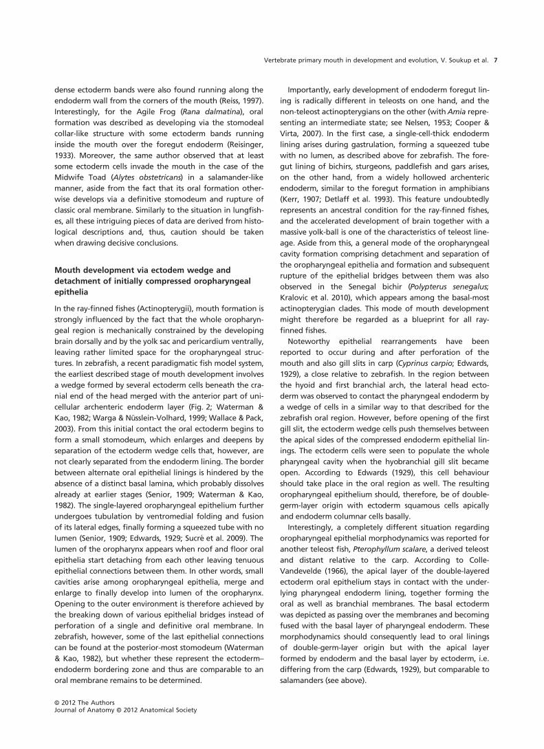
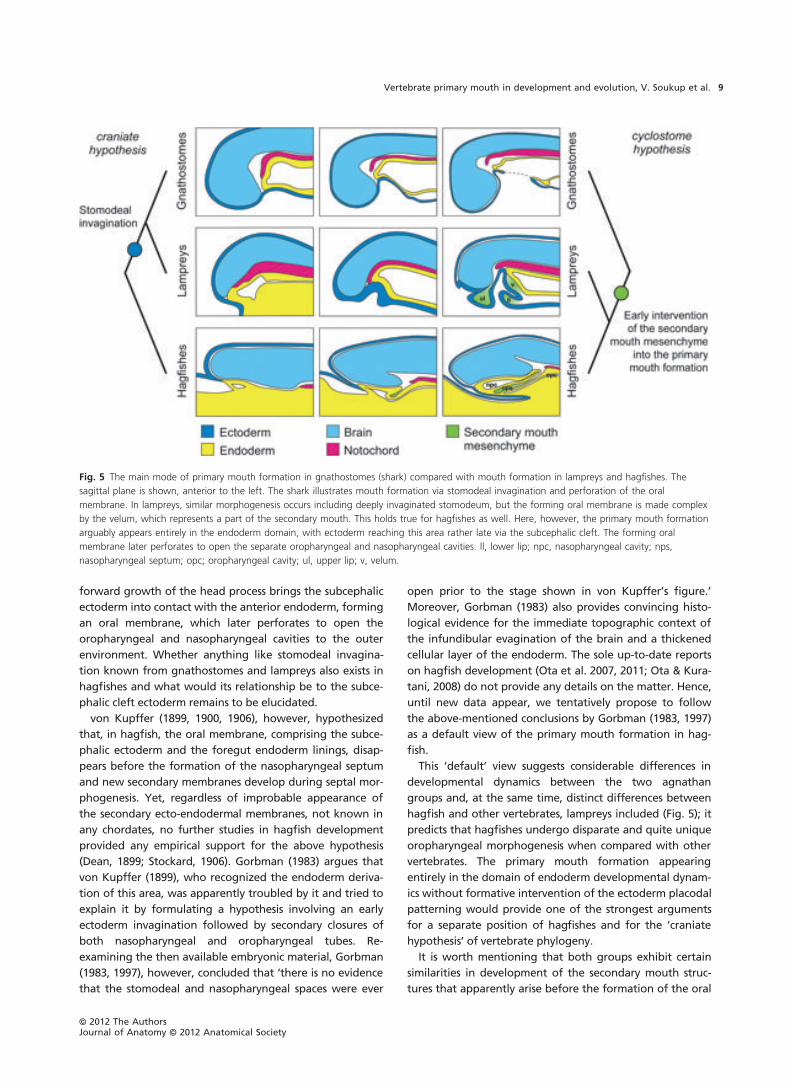
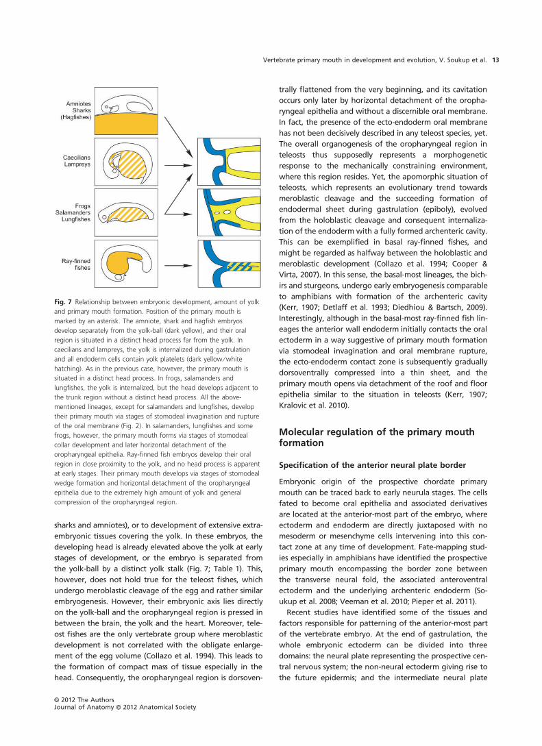
Fig. 5 The main mode of primary mouth formation in gnathostomes (shark) compared with mouth formation in lampreys and hagfishes. The
sagittal plane is shown, anterior to the left. The shark illustrates mouth formation via stomodeal invagination and perforation of the oral
membrane. In lampreys, similar morphogenesis occurs including deeply invaginated stomodeum, but the forming oral membrane is made complex
by the velum, which represents a part of the secondary mouth. This holds true for hagfishes as well. Here, however, the primary mouth formation
arguably appears entirely in the endoderm domain, with ectoderm reaching this area rather late via the subcephalic cleft. The forming oral
membrane later perforates to open the separate oropharyngeal and nasopharyngeal cavities. ll, lower lip; npc, nasopharyngeal cavity; nps,
nasopharyngeal septum; opc; oropharyngeal cavity; ul, upper lip; v, velum.
ªª 2012 The AuthorsJournal of Anatomy ªª 2012 Anatomical Society
Vertebrate primary mouth in development and evolution, V. Soukup et al. 9
opening, i.e. at the stage when the primary mouth still
undergoes development. Namely, this involves the nasopha-
ryngeal septum in hagfish and velum in lampreys augment-
ing the ectoderm–endoderm contact zone by their growth.
Although in hagfish and lamprey the respective structures
form in the domain of the mandibular arch, their morpho-
genesis differs considerably. The ammocoete velum repre-
sents two lateral dorsoventrally placed flanks between the
oral and pharyngeal cavities, and it is reminiscent of the for-
mer position of the oral membrane (Dohrn, 1886; Damas,
1944). The velum of hagfishes, on the other hand, very
probably arises within the endoderm lining and forms as
two ventral outgrowths from the dorsal pharyngeal wall
(von Kupffer, 1900; Stockard, 1906). Yet, the final word on
this matter must be postponed; new developmental data
on hagfish are needed.
Primary mouth formation from thephylogenetic perspective
Mapping the above-surveyed alternatives of the primary
mouth formation onto a phylogenetic tree of vertebrates
(Fig. 6) reveals several intriguing issues.
1. First, a support for the basal divergence of hagfishes
and vertebrates (lampreys + gnathostomes) if the
‘default view’ of the hagfish mouth development (sensu;
Gorbman, 1983) is accepted. The conditions characteriz-
ing the hagfish are: (a) a key role of early differentiation
of the anterior endoderm with formation of the endo-
derm nasopharyngeal cavity and its separation by the
nasopharyngeal septum from the ventral endoderm
oropharyngeal cavity; (b) delayed persistence of oral
membrane without deep stomodeal invagination; (c)
and opening of the mouth cavity via rupture of the oral
membrane – a character shared with most of the other
vertebrates.
2. Second, lampreys exhibit essential differences with
respect to gnathostomes, particularly in: (a) a strict sepa-
ration of stomodeal ectoderm from the nasohypophyseal
plate containing olfactoric and adenohypophyseal pla-
codes, which form a common developmental unit retain-
ing its primary topographic position during evolution.
The topographic separation of stomodeal region from
the nasohypophyseal plate in lamprey resembles the sit-
uation in the hagfish, though these clades supposedly
differ both in origin and developmental dynamics of
these structures. The lamprey corresponds to gnathosto-
mes, or the hypothetical gnathostome ancestor, in (b) a
deep stomodeal invagination resulting in a bilaminar
oral membrane.
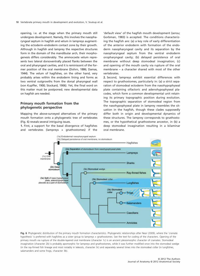
Fig. 6 Phylogenetic distribution of the primary mouth formation characteristics. Phylogenetic relationships after Near (2009), where the ‘craniate
hypothesis’ is preferred with hagfishes as a sister group to lampreys + gnathostomes. See the text for coding of the characters. Opening of the
primary mouth via rupture of the double-layered oral membrane (character 1c) is an ancient plesiomorphic character of craniates. Stomodeal
invagination (character 2b) is probably apomorphic for lampreys and gnathostomes, while it was further modified once into the stomodeal wedge
(in the ray-finned fish lineage and most notably in teleosts, character 3c) and separately several times into the stomodeal collar (in lungfishes,
salamanders and some frogs, character 3b).
ªª 2012 The AuthorsJournal of Anatomy ªª 2012 Anatomical Society
Vertebrate primary mouth in development and evolution, V. Soukup et al.10
3. Third, because the arrangements corresponding to
character (2b) appear in the vast majority of vertebrate
clades, we propose it a synapomorphy of vertebrates and
a plesiomorphy (ancestral state) of gnathostomes. The
major apomorphy of gnathostomes is then: (a) a separa-
tion of the nasohypophyseal plate and incorporation of
the adenohypophyseal anlage into the expanding stomo-
deal field. Besides the common mode of primary mouth
formation in gnathostomes (i.e. characters 1c + 2b + 3a),
at least two other modes evolved: (b) a process of stomo-
deal involution followed by formation of the stomodeal
collar in salamanders, lungfishes and some frogs; and (c)
an intimate connection between ectoderm and endoderm
(stomodeal wedge), with a subsequent dissociation of
roof and floor oropharyngeal epithelia in ray-finned
fishes. The extent of variation and taxon-specific arrange-
ment of the mode of mouth formation among ray-finned
fishes are unfortunately largely unknown, although this
lineage contains half of all vertebrate species. However,
despite the scarcity of information, the character (c) has
probably been acquired already at the base of the ray-
finned fish clade.
Interestingly, character (3b) probably evolved several
times independently in lineages leading to lungfishes, sala-
manders and ⁄or frogs (Fig. 6) and, thus, represents a conver-
gently acquired homoplastic trait. All these lineages
undergo comparable morphogenesis of the pharyngeal
region (including extensive development of branchial arches
or larval external gills), and their early ontogeny (namely
the content of yolk, gastrulation or endoderm formation) is
markedly alike. We therefore expect that the respective
mode of primary mouth formation is influenced by a set of
various contextual factors, one of them evidently repre-
sented by the amount of yolk and by spatial and molecular
settings of the early developing embryo, as discussed below.
General patterns of gastrulation andparticularly the yolk content of embryosprefigure modes of primary mouthdevelopment
In the previous section, three main modes of mouth forma-
tion were identified for vertebrates (Fig. 2). In order to
reveal factors determining and constraining the mouth in
development and evolution, the formation of prospective
oral regions was followed from the time of gastrulation,
and developmental correlates of mouth morphogenesis
were explored (Fig. 8; Table 1).
During the process of gastrulation, chordate embryos
must bring their endoderm into the future anterior region
so that a direct contact zone between the ectoderm and
endoderm can be established for the prospective mouth for-
mation (Fig. 1). However, particular patterns of endoderm
formation vary and strongly depend upon the amount and
positioning of yolk in eggs and embryos. Therefore, in
groups, where the yolk areas are fully internalized into the
embryo during gastrulation and where the whole egg cyto-
plasm is cleaved (mesolecithal eggs, holoblastic develop-
ment), the endoderm is formed with a distinct archenteron,
a situation seen in amphibians, basal ray-finned fishes, lam-
preys or lungfishes (Table 1). The archenteric cavity then
progresses into the lumen of the whole alimentary canal,
and its anterior-most lining directly contacts the surface
ectoderm. This contact demarcates the position of future
mouth that opens anteriorly. Alternatively, in those groups,
where the yolk (vegetal) part of eggs is uncleaved and
serves for nutrition only, the embryo develops from the
cleaved animal part itself (telolecithal eggs, meroblastic
development), no foregut cavity forms initially, but a com-
pact archenteric layer or archenteron mass develops in the
anterior part of the head instead. The foregut cavity has to
be formed secondarily (Nelsen, 1953), for example, as
described above for teleosts, via ventromedial bending of
the lateral edges of the foregut endoderm sheet and their
subsequent fusion into a tube. This situation is known for
hagfishes, sharks, teleosts and amniotes.
Both protochordate groups (cephalochordates and uro-
chordates) undergo embryonic development from small
eggs with a minimum amount of yolk and equal holoblastic
cleavage, which is in strong contrast to the vertebrate
embryos where such a situation is achieved only secondarily
in placental mammals. Vertebrates, on the contrary, display
increased maternal investments into their offspring by mas-
sive deposition of yolk into the eggs (Takeuchi et al. 2009),
which necessarily leads to different cleavage and gastrula-
tive cellular behaviour. These two processes are then exem-
plified either by unequal holoblastic cleavage with an
amphibian-type gastrulation, or meroblastic cleavage with
an amniote-type gastrulation. It is beyond the scope of
this paper to discuss the archetypal condition for verte-
brates in detail as both conditions may, based on the distri-
bution on the cladogram, represent an ancestral character
state. However, the holoblastic development is generally
regarded plesiomorphic for vertebrates (Collazo et al. 1994;
Takeuchi et al. 2009).
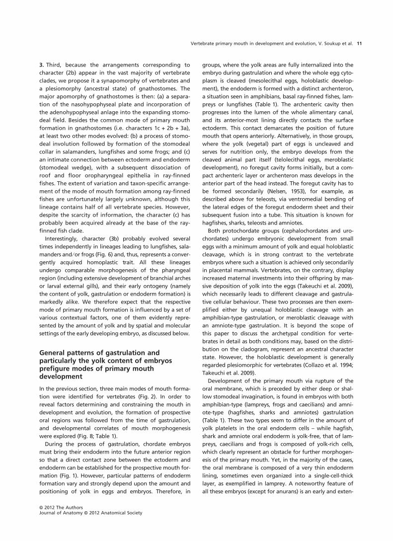
Development of the primary mouth via rupture of the
oral membrane, which is preceded by either deep or shal-
low stomodeal invagination, is found in embryos with both
amphibian-type (lampreys, frogs and caecilians) and amni-
ote-type (hagfishes, sharks and amniotes) gastrulation
(Table 1). These two types seem to differ in the amount of
yolk platelets in the oral endoderm cells – while hagfish,
shark and amniote oral endoderm is yolk-free, that of lam-
preys, caecilians and frogs is composed of yolk-rich cells,
which clearly represent an obstacle for further morphogen-
esis of the primary mouth. Yet, in the majority of the cases,
the oral membrane is composed of a very thin endoderm
lining, sometimes even organized into a single-cell-thick
layer, as exemplified in lamprey. A noteworthy feature of
all these embryos (except for anurans) is an early and exten-
ªª 2012 The AuthorsJournal of Anatomy ªª 2012 Anatomical Society
Vertebrate primary mouth in development and evolution, V. Soukup et al. 11

sive growth of the head process, which removes the pro-
spective primary mouth far from the trunk. We speculate
that this morphogenetic event enables development of the
primary mouth at a distance from the yolk-rich trunk region
and a concurrent participation of only a small number of
endoderm cells (Fig. 7). Indeed, this mode of primary mouth
formation, although modified in terms of position and
development, is found in urochordates as well as in cepha-
lochordates (Fig. 1), and represents a plesiomorphic condi-
tion for vertebrates (Legros, 1898; Goppert, 1906; Manni
et al. 2005; Veeman et al. 2010). Within vertebrates, such a
mode of primary mouth formation represents a ground
state condition from which the other modes are derived.
One such derived mode is the formation of primary
mouth via the stomodeal collar and horizontal detachment
of oropharyngeal epithelia, which occurs in lineages with
embryos undergoing holoblastic cleavage and amphibian-
type gastrulation; more specifically, in those lineages where
the primary mouth development does not correlate with
the formation of a distinct head-fold during early embryo-
genesis (Fig. 7; Table 1). The oral region is meanwhile filled
by a mass of yolk-laden cells, which resides within the man-
dibular arch domain and abuts the oral ectoderm anteri-
orly. On account of the accumulation of oral endoderm, no
deep invagination forming a stomodeum can develop and,
consequently, the ectoderm cells tend to undergo involu-
tion in a form of deep epithelial layers (Fig. 3). Shallow sto-
modeum and multilayered oral endoderm can be found
also in Xenopus (Dickinson & Sive, 2006), and it should be
emphasized that the default classic mode of mouth forma-
tion via invaginating stomodeum and rupture of the oral
membrane might not be present in all anuran species and
that, instead, involution of the inner ectoderm layer and
the presence of the stomodeal collar-like structure might be
a more common feature than recently appreciated.
Morphogenesis of the stomodeal collar apparently repre-
sents an alternative way to bring the ectoderm inside the
mouth in those lineages, where yolk cells were deposited
within the oral region and thus block the formation of
deep stomodeal ectoderm invagination.
Meroblastic development often leads to embryos consist-
ing of almost transparent sheets and layers (in hagfishes,
Table 1 Correlations between the modes of primary mouth formation and some key features of embryonic development in individual vertebrate
lineages.
The comparative features of the mode of primary mouth formation include: first, endoderm during gastrulation, which can primarily
represent a hollow pocket with an archenteron or a single sheet archenteric layer (hypoderm in amniotes); second, position of
embryo and its connection to the yolk, which can be manifested in three ways – the yolk can be internalized and become a part of
the embryonic body, the embryo can be connected to the yolk-ball by a yolk stalk, or the embryo can lie directly on the yolk-ball
without a yolk stalk; and third, head process, which may or may not arise from the main yolky trunk region during early embryonic
development.
ªª 2012 The AuthorsJournal of Anatomy ªª 2012 Anatomical Society
Vertebrate primary mouth in development and evolution, V. Soukup et al.12
sharks and amniotes), or to development of extensive extra-
embryonic tissues covering the yolk. In these embryos, the
developing head is already elevated above the yolk at early
stages of development, or the embryo is separated from
the yolk-ball by a distinct yolk stalk (Fig. 7; Table 1). This,
however, does not hold true for the teleost fishes, which
undergo meroblastic cleavage of the egg and rather similar
embryogenesis. However, their embryonic axis lies directly
on the yolk-ball and the oropharyngeal region is pressed in
between the brain, the yolk and the heart. Moreover, tele-
ost fishes are the only vertebrate group where meroblastic
development is not correlated with the obligate enlarge-
ment of the egg volume (Collazo et al. 1994). This leads to
the formation of compact mass of tissue especially in the
head. Consequently, the oropharyngeal region is dorsoven-
trally flattened from the very beginning, and its cavitation
occurs only later by horizontal detachment of the oropha-
ryngeal epithelia and without a discernible oral membrane.
In fact, the presence of the ecto-endoderm oral membrane
has not been decisively described in any teleost species, yet.
The overall organogenesis of the oropharyngeal region in
teleosts thus supposedly represents a morphogenetic
response to the mechanically constraining environment,
where this region resides. Yet, the apomorphic situation of
teleosts, which represents an evolutionary trend towards
meroblastic cleavage and the succeeding formation of
endodermal sheet during gastrulation (epiboly), evolved
from the holoblastic cleavage and consequent internaliza-
tion of the endoderm with a fully formed archenteric cavity.
This can be exemplified in basal ray-finned fishes, and
might be regarded as halfway between the holoblastic and
meroblastic development (Collazo et al. 1994; Cooper &
Virta, 2007). In this sense, the basal-most lineages, the bich-
irs and sturgeons, undergo early embryogenesis comparable
to amphibians with formation of the archenteric cavity
(Kerr, 1907; Detlaff et al. 1993; Diedhiou & Bartsch, 2009).
Interestingly, although in the basal-most ray-finned fish lin-
eages the anterior wall endoderm initially contacts the oral
ectoderm in a way suggestive of primary mouth formation
via stomodeal invagination and oral membrane rupture,
the ecto-endoderm contact zone is subsequently gradually
dorsoventrally compressed into a thin sheet, and the
primary mouth opens via detachment of the roof and floor
epithelia similar to the situation in teleosts (Kerr, 1907;
Kralovic et al. 2010).
Molecular regulation of the primary mouthformation
Specification of the anterior neural plate border
Embryonic origin of the prospective chordate primary
mouth can be traced back to early neurula stages. The cells
fated to become oral epithelia and associated derivatives
are located at the anterior-most part of the embryo, where
ectoderm and endoderm are directly juxtaposed with no
mesoderm or mesenchyme cells intervening into this con-
tact zone at any time of development. Fate-mapping stud-
ies especially in amphibians have identified the prospective
primary mouth encompassing the border zone between
the transverse neural fold, the associated anteroventral
ectoderm and the underlying archenteric endoderm (So-
ukup et al. 2008; Veeman et al. 2010; Pieper et al. 2011).
Recent studies have identified some of the tissues and
factors responsible for patterning of the anterior-most part
of the vertebrate embryo. At the end of gastrulation, the
whole embryonic ectoderm can be divided into three
domains: the neural plate representing the prospective cen-
tral nervous system; the non-neural ectoderm giving rise to
the future epidermis; and the intermediate neural plate
Fig. 7 Relationship between embryonic development, amount of yolk
and primary mouth formation. Position of the primary mouth is
marked by an asterisk. The amniote, shark and hagfish embryos
develop separately from the yolk-ball (dark yellow), and their oral
region is situated in a distinct head process far from the yolk. In
caecilians and lampreys, the yolk is internalized during gastrulation
and all endoderm cells contain yolk platelets (dark yellow ⁄white
hatching). As in the previous case, however, the primary mouth is
situated in a distinct head process. In frogs, salamanders and
lungfishes, the yolk is internalized, but the head develops adjacent to
the trunk region without a distinct head process. All the above-
mentioned lineages, except for salamanders and lungfishes, develop
their primary mouth via stages of stomodeal invagination and rupture
of the oral membrane (Fig. 2). In salamanders, lungfishes and some
frogs, however, the primary mouth forms via stages of stomodeal
collar development and later horizontal detachment of the
oropharyngeal epithelia. Ray-finned fish embryos develop their oral
region in close proximity to the yolk, and no head process is apparent
at early stages. Their primary mouth develops via stages of stomodeal
wedge formation and horizontal detachment of the oropharyngeal
epithelia due to the extremely high amount of yolk and general
compression of the oropharyngeal region.
ªª 2012 The AuthorsJournal of Anatomy ªª 2012 Anatomical Society
Vertebrate primary mouth in development and evolution, V. Soukup et al. 13
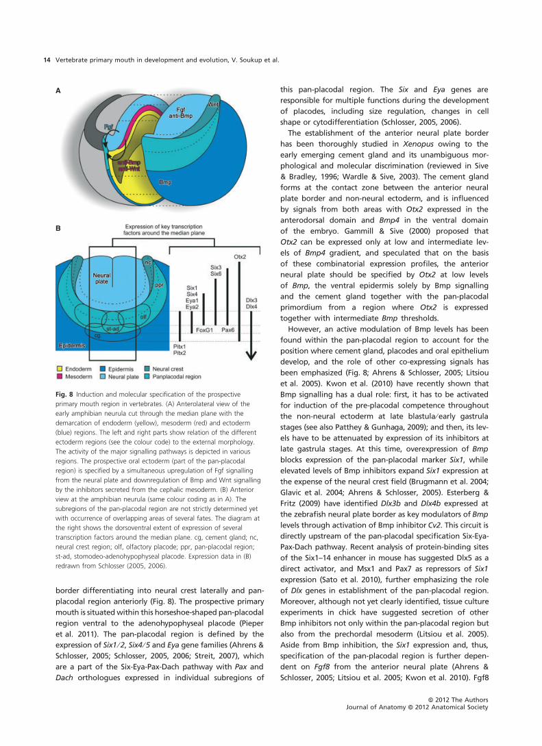
border differentiating into neural crest laterally and pan-
placodal region anteriorly (Fig. 8). The prospective primary
mouth is situatedwithin this horseshoe-shaped pan-placodal
region ventral to the adenohypophyseal placode (Pieper
et al. 2011). The pan-placodal region is defined by the
expression of Six1 ⁄2, Six4 ⁄5 and Eya gene families (Ahrens &
Schlosser, 2005; Schlosser, 2005, 2006; Streit, 2007), which
are a part of the Six-Eya-Pax-Dach pathway with Pax and
Dach orthologues expressed in individual subregions of
this pan-placodal region. The Six and Eya genes are
responsible for multiple functions during the development
of placodes, including size regulation, changes in cell
shape or cytodifferentiation (Schlosser, 2005, 2006).
The establishment of the anterior neural plate border
has been thoroughly studied in Xenopus owing to the
early emerging cement gland and its unambiguous mor-
phological and molecular discrimination (reviewed in Sive
& Bradley, 1996; Wardle & Sive, 2003). The cement gland
forms at the contact zone between the anterior neural
plate border and non-neural ectoderm, and is influenced
by signals from both areas with Otx2 expressed in the
anterodorsal domain and Bmp4 in the ventral domain
of the embryo. Gammill & Sive (2000) proposed that
Otx2 can be expressed only at low and intermediate lev-
els of Bmp4 gradient, and speculated that on the basis
of these combinatorial expression profiles, the anterior
neural plate should be specified by Otx2 at low levels
of Bmp, the ventral epidermis solely by Bmp signalling
and the cement gland together with the pan-placodal
primordium from a region where Otx2 is expressed
together with intermediate Bmp thresholds.
However, an active modulation of Bmp levels has been
found within the pan-placodal region to account for the
position where cement gland, placodes and oral epithelium
develop, and the role of other co-expressing signals has
been emphasized (Fig. 8; Ahrens & Schlosser, 2005; Litsiou
et al. 2005). Kwon et al. (2010) have recently shown that
Bmp signalling has a dual role: first, it has to be activated
for induction of the pre-placodal competence throughout
the non-neural ectoderm at late blastula ⁄early gastrula
stages (see also Patthey & Gunhaga, 2009); and then, its lev-
els have to be attenuated by expression of its inhibitors at
late gastrula stages. At this time, overexpression of Bmp
blocks expression of the pan-placodal marker Six1, while
elevated levels of Bmp inhibitors expand Six1 expression at
the expense of the neural crest field (Brugmann et al. 2004;
Glavic et al. 2004; Ahrens & Schlosser, 2005). Esterberg &
Fritz (2009) have identified Dlx3b and Dlx4b expressed at
the zebrafish neural plate border as key modulators of Bmp
levels through activation of Bmp inhibitor Cv2. This circuit is
directly upstream of the pan-placodal specification Six-Eya-
Pax-Dach pathway. Recent analysis of protein-binding sites
of the Six1–14 enhancer in mouse has suggested Dlx5 as a
direct activator, and Msx1 and Pax7 as repressors of Six1
expression (Sato et al. 2010), further emphasizing the role
of Dlx genes in establishment of the pan-placodal region.
Moreover, although not yet clearly identified, tissue culture
experiments in chick have suggested secretion of other
Bmp inhibitors not only within the pan-placodal region but
also from the prechordal mesoderm (Litsiou et al. 2005).
Aside from Bmp inhibition, the Six1 expression and, thus,
specification of the pan-placodal region is further depen-
dent on Fgf8 from the anterior neural plate (Ahrens &
Schlosser, 2005; Litsiou et al. 2005; Kwon et al. 2010). Fgf8
A
B
Fig. 8 Induction and molecular specification of the prospective
primary mouth region in vertebrates. (A) Anterolateral view of the
early amphibian neurula cut through the median plane with the
demarcation of endoderm (yellow), mesoderm (red) and ectoderm
(blue) regions. The left and right parts show relation of the different
ectoderm regions (see the colour code) to the external morphology.
The activity of the major signalling pathways is depicted in various
regions. The prospective oral ectoderm (part of the pan-placodal
region) is specified by a simultaneous upregulation of Fgf signalling
from the neural plate and downregulation of Bmp and Wnt signalling
by the inhibitors secreted from the cephalic mesoderm. (B) Anterior
view at the amphibian neurula (same colour coding as in A). The
subregions of the pan-placodal region are not strictly determined yet
with occurrence of overlapping areas of several fates. The diagram at
the right shows the dorsoventral extent of expression of several
transcription factors around the median plane. cg, cement gland; nc,
neural crest region; olf, olfactory placode; ppr, pan-placodal region;
st-ad, stomodeo-adenohypophyseal placode. Expression data in (B)
redrawn from Schlosser (2005, 2006).
ªª 2012 The AuthorsJournal of Anatomy ªª 2012 Anatomical Society
Vertebrate primary mouth in development and evolution, V. Soukup et al.14
together with Bmp inhibitors, but not Fgf8 alone, are able
to induce ectopic Six1 expression in anteroventral ectoderm,
whereas knock-down of Fgf signalling results in loss of pan-
placodal markers in Xenopus (Ahrens & Schlosser, 2005).
Moreover, canonical Wnt pathway has been shown to
direct the decision of neural plate border into either pan-
placodal or neural crest fates (Brugmann et al. 2004; Litsiou
et al. 2005). The Wnt ⁄b-catenin antagonist Dkk1 secreted
from the prechordal mesoderm is required for preventing
the formation of neural crest in the transverse neural fold,
which is normally fated to become the pan-placodal region
(Carmona-Fontaine et al. 2007). Taken together, interplay
between activated Fgf and inhibited Bmp and Wnt signal-
ling is a basis for specification of the pan-placodal region
and activation of the Six-Eya-Pax-Dach pathway (Fig. 8A).
Once specified, the pan-placodal region is further subdi-
vided into distinct cell populations giving rise to a variety of
placodes, head epidermis, cement gland and primary
mouth ectoderm (Pieper et al. 2011). During the course of
embryonic development, the regions giving rise to these
organs are first blurred (meaning that a single cell can con-
tribute to several organs), but the respective fates gradually
become spatially restricted (Whitlock & Westerfield, 2000;
Pieper et al. 2011).
Given that the similar induction and specification mecha-
nisms of the pan-placodal region have been identified in
different vertebrate species (Xenopus, zebrafish, chick and
mouse), the pan-placodal region almost certainly seems to
represent a conserved spatiotemporal domain of vertebrate
embryonic period (Schlosser, 2006). In urochordates, the
vertebrate sister group, Six and Eya genes are expressed in a
similar manner, although they may provide different
placode-specific functions (Bassham & Postlethwait, 2005;
Mazet et al. 2005; Schlosser, 2007), while in amphioxus,
these genes are expressed in endodermal derivatives and
the vertebrate type pan-placodal region is not found (Ko-
zmik et al. 2007; Schlosser, 2007). The pan-placodal region
thus probably evolved in the common ancestor of verte-
brates and urochordates, and represents a defining feature
of the taxonomic group Olfactoria.
Specification of the primary mouth and the role ofPitx genes in oral development
Although, due to scarcity of detailed comparative informa-
tion, hypothesizing on the structure of the molecular regu-
lation controlling the primary mouth formation would be
premature, some of its components can almost certainly be
identified. First, this concerns the essential role of Pitx sig-
nalling. As described above, the prospective primary mouth
forms from the median portion of the pan-placodal ecto-
derm and the rostral foregut endoderm. Such an extreme
anterior position of the oral region is specifically marked by
the expression of the pituitary homeobox (Pitx, formerly
Ptx) genes (Fig. 8B; Dickinson & Sive, 2007). Pitx genes
belong to a family of paired-like homeodomain class of
transcription factors, which includes Pitx1, Pitx2 and Pitx3
paralogues. These genes can produce a number of different
proteins thanks to alternative splicing and alternative trans-
lation initiation sites (Gage et al. 1999b; Shiratori et al.
2001; Cox et al. 2002; Angotzi et al. 2008; Lamba et al.
2008). Pitx genes are involved in morphogenesis of diverse
organs, including eye, brain, heart or limbs, and are respon-
sible for early embryonic patterning and left–right asymme-
try (Ryan et al. 1998; Gage et al. 1999a; Lanctot et al.
1999b; Lin et al. 1999; Lu et al. 1999; Burdine & Schier,
2000; Shapiro et al. 2004). Most notably, however, Pitx par-
alogues are expressed at the anterior part of the pan-
placodal ectoderm at neurula stages (Schweickert et al.
2001a; Jaszczyszyn et al. 2007; Angotzi et al. 2008), and
their expression is subsequently restricted to the ectoderm
anlagen of the developing stomodeum and adenohypophy-
sis and endoderm anterior pharynx. Further on, the Pitx-
expressing epithelia contribute to vertebrate ectoderm and
endoderm derivatives of the oral cavity, like tooth germs,
tongue or palate (Lanctot et al. 1997, 1999a; St. Amand
et al. 2000; Fraser et al. 2004).
While the functions of Pitx genes during the morphogen-
esis of organs associated with the mouth are becoming
clearer (see below), their roles in the development of pri-
mary mouth are still to be elucidated. This might partially
be due to the relatively simple morphogenesis of the invagi-
nating stomodeum and consequently a lack of morphologi-
cally clearly discernible developmental landmarks when
compared with the morphogenesis of, for example, teeth or
the adenohypophysis. Therefore, the roles of Pitx genes in
primary mouth morphogenesis can only be inferred by anal-
ogy to their known functions in associated organs.
Functional studies in vertebrates have demonstrated the
requirements of Pitx genes for the development of the mur-
ine adenohypophysis and Xenopus cement gland. All three
Pitx genes are expressed in the Xenopus primary mouth
and adenohypophysis, while only Pitx1 and Pitx2
paralogues can be found in the early developing cement
gland (Hollemann & Pieler, 1999; Pommereit et al. 2001;
Schweickert et al. 2001b). Overexpression of either Pitx1 or
Pitx2c induces formation of enlarged or ectopic cement
glands, while more severe phenotypes display reduced
trunk and tail regions (Chang et al. 2001; Schweickert et al.
2001b). Double knock-down of both of these genes, on the
other hand, inhibits the formation of ectopically induced
cement glands (Schweickert et al. 2001b). Pitx genes are
thus both necessary and sufficient for the induction of
Xenopus cement gland formation. Concerning the adeno-
hypophysis, different Pitx paralogues are required for its
development in different vertebrates, showing conserved
functions and evolutionary shuffling of these genes in pitui-
tary organogenesis (Gage et al. 1999a; Hollemann & Pieler,
1999; Lanctot et al. 1999a; Szeto et al. 1999; Pommereit
et al. 2001; Schweickert et al. 2001b; Suh et al. 2002;
ªª 2012 The AuthorsJournal of Anatomy ªª 2012 Anatomical Society
Vertebrate primary mouth in development and evolution, V. Soukup et al. 15
Jaszczyszyn et al. 2007; Angotzi et al. 2008). The adenohy-
pophysis in mouse is dependent on the expression of Pitx1
and Pitx2 paralogues with cooperative and functionally
overlapping functions (Suh et al. 2002; Charles et al. 2005).
While mice deficient in Pitx1 show milder pituitary pheno-
types with normal formation of Rathke’s pouch, Pitx2) ⁄ )
mutant embryos have relatively normal Rathke’s pouch as
well, but its later expansion is arrested and increased cell
death occurs (Gage et al. 1999a; Szeto et al. 1999; Charles
et al. 2005). A very small Rathke’s pouch forms after disrup-
tion of both genes, suggesting a synergistic action of these
paralogues and specific Pitx dosage requirements for
proper pituitary morphogenesis (Suh et al. 2002).
Pitx genes have multiple roles in pituitary development
by regulating the expression of different downstream fac-
tors at different time points (Suh et al. 2002). At early
stages, when the prospective murine adenohypophysis is in
the form of Rathke’s pouch and therefore morphologically
resembles a much earlier invagination of the stomodeum,
Pitx1 and Pitx2 regulate the expression of factors responsi-
ble for proliferation and Rathke’s pouch expansion. Pitx2
expression is induced at early to late G1 phase of the cell
cycle by Wnt ⁄b-catenin signalling, and directly co-activates
the expression of growth-control genes like Cyclin D1,
Cyclin D2 and c-Myc (Kioussi et al. 2002; Baek et al. 2003).
Pitx2 as well as cell cycle-control mRNAs represent unstable
molecules with a rapid turnover and short half-life, a fea-
ture that enables quick and precise response to the chang-
ing developmental and genetic cues. After activation of
Pitx2 expression by b-catenin, the Pitx2 protein acts as a sta-
bilizing factor of both Pitx2 mRNA and the cell cycle-control
mRNAs like Cyclin D1, Cyclin D2 and c-Jun (Briata et al.
2003). The early adenohypophyseal morphogenesis is there-
fore regulated by Wnt ⁄b-catenin signalling at both
transcriptional as well as RNA stabilization levels.
A question thus arises as to whether the Wnt ⁄b-catenin ⁄Pitx pathway identified as triggering proliferation
in the adenohypophysis functions in a similar way also dur-
ing the invaginating stomodeum. Pitx orthologues have
been found at the median pan-placodal region and the
developing primary mouth in a number of vertebrates
(Lanctot et al. 1997; Schweickert et al. 2001b; Boorman &
Shimeld, 2002a; Jaszczyszyn et al. 2007; Angotzi et al. 2008).
However, Wnt signalling is specifically downregulated in
the pan-placodal region by secretion of its inhibitor Dkk1
from the cranial mesoderm (Fig. 8A; Carmona-Fontaine
et al. 2007), and it has to be inhibited also at later stages
during the process of dissolution of the basal lamina
between ectoderm and endoderm of the oral membrane
prior to opening of the primary mouth (Dickinson & Sive,
2009). The Pitx expression in the primary mouth is therefore
probably regulated by a mechanism different from the
Wnt ⁄b-catenin signalling. During the early patterning of
the murine mandibular arch, the Pitx1 and Pitx2 are posi-
tively regulated by Fgf8 and negatively regulated by Bmp4
(St. Amand et al. 2000). These genes in turn regulate expres-
sions of Fgf8 in positive and Bmp4 in negative feedback
loops, a mechanism responsible for proper positioning of
teeth (St. Amand et al. 2000; Liu et al. 2003). In the mouse
upper and lower jaw epithelia, the Pitx2-expressing cells dis-
play increased proliferative and migratory properties so that
their daughter cells expand from the mandibular arch area
both outwards into the facial epithelium and inwards con-
tributing to the roof and floor epithelial linings of the oral
cavity (Liu et al. 2003). The establishment and maintenance
of the zone of expression of Pitx genes in the anterior neu-
ral plate border may therefore be activated by the Fgf sig-
nalling similar to its activation in the mandibular arch and
teeth, and may in turn repress Bmp in this region. Similarly
to Pitx genes, the activation of Six1 during establishment of
the pan-placodal region is also dependent on both synergis-
tic activation of Fgf and repression of Bmp signalling
(Ahrens & Schlosser, 2005; Litsiou et al. 2005; Kwon et al.
2010).
Whatever induces the Pitx genes in the embryonic
anterior region, their expression has been identified in a
number of vertebrate species from lamprey to mouse, sug-
gesting a conserved function of Pitx genes in the early spec-
ification of the stomodeo-adenohypophyseal ectoderm and
anterior pharyngeal endoderm. Moreover, Pitx orthologues
have been found associated with the oral apparatus of uro-
chordates and cephalochordates (Yasui et al. 2000; Boor-
man & Shimeld, 2002b; Bassham & Postlethwait, 2005;
Christiaen et al. 2005), illuminating their archetypal instruc-
tive roles for stomodeal invagination, adenohypophyseal
placode development and oropharyngeal morphogenesis in
chordates.
Concluding remarks: phylogeneticimplications of primary mouth formation
Until now, our interest in comparative aspects of primary
mouth formation was restricted to vertebrates. Yet to
reveal the phylogenetic background of the involved devel-
opmental pathways requires a comparison with the path-
ways in basal chordates. Although detailed information on
patterns and processes of primary mouth formation is avail-
able for just a few species and the actual taxonomical cover-
age is quite scarce for the discussion of the phylogenetic
implications, several interpretations do arise.
First, the early steps of mouth formation putatively rep-
resent a shared trait of vertebrates and urochordates. Both
vertebrates and urochordates show a topographically inti-
mate connection between the stomodeum and adenohy-
pophyseal placode appearing in identical positions: at the
anterior margin of pan-placodal region in vertebrates; and
at the anterior margin of neuropore in urochordates
(Manni et al. 2004, 2005; Schlosser, 2007). In both lineages,
therefore, the primary mouth is associated with the invag-
ination of the epithelial cell population derived from the
ªª 2012 The AuthorsJournal of Anatomy ªª 2012 Anatomical Society
Vertebrate primary mouth in development and evolution, V. Soukup et al.16
stomodeal ectoderm and formation of distinct stomo-
deum. In contrast, in amphioxus, the mouth forms as an
endoderm outpocketing in the topographic and structural
context of the primary pharyngeal slits at the left side of
the anterior pharynx. The only putative placode appearing
in amphioxus, the preoral ciliary pit, invaginates only after
completion of primary mouth formation, where it forms
the Hatschek’s pit, i.e. the homologue of vertebrate ade-
nohypophysis (Gorbman, 1999; Holland & Holland, 2001).
In this regard, the correspondence of primary mouth for-
mation seems to support monophyly of Olfactoria (verte-
brates + urochordates) and distinct position of
cephalochordates (Fig. 1). The essential separation of
mouth formation from the morphogenetic processes of
pharyngotremia can tentatively be considered as the key
apomorphy of olfactors. Of course, it should be remem-
bered that the vertebrate pharyngeal region exhibits a
number of apomorphies as well.
The plesiomorphic condition for deuterostomes indicated
by initial endoderm outpocketings, which adopt a narrow
slit-like morphology and are elaborated along the dorsoven-
tral axis (Graham et al. 2005), is to form a pharyngeal appa-
ratus designed for filter-feeding and ventilation by cilliary
propulsion. In both respects, its efficiency increases with the
number of slits and height of cilia-bearing interpouch bars,
while no special requirements are imposed upon shape and
size of oral opening. The branchial basket in cephalochor-
dates and urochordates reflects this scheme (Swalla & Smith,
2008). In contrast, the pharyngeal apparatus in vertebrates
is exclusively designed for the ventilation by branchiomeric
musculature attached to rigid branchial bars. Here, the
capacity of ventilation is positively related to the volume of
the musculature and the extent of particular pouches. The
pharyngeal region of vertebrates consequently tends to
exhibit a reduced number of particular slits, extensive
enlargements of their volumes and the height of branchial
region. This also characterizes the earliest sister fossil taxon
of vertebrates, the Cambrian genus Haikouella (Mallatt &
Chen, 2003). Here, enlargement of the branchial region is
accompanied by the enlargement of oral opening and
rostral expansion of a suprapharyngeal brain. It can be con-
sequently expected that mouth formation involved both
the invagination of ectoderm population at the anterior
margin of expanding brain as well as the instructive role of
endoderm. The situation inHaikouella thus fits the develop-
mental evidence demonstrating that: first, the source ecto-
derm population for oral formation appears at the anterior
margin of neural plate within the zone of the primordial
pan-placodal field (Schlosser, 2005, 2006; Christiaen et al.
2007), but also that, second, the morphogenesis of the pha-
ryngeal endoderm may play the role of the key patterning
factor (Graham et al. 2005) preforming the space for stomo-
deal invagination. Third, we can also hypothesize that the
signalling modules including co-expression of Pitx transcrip-
tion factors had already been established.
The ultimate role of endoderm morphogenesis in mouth
formation is supposedly retained in hagfishes and, as dis-
cussed above, the hagfish development in that respect may
provide a very robust argument for the ‘craniate hypothesis’
of vertebrate phylogeny. Yet, validity of that statement is
essentially dependent upon data on early development in
these animals, which are unfortunately still missing. Corre-
spondingly, little is also known about primary mouth forma-
tion in the vast majority of other vertebrate clades. The
precise developmental data both in taxonomic and method-
ological respects are just what we urgently need in order to
uncover dissimilarities in developmental processes produc-
ing morphological variation and being responsible for
evolutionary change.
Acknowledgements
We would like to thank Zerina Johanson and Anthony Graham
for their kind invitation to participate in this volume; we are
grateful to our colleagues and to anonymous reviewers for their
helpful comments on the manuscript. The research reported
here was supported by MSMT project 0021620828 and GACR
206 ⁄ 09 ⁄ 1007.
References
Adams AE (1924) An experimental study of the development of
the mouth in the amphibian embryo. J Exp Zool 40, 311–379.
Adams AE (1931) Some effects of the removal of endoderm
from the mouth region of the early Amblystoma punctatum
embryos. J Exp Zool 58, 147–163.
Ahrens K, Schlosser G (2005) Tissues and signals involved in the
induction of placodal Six1 expression in Xenopus laevis. Dev
Biol 288, 40–59.
Angotzi AR, Ersland KM, Mungpakdee S, et al. (2008)
Independent and dynamic reallocation of pitx gene
expression during vertebrate evolution, with emphasis on fish
pituitary development. Gene 417, 19–26.
Baek SH, Kioussi C, Briata P, et al. (2003) Regulated subset of G1
growth-control genes in response to derepression by the Wnt
pathway. Proc Natl Acad Sci USA 100, 3245–3250.
Balinsky BI (1947) Korrelationen in den Entwicklung der Mund-
und Kiemenregion und des Darmkanales bei den Amphibien.
Roux Arch Entw Mech 143, 365–395.
Ballard WW, Mellinger J, Lachenault H (1993) A series of normal
stages for development of Scyliorhinus canicula, the lesser
spotted dogfish (Chondrichthyes: Scyliorhinidae). J Exp Zool
267, 318–336.
Bassham S, Postlethwait JH (2005) The evolutionary history of
placodes: a molecular genetic investigation of the larvacean
urochordate Oikopleura dioica. Development 132, 4259–
4272.
de Beer GR (1947) The differentiation of neural crest cells into
visceral cartilages and odontoblasts in Amblystoma, and
re-examination of the germ-layer theory. Proc R Soc Lond B
Biol Sci 134, 377–398.
Boorman CJ, Shimeld SM (2002a) Cloning and expression of a
Pitx homeobox gene from the lamprey, a jawless vertebrate.
Dev Genes Evol 212, 349–353.
ªª 2012 The AuthorsJournal of Anatomy ªª 2012 Anatomical Society
Vertebrate primary mouth in development and evolution, V. Soukup et al. 17
Boorman CJ, Shimeld SM (2002b) Pitx homeobox genes in Ciona
and amphioxus show left-right asymmetry is a conserved
chordate character and define the ascidian adenohypophysis.
Evol Dev 4, 354–365.
Bordzilovskaya NP, Dettlaff TA, Duhon ST, et al. (1989)
Developmental-stage series of axolotl embryos. In:
Developmental Biology of the Axolotl. (eds Armstrong JB,
Malacinski GM), pp. 201–219. Oxford: Oxford University Press.
Briata P, Ilengo C, Corte G, et al. (2003) The Wnt ⁄ beta-catenin fi Pitx2 pathway controls the turnover of Pitx2 and
other unstable mRNAs. Mol Cell 12, 1201–1211.
Brugmann SA, Pandur PD, Kenyon KL, et al. (2004) Six1
promotes a placodal fate within the lateral neurogenic
ectoderm by functioning as both a transcriptional activator
and repressor. Development 131, 5871–5881.
Burdine RD, Schier AF (2000) Conserved and divergent
mechanisms in left-right axis formation. Genes Dev 14, 763–776.
Carmona-Fontaine C, Acuna G, Ellwanger K, et al. (2007) Neural
crests are actively precluded from the anterior neural fold by
a novel inhibitory mechanism on Dickkopf1 secreted by the
prechordal mesoderm. Dev Biol 309, 208–221.
Chang WY, KhosrowShahian F, Chang R, et al. (2001) xPitx1
plays a role in specifying cement gland and head during early
Xenopus development. Genesis 29, 78–90.
Charles MA, Suh H, Hjalt TA, et al. (2005) PITX genes are
required for cell survival and Lhx3 activation. Mol Endocrinol
19, 1893–1903.
Chibon P (1970) L’origine de l’organe adamantin des dents.
Etude an mayen du marquage nucleaire de l’ectoderme
stomodeal. Ann Embryol Morph 3, 203–213.
Christiaen L, Bourrat F, Joly JS (2005) A modular cis-regulatory
system controls isoform-specific pitx expression in ascidian
stomodaeum. Dev Biol 277, 557–566.
Christiaen L, Jaszczyszyn Y, Kerfant M, et al. (2007) Evolutionary
modification of mouth position in deuterostomes. Semin Cell
Dev Biol 18, 502–511.
Collazo A, Bolker JA, Keller R (1994) A phylogenetic perspective
on teleost gastrulation. Am Nat 144, 133–152.
Colle-Vandevelde A (1966) Sur le develloppement embryonnaire
de l’epithelium bucco-pharyngien chez Pterophyllum scalare
(Teleosteen). C R Assoc Anat 51, 243–249.
Cook MH, Neal HV (1921) Are taste-buds of elasmobranchs
endodermal in origin? J Comp Neurol 33, 45–63.
Cooper MS, Virta VC (2007) Evolution of gastrulation in the ray-
finned (actinopterygian) fishes. J Exp Zool Mol Dev Evol 308B,
591–608.
Cox CJ, Espinoza HM, McWilliams B, et al. (2002) Differential
regulation of gene expression by PITX2 isoforms. J Biol Chem
277, 25 001–25 010.
Damas H (1944) Recherches sur le developpement de Lampetra
fluviatilis L. Contribution a l’etude de la cephalogenese des
Vertebres. Arch Biol 55, 1–284.
Dean B (1899) On the embryology of Bdellostoma stouti.
A general account of myxinoid development from the egg
and segmentation to hatching. In: Festschrift zum
siebenzigsten Geburtstag von Carl von Kupffer. pp. 221–276,
Jena: Gustav Fischer.
Delarbre C, Gallut C, Barriel V, et al. (2002) Complete
mitochondrial DNA of the hagfish, Eptatretus burgeri: the
comparative analysis of mitochondrial DNA sequences strongly
supports the cyclostome monophyly. Mol Phylogenet Evol 22,
184–192.
Detlaff TA, Ginsburg AS, Schmalhausen OI (1993) Sturgeon
Fishes: Developmental Biology and Aquaculture. Berlin,
Heidelberg: Springer.
Dickinson AJ, Sive H (2006) Development of the primary mouth
in Xenopus laevis. Dev Biol 295, 700–713.
Dickinson A, Sive H (2007) Positioning the extreme anterior in
Xenopus: cement gland, primary mouth and anterior
pituitary. Semin Cell Dev Biol 18, 525–533.
Dickinson AJ, Sive HL (2009) The Wnt antagonists Frzb-1 and
Crescent locally regulate basement membrane dissolution
in the developing primary mouth. Development 136, 1071–
1081.
Diedhiou S, Bartsch P (2009) Staging of the early development
of Polypterus (Cladistia: Actinopterygii). In: Development of
Non-Teleost Fishes. (eds Kunz-Ramsay YW, Luer CA, Kapoor
BG), pp. 104–169, Enfield: Science Publishers.
Dohrn A (1886) Studien zur Urgeschichte des Wirbeltierkorpers.
XII. Thyreoidea und Hypobranchialrinne, Spritzlochsack und
Pseudobranchialrinne bei Fischen, Ammocoetes und
Tunicaten. Mitteil Zool St Neapel 7, 301–337.
Donoghue PCJ, Sansom IJ (2002) Origin and early evolution of
vertebrate skeletonization. Microsc Res Tech 59, 352–372.
Drysdale TA, Elinson RP (1991) Development of the Xenopus
laevis hatching gland and its relationship to surface ectoderm
patterning. Development 111, 469–478.
Eagleson GW, Jenks BG, Van Overbeeke AP (1986) The pituitary
adrenocorticotropes originate from neural ridge tissue in
Xenopus laevis. J Embryol Exp Morphol 95, 1–14.
Edwards LF (1929) The origin of the pharyngeal teeth of the
carp (Cyprinus carpio Linnaeus). Ohio J Sci 29, 93–130.
Esterberg R, Fritz A (2009) dlx3b ⁄ 4b are required for the
formation of the preplacodal region and otic placode through
local modulation of BMP activity. Dev Biol 325, 189–199.
Fraser GJ, Graham A, Smith MM (2004) Conserved deployment
of genes during odontogenesis across osteichthyans. Proc R
Soc Lond B Biol Sci 271, 2311–2317.
Fraser GJ, Cerny R, Soukup V, et al. (2010) The odontode
explosion: the origin of tooth-like structures in vertebrates.
BioEssays 32, 808–817.
Gage PJ, Suh H, Camper SA (1999a) Dosage requirement of
Pitx2 for development of multiple organs. Development 126,
4643–4651.
Gage PJ, Suh H, Camper SA (1999b) The bicoid-related Pitx gene
family in development. Mamm Genome 10, 197–200.
Gammill LS, Sive H (2000) Coincidence of otx2 and BMP4
signaling correlates with Xenopus cement gland formation.
Mech Dev 92, 217–226.
Gess RW, Coates MI, Rubidge BS (2006) A lamprey from the
Devonian period of South Africa. Nature 443, 981–984.
Glavic A, Maris HS, Gloria Feijoo C, et al. (2004) Role of BMP
signaling and the homeoprotein iroquois in the specification
of the cranial placodal field. Dev Biol 272, 89–103.
Goppert E (1906) Die Entwickelung des Mundes und der
Mundhohle mit Drusen und Zunge: die Entwickelung der
Schwimmblase, der Lunge und des Kehlkopfes bei den
Wirbeltieren. In: Handbuch der vergleichenden und
experimentellen Entwickelungslehre der Wirbeltiere Bd. 2 Teil
1 (ed. Hertwig O), pp. 1–108. Jena: Gustav Fischer.
Gorbman A (1983) Early development of the hagfish pituitary
gland: evidence for the endodermal origin of the
adenohypophysis. Am Zool 23, 639–654.
Gorbman A (1997) Hagfish development. Zool Sci 14, 375–390.
ªª 2012 The AuthorsJournal of Anatomy ªª 2012 Anatomical Society
Vertebrate primary mouth in development and evolution, V. Soukup et al.18

Gorbman A (1999) Brain – Hatschek’s pit relationships in
amphioxus species. Acta Zool 80, 301–305.
Gorbman A, Tamarin A (1985) Early development of oral,
olfactory and adenohypophyseal structures of agnathans and
its evolutionary implications. In: Evolutionary Biology of
Primitive Fishes. (eds Foreman R, Gorbman A, Dodd J, Olsson
R), pp. 165–185, New York: Plenum Press.
Gorbman A, Tamarin A (1986) Pituitary development in
cyclostomes compared to higher vertebrates. In: Pars Distalis
of the Pituitary Gland. (eds Yoshimura F, Gorbman A), pp.
3–14, Amsterdam: Elsevier.
Graham A, Smith A (2001) Patterning the pharyngeal arches.
BioEssays 23, 54–61.
Graham A, Okabe M, Quinlan R (2005) The role of the
endodermin the development and evolution of the pharyngeal
arches. J Anat 207, 479–487.
Greil A (1905) Ueber die Genese der Mundhohlenschleimhaut
der Urodelen. Verh anat Ges 19, 25–37.
Greil A (1913) Entwickelungsgeschichte des Kopfes und des
Blutgefasssystems von Ceratodus forsteri. I.
Gesammtentwickelung bis zum Beginn der Blutzirkulation.
Denkschr med-naturwiss Ges Jena 4, 661–934.
Hatschek B (1893) The Amphioxus and its Development.
London: Swan Sonnenschein.
Holland LZ, Holland ND (2001) Evolution of neural crest and
placodes: amphioxus as a model for the ancestral vertebrate?
J Anat 199, 85–98.
Hollemann T, Pieler T (1999) Xpitx-1: a homeobox gene
expressed during pituitary and cement gland formation of
Xenopus embryos. Mech Dev 88, 249–252.
Honma Y, Chiba A, Welsch U (1990) Development of the
hypophysis of the arctic lamprey, Lampetra japonica. Fish
Physiol Biochem 8, 355–364.
Janvier P (1981) The phylogeny of the Craniata, with particular
reference to the significance of fossil ‘‘agnathans’’. J Vertebr
Paleontol 1, 121–159.
Janvier P (1996) Early Vertebrates. Oxford Monographs on
Geology and Geophysics 33. Oxford: Clarendon Press.
Jaszczyszyn Y, Haeussler M, Heuze A, et al. (2007) Comparison
of the expression of medaka (Oryzias latipes) pitx genes with
other vertebrates shows high conservation and a case of
functional shuffling in the pituitary. Gene 406, 42–50.
Johnston JB (1910) The limit between ectoderm and entoderm in
the mouth, and the origin of taste buds. Am J Anat 10, 41–67.
Kardong KV (1995) Vertebrates. Comparative Anatomy,
Function, Evolution. Dubuque: Wm. C. Brown.
Kemp A (2002) Unique dentition of lungfish. Microsc Res Tech
59, 435–448.
Kerr JG (1902) The development of Lepidosiren paradoxa III.
Development of the skin and its derivatives. Quart J Micr Sci
46, 418–459.
Kerr JG (1907) The development of Polypterus senegalus Cuv. by
J. Graham Kerr, University of Glasgow. In: The work of John
Samuel Budgett, Balfour Student of the University of
Cambridge: Being a Collection of His Zoological Papers,
together with a Biographical Sketch by A. E. Shipley, F.R.S.,
and Contributions by Richard Assheton, Edward J. Bles,
Edward T. Browne, J. Herbert Budgett and J. Graham Kerr (ed.
Kerr JG), pp. 195–290. Cambridge: Cambridge University Press.
Kerr JG (1910) On certain features in the development of the
alimentary canal in Lepidosiren and Protopterus. Quart J Micr
Sci 54, 483–518.
Kingsley JS, Thyng F (1904) The hypophysis of Amblystoma.
Tufts Coll Stud 1, 363–378.
Kioussi C, Briata P, Baek SH, et al. (2002) Identification of a
Wnt ⁄Dvl ⁄ beta-Catenin fi Pitx2 pathway mediating cell-type-
specific proliferation during development. Cell 111, 673–685.
Kozmik Z, Holland ND, Kreslova J, et al. (2007) Pax-Six-Eya-Dach
network during amphioxus development: conservation in
vitro but context specificity in vivo. Dev Biol 306, 143–159.
Kralovic M, Horacek I, Cerny R (2010) Mouth development in
the Senegal bichir Polypterus senegalus does not involve the
oropharyngeal membrane: possible implications for the ecto-
endoderm boundary and tooth initiation. J Appl Ichthyol 26,
179–182.
von Kupffer C (1899) Zur Kopfentwicklung von Bdellostoma.
Sitzungsber Ges Morph Phys Munchen 15, 21–35.
von Kupffer C (1900) Studien zur vergleichende
Enwicklungsgeschichte des Kopfes der Kranioten 4. Zur
Kopfentwicklung von Bdellostoma. Munchen, Leipzig: J. F.
Lehmann.
von Kupffer C (1906) Die Morphogenie des Centralnervensystems.
In: Handbuch der vergleichenden Entwicklungslehre der
Wirbeltiere, Bd. 2, Teil 3 (ed. Hertwig O), pp. 1–272. Jena:
Gustav Fischer.
Kuraku S, Hoshiyama D, Katoh K, et al. (1999) Monophyly of
lampreys and hagfishes supported by nuclear DNA-coded
genes. J Mol Evol 49, 729–735.
Kuratani S, Nobusada Y, Horigome N, et al. (2001) Embryology
of the lamprey and evolution of the vertebrate jaw: insights
from molecular and developmental perspectives. Philos Trans
R Soc Lond B 356, 1615–1632.
Kwon HJ, Bhat N, Sweet EM, et al. (2010) Identification of early
requirements for preplacodal ectoderm and sensory organ
development. PLoS Genet 6, e1001133.
Lamba P, Hjalt TA, Bernard DJ (2008) Novel forms of Paired-like
homeodomain transcription factor 2 (PITX2): generation by
alternative translation initiation and mRNA splicing. BMC Mol
Biol 9, 31.
Lanctot C, Lamolet B, Drouin J (1997) The bicoid-related
homeoprotein Ptx1 defines the most anterior domain of the
embryo and differentiates posterior from anterior lateral
mesoderm. Development 124, 2807–2817.
Lanctot C, Gauthier Y, Drouin J (1999a) Pituitary homeobox 1
(Ptx1) is differentially expressed during pituitary development.
Endocrinology 140, 1416–1422.
Lanctot C, Moreau A, Chamberland M, et al. (1999b) Hindlimb
patterning and mandible development require the Ptx1 gene.
Development 126, 1805–1810.
Landacre FL (1921) The fate of the neural crest in the head of
the Urodeles. J Comp Neurol 33, 1–43.
Lankester ER, Willey A (1890) The development of the atrial
chamber of Amphioxus. Quart J Micr Sci 31, 445–466.
Legros R (1898) Developpement de la cavite buccale de
l’Amphioxus lanceolatus I. Contribution a l’etude de la
morphologie de la tete. Arch Anat Micr 1, 497–542.
Lin CR, Kioussi C, O’Connell S, et al. (1999) Pitx2 regulates lung
asymmetry, cardiac positioning and pituitary and tooth
morphogenesis. Nature 401, 279–282.
Litsiou A, Hanson S, Streit A (2005) A balance of FGF, BMP and
WNT signalling positions the future placode territory in the
head. Development 132, 4051–4062.
Liu W, Selever J, Lu MF, et al. (2003) Genetic dissection of Pitx2 in
craniofacial development uncovers new functions in branchial
ªª 2012 The AuthorsJournal of Anatomy ªª 2012 Anatomical Society
Vertebrate primary mouth in development and evolution, V. Soukup et al. 19

arch morphogenesis, late aspects of tooth morphogenesis and
cell migration. Development 130, 6375–6385.
Løvtrup S (1977) The Phylogeny of Vertebrata. New York: Wiley.
Lu MF, Pressman C, Dyer R, et al. (1999) Function of Rieger
syndrome gene in left-right asymmetry and craniofacial
development. Nature 401, 276–278.
Mallatt J (1996) Ventilation and the origin of jawed vertebrates:
a new mouth. Zool J Linn Soc 117, 329–404.
Mallatt J, Chen JY (2003) Fossil sister group of craniates:
predicted and found. J Morphol 258, 1–31.
Mallatt J, Sullivan J (1998) 28S and 18S rDNA sequences support
the monophyly of lampreys and hagfishes. Mol Biol Evol 15,
1706–1718.
Manni L, Lane NJ, Joly J-S, et al. (2004) Neurogenic and non-
neurogenic placodes in ascidians. J Exp Zool Mol Dev Evol
302B, 483–504.
Manni L, Agnoletto A, Zaniolo G, et al. (2005) Stomodeal and
neurohypophysial placodes in Ciona intestinalis: insights into
the origin of the pituitary gland. J Exp Zool Mol Dev Evol
304B, 324–339.
Marcus E (1930) Zur Entwicklungsgeschichte des Vorderdarmes
der Amphibien. Zool Jahrb Anat 52, 405–486.
Mazet F, Hutt JA, Milloz J, et al. (2005) Molecular evidence from
Ciona intestinalis for the evolutionary origin of vertebrate
sensory placodes. Dev Biol 282, 494–508.
McCauley DW, Kuratani S (2008) Cyclostome studies in the
context of vertebrate evolution. Zool Sci 25, 953–954.
Miller SA, Olcott CW (1989) Cell proliferation in chick oral
membrane lags behind that of adjacent epithelia at the time
of rupture. Anat Rec 223, 204–208.
Miller SA, Favale AM, Knohl SJ (1993) Role for differential cell
proliferation in perforation and rupture of chick pharyngeal
closing plates. Anat Rec 237, 408–414.
Near TJ (2009) Conflict and resolution between phylogenies
inferred from molecular and phenotypic data sets for hagfish,
lampreys, and gnathostomes. J Exp Zool Mol Dev Evol 312B,
749–761.
Nelsen OE (1953) Comparative Embryology of the Vertebrates.
New York: McGraw-Hill
Nikitina N, Sauka-Spengler T, Bronner-Fraser M (2009) Chapter
1. Gene regulatory networks in neural crest development and
evolution. Curr Top Dev Biol 86, 1–14.
Ota KG, Kuratani S (2006) The history of scientific endeavours
towards understanding hagfish embryology. Zool Sci 23, 403–
418.
Ota KG, Kuratani S (2008) Developmental biology of hagfishes,
with a report on newly obtained embryos of the Japanese
inshore hagfish, Eptatretus burgeri. Zool Sci 25, 999–1011.
Ota KG, Kuraku S, Kuratani S (2007) Hagfish embryology with
reference to the evolution of the neural crest. Nature 446,
672–675.
Ota KG, Fujimoto S, Oisi Y, et al. (2011) Identification of
vertebra-like elements and their possible differentiation from
sclerotomes in the hagfish. Nat Commun 2, 373.
Patthey C, Gunhaga L (2009) Specification and regionalisation
of the neural plate border. Eur J Neurosci 34, 1516–1528.
Pieper M, Eagleson GW, Wosniok W, et al. (2011) Origin and
segregation of cranial placodes in Xenopus laevis. Dev Biol
360, 257–275.
Piotrowski T, Nusslein-Volhard C (2000) The endoderm plays an
important role in patterning the segmented pharyngeal
region in zebrafish (Danio rerio). Dev Biol 225, 339–356.
Poelmann RE, Dubois SV, Hermsen C, et al. (1985) Cell
degeneration and mitosis in the buccopharyngeal and
branchial membranes in the mouse embryo. Anat Embryol
171, 187–192.
Pommereit D, Pieler T, Hollemann T (2001) Xpitx3: a member of
the Rieg ⁄ Pitx gene family expressed during pituitary and lens
formation in Xenopus laevis. Mech Dev 102, 255–257.
Reisinger E (1933) Entwicklungsgeschichtliche Untersuchungen
am Amphibienvorderdarm (Gleichzeitig ein Beitrag zur
Keimblattspezifitat und zur prospektiven Bedeutung des
Mesektoderms). Roux Arch EntwMech 129, 445–501.
Reiss JO (1997) Early development of chondrocranium in the
tailed frog Ascaphus truei (Amphibia: Anura): implications
for anuran palatoquadrate homologies. J Morphol 231,
63–100.
Richardson MK, Admiral J, Wright GM (2010) Developmental
anatomy of lampreys. Biol Rev 85, 1–33.
Romer AS, Parsons TS (1986) The Vertebrate Body, 6th edn.
Philadelphia, PA: Saunders.
Ryan AK, Blumberg B, Rodriguez-Esteban C, et al. (1998) Pitx2
determines left-right asymmetry of internal organs in
vertebrates. Nature 394, 545–551.
Sato S, Ikeda K, Shioi G, et al. (2010) Conserved expression of
mouse Six1 in the pre-placodal region (PPR) and
identification of an enhancer for the rostral PPR. Dev Biol
344, 158–171.
Schlosser G (2005) Evolutionary origins of vertebrate placodes:
insights from developmental studies and from comparisons
with other deuterostomes. J Exp Zool Mol Dev Evol 304B, 347–
399.
Schlosser G (2006) Induction and specification of cranial
placodes. Dev Biol 294, 303–351.
Schlosser G (2007) How old genes make a new head:
redeployment of Six and Eya genes during the evolution of
vertebrate cranial placodes. Integr Comp Biol 47, 343–359.
Schlosser G, Ahrens K (2004) Molecular anatomy of placode
development in Xenopus laevis. Dev Biol 271, 439–466.
Schweickert A, Deissler K, Blum M, et al. (2001a) Pitx1 and
Pitx2c are required for ectopic cement gland formation in
Xenopus laevis. Genesis 30, 144–148.
Schweickert A, Steinbeisser H, Blum M (2001b) Differential
gene expression of Xenopus Pitx1, Pitx2b and Pitx2c during
cement gland, stomodeum and pituitary development. Mech
Dev 107, 191–194.
Senior HD (1909) The development of the heart in shad (Alosa
spadissima, Wilson). Am J Anat 9, 211–262.
Shapiro MD, Marks ME, Peichel CL, et al. (2004) Genetic and
developmental basis of evolutionary pelvic reduction in
threespine sticklebacks. Nature 428, 717–723.
Shiratori H, Sakuma R, Watanabe M, et al. (2001) Two-step
regulation of left-right asymmetric expression of Pitx2:
initiation by nodal signaling and maintenance by Nkx2. Mol
Cell 7, 137–149.
Sive H, Bradley L (1996) A sticky problem: the Xenopus cement
gland as a paradigm for anteroposterior patterning. Dev Dyn
205, 265–280.
Sobkow L, Epperlein HH, Herklotz S, et al. (2006) A germline GFP
transgenic axolotl and its use to track cell fate: dual origin of
the fin mesenchyme during development and the fate of blood
cells during regeneration. Dev Biol 290, 386–397.
Soukup V, Epperlein HH, Horacek I, et al. (2008) Dual epithelial
origin of vertebrate oral teeth. Nature 455, 795–798.
ªª 2012 The AuthorsJournal of Anatomy ªª 2012 Anatomical Society
Vertebrate primary mouth in development and evolution, V. Soukup et al.20
St. Amand TR, Zhang Y, Semina EV, et al. (2000) Antagonistic
signals between BMP4 and FGF8 define the expression of Pitx1
and Pitx2 inmouse tooth-forming anlage.Dev Biol 217, 323–332.
Stock DW (2001) The genetic basis of modularity in the
development and evolution of the vertebrate dentition. Philos
Trans R Soc Lond B 356, 1633–1653.
Stockard CR (1906) The development of the mouth and gills of
Bdellostoma stouti. Am J Anat 5, 481–517.
Streit A (2007) The preplacodal region: an ectodermal domain
with multipotential progenitors that contribute to sense organs
and cranial sensory ganglia. Int J Dev Biol 51, 447–461.
Stroer WFH (1933) Experimentelle Untersuchungen uber die
Mundentwicklung bei den Urodelen. Roux Arch EntwMech
130, 131–186.
Sucre E, Charmantier-Daures M, Grousset E, et al. (2009) Early
development of the digestive tract (pharynx and gut) in the
embryos and pre-larvae of the European sea bass
Dicentrarchus labrax. J Fish Biol 75, 1302–1322.
Suh H, Gage PJ, Drouin J, et al. (2002) Pitx2 is required at multiple
stages of pituitary organogenesis: pituitary primordium
formation and cell specification. Development 129, 329–337.
Swalla BJ, Smith AB (2008) Deciphering deuterostome
phylogeny: molecular, morphological and palaeontological
perspective. Philos Trans R Soc Lond B Biol Sci 363, 1557–1568.
Szeto DP, Rodriguez-Esteban C, Ryan AK, et al. (1999) Role of
the Bicoid-related homeodomain factor Pitx1 in specifying
hindlimb morphogenesis and pituitary development. Genes
Dev 13, 484–494.
Takahama H, Sasaki F, Watanabe K (1988) Morphological changes
in the oral (buccopharyngeal) membrane in urodelan embryos:
development of the mouth opening. J Morphol 195, 59–69.
Takeuchi M, Takahashi M, Okabe M, et al. (2009) Germ layer
patterning in bichir and lamprey; an insight into its evolution
in vertebrates. Dev Biol 332, 90–102.
Teipel H (1932) Beitrag zur Kenntniss der Gymnophionen XVI.
Die Zunge. Zeitsch geschicht Anat I 98, 727–746.
Urata M, Yamaguchi N, Henmi Y, et al. (2007) Larval
development of the oriental lancelet, Branchiostoma belcheri,
in laboratory mass culture. Zool Sci 24, 787–797.
Veeman MT, Newman-Smith E, El-Nachef D, et al. (2010) The
ascidian mouth opening is derived from the anterior
neuropore: reassessing the mouth ⁄ neural tube relationship in
chordate evolution. Dev Biol 344, 138–149.
Veitch E, Begbie J, Schilling TF, et al. (1999) Pharyngeal arch
patterning in the absence of neural crest. Curr Biol 9, 1481–
1484.
Wallace KN, Pack M (2003) Unique and conserved aspects of gut
development in zebrafish. Dev Biol 255, 12–29.
Wardle FC, Sive HL (2003) What’s your position? The Xenopus
cement gland as a paradigm of regional specification.
BioEssays 25, 717–726.
Warga RM, Nusslein-Volhard C (1999) Origin and development
of the zebrafish endoderm. Development 126, 827–838.
Watanabe K, Sasaki F, Takahama H (1984) The ultrastructure of
oral (buccopharyngeal) membrane formation and rupture in
the anuran embryo. Anat Rec 210, 513–524.
Waterman RE (1977) Ultrastructure of oral (buccopharyngeal)
membrane formation and rupture in the hamster embryo. Dev
Biol 58, 219–229.
Waterman RE (1985) Formation and perforation of closing
plates in the chick embryo. Anat Rec 211, 450–457.
Waterman RE, Balian G (1980) Indirect immunofluorescent
staining of fibronectin associated with the floor of the
foregut during formation and rupture of the oral membrane
in the chick embryo. Anat Rec 198, 619–635.
Waterman RE, Kao R (1982) Formation of the mouth opening in
the zebrafish embryo. In: Scanning Electron Microscopy ⁄1982 ⁄ III. (ed. O’Hare AMF), pp. 1249–1257. Chicago, IL: SEM.
Waterman RE, Schoenwolf GC (1980) The ultrastructure of oral
(buccopharyngeal) membrane formation and rupture in the
chick embryo. Anat Rec 197, 441–470.
Whitlock KE, Westerfield M (2000) The olfactory placodes of the
zebrafish form by convergence of cellular fields at the edge
of the neural plate. Development 127, 3645–3653.
Willey A (1891) The later larval development of amphioxus.
Quart J Micr Sci 32, 183–234.
Yasui K, Kaji T (2008) The lancelet and ammocoete mouths.
Zool Sci 25, 1012–1019.
Yasui K, Zhang S, Uemura M, et al. (2000) Left-right asymmetric
expression of BbPtx, a Ptx-related gene, in a lancelet species
and the developmental left-sidedness in deuterostomes.
Development 127, 187–195.
ªª 2012 The AuthorsJournal of Anatomy ªª 2012 Anatomical Society
Vertebrate primary mouth in development and evolution, V. Soukup et al. 21