joints of the lower limb - judoctors · pdf file2/7/2013 · joints of the lower...
TRANSCRIPT

Joints of the lower limb

Hip jointSynovial ball-and-socket joint
a- head of femur
b- lunate surface of acetabulum
2-Articular surfaces:
1-Type:
3-Nerve Supply:Femoral nerve
Obturator nerve
Sciatic nerve
Which is deepened by the fibrocartilaginous
labrum acetabulare

posteriorly,
to the femoral neck about
0.5 in (12mm) from the
trochanteric crest.
From this distal
attachment, capsular fibres
are reflected on to the
femoral
neck as retinacula and
provide one pathway for
the blood supply to the
femoral head
4-The capsule of the hip is attached
proximally to the margins of the acetabulum
Distally, it is attached along
the trochanteric line, the bases
of the greater and lesser
trochanters
Capsule

5-The synovial membrane
of the hip joint
lines the fibrous layer as well
as any intracapsular bony
surfaces not lined with
articular cartilage
Thus, where the fibrous layer
attaches to the femur, the synovial
membrane reflects proximally
along the femoral neck to the edge
of
the femoral head. The synovial
folds (retinacula), which
reflect superiorly along the
femoral neck as longitudinal
bands, contain subsynovial
ret inacular arteries (branches
of the medial and a few from
the lateral femoral circumflex
artery), which supply the head
and neck of the
femur
important

6-Subsynovial retinacular arteries
(branches of the medial and a few from the lateral femoral
circumflex artery), which supply the head and neck of the femur
Posterior viewAnterior view

Blood supply of the head of the femur
-Acetabular (foveolar)
br. of post division of
obturator a. (patent in
approx. 30% )

1-Medial and lateral
circumflex femoral
arteries
The main blood supply
is from
the retinacular arteries
arising as branches from
the circumflex femoral
arteries (especially the
medial circumflex
femoral artery).
2-Artery to the
head of femur, a
branch of the
obturator artery
that traverses
the ligament of
the head.
Blood supply of the head of the femur


The neck may break
1-immediately beneath the head
subcapital2-near its midpoint
cervical 3-adjacent to the trochanters
basal4-the fracture line
may pass between, along or just below
the trochanters
pretrochanteic
The upper end of the femur is a common site
for fracture
in the elderly

Neck fracture will result in

MRI
revealing
Left
Femoral
neck
Fracture

c-Ischiofemoral:
limits extension
7-MAIN LIGAMENTS OF THE HIP JOINT
a-Iliofemoral: is a strong, inverted
Y-shaped ligament.
Prevents hyperextension of hip joint during
standing
b-Pubofemoral: limits
extension and abduction

D-The ligament of head of
femur ligamentum teres
primarily a synovial fold
conducting a blood vessel, is
weak and of little
importance in strengthening
the hip joint
Its wide end attaches to the
margins of the acetabular notch
and the transverse acetabular
ligament; its narrow end
attaches to the femur at the
fovea for the ligament of the
head of femur. Usually, the
ligament contains a small artery
to the head of the femur.
The
non-articular lower part of the acetabulum,
the acetabular notch, is closed off
below by the
E-transverse acetabular ligament
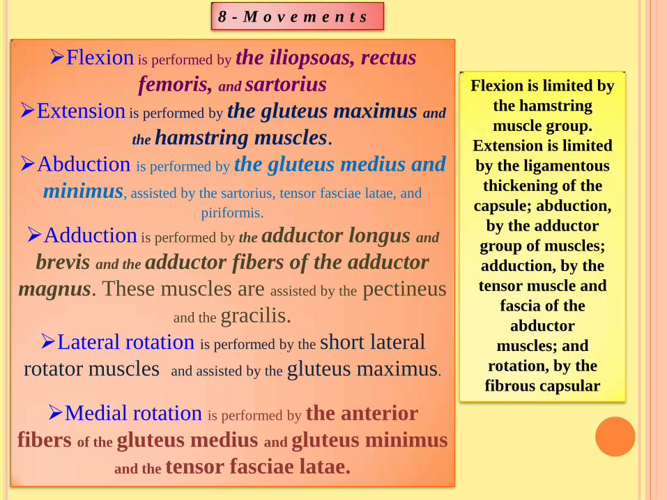
8 - M o v e m e n t s
Flexion is performed by the iliopsoas, rectus
femoris, and sartorius
Extension is performed by the gluteus maximus and
the hamstring muscles.
Abduction is performed by the gluteus medius and
minimus, assisted by the sartorius, tensor fasciae latae, and
piriformis.
Adduction is performed by the adductor longus and
brevis and the adductor fibers of the adductor
magnus. These muscles are assisted by the pectineus
and the gracilis.
Lateral rotation is performed by the short lateral
rotator muscles and assisted by the gluteus maximus.
Medial rotation is performed by the anterior
fibers of the gluteus medius and gluteus minimus
and the tensor fasciae latae.
Flexion is limited by
the hamstring
muscle group.
Extension is limited
by the ligamentous
thickening of the
capsule; abduction,
by the adductor
group of muscles;
adduction, by the
tensor muscle and
fascia of the
abductor
muscles; and
rotation, by the
fibrous capsular
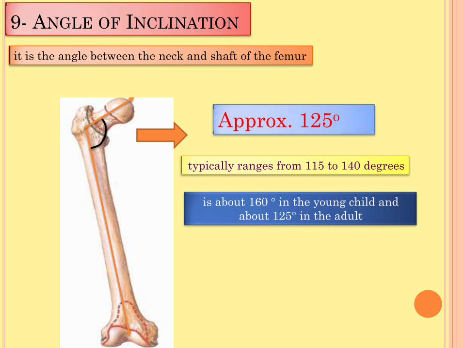
9- ANGLE OF INCLINATION
Approx. 125o
it is the angle between the neck and shaft of the femur
typically ranges from 115 to 140 degrees
is about 160 ° in the young child and
about 125° in the adult
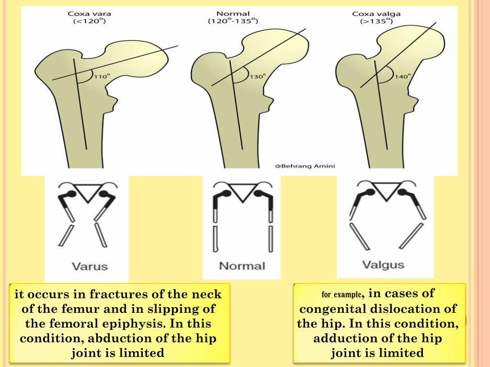
, in cases of for example
congenital dislocation of
the hip. In this condition,
adduction of the hip
joint is limited
it occurs in fractures of the neck
of the femur and in slipping of
the femoral epiphysis. In this
condition, abduction of the hip
joint is limited
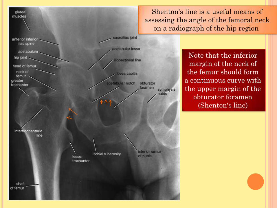
Note that the inferior
margin of the neck of
the femur should form
a continuous curve with
the upper margin of the
obturator foramen
(Shenton's line)
Shenton's line is a useful means of
assessing the angle of the femoral neck
on a radiograph of the hip region

10-There is a pattern of hip injuries;
in old age
fracture of the neck of the femur again becomes the usual
lesion
in adult life the hip dislocates
schoolboys may displace the epiphysis
of the femoral head
In children may sustain
greenstick fractures of the femoral
neck

Dislocation of the hip
The hip is usually dislocated backwards and this is
produced by a force applied along the femoral shaft
with the hip in the flexed position (e.g. the
knee striking against the opposite seat or in car
accedent
The sciatic nerve, is in a close posterior relation with the hip joint
therefore, it is in a danger of
damage in these injuries

K n e e J o i n tIs the most complicated joint in the body
Consists of two condylar joints between:
The medial and lateral condyles of the
femur and The condyles of the tibia
and a gliding joint
between the patella and the patellar surface
of the femur
Note that the fibula is not directly involved in
the joint.
TypeThe joint between the femur and tibia is a
synovial joint of the hinge variety, but
some degree of rotatory movement is possible.
The joint between the patella and femur is a
synovial joint of the plane gliding variety.

Notice that the
lateral condyle of
femur is a bit longer
than the medial
why?!
Lateral condyle of femur
(OUTR)Medial condyle of femur
(INNER)
THE INNER IS THINERTHE OUTER IS STOUTER
prevents lateral dislocation
of the patella
Longer than the medial

Locking mechanism
When standing, the knee joint is 'locked' which reduces the amount of muscle work needed to
maintain the standing position
The locking mechanism is achieved by medial rotation of the femur on the tibia
during extension. Medial rotation and full extension tighten all the associated ligaments
Another feature that keeps the knee extended when standing is that the body's center of
gravity is positioned along a vertical line that passes anterior to the knee joint.
The extended knee is said to be in the
locked position
Before flexion of the knee joint can occur, it is essential that the major ligaments be
untwisted to permit movements between the joint surfaces.
This unlocking or untwisting process is accomplished by the popliteus
muscle, which laterally rotates the femur on the tibia
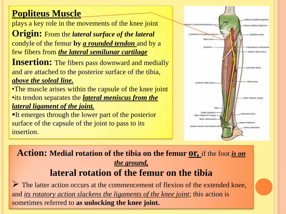
Popliteus Muscleplays a key role in the movements of the knee joint
Origin: From the lateral surface of the lateral
condyle of the femur by a rounded tendon and by a
few fibers from the lateral semilunar cartilage
Insertion: The fibers pass downward and medially
and are attached to the posterior surface of the tibia,
above the soleal line.
•The muscle arises within the capsule of the knee joint
•its tendon separates the lateral meniscus from the
lateral ligament of the joint.
It emerges through the lower part of the posterior
surface of the capsule of the joint to pass to its
insertion.
Action: Medial rotation of the tibia on the femur or, if the foot is on
the ground,
lateral rotation of the femur on the tibia
The latter action occurs at the commencement of flexion of the extended knee,
and its rotatory action slackens the ligaments of the knee joint; this action is
sometimes referred to as unlocking the knee joint.

Capsule1-The capsule is attached to the margins of the
articular surfaces
2- surrounds the sides and posterior aspect of
the joint.
3-On the front of the joint, the capsule is
absent permitting the synovial membrane to
pouch upward beneath the quadriceps tendon,
forming
the suprapatellar bursa 4-On each side of the patella, the capsule is
strengthened by expansions from the tendons
of vastus lateralis and medialis.
5- Behind the joint, the capsule is strengthened
by an expansion of the semimembranous
muscle called the oblique popliteal
ligament 6-An opening in the capsule behind the lateral
tibial condyle permits the tendon of the
popliteus to emerge
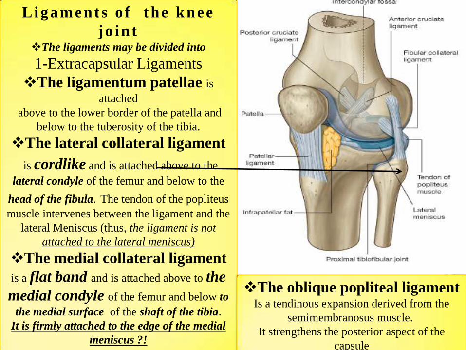
Li gam e nt s o f t he k ne e
j o i n tThe ligaments may be divided into
1-Extracapsular Ligaments
The ligamentum patellae is
attached
above to the lower border of the patella and
below to the tuberosity of the tibia.
The lateral collateral ligament
is cordlike and is attached above to the
lateral condyle of the femur and below to the
head of the fibula. The tendon of the popliteus
muscle intervenes between the ligament and the
lateral Meniscus (thus, the ligament is not
attached to the lateral meniscus)
The medial collateral ligament
is a flat band and is attached above to the
medial condyle of the femur and below to
the medial surface of the shaft of the tibia.
It is firmly attached to the edge of the medial
meniscus ?!
The oblique popliteal ligament Is a tendinous expansion derived from the
semimembranosus muscle.
It strengthens the posterior aspect of the
capsule

2-Intracapsular LigamentsThe cruciate ligamentsThey are named anterior and posterior, according to their
tibial attachments
The cruciate ligaments are the main bond between the femur
and the tibia during the joint's range of movement.
Anterior Cruciate LigamentIs attached to the anterior intercondylar area of the tibia
and passes upward, backward, and laterally, to be attached
to the posterior part of the medial surface of the lateral
femoral condyle
Prevents posterior displacement of the femur
on the tibia. With the knee joint flexed, the anterior cruciate
ligament prevents the tibia from being pulled anteriorly.
Posterior Cruciate LigamentIs attached to the posterior intercondylar area of the
tibia and passes upward, forward, and medially to be
attached to the anterior part of the lateral surface of the
medial femoral condyle
Prevents anterior displacement of the femur
on the tibia. With the knee joint flexed, the posterior cruciate
ligament prevents the tibia from being pulled posteriorly.

MenisciMedial and lateral menisci are C-
shaped sheets of fibrocartilage.
Their function is to deepen the
articular surfaces of the tibial
condyles to receive the convex
femoral condyles;
They also serve as cushions
between the two bones
Each meniscus is attached to the
upper surface of the tibia by
anterior and posterior horns.
Anterior
Posterior

movements of the knee joint
Flexion
The biceps femoris, semitendinosus, and semimembranosus muscles,
assisted by the gracilis, and sartorius, produce flexion.
Flexion is limited by the contact of the back of the leg with the thigh.
Extension
The quadriceps femoris.
Extension is limited by the tension of all the major ligaments of the
joint.
Medial Rotation
The sartorius, gracilis, and semitendinosus
Lateral Rotation
The biceps femoris
Note:
The stability of the knee joint depends on the tone of the strong muscles
acting on the joint and the strength of the ligaments.

Ankle JointType
The ankle is a synovial hinge joint.
Articulation
the lower end of the tibia, the two malleoli, and
the body of the talus
Dorsiflextion is performed by the tibialis
anterior, extensor hallucis longus, extensor
digitorum longus, and peroneus tertius.
(muscles of the anterior compartment of the
leg)
Plantar flexion is performed by the
gastrocnemius, soleus, plantaris, peroneus
longus, peroneus brevis, tibialis posterior,
flexor digitorum longus, and flexor hallucis
longus. (all the muscles of lateral and
posterior compartment except popliteus
muscle)
The medial, or deltoid, ligament
The lateral ligament
Ligaments
Movements
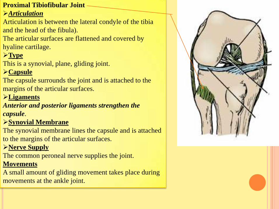
Proximal Tibiofibular Joint
Articulation
Articulation is between the lateral condyle of the tibia
and the head of the fibula).
The articular surfaces are flattened and covered by
hyaline cartilage.
Type
This is a synovial, plane, gliding joint.
Capsule
The capsule surrounds the joint and is attached to the
margins of the articular surfaces.
Ligaments
Anterior and posterior ligaments strengthen the
capsule.
Synovial Membrane
The synovial membrane lines the capsule and is attached
to the margins of the articular surfaces.
Nerve Supply
The common peroneal nerve supplies the joint.
Movements
A small amount of gliding movement takes place during
movements at the ankle joint.
Patellar Dislocations
The patella is a sesamoid bone lying
within the quadriceps tendon. The
importance of the lower horizontal fibers
of the vastus medialis and the large size
of the lateral condyle of the femur in
preventing lateral displacement of the
patella has been emphasized. Congenital
recurrent dislocations of the patella are
caused by underdevelopment of the
lateral femoral condyle. Traumatic
dislocation of the patella results from
direct trauma to the quadriceps
attachments of the patella (especially the
vastus medialis), with or without
fracture of the patella

Distal Tibiofibular Joint
Articulation
Articulation is between the fibular notch at the
lower end of the tibia and the lower end of the
fibula
Type
The distal tibiofibular joint is
a fibrous jointCapsule
There is no capsule.
Ligaments
1-The interosseous ligament is a strong, thick
band of fibrous tissue that binds the two bones
together.
2-The anterior and posterior ligaments are
flat bands of fibrous tissue connecting the two
bones together in front and behind the
interosseous ligament
3-The inferior transverse ligament

Tarsal Joints1-Subtalar JointThe subtalar joint is the posterior joint between the talus and the calcaneum.
Articulation
Articulation is between the inferior surface of the body of the talus and the
facet on the middle of the upper surface of the calcaneum .
Type
These joints are synovial, of the plane variety
Ligaments
Medial and lateral (talocalcaneal) ligaments strengthen the capsule.
The interosseous (talocalcaneal) ligament is strong and is the main bond of
union between the two bones. It is attached above to the sulcus tali and below
to the sulcus calcanei..
Movements
Gliding and rotatory movements are possible

Talocalcaneonavicular Joint-2is the anterior joint between the talus and the calcaneum and also
involves the navicular bone
Articulation
Articulation is between the rounded head of the talus, the upper
surface of the sustentaculum tali, and the posterior concave
surface of the navicular bone.
Type
The joint is a synovial joint..
Ligaments
is strong and runs from ligament calcaneonavicularThe plantar
the anterior margin of the sustentaculum tali to the inferior surface
and tuberosity of the navicular bone. The superior surface of the
ligament is covered with fibrocartilage and supports the head of
the talus..
Movements
Gliding and rotatory movements are possible.
Calcaneocuboid Joint-3Articulation
Articulation is between the anterior end of the calcaneum and the
posterior surface of the cuboid
The calcaneocuboid joint is synovial, of the plane variety.
Ligaments
The bifurcated ligament