joints of the lower limb i - medicinebau.com...joints of the lower limb : •associated ligaments...
TRANSCRIPT

Joints of the Lower Limb I
Done by: Dina Sawadha
& Mohammad Abukabeer
كالم الدكتور المضاف تحته خط

1-Sacroiliac Joint
• Auricular surfaces of the sacrum and the iliac bone
• No movement; transmit body weight from vertebral column to pelvis (immobile joint)
• In elderly people synovial cavity disappear and becomes fibrous joint
• Nerve supply: sacral spinal nerves
• Mixed joint (fibrous & synovial)
• Auricular surface is covered with hyline cartilage (synovial part of the joint)
• Tuberosities are connected to each other by interosseous ligaments (fibrous part of the joint)
• Only during delivery , under the control of certain hormones there’ll be relaxation in the ligaments granting some mobility to the joint
Joints of the lower limb :

• Associated ligaments • Posterior sacroiliac ligament
• Interosseous sacroiliac ligament (between tuberosities of sacrum and iliac bone)
• Anterior sacroiliac ligament
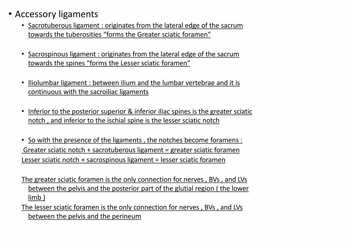
• Accessory ligaments • Sacrotuberous ligament : originates from the lateral edge of the sacrum
towards the tuberosities “forms the Greater sciatic foramen“
• Sacrospinous ligament : originates from the lateral edge of the sacrum towards the spines “forms the Lesser sciatic foramen“
• Iliolumbar ligament : between ilium and the lumbar vertebrae and it is continuous with the sacroiliac ligaments
• Inferior to the posterior superior & inferior iliac spines is the greater sciatic notch , and inferior to the ischial spine is the lesser sciatic notch
• So with the presence of the ligaments , the notches become foramens :
Greater sciatic notch + sacrotuberous ligament = greater sciatic foramen
Lesser sciatic notch + sacrospinous ligament = lesser sciatic foramen
The greater sciatic foramen is the only connection for nerves , BVs , and LVs between the pelvis and the posterior part of the glutial region ( the lower limb )
The lesser sciatic foramen is the only connection for nerves , BVs , and LVs between the pelvis and the perineum

2-Hip Joint :
• Synovial joint
• The depth of the acetabulum is increased by the acetabular labrum (rim)
• Acetabular labrum is a C-shaped ring , and the discontinuation is located inferiorly and called “acetabular notch” and is completed by the transverse acetabular ligament , and the space under the ligament will be a pathway for the vital components to nourish the joint structures
• Head of the femur and the acetabulum • Type : Ball and socket joint
• Movements : All movements Most movable joint next to shoulder
• On standing transmits body weight through hip bone to head & neck of femur
• Nerve supply : femoral, obturator, and sciatic nerves and nerve to the
quadratus femoris muscle

Components of the hip joint : • Head of femur : Fovea for lig. of femoral head , No
articular cartilage
• Neck of femur
• Acetabulum : Lunate surface ,Articular part • Acetabular fossa : No articular part , Filled with adipose
tissue (fat pad)
• Acetabular rim : with theTransverse acetabular ligament
forms Acetabular notch

Hip Joint: Capsule
• Proximal attachment • Acetabular labrum
• Transverse ligament
• Distal attachment • Anteriorly
• Intertrochanteric line
• Posteriorly • Free edge
Attaches at the external edge of the acetabulum (proximal attachment) and The neck of femur anteriorly (distal attachment) On the posterior side of neck of femur there is a free edge that the capsule will not attach to .

Hip Joint: Ligaments
• Intracapsular ligaments : increase the fixation (strength) of the joint ; • Transverse acetabular ligament
• Bridge the notch
• Entrance of BVs
• Ligament of the head of the femur • Transverse lig. & edges of notch
• Fovea capitis
Most joints don’t have intracapsular ligaments

• Extracapsular ligaments : thickened strong ligaments
surrond the capsule all around , they limit the hyperextension because they all have spiral fibers
A- Iliofemoral ligament : huge & the Strongest , inverted
Y‐shape
• Attachments
• Anterior inferior iliac spine (AIIS) “origin”
• Intertrochanteric line (ITL) “insertion”
• Covers the Superior and anterior areas
• Function : Prevent hyperextension during standing
B- Pubofemoral ligament : • Triangular shape
• Attachments
• Superior ramus of pubis “origin”
• Inferior part of intertrochanteric line “insertion”
• Function
• Limits extension and abduction
• Covers the Anterior and inferior areas

C- Ischiofemoral ligament : • The weakest because of the presence of the strong muscles
• Spiral shape
• Attachments
• Ischial part of acetabular rim “origin”
• Greater trochanter “insertion”
• Limits extension and medial rotation

Hip Joint: Ligaments
• Relative strength of ligaments compared to muscles
• Anteriorly
• Strong ligament and weak muscles
• Posteriorly • Weak ligament and strong muscles

Hip Joint : Synovial Membrane
• Attached to articular surfaces
• Lines fibrous capsule
• Completely inclose the synovial fluid inside the synovial cavity
• Ligaments are included in the capsule , but not in the synovial cavity
• Covers • Transverse lig.
• Ligament of head of the femur
• Pad of fat in acetabular fossa
• Neck of femur
• Synovial fold (retinaculum) : thickening of the synovial membrane at the free edge of the capsule posteriorly
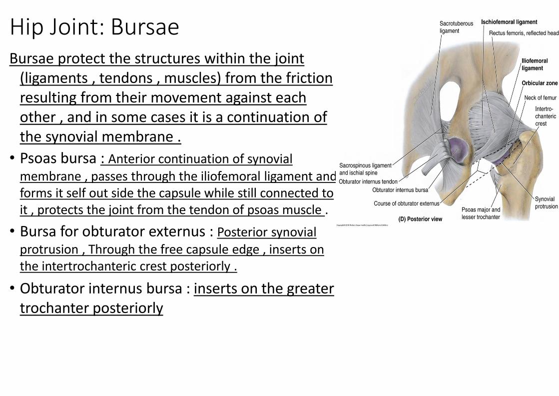
Hip Joint: Bursae Bursae protect the structures within the joint
(ligaments , tendons , muscles) from the friction resulting from their movement against each other , and in some cases it is a continuation of the synovial membrane .
• Psoas bursa : Anterior continuation of synovial membrane , passes through the iliofemoral ligament and forms it self out side the capsule while still connected to it , protects the joint from the tendon of psoas muscle .
• Bursa for obturator externus : Posterior synovial protrusion , Through the free capsule edge , inserts on the intertrochanteric crest posteriorly .
• Obturator internus bursa : inserts on the greater trochanter posteriorly

Hip Joint: Movements
Muscles الدكتور حكى اتركوها لبعد ال

Hip Joint: Relations
• Anteriorly : • Content of femoral triangle :
Femoral artery , vein and nerve
• Posteriorly : Sciatic nerve is close to the joint
and supplies all of the lower limb
• Superiorly :
gluteus muscles
• Inferiorly :
mostly obturator externus
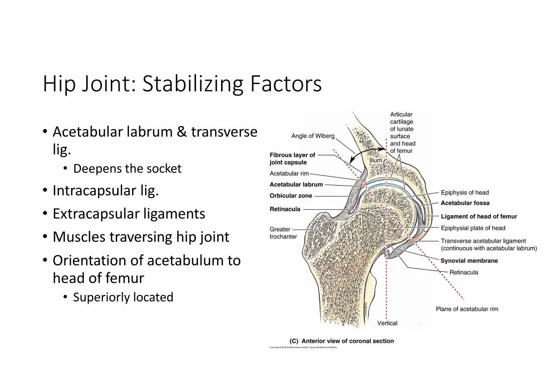
Hip Joint: Stabilizing Factors
• Acetabular labrum & transverse lig. • Deepens the socket
• Intracapsular lig.
• Extracapsular ligaments
• Muscles traversing hip joint
• Orientation of acetabulum to head of femur • Superiorly located

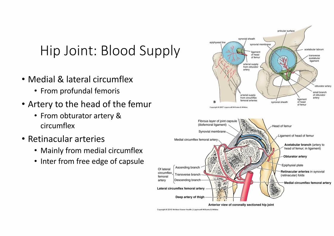
Hip Joint: Blood Supply
• Medial & lateral circumflex • From profundal femoris
• Artery to the head of the femur • From obturator artery &
circumflex
• Retinacular arteries • Mainly from medial circumflex
• Inter from free edge of capsule
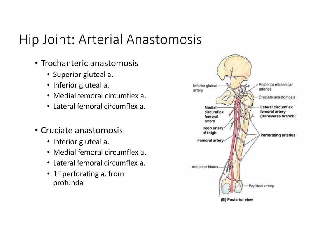
Hip Joint: Arterial Anastomosis
• Trochanteric anastomosis • Superior gluteal a.
• Inferior gluteal a.
• Medial femoral circumflex a.
• Lateral femoral circumflex a.
• Cruciate anastomosis
• Inferior gluteal a.
• Medial femoral circumflex a.
• Lateral femoral circumflex a.
• 1st perforating a. from profunda

Hip Joint: Fracture • Fracture of femoral neck
• Disruption of blood supply to the head
• Avascular necrosis • Blood supply from artery to the head of the femur usually is not enough
• In elderly
• Female > Male • Osteoporosis

Surgical Hip Replacement
• In traumatic injuries or degenerative diseases
• Replace head and neck of femur
• Often acetabulum lined by metal or plastic socket
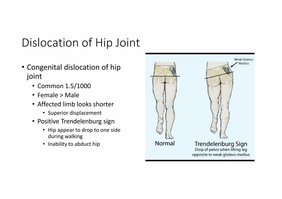
Dislocation of Hip Joint
• Congenital dislocation of hip joint • Common 1.5/1000
• Female > Male
• Affected limb looks shorter • Superior displacement
• Positive Trendelenburg sign • Hip appear to drop to one side
during walking
• Inability to abduct hip
Dislocation of Hip Joint
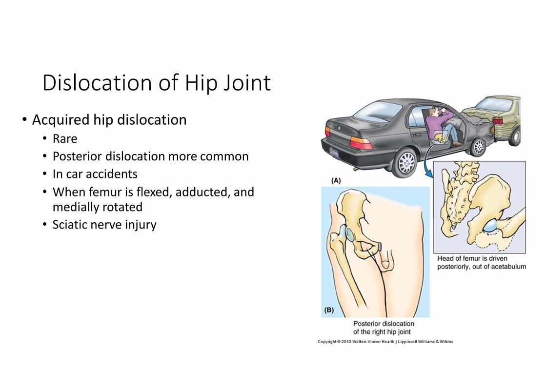
• Acquired hip dislocation • Rare
• Posterior dislocation more common
• In car accidents
• When femur is flexed, adducted, and medially rotated
• Sciatic nerve injury
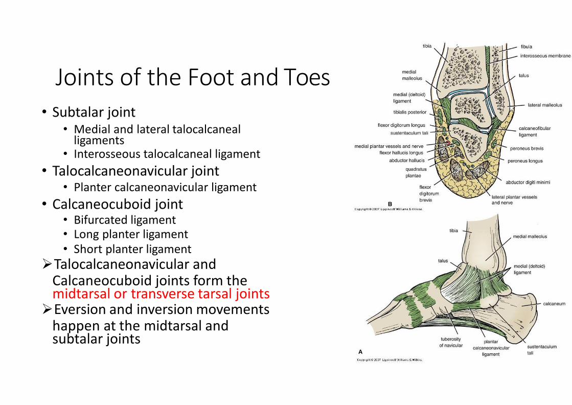
Joints of the Foot and Toes • Subtalar joint
• Medial and lateral talocalcaneal ligaments
• Interosseous talocalcaneal ligament
• Talocalcaneonavicular joint • Planter calcaneonavicular ligament
• Calcaneocuboid joint • Bifurcated ligament • Long planter ligament • Short planter ligament
Talocalcaneonavicular and Calcaneocuboid joints form the midtarsal or transverse tarsal joints
Eversion and inversion movements happen at the midtarsal and subtalar joints

Joints of the Foot and Toes
• Cuneonavicular joint • Dorsal and planter ligaments
• Cuboideonavicular joint • Dorsal, planter and interosseous ligaments
• Intercuneiform and cuneocuboid joints • Dorsal, planter and interosseous ligaments • Same cavity with cuneonavicular joint
• Tarsometatarsal and intermetatarsal joints • Dorsal, planter and interosseous ligaments • For big toe there is separate cavity
• Metatarsophalangeal and interphalangeal joints • Deep transverse ligaments