joint and msk ultrasound - cme.dmu.edu
TRANSCRIPT
Joint and MSK UltrasoundTodd M Sexton, DO
UnityPoint Health- Des Moines

Disclosure
• I do not have any relevant financial conflicts with commercial interest companies to disclose.

About MeI am an emergency medicine physician practicing in Des Moines, with UnityPoint Health. I am a Des Moines native and returned to the area in 2019. I completed my emergency medicine residency at the University of Iowa, medical school at KCOM in Kirksville, Missouri.
I assisted in development of new ultrasound curriculum at the University of Iowa EM program, and nursing US IV access at UIHC. Completed US based research in fluid resuscitation and sepsis.

Shoulder UltrasoundDiagnosis and Joint Injections
Shoulder Dislocations
• Ultrasound can be used to visualize the glenohumeral joint and AC joint
• Curvilinear or Linear probe can be used• Exam is best performed on the posterior aspect, place your
screen in front of the patient if possible• Limitations include that you are not identifying possible Hill-
Sachs or Bankart deformities
Shoulder Anatomy
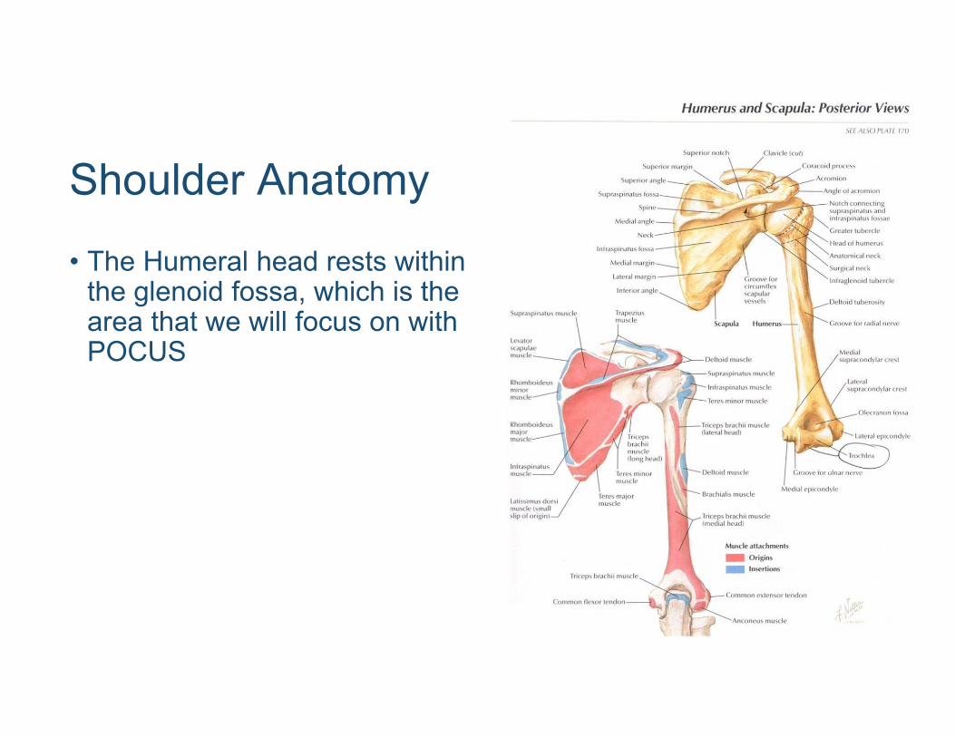
• The Humeral head rests within the glenoid fossa, which is the area that we will focus on with POCUS
Shoulder Anatomy
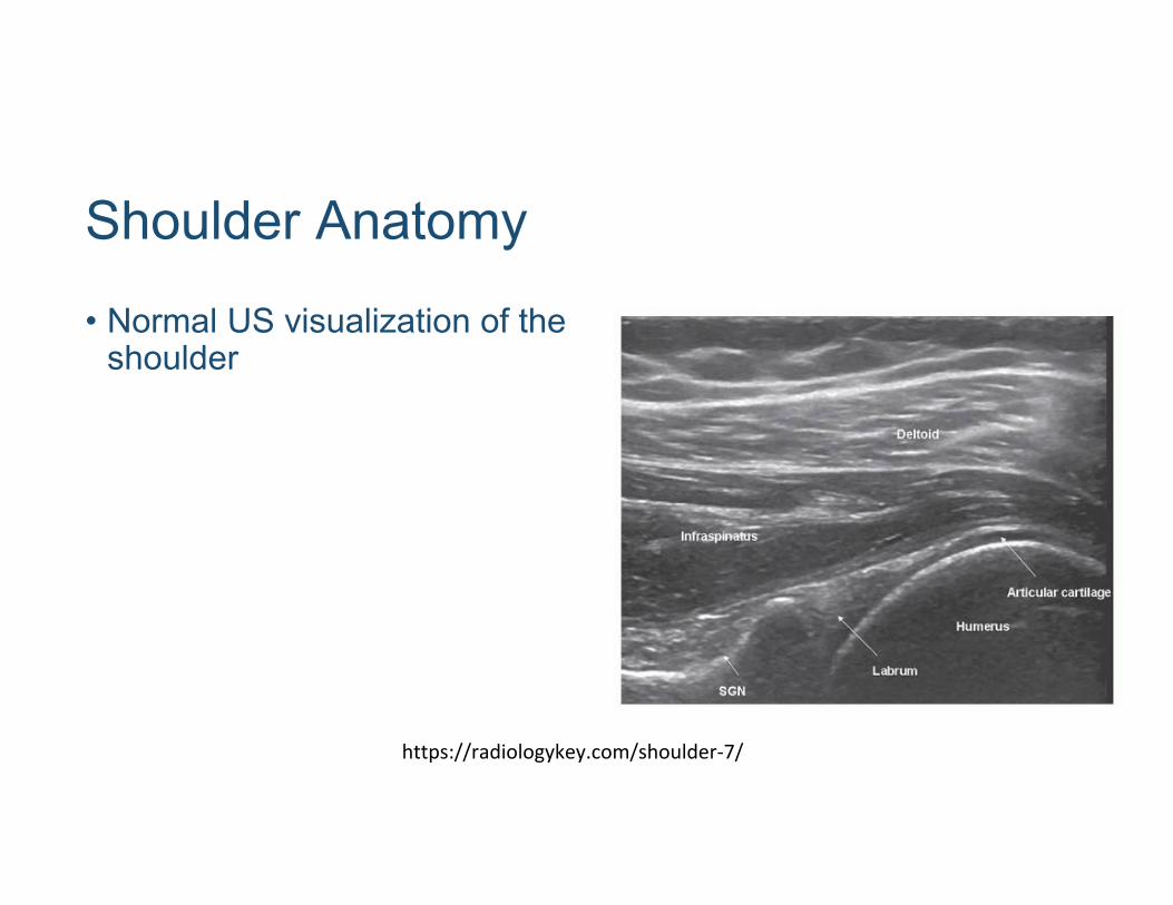
• Normal US visualization of the shoulder
https://radiologykey.com/shoulder‐7/

Shoulder Anatomy
• Anterior Glenohumeral dislocation
http://brownemblog.com/blog‐1/2016/11/30/pocus‐shoulder‐dislocation
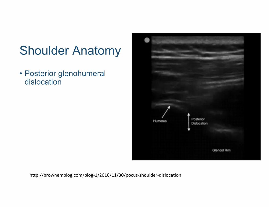
Shoulder Anatomy
• Posterior glenohumeral dislocation
http://brownemblog.com/blog‐1/2016/11/30/pocus‐shoulder‐dislocation
Shoulder Dislocations
• Often procedural sedation is employed for reduction of glenohumeral dislocations
• Alternatively, intraarticular lidocaine can be administered with improved pain control
• This reduces many of the time and labor intensive aspects of procedural sedation

Intra-Articular LidocainePosition yourself in the same manner that you would to visualize the joint
Ensure that you have a long needle, 22G is preferred (you may need a spinal needle)
As always, prepare your injection site
Patient’s may feel a sharp pain as you enter the joint space
Anesthetic should freely flow when you are in the joint space without resistance


AC Separation
• The acromioclavicular joint is easily identified with the linear probe as it is relatively superficial in most patients
• As with the shoulder you can also administer anesthetic into this joint for pain control in these patients
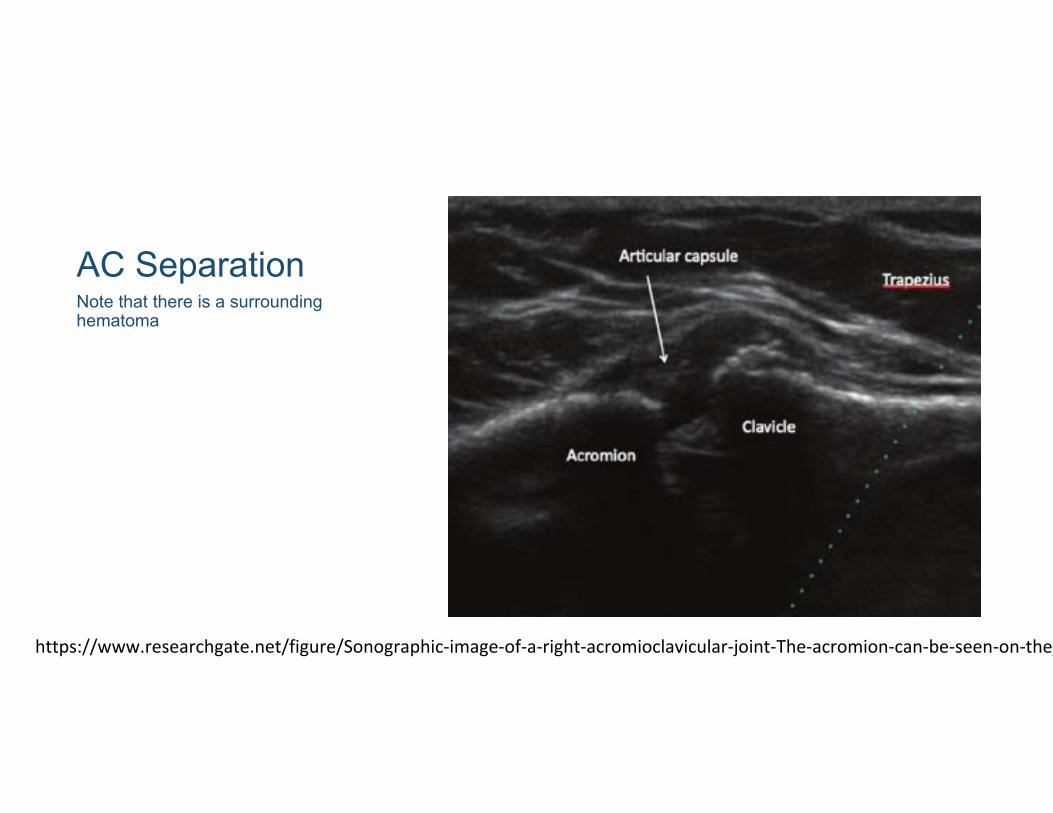
AC SeparationNote that there is a surrounding hematoma
https://www.researchgate.net/figure/Sonographic‐image‐of‐a‐right‐acromioclavicular‐joint‐The‐acromion‐can‐be‐seen‐on‐the_
Knee UltrasoundDiagnostics and arthrocentesis
Knee Trauma
• Knee trauma can result in multiple different pathologies• Ultrasound can help give real time visualization of anatomy that
is not identified on plain films• POCUS can also aid in identifying occult fractures such as tibial
plateau fractures

Knee AnatomyQuadriceps are divided into four muscles which join together to insert onto the patella
Quadriceps tendon tears usually occur just above the insertion
Less common than a patellar tendon tear
https://coreem.net/core/quadriceps‐tendon‐rupture/
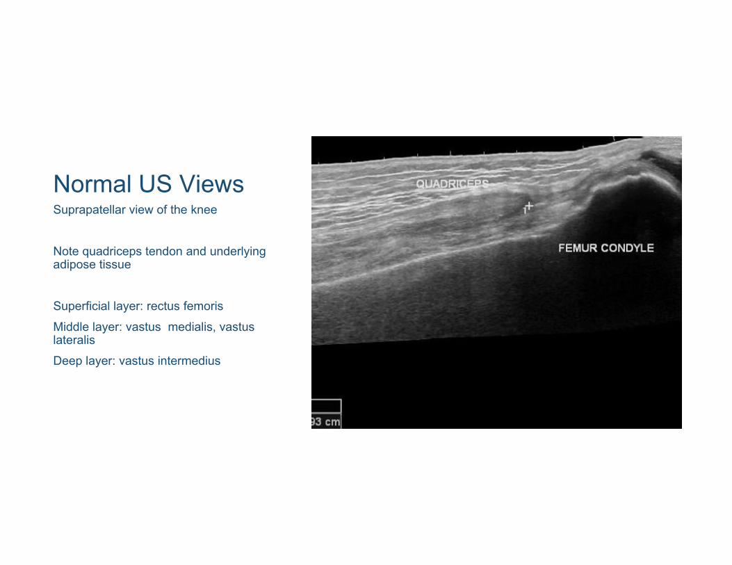
Normal US ViewsSuprapatellar view of the knee
Note quadriceps tendon and underlying adipose tissue
Superficial layer: rectus femorisMiddle layer: vastus medialis, vastus lateralis Deep layer: vastus intermedius

Quadriceps Tendon RuptureUltrasound can provide direct visualization of a tendon rupture, and sometime an associated hematoma
https://coreem.net/core/quadriceps‐tendon‐rupture/

Patellar Tendon RupturePatellar tendon ruptures also frequently occur near the attachment of the inferior pole of the patella
https://coreem.net/core/patella‐tendon‐rupture/
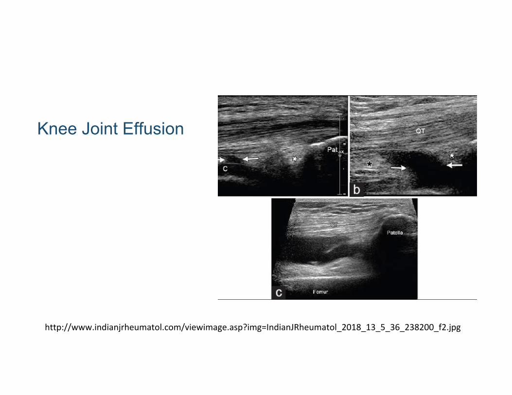
Knee Joint Effusion
http://www.indianjrheumatol.com/viewimage.asp?img=IndianJRheumatol_2018_13_5_36_238200_f2.jpg

Knee arthrocentesisLateral access to the knee joint can be easily obtained with palpating the patella and joint recess
Suprapatellar access can also be easily obtained using a linear transducerNeedle entrance through the potential space lateral to the quadriceps tendon
http://www.indianjrheumatol.com/viewimage.asp?img=IndianJRheumatol_2018_13_5_36_238200_f6.jpg

Hip Ultrasound

Common Hip Pathologies
• POCUS is particularly useful in pediatric patients• Can be used to evaluate for effusions, and to a lesser degree,
bony abnormalities• Transient synovitis and septic arthritis

Normal Hip POCUSPlace the patient supineUse the linear or curvelinear probePlace the probe in the sagittal plane and move superiority until you identify the femoral head
https://www.acep.org/how‐we‐serve/sections/emergency‐ultrasound/news/april‐2018/tips‐‐tricks‐ultrasound‐in‐the‐diagnosis‐o
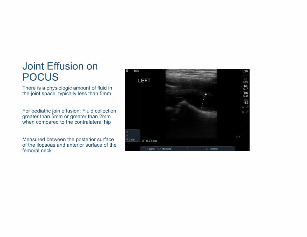
Joint Effusion on POCUSThere is a physiologic amount of fluid in the joint space, typically less than 5mm
For pediatric join effusion: Fluid collection greater than 5mm or greater than 2mm when compared to the contralateral hip
Measured between the posterior surface of the ilopsoas and anterior surface of the femoral neck
Ankle Ultrasound
Ankle Ultrasound Utilization
• Identify Achilles’ tendon rupture• Identify joint effusions
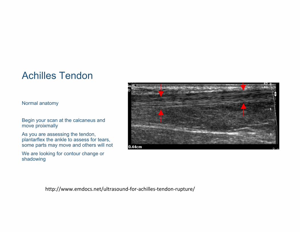
Achilles Tendon
Normal anatomy
Begin your scan at the calcaneus and move proixmallyAs you are assessing the tendon, plantarflex the ankle to assess for tears, some parts may move and others will notWe are looking for contour change or shadowing
http://www.emdocs.net/ultrasound‐for‐achilles‐tendon‐rupture/
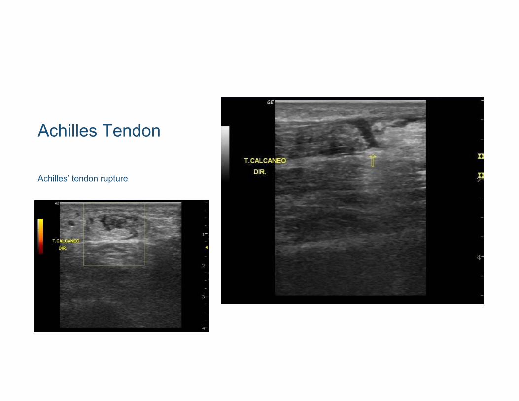
Achilles Tendon
Achilles’ tendon rupture

Ankle Arthocentesis
Place the foot in slight plantarflexionSlide the probe distally along the tibia in sagittal orientation, identify the tibialis anterior tendonVisualize the tibial-talar joint spaceUse a medial to lateral approach with your needle (you may use in-plane if possible)
http://highlandultrasound.com/ankle‐arthrocentesis

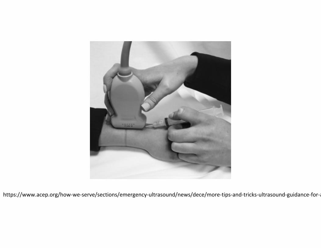
Wrist ArthocentesisBONUS!
Wrists can be difficult to obtain synovial fluid from, and can frequently have a dry tap.
Place the patient with their palm downProbe will be sagittal over the distal radiusIdentify the joint space between the radius and scaphoid/lunateAdvance your needle in plane
https://www.acep.org/how‐we‐serve/sections/emergency‐ultrasound/news/dece/more‐tips‐and‐tricks‐ultrasound‐guidance‐for‐a
https://www.acep.org/how‐we‐serve/sections/emergency‐ultrasound/news/dece/more‐tips‐and‐tricks‐ultrasound‐guidance‐for‐a

Foreign Body Retrieval
Foreign Body Retrieval
• FBs are a common ED complaint that can result in a relatively simple and efficient disposition
• POCUS can aid in identifying these FBs and removing them• Not all FBs are radio-opaque, but may be visualized with US• Real time investigation of soft tissues

Foreign Body Retrieval
• Wood splinters are one example of objects which may not appear on plain films
• US can investigate the area while physical exam is being performed and can aid in real time visualization of retrieval
• Less trauma as we are not searching blindly
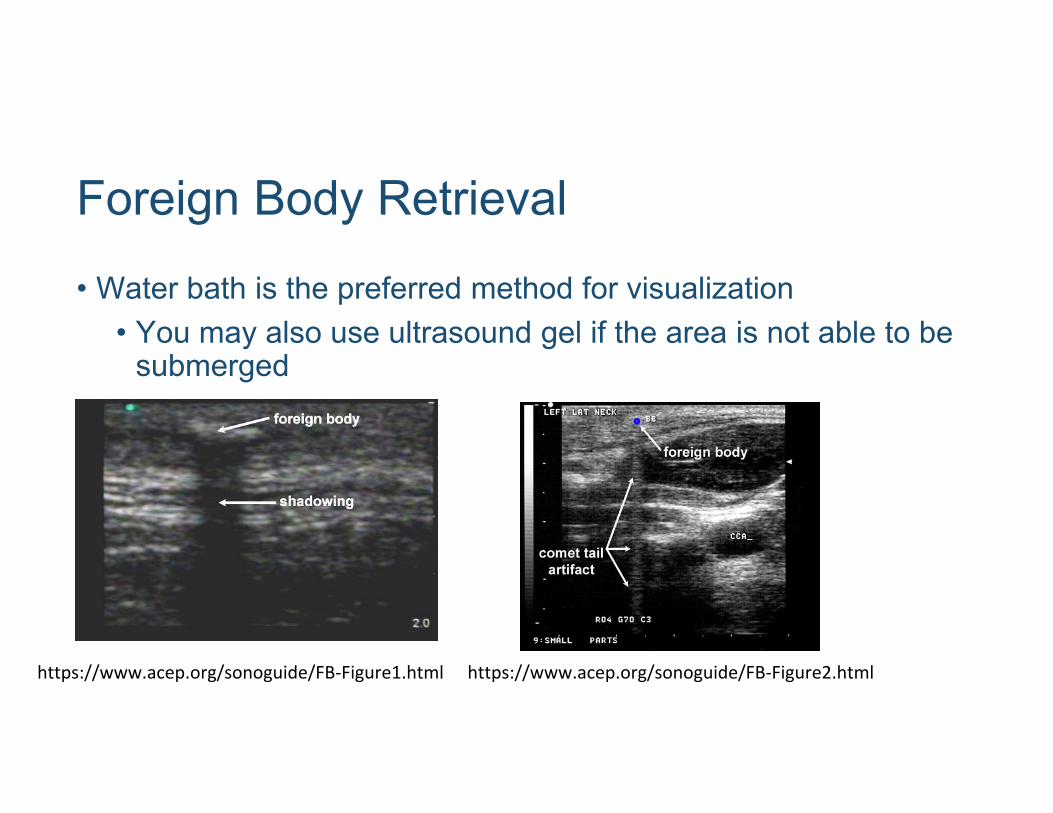
Foreign Body Retrieval
• Water bath is the preferred method for visualization• You may also use ultrasound gel if the area is not able to be
submerged
https://www.acep.org/sonoguide/FB‐Figure1.html https://www.acep.org/sonoguide/FB‐Figure2.html

Soft Tissue InfectionsCellulitis, abscess, necrotizing infections
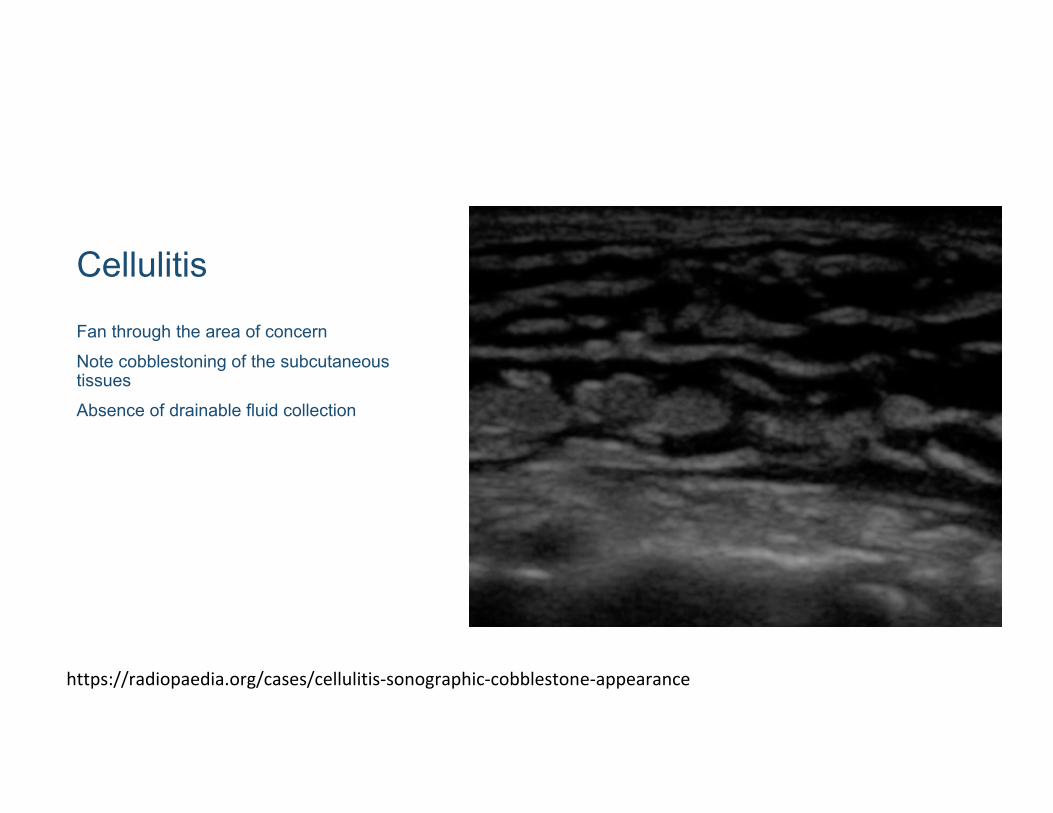
Cellulitis
Fan through the area of concernNote cobblestoning of the subcutaneous tissuesAbsence of drainable fluid collection
https://radiopaedia.org/cases/cellulitis‐sonographic‐cobblestone‐appearance
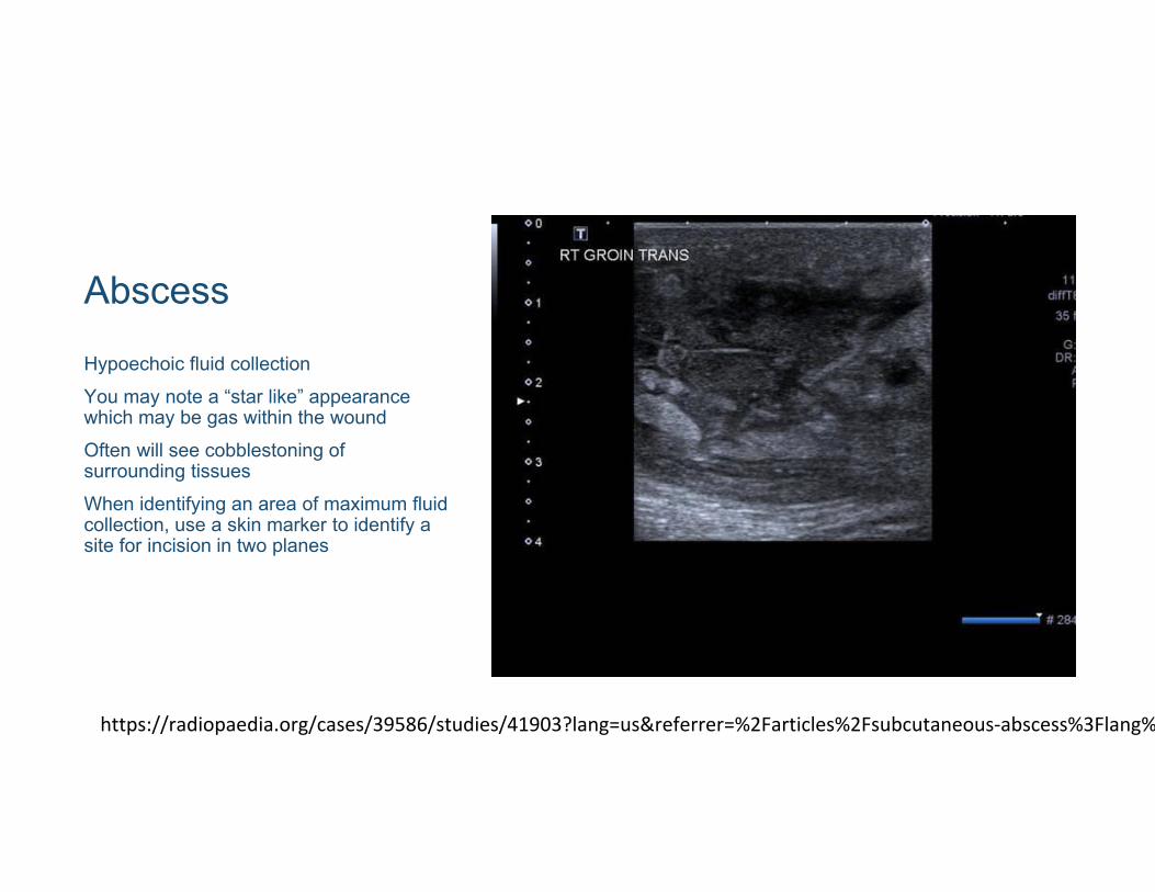
Abscess
Hypoechoic fluid collectionYou may note a “star like” appearance which may be gas within the woundOften will see cobblestoning of surrounding tissuesWhen identifying an area of maximum fluid collection, use a skin marker to identify a site for incision in two planes
https://radiopaedia.org/cases/39586/studies/41903?lang=us&referrer=%2Farticles%2Fsubcutaneous‐abscess%3Flang%
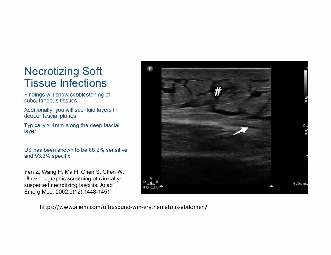
Necrotizing Soft Tissue InfectionsFindings will show cobblestoning of subcutaneous tissues Additionally, you will see fluid layers in deeper fascial planesTypically > 4mm along the deep fascial layer
US has been shown to be 88.2% sensitive and 93.3% specific
Yen Z, Wang H, Ma H, Chen S, Chen W. Ultrasonographic screening of clinically-suspected necrotizing fasciitis. Acad Emerg Med. 2002;9(12):1448-1451.
https://www.aliem.com/ultrasound‐win‐erythematous‐abdomen/

Questions?

Thank you!Questions? Comments? Email: [email protected]