joint acne image grading and counting via label...
TRANSCRIPT

Joint Acne Image Grading and Counting via Label Distribution Learning
Xiaoping Wu1∗, Ni Wen2∗, Jie Liang1, Yu-Kun Lai3, Dongyu She1, Ming-Ming Cheng1, Jufeng Yang1☞
1College of Computer Science, Nankai University 2Beijing Tsinghua Changgung Hospital3School of Computer Science and Informatics, Cardiff University
[email protected], [email protected], [email protected], [email protected],
[email protected], {cmm, yangjufeng}@nankai.edu.cn
Abstract
Accurate grading of skin disease severity plays a crucial
role in precise treatment for patients. Acne vulgaris, the
most common skin disease in adolescence, can be graded
by evidence-based lesion counting as well as experience-
based global estimation in the medical field. However, due
to the appearance similarity of acne with close severity,
it is challenging to count and grade acne accurately. In
this paper, we address the problem of acne image analy-
sis via Label Distribution Learning (LDL) considering the
ambiguous information among acne severity. Based on the
professional grading criterion, we generate two acne label
distributions considering the relationship between the sim-
ilar number of lesions and severity of acne, respectively.
We also propose a unified framework for joint acne image
grading and counting, which is optimized by the multi-task
learning loss. In addition, we further build the ACNE04
dataset with annotations of acne severity and lesion number
of each image for evaluation. Experiments demonstrate that
our proposed framework performs favorably against state-
of-the-art methods. We make the code and dataset publicly
available at https://github.com/xpwu95/ldl.
1. Introduction
Automatic grading of skin disease severity is of great im-
portance in the medical field. Acne vulgaris, commonly
named acne, is the most common skin disease, which has
prevalence peaks during adolescence [28, 33]. About 80%adolescents suffer from acne [9] and the symptoms last
through adulthood in 3% men and 12% women [23]. There-
fore, there are a massive number of acne patients that need
specific treatment imminently, since acne may also leave
scars and pigmentation and often leads to considerable in-
ferior and depressed emotions [48]. The severity of acne
∗Equal contributions
Figure 1. Image examples (a) and their corresponding label distri-
butions (b, c). The x-axes in (b, c) indicate the number of lesions
and acne severity, respectively. The label distributions cover sev-
eral neighboring labels which represent the degree each label de-
scribes the instance (denoted as “D”). Acne images can be divided
into different severity levels according to the lesion numbers [24]
(d). Different colors indicate different severity levels, e.g., blue is
for mild, and green is for moderate.
is essential for dermatologists to make a precise and stan-
dardized treatment decision [24]. Besides, junior dermatol-
ogists also need an objective and reliable diagnosis for refer-
ence. A standard criterion used by dermatologists for grad-
ing acne severity is the Hayashi criterion [24], which com-
bines the outcome measures of lesion counting and global
assessment. Specifically, acne can be graded into four lev-
els of severity, i.e. mild, moderate, severe, and very severe,
according to the number of lesions.
In the past years, substantial advances have been made
for acne lesion analysis [2, 6, 16]. Most methods indi-
rectly focus on the classification or detection of acne le-
sions and generally rely on hand-crafted features. For ex-
ample, Abas et al. [1] employ discrete wavelet frames and
gray-level co-occurrence matrices to extract features for de-
tecting acne lesions. Recently, Convolutional Neural Net-
10642
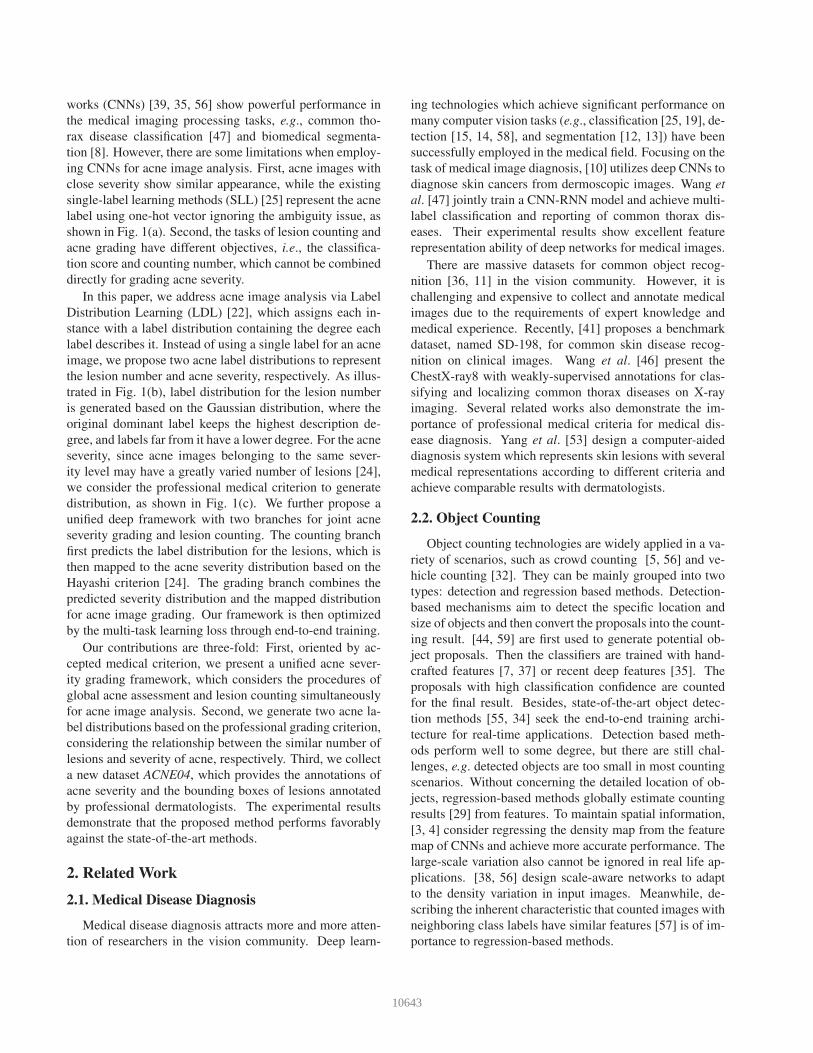
works (CNNs) [39, 35, 56] show powerful performance in
the medical imaging processing tasks, e.g., common tho-
rax disease classification [47] and biomedical segmenta-
tion [8]. However, there are some limitations when employ-
ing CNNs for acne image analysis. First, acne images with
close severity show similar appearance, while the existing
single-label learning methods (SLL) [25] represent the acne
label using one-hot vector ignoring the ambiguity issue, as
shown in Fig. 1(a). Second, the tasks of lesion counting and
acne grading have different objectives, i.e., the classifica-
tion score and counting number, which cannot be combined
directly for grading acne severity.
In this paper, we address acne image analysis via Label
Distribution Learning (LDL) [22], which assigns each in-
stance with a label distribution containing the degree each
label describes it. Instead of using a single label for an acne
image, we propose two acne label distributions to represent
the lesion number and acne severity, respectively. As illus-
trated in Fig. 1(b), label distribution for the lesion number
is generated based on the Gaussian distribution, where the
original dominant label keeps the highest description de-
gree, and labels far from it have a lower degree. For the acne
severity, since acne images belonging to the same sever-
ity level may have a greatly varied number of lesions [24],
we consider the professional medical criterion to generate
distribution, as shown in Fig. 1(c). We further propose a
unified deep framework with two branches for joint acne
severity grading and lesion counting. The counting branch
first predicts the label distribution for the lesions, which is
then mapped to the acne severity distribution based on the
Hayashi criterion [24]. The grading branch combines the
predicted severity distribution and the mapped distribution
for acne image grading. Our framework is then optimized
by the multi-task learning loss through end-to-end training.
Our contributions are three-fold: First, oriented by ac-
cepted medical criterion, we present a unified acne sever-
ity grading framework, which considers the procedures of
global acne assessment and lesion counting simultaneously
for acne image analysis. Second, we generate two acne la-
bel distributions based on the professional grading criterion,
considering the relationship between the similar number of
lesions and severity of acne, respectively. Third, we collect
a new dataset ACNE04, which provides the annotations of
acne severity and the bounding boxes of lesions annotated
by professional dermatologists. The experimental results
demonstrate that the proposed method performs favorably
against the state-of-the-art methods.
2. Related Work
2.1. Medical Disease Diagnosis
Medical disease diagnosis attracts more and more atten-
tion of researchers in the vision community. Deep learn-
ing technologies which achieve significant performance on
many computer vision tasks (e.g., classification [25, 19], de-
tection [15, 14, 58], and segmentation [12, 13]) have been
successfully employed in the medical field. Focusing on the
task of medical image diagnosis, [10] utilizes deep CNNs to
diagnose skin cancers from dermoscopic images. Wang et
al. [47] jointly train a CNN-RNN model and achieve multi-
label classification and reporting of common thorax dis-
eases. Their experimental results show excellent feature
representation ability of deep networks for medical images.
There are massive datasets for common object recog-
nition [36, 11] in the vision community. However, it is
challenging and expensive to collect and annotate medical
images due to the requirements of expert knowledge and
medical experience. Recently, [41] proposes a benchmark
dataset, named SD-198, for common skin disease recog-
nition on clinical images. Wang et al. [46] present the
ChestX-ray8 with weakly-supervised annotations for clas-
sifying and localizing common thorax diseases on X-ray
imaging. Several related works also demonstrate the im-
portance of professional medical criteria for medical dis-
ease diagnosis. Yang et al. [53] design a computer-aided
diagnosis system which represents skin lesions with several
medical representations according to different criteria and
achieve comparable results with dermatologists.
2.2. Object Counting
Object counting technologies are widely applied in a va-
riety of scenarios, such as crowd counting [5, 56] and ve-
hicle counting [32]. They can be mainly grouped into two
types: detection and regression based methods. Detection-
based mechanisms aim to detect the specific location and
size of objects and then convert the proposals into the count-
ing result. [44, 59] are first used to generate potential ob-
ject proposals. Then the classifiers are trained with hand-
crafted features [7, 37] or recent deep features [35]. The
proposals with high classification confidence are counted
for the final result. Besides, state-of-the-art object detec-
tion methods [55, 34] seek the end-to-end training archi-
tecture for real-time applications. Detection based meth-
ods perform well to some degree, but there are still chal-
lenges, e.g. detected objects are too small in most counting
scenarios. Without concerning the detailed location of ob-
jects, regression-based methods globally estimate counting
results [29] from features. To maintain spatial information,
[3, 4] consider regressing the density map from the feature
map of CNNs and achieve more accurate performance. The
large-scale variation also cannot be ignored in real life ap-
plications. [38, 56] design scale-aware networks to adapt
to the density variation in input images. Meanwhile, de-
scribing the inherent characteristic that counted images with
neighboring class labels have similar features [57] is of im-
portance to regression-based methods.
10643

Figure 2. Pipeline of the proposed framework. The input image is resized and passed through the CNN backbone model (ResNet-50 [25]).
Then the framework is divided into two branches. The grading branch globally estimates the severity of acne. The counting branch first
predicts the label distribution of acne lesion count. Then it is converted into the label distribution of acne severity based on Eq. 3. The
counting model simultaneously grades the acne severity and predicts the lesion number to provide acne diagnosis evidence. Finally, the
prediction results from global grading and local counting models are merged, oriented by the medical criterion.
2.3. Label Distribution Learning
To cope with the issue of label ambiguity existing in
traditional single label learning, Geng et al. [22, 45] pro-
pose a new machine learning paradigm, i.e., label distribu-
tion learning. Instead of assigning a single label [25], LDL
covers a certain number of adjacent labels where each la-
bel represents different description degree to the instance,
respectively. Recently, LDL has been used to well ad-
dress label ambiguities in various tasks, e.g., age estima-
tion [26, 27, 54], head pose estimation [21], and visual sen-
timent analysis [51, 52]. Many methods [49, 40, 18, 50]
successfully employ the Gaussian function to generate la-
bel distribution. Geng et al. [20] propose adaptive label
distribution learning (ALDL) to generate label distributions
with different shapes for each age, i.e., different variance
parameter in the Gaussian function for each class. In this
particular scenario that the aging process varies at different
aging stages, the ALDL performs well with sufficient la-
beled training data. Subsequently, Hou et al. [27] propose
a semi-supervised ALDL method that utilizes the unlabeled
data to solve the data-scale problem. In addition, Gao et
al. [17] employ deep CNNs for label distribution learning
by minimizing the KL divergence between the predicted
and ground-truth distributions. In the multi-task learning
scenario, our framework also explores the latent fusion of
multiple tasks in both training and testing phases.
3. Method
Fig. 2 illustrates the pipeline of our proposed method
with two branches for joint acne severity grading and le-
sion counting. Given N input training images with corre-
sponding single labels of acne severity and ground-truths
of lesion counts {(x1, y1, z1), · · · , (xN , yN , zN )}, where
yi ∈ [1, · · · , Y ] and zi ∈ [1, · · · , Z]. Y and Z denote the
class number of acne severity levels and max number of
lesion counts, respectively. The goal of our framework is
to simultaneously output the grading result of acne severity
and the counting result of lesions as diagnostic evidence.
The following subsections introduce details of these two
tasks respectively and the final multi-task learning strategy.
3.1. Label Distribution Generation
For the input image xi, we utilize the Gaussian function
following [17] to generate the label distribution for the le-
sion counting task. The description degree of one particular
count label of acne lesions cj to the instance xi can be de-
fined as:
dcjxi
=1√
2πσMexp
(
− (cj − zi)2
2σ2
)
, (1)
where j ∈ [1, · · · , Z] and all the labels are utilized to de-
scribe the instance. The standard deviation σ is a hyper-
parameter which controls the distribution amplitude, which
is set as 3 in this paper. Let the vector dcxi
= [dc1xi, · · · , dcZ
xi]
denote the label distribution of the instance xi in the count-
ing task. The label distribution dcxi
generated by a Gaussian
function has two properties. The first is that dcjxi
∈ [0, 1]
and∑Z
j=1 dcjxi
= 1. The normalization factor M ensures
this property, where
M =1√2πσ
Z∑
j=1
exp
(
− (cj − zi)2
2σ2
)
. (2)
The other is that the ground-truth label of lesion count
describes the highest degree, i.e., the description degree
10644

dzixi
� dcjx . The count label farther from the ground-truth
has lower description degree.
In the grading task, the label distribution is converted
from the counting task. Specifically, Table 1 shows that
the count of acne lesions in an image can be mapped to a
specific class of acne severity according to the medical cri-
terion [24]. Then the description degree dskxi
of the acne
severity label to instance xi can be defined as the sum of
the description degrees of the lesion count labels that be-
long to the corresponding mapping interval φ(k) based on
the medical criterion:
dskxi
=∑
j∈φ(k)
dcjxi, (3)
where k ∈ [1, · · · , Y ]. The label distribution dsxi
=[ds1
xi, · · · , dsY
xi] used for the grading task also satisfies the
aforementioned two properties similar to the counting task.
3.2. Lesion Counting
For the input instance xi, its predicted probability of be-
longing to each class j ∈ {1, · · · , Z} is calculated as:
p(j)i =
exp(θj)∑Z
m=1 exp(θm), (4)
where θj is the predicted score corresponding to the j-th
class outputted from the last fully connected layer. We ap-
ply the KL loss following [17] to minimize the deviation
between ground-truth label distribution dcxi
and predicted
label distribution pci = [p
(1)i , · · · , p(Z)
i ] in the counting task:
Lcnt(xi, zi) = −Z∑
j=1
(
dcjxi
ln p(j)i
)
. (5)
Observed from the Hayashi criterion [24], we can find
that counting has certain practical significance in the task of
acne severity grading. The information of lesion count can
latently classify the acne into one of the four severity levels
(i.e., mild, moderate, severe and very severe) according to
the mapping intervals φ(k) as shown in Table 1. Hence we
further convert the predicted counting result p(c)i to grad-
ing result psi = [
∑
j∈φ(1) p(j)i , · · · ,
∑
j∈φ(Y ) p(j)i ] based on
Eq. 3. Then the loss of label distribution of grading results
converted from the counting results can be defined as:
Lcnt2cls(xi, yi) = −Y∑
k=1
⎛
⎝dskxi
ln∑
j∈φ(k)
p(j)i
⎞
⎠ . (6)
3.3. Acne Severity Grading
The previous section shows that the counting task can
provide the results of acne severity and lesion number si-
multaneously. Yet the procedure of global acne severity
grading is also necessary, since the Hayashi criterion [24]
grades the acne severity based on the combination of global
and local diagnosis results.
For the i-th input instance xi, its predicted probability of
belonging to each class k ∈ {1, · · · , Y } is calculated as:
p(k)i =
exp(δk)∑Y
n=1 exp(δn), (7)
where δk is the predicted score corresponding to the k-th
class outputted from the last fully connected layer. The dis-
tribution loss of psi = [p
(1)i , · · · , p(Y )
i ] in the KL divergence
form can be defined as:
Lcls(xi, yi) = −Y∑
k=1
(
dskxi
ln p(k)i
)
. (8)
3.4. Multi-Task Learning Model
Different losses or tasks mentioned above guide the
model to focus on different aspects of acne images. For ex-
ample, the classification loss makes global estimation and
the counting loss tends to explore local information of spe-
cific lesions. A unified multi-task learning strategy latently
leads the model to learn more robust and discriminative de-
scription of features and classifier.
Our model combines the advantages of both global and
local features for visual acne representations both in train-
ing and testing phases. At the training procedure, the multi-
task learning loss is defined as:
Li(xi, yi, zi) =(1− λ)Lcnt(xi, zi) (9)
+λ
2(Lcls(xi, yi) + Lcnt2cls(xi, yi)) ,
where the hyper-parameters of λ is the trade-off between
counting and grading tasks.
At the testing phase, the model merges classification re-
sults from grading task pyi and counting task p
yi for the in-
stance xi. The final diagnosis takes the average of them12 (p
yi + p
yi ). In this way, our method achieves an end-to-
end procedure that simultaneously grades the acne severity
and provides diagnostic evidence of lesion counts. Besides,
it combines the global estimation and lesion counting tasks
both in training and testing phases.
4. Experiments
In this section, we detail the experiment settings, param-
eters, ablation analysis, and comparison with the state-of-
the-art methods.
4.1. Dataset & Evaluation Metrics
For verifying our algorithm and promoting further study
on medical disease grading, we build an acne severity grad-
ing dataset named ACNE04. The ACNE04 dataset includes
10645

the annotations of local lesion numbers and global acne
severity. When the experts are making a diagnosis, the im-
ages with acne lesions are collected by a digital camera with
the consent of patients. Following the requirements of the
Hayashi grading criterion [24], all images are taken at an
approximately 70-degree angle from the front of patients.
Then the experts manually annotate the images with our
provided annotation tool. Fig. 3 shows several example im-
ages with annotations. Under the challenges both on the
procedures of data collection and annotation, the ACNE04
contains 1, 457 images with 18, 983 bounding boxes of le-
sions. For evaluating, we split the dataset into 80% training
set and 20% testing set, containing 1, 165 and 292 images,
respectively, as shown in Table 1.
Following previous methods, we select different evalua-
tion metrics for the tasks of classification and object count-
ing, respectively. The commonly utilized accuracy and pre-
cision are applied to evaluate the classification performance.
Considering that our work of the acne severity grading is re-
lated to medical image processing, we additionally choose
several important metrics from the medical field, includ-
ing sensitivity, specificity, and Youden Index. Sensitivity
is typically named recall or true positive rate in the vision
community. Specificity is the true negative rate, which re-
flects the ability to correctly rule out a disease. Youden In-
dex equals (Sensitivity + Specificity − 1) with a range of
[−1, 1], which represents the comprehensive ability of di-
agnosis. Larger Youden Index indicates higher diagnostic
value and the diagnosis is entirely meaningless if it is less
than or equal to 0. We adopt mean absolute error (MAE)
and root mean squared error (MSE) to evaluate the object
counting performance [56, 4].
4.2. Implementation Details
The backbone of our architecture is ResNet-50 [25] with
the parameters pre-trained on the ImageNet [36] dataset.
Before training the network, we resize the input image to
224× 224× 3 pixels and normalize it to the range of [0, 1]in RGB channels, respectively. We choose Stochastic Gra-
dient Descent (SGD) with the mini-batch of 32 as the model
optimizer and train the model for 120 epochs ensuring that
the average loss on the training set is stable. The momentum
and weight decay are set to 0.9 and 5e-4, respectively. We
start the learning rate at 0.001 and decay it by 0.5 every 30epochs. Our algorithm runs on an NVIDIA TITAN X GPU
with 12GB VRAM. Notice that 5-fold cross-validation is
applied for robust evaluation. Our proposed algorithm is
implemented based on the PyTorch framework.
4.3. Parameters
In this section, we experimentally discuss the setting of
λ parameter. The λ parameter is the trade-off between the
acne grading and lesion counting tasks. Larger λ makes
Figure 3. Examples in the ACNE04 dataset. The numbers under
each image denote the ground-truth severity and lesion number,
respectively. The yellow bounding boxes represent the lesion po-
sitions.
Table 1. Medical criterion [24] and statistics of training and testing
splits of the ACNE04 dataset. The criterion represents the relation-
ship between severity class and lesion number.
Class CriterionTraining Testing
Image Lesion Image Lesion
Mild 1 ∼ 5 410 858 103 221
Moderate 6 ∼ 20 506 4,547 127 1,123
Severe 21 ∼ 50 146 3,857 36 890
Very severe > 50 103 5,965 26 1,522
Total - 1,165 15,227 292 3,756
the model pays more attention on the counting task. We
evaluate the proposed algorithm performance under differ-
ent settings of λ from 0.1 to 0.9 using the accuracy and
MAE metrics. As illustrated in Fig. 4, with an increasing
λ, the model performs better within a certain range. The
model gains the best performances on the two evaluation
metrics when λ = 0.6. So we choose λ = 0.6 as the final
parameter setting.
4.4. Ablation Studies
In this section, we analyze the efficiency of each com-
ponent in our proposed method. The conventional SLL uti-
lizes a single label to represent the instance. Table 2 shows
that this learning scheme achieves the accuracy of 78.42%on the acne grading task and the MAE of 4.16 on the lesion
counting task. The counting results are converted into corre-
sponding acne severity guided by the Hayashi criterion [24]
and achieves the accuracy of 75.69%.
We first introduce the LDL into the grading and count-
ing tasks, respectively. On the grading task, the LDL gains
slightly with improved accuracy of 0.89% compared with
the SLL. Yet the very low standard deviation (less than 0.1)
10646

indicates that it can achieve more reliable acne grading re-
sults. On the counting task, the LDL improves the model
performance in the MAE by 0.92. In addition, the con-
verted grading results gain significant improvements on all
the evaluation metrics, e.g., by 5.40% for the accuracy, even
better than the direct grading procedure. This indicates that
the label distribution of the lesion number has the latent ca-
pacity to represent the continuous features of acne images.
The improvements also demonstrate that it is reasonable
and capable to discriminate acne severity via counting le-
sions using computer vision. In the third row, we propose
to explore the label distribution for grading as introduced in
section 3.1 via the correlation of these two tasks. The stan-
dard LDL assigns all the instances with label distributions
of the same shape, while our proposed label distribution for
the grading task is dynamically generated and achieves the
grading accuracy of 82.05%. Then we combine the tasks
of acne grading and lesion counting in the sixth row, which
improves the performance on all the metrics. This indicates
that the multi-task learning pattern based on the medical cri-
terion can latently benefit the problem of acne severity grad-
ing. Furthermore, in the seventh line, the introduction of
Lcnt2cls loss and the average of the grading pyi and counting
pyi results bring performance improvement to the model, be-
cause these two processes benefit the consistency between
the counting and classification tasks and make the training
and testing procedures of our method more stable.
4.5. Comparison with Classification Methods
As shown in Table 3, we compare our method with
three types of methods, including LDL, hand-crafted fea-
ture (HF), and deep feature (DF) based methods. The LDL
methods contain PT-Bayes, PT-SVM, AA-kNN, AA-BP,
SA-IIS, SA-BFGS, and SA-CPNN [22]. We extract fea-
ture representations from the last fully connected layer of
the ResNet-50 [25]. Gao et al. [17] also propose a CNN-
based deep LDL (DLDL) which is consistent with the LDL
method in Table 2. Hand-crafted feature based methods
consist of SIFT [30], HOG [7], GABOR [31], and color
histogram (CH) [42]) representations. Extracted features
are sent into a Support Vector Machine (SVM) classifier.
Deep feature based methods consist of VGGNet-16 [39],
Inception-v3 [43], and ResNet-50 [25].
We compare our method with several LDL methods as
shown in Table 3. SA-BFGS [22] performs better than
other classifiers, although the grading accuracy of 76.16 is
lower than basic deep ResNet-50 [25] model. The DLDL
performs the best because the DLDL trains an end-to-end
CNN model. However, the standard label distribution, i.e.,
assigning all the instances with the label distribution of
the same shape, is more suitable for totally ordered labels
such as the age. The labels of acne severity are ordered,
yet it is too raw to represent the large intra-class variance.
0.1 0.2 0.3 0.4 0.5 0.6 0.7 0.8 0.92.8
2.9
3.0
3.1
3.2
3.3
74
76
78
80
82
84
86
MA
E
Acc
urac
y
Accuracy) MAE)
Figure 4. Model performance of our proposed method with differ-
ent λ parameter.
Table 2. Ablation experiments demonstrating the effectiveness of
different modules on the ANCE04 dataset. “YI” indicates the
Youden Index metric. “G” and “C” denote the grading and count-
ing tasks, respectively. The values following ‘±’ are standard de-
viations.
Method MAE ↓ Precision ↑ YI ↑ Accuracy ↑
SLL(G) - 75.81±2.56 67.21±4.11 78.42±2.11
LDL(G) - 78.51±0.03 68.81±0.05 79.31±0.02
Ours(G) - 80.56±0.02 71.67±0.05 82.05±0.02
SLL(C) 4.16±0.11 76.04±1.57 66.23±3.42 75.69±1.26
LDL(C) 3.24±0.14 80.39±0.02 70.65±0.01 81.09±0.01
Ours(G+C) 3.01±0.17 83.12±0.03 72.88±0.04 82.53±0.01
Ours 2.93±0.18 84.37±0.02 75.32±0.02 84.11±0.01
Our method explores the label distribution from the lesion
counting task with continuous features for the acne severity
grading and achieves the best performance.
Furthermore, compared with deep feature based meth-
ods, hand-crafted features perform poorly in all evaluation
metrics. Different stages of acne have distinct variances
between lesions in texture, color, and border. For exam-
ple, acne lesions should have deeper color (e.g., crimson or
black) in the late stage compared with the early stage, while
these low-level features are not enough to discriminate dif-
ferent acne severity. Since every severity level of acne will
experience the same procedure from the early stage to re-
covery. Even for more severe acne, the lesions may present
richer and clearer color etc. to a certain degree. The poor
performance of YI metric also indicates that the diagnostic
results from hand-crafted feature based methods have very
low reference value.
In contrast, deep features represent the acne via high-
level semantic information and perform better. ResNet-
50 [25] achieves the best performance in basic CNN models
(i.e., the accuracies of 3.2% and 2.0% over the other two
10647

Table 3. Comparison with the label distribution learning methods (PT-Bayes, PT-SVM, AA-kNN, AA-BP, SA-IIS, SA-BFGS, SA-CPNN,
DLDL), hand-crafted feature based classification methods (SIFT, HOG, GABOR, CH), and deep methods (VGGNet, Inception, ResNet).
The values following ‘±’ are standard deviations.
Criterion PT-Bayes PT-SVM AA-kNN AA-BP SA-IIS SA-BFGS SA-CPNN DLDL [17]
Precision 45.31±0.09 44.60±0.07 67.61±0.13 65.36±0.10 60.45±0.04 73.85±0.03 47.60±0.17 78.51±0.03
Specificity 79.39±0.03 83.04±0.03 87.73±0.07 87.37±0.02 85.93±0.01 91.01±0.01 80.40±0.03 92.24±0.01
Sensitivity 45.06±0.12 46.05±0.05 67.33±0.15 58.65±0.10 60.17±0.05 72.03±0.03 47.15±0.08 78.57±0.05
Youden Index 24.44±0.15 29.10±0.08 55.05±0.22 46.02±0.11 46.10±0.06 63.03±0.04 27.55±0.10 68.81±0.05
Accuracy 45.38±0.07 48.15±0.11 68.15±0.17 66.44±0.04 63.22±0.02 76.16±0.03 46.92±0.08 79.31±0.02
Criterion SIFT [30] HOG [7] GABOR [31] CH [42] VGGNet [39] Inception [43] ResNet [25] Ours
Precision 42.59±2.14 39.10±5.30 45.35±5.58 43.40±4.20 72.65±3.42 74.26±3.26 75.81±2.56 84.37±0.02
Specificity 78.44±1.10 77.91±1.53 79.89±1.58 78.70±1.06 90.60±0.71 90.95±0.68 91.85±0.77 93.80±0.00
Sensitivity 39.09±4.47 38.10±5.33 41.78±5.47 41.27±2.01 72.71±2.60 72.77±2.61 75.36±3.39 81.52±0.02
Youden Index 17.53±5.38 16.01±6.80 21.67±7.02 19.97±2.91 63.31±3.19 63.72±2.92 67.21±4.11 75.32±0.02
Accuracy 45.89±2.16 41.30±6.02 48.22±4.20 47.47±2.39 75.17±1.97 76.44±1.77 78.42±2.11 84.11±0.01
Table 4. Comparison with the state-of-the-art counting methods on the ACNE04 datasets. The values following ‘±’ are standard deviations.
Methods MAE ↓ MSE ↓ Precision ↑ Specificity ↑ Sensitivity ↑ Youden Index ↑ Accuracy ↑
F-RCNN [35] 6.70±0.28 11.51±0.37 56.91±9.15 90.32±0.86 61.01±3.90 51.34±4.66 73.97±1.88
RefineDet [55] 5.82±0.53 10.14±0.49 72.20±1.70 89.53±0.60 66.03±5.10 55.56±5.69 72.09±1.46
YOLOv3 [34] 6.69±0.28 11.35±0.13 67.01±0.09 85.96±0.71 51.68±4.58 37.63±5.29 63.70±1.37
MCNN [56] 5.28±0.20 7.76±0.29 63.97±3.89 82.84±1.40 46.22±3.34 29.07±4.62 58.01±3.26
Ours 2.93±0.18 5.42±0.66 84.37±0.02 93.80±0.00 81.52±0.02 75.32±0.02 84.11±0.01
models respectively). Besides, it obtains at least 30% ac-
curacy boost over hand-crafted feature based methods. Our
method outperforms ResNet-50 in accuracy by a significant
5.69% and YI by 8.11%. This demonstrates the advantages
of label distribution and multi-task learning strategies ori-
ented by the professional medical criterion. The very low
standard deviations further indicate the stability of our pro-
posed method. Our method can benefit mining discrimina-
tive feature representation for acne images.
4.6. Comparison with Counting Methods
We compare with detection and regression based count-
ing methods respectively. Table 4 illustrates the acne sever-
ity grading results, which are converted by counted lesion
numbers based on the medical criterion [24], and lesion
counting results respectively. Object detection methods
such as Faster R-CNN [35] etc. generally outperform re-
gression methods in the grading task due to the sparsity of
acne lesions. However, detection based methods are un-
stable when the object size is small. For example, Faster
R-CNN [35] achieves the best accuracy of 73.97% among
the compared counting methods. But the poor performance
such as precision and sensitivity indicates detection based
methods are hard to achieve balanced results in each cate-
gory. Specifically, as shown in Fig. 5, Faster RCNN (de-
noted as F-RCNN) achieves the accuracy of 12.44% on the
acne severity of ‘severe’, while only achieving the accu-
racy of 4.62% on the acne severity of ‘very severe’. The
regression-based MCNN [56] achieves the best counting re-
sults especially in the MSE metric compared with detection
based methods. However, the poor grading performance
shows this method ignores the different weights between
acne severity levels when counting lesions, i.e., the interval
corresponding to different severity levels of acne is differ-
ent. And our method can not only achieve excellent count-
ing results but also balance the counting loss among differ-
ent acne severity via label distributions. As shown in Ta-
ble 4, our method outperforms compared methods both on
classification and counting tasks. In addition, as illustrated
in Fig. 5(b) and Fig. 5(c), our method achieves lower count-
ing errors both on average and for each severity level of
acne. Especially the significant improvement on the sever-
ity of ‘very severe’ demonstrates the stability of our method.
4.7. Comparison with Dermatologists
To validate the practical application value of our diag-
nostic system, we compare our method with 2 professional
dermatologists and 2 general doctors. After getting famil-
iarized with the Hayashi criterion [24], each doctor tests
on 700 acne images which are randomly sampled from the
10648

0
5
10
15
20
25
30
mild moderate severe very severe avg
MA
E
F-RCNN RefineDet YOLOv3 MCNN Ours
(c)
25
35
45
55
65
75
85
mild moderate severe very severe avg
Acc
urac
y (%
)
GABOR ResNet-50 DLDL F-RCNN Ours
(a)
0
5
10
15
20
25
30
mild moderate severe very severe avg
MSE
F-RCNN RefineDet YOLOv3 MCNN Ours
(b)
Figure 5. Classification (a) and counting (b, c) results of different methods on four severity levels of acne and average performance.
Table 5. Comparison of the doctors on the ACNE04 datasets.
‘Derm’ denotes professional dermatologists who are knowledge-
able in the dermatology field. ‘GD’ denotes general doctors who
are not specialized in dermatology. The values following ‘±’ are
standard deviations.
Criterion GD 1 GD 2 Derm 1 Derm 2 Ours
Precision 62.87 62.07 77.33 82.95 84.37±0.02
Specificity 84.11 86.98 90.66 92.16 93.80±0.00
Sensitivity 55.27 68.33 72.56 78.27 81.52±0.02
Youden Index 39.38 55.31 63.22 70.43 75.32±0.02
Accuracy 58.43 63.14 75.29 79.43 84.11±0.01
ACNE04 dataset. We report the grading results of each doc-
tor in Table 5. We can observe that expert knowledge is
of great importance in the acne grading procedure. Gen-
eral doctors show mediocre performance on each evalua-
tion metric. The poor results of Youden Index value and
accuracy indicate that they have a weak ability to clearly
distinguish different severity levels of acne without expert
knowledge. The dermatologists achieve better performance
on all metrics. The gap in their results is due to the differ-
ence of acne diagnosis experience and personal subjectivity.
Our method achieves the dermatologist level perfor-
mance and even exceeds the two dermatologists to a cer-
tain degree. This demonstrates that our method can provide
valuable diagnosis evidence for doctors or patients. While
the grading of acne severity is still a challenging task, as
the examples illustrated in Fig. 6, the counting result can
be regarded as the confident evidence for final acne grad-
ing. When the results of the classification and the number
are inconsistent, they can also refer to each other such as
Fig. 6(b). However, when both the results of classification
and counting are wrong such as Fig. 6(c), we may need to
introduce more prior expert knowledge or medical criteria
to the diagnostic system.
5. Conclusion
In this work, we present a unified framework which can
simultaneously learn to grade global acne severity and count
Figure 6. Examples of the counting and classification results. The
numbers under the images denote the ground-truths of the acne
severity and lesion number. The numbers in parentheses repre-
sent classification probability corresponding to true severity and
estimated lesion number, respectively. The red font denotes the
wrong prediction.
local lesions. Oriented by the professional medical crite-
rion, our method has two branches addressing grading and
counting tasks. Our method learns the continuous feature
representation of the acne image from the lesion counting
task. Then it learns the severity label distribution to effi-
ciently grade the acne image. To verify the effectiveness of
the proposed method, we collect a dataset named ACNE04.
We invite several experts to manually annotate the lesion
bounding boxes and severity grade. Results indicate that
our method can achieve dermatologist level performance
and provide accurate diagnostic reference.
Acknowledgment
This work was supported by the NSFC (No. 61876094,
U1933114), Natural Science Foundation of Tianjin, China
(No. 18JCYBJC15400, 18ZXZNGX00110), the Open
Project Program of the National Laboratory of Pattern
Recognition (NLPR), and the Fundamental Research Funds
for the Central Universities.
10649

References
[1] Fazly Salleh Abas, Benjamin Kaffenberger, Joseph
Bikowski, and Metin N Gurcan. Acne image analysis:
Lesion localization and classification. In SPIE, 2016.
[2] Nasim Alamdari, Kouhyar Tavakolian, Minhal Alhashim,
and Reza Fazel-Rezai. Detection and classification of acne
lesions in acne patients: A mobile application. In EIT, 2016.
[3] Lokesh Boominathan, Srinivas S.S. Kruthiventi, and
R. Venkatesh Babu. Crowdnet: A deep convolutional net-
work for dense crowd counting. In ACM MM, 2016.
[4] Xinkun Cao, Zhipeng Wang, Yanyun Zhao, and Fei Su. Scale
aggregation network for accurate and efficient crowd count-
ing. In ECCV, 2018.
[5] Antoni B. Chan and Nuno Vasconcelos. Counting people
with low-level features and bayesian regression. IEEE Trans-
actions on Image Processing, 21(4):2160–2177, 2012.
[6] Thanapha Chantharaphaichi, Bunyarit Uyyanonvara, Chan-
jira Sinthanayothin, and Akinori Nishihara. Automatic acne
detection for medical treatment. In ICTES, 2015.
[7] Navneet Dalal and Bill Triggs. Histograms of oriented gra-
dients for human detection. In CVPR, 2005.
[8] Adrian V. Dalca, John Guttag, and Mert R. Sabuncu.
Anatomical priors in convolutional networks for unsuper-
vised biomedical segmentation. In CVPR, 2018.
[9] Brigitte Dreno and Florence Poli. Epidemiology of acne.
Dermatology, 206(1):7–10, 2003.
[10] Andre Esteva, Brett Kuprel, Roberto A. Novoa, Justin Ko,
Susan M. Swetter, Helen M. Blau, and Sebastian Thrun.
Dermatologist-level classification of skin cancer with deep
neural networks. Nature, 542(7639):115–118, 2017.
[11] Deng-Ping Fan, Ming-Ming Cheng, Jiang-Jiang Liu, Shang-
Hua Gao, Qibin Hou, and Ali Borji. Salient objects in clut-
ter: Bringing salient object detection to the foreground. In
ECCV, 2018.
[12] Deng-Ping Fan, Ming-Ming Cheng, Yun Liu, Tao Li, and Ali
Borji. Structure-measure: A new way to evaluate foreground
maps. In ICCV, 2017.
[13] Deng-Ping Fan, Cheng Gong, Yang Cao, Bo Ren, Ming-
Ming Cheng, and Ali Borji. Enhanced-alignment measure
for binary foreground map evaluation. In IJCAI, 2018.
[14] Deng-Ping Fan, Zheng Lin, Jia-Xing Zhao, Yun Liu,
Zhao Zhang, Qibin Hou, Menglong Zhu, and Ming-Ming
Cheng. Rethinking RGB-D salient object detection: Mod-
els, datasets, and large-scale benchmarks. arXiv preprint
arXiv:1907.06781, 2019.
[15] Deng-Ping Fan, Wenguan Wang, Ming-Ming Cheng, and
Jianbing Shen. Shifting more attention to video salient object
detection. In CVPR, 2019.
[16] Maroni Gabriele, Ermidoro Michele, and Previdi Fabio. Au-
tomated detection, extraction and counting of acne lesions
for automatic evaluation and tracking of acne severity. In
SSCI, 2017.
[17] Bin-Bin Gao, Chao Xing, Chen-Wei Xie, Jianxin Wu, and
Xin Geng. Deep label distribution learning with label ambi-
guity. IEEE Transactions on Image Processing, 26(6):2825–
2838, 2017.
[18] Bin-Bin Gao, Hong-Yu Zhou, Jianxin Wu, and Xin Geng.
Age estimation using expectation of label distribution learn-
ing. In IJCAI, 2018.
[19] Shang-Hua Gao, Ming-Ming Cheng, Kai Zhao, Xin-Yu
Zhang, Ming-Hsuan Yang, and Philip Torr. Res2Net: A new
multi-scale backbone architecture. IEEE TPAMI, 2019.
[20] Xin Geng, Qin Wang, and Yu Xia. Facial age estimation by
adaptive label distribution learning. In PR, 2014.
[21] Xin Geng and Yu Xia. Head pose estimation based on mul-
tivariate label distribution. In CVPR, 2014.
[22] Xin Geng, Chao Yin, and Zhi-Hua Zhou. Facial age estima-
tion by learning from label distributions. IEEE Transactions
on Pattern Analysis and Machine Intelligence, 35(10):2401–
2412, 2013.
[23] V. Goulden, G.I. Stables, and W.J. Cunliffe. Prevalence of
facial acne in adults. Journal of the American Academy of
Dermatology, 41(4):577–580, 1999.
[24] Nobukazu Hayashi, Hirohiko Akamatsu, Makoto
Kawashima, and Acne Study Group. Establishment of
grading criteria for acne severity. The Journal of Dermatol-
ogy, 35(5):255–260, 2008.
[25] Kaiming He, Xiangyu Zhang, Shaoqing Ren, and Jian Sun.
Deep residual learning for image recognition. In CVPR,
2016.
[26] Zhouzhou He, Xi Li, Zhongfei Zhang, Fei Wu, Xin Geng,
Yaqing Zhang, Ming-Hsuan Yang, and Yueting Zhuang.
Data-dependent label distribution learning for age estima-
tion. IEEE Transactions on Image Processing, 26(8):3846–
3858, 2017.
[27] Peng Hou, Xin Geng, Zeng-Wei Huo, and Jia-Qi Lv. Semi-
supervised adaptive label distribution learning for facial age
estimation. In AAAI, 2017.
[28] Daniel P. Krowchuk. Managing acne in adolescents. Pedi-
atric Clinics of North America, 47(4):841–857, 2000.
[29] Xialei Liu, Joost van de Weijer, and Andrew D Bagdanov.
Leveraging unlabeled data for crowd counting by learning to
rank. In CVPR, 2018.
[30] David G. Lowe. Distinctive image features from scale-
invariant keypoints. International Journal of Computer Vi-
sion, 60(2):91–110, 2004.
[31] Rajiv Mehrotra, Kameswara Rao Namuduri, and Nagarajan
Ranganathan. Gabor filter-based edge detection. Pattern
Recognition, 25(12):1479–1494, 1992.
[32] Daniel Onoro-Rubio and Roberto J. Lopez-Sastre. Towards
perspective-free object counting with deep learning. In
ECCV, 2016.
[33] P.E. Pochi. The pathogenesis and treatment of acne. Annual
Review of Medicine, 41(1):187–198, 1990.
[34] Joseph Redmon and Ali Farhadi. YOLOv3: An incremental
improvement. arXiv preprint arXiv:1804.02767, 2018.
[35] Shaoqing Ren, Kaiming He, Ross Girshick, and Jian Sun.
Faster R-CNN: Towards real-time object detection with re-
gion proposal networks. In NIPS, 2015.
[36] Olga Russakovsky, Jia Deng, Hao Su, Jonathan Krause, San-
jeev Satheesh, Sean Ma, Zhiheng Huang, Andrej Karpathy,
Aditya Khosla, Michael Bernstein, et al. Imagenet large
scale visual recognition challenge. International Journal of
Computer Vision, 115(3):211–252, 2015.
10650

[37] Payam Sabzmeydani and Greg Mori. Detecting pedestrians
by learning shapelet features. In CVPR, 2007.
[38] Deepak Babu Sam, Shiv Surya, and R. Venkatesh Babu.
Switching convolutional neural network for crowd counting.
In CVPR, 2017.
[39] Karen Simonyan and Andrew Zisserman. Very deep convo-
lutional networks for large-scale image recognition. arXiv
preprint arXiv:1409.1556, 2014.
[40] Kai Su and Xin Geng. Soft facial landmark detection by label
distribution learning. In AAAI, 2019.
[41] Xiaoxiao Sun, Jufeng Yang, Ming Sun, and Kai Wang. A
benchmark for automatic visual classification of clinical skin
disease images. In ECCV, 2016.
[42] Michael J. Swain and Dana H. Ballard. Color indexing. In-
ternational Journal of Computer Vision, 7(1):11–32, 1991.
[43] Christian Szegedy, Vincent Vanhoucke, Sergey Ioffe, Jon
Shlens, and Zbigniew Wojna. Rethinking the inception ar-
chitecture for computer vision. In CVPR, 2016.
[44] Jasper R.R. Uijlings, Koen E.A. Van De Sande, Theo Gev-
ers, and Arnold W.M. Smeulders. Selective search for ob-
ject recognition. International Journal of Computer Vision,
104(2):154–171, 2013.
[45] Jing Wang and Xin Geng. Theoretical analysis of label dis-
tribution learning. In AAAI, 2019.
[46] Xiaosong Wang, Yifan Peng, Le Lu, Zhiyong Lu, Mo-
hammadhadi Bagheri, and Ronald M Summers. Chestx-
ray8: Hospital-scale chest x-ray database and benchmarks
on weakly-supervised classification and localization of com-
mon thorax diseases. In CVPR, 2017.
[47] Xiaosong Wang, Yifan Peng, Le Lu, Zhiyong Lu, and
Ronald M. Summers. Tienet: Text-image embedding net-
work for common thorax disease classification and reporting
in chest x-rays. In CVPR, 2018.
[48] Hywel C. Williams, Robert P. Dellavalle, and Sarah Garner.
Acne vulgaris. The Lancet, 379(9813):361–372, 2012.
[49] Changdong Xu and Xin Geng. Hierarchical classification
based on label distribution learning. In AAAI, 2019.
[50] Jufeng Yang, Liyi Chen, Le Zhang, Xiaoxiao Sun, Dongyu
She, Shao-Ping Lu, and Ming-Ming Cheng. Historical
context-based style classification of painting images via label
distribution learning. In ACM MM, 2018.
[51] Jufeng Yang, Dongyu She, and Ming Sun. Joint image emo-
tion classification and distribution learning via deep convo-
lutional neural network. In IJCAI, 2017.
[52] Jufeng Yang, Ming Sun, and Xiaoxiao Sun. Learning visual
sentiment distributions via augmented conditional probabil-
ity neural network. In AAAI, 2017.
[53] Jufeng Yang, Xiaoxiao Sun, Jie Liang, and Paul L Rosin.
Clinical skin lesion diagnosis using representations inspired
by dermatologist criteria. In CVPR, 2018.
[54] Xu Yang, Xin Geng, and Deyu Zhou. Sparsity conditional
energy label distribution learning for age estimation. In IJ-
CAI, pages 2259–2265, 2016.
[55] Shifeng Zhang, Longyin Wen, Xiao Bian, Zhen Lei, and
Stan Z. Li. Single-shot refinement neural network for object
detection. In CVPR, 2018.
[56] Yingying Zhang, Desen Zhou, Siqin Chen, Shenghua Gao,
and Yi Ma. Single-image crowd counting via multi-column
convolutional neural network. In CVPR, 2016.
[57] Zhaoxiang Zhang, Mo Wang, and Xin Geng. Crowd count-
ing in public video surveillance by label distribution learn-
ing. Neurocomputing, 166:151–163, 2015.
[58] Jia-Xing Zhao, Yang Cao, Deng-Ping Fan, Ming-Ming
Cheng, Xuan-Yi Li, and Le Zhang. Contrast prior and fluid
pyramid integration for RGBD salient object detection. In
CVPR, 2019.
[59] C. Lawrence Zitnick and Piotr Dollar. Edge boxes: Locating
object proposals from edges. In ECCV, 2014.
10651