johnson et al. 1 et al. 1 associative learning over trials activates the hippocampus in healthy...
TRANSCRIPT

Johnson et al. 1
Associative Learning Over Trials Activates the Hippocampus in Healthy
Elderly but not Mild Cognitive Impairment
Sterling C. Johnson, Ph.D.1,2 Taylor W. Schmitz, BS1,2 Sanjay Asthana, MD1,2 Mark A. Gluck, Ph.D.3 Catherine Myers, Ph.D.3
1Geriatric Research Education and Clinical Center, Wm. S. Middleton VA Hospital, Madison, WI, USA 2University of Wisconsin Medical School, Madison WI
3Rutgers University-Newark
Address Editorial Correspondence to: Sterling C. Johnson, Ph.D. Wm. S. Middleton VA Medical Center 2500 Overlook Terrace (11G), GRECC Madison, WI, 53705 Phone: 608-256-1901 x 11946 Fax: 608-280-7165 Email: [email protected]
Johnson et al. 2
Abstract
The ability to form associations between choice alternatives and their
contingent outcomes is an important aspect of learning that may be sensitive to
hippocampal dysfunction in memory disorders of aging such as amnestic Mild
Cognitive Impairment (MCIa), or early Alzheimer Disease. In this preliminary
study we examined brain activation using functional magnetic resonance imaging
(fMRI) in twelve healthy elderly participants and nine patients with MCIa during
an associative learning task. Using a high-field 3.0 Tesla MRI scanner, we
examined the dynamic neural response during associative learning over trials.
The slope of signal attenuation associated with learning was analyzed for
differences between groups within an a-priori defined hippocampal region.
Results indicated dynamic signal attenuation associated with learning in the
healthy elderly sample, but not in MCIa. The absence of an associative learning
effect in the MCIa sample reaffirms an important link between the learning
difficulties that are commonly encountered in MCIa and the mesial temporal
region.

Johnson et al. 3
The hippocampal region, including hippocampus, entorhinal cortex, and nearby
brain structures, is affected early in the course of Alzheimer’s disease (AD) (Jack
et al., 2000; Jack et al., 1998; Jack et al., 1999). Amnestic Mild Cognitive
Impairment (MCIa) is a putative prodromal syndrome to AD in which learning and
memory related cognitive abilities are the predominant impairment, but the
patient has not yet lost the functional capacity to warrant a diagnosis of dementia
(Petersen, 2004; Winblad et al., 2004). Characterizing the hippocampus in MCIa
may yield important new ways to predict whether a patient will eventually develop
AD (Grundman et al., 2003; Muller et al., 2006).
Quantitative structural MRI studies indicate that MCI patients exhibit volume loss
in the hippocampal and other mesial temporal lobe structures (MTL) (Dickerson
et al., 2001), but volumetric studies do not provide a means to make direct
inferences about the functionality of this region. Functional MRI (fMRI) may be
useful in this regard and has previously been used to assess the functionality of
the MTL in people with suspected hippocampal compromise such as persons
with MCIa (Dickerson et al., 2005; Johnson et al., 2006; Machulda et al., 2003).
A recent fMRI study with MCI has shown an adaptation response in the
hippocampal region associated with stimulus repetition (Johnson et al., 2004).
The effect was found for healthy elderly persons with intact learning abilities, but
not in similarly aged persons who had exhibited memory decline. That study joins

Johnson et al. 4
a growing body of functional imaging evidence suggesting that the intact
hippocampus is active during encoding of new information (LePage, Habib, &
Tulving, 1998; Saykin et al., 1999; Tulving & Markowitsch, 1998; Wagner,
Koutstaal, & Schacter, 1999), a function that is compromised by MCIa.
In MCI populations, some studies have found that the MTL is less
responsive relative to controls (Johnson et al., 2004; Johnson et al., 2006;
Machulda et al., 2003; Petrella et al., 2006; Small, Perera, DeLaPaz, Mayeux, &
Stern, 1999), though other studies have shown that the MTL is paradoxically
more active in MCI than controls (Dickerson et al., 2004; Dickerson et al., 2005) .
Several explanations are possible for the discrepant findings including variation
in the type of tasks that are used, differences in difficulty level between patient
and control groups, differences in the choice of statistical analysis, and
differences in the severity of deficit in the patient group. For example Celone et
al., (2006) found that less impaired MCI subjects showed greater than normal
magnitude of activation, while more impaired MCI subjects exhibited smaller
signal change compared to controls. More work is needed to clarify the cerebral
activation issues in MCI if fMRI is ever to be useful as a clinical tool in this
population.
To date, the majority of fMRI studies examining MCI have utilized
declarative memory tasks to measure MTL signal change over discrete trials of
episodic stimuli (e.g. novel and previously learned items). However, a broad
body of literature in animals and humans employing tasks of associative learning
has implicated the mesial temporal lobe in forming relationships between choices

Johnson et al. 5
and their contingent outcomes. For example, non-demented elderly with
hippocampal atrophy can learn a series of object discriminations as well as non-
atrophied peers, but they are seriously impaired on a subsequent generalization
test in which familiar information is presented in novel recombinations or novel
contexts (Myers et al., 2002; Myers et al., 2003). Myers et al., (2002)
implemented an eight-pair concurrent visual discrimination task. On each trial,
subjects saw a pair of objects that differed in color or shape but not both (e.g. red
square vs. red triangle, or blue diamond vs. green diamond). The left-right
ordering of the two objects was random; one member of each pair was arbitrarily
designated as rewarding. Over repeated trial blocks, subjects learned to choose
the rewarded member of each pair. Non-demented elderly with and without
hippocampal atrophy could learn this at equivalent speeds. This learning phase
was followed by an unsignaled transfer phase, in which relevant stimulus
dimensions remained the same, but irrelevant features were altered (e.g. yellow
square vs. yellow triangle, or blue circle vs. green circle). Individuals with no
hippocampal atrophy transferred with near-ceiling accuracy, continuing to choose
the previously-rewarded stimulus feature (e.g. square beats triangle, regardless
of color). Individuals with atrophy performed significantly worse on transfer.
Data from a subsequent study indicate that these atrophied subjects had not
simply forgotten the earlier information, but were specifically impaired at
generalizing that learning to new contexts (Myers et al., 2003). The results of
these studies suggest that the hippocampal region may not be required for
simple stimulus-response learning, but it normally participates during this

Johnson et al. 6
learning, helping to establish stimulus representations that are sufficient to
support subsequent generalization when task demands change (Gluck & Myers,
1993, 2001). Individuals with hippocampal atrophy are therefore able to learn
but, without full benefit of hippocampal-region mediation during that learning, are
impaired at subsequent transfer.
The objective of this fMRI study was to build upon the prior results of
Myers et al., (2002; 2003) by examining MTL signal changes during associative
learning over trials in a concurrent discrimination task adapted to fMRI that was
similar to the learning phase of Myers et al., (2002). Given the results of the
aforementioned fMRI studies, the right hippocampus was predicted to exhibit
blood-oxygen-level-dependent (BOLD) signal attenuation associated with
learning the correct response in this associative learning paradigm. Furthermore,
the slope of attenuation was expected to differentiate MCIa patients from healthy
controls.
Methods
Participants
Twenty-one participants were studied and are described in Table 1.
MCI: Nine participants met criteria for MCI diagnosis. Our diagnostic criteria for
MCI were consistent with those proposed by Winblad et al. (2004), and they
included: a) presence of memory complaints by participant and/or informant, b)
cognitive deficits on memory testing together with subjective report of decline
over time by participant and/or information, c) intact functional status, d) cognitive

Johnson et al. 7
and functional status not consistent with a diagnosis of dementia. All MCI
participants were classified as amnestic on the basis of objective cognitive
testing, though some showed deficits in other domains as well. The patients
were referred from the several memory disorders clinics at a large university-
based medical center. These subjects, as well as the controls, received a battery
of neuropsychological tests as part of this study to verify and document the
extent of any selective memory impairment. In addition, T2-weighted images
acquired as part of this study were required to be rated as normal by a
neuroradiologist for final inclusion of the participants.
Prior to inclusion in this study, the MCI patients were presented to a
diagnostic consensus panel (consisting of experienced physicians and
neuropsychologists in dementia research and clinical practice) for support of the
diagnosis. Clinical information presented to the panel included medical history,
laboratory tests, brain imaging findings, neurologic examination and
neuropsychological test results. Exclusion criteria for this study consisted of
Hachinski score (Hachinski, 1990) greater than four, abnormal brain imaging
findings, prior neurological disease or neurosurgery, prior major psychiatric
disorder, chronic major medical conditions such as poorly controlled diabetes,
poorly controlled hypertension, or cardiac disease.
Twelve controls were also studied. The same exclusion criteria applied.
Additionally, the controls were required to demonstrate neuropsychological
evidence of normal cognitive function at the time of the study (no cognitive score
could be more than one standard deviation below the normative mean for the

Johnson et al. 8
subject’s age). The demographic characteristics and neuropsychological data for
the controls as well as the MCIs are presented in Table 1. The controls were
recruited by advertisement, physician referral, and by selected mailings to a local
registry of healthy elderly who had previously expressed an interest in
participating in research.
Procedures
Neuropsychology: All participants received a cognitive battery including
the Mini Mental State Exam (Folstein, Folstein, & McHugh, 1975), Brief
Visuospatial Memory Test-Revised (Benedict, 1997), Rey Auditory Verbal
Learning Test (Spreen & Strauss, 1998), Boston Naming Test (Goodglass &
Kaplan, 1987), Controlled Oral Word Association Test, Trail Making Test (Reitan
& Wolfson, 1993), and an estimate of verbal intellectual ability—The Wide Range
Achievement Test-3rd Edition, Reading Recognition Subtest (Jastak Associates,
1993).
Functional MRI Setup: Participants were provided with instruction on the
fMRI task and underwent practice prior to scanning. They were then situated on
the bed of a GE long bore 3.0 Tesla MRI scanner, equipped with a two button
MR-compatible response device, and fitted with a high-resolution goggle system
containing vision correction and screen resolution set at 800 X 600 from
Resonance Technology (Northridge, CA, USA). Head motion was constrained
by foam padding. The software Presentation (http://www.neurobs.com) was
used to deliver visual stimuli and response feedback from a PC computer via the

Johnson et al. 9
goggle system and also record the responses through the button-box connected
to the serial port of the stimulus-delivery computer. A BNC cable connecting the
scanner to the computer enabled Presentation to detect each and every slice
acquisition from the scanner, thereby enabling precise synchrony between slice
acquisition and event-related stimulus delivery.
fMRI Task: We have adapted the concurrent discrimination task
described by Myers et al. (2002) for event-related fMRI. Subjects were
presented with six individually presented pairs of abstract shapes that repeated
six times over the course of the scan. In addition to the object pairs, a control
condition was presented in which subjects saw two gray squares side by side;
one of the squares was clearly marked as the correct choice; the subject’s task
was merely to choose that square. Feedback was provided for both the object
and control trials, as shown in Figure 1. As in Myers et al. (2002), if the subject’s
response was correct, the chosen object was raised to reveal a smiley face. If
incorrect, the chosen object was raised but no smiley face appeared. Target
locations were counterbalanced.
Trials were presented as discrete events. The stimulus onset asynchrony
ranged from 5 to 18 seconds (mean of 7 seconds). The trials were jittered around
the TR (+/- 0, .15 or .5 TR). The concurrent discrimination (choice) trials were a
maximum of 4 seconds and ended with response selection. The feedback trial
immediately ensued indicating whether the response was correct and lasted for
one second duration. In the rare event a choice was not made in the 4 second
time frame, the feedback trial consisted of a reminder to respond quickly.

Johnson et al. 10
Subjects were provided with instructions and practice prior to scanning to ensure
they understood the task.
Imaging Protocol: MR imaging was performed using a General Electric
3.0 Tesla SIGNA (Waukesha, WI) MRI system. A T2* gradient-echo, echo-planar
imaging (EPI) pulse sequence was used to obtain BOLD fMRI data. The
homogeneity of the static magnetic field (B0) in the brain was optimized using
higher order shims prior to the functional trials. The BOLD EPI parameters were
as follows: echo time (TE) = 30 ms; repetition time (TR) = 2000 ms; flip angle =
90 degrees; acquisition matrix = 64 x 64 voxels; field of view (FOV) = 240 mm.
Thirty sagittal slices of brain were acquired within each TR. Voxel resolution was
3.75 x 3.75 x 4 mm, with a 1 mm skip between slices. 152 temporal volume
images were collected, of which the initial 3 image volumes acquired during the
first 6 s were discarded.
Anatomic Imaging: In the same session as the functional scans, T1 and
T2 weighted anatomic images were acquired and later viewed by a
neuroradiologist for structural abnormalities including gliosis and ischemic
disease that might have warranted exclusion and/or clinical follow-up.
The T2 weighted scan sequence was a 2D axially prescribed fast recovery
fast spin echo pulse sequence with the following parameters: slice thickness 1.7
mm with .3 mm skip; TE= 90.8; TR=9000; NEX=2; FOV = 240 mm; matrix =
256x128. Image acquisition time was approximately 5 minutes.
A 3D IR-prepped fast gradient echo pulse sequence was employed to
obtain high-resolution T1-weighted structural images. In order to obtain whole-

Johnson et al. 11
brain coverage, the imaging parameters were as follows: inversion time= 600 ms,
fast gradient echo read-out with TR/TE/flip = 9 ms/1.8 ms/20 degrees; acquisition
matrix = 256 x 192 x124 (axial 256 x 192 in-plane, interpolated to 256 x 256);
FOV = 240 mm; slice thickness = 1.2 mm (124 slices); +/- 16 kHz receiver
bandwidth; acquisition time 7.5 minutes.
Field Mapping: Even after high-order shimming, there are residual
magnetic field (B0) inhomogeneities across the brain that cause regional image
distortions in echo planar images such as in the inferior prefrontal regions near
the mesial temporal lobe and in the frontal and ethmoid sinuses. Image
distortions were corrected by measuring 3D field maps across the brain
(co-planar with the fMRI slices). This was accomplished by measuring the phase
of non-EPI gradient echo images at two echo times (7 and 10 ms). The phase
difference between the two echo images is proportional to the static field
inhomogeneity (Jezzard & Balaban, 1995). The warp correction was performed
using FSL3.2 (Jenkinson, 2003).
Image Processing
Following echo-planar image reconstruction the files were converted to
Analyze7.5 file format and reoriented to the axial plane. The 4D image time-
series was motion-corrected to overcome minor head movement during the scan.
The field map from each subject (described above) was then applied to each
image in the time series to correct for echo-planar related distortions in image
space. The images were then normalized into a standard atlas space (using the

Johnson et al. 12
T2* weighted template provided through SPM2), and then smoothed with an 8
mm full-width at half-maximum Gaussian kernel.
Functional Image Analysis: All image analyses were performed in SPM2
(www.fil.ion.ucl.ac.uk/spm). Analyses of the time-series data were performed on
individual participants using a boxcar model convolved with the canonical
hemodynamic response function. The statistical model included high frequency
signal filtering (high pass filter = 128 seconds) and the AR1 method of accounting
for temporal autocorrelation (Kiebel & Holmes, 2004). The time series data were
statistically analyzed using the general linear model (Friston et al., 1995). Each
participant’s performance accuracy scores were used in a subject-specific
regressor (parametric modulation) according to the following rule. Presentation of
the initial pair (regardless of whether or not the answer was guessed correctly)
and all incorrect responses, were weighted with a score of 1 (meaning not
learned; the initial items were always model as not learned because the subject
had not yet received feedback by which learning could occur). The first two
subsequent correct responses were weighted with a score of 0.5. All other
subsequent and consecutive correct responses were weighted with a score of 0
(denoting the correct item in the pair had been learned). This rule explicitly
equates learning with gradual signal attenuation and was used to feature brain
locations where pair specific adaptation or signal attenuation occurs as learning
takes place over trials. First-level contrast images, characterizing individual
participant’s average slope of signal change associated with learning were then
entered into a second-level random effects analysis.
Johnson et al. 13
Slope differences between groups were tested using a two-sample t-test
model. For this analysis, an anatomical mask was selected based on prior results
with other memory encoding tasks (Johnson et al., 2004; Johnson et al., 2006;
Trivedi et al., 2006) implicating the right side for nonverbal encoding tasks. The a
priori ROI was defined once only, on a template brain in standard atlas space,
and included the right hippocampus and subiculum from the amygdala to the tail.
The parahippocampal and fusiform gyri were not included. The results of the two
sample t-test were thus only assessed within the mask region. The purpose of
the mask was to impose a more stringent hypothesis driven restriction on the
number of simultaneous comparisons based on our prior work. Because of the
restricted region of interest (ROI) analysis, uncorrected p-values were used
(p<.01).
Voxel-based morphometry: The T1 volume was subsequently used for
voxel-based morphometry (VBM) to determine whether there were volumetric
differences between groups that might account for any observed fMRI
differences. The VBM procedure utilized a standard approach (Good et al.,
2001) that included optimized normalization to standard atlas space (and
resampled at 2 mm isotropic voxels), modulation of the normalized image by the
Jacobian determinants in order to preserve volume information, followed by
spatial smoothing to 12 mm. The Gaussian smoothing function differed from that
of the functional images because the intrinsic smoothness of the high resolution
structural scans was less than that of the functional scans. A comparable two
sample t-test was performed between groups within the previously defined

Johnson et al. 14
search region to determine if atrophy was accounting for the differences in the
fMRI data.
Results
Neuropsychological Results
The cognitive data are presented in Table 1 for the MCI and control
groups. Age, education and Wide Range Achievement Test Reading Standard
Score (an estimate of maximal verbal IQ) were not statistically different. Group
differences on the Boston Naming Test raw scores approach significance
(p=.052). The learning and delayed recall components of the Rey Auditory Verbal
Learning Test and the Brief Visuospatial Memory Test-Revised were all
significantly different between groups, with the MCI participants performing more
poorly (p<.001). Similarly, MCI subjects were significantly different on the
MMSE, COWAT, and Trailmaking Test B (p<.05).
Behavioral Results:
Plots of reaction time and performance accuracy are shown in Figure 2 and
Table 2. The plots reveal that both groups responded progressively quicker over
trials and with greater accuracy. Repeated-measures ANOVAs were
implemented to determine whether performance differed by group in magnitude
or slope over trials. For reaction time (RT), the effect of group was non-
significant (F [1,19]= 1.78, p=.19), though qualitatively the controls exhibited
shorter RTs. The effect of trials was significant (F[1,19] = 13.12, p=.002),
Johnson et al. 15
indicating a learning effect. There was no trial by group interaction (F[1,19]=1.55,
p=.182). With regard to the accuracy data, there was a significant effect of group
(F[1,19]= 11.41, p=.003), with controls being more accurate. There was also a
significant effect of trials (F[1,19] = 21.27, p<.001), indicating that accuracy
improved with repetition and feedback. The trial by group interaction was again
not significant (F[1,19] =1.51, p=.195).
Imaging Results:
The group analysis within the hippocampal ROI indicated significant
differences in the slope of learning related BOLD signal over trials. The controls
exhibited a steeper negative slope than did the MCI participants. This effect was
observed in two clusters located in the hippocampal body (x,y,z: 30, -28, -8;
t=3.29, punc=.002) and anterior hippocampus (x,y,z: 36, -8, -26; t=3.27,
punc=.002). These results are shown in Figure 3. This figure depicts the statistical
parametric map indicating the two clusters within the MTL search region where
the control group exhibited a steeper slope associated with learning over trials
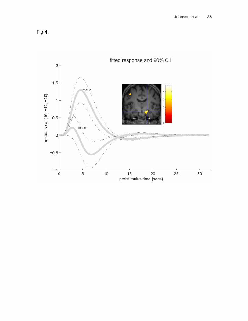
than the MCI patients. Figure 4 illustrates the shape of the average
hemodynamic response early in the task and again upon the final trial for a single
subject. The hemodynamic response is observed to decrease as a function of
trial in the hippocampal region.
In order to determine whether the group results were due to atrophy in the
region of interest we conducted a voxel-based morphometry analysis with the
same apriori ROI and extracted the VBM atrophy values underlying the activation

Johnson et al. 16
coordinates. This was accomplished by extracting the principal component of a
small sphere (2mm radius) at the maxima in the two activation clusters and then
extracting the atrophy signal from the VBM data at the same locations in the
same fashion. A small homogenous ROI was selected because we were only
concerned with gray matter at the site of peak activity differences. This was a
conservative approach for determining gray matter values at peak activation
maxima; a larger ROI may have obfuscated volumetric findings under the fMRI
maxima. The extracted ROI values were then entered into a correlation analysis.
In the anterior cluster there was a relationship between fMRI signal and gray
matter volume (GMV; r=.55, p=.009). The posterior hippocampal cluster did not
show this relationship (r=.05, p=.86), suggesting that fMRI signal and GMV were
not related at this location in these data.
Discussion
In this report we have examined the dynamic neural response during associative
learning in a group of MCI patients and controls. Overall, the MCI patients
expectedly performed worse than controls on a number of neuropsychological
measures of encoding and recall abilities, as well as on the concurrent
discrimination fMRI task itself. The imaging results were consistent with our
initial hypotheses indicating a difference in learning-related BOLD attenuation in
the right hippocampus. The controls exhibited a significant learning effect while
the MCI patients exhibited very little learning effect. The results are consistent
with prior work using repeating trials paradigms (Johnson et al., 2004; Rand-

Johnson et al. 17
Giovannetti et al., 2006) in elderly adults. Furthermore, these findings fit well with
other studies of dynamic hippocampal adaptation over successive trials during
learning acquisition (Dolan & Fletcher, 1999; Henson, Cansino, Herron, Robb, &
Rugg, 2003; Henson & Rugg, 2003; Strange, Fletcher, Henson, Friston, & Dolan,
1999).
The earlier study by Myers et al. (2002) found that individuals with
hippocampal atrophy, a risk factor for subsequent cognitive decline, performed
worse than non-atrophied controls on the transfer portion of the concurrent
discrimination task. The authors attributed impairment to reduced hippocampal-
region processing during the initial learning stages. Since hippocampal atrophy
is a risk factor for cognitive decline, including MCI, we expected that MCI patients
would show reduced learning-related hippocampal signal change. Our results
confirmed this prediction. The findings are consistent with the idea that the
hippocampal-region is normally involved in stimulus-response learning, even if
such simple learning is possible without hippocampal involvement. Similarly, in
animals, classical conditioning may be spared following hippocampal lesion (e.g.
Schmaltz & Theios, 1972), but electrophysiological recordings show that, when
present, the hippocampus is active during this simple stimulus-response learning
(Berger, Rinaldi, Weisz, & Thompson, 1983). Our results are also consistent with
the idea that hippocampal-region activation during this simple learning task may
facilitate subsequent transfer when task demands change; this is consistent with
the idea that learning in animals and humans with hippocampal-region damage is
Johnson et al. 18
often “inflexible” or “hyperspecific” (Eichenbaum, Otto, & Cohen, 1994; Gluck &
Myers, 1993, 2001; Schacter, 1985).
Others have examined the hippocampal response during transitive
inference—after initial learning of stimulus response associations have taken
place. Heckers et al. (2004) found the anterior hippocampal region was active
while making relational or transitive inferences on information already in
declarative memory (see also Preston, Shrager, Dudukovic, & Gabrieli, 2004). A
more recent study had extended that paradigm to schizophrenia and determined
that transitive inference, or transfer, was impaired in patients compared to
controls (Ongur et al., 2006). These studies all conducted fMRI after acquisition
of information had already occurred, whereas the current study imaged the brain
during the dynamic temporal course of acquisition over repetition and feedback.
Our results show that hippocampal attenuation during concurrent
discrimination learning was detected in the right MTL for this visuospatial
associative encoding task. We expected effects in the right-hemisphere due to
the visuospatial modality of item presentation (Golby et al., 2001). Further,
Henson, et al. (2003) reported a series of studies on repeating stimuli and
concluded that across a variety of stimulus materials, the right MTL was more
sensitive to stimulus repetitions than the left. Strange et al. (1999) also reported
specific right hippocampal attenuation in healthy young adults during learning of
visuospatial material.
Functional MRI studies in patient groups are prone to a number of
potentially confounding factors. The coupling between neural activity and the

Johnson et al. 19
hemodynamic response may be variably reduced in the patient group such that
the hemodynamic response is no longer an accurate measure of neural activity.
Another possible issue is that patients with cognitive impairment may approach
the task with different strategies than controls. Examination of Figure 2 indicates
that the MCI subjects exhibited a similar learning slope to controls, but indeed
their accuracy was significantly lower. In this study we attempted to deal with this
possible issue by incorporating the learning slope directly into the analysis. The
input contrast images into the group analysis represented learning-associated
signal change where learning of the correct choice in each pair was associated
with signal reduction in the MTL. Another possible approach (but only useful at
the group level) would have been to use the behavioral learning data as a
covariate in the statistical analysis.
The samples in this preliminary study were small and thus raise the
possibility of vulnerability to outliers, and simultaneously, low statistical power to
detect true effects. The small number of subjects prevented more elaborate
regression analyses modeling other demographic or general cognitive status in
the fMRI analysis. Another limitation may have been incurred by the relatively
small number of fMRI acquisitions from which event averages were calculated.
More acquisition time points would likely improve detection of subtle BOLD
adaptation over trials. Finally, others have pointed out that the accuracy of spatial
normalization of the hippocampus to standard MNI space may be diminished in
MCI and AD (Dickerson et al., 2005; Krishnan, Slavin, Tran, Doraiswamy, &
Johnson et al. 20
Petrella, 2006) and this might have introduced spatial variability, affecting the
final result.
Despite these limitations, the findings presented in this study are
consistent with the hypothesis that hippocampal signal decreases with
acquisition of stimulus-response associations derived through feedback.
Although prior learning-based fMRI studies in MCI have modeled repetition
(Johnson et al., 2004; Rand-Giovannetti et al., 2006), none have modeled
feedback-based learning over trials as was done in the present report. Whether
the fMRI-measured hippocampal encoding response can predict subsequent
successful transfer is still an unanswered question that will be addressed by our
laboratories. Additionally, further studies with MCI patients are needed to
differentiate the role of the MTL during encoding of specific stimulus-response
associations from its role in transfer of relevant information to new items.

Johnson et al. 21
Acknowledgements:
This study was supported by R01 AG021155 and a Merit Review grant from the
Department of Veterans Affairs to SCJ. We thank Andrew Alexander, Ph.D.,
Howard Rowley, MD, Britta Torgerson, Justin Dunker, Michael Anderle and Ron
Fisher for their assistance with this research.

Johnson et al. 22
References:
Benedict, R. (1997). Brief Visuospatial Memory Test-Revised. Lutz, FL:
Psychological Assessment Resources Inc.
Berger, TW, Rinaldi, PC, Weisz, DJ, & Thompson, RF. (1983). Single-unit
analysis of different hippocampal cell types during classical conditioning of rabbit
nictitating membrane response. J Neurophysiol, 50(5), 1197-1219.
Celone, KA, Calhoun, VD, Dickerson, BC, Atri, A, Chua, EF, Miller, SL, et al.
(2006). Alterations in memory networks in mild cognitive impairment and
Alzheimer's disease: an independent component analysis. J Neurosci, 26(40),
10222-10231.
Dickerson, BC, Goncharova, I, Sullivan, MP, Forchetti, C, Wilson, RS,
Bennett, DA, et al. (2001). MRI-derived entorhinal and hippocampal atrophy in
incipient and very mild Alzheimer's disease. Neurobiol Aging, 22(5), 747-754.
Dickerson, BC, Salat, DH, Bates, JF, Atiya, M, Killiany, RJ, Greve, DN, et al.
(2004). Medial temporal lobe function and structure in mild cognitive impairment.
Ann Neurol, 56(1), 27-35.
Dickerson, BC, Salat, DH, Greve, DN, Chua, EF, Rand-Giovannetti, E,
Rentz, DM, et al. (2005). Increased hippocampal activation in mild cognitive
impairment compared to normal aging and AD. Neurology, 65(3), 404-411.
Dolan, RJ, & Fletcher, PF. (1999). Encoding and retrieval in human medial
temporal lobes: an empirical investigation using functional magnetic resonance
imaging (fMRI). Hippocampus, 9(1), 25-34.

Johnson et al. 23
Eichenbaum, H, Otto, T, & Cohen, NJ. (1994). Two functional components of
the hippocampal memory system. Behavioral and Brain Sciences, 17(3), 449.
Folstein, MF, Folstein, SE, & McHugh, PR. (1975). "Mini Mental State": a
practical method for grading the cognitive state of patients for the clinician.
Journal of Psychiatry Research, 12, 189-198.
Friston, KJ, Holmes, AP, Worsley, KJ, Poline, JP, Frith, CD, & Frackowiak,
RSJ. (1995). Statistical parametric maps in functional imaging: A general linear
approach. Human Brain Mapping, 2, 189-210.
Gluck, MA, & Myers, CE. (1993). Hippocampal mediation of stimulus
representation: a computational theory. Hippocampus, 3(4), 491-516.
Gluck, MA, & Myers, CE. (2001). Gateway to Memory: An Introduction to
Neural Network Modeling of the Hippocampus in Learning and Memory.
Cambridge, MA: MIT Press.
Golby, AJ, Poldrack, RA, Brewer, JB, Spencer, D, Desmond, JE, Aron, AP, et
al. (2001). Material-specific lateralization in the medial temporal lobe and
prefrontal cortex during memory encoding. Brain, 124(Pt 9), 1841-1854.
Good, CD, Johnsrude, IS, Ashburner, J, Henson, RN, Friston, KJ, &
Frackowiak, RS. (2001). A voxel-based morphometric study of ageing in 465
normal adult human brains. Neuroimage, 14(1 Pt 1), 21-36.
Goodglass, H, & Kaplan, E. (1987). The assessment of aphasia and related
disorders (2nd ed.). Philadelphia: Lea & Febiger.
Grundman, M, Jack, CR, Jr., Petersen, RC, Kim, HT, Taylor, C, Datvian, M,
et al. (2003). Hippocampal volume is associated with memory but not
Johnson et al. 24
nonmemory cognitive performance in patients with mild cognitive impairment. J
Mol Neurosci, 20(3), 241-248.
Hachinski, VC. (1990). The decline and resurgence of vascular dementia.
Can-Med-Assoc-J., 142(2), 107-111.
Heckers, S, Zalesak, M, Weiss, AP, Ditman, T, & Titone, D. (2004).
Hippocampal activation during transitive inference in humans. Hippocampus,
14(2), 153-162.
Henson, RN, Cansino, S, Herron, JE, Robb, WG, & Rugg, MD. (2003). A
familiarity signal in human anterior medial temporal cortex? Hippocampus, 13(2),
301-304.
Henson, RN, & Rugg, MD. (2003). Neural response suppression,
haemodynamic repetition effects, and behavioural priming. Neuropsychologia,
41(3), 263-270.
Jack, CR, Petersen, RC, Xu, Y, O'Brien, PC, Smith, GE, Ivnik, RJ, et al.
(2000). Rates of hippocampal atrophy correlate with change in clinical status in
aging and AD. Neurology, 55(4), 484-489.
Jack, CR, Petersen, RC, Xu, Y, O'Brien, PC, Smith, GE, Ivnik, RJ, et al.
(1998). Rate of medial temporal lobe atrophy in typical aging and Alzheimer's
disease. Neurology, 51(4), 993-999.
Jack, CR, Petersen, RC, Xu, YC, O'Brien, PC, Smith, GE, Ivnik, RJ, et al.
(1999). Prediction of AD with MRI-based hippocampal volume in mild cognitive
impairment. Neurology, 52(7), 1397-1403.

Johnson et al. 25
Jastak Associates. (1993). Wide Range Achievement Test-Third Edition.
Wilmington: Wide Range Inc.
Jenkinson, M. (2003). Fast, automated, N-dimensional phase-unwrapping
algorithm. Magn Reson Med, 49(1), 193-197.
Jezzard, P, & Balaban, RS. (1995). Correction for geometric distortion in
echo planar images from B0 field variations. Magn Reson Med, 34(1), 65-73.
Johnson, SC, Baxter, L, Susskind-Wilder, L, Connor, DJ, Sabbagh, MN, &
Caselli, RJ. (2004). Hippocampal adaptation to face repetition in healthy elderly
and mild cognitive impairment. Neuropsychologia, 42(7), 980-989.
Johnson, SC, Schmitz, TW, Moritz, CH, Meyerand, ME, Rowley, HA,
Alexander, AL, et al. (2006). Activation of brain regions vulnerable to Alzheimer's
disease: the effect of mild cognitive impairment. Neurobiol Aging, 27(11), 1604-
1612.
Kiebel, S, & Holmes, A. (2004). The general linear model. In R. Frackowiak,
K. Friston, C. Frith, R. Dolan, C. Price, S. Zeki, J. Ashburner & W. Penny (Eds.),
Human Brain Function (2nd ed., pp. 725-760). London: Elsevier.
Krishnan, S, Slavin, MJ, Tran, TT, Doraiswamy, PM, & Petrella, JR. (2006).
Accuracy of spatial normalization of the hippocampus: implications for fMRI
research in memory disorders. Neuroimage, 31(2), 560-571.
LePage, M, Habib, R, & Tulving, E. (1998). Hippocampal PET activations of
memory encoding and retrieval: The HIPER model. Hippocampus, 8, 313-322.

Johnson et al. 26
Machulda, MM, Ward, HA, Borowski, B, Gunter, JL, Cha, RH, O'Brien, PC, et
al. (2003). Comparison of memory fMRI response among normal, MCI, and
Alzheimer's patients. Neurology, 61(4), 500-506.
Muller, MJ, Greverus, D, Weibrich, C, Dellani, PR, Scheurich, A, Stoeter, P,
et al. (2006). Diagnostic utility of hippocampal size and mean diffusivity in
amnestic MCI. Neurobiol Aging.
Myers, CE, Kluger, A, Golomb, J, Ferris, S, de Leon, MJ, Schnirman, G, et al.
(2002). Hippocampal atrophy disrupts transfer generalization in nondemented
elderly. J Geriatr Psychiatry Neurol, 15(2), 82-90.
Myers, CE, Shohamy, D, Gluck, MA, Grossman, S, Kluger, A, Ferris, S, et al.
(2003). Dissociating hippocampal versus basal ganglia contributions to learning
and transfer. J Cogn Neurosci, 15(2), 185-193.
Ongur, D, Cullen, TJ, Wolf, DH, Rohan, M, Barreira, P, Zalesak, M, et al.
(2006). The neural basis of relational memory deficits in schizophrenia. Arch Gen
Psychiatry, 63(4), 356-365.
Petersen, RC. (2004). Mild cognitive impairment as a diagnostic entity. J
Intern Med, 256(3), 183-194.
Petrella, JR, Krishnan, S, Slavin, MJ, Tran, TT, Murty, L, & Doraiswamy, PM.
(2006). Mild cognitive impairment: evaluation with 4-T functional MR imaging.
Radiology, 240(1), 177-186.
Preston, AR, Shrager, Y, Dudukovic, NM, & Gabrieli, JD. (2004).
Hippocampal contribution to the novel use of relational information in declarative
memory. Hippocampus, 14(2), 148-152.

Johnson et al. 27
Rand-Giovannetti, E, Chua, EF, Driscoll, AE, Schacter, DL, Albert, MS, &
Sperling, RA. (2006). Hippocampal and neocortical activation during repetitive
encoding in older persons. Neurobiol Aging, 27(1), 173-182.
Reitan, RM, & Wolfson, D. (1993). The Halstead-Reitan Neuropsychological
Test Battery: Theory and clinical interpretation (2 ed.). Tucson: Neuropsychology
Press.
Saykin, AJ, Johnson, SC, Flashman, LA, McAllister, TW, Sparling, MB,
Darcey, TM, et al. (1999). Functional differentiation of medial temporal and
frontal regions involved in processing novel and familiar words: An fMRI study.
Brain, 122, 1963-1971.
Schacter, DL. (1985). Multiple forms of memory in humans and animals. In N.
Weingberger, J. L. McGaugh & G. Lynch (Eds.), Memory Systems of the Brain:
Animal and Human Cognitive Processes (pp. 351-379). New York: Guilford
Press.
Schmaltz, LW, & Theios, J. (1972). Acquisition and extinction of a classically
conditioned response in hippocampectomized rabbits (Oryctolagus cuniculus). J
Comp Physiol Psychol, 79(2), 328-333.
Small, S, Perera, GM, DeLaPaz, R, Mayeux, R, & Stern, Y. (1999).
Differential regional dysfunction of the hippocampal formation among elderly with
memory decline and Alzheimer's disease. Ann Neurol, 45(4), 466-472.
Spreen, O, & Strauss, E. (1998). A Compendium of Neuropsychological
Tests. New York: Oxford.

Johnson et al. 28
Strange, BA, Fletcher, P, Henson, R, Friston, K, & Dolan, R. (1999).
Segregating the functions of the human hippocampus. Procedings of the National
Academy of Sciences, 96, 4034-4039.
Trivedi, MA, Schmitz, TW, Ries, ML, Torgerson, BM, Sager, MA, Hermann,
BP, et al. (2006). Reduced hippocampal activation during episodic encoding in
middle-aged individuals at genetic risk of Alzheimer's disease: a cross-sectional
study. BMC Med, 4, 1.
Tulving, E, & Markowitsch, HJ. (1998). Episodic and declarative memory: role
of the hippocampus. Hippocampus, 8(3), 198-204.
Wagner, AD, Koutstaal, W, & Schacter, DL. (1999). When encoding yields
remembering: insights from event-related neuroimaging. Philos Trans R Soc
Lond B Biol Sci, 354(1387), 1307-1324.
Winblad, B, Palmer, K, Kivipelto, M, Jelic, V, Fratiglioni, L, Wahlund, LO, et
al. (2004). Mild cognitive impairment--beyond controversies, towards a
consensus: report of the International Working Group on Mild Cognitive
Impairment. J Intern Med, 256(3), 240-246.
Johnson et al. 29
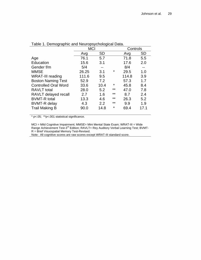
Table 1. Demographic and Neuropsychological Data. MCI Controls Avg SD Avg SD Age 76.1 5.7 71.8 5.5 Education 15.6 3.1 17.6 2.0 Gender f/m 5/4 -- 8/4 -- MMSE 26.25 3.1 * 29.5 1.0 WRAT-III reading 111.6 9.5 114.8 3.9 Boston Naming Test 52.9 7.2 57.3 1.7 Controlled Oral Word 33.6 10.4 * 45.8 8.4 RAVLT total 28.0 5.2 ** 47.0 7.8 RAVLT delayed recall 2.7 1.6 ** 8.7 2.4 BVMT-R total 13.3 4.6 ** 26.3 5.2 BVMT-R delay 4.3 2.2 ** 9.9 1.9 Trail Making B 90.0 14.8 * 69.4 17.1 * p<.05; **p<.001 statistical significance. MCI = Mild Cognitive Impairment; MMSE= Mini Mental State Exam; WRAT-III = Wide Range Achievement Test-3rd Edition; RAVLT= Rey Auditory Verbal Learning Test; BVMT-R = Brief Visuospatial Memory Test-Revised. Note: All cognitive scores are raw scores except WRAT-III standard score.

Johnson et al. 30
Table 2. Task accuracy over trials Controls MCI
Trial MEAN SD MEAN SD 1 0.63 0.24 0.44 0.20 2 0.72 0.26 0.41 0.30 3 0.74 0.21 0.56 0.19 4 0.88 0.13 0.59 0.25 5 0.85 0.13 0.61 0.17 6 0.83 0.16 0.78 0.24
Note: scores represent mean proportion correct.

Johnson et al. 31
Figure Captions
Figure 1. Concurrent visual discrimination task. (A) On each trial the subject
sees a pair of objects and is asked to choose the left or right object. (B) The
chosen object is raised and, if the subject’s choice was correct a smiley face is
revealed; (C) when the incorrect object is chosen the object is raised to reveal
no smiley face; (D) In the control task, the correct choice was always apparent;
(E) The chosen object is raised to reveal the smiley face.
Figure 2. Behavioral results. Plots of reaction time (Top) and accuracy (Bottom),
for the control (squares) and MCIa (circles) groups. See also Table 2.
Figure 3. Statistical parametric map of the two-sample t-test delineating group
differences between Controls and MCIa. The result is overlaid on an atlas
brain in MCI space. The analysis was constrained to the right hippocampus
based on prior studies. The ROI search region is in white on the images, and
encompasses the statistical results (hot colors). The analysis resulted in two
clusters in the body and head of the right hippocampus. See the text for
coordinates and statistics. The right side of the brain is on the right side of the
image.

Johnson et al. 32
Figure 4. Peristimulus plots indicating a decrease in the hemodynamic response
as a function of trial in the mesial temporal lobe for a healthy control subject. The
mean time-course of the second trial (the first trial after feedback) and final trial
are shown with solid lines. The 90% confidence intervals are represented by
dashed lines. The coronal image inlay shows the subject’s SPM of learning over
trials overlaid on the T1 volume. The color scale is the magnitude of the t-
statistic.

Johnson et al. 33
Fig 1.

Johnson et al. 34
Fig. 2

Johnson et al. 35
Figure 3
Johnson et al. 36
Fig 4.