joão paulo batista machado insights into regulatory mechanisms of
TRANSCRIPT

JOÃO PAULO BATISTA MACHADO
INSIGHTS INTO REGULATORY MECHANISMS OF THE NIK-MEDIATED ANTIVIRAL DEFENSE: NEW COMPONENTS AND THE MOLECULAR
BASES OF THE DEFENSE
Tese apresentada à Universidade Federal de Viçosa, como parte das exigências do Programa de Pós-Graduação em Bioquímica Aplicada, para obtenção do título de Doctor Scientiae.
VIÇOSA MINAS GERAIS – BRASIL
2015

JOÃO PAULO BATISTA MACHADO
INSIGHTS INTO REGULATORY MECHANISMS OF THE NIK-MEDIATED ANTIVIRAL DEFENSE: NEW COMPONENTS AND THE MOLECULAR
BASES OF THE DEFENSE
Tese apresentada à Universidade Federal de Viçosa, como parte das exigências do Programa de Pós-Graduação em Bioquímica Aplicada, para obtenção do título de Doctor Scientiae.
APROVADA: 20 de julho de 2015. ______________________________ ______________________________ Marcia Flores da Silva Ferreira Poliane Alfenas Zerbini ______________________________ ______________________________ Pedro Augusto Braga dos Reis Maximiller Dal-Bianco Lamas Costa (Coorientador)
______________________________ Elizabeth Pacheco Batista Fontes
(Orientadora)

ii
Aos meus amados pais Antonio e
Maria Lucia, aos meus irmãos
Elisangela, Edilâine e Marco Antônio,
e a minha querida sobrinha Estela
DEDICO

iii
AGRADECIMENTOS
Agradeço a Deus, por ter me dado força e perseverança e guiado meus
passos para chegar até aqui, por providenciar tudo que sou e que tenho, e por me
ensinar a reconhecer, nas coisas simples do dia-a-dia, sua providência divina.
Aos meus pais Antonio e Maria Lucia, meus exemplos de vida. Posso estudar
a vida inteira que nunca terei a sabedoria de vocês! Agradeço por sempre me
apoiarem e acreditarem em mim. Agradeço também por não cessarem de rezar e
interceder por mim. A oração de vocês me fez forte principalmente nos momentos
em que o desânimo me fazia fraco.
Aos meus irmãos Elisangela, Edilâine e Marco Antônio, por me incentivarem
em cada nova etapa da minha vida. Tenho muito orgulho de vocês!
À professora Elizabeth, exemplo de dedicação e competência profissional,
pela orientação e pelas inúmeras oportunidades de aprendizado oferecidas durante
estes nove anos que passei em seu laboratório. Agradeço também pela oportunidade
de desenvolver este projeto, que permitiu meu crescimento profissional, e por permitir
que em seu laboratório eu pudesse dar os primeiros passos na pesquisa.
À Universidade Federal de Viçosa, por todas as oportunidades de estudo
oferecidas durante toda minha vida acadêmica.
Ao Conselho Nacional de Desenvolvimento Científico e Tecnológico (CNPq)
pelo apoio financeiro.
À minha namorada Analice, pela companhia, parceria, amizade, apoio e
compreensão em todos os momentos, principalmente nesta fase final da minha tese.
Agradeço pelos valiosos conselhos que sempre vinham em momentos oportunos.
Aos amigos Pedro, Pato, Claudinha, Priscila, Maiana, Cris, Lucas, Giselle,
Iara, Bianca, Janaína, por me incentivarem durante todos os momentos. Obrigado

iv
por aturarem minhas reclamações, minha chatice e meu mau humor, e por todos os
momentos divertidos que passamos durante este tempo.
Ao estudante de iniciação científica Marcos, que além de um estagiário, foi
um amigo. Agradeço por toda ajuda na condução dos experimentos realizados nos
primeiros anos do meu doutorado.
Aos integrantes do Laboratório de Biologia Molecular de Plantas, os antigos
e os atuais, pela prazerosa convivência durante estes anos e por tornarem cada dia
de trabalho uma oportunidade única de aprendizado.
À Marlene, à Gláucia e ao Eduardo, pela presteza.
E a todos que passaram pela minha vida durante a realização deste trabalho
e que deixaram sua contribuição para a concretização do mesmo.

v
ÍNDICE
RESUMO ......................................................................................................... vii
ABSTRACT ...………………………………………………………………………. ix
INTRODUÇÃO GERAL ................................................................................... 1
CAPÍTULO I - REVISÃO DE LITERATURA ..………………………………....... 5
Geminivírus .............................................................................................. 5
Interação geminivírus-hospedeiro ............................................................ 11
Interações de fatores do hospedeiro com as proteínas de movimento MP
e NSP ....................................................................................................... 17
Sisteme imune de plantas ........................................................................ 21
Imunidade induzida por PAMP (PTI) ................................................ 22
Imunidade induzida por efetores (ETI) ............................................. 25
Resistência sistêmica adquirida (SAR) ............................................ 30
Defesas mediadas por RNAi ............................................................ 31
Referências bibliográficas ........................................................................ 36
CHAPTER II - DOWSTREAM EVENTS IN THE NIK-MEDIATED DEFENSE
ASSOCIATED WITH RESISTANCE TO BEGOMOVIRUS .............................. 53
Abstract .................................................................................................... 54
Introduction .............................................................................................. 55
Results ..................................................................................................... 59
Identification and isolation of a RPL10 partner, designated LIMYB
(L10-Interacting Myb domain-containing protein) .............................. 59
LIMYB interacts with RPL10 in vivo and displays overlapping
expression profiles with RPL10 and NIK1 .......................................... 60
LIMYB and RPL10 transactivate promoters of ribosomal protein
genes ................................................................................................ 62
Constitutive activation of NIK confers tolerance to begomovirus
infection in tomato but does not impair development ........................ 64
The enhanced tolerance to begomovirus displayed by the T474D-
expressing lines may be associated with the translational control
branch of the NIK-mediated antiviral responses ................................ 67
Discussion ................................................................................................ 68
Material and methods .............................................................................. 71
Plasmid constructs ........................................................................... 71

vi
Yeast two-hybrid screening .............................................................. 74
Phylogenetic analysis ....................................................................... 75
Plant material, growth conditions and transformation ....................... 75
Co-immunoprecipitation assay ......................................................... 76
Bimolecular fluorescent complementation (BiFC) ............................ 76
Histochemical in situ localization of GUS in Arabidopsis seedlings.. 77
RT-PCR and real time RT-PCR analyses ........................................ 77
Chromatin immunoprecipitation (ChIP) assay .................................. 78
Luciferase reporter gene assay ........................................................ 78
Infectivity assays .............................................................................. 79
Quantitation of viral DNA in infected plants ...................................... 79
Physiological measurements of tomato transgenic lines .................. 80
Estimation of total carotenoids, lycopene, and β-carotene ............... 81
In vivo labeling of leaf proteins ......................................................... 81
Polysome fractionation ..................................................................... 81
References ............................................................................................... 83
Figures ..................................................................................................... 87
CHAPTER III - NIK1, A HOST FACTOR SPECIALIZED IN ANTIVIRAL
DEFENSE OR A NOVEL GENERAL REGULATOR OF PLANT IMMUNITY?.. 101
Abstract .................................................................................................... 102
Introduction .............................................................................................. 103
Like BAK1, NIK1 belongs to the subfamily II of leucine-rich repeats (LRR)
receptor-like kinases (RLK) ...................................................................... 104
The NIK1/RPL10 module transduces a defense response to
begomoviruses.......................................................................................... 106
The mechanism of NIK1-mediated antiviral defense is underscored by
suppression of host global translation: a new paradigm for antiviral
defenses in plants ..................................................................................... 109
NIK may be a general negative co-receptor in signaling pathways: a
property that may impact negatively defense against other pathogens ..... 112
Conclusions and outlook .......................................................................... 114
Acknowledgements .................................................................................. 114
References ............................................................................................... 116
Figure legends ......................................................................................... 122
CONCLUSÕES GERAIS ................................................................................. 126
ANEXOS ......................................................................................................... 128

vii
RESUMO
MACHADO, João Paulo Batista, D.Sc., Universidade Federal de Viçosa, julho de 2015. Introspecções sobre os mecanismos regulatórios da via de sinalização antiviral mediada por NIK: novos componentes e bases moleculares da defesa. Orientadora: Elizabeth Pacheco Batista Fontes. Coorientadores: Anésia Aparecida dos Santos e Maximiller Dal-Bianco Lamas Costa.
A proteína NIK (NSP-interacting kinase), identificada por interagir com a proteína
NSP (nuclear shuttle protein) de geminivírus, apresenta características estruturais,
bioquímicas e biológicas condizentes com um autêntico receptor cinase envolvido na
resposta de defesa à infecção por geminivírus. A ativação deste receptor imune
resulta na fosforilação da proteína ribossomal L10 (RPL10) e na subsequente
relocação nuclear desta proteína ribossomal. Apesar dos avanços obtidos com a
identificação de RPL10, outras conexões moleculares que ligam a ativação de NIK à
resposta antiviral ainda são desconhecidas, bem como a natureza deste mecanismo
de defesa. Nesta investigação, foi identificado um novo componente efetor
downstream da via de sinalização mediada por NIK, um fator de transcrição
denominado LIMYB (L10-Interacting Myb domain-containing protein), isolado pela
sua capacidade de interagir com RPL10 pelo sistema de duplo híbrido em leveduras.
Ensaios de co-imunoprecipitação e de complementação de fluorescência bimolecular
(BiFC) mostraram que o complexo RPL10-LIMYB ocorre estavelmente na planta,
mais precisamente no núcleo das células vegetais. Estudos de caracterização
funcional mostraram que LIMYB atua como um autêntico fator de transcrição,
ligando-se ao promotor e inibindo a expressão de genes de proteínas ribossomais.
Estes resultados sugerem que o mecanismo de defesa antiviral mediado por NIK
baseia-se na supressão da tradução do hospedeiro. Para examinar esta hipótese,
inicialmente foi avaliado os níveis de tradução em linhagens de tomate
superexpressando um mutante superativo de AtNIK1 (T474D). Os resultados
mostraram que as linhagens transgênicas apresentaram menor acúmulo de
proteínas recentemente sintetizadas quando comparadas com plantas WT.
Posteriormente, as linhagens de tomate superexpressando T474D foram infectadas
com duas espécies de begomovírus altamente divergentes, ToYSV (Tomato yellow
spot virus) e ToSRV (Tomato severe rugose virus). As linhagens transgênicas
apresentaram sintomas atenuados ou foram assintomáticas. Estas observações
foram associadas com um atraso na infecção viral, com taxas de infecção menores
e com a redução no acúmulo de DNA viral em folhas sistêmicas infectadas. Apesar

viii
do fenótipo de tolerância exibido pelas linhagens superexpressando T474D e dos
menores índices de síntese proteica, estas linhagens não apresentaram diferenças
no desenvolvimento, no desempenho fisiológico e nas características horticulturais
quando comparadas com plantas WT ou com linhagens superexpressando AtNIK1.
Finalmente, foi determinado se a redução nos níveis de tradução global do
hospedeiro causado pela superexpressão de T474D poderia prejudicar a síntese de
proteínas virais. Para isto, frações polissomais foram isoladas a partir de linhagens
WT e T474D infectadas com ToYSV e a presença de mRNA viral foi examinada
nestas frações. Os resultados mostraram uma menor associação do mRNA que
codifica a proteína do capsídeo viral nas frações de polissomos de linhagens T474D
quando comparadas àquelas de linhagens não transformadas. Coletivamente, estes
resultados indicam que begomovírus não são capazes de manter altos níveis de
tradução de mRNA virais nas linhagens superexpressando o mutante T474D,
indicando que a supressão global da síntese de proteínas, identificada nestes
genótipos, pode proteger eficientemente as células contra a infecção por estes vírus.
Entretanto, estudos da variação global na expressão gênica de plantas nik1 alelos
nulos mostraram que a perda da função de NIK1 promoveu a indução de
componentes do hub principal da sinalização a brassinosteróide e de hubs envolvidos
na sinalização ao ácido salicílico e na imunidade antibacteriana. Estes resultados
sugerem que NIK1 pode funcionar como um regulador negativo em vias de
sinalização de desenvolvimento e imunidade, apesar de sua propriedade antiviral.

ix
ABSTRACT
MACHADO, João Paulo Batista, D.Sc., Universidade Federal de Viçosa, July, 2015. Insights into regulatory mechanisms of the NIK-mediated antiviral defense: new components and the molecular bases of the defense. Adviser: Elizabeth Pacheco Batista Fontes. Co-advisers: Anésia Aparecida dos Santos and Maximiller Dal-Bianco Lamas Costa.
The NSP-interacting kinase (NIK) protein, identified through interaction with the
geminivirus nuclear shuttle protein (NSP), displays structural, biochemical and
biological characteristics consistent with an authentic receptor kinase involved in
defense response against geminivirus infection. The activation of this immune
receptor leads to phosphorilation of the ribosomal protein L10 (RPL10) and in the
subsequent nuclear relocation of this ribosomal protein. Apart from the identification
of RPL10 as a downtream component of the defense signaling, others molecular
connections that link the NIK activation to the antiviral response, as well as the nature
of this defense mechanism, remain to be determined. In this investigation, a new
downstream effector component of the NIK-mediated signaling pathway, a
transcription fator designated LIMYB (L10-Interacting Myb domain-containing
protein), was identified by its capacity to interact with RPL10 through yeast two-hybrid
system. Co-immunoprecipitation and bimolecular fluorescence complementation
assays showed that RPL10-LIMYB complex occurs stably in the plant, specifically in
the nucleus of plant cells. Functional characterization studies showed that LIMYB acts
as an authentic transcription fator, binding to and inhibiting expression of ribosomal
protein promoters. These results suggest that NIK-mediated antiviral defense
mechanism is based on host translation suppression. To examine this hypothesis, the
translation levels in tomato lines overexpressing the constitutively active mutant of
AtNIK1 (T474D) was initially assessed. The results demonstrated that transgenic lines
had lower accumulation of newly synthesized proteins when comparated to WT
plants. Subsequently, T474D-overexpressing tomato lines were infected with two
highly divergent begomovirus species, ToYSV (Tomato yellow spot virus) and ToSRV
(Tomato severe rugose virus). The transgenic lines either displayed attenuated
symptons or were asymptomatic. These findings were associated with delay in viral
infections, lower infection rates and reduction in viral DNA accumulation in
systemically infected leaves. Despite the tolerance phenotype and lower rates of
protein synthesis displayed by T474D-overexpressing lines, no difference in the
development, physiological performance and horticultural traits was observed

x
between T474D-overexpressing and WT or AtNIK1-overexpressing lines. Finally,
whether the reduction in overall levels of host translation caused by overexpression
of T474D could affect the viral proteins synthesis was examined. For this purpose,
polysomal fractions were isolated from WT and T474D lines infected with ToYSV and
examined for the presence of viral mRNA. The results showed lower association of
mRNA encoding viral capsid protein at the polysomes fractions of T474D lines when
comparated to that of untransformed lines. Collectively, these results indicate that
begomovirus are not able to maintain high levels of viral mRNA translation into
T474D-overexpressing lines, indicating that the global suppression of the protein
synthesis identified in these genotypes may efficiently protect cells against infection
by these virus. However, studies of global changes in gene expression of nik1 null
alleles revealed that loss of NIK1 function promoted induction of components of the
main brassinosteroid signaling hub and of hubs involved in salicylic acid signaling and
antibacterial immunity. These results suggest that NIK1 may function as a negative
regulator in development and immunity signaling pathways, despite its antiviral
property.

1
INTRODUÇÃO GERAL
As plantas, devido a sua natureza séssil, estão constantemente submetidas
a condições externas adversas, como por exemplo, seca, calor, salinidade, frio,
ataque de predadores e infecções por patógenos, que afetam negativamente o seu
crescimento e desenvolvimento, resultando em perdas consideráveis na
produtividade em condições de campo. Dentre os fatores bióticos, infecções por
geminivírus merecem destaque, pois limitam severamente a produção de uma
variedade de sistemas de cultivo por todo o mundo. No Brasil, a incidência de
doenças causadas por geminivírus em tomateiros tem aumentado drasticamente nas
últimas duas décadas, tornando-se uma das principais ameaças para o seu cultivo.
Os métodos convencionais de controle da propagação de geminivírus que se
baseiam no combate ao inseto vetor pela utilização de inseticidas têm sido mal-
sucedidos em atenuar o impacto da infecção viral. Deste modo, o desenvolvimento
de genótipos resistentes por meio de engenharia genética representa um método
mais confiável e eficaz no manejo de doenças causadas por estes vírus.
Considerando a ocorrência de infecções mistas e as altas taxas de mutação e
recombinação que permitem aos geminivírus se adaptarem rapidamente a novos
hospedeiros e ambientes, torna-se essencial que as estratégias empregadas tenham
caráter geral, capazes de atingir diversos componentes deste grupo viral. Diante
disto, um melhor entendimento das interações geminivírus-hospedeiro e seu efeito
na infecção viral são cruciais para o desenvolvimento de uma metodologia efetiva no
controle da indução de doenças causadas por estes vírus.
Dentro desse contexto, as proteínas NIKs (NSP-interacting kinase) de soja,
tomate e Arabidopsis foram identificadas por interagir com a proteína NSP (nuclear
shuttle protein) de diferentes begomovírus, pertencentes ao gênero Begomovirus da
família Geminiviridae (Fontes et al., 2004; Mariano et al., 2004; Sakamoto et al.,

2
2012). NIKs são proteínas localizadas na membrana plasmática que apresentam
uma série de propriedades estruturais, bioquímicas e biológicas características de
autênticos receptores Ser/Thr cinase que medeiam a transdução de sinal através da
membrana plasmática (Carvalho et al., 2008c; Fontes et al., 2004, Santos et al.,
2009). A interação de NSP com a alça de ativação de NIK inibe a sua atividade
cinase, e a perda da função do gene NIK ou mutações que comprometem a
capacidade de autofosforilação deste receptor aumentam a suscetibilidade à
infecção por geminivírus (Fontes et al., 2004; Santos et al., 2009). Por outro lado, a
superexpressão de NIK em Arabidopsis ou tomateiros atenua o desenvolvimento de
sintomas e atrasa a infecção viral (Carvalho et al., 2008c; Fontes et al., 2004).
Progressos no entendimento molecular que conecta a ativação do receptor imune
NIK à resposta antiviral foram obtidos com a identificação da proteína ribossomal L10
(RPL10) como componente downstream desta via de sinalização (Rocha et al.,
2008). A ativação de NIK resulta na fosforilação de RPL10 e no redirecionamento da
proteína ribossomal para o núcleo onde pode atuar montando uma resposta de
defesa antiviral. Em células infectadas, a localização nuclear de RPL10 mediada por
NIK é bloqueada, resultando no acúmulo da proteína ribossomal em corpos
puntiformes dispersos no citoplasma (Carvalho et al., 2008c). Coletivamente, estes
resultados sugerem que NIK esteja envolvida em uma via de resposta de defesa
antiviral, e que esta via pode ser um alvo molecular em potencial para conferir
tolerância a diferentes begomovírus.
NIK pertence à família de receptores-like cinases (RLK) contendo repetições
ricas em leucina (LRR) em seu domínio extracelular. Considerando a identidade de
sequência, o número de cópias e a disposição dos motivos LRR, as proteínas NIK
(NIK1, NIK2 e NIK3) foram classificadas dentro da subfamília LRRII-RLK (Zhang et
al., 2006). Esta subfamília é composta ainda por proteínas envolvidas no
desenvolvimento e defesa, e por proteínas cujas funções são desconhecidas. O
membro mais bem estudado desta subfamília é BAK1/AtSERK3 (BRI1-associated

3
kinase 1/SOMATIC EMBRYOGENESIS RECEPTOR KINASE 3), que desempenha
uma função importante na imunidade inata de plantas contra patógenos, atuando
como coreceptor de diversos receptores que reconhecem padrões (PRR)
moleculares associados a patógenos (PAMP), induzindo PTI (PAMP-triggered
immunity). Apesar da similaridade estrutural verificada entre NIK e BAK1, o
mecanismo de defesa antiviral mediado por NIK diverge daquele induzido por BAK1,
não se encaixando no modelo elicitor-receptor de resistência. Além da sua função na
imunidade inata, BAK1 contribui para o desenvolvimento da planta por meio da sua
interação com BRI1 (BRASSINOSTEROID INSENSITIVE 1), um LRR-RLK que
reconhece o fitohôrmonio brassinosteróide. Uma vez que os componentes SERK da
subfamília LRRII-RLK são encontrados em associação dependente de estímulo com
LRR-RLK que se ligam a ligantes, é razoável pressupor que NIK, por integrar esta
subfamília, participe também de outras vias de sinalização mediadas por LRR-RLKs
ainda não identificados.
A ativação do receptor imune NIK resulta na indução de uma resposta de
defesa antiviral. Apesar do conhecimento obtido com a identificação de RPL10 como
membro integrante desta via de sinalização, componentes nucleares efetores
downstream não foram identificados e diversas lacunas, como a natureza do
mecanismo da resposta de defesa, ainda estão presentes. Deste modo, esta
investigação teve como objetivo principal a identificação e elucidação da via de
sinalização mediada por NIK1 que leva à tolerância a geminivírus. Este objetivo é
explorado principalmente no segundo capítulo, cujos resultados apresentados
colaboraram para publicações em Nature (Zorzatto et al., 2015) e Plant
Biotechnology Journal (Brustolini et al., 2015) e em cujos manuscritos o autor dessa
tese de doutorado compartilha a primeira autoria. O terceiro capítulo, publicado em
BioEssays (Machado et al., 2015), descreve o estado da arte do mecanismo de
sinalização antiviral mediado por NIK, bem como uma possível participação deste
receptor como um regulador negativo em vias de sinalização imune e de

4
desenvolvimento. O primeiro capítulo refere-se a um levantamento bibliográfico sobre
geminivírus e suas interações com fatores do hospedeiro, bem como uma revisão
sobre sistema imune de plantas.

5
CAPÍTULO I
REVISÃO DE LITERATURA
Geminivírus
A família Geminiviridae constitui um grupo de vírus de plantas de grande
importância econômica, pois infectam culturas agronômicas relevantes (Moffat,
1999). Muitas das doenças causadas por estes vírus representam uma ameaça séria
para a segurança alimentar de países em desenvolvimento nas regiões tropicais e
subtropicais do mundo (Rey et al., 2012; Rybicki e Pietersen, 1999). Na América
latina, que apresenta uma das maiores diversidades e distribuição de geminivírus
transmitidos por mosca branca, a produção de alimentos básicos é altamente afetada
(Morales, 2006). A produção de tomate no Brasil é reduzida em até 100% em
algumas regiões devido à infecção por geminivírus (Fernandes et al., 2008; Ribeiro
et al., 2003).
Geminivírus são vírus de DNA fita simples (ssDNA) circular, compostos por
um ou dois componentes genômicos encapsulados individualmente em uma partícula
icosaédrica geminada. Até o momento, sete gêneros foram reconhecidos dentro da
família Geminiviridae: Mastrevirus, Eragrovirus, Becurtovirus, Curtovirus,
Topocuvirus, Turncurtovirus e Begomovirus (Varsani et al., 2014), que podem ser
diferenciados de acordo com o tipo de inseto vetor do vírus, gama de hospedeiros
(monocotiledôneas ou dicotiledôneas), organização genômica e relacionamento
filogenético (Brown et al., 2012; Varasani et al., 2014). Dentre os gêneros de
geminivírus, o Begomovirus possui o maior número de representantes e inclui vírus
transmitidos pela mosca branca Bemisia tabaci para várias espécies de
dicotiledôneas (Fauquet et al., 2008). Os begomovírus podem ser bipartidos,

6
possuindo dois componentes genômicos, ou monopartidos, com apenas um
componente de DNA, mas com dois genes adicionais, ambos codificando proteínas
envolvidas no movimento do vírus (Rojas et al., 2001). A maioria dos begomovírus
encontrados no Brasil é bipartida, ou seja, possui dois componentes genômicos
denominados DNA-A e DNA-B (Ribeiro et al., 2003).
Os dois componentes genômicos de begomovírus compartilham pouca
identidade de sequência, com exceção de uma região de aproximadamente 200
nucleotídeos com identidade tipicamente maior que 85%, conhecida como região
comum (CR; Briddon et al., 2010). A CR abriga uma estrutura em grampo, que
contém, dentro do loop, uma sequência de nove nucleotídeos (TAATATTAC) que
define a origem de replicação do DNA, e sequências repetidas (conhecidas como
iterons), que são reconhecidas pela proteína viral Rep (replication-associated protein)
e requeridas para a replicação viral (Argüello-Astorga e Ruiz-Medrano, 2001; Hanley-
Bowdoin et al., 2000). A CR funciona na manutenção da integridade do genoma
dividido, garantindo que a replicação de ambos componentes possa ser iniciada pela
proteína Rep (Fontes et al., 1994). O reconhecimento por Rep é considerado
específico do vírus (Argüello-Astorga et al., 1994); no entanto, em alguns casos, a
proteína Rep codificada pelo DNA-A de um determinado vírus reconhece o DNA-B
de um vírus diferente, resultanto em um fenômeno conhecido como
pseudorecombinação (Rojas et al., 2005).
O ciclo de replicação de geminivírus inclui um primeiro estágio no qual o DNA
genômico fita simples (ssDNA) é convertido a fita dupla (dsDNA), um processo
realizado por DNA polimerases do hospedeiro. O dsDNA produzido é transcrito pela
RNA polimerase II do hospedeiro, permitindo a síntese da proteína Rep. Esta
proteína inicia a replicação viral, que ocorre por uma combinação de replicação por
círculo rolante e replicação dependente de recombinação (Jeske et al., 2001; Preiss
e Jeske, 2003). O ssDNA circular nascente pode ser convertido em dsDNA para

7
entrar novamente no ciclo de replicação ou pode ser empacotado em vírions, após a
síntese da proteína CP (coat protein).
Devido ao pequeno tamanho de seu genoma, geminivírus codificam de quatro
a oito proteínas, algumas das quais evoluíram para proteínas multifuncionais que,
juntamente com a presença de genes sobrepostos, contribuem para a economia
genética apresentada por estes vírus (Fondong, 2013). O DNA-A da maioria dos
begomovírus contém cinco genes, um na fita de sentido viral (AV1) e quatro na fita
de sentido complementar (AC1, AC2, AC3 e AC4), que codificam para as funções
requeridas na replicação do DNA viral, no controle da expressão gênica, na
supressão das defesas do hospedeiro e na encapsidação (Rojas et al., 2005). O gene
AV1 codifica a proteína CP, a única proteína estrutural de geminivírus (Stanley e Gay,
1983). Além do empacotamento do genoma viral, a proteína CP está associada com
a transmissão pelo inseto vetor (Briddon et al., 1990). Sequências importantes para
a transmissão pelo inseto vetor têm sido mapeadas na parte central da proteína CP
(Hohnle et al., 2001; Noris et al., 1998). Além disso, CP interage com a chaperonina
GroEL de Arsenophonus e Hamiltonella, bactérias endossimbiontes encontradas no
intestino médio de B. tabaci, e com HSP16 (heat shock protein 16), codificada pela
mosca branca, presumivelmente para proteger os vírions de proteólise rápida na
hemolinfa do inseto vetor (Gottlieb et al., 2010; Ohnesorge e Bejarano, 2009; Rana
et al., 2012).
A proteína Rep, codificada pelo gene AC1, é conservada em sequência,
posição e função (Hanley-Bowdoin et al., 2004) e consiste na única proteína
essencial para o processo de replicação (Elmer et al., 1998; Fontes et al., 1994).
Essa proteína se liga especificamente ao intermediário dsDNA durante o
reconhecimento da origem de replicação viral e cliva o motivo em grampo que se
localiza na região intergênica, dentro da CR, para iniciar a replicação via círculo
rolante (Heyraud-Nitschke et al., 1995). Rep possui também a atividade de ligase,
unindo as extremidades do grampo após a terminação da replicação (Fontes et al.,

8
1994; Heyraud-Nitschke et al., 1995). Além da replicação, tem sido demonstrado que
a proteína Rep de alguns geminivírus, incluindo os do gênero Begomovirus, está
envolvida na regulação de genes virais, reprimindo a expressão de genes de sentido
complementar (Eagle et al., 1994). Estudos mais recentes têm sugerido que a
proteína Rep apresenta também atividade supressora do silenciamento gênico
(Rodríguez-Negrete et al., 2013).
A proteína TrAP (transcriptional activator protein), codificada pelo gene AC2,
ativa a transcrição dos genes de sentido viral, AV1 e BV1 (Rojas et al., 2005;
Shivaprasad et al., 2005). TrAP não se liga especificamente ao dsDNA (Hartitz et al.,
1999). Portanto, acredita-se que esta proteína seja direcionada para as regiões
promotoras responsivas por meio de interações com proteínas celulares (Hartitz et
al., 1999; Lacatus e Sunter, 2009). Resultados que fortalecem esta hipótese foram
fornecidos em trabalhos onde a proteína de Arabidopsis PEAPOD2 (AtPPD2),
também conhecida como AtTIFY4B, interagiu com sequências promotoras dos genes
AV1 de CaLCuV (Cabbage leaf curl virus) e de TGMV (Tomato golden mosaic virus;
Lacatus e Sunter, 2009), e BV1 de TGMV (Berger e Sunter 2013). A expressão de
TIFY4B aumenta em resposta à infecção viral e correlaciona com o nível aumentado
de mRNA de AV1 (Chung e Sunter, 2014). Isto implica que TrAP pode influenciar
também a expressão de genes do hospedeiro por meio de suas interações. TrAP
atua também como um fator de patogenicidade, comprometendo a funcionalidade de
CSN (COP9 signalosome), um complexo protéico que atua na via ubiquitina-
proteassomo e que regula diversos processos celulares (Lozano-Durán et al.,
2011a). Além disto, TrAP pode atuar na supressão do silenciamento gênico
(Buchmann et al., 2009; Trinks et al., 2005; Wang et al., 2003; Zhang et al., 2011).
O gene AC3 codifica a proteína REn (replication enhancer protein), uma
proteína que, embora não seja essencial para a replicação, aumenta o acúmulo do
DNA viral e o desenvolvimento dos sintomas em plantas infectadas (Hanley-Bowdoin
et al., 2000; Settlage et al., 2001). Além disto, a proteína REn de ToLCKeV (Tomato

9
leaf curl Kerala virus) interage com Rep e reforça a atividade ATPase mediada por
Rep (Pasumarthy et al., 2010), confirmando assim o seu papel na replicação do DNA
viral. A proteína AC4, codificada pelo gene AC4, possui a capacidade de suprimir o
silenciamento de RNA (Chellappan et al., 2005).
O DNA-B contém dois genes, BV1 na fita de sentido viral e BC1 na fita de
sentido complementar, que codificam as proteínas de movimento NSP (nuclear
shuttle protein) e MP (movement protein), respectivamente (Hanley-Bowdoin et al.,
2000). Estas proteínas estão envolvidas com o movimento do vírus durante a
infecção e afetam a patogenicidade viral (Rojas et al., 2005; Zhou et al., 2007).
As proteínas MP e NSP são as únicas proteínas consideradas de movimento,
uma vez que mutações que inativam os genes correspondentes levam à perda
completa de infectividade em plantas, sem causar qualquer efeito aparente na
replicação do genoma viral (Lazarowitz, 1992). Estudos bioquímicos, moleculares e
celulares têm mostrado que NSP se liga ao DNA viral fita simples e o transporta do
núcleo para o citoplasma, na forma de um complexo NSP-DNA viral, ao passo que
MP facilita o movimento célula-a-célula por meio de sua interação cooperativa com
NSP de begomovírus bipartidos (Hehnle et al., 2004; Lazarowitz e Beachy, 1999;
Pascal et al., 1994; Rojas et al., 1998; Sanderfoot e Lazarowitz, 1995, 1996).
Evidências que NSP se liga ao DNA viral de um modo independente de sequência
tem sido mostrado para SqLCV (Squash leaf curl virus; Pascal et al., 1994), BDMV
(Bean dwarf mosaic virus; Rojas et al., 1998) e AbMV (Abutilon mosaic virus; Hehnle
et al., 2004). Para se movimentar célula-a-célula e a longas distâncias, o complexo
NSP-DNA viral é retido pela MP no citoplasma e redirecionado através da parede
celular para uma célula adjacente não infectada, onde NSP direciona o genoma viral
para o núcleo para novos ciclos de replicação (Hehnle et al., 2004; Pascal et al.,
1994; Sanderfoot e Lazarowitz, 1995; Zhang et al., 2001). Resultados recentes têm
sugerido que a cooperação MP-NSP, levando à exportação do DNA viral nascente,
segue um modelo de transporte “couple-skating” (Frischmuth et al., 2007; Hehnle et

10
al., 2004; Zhang et al., 2001), no qual MP se liga ao complexo NSP-DNA na face
citoplasmática da membrana plasmática ou vesículas microssomais e permite a
transferência para uma célula adjacente (Kleinow et al., 2009b). Plantas transgênicas
expressando MP apresentam sintomas típicos de infecção, enquanto que aquelas
expressando NSP possuem aparência normal (Ingham et al., 1995; Pascal et al.,
1993). Além disso, NSP e MP diferem quanto à localização subcelular. Em plantas
infectadas, NSP localiza-se no núcleo (Pascal et al., 1994; Sanderfoot e Lazarowitz,
1995) e MP encontra-se na membrana plasmática (Pascal et al., 1993).
Os begomovírus estão associados com duas classes de satélites,
denominadas alfassatélites e betassatélites (Briddon e Stanley, 2006). Os
betassatélites são capazes de reforçar a eficiência de infecção, o acúmulo e os
sintomas do vírus auxiliar (Jyothsna et al., 2013; Patil e Fauquet, 2010; Saeed et al.,
2005), provavelmente devido a atividade supressora do silenciamento de βC1,
proteína codificada por estes satélites (Cui et al., 2005). Os alfassatélites, também
denominados DNA 1, codificam sua própria proteína Rep, que desencadeia sua
replicação independente do vírus auxiliar. Entretanto, os alfassatélites dependem do
begomovírus auxiliar para sua infecção sistêmica e transmissão pelo inseto vetor
(Nawaz-ul-Rehman e Fauquet, 2009). Em alguns casos, alfassatélites suprimem os
sintomas virais (Patil e Fauquet, 2010), em outros, suprimem a atividade de
silenciamento gênico (Nawaz-ul-Rehman et al., 2010). Os betassatélites ocorrem
apenas no Velho Mundo, e a grande maioria tem sido identificada em associação
com begomovírus monopartidos (Briddon e Stanley, 2006). Os alfassatélites, por sua
vez, além de se associarem com begomovírus monopartidos associados com
betassatélites no Velho Mundo (Nawaz-ul-Rehman e Fauquet, 2009), também podem
se associar com begomovírus ocorrendo no Novo Mundo (Jeske et al., 2014;
Paprotka et al., 2010; Romay et al., 2010).

11
Interação geminivírus-hospedeiro
Por apresentar uma capacidade codificante limitante, os geminivírus
dependem extensivamente das maquinarias celulares do hospedeiro e interagem
com um grande número de proteínas de plantas durante o processo de infecção viral
(Hanley-Bowdoin et al., 2013). Geminivírus reprogramam o ciclo celular de células
infectadas para induzir a replicação do genoma viral, alteram o padrão de expressão
gênica, inibem as vias de morte celular, alteram o tráfego de macromoléculas e
interferem com a sinalização celular e turnover de proteínas para redirecionar ou
bloquear as defesas do hospedeiro e a sinalização hormonal (Hanley-Bowdoin et al.,
2013).
Geminivírus infectam tipicamente células totalmente diferenciadas, que já
saíram do ciclo de divisão celular e que apresentam quantidades insignificantes de
enzimas envolvidas na replicação do DNA (Nagar et al., 1995). Portanto, estes vírus
devem criar um ambiente celular favorável à replicação por meio de interações
complexas entre fatores virais e fatores do hospedeiro (Desvoyes et al., 2006). Uma
vez que a proteína viral Rep consiste em um fator chave para replicação viral, é
provável que ela tenha um papel fundamental no recrutamento e montagem do
replissomo viral, um complexo que inclui proteínas virais e fatores do hospedeiro
envolvidos na replicação do DNA, reparo e outras funções nucleares (Hanley-
Bowdoin et al., 2013). É provável que a proteína REn, que aumenta o acúmulo de
DNA viral e interage com Rep e fatores de replicação do hospedeiro (Settlage et al.,
2005), também faça parte do replissomo viral. Tanto Rep quanto REn se ligam a
PCNA (proliferating cell nuclear antigen; Bagewadi et al., 2004; Castillo et al., 2003),
cujo acúmulo em células vegetais diferenciadas é induzido após infecção por
geminivírus (Nagar et al., 1995). PCNA forma um anel trimérico ao redor do DNA,
atuando como um grampo deslizante que aumenta a processividade da DNA
polimerase e modula a interação de outras proteínas com o DNA. De fato, PCNA

12
interage com diversas proteínas envolvidas em processos celulares importantes,
como a replicação e reparo do DNA e regulação do ciclo celular (Maga e Hubscher,
2003). Também crítica para a replicação do DNA viral é a interação entre Rep e RFC
(replication factor C), que forma um complexo com PCNA antes da ligação à
extremidade 3’ do DNA e que catalisa o carregamento de PCNA na junção
oligonucleotídeo iniciador-molde (Gomes et al., 2001; Luque et al., 2002). Rep
interage também com RPA (replication protein A; Singh et al., 2007), uma proteína
chave no recrutamento de proteínas importantes do aparato de replicação, tais como
DNA polimerase α, RFC e PCNA (Loor et al., 1997). Estudos mais recentes têm
investigado a funcionalidade de RPA na estabilização da estrutura em grampo, alvo
da atuação de Rep no início do processo de replicação (Singh et al., 2007). Além
disto, Rep interage com RAD54 e RAD51, proteínas envolvidas no processo de
recombinação homóloga (Kaliappan et al., 2012; Suyal et al., 2013a). Apesar destas
interações indicarem uma possível participação de RAD54 e RAD51 na replicação
viral dependente de recombinação, estudos mais recentes utilizando linhagens de
Arabidopsis rad54 alelos nulos demosntraram especificamente que RAD54 não é
essencial para o processo de replicação viral (Richter et al., 2015). Tem sido
demonstrado que o dsDNA de geminivírus forma minicromossomos por meio da
associação com nucleossomos (Pilartz e Jeske, 2003). Rep interage com a histona
H3, e esta interação pode estar envolvida no deslocamento dos nucleossomos do
DNA viral, o que permitiria o acesso da maquinaria de replicação e transcrição (Kong
e Hanley-Bowdoin, 2002).
Rep se liga ainda com GRIMP (geminivirus Rep-interacting motor protein;
Kong e Hanley-Bowdoin, 2002), uma cinesina mitótica, e com MCM2
(minichromosome maintenance protein 2; Suyal et al., 2013b), uma subunidade do
complexo MCM, que funciona como uma possível helicase em eucariotos (Bochman
e Schwacha, 2008). Assim como outros fatores do hospedeiro, GRIMP também é
induzida após a infecção por geminivírus. Cinesinas são proteínas motoras que se

13
ligam ao aparato do fuso mitótico, e, portanto, Rep poderia impedir a progressão do
ciclo celular para a fase mitótica e favorecer a ocorrência de endociclos por meio da
interação com GRIMP (Desvoyes et al., 2006; Kong e Hanley-Bowdoin, 2002).
Plantas de Arabidopsis mcm2 alelos nulos apresentam uma redução na eficiência da
replicação de MYMIV (Mungbean yellow mosaic India virus; Suyal et al., 2013b). No
entanto, o mecanismo pelo qual MCM2 influencia a replicação de MYMIV não é
conhecido. Além disto, o papel de todo o complexo MCM na replicação de
geminivírus continua a ser investigado (Rizvi et al., 2015).
A interação de Rep com duas serina/treonina cinases proximamente
relacionadas, denominadas GRIK1 (geminivirus Rep-interacting kinase1) e GRIK2,
representa outro nível de interação entre fatores do hospedeiro e fatores virais (Kong
e Hanley-Bowdoin, 2002; Shen e Hanley-Bowdoin, 2006). GRIKs são proteínas
cinases que se ligam à subunidade catalítica de SNRK1 (SNF1-related protein kinase
1) e fosforilam um resíduo de treonina presente em seu loop de ativação. A
fosforilação mediada por GRIKs resulta na ativação da atividade cinase de SNRK1
(Shen et al., 2009). SNRK1 desempenha um papel central na coordenação do
balanço energético e metabolismo de nutrientes, promovendo o catabolismo celular
e inibindo a síntese macromolecular em resposta à limitação de nutrientes (Baena-
Gonzalez et al., 2007). Além disto, SNRK1 interage com a proteína TrAP e com a
proteína βC1 de betassatélites (Hao et al., 2003; Shen et al., 2011). A interação TrAP-
SNRK1 leva à inativação da proteína cinase (Hao et al., 2003), enquanto a interação
βC1-SNRK1 resulta na fosforilação da proteína viral, atrasando o estabelecimento da
infecção viral (Shen et al., 2011). Em um trabalho mais recente, Shen et al. (2014)
mostraram que a proteína viral TrAP também é alvo de fosforilação por SNRK1. A
forma fosfomimética de TrAP apresentou um atraso no surgimento de sintomas e no
acúmulo de DNA viral durante infecção de Arabidopsis por CaLCuV (Shen et al.,
2014). O papel de GRIK e SNRK1 durante o processo de infecção ainda não está
claro. Provavelmente, as proteínas cinases GRIKs desencadeiam uma cascata de

14
sinalização por meio de diversos mecanismos, como por exemplo, a fosforilação de
SNRK1, levando a uma resposta de defesa por meio da ativação da maquinaria
metabólica (Shen e Hanley-Bowdoin, 2006).
Geminivírus alteram o controle transcricional do hospedeiro para induzir a
produção da maquinaria de síntese do DNA do hospedeiro (Hanley-Bowdoin, et al.,
2004). Análise do transcriptoma de Arabidopsis infectada com CaLCuV mostrou que
a infecção por geminivírus altera a expressão de genes associados ao ciclo celular,
ativando preferencialmente genes que são expressos durante a fase S e G2 do ciclo
celular e inibindo genes que são ativos nas fases G1 e M (Ascencio-Ibáñez et al.,
2008). Um conjunto limitado de genes do ciclo celular central, associados com a
reentrada no ciclo celular, com a fase G1 tardia, com a fase S e com a fase G2 inicial,
apresentou um aumento no nível de expressão, enquanto que genes do ciclo celular
central ligados a fase G1 inicial e G2 tardia apresentaram os níveis de transcritos
reduzidos (Ascencio-Ibáñez et al., 2008). A análise do transcriptoma realizada por
Ascencio-Ibáñez et al. (2008) mostrou também que a infecção por geminivírus
estimula a expressão de genes do hospedeiro associados ao estresse genotóxico,
incluindo genes que codificam proteínas envolvidas no reparo e recombinação do
DNA. A ativação de genes do ciclo celular central e de genes envolvidos no reparo
do DNA durante a infecção por CaLCuV é consistente com a utilização dos
mecanismos de replicação por círculo rolante e de replicação dependente de
recombinação para amplificação do genoma de geminivírus (Jeske et al., 2001).
A proteína RBR (retinoblastoma-related protein) consiste em um regulador
chave do ciclo celular em plantas. Esta proteína interage com o fator de transcrição
E2F, inibindo a expressão de genes que codificam proteínas envolvidas na replicação
do DNA do hospedeiro (Argüello-Astorga et al., 2004). Durante o ciclo celular normal,
RBR é regulado por fosforilação, desestabilizando sua ligação com E2F, o que
permite a transcrição de genes alvo deste transfator durante a fase G1 tardia.
Inativação de RBR para permitir a entrada na fase S representa uma característica

15
conservada de vírus de DNA que infectam plantas e animais (Hanley-Bowdoin et al.,
2013). Deste modo, a interação entre Rep e RBR interfere com a interação entre RBR
e E2F, liberando o fator de transcrição para ativar a expressão de genes necessários
para a replicação do DNA, incluindo PCNA (Argüello-Astorga et al., 2004; Egelkrout
et al., 2001; Kong et al., 2000). A interação REn-RBR pode também estar envolvida
na liberação de RBR da inibição em folhas terminalmente diferenciadas (Settlage et
al., 2001). REn interage também com SlNAC1 (Solanum lycopersicum NAC1; Selth
et al., 2005). Os níveis de SlNAC1 são maiores em células infectadas com ToLCV
(Tomato leaf curl virus), sugerindo que NAC1 esteja envolvido na replicação do DNA
viral, possivelmente por meio da interação com REn (Selth et al., 2005).
Geminivírus alteram a maquinaria de ubiquitinação para obterem uma
infecção completa. A modificação de proteínas pela ligação de ubiquitina e ubiquitina-
like representa eventos de modificações pós-traducionais que modulam a função da
proteína e regulam muitos processos na planta, incluindo desenvolvimento, ciclo
celular e resposta a estresse biótico e abiótico (Castro et al., 2012; Marino et al.,
2006). O complexo SCF (SKP1, CUL1/CDC53, F box proteins) E3 ligase parece ser
um alvo importante durante a infecção por geminivírus, uma vez que proteínas virais
sequestram ou interferem com a função deste complexo (Lozano-Durán et al., 2011a,
2011b). Estudos realizados por Lozano-Durán et al. (2011b) mostraram que a
infecção por TYLCSV (Tomato yellow leaf curl Sardinia virus) é comprometida
quando há o silenciamento de ASK2 (S-phase kinase-associated protein 1) e CSN3
(COP9 signalosome 3). ASK2 pertence a uma família gênica que codifica proteínas
SKP1-like em Arabidopsis, podendo ser montadas em diferentes complexos SCF, e
que desempenham importante papel em um grande número de processos celulares
(Liu et al., 2004; Umezawa et al., 2004). A proteína CSN3 é uma das oito subunidades
do complexo CSN (COP9 signalosome). Este complexo atua derrubilando o
componente culina, regulando, deste modo, a atividade do complexo CRL (Cullin
RING ligases), incluindo o complexo SCF (Hotton e Callis, 2008). A proteína TrAP de

16
geminivírus também interfere com a atividade de derrubilação do complexo CSN
sobre Culina 1, por meio da sua interação com CSN5A (COP9 signalosome 5A), a
subunidade catalítica do complexo CSN (Lozano-Durán et al., 2011a). A interação
TrAP-CSN altera processos celulares regulados pelos complexos SCF, incluindo a
sinalização ao jasmonato (Lozano-Durán et al., 2011a). Além disto, tem sido
demonstrado que a interação entre a proteína βC1, codificada pelo betassatélite
associado com CLCuMV (Cotton leaf curl Multan virus), e uma enzima E2 ubiquitina
conjugadora de tomateiros, denominada SlUBC3, é necessária para a indução dos
sintomas específicos causados pelo betassatélite, possivelmente por meio da
perturbação da via ubiquitina-proteassomo (Eini et al., 2009). Considerando que os
complexos SCF ligases são reguladores chave de muitos processos celulares, a
capacidade de geminivírus em sequestrar estes complexos representa uma
estratégia poderosa para modulação das funções do hospedeiro (Hanley-Bowdoin et
al., 2013)
A infecção viral pode levar também a alterações do padrão de sumoilação de
fatores celulares. Rep se liga a enzima E2 conjugadora de SUMO (SCE1) por meio
de resíduos de lisina (K68 e K96) localizados em sua região N-terminal. Mutações
que alteram a associação de Rep com SCE1 resultam na redução ou eliminação da
infectividade do vírus, bem como na redução do acúmulo viral, sugerindo que a
interação Rep-SCE1 é necessária para a replicação do DNA viral (Castillo et al.,
2004; Lozano-Durán et al., 2011b). A expressão ectópica de Rep não altera o padrão
geral de sumoilação das proteínas da planta; no entanto, a detecção de alterações
específicas indica que Rep modifica o estado de sumoilação de algumas proteínas
do hospedeiro, criando um ambiente mais favorável para a replicação viral (Lozano-
Durán et al., 2011b).

17
Interações de fatores do hospedeiro com as proteínas de movimento MP
e NSP
Para o estabelecimento de uma infecção produtiva, os geminivírus devem
replicar-se no núcleo das células, mover-se célula-a-célula e, finalmente, mover-se
por toda a planta via transporte mediado pelo floema. Neste processo, os geminivírus
precisam transpor duas barreiras distintas, impostas pelo envelope nuclear e pela
parede celular, para assim infectar a planta de forma sistêmica (Rojas et al., 2005).
Considerando a importante função de NSP no transporte do vírus na planta, acredita-
se que essa proteína interaja com proteínas do hospedeiro nos diversos
compartimentos celulares (Lazarowitz e Beachy, 1999). Da mesma forma, o
transporte célula-a-célula de geminivírus representa um mecanismo com vários
passos, no qual a proteína MP tem que mediar uma série de funções distintas: (i)
ligação ao complexo DNA/NSP, (ii) direcionamento e modificação do plasmodesma,
(iii) passagem através do plasmodesma e (iv) liberação do substrato transportado
após a transferência ser completada (Lucas, 2006). Durante estes processos, uma
série de interações de MP com proteínas do hospedeiro são esperadas. Deste modo,
para entender melhor os mecanismos do movimento inter e intracelular dos vírus,
vários grupos têm buscado identificar as proteínas do hospedeiro que podem estar
envolvidas nestes processos, baseando-se na sua habilidade de ligação às proteínas
de movimento viral (Oparka, 2004).
A identificação de três sítios de fosforilação na porção C-terminal da proteína
MP de AbMV, que apresentam um impacto no desenvolvimento dos sintomas e/ou
no acúmulo do DNA viral (Kleinow et al., 2009a), indica uma possível regulação de
diversas funções de MP por proteínas cinases do hospedeiro ainda desconhecidas.
A chaperone cpHSC70-1 de Arabidopsis, uma proteína de choque térmico cognata
de 70 kDa (cpHSC70-1) codificada no núcleo e direcionada para os plastídeos,
interage com o domínio N-terminal de MP de AbMV por meio do sistema de duplo

18
híbrido em leveduras (Krenz et al., 2010). A interação entre estas proteínas foi
avaliada in vivo, e estudos, utilizando linhagens de Nicotiana benthamiana
silenciadas para esta chaperone, sugerem um envolvimento de cpHSC70 no ciclo de
infecção do AbMV (Krenz et al., 2010). Outra proteína, uma sinaptotagmina (SYTA),
foi identificada também pelo sistema de duplo híbrido em leveduras, utilizando a
proteína MP de CaLCuV como isca (Lewis e Lazarowitz, 2010). A interação entre
SYTA e MP de CaLCuV e de TMV (Tobacco mosaic virus), um vírus não relacionado,
foi confirmada in vitro. Estudos com linhagens knockdown para SYTA, bem como a
utilização de uma forma dominante negativa da mesma, mostraram que esta proteína
regula tanto a reciclagem de endossomos quanto a atividade de MP no movimento
viral célula-a-célula (Lewis e Lazarowitz, 2010). Para determinar se SYTA tem um
papel central na regulação do tráfego célula-a-célula de um número mais amplo de
vírus, Uchiyama et al. (2014) estenderam os estudos utilizando vírus de plantas que
empregam diferentes estratégias de movimento. Os resultados mostraram que a
infecção sistêmica e o tráfego célula-a-célula das proteínas de movimento do
potivírus TuMV (Turnip mosaic virus) e o do tobamovírus TVCV (Turnip vein clearing
virus) foram atrasadas em mutantes syta-1 knockdown. Em contraste, a infecção
sistêmica do caulimovírus CaMV (Cauliflower mosaic virus) não foi inibida em syta-1.
O fato de MP de diferentes vírus interagirem com SYTA sugere que essa proteína
seja um regulador chave de movimento intercelular de vírus de planta, sendo
necessária para a habilidade de diversas MPs em alterarem o plasmodesma e mediar
à propagação célula-a-célula (Uchiyama et al., 2014). Ensaios de Gel overlay e de
co-imunoprecipitação, in vitro e in vivo, mostraram interação de H3 com MP e NSP
de BDMV, bem como com CPs de diferentes geminivírus (Zhou et al., 2011). H3,
expressa transientemente em protoplastos de Nicotiana tabacum e em folhas de N.
benthamiana, co-localizou com NSP no núcleo, e na presença de MP foi
redirecionada para a periferia celular e plasmodesmas. Complexos formados de H3,
NSP, MP e DNA viral foram recuperados por co-imunopreciptação de folhas de N.

19
benthamiana expressando transientemente H3. Os resultados suportam um modelo
no qual H3 faz parte de um complexo de movimento que é estruturado no núcleo e
transferido para a periferia celular e plasmodesmas (Zhou et al., 2011).
A proteína NSP codificada por CaLCuV interage diretamente com a proteína
NSI (nuclear shuttle protein interactor) de Arabidopsis, uma acetiltransferase nuclear
(McGarry et al., 2003) capaz de acetilar as histonas H2A e H3 in vitro, bem como a
proteína capsidial viral. Apesar da interação, a proteína NSP não serve como
substrato para esta acetiltransferase (McGarry et al., 2003). Entretanto, foi mostrado
que NSP pode inibir a atividade de NSI por interferir com a sua montagem em
complexos ativos, sugerindo um mecanismo no qual NSP possa recrutar NSI para
regular a exportação nuclear do genoma viral (Carvalho et al., 2006).
Florentino et al. (2006) identificaram uma proteína de Arabidopsis,
denominada NsAK (NSP-associated Kinase), capaz de interagir especificamente
com NSP de CaLCuV. NsAK pertence à famíia de receptores do tipo serina/ treonina
cinases (RLK, receptor-like kinase) classificada como PERK-like. Foi demonstrado
que NSP funciona como substrato de NsAK e que a perda de função desta cinase
reduz a eficiência de infecção e atenua o desenvolvimento de sintomas. Isto indica
que NsAK atua como um contribuidor positivo para a infecção de geminivírus
podendo atuar na regulação da função de NSP.
Outra proteína de Arabidopsis foi identificada por interagir, in vitro e in vivo,
com a proteína NSP de geminivírus (Carvalho et al., 2008a). Esta proteína,
denominada NIG (NSP-interacting GTPase), se localiza no citoplasma, com um
evidente acúmulo ao redor do envelope nuclear em células vegetais, apresenta
ligação específica a GTP e possui atividade de GTPase intrínseca (Carvalho et al.,
2008a). A co-expressão transiente de NIG e NSP em folhas de tabaco resulta no
redirecionamento da proteína viral do núcleo para o citoplasma. Além disso, a
superexpressão de NIG em plantas transgênicas acarreta em um aumento da
suscetibilidade à infecção por geminivírus (Carvalho et al., 2008a). Diante disto, foi

20
proposto um modelo, no qual NIG se ligaria à NSP no lado citoplasmático do
complexo poro nuclear e facilitaria o transporte intracelular do complexo DNA-NSP
do envelope nuclear para a periferia celular, onde ocorreria sua substituição por MP
(Carvalho et al., 2008b).
A interação entre membros da família LRR-RLK (leucine rich-repeat -
receptor-like kinase), denominados NIK (NSP-interacting kinase), e NSP de
geminivírus é conservada entre homólogos de NIK de diferentes espécies
hospedeiras e NSP de diferentes geminivírus (Mariano et al., 2004). NIKs são
proteínas cinases autênticas, com propriedades bioquímicas de receptores de
sinalização. A ligação de NSP ao loop de ativação (A-loop) de NIK suprime sua
atividade cinase, impedindo a autofosforilação de resíduos de treonina regulatórios,
que, de outra forma, levariam à ativação do receptor (Fontes et al., 2004). Da mesma
forma, mutações no A-loop de NIK, que impedem a sua autofosforilação, prejudicam
a capacidade de NIK em elicitar a resposta de defesa contra geminivírus (Santos et
al., 2009). Além de ser inibida por NSP, a perda da função do gene NIK aumenta a
suscetibilidade à infecção por geminivírus, e a superexpressão de NIK1 de
Arabidopsis em plantas de tomate atenua o desenvolvimento de sintomas e atrasa a
infecção por ToYSV (Tomato yellow spot virus). Estes resultados sugerem que NIK
esteja envolvida em resposta de defesa antiviral (Fontes et al., 2004). Progressos
recentes no sentido de elucidar a sinalização antiviral mediada por NIK inclui a
identificação da proteína ribossomal L10 (RPL10) como um efetor downstream na via
(Carvalho et al., 2008c; Rocha et al., 2008). A fosforilação de RPL10 por NIK promove
a translocação da proteína ribossomal para o núcleo, onde ela pode funcionar na
montagem de uma resposta de defesa que afeta negativamente a infecção viral
(Carvalho et al., 2008c).

21
Sistema imune de plantas
A interação dinâmica entre patógeno e planta hospedeira resulta de milhões
de anos de co-evolução. Esta luta é geralmente descrita em termos de uma “corrida
armamentista”, um termo apropriado, considerando o investimento necessário e o
significando para ambos os lados (Malinovsky et al., 2014). O objetivo da planta é
manter-se saudável e fértil, já a estratégia dos patógenos varia. Microrganismos
biotróficos e hemibiotróficos dependem do hospedeiro vivo e inconsciente de sua
presença, pelo menos até estágios mais avançados da infecção. Por sua vez, os
microrganismos necrotróficos vivem e se alimentam de células mortas, podendo,
portanto, utilizar uma estratégia de ataque mais forte (Davidsson et al., 2013).
A linha de frente do sistema de defesa de plantas consiste de barreiras físicas
e químicas, tais como cutícula, parede celular, e compostos antimicrobianos
produzidos constitutivamente. Para penetrar esta barreira física, patógenos tem
evoluído um arsenal de enzimas que degradam a parede celular e que são fatores
de virulência chaves (Davidsson et al., 2013; Nuhse, 2012). Os agentes patogênicos
que conseguem romper estas barreiras físicas e químicas pré-formadas, tem de
enfrentar, em seguida, o sistema imunológico da planta, sistema de vigilância
elaborado, no qual sentinelas moleculares operam para ativar as respostas de
resistência (Jones e Dangl, 2006).
Entre os mecanismos pelos quais as plantas respondem a patógenos, a
resposta imune inata é um dos mais importantes e emprega um sistema de
percepção em dois níveis (Dodds e Rathjen, 2010). O primeiro deles é mediado por
PRRs (pattern recognition receptors) localizados na superfície celular e que
desencadeiam a imunidade induzida por PAMP (PTI; PAMP-triggered immunity). O
segundo nível envolve receptores imunes intracelulares que reconhecem, direta ou
indiretamente, efetores de virulência secretados no interior da célula do hospedeiro

22
pelo patógeno, induzindo, deste modo, a imunidade induzida por efetores (ETI;
effector-triggered immunity; Jones e Dangl, 2006).
Imunidade induzida por PAMP (PTI)
PTI baseia-se na percepção do patógeno por meio do reconhecimento de
PAMPs (pathogen-associated molecular patterns) por PRRs. PAMPs são moléculas
derivadas do patógeno, essenciais para estes organismos, mas que podem ser
reconhecidas pelas plantas. PAMPs incluem proteínas, como por exemplo, flagelina
e fator de elongação Tu (EFTu); carboidratos, entre eles quitina; lipopolissacarídeos,
entre outros (Macho e Zipfel, 2014). As plantas são capazes também de detectar
DAMPs (damage-associated molecular patterns), produtos de degradação
resultantes da ação de patógenos invasores ou peptídeos endógenos que são
liberados pelas plantas decorrente do ataque por patógeno (Boller e Felix, 2009). O
reconhecimento de DAMPs também desencadeia resposta imune similar à resposta
PTI (Yamaguchi e Huffaker, 2011). PRRs são, geralmente, RLKs ligados à
membrana plasmática, ou RLPs (receptor-like proteins) com domínio extracelular que
permite a percepção de PAMPs (Bohm et al., 2014). Poucos pares PRR/PAMPs têm
sido reconhecidos, entre eles FLS2 (flagellin-sensitive 2), que reconhece o N-terminal
de flagelina, representado pelos vinte e dois aminoácidos do epítopo fls22 de
Pseudomonas aeruginosa (Gomez-Gomez e Boller, 2000) e EFR (EF-Tu receptor),
que percebe o fator de elongação Tu por meio de um epítopo com dezoito resíduos
de aminoácidos, denominado elf18 de Escherichia coli (Zipfel et al., 2006). A
percepção de PAMP resulta na formação de complexos receptores imunes e em
diferentes reações de autofosforilação e transfosforilação envolvendo estes
receptores, processos necessários para o desencadeamento da sinalização
intracelular (Macho e Zipfel, 2014). Além disto, plantas apresentam RLCKs (receptor-

23
like cytoplasmic kinases), que se associam funcionalmente e/ou fisicamente com
RLKs para transmitir a sinalização intracelular por meio de eventos de fosforilações
(Lin et al., 2013)
Após a detecção do patógeno por PRRs, plantas são capazes de induzir
numerosos mecanismos de defesa, incluindo fechamento estomático para limitar a
entrada de bactéria (Sawinski et al., 2013); influxo de íons Ca2+ extracelular para o
citosol, onde atua como mensageiro secundário sendo percebido por sensores de
ligação a Ca2+ que podem mediar à transdução de sinal downstream (Ranf et al.,
2011); produção e secreção de compostos antimicrobianos incluindo fitoalexinas,
entre eles a camalexina (Ahuja et al., 2012); produção de espécies reativas de
oxigênio (ROS), que possui efeito tóxico sobre patógenos (O’Brien et al., 2012) e
deposição de calose, que forma uma barreira química e física contra a invasão de
agentes patogênicos (Underwood, 2012). A indução destes mecanismos de defesa
baseia-se numa rede complexa de vias de sinalização.
Além dos eventos de autofosforilação e transfosforilação de complexos
receptores imunes, outras proteínas cinases são rapidamente ativadas após a
percepção de PAMP. A maioria delas pertence às famílias de proteínas cinases
CDPK (calcium-dependent protein kinases) e MAPK (mitogen-activated protein
kinases), e são elementos chave que permitem a regulação de um grande número
de proteínas alvo, como por exemplo, fatores de transcrição, enzimas metabólicas,
proteínas de membrana plasmática e proteínas do citoesqueleto (Rayapuram et al.,
2014). Durante PTI, CDPKs atuam como sensores de Ca2+ e dependem destes íons
para sua ativação. Algumas CDPKs são ativadas transientemente após tratamento
com flg22, entre elas CPK4, CPK5, CPK6 e CPK11 (Boudsocq e Sheen, 2013).
CPK4/5/6/11 estão envolvidas na explosão oxidativa e na reprogramação
transcricional mediada por fls22 e na resistência induzida por fls22 para o patógeno
bacteriano Pseudomonas syringae (Boudsocq e Sheen, 2013; Romeis e Herde,
2014). Vários substratos de CDPK foram identificados, como por exemplo ACS2 (1-

24
aminocyclopropane-1-carboxylic acid synthase), uma enzima limitante na síntese de
etileno (ET; Boudsocq e Sheen, 2013; Kamiyoshihara et al., 2010; Schulz et al., 2013)
e RBOHD (respiratory burst oxidase homolog D), uma NADPH localizada na
membrana plasmática, responsável pela explosão oxidativa induzida por PAMP
(Dubiella et al., 2013; Ranf et al., 2011).
As cascatas de sinalização MAPK também funcionam na transmissão de
sinal, traduzindo um estímulo extracelular em respostas apropriadas, por meio de
fosforilações diferenciais. Nestas cascatas de sinalização, MAP-cinase-cinase-
cinase (MAPKKKs) fosforilam MAP-cinase-cinase (MAPKKs), que por sua vez
fosforilam MAP-cinase (MAPKs; Rasmussen et al., 2012). Quase a metade dos
substratos de MAPK são fatores transcricionais, o que destaca o envolvimento de
MAPKs na reprogramação transcricional que ocorre durante a defesa (Mao et al.,
2011; Meng et al., 2013). Um exemplo da ligação entre a transdução de sinal e a
implementação de mecanismos de defesa induzidos pela PTI consiste na cascata de
MAPK ativando a produção e secreção de compostos antimicrobianos. MAPK3,
MAPK4 e MAPK6 fosforilam o fator de transcrição WRKY33 (Andreasson et al., 2005;
Mao et al., 2011). Análises genéticas mostraram que WRKY33 é essencial para a
biossíntese de calamexina, uma fitoalexina, após a infecção por diferentes patógenos
e após a sinalização MPK3/MPK6 (Mao et al., 2011; Qiu et al., 2008). Além disto, tem
sido demonstrado que WRKY33 controla a biossíntese de camalexina por meio da
sua interação com MPK4. WRKY33 forma um complexo nuclear com MPK4 e MKS1
(Qiu et al., 2008). Após a infecção por bactéria, a fosforilação de MKS1 por MPK4
induz a liberação do complexo MKS1-WRKY33, que ativa, então, a expressão do
gene PAD3 (phytoalexin deficient 3), envolvido no último passo da biossíntese de
calamexina (Andreasson et al., 2005; Mao et al., 2011; Qiu et al., 2008).
Os fitohormônios, entre eles ácido salicílico (SA), ácido jasmônico (JA) e ET,
constituem outra classe importante de moléculas sinalizadoras envolvidas na
regulação de respostas de defesa de plantas a patógenos. A sinalização do SA está

25
geralmente envolvida na defesa contra patógenos biotróficos e hemibiotróficos,
enquanto que a sinalização do JA e ET são geralmente importantes contra patógenos
necrotróficos (Glazebrook, 2005). As sínteses de SA, JA, e ET são acionadas após
a percepção de PAMP, e uma vez produzidos, estes fitohormômios são reconhecidos
pelos seus receptores, que transmitem diferentes sinalizações e respostas imunes
(Alazem e Lin, 2014). Estes hormônios que regulam as vias de defesa da planta
possuem efeitos antagônicos. Por exemplo, a indução da via de sinalização do SA
pode reprimir a via do JA/ET, enquanto que a indução da via JA/ET reprime a
expressão de certos genes downstream da via de sinalização do SA (Alazem e Lin,
2014). Em particular, evidências crescentes indicam que ET está proximamente
associado com a via de sinalização de PTI. Por exemplo, a ativação de MPK6 por
flg22 estabiliza ACS2 e ACS6, enzimas limitantes para a síntese de ET (Liu e Zhang,
2004). ET exerce sua regulação nas respostas de defesa por meio de EIN3 (Ethylene
insensitive 3) e EIL1 (EIN3-like 1), dois fatores de transcrição relacionados (Guo e
Ecker, 2003), que, por exemplo, regulam positivamente a transcrição de FLS2
(Boutrot et al., 2010; Mersmann et al., 2010). EIN3/EIL1 também regulam
negativamente a imunidade dependente de SA, ligando-se a região promotora de
SID2 (Salicylic acid induction deficient 2), que controla a biossíntese de SA (Chen et
al., 2009).
Imunidade induzida por efetores (ETI)
As respostas desencadeadas pela PTI normalmente evitam que micróbios
não adaptados infectem o hospedeiro, representando uma importante barreira contra
o desenvolvimento de doenças. Por sua vez, patógenos apresentam uma notável
propensão a se adaptar a determinados genótipos hospedeiros, evoluindo um
conjunto de fatores de virulência, conhecidos como efetores, dos quais alguns são

26
enviados para dentro da célula do hospedeiro para interferirem com a PTI,
amenizando a defesa basal (Cui et al., 2015). Os efeitos causados pelos efetores
impulsionaram a coevolução da interação hospedeiro-patógeno, e as plantas, como
uma forma de contra defesa, desenvolveram proteínas de resistência (R) que
reconhecem alguns dos efetores e ativam a defesa da planta. Efetores que são
reconhecidos por uma proteína R correspondente são chamados de proteínas de
avirulência (Avr; Alfano e Collmer, 1997), enquanto que o patógeno que transporta
esta proteína Avr, que desencadeia a resistência da planta, é denominado de
avirulento. A detecção de efetores liberados por um patógeno avirulento induzirá uma
resposta de resistência vigorosa, denominada ETI, que interrompe o crescimento do
patógeno (Jacob et al., 2013; Jones e Dangl, 2006). ETI reintegra e amplifica a
reprogramação transcricional basal e as defesas antimicrobianas desencadeada pela
PTI, e está geralmente associada com morte celular localizada, denominada resposta
hipersensível (HR; Jones e Dangl, 2006).
A maioria das proteínas R são receptores intracelulares NB-LRR (nucleotide
binding-leucine rich repeat), que são subclassificados de acordo com a variabilidade
do seu domínio N-terminal em CC-NB-LRR (coiled-coil-NB-LRR) ou TIR-NB-LRR
(toll/interleukin 1 receptor-like-NB-LRR). Estas proteínas receptoras funcionam como
interruptores moleculares que alternam entre uma forma fechada inativa e uma forma
aberta ativa. A forma inativa se liga ao ADP, com o domínio N-terminal, TIR ou CC,
e o domínio C-terminal, LRR, cooperando para inibir estereoquimicamente o domínio
NB e a consequente troca de ADP por ATP. Após a indução, as proteínas R passam
por modificações conformacionais que expõem o domínio NB, antes oculto por
interações intramoleculares, levando à troca de ADP por ATP e a consequente
ativação da proteína R, que inicia a sinalização ETI (Cui et al., 2015; Griebel et al.,
2014). Em resposta à infecção por patógenos, proteínas R induzem respostas
fisiológicas locais dramáticas, incluindo explosão oxidativa, produção e acúmulo de
SA e expressão de genes relacionados à patogenicidade (PR) e morte celular
27
programada (PCD), ao custo de alterar processos fisiológicos normais (Wu et al.,
2015). Portanto, vários mecanismos do hospedeiro devem atuam para restringir a
expressão de genes R e evitar ativação inapropriada de proteínas R (Gloggnitzer et
al., 2014; Palma et al., 2010; Staiger et al., 2013).
O primeiro passo para ativar a ETI após infecção por um patógeno avirulento
consiste no reconhecimento das proteínas Avr que foram liberadas no interior das
células do hospedeiro. As plantas desenvolveram mecanismos distintos de
reconhecer efetores. O modelo de interação direta, ou gene por gene, preconiza que
pares complementares de genes da planta hospedeira e do patógeno determinam a
especificidade de resistência à doença (Flor, 1971). Uma interpretação molecular do
modelo gene por gene em termos de reconhecimento proteína R-efetor é que
interação específica entre o receptor e seu efetor cognato desencadeia resistência.
De fato, apenas alguns casos de reconhecimento direto têm sido demonstrados
(Catanzariti et al., 2010; Cesari et al., 2013; Deslandes et al., 2003; Jia et al., 2000).
Em outras respostas de resistência desencadeada por efetores, variações de
um modelo de reconhecimento indireto do efetor explicam mais satisfatoriamente os
resultados de interação molecular (Collier e Moffett, 2009; Takken e Goverse, 2012).
Neste modelo de interação indireta, ou modelo guarda, o receptor é ativado
indiretamente por efetores do patógeno, que modificam fatores do hospedeiro ligados
a proteínas R e monitorados (ou guardados) por elas (Collier e Moffett, 2009; Takken
e Goverse, 2012). Uma vez ativadas, as proteínas R desencadeiam uma variedade
de respostas, como por exemplo, explosão oxidativa, influxo de Ca2+, cascatas de
MAPK, recrutamento de fatores de transcrição, reprogramação transcricional
resultando na expressão de genes de defesa e produção de fitohormônios (Wu et al.,
2015). As mudanças transcricionais causadas pela ETI se sobrepõem
consideravelmente aquelas causadas pela PTI (Navarro et al., 2004). Esta
observação sugere que a mesma maquinaria central é utilizada por diferentes

28
receptores para reprogramar transcricionalmente as células. Apesar das respostas
causadas pela PTI e ETI, tais como produção de ROS, influxo de Ca2+ e ativação de
MAPK, serem qualitativamente similares, na ETI elas são amplificadas, ou possuem
maior duração do que na PTI (Gao et al., 2013; Tsuda e Katagiri, 2010). Deste modo,
a sinalização da ETI durante a infecção por patógeno parece operar impulsionando
a rede de resposta imune basal desencadeada pela PTI, mas emitindo respostas
mais fortes e/ou mais duradouras (Cui et al., 2015). Uma possibilidade diferente, mas
não exclusiva, é que a sinalização desencadeada na ETI opera desfazendo os efeitos
repressores do sistema de resposta PTI. Durante PTI, um equilíbrio é normalmente
atingido entre ativação de reguladores positivos e negativos da imunidade a fim de
manter o crescimento da planta e não reagir de forma exagerada a micróbios
inofensivos (Bohm et al., 2014). Ao contrário, efetores são assinaturas claras da
presença de patógeno, e, portanto, durante ETI, as plantas podem relaxar os freios
da regulação negativa da PTI, resultando em respostas imunes mais fortes (Cui et
al., 2015). Diante disto, é sugerido um modelo em que o papel central das proteínas
R ativadas consiste em remover, direta ou indiretamente, as restrições no programa
transcricional da PTI, amplificando, assim, a resistência (Cui et al., 2015).
A característica marcante da ETI consiste na indução rápida de PCD no local
de infecção, um processo de liberação de proteínas antibacterianas vacuolares no
apoplasto, resultante da fusão entre a membrana do vacúolo e a membrana
plasmática. O objetivo principal da resposta hipersensitiva (HR) consiste em isolar e
impedir a proliferação do patógeno invasor (Wu et al., 2014). Esta estratégia de
defesa é eficaz contra patógenos virais, bacterianos, fúngicos, bem como contra
oomiceto e nematóides, que se alimentam de células vegetais vivas. Em Arabidopsis,
a PCD está sob a regulação do SA, NPR1 (nonexpresser of PR genes 1), um
supressor da morte celular, e de NPR3 e NPR4, receptores do SA. Baixos níveis de
SA suprimem a morte celular, enquanto que o acúmulo de SA induz este processo.
NPR3 e NPR4 interagem com NPR1 e Cullina 3 E3 ligase e funcionam como

29
proteínas adaptadoras para facilitar a poliubiquitinação e subsequente degradação
de NPR1 pelo proteassomo 26S (Fu et al., 2012). NPR4 tem uma elevada afinidade
por SA, enquanto que NPR3 possui uma baixa afinidade por este hormônio. Altos
níveis de SA no centro da zona de morte celular interrompe a interação entre NPR1
e NPR4, mas favorece a interação entre NPR1 e NPR3. Então, NPR3 promove a
degradação de NPR1 a um nível que não é suficiente para suprimir a morte celular.
Em células vizinhas, o nível intermediário de SA desfaz a interação NPR1-NPR4,
mas não é alto o suficiente para promover a interação NPR1-NPR3. Como resultado,
NPR3 e NPR4 não podem mediar a degradação de NPR1. Assim, o alto nível de
NPR1 nas células vizinhas suprime a propagação da morte celular e interage com
fatores transcricionais para ativar as respostas de defesa da planta (Fu et al., 2012).
Agentes patogênicos diferentes (vírus, bactéria, fungo, por exemplo)
codificam diversas proteínas Avr, possuem diferentes elicitores e utilizam estratégias
de infecção distintas; entretanto, alteram, de forma similar, o estado metabólico,
fisiológico e celular do hospedeiro. Apesar da diversidade, os inúmeros efetores
injetados pelos patógenos no interior da célula para promover virulência podem
alvejar poucos hubs conservados na rede de sinalização que controlam a defesa da
planta, metabolismo e sinalização (Pritchard e Birch, 2011). De fato, um estudo
recente que utilizou o sistema de duplo híbrido para identificar interações físicas entre
proteínas de Arabidopsis relacionadas à imunidade (incluindo 30 proteínas R) e
efetores de dois patógenos evolutivamente separados, mostrou que efetores
patogênicos podem convergir para um conjunto limitado de proteínas do hospedeiro
que são hubs altamente interconectados com funções reguladoras importantes na
sinalização imune de plantas (Mukhtar et al., 2011).

30
Resistência sistêmica adquirida (SAR)
Patógenos avirulentos desencadeiam não apenas respostas de defesa local,
mas também induzem respostas de defesa sistêmica em tecidos distantes do local
da infecção, afim de proteger o restante da planta de infecções secundárias (Durrant
e Dong, 2004). O estabelecimento desta imunidade sistêmica mais duradoura,
chamada resistência sistêmica adquirida (SAR), envolve o transporte de várias
moléculas sinal, como por exemplo, SA, MeSA (ácido metil-salicílico), AzA (ácido
azeláico), G3P (gliceraldeído-3-fosfato) e DA (dehydroabietinal) do sítio de infecção
inicial para toda a planta (Chanda et al., 2011; Chaturvedi et al., 2012; Metraux et al.,
1990; Park et al., 2007). A percepção destes sinais em tecidos sistêmicos conduz ao
acúmulo de SA, que conduz a reprogramação transcricional de diversos genes,
incluindo aqueles que codificam proteínas PR (van Loon et al., 2005; Park et al.,
2007). Esta reprogramação transcricional é altamente dependente do fator de
transcrição NPR1, que além de ativar a expressão de inúmeros genes relacionados
a imunidade (tais como os genes PR) e de genes que codificam fatores de transcrição
que iniciam cascatas adicionais de transcrição, também suprime genes envolvidos
em processos celulares básicos, como a fotossíntese, privilegiando, assim, resposta
imune ao custo do crescimento da planta (Sugano et al., 2010; Wang et al., 2006).
NPR1 também participa na sobreposição entre as vias de defesa dependentes de JA
e SA, facilitando o estabelecimento de respostas imunes adequadas, dependentes
da natureza do patógeno e do estágio da infecção (Spoel et al., 2003). A memória
imune conferida pela SAR em plantas pode durar de semanas a meses, e
possivelmente até mesmo durante todo o período de crescimento da planta (Fu e
Dong, 2013)

31
Defesas mediadas por RNAi
O silenciamento de RNA consiste em um mecanismo de regulação gênica
com implicações fundamentais em muitos processos biológicos. Este mecanismo é
induzido pela presença de RNA fita dupla (dsRNA), que é processado por diferentes
ribonucleases, denominadas Dicer-like (DCL) em plantas, em pequenos RNAs (21-
24 nucleotídeos), que são incorporados em complexos de silenciamento
citoplasmático induzido por RNA (RISC), cujo componente catalítico chave
corresponde a um membro da família de proteínas argonauta (AGO). Estes
complexos, guiados pela especificidade de sequência dos pequenos RNAs (sRNAs),
medeiam inibição transcricional, clivagem do transcrito, metilação do DNA, ou
alguma combinação destes processos, dependendo das proteínas AGO e dos
pequenos RNAs que eles contêm (Kamthan et al., 2015). Plantas possuem três vias
de silenciamento caracterizadas: (i) a via de silenciamento pós-transcricional ou
citoplasmática (PTGS), resultando na degradação de um mRNA complementar alvo;
(ii) a via de microRNA (miRNA) desencadeada por precursores de miRNA
especificados por genes não codificadores de proteínas; (iii) silenciamento gênico
transcricional e heterocromático (TGS) mediado pela metilação de histonas e DNA,
direcionada por pequenos RNAs de interferência (siRNA; Kamthan et al., 2015).
O silenciamento de RNA, além de sua função no controle da expressão
gênica, constitui um mecanismo de defesa que plantas e outros eucariotos utilizam
para proteger seus genomas de ácidos nucléicos invasores, tais como vírus,
transposons e transgenes (Incarbone e Dunoyer, 2013). A maioria dos vírus que
infectam plantas possuem genoma de RNA, que comumente possui elementos de
estrutura secundária em fita dupla e/ou produzem intermediários de dsRNA por meio
da atividade de RNA polimerases dependentes de RNA (RDRs) durante os passos
de replicação (Qi et al., 2009). Estes dsRNAs virais são processados principalmente
por DCL4 ou DCL2, produzindo pequenos RNAs derivados do vírus (vsRNA) com 21

32
ou 22 nucleotídeos de extensão, respectivamente (Blevins et al., 2006). Os vsRNAs
são subsequentemente recrutados, principalmente por AGO1 e AGO2, para
direcionar PTGS de RNAs virais como parte de RISCs antivirais (Harvey et al., 2011;
Zhang et al., 2006).
A infecção por vírus de DNA, como por exemplo geminivírus, também produz
dsRNA por meio de transcrição convergente bidirecional (Aregger et al., 2012). Em
plantas infectadas com vírus de DNA, são produzidas também grandes quantidades
de vsRNAs com 24 nucleotídeos de extensão, que direcionam o TGS por meio de
metilação do DNA direcionada por RNA (RdDM; Blevins et al., 2006). A produção de
siRNA heterocromático de 24-nt envolve a RNA polimerase IV, que produz transcritos
fita simples a partir do genoma viral (Huang et al., 2009). Estes RNAs de fita simples
(ssRNAs) são convertidos em dsRNA por meio da atividade de RDR2, e
subsequentemente clivados por DCL3, produzindo siRNAs de 24-nt de extensão.
Estes siRNA são incorporados em complexos RISC e funcionam como guias para
metilação de citosinas e condensação da cromatina de epissomos virais nucleares e
minicromossomos, principalmente por meio da atividade de AGO4 (Raja et al., 2008;
Yadav e Chattopadhyay, 2011). A correlação inversa entre o nível de DNA viral
metilado e a severidade dos sintomas virais sugere que a defesa de plantas contra
vírus de DNA depende também da via de RdDM (Incarbone e Dunoyer, 2013).
Para lidar com a elevada taxa de replicação e movimento do vírus, as plantas
têm evoluído também meios para amplificar a resposta de silenciamento antiviral. Isto
ocorre por meio da produção de vsRNAs secundários, que, diferentemente dos
vsRNAs primários, são produzidos por meio da atividade de RDRs celulares, que
convertem ssRNA em novos substratos dsRNA para processamento por DCLs
(Donaire et al., 2008; Wang et al., 2011). Em Arabidopsis, RDR1, RDR6 e, em menor
contribuição, RDR2 estão envolvidas neste passo de amplificação de acúmulo de
vsRNA (Qu, 2010; Wang et al., 2010). Além disto, o sinal de silenciamento de RNA
antiviral pode também se propagar a partir do local de iniciação para os tecidos

33
circundantes, primeiramente por meio dos plasmodesmas, e, em seguida, pelo
floema, semelhante a SAR (Palauqui et al., 1997). Em plantas, a natureza dos ácidos
nucléicos de silenciamento móvel que transmite a especificidade de sequência foi
inequivocamente atribuída a siRNA (Molnar et al., 2010). Este aspecto autônomo do
silenciamento de RNA representa uma arma sistêmica desta reação antiviral, em que
a transmissão de vsRNAs móveis à frente da infecção antecipa o silenciamento
antiviral em células que ainda não foram infectadas. Consequentemente, replicação
ou movimento de patógenos nestas células são atrasados ou impedidos (Havelda et
al., 2003).
Embora o silenciamento de RNA tenha evoluído para ser uma potencial
estratégia de defesa antiviral, a maioria dos vírus codificam pelo menos uma proteína
supressora do silenciamento, que compromete a via de silenciamento de RNA da
planta hospedeira, neutralizando as respostas antivirais da planta (Bisaro, 2006).
Supressores virais do silenciamento (VRS) interferem com a via de siRNA em
diferentes pontos e por meio de diversos mecanismos, incluindo prejuízos na
biossíntese de siRNA, defeitos na incorporação de siRNA no complexo RISC,
degradação de AGO, aprisionamento de siRNA e supressão da amplificação de
siRNA (Burgyán e Havelda, 2011).
Infecções por geminivirus estão associadas com quantidades abundantes de
siRNAs com 24 nt de comprimento, que direcionam a metilação do DNA (Akbergenov
et al., 2006). Entretanto, esta defesa antiviral pode ser suprimida por proteínas
codificadas por geminivírus e por satélites associados a geminivírus (Buchmann et
al., 2009; Yang et al., 2011). A proteína TrAP se liga e inibe ADK (adenosine kinase),
enzima necessária para a síntese de SAM (S-adenenosylmethionine), que fornece o
grupo metil para a metilação do DNA (Wang et al., 2005). A proteína C2 de um
curtovírus interage com SAMDC1 (S-adenosylmethionine decarboxylase 1), enzima
que catalisa a conversão de SAM para a forma descarboxilada (dcSAM; Zhang et al.,
2011). A interação C2-SAMDC1 atenua a degradação de SAMDC1 mediada pelo

34
proteassomo, o que aumenta a razão dsSAM/SAM suficientemente para inibir a
metilação do DNA de BSCTV (Beet severe curly top virus; Zhang et al., 2011).
Adicionalmente, tem sido mostrado que a proteína βC1 também interfere no ciclo de
metilação, inibindo a atividade de SAHH (S-adenosyl homocysteine hydrolase), uma
enzima do ciclo de metilação necessária para a síntese de SAM, molécula doadora
de grupos metil (Yang et al., 2011). Geminivírus prejudicam a metilação do DNA
também por meio da redução da expressão de DNA metiltransferases. A proteína
Rep apresenta atividade supressora do TGS reprimindo a expressão de MET1
(Methyltransferase 1) e CMT3 (Chromomethylase 3), DNA metiltransferases
responsáveis pela manutenção da metilação simétrica (Rodríguez-Negrete et al.,
2013). Mais recentemente, a proteína AC5 de MYMIV foi relacionada com a
supressão do TGS, reprimindo a expressão de DRM2 (Domains rearranged
methyltransferase 2; Li et al., 2015), uma DNA metiltransferase que está envolvida
no estabelecimento e manutenção da metilação assimétrica em sítios CHH (onde H
= A, C ou T; Law e Jacobsen, 2010).
Além da supressão do TGS, proteínas virais interferem também na via de
PTGS em diferentes etapas, como produção de siRNA, carregamento de siRNA no
complexo efetor, e expressão de proteínas envolvidas na via relacionada ao
silenciamento gênico (Burgyán e Havelda, 2011; Diaz-Pendon e Ding, 2008; Raja et
al., 2010;). Resultados de perfil transcricional em protoplastos de Arabidopsis
transfectados com TrAP de ACMV (African cassava mosaic virus) ou de MYMV
(Mungbean yellow mosaic virus) sugeriram que esta proteína viral pode suprimir o
silenciamento gênico por meio da ativação da expressão de uma proteína celular,
WEL1 (Werner exonuclease-like 1 protein), que pode funcionar como um regulador
negativo endógeno do sistema (Trinks et al., 2005). Além disso, TrAP induz a
expressão e interage com uma calmodulina-like (At-rgsCaM) de Arabidopsis (Chung
et al., 2014), relacionada a reguladores conhecidos do silenciamento de RNA em
tomate e N. tabacum (Anandalakshmi et al., 2000). Da mesma forma, a atividade

35
supressora viral do silenciamento de RNA de βC1, codificada pelo DNA satélite de
TYLCCNV (Tomato yellow leaf curl China virus), é dependente de uma calmodulina-
like de N. benthamiana, Nb-rgsCaM. A indução de Nb-rgsCaM por βC1 resultou na
supressão do silenciamento de RNA, provavelmente por meio da redução dos níveis
de mRNA de RDR6 (Li et al., 2014). Estes resultados demonstram que a supressão
do PTGS realizado por βC1 é mediado por meio de supressores endógenos do
hospedeiro de silenciamento de RNA (ESR). Já a proteína AC4 de ACMV e de vírus
East African cassava mosaic virus (EACMV)-like suprime o PTGS (Fondong et al.,
2007; Vanitharani et al., 2004) pela ligação a miRNA e a siRNA (Chellappan et al.,
2005). A proteína AC5 de MYMIV, além do envolvimento na supressão do TGS citado
acima, atua também suprimindo o PTGS induzido por ssRNA, mas não o PTGS
induzido por dsRNA, indicando que esta proteína viral pode suprimir a formação de
dsRNA, sem interferir com a ação de siRNAs (Li et al., 2015).

36
Referências bibliográficas
Ahuja, I., Kissen, R., Bones, A.M. (2012) Phytoalexins in defense against pathogens. Trends Plant Sci, 17, 73-90. Akbergenov, R., Si-Ammour, A., Blevins, T., Amin, I., Kutter, C., Vanderschuren, H., Zhang, P., Gruissem, W., Meins, F. Jr., Hohn, T., Pooggin, M.M. (2006) Molecular characterization of geminivirus-derived small RNAs in different plant species. Nucleic Acids Res, 34, 462-471. Alazem, M., Lin, N-S. (2014) Roles of plant hormones in the regulation of host–virus interactions. Mol Plant Pathol, 16, 529-540. Alfano, J.R., Collmer, A. (1997) The type III (Hrp) secretion pathway of plant pathogenic bacteria: trafficking harpins, Avr proteins, and death. J Bacteriol, 179, 5655-5662. Amin, I., Patil, B.L., Briddon, R.W., Mansoor, S., Fauquet, C.M. (2011) A common set of developmental miRNAs are upregulated in Nicotiana benthamiana by diverse begomoviruses. Virol J, 8, 143. Anandalakshmi, R., Marathe, R., Ge, X., Herr Jr., J.M., Mau, C., Mallory, A., Pruss, G., Bowman, L., Vance, V.B. (2000) A calmodulin-related protein that suppresses posttranscriptional gene silencing in plants. Science, 290, 142-144. Andreasson, E., Jenkins, T., Brodersen, P., Thorgrimsen, S., Petersen, N.H., Zhu, S., Qiu, J.L., Micheelsen, P., Rocher, A., Petersen, M., Newman, M-A., Nielses, H.B., Hirt, H., Somssich, I., Mattsson, O., Mundy, J. (2005) The MAP kinase substrate MKS1 is a regulator of plant defense responses. EMBO J, 24, 2579-2589. Aregger, M., Borah, B.K., Seguin, J., Rajeswaran, R., Gubaeva, E.G., Zvereva, A.S., Windels, D., Vazquez, F., Blevins, T., Farinelli, L., Pooggin, M.M. (2012) Primary and secondary siRNAs in geminivirus-induced gene silencing. PLoS Pathog, 8, e1002941. Argüello-Astorga, G., Herrera-Estrella, L., Rivera-Bustamante, R. (1994) Experimental and theoretical definition of geminivirus origin of replication. Plant Mole Biol, 26, 553-556. Argüello-Astorga, G., Lopez-Ochoa, L., Kong, L.J., Orozco, B.M., Settlage, S.B., Hanley-Bowdoin, L. (2004) A novel motif in geminiviral replication proteins interacts with the plant retinoblastoma related protein. J Virol, 78, 4817-4826. Argüello-Astorga, G.R., Ruiz-Medrano, R. (2001) An iteron-related domain is associated to motif 1 in the replication proteins of geminiviruses: identification of potential interacting amino acid-base pairs by a comparative approach. Arch Virol, 146, 1465-1485. Ascencio-Ibáñez, J.T., Sozzani, R., Lee, T-J., Chu, T-M., Wolfinger, R.D., Cella, R., Hanley-Bowdoin, L. (2008) Global Analysis of Arabidopsis Gene Expression Uncovers a Complex Array of Changes Impacting Pathogen Response and Cell Cycle during Geminivirus Infection. Plant Physiol, 148, 436-454.

37
Baena-Gonzalez, E., Rolland, F., Thevelein, J.M., Sheen, J. (2007) A central integrator of transcription networks in plant stress and energy signalling. Nature, 448, 938-942. Bagewadi, B., Chen, S., Lal, S.K., Choudhury, N.R., Mukherjee, S.K. (2004) PCNA interacts with Indian mung bean yellow mosaic virus rep and downregulates Rep activity. J Virol, 78, 11890-11903. Berger, M.R., Sunter, G. (2013) Identification of sequences required for AL2-mediated activation of the Tomato golden mosaic virus-yellow vein BR1 promoter. J Gen Virol, 94, 1398-1406. Bisaro, D.M. (2006) Silencing suppression by geminivirus proteins. Virology, 344, 158-168. Blevins, T., Rajeswaran, R., Shivaprasad, P.V., Beknazariants, D., Si-Ammour, A., Park, H.S., Vazquez, F., Robertson, D., Meins, F.Jr., Hohn, T., Pooggin, M.M. (2006) Four plant Dicers mediate viral small RNA biogenesis and DNA virus induced silencing. Nucleic Acids Res, 34, 6233-6246. Bochman, M.L., Schwacha, A. (2008) The MCM2-7 complex has in vitro helicase activity. Mol Cell, 31, 287-293. Bohm, H., Albert, I., Fan, L., Reinhard, A., Nurnberger, T. (2014) Immune receptor complexes at the plant cell surface. Curr Opin Plant Biol, 20, 47-54. Boller, T., Felix, G. (2009) A renaissance of elicitors: perception of microbe-associated molecular patterns and danger signals by pattern-recognition receptors. Annu Rev Plant Biol, 60, 379-406. Boudsocq, M., Sheen, J. (2013) CDPKs in immune and stress signaling. Trends Plant Sci, 18, 30-40. Boutrot, F., Segonzac, C., Chang, K.N., Qiau, H., Ecker, J.R., Zipfel, C., Rathjen, J.P. (2010) Direct transcriptional control of the Arabidopsis immune receptor FLS2 by the ethylene-dependent transcription factors EIN3 and EIL1. Proc Natl Acad Sci USA, 107, 14502-14507. Briddon, R.W., Patil, B.L., Bagewadi, B., Nawaz-ul-Rehman, M.S., Fauquet, C.M. (2010) Distinct evolutionary histories of the DNA-A and DNA-B components of bipartite begomoviruses. BMC Evol Biol, 10, 97. Briddon, R.W., Pinner, M.S., Stanley, J., Markham, P.G. (1990) Geminivirus coat protein replacement alters insect specificity. Virology, 177, 85-94. Briddon, R.W., Stanley, J. (2006) Subviral agents associated with plant single-stranded DNA viruses. Virology, 344, 198-210. Brown, J.K., Fauquet, C.M., Briddon, R.W., Zerbini, F.M., Moriones, E., Navas-Castillo, J. (2012) Family Geminiviridae. In Virus Taxonomy 9th Report of the International committee on taxonomy of viruses. Edited by King, A.M.Q., Adams, M.J., Carstens, E.B., Lefkowitz, E.J. Elsevier Academic Press, London, 351-373.

38
Buchmann, R.C., Asad, S., Wolf, J.N., Mohannath, G., Bisaro, D.M. (2009) Geminivirus AL2 and L2 proteins suppress transcriptional gene silencing and cause genome-wide reductions in cytosine methylation. J Virol, 83, 5005-5013. Burgyán, J., Havelda, Z. (2011) Viral suppressors of RNA silencing. Trends Plant Sci, 16, 265-272. Carvalho, C.M., Fontenelle, M.R., Florentino, L.H., Santos, A.A., Zerbini, F.M., Fontes, E.P.B. (2008a) A novel nucleocytoplasmic traffic GTPase identified as a functional target of the bipartite geminivirus nuclear shuttle protein. Plant J, 5, 869-80. Carvalho, C.M., Machado, J.P.B., Zerbini, F.M., Fontes, E.P.B. (2008b) NSP-interacting GTPase: A cytosolic protein as cofactor for nuclear shuttle proteins. Plant Signal Behav, 3, 752-754. Carvalho, C.M., Santos, A.A., Pires, S.R., Rocha, C.S., Saraiva, D.I., Machado, J.P.B., Mattos, E.C., Fietto, L.G., Fontes, E.P.B. (2008c) Regulated Nuclear Trafficking of rpL10A Mediated by NIK1 Represents a Defense Strategy of Plant Cells against Virus. PLoS Pathog, 4, e1000247. Carvalho, M.F., Turgeon, R., Lazarowitz, S.G. (2006) The Geminivirus Nuclear Shuttle Protein NSP Inhibits the Activity of AtNSI, a Vascular-Expressed Arabidopsis Acetyltransferase Regulated with the Sink-to-Source Transition. Plant Physiol, 140, 1317-1330. Castillo, A.G., Collinet, D., Deret, S., Kashoggi, A., Bejarano, E.R. (2003) Dual interaction of plant PCNA with geminivirus replication accessory protein (Ren) and viral replication protein (Rep). Virology, 312, 381-394. Castillo, A.G., Kong, L.J., Hanley-Bowdoin, L., Bejarano, E.R. (2004) Interaction between a geminivirus replication protein and the plant sumoylation system. J Virol, 78, 2758-2769. Castro, P.H., Tavares, R.M., Bejarano, E.R., Azevedo, H. (2012) SUMO, a heavyweight player in plant abiotic stress responses. Cell Mol Life Sci, 69, 3269-3283. Catanzariti, A.M., Dodds, P.N., Ve T., Kobe, B., Ellis, J.G., Staskawicz, B.J. (2010) The AvrM effector from flax rust has a structured C-terminal domain and interacts directly with the M resistance protein. Mol Plant-Microbe Interact, 23, 49-57. Cesari, S., Thilliez, G., Ribot, C., Chalvon, V., Michel, C., Jauneau, A., Rivas, S., Alaux, L., Kanzaki, H., Okuyama, Y., Morel, J-B., Fournier, E., Tharreau, D., Terauchi, R., Kroj, T. (2013) The rice resistance protein pair RGA4/RGA5 recognizes the Magnaporthe oryzae effectors AVR-Pia and AVR1-CO39 by direct binding. Plant Cell, 25, 1463-1481. Chanda, B., Xia, Y., Mandal, M.K., Yu, K., Sekine, K.T., Gao, Q.M., Selote, D., Hu, Y., Stromberg, A., Navarre, D., Kachroo, A., Kachroo, P. (2011) Glycerol-3-phosphate is a critical mobile inducer of systemic immunity in plants. Nat Genet, 43, 421-27. Chapman, E.J., Prokhnevsky, A. I., Gopinath, K., Dolja, V. V., Carrington, J.C. (2004) Viral RNA silencing suppressors inhibit the microRNA pathway at an intermediate step. Genes Dev, 18, 1179-1186.

39
Chaturvedi, R., Venables, B., Petros, R.A., Nalam, V., Li, M., Wang, X., Takemoto, L.J., Shah, J. (2012) An abietane diterpenoid is a potente activator of systemic acquired resistance. Plant J, 71, 161-72. Chellappan, P., Vanitharani, R., Fauquet, C.M. (2005) MicroRNA-binding viral protein interferes with Arabidopsis development. Proc Natl Acad Sci USA, 102, 10381-10386. Chen, H., Xue, L., Chintamannani, S., Germain, H., Lin, H., Cui, H., Cai, R., Zuo, J., Tang, X., Li, X., Guo, H., Zhou, J.M. (2009) ETHYLENE INSENSITIVE3 and ETHYLENE INSENSITIVE3-LIKE1 repress SALICYLIC ACID INDUCTION DEFICIENT2 expression to negatively regulate plant innate immunity in Arabidopsis. Plant Cell, 21, 2527-2540. Chung, H.Y., Lacatus, G., Sunter, G. (2014) Geminivirus AL2 protein induces expression of, and interacts with, a calmodulin-like gene, an endogenous regulator of gene silencing. Virology, 460, 108-118. Chung, H.Y., Sunter, G. (2014) Interaction between the transcription factor AtTIFY4B and begomovirus AL2 protein impacts pathogenicity. Plant Mol Biol, 86, 185-200. Collier, S.M., Moffett, P. (2009) NB-LRRs work a “bait and switch” on pathogens. Trends Plant Sci, 14, 521-529. Cui, H., Tsuda, K., Parker, J.E. (2015) Effector-Triggered Immunity: From Pathogen Perception to Robust Defense. Annu Rev Plant Biol, 66, 487-511. Cui, X., Li, G., Wang, D., Hu, D., Zhou, X. (2005) A begomovirus DNAβ-encoded protein binds DNA, functions as a suppressor of RNA silencing, and targets the cell nucleus. J Virol, 79, 10764-10775. Davidsson, P.R., Kariola, T., Niemi, O., Palva, E.T. (2013) Pathogenicity of and plant immunity to soft rot pectobacteria. Front Plant Sci, 4, 191. Deslandes, L., Olivier, J., Peeters, N., Feng, D.X., Khounlotham, M., Boucher, C., Somssich, I., Genin, S., Marco, Y. (2003) Physical interaction between RRS1-R, a protein conferring resistance to bacterial wilt, and PopP2, a type III effector targeted to the plant nucleus. Proc Natl Acad Sci USA, 100, 8024-8029. Desvoyes, B., Ramirez-Parra, E., Xie, Q., Chua, N.H., Gutierrez, C. (2006) Cell type-specific role of the retinoblastoma/E2F pathway during Arabidopsis leaf development. Plant Physiol, 140, 67-80. Diaz-Pendon, J.A., Ding, S.W. (2008) Direct and indirect roles of viral suppressors of rna silencing in pathogenesis. Annu Rev Phytopathol, 46, 303-326. Dodds, P.N., Rathjen, J.P. (2010) Plant immunity: towards an integrated view of plant-pathogen interactions. Nat Rev Genet, 11, 539-548. Donaire, L., Barajas, D., Martínez-García, B., Martínez-Priego, L., Pagán, I., Llave, C. (2008) Structural and genetic requirements for the biogenesis of Tobacco rattle virus-derived small interfering RNAs. J Virol, 82, 5167-5177. Dubiella, U., Seybold, H., Durian, G., Komander, E., Lassig, R., Witte, C.P., Schulze, W.X., Romeis, T. (2013) Calcium-dependent protein kinase/NADPH oxidase

40
activation circuit is required for rapid defense signal propagation. Proc Natl Acad Sci USA, 110, 8744-8749. Durrant, W.E., Dong, X. (2004) Systemic acquired resistance. Annu Rev Phytopathol, 42, 185-209. Eagle, P.A., Orozco, B.M., Hanley-Bowdoin, L. (1994) A DNA sequence required for geminivirus replication also mediates transcriptional regulation. Plant Cell, 6, 1157-1170. Egelkrout, E.M., Robertson, D., Hanley-Bowdoin, L. (2001) Proliferating cell nuclear antigen transcription is repressed through an E2F consensus element and activated by geminivirus infection in mature leaves. Plant Cell, 13, 1437-1452. Eini, O., Dogra, S., Selth, L.A., Dry, I.B., Randles, J.W., Rezaian, M.A. (2009) Interaction with a host ubiquitin-conjugating enzyme is required for the pathogenicity of a geminiviral DNA βsatellite. Mol Plant Microbe Interact, 22, 737-746. Elmer, J.S., Brand, L., Sunter, G., Gardiner, W., Bisaro, D.M., Rogers, S.G. (1998) Genetic analysis of the tomato golden mosaic virus II. The product of the AL1 coding sequence is required for replication. Nucleic Acids Resort, 16, 7043-7060. Fauquet, C.M., Briddon, R.W., Brown, J.K., Moriones, E., Stanley, J., Zerbini, M., Zhou, X. (2008) Geminivirus strain demarcation and nomenclature. Arch Virol, 153, 783-821. Fernandes, F.R., de Albuquerque, L.C., de Britto Giordano, L., Boiteux, L.S., de Avila, A.C., Inoue-Nagata, A.K. (2008) Diversity and prevalence of Brazilian bipartite Begomovirus species associated to tomatoes. Virus Genes, 36, 251-258. Finnegan, E.J., Matzke, M.A. (2003) The small RNA world. J Cell Sci, 116, 4689-4693. Flor, H.H. (1971) Current status of the gene-for-gene concept. Annu Ver Phytopathol, 9, 275-296. Florentino, L.H., Santos, A.A., Fontenelle, M.R., Pinheiro, G.L., Zerbini, F.M., Baracat-Pereira, M.C., Fontes, E.P.B. (2006) A PERK-Like receptor kinase interacts with the geminivirus nuclear shuttle protein and potentiates viral infection. J Virol, 80, 6648-6656. Fondong, V.N. (2013) Geminivirus protein structure and function. Mol Plant Pathol, 14, 635-649. Fondong, V.N., Reddy, R.V., Lu, C., Hankoua, B., Felton, C., Czymmek, K., Achenjang, F. (2007) The consensus N-myristoylation motif of a geminivirus AC4 protein is required for membrane binding and pathogenicity. Mol Plant–Microbe Interact, 20, 380-391. Fontes, E.P.B., Eagle, P.A., Sipe, P.S., Luckow, V.A., Hanley-Bowdoin, L. (1994) Interaction between a geminivirus replication protein and origin DNA is essential for viral replication. J Biol Chem, 269, 8459-8465.

41
Fontes, E.P.B., Santos, A.A., Luz, D.F., Waclawovsky, A.J., Chory, J. (2004) The geminivirus nuclear shuttle protein is a virulence factor that suppresses transmembrane receptor kinase activity. Genes Dev, 18, 2545-2556. Frischmuth, S., Wege, C., Hulser, D., Jeske, H. (2007) The movement protein BC1 promotes redirection of the nuclear shuttle protein BV1 of Abutilon mosaic geminivirus to the plasma membrane in fission yeast. Protoplasma, 230, 117-123. Fu, Z.Q., Dong, X. (2013) Systemic Acquired Resistance: Turning Local Infection into Global Defense. Annu Rev Plant Biol, 64, 839-863. Fu, Z.Q., Yan, S., Saleh, A., Wang, W., Ruble, J., Oka, N., Mohan, R., Spoel, S.H., Tada, Y., Zheng, N., Dong, X. (2012) NPR3 and NPR4 are receptors for the immune signal salicylic acid in plants. Nature, 486, 228-232. Gao, X., Chen, X., Lin, W., Chen, S., Lu, D., Niu, Y., Li, L., Cheng, C., McCormack, M., Sheen, J., Shan, L., He, P. (2013) Bifurcation of Arabidopsis NLR immune signaling via Ca2+-dependent protein kinases. PLoS Pathog, 9, e1003127. Glazebrook, J. (2005) Contrasting mechanisms of defense against biotrophic and necrotrophic pathogens. Annu Rev Phytopathol, 43, 205-227. Gloggnitzer, J., Akimcheva, S., Srinivasan, A., Kusenda, B., Riehs, N., Stampfl, H., Bautor, J., Dekrout, B., Jonak, C., Jiménez-Gómez, J.M., Parker, J.E., Riha, K. (2014) Nonsense-mediated mRNA decay modulates immune receptor levels to regulate plant antibacterial defense. Cell Host Microbe, 16, 376-390. Gomes, X.V., Schmidt, S.L., Burgers, P.M. (2001) ATP utilization by yeast replication factor C. II. Multiple stepwise ATP binding events are required to load proliferating cell nuclear antigen onto primed DNA. J Biol Chem, 276, 34776-34783. Gomez-Gomez, L., Boller, T. (2000) FLS2: an LRR receptor-like kinase involved in the perception of the bacterial elicitor flagellin in Arabidopsis. Mol Cell, 5, 1003-1011. Gottlieb, Y., Zchori-Fein, E., Mozes-Daube, N., Kontsedalov, S., Skaljac, M., Brumin, M., Sobol, I., Czosnek, H., Vavre, F., Fleury, F., Ghanim, M. (2010) The transmission efficiency of Tomato yellow leaf curl virus by the whitefly Bemisia tabaci is correlated with the presence of a specific symbiotic bacterium species. J Virol, 84, 9310-9317. Griebel, T., Maekawa, T., Parker, J.E. (2014) NOD-like receptor cooperativity in effector-triggered immunity. Trends Immunol, 35, 562-570. Guo, H.W., Ecker, J.R. (2003) Plant responses to ethylene gas are mediated by SCF (EBF1/EBF2)-dependent proteolysis of EIN3 transcription factor. Cell, 115, 667-677. Hanley-Bowdoin, L., Bejarano, E.R., Robertson, D., Mansoor, S. (2013) Geminiviruses: masters at redirecting and reprogramming plant processes. Nat Rev Microbiol, 11, 777-788. Hanley-Bowdoin, L., Settlage, S.B., Orozco, B.M., Nagar, S., Robertson, D. (2000) Geminiviruses: models for plant DNA replication, transcription, and cell cycle regulation. Crit Rev Biochem Mol Biol, 35, 105-140.

42
Hanley-Bowdoin, L., Settlage, S.B., Robertson, D. (2004) Reprogramming plant gene expression: a prerequisite to geminivirus DNA replication. Mol Plant Pathol, 5, 149-156. Hao, L., Wang, H., Sunter, G., Bisaro, D.M. (2003) Geminivirus AL2 and L2 proteins interact with and inactivate SNF1 kinase. Plant Cell, 15, 1034-1048. Hartitz, M.D., Sunter, G., Bisaro, D.M. (1999) The geminivirus transactivator (TrAP) is a zinc-binding phosphoprotein with an acidic activation domain. Virology, 263, 1-14. Harvey, J.J., Lewsey, M.G., Patel, K., Westwood, J., Heimstädt, S., Carr, J.P., Baulcombe, D.C. (2011) An antiviral defense role of AGO2 in plants. PLoS ONE, 6, e14639. Havelda, Z., Hornyik, C., Crescenzi, A., Burgyán, J. (2003) In situ characterization of Cymbidium ringspot tombusvirus infection-Induced posttranscriptional gene silencing in Nicotiana benthamiana. J Virol, 77, 6082-6086. Hehnle, S., Wege, C., Jeske, H. (2004) Interaction of DNA with the movement proteins of geminiviruses revisited. J Virol, 78, 7698-7706. Höhnle, M., Hofer, P., Bedford, I.D., Briddon, R.W., Markham, P.G., Frischmuth, T. (2001) Exchange of three amino acids in the coat protein results in eficiente whitefly transmission of a nontransmissible Abutilon mosaic virus isolate. Virology, 290, 164-171. Hotton, S.K., Callis, J. (2008) Regulation of cullin RING ligases. Annu Rev Plant Biol, 59, 467-489. Huang, L., Jones, A.M., Searle, I., Patel, K., Vogler, H., Hubner, N.C., Baulcombe, D.C. (2009) An atypical RNA polymerase involved in RNA silencing shares small subunits with RNA polymerase II. Nat Struct Mol Biol, 16, 91-93. Incarbone, M., Dunoyer, P. (2013) RNA silencing and its suppression: novel insights from in planta analyses. Trends Plant Sci, 18, 382-392. Ingham, D.J., Pascal, E., Lazarowitz, S.G. (1995) Both bipartite geminivirus movement proteins define viral host range, but only BL1 determines viral pathogenicity. Virology, 207, 191-204. Jacob, F., Vernaldi, S., Maekawa, T. (2013) Evolution and conservation of plant NLR functions. Front Immunol, 4, 297. Jeske, H., Kober, S., Schäfer, B., Strohmeier, S., (2014) Circomics of Cuban geminiviruses reveals the first alpha-satellite DNA in the Caribbean. Virus Genes, 49, 312-324. Jeske, H., Lutgemeier, M., Preisz, W. (2001) DNA forms indicate rolling circle and recombination-dependent replication of Abutilon mosaic virus. EMBO J, 20, 6158-6167. Jia, Y., McAdams, S.A., Bryan, G.T., Hershey, H.P., Valent, B. (2000) Direct interaction of resistance gene and avirulence gene products confers rice blast resistance. EMBO J, 19, 4004-4014.

43
Jones, J.D., Dangl, J.L. (2006) The plant immune system. Nature, 444, 323-329. Jyothsna, P., Haq, Q.M., Singh, P., Sumiya, K.V., Praveen, S., Rawat, R., Briddon, R.W. and Malathi, V.G. (2013) Infection of Tomato leaf curl New Delhi virus (ToLCNDV), a bipartite begomovirus with betasatellites, results in enhanced level of helper virus components and antagonistic interaction between DNA B and betasatellites. Appl Microbiol Biotechnol, 97, 5457-5471. Kaliappan, K., Choudhury, N.R., Suyal, G., Mukherjee, S.K. (2012) A novel role for RAD54: this host protein modulates geminiviral DNA replication. FASEB J, 26, 1142-1160. Kamiyoshihara, Y., Iwata, M., Fukaya, T., Tatsuki, M., Mori, H. (2010) Turnover of LeACS2, a wound-inducible 1-aminocyclopropane-1-carboxylic acid synthase in tomato, is regulated by phosphorylation/dephosphorylation. Plant J, 64,140-150. Kamthan, A., Chaudhuri, A., Kamthan, M., Datta, A. (2015) Small RNAs in plants: recent development and application for crop improvement. Front Plant Sci, 6, 208. Kasschau, K. D., Xie, Z., Allen, E., Llave, C., Chapman, E. J., Krizan, K. A., Carrington, J. C. (2003) P1/HC-Pro, a viral suppressor of RNA silencing, interferes with Arabidopsis development and miRNA function. Dev Cell, 4, 205-217. Kleinow, T., Nischang, M., Beck, A., Kratzer, U., Tanwir, F., Preiss, W., Kepp, G., Jeske, H. (2009a) Three C-terminal phosphorylation sites in the Abutilon mosaic virus movement protein affect symptom development and viral DNA accumulation. Virology, 390, 89-101. Kleinow, T., Tanwir, F., Kocher, C., Krenz, B., Wege, C., Jeske, H. (2009b) Expression dynamics and ultrastructural localization of epitope-tagged Abutilon mosaic virus nuclear shuttle and movement proteins in Nicotiana benthamiana cells. Virology, 391, 212-220. Kong, L-J., Hanley-Bowdoin, L. (2002) A geminivirus replication protein interacts with a protein kinase and a motor protein that display different expression patterns during plant development and infection. Plant Cell, 14, 1817-1832. Kong, L-J., Orozco, B.M., Roe, J.R., Nagar, S., Ou, S., Feiler, H.S., Durfee, T., Miller, A.B., Gruissem, W., Robertson, D., Hanley-Bowdoin, L. (2000) A geminivirus replication protein interacts with the retinoblastoma protein through a novel domain to determine symptoms and tissue specificity of infection in plants. EMBO J, 19, 3485-3495. Krenz, B., Windeisen, V., Wege, C., Jeske, H., Kleinow, T. (2010) A plastid-targeted heat shock cognate 70 kDa protein interacts with the Abutilon mosaic virus movement protein. Virology, 401, 6-17. Lacatus, G., Sunter, G. (2009) The Arabidopsis PEAPOD2 transcription fator interacts with geminivirus AL2 protein and the coat protein promoter. Virology, 392, 196-202. Law, J.A., Jacobsen, S.E. (2010) Establishing, maintaining and modifying DNA methylation patterns in plants and animals. Nat Rev Genet, 11, 204-220.
44
Lazarowitz, S.G. (1992) Geminiviruses: genomes structure and gene function. Crit Rev Plant Sci, 11, 327-349. Lazarowitz, S.G., Beachy, R.N. (1999) Viral movement proteins as probes for intracellular and intercellular trafficking in plants. Plant Cell, 11, 535-548. Lewis, J.D., Lazarowitz, S.G. (2010) Arabidopsis synaptotagmin SYTA regulates endocytosis and virus movement protein cell-to-cell transport. Proc Nat Acad Sci USA, 107, 2491-2496. Li, F., Huang, C., Li , Z., Zhou, X. (2014) Suppression of RNA Silencing by a Plant DNA Virus Satellite Requires a Host Calmodulin-Like Protein to Repress RDR6 Expression. PLoS Pathog, 10, e1003921. Li, F., Xu, X., Huang, C., Gu, Z., Cao, L., Hu, T., Ding, M., Li, Z., Zhou, X. (2015) The AC5 protein encoded by Mungbean yellow mosaic India virus is a pathogenicity determinant that suppresses RNA silencing-based antiviral defenses. New Phytol, doi: http://dx.doi.org/10.1111/nph.13473. Lin, W., Ma, X., Shan, L., He, P. (2013) Big roles of small kinases: the complex functions of receptor-like cytoplasmic kinases in plant immunity and development. J Integr Plant Biol, 55, 1188-1197. Liu, F., Ni, W., Griffith, M.E., Huang, Z., Chang, C., Peng, W., Ma, H., Xie, D. (2004) The ASK1 and ASK2 genes are essential for Arabidopsis early development. Plant Cell, 16, 5-20. Liu, Y., Zhang, S. (2004) Phosphorylation of 1-aminocyclopropane-1-carboxylic acid synthase by MPK6, a stress-responsive mitogen-activated protein kinase, induces ethylene biosynthesis in Arabidopsis. Plant Cell, 16, 3386-3399. Loor, G., Zhang, S., Zhang, P., Toomey, N.L., Lee, M.Y.W. (1997) Identification of DNA replication and cell cycle proteins that interact with PCNA. Nucleic Acids Res, 25, 5041-5046. Lozano-Durán, R., Rosas-Díaz, T., Gusmaroli, G., Luna, A.P., Taconnat, L., Deng, X.W., Bejarano, E.R. (2011a) Geminiviruses subvert ubiquitination by altering CSN-mediated derubylation of SCF E3 ligase complexes and inhibit jasmonate signaling in Arabidopsis thaliana. Plant Cell, 23, 1014-1032. Lozano-Durán, R., Rosas-Díaz, T., Luna, A.P., Bejarano, E.R. (2011b) Identification of Host Genes Involved in Geminivirus Infection Using a Reverse Genetics Approach. PLoS ONE, 6, e22383. Luque, A., Sanz-Burgos, A.P., Ramirez-Parra, E., Castellano, M.M., Gutierrez, C. (2002) Interaction of geminivirus Rep protein with replication factor C and its potential role during geminivirus DNA replication. Virology, 302, 83-94. Macho, A.P., Zipfel, C. (2014) Plant PRRs and the activation of innate immune signaling. Mol Cell, 54, 263-272. Maga, G., Hubscher, U. (2003) Proliferating cell nuclear antigen (PCNA): a dancer with many partners. J Cell Sci, 116, 3051-3060.

45
Malinovsky, F.G., Fangel, J.U., Willats, W.G.T. (2014) The role of the cell wall in plant immunity. Front Plant Sci, 5,178. Mao, G., Meng, X., Liu, Y., Zheng, Z., Chen, Z., Zhang, S. (2011) Phosphorylation of a WRKY transcription factor by two pathogenresponsive MAPKs drives phytoalexin biosynthesis in Arabidopsis. Plant Cell, 23,1639-1653. Mariano, A.C., Andrade, M.O., Santos, A.A., Carolino, S.M.B., Oliveira, M.L., Baracat-Pereira, M.C., Brommonshenkel, S.H., Fontes, E.P.B. (2004) Identification of a novel receptor-like protein kinase that interacts with a geminivirus nuclear shuttle protein. Virology, 318, 24-31. Marino, D., Peeters, N., Rivas, S. (2012) Ubiquitination during plant immune signaling. Plant Physiol, 160, 15-27. McGarry, R.C., Barron, Y.D., Carvalho, M.F., Hill, J.E., Gold, D., Cheung, E., Kraus, W.L., Lazarowitz, S.G. (2003) A novel Arabidopsis acetyltransferase interacts with the geminivirus movement protein NSP. Plant Cell, 15, 1605-1618. Meng, X., Zhang, S. (2013) MAPK cascades in plant disease resistance signaling. Annu Rev Phytopathol, 51, 245-266. Mersmann, S., Bourdais, G., Rietz, S., Robatzek, S. (2010) Ethylene signaling regulates accumulation of the FLS2 receptor and is required for the oxidative burst contributing to plant immunity. Plant Physiol, 154, 391-400. Metraux, J.P., Signer, H., Ryals, J., Ward, E., Wyss-Benz, M., Gaudin, J., Raschdorf, K., Schmid, E., Blum, W., Inverardi, B. (1990) Increase in salicylic acid at the onset of systemic acquired resistance in cucumber. Science, 250, 1004-1006. Moffat, A.C. (1999) Plant pathology-geminiviruses emerge as serious crop threat. Science, 286, 1835-1835. Molnar, A., Melnyk, C.W., Bassett, A., Hardcastle, T.J., Dunn, R., Baulcombe, D.C. (2010) Small silencing RNAs in plants are mobile and direct epigenetic modification in recipient cells. Science, 328, 872-875. Morales, F.J. (2006) History and current distribution of begomoviruses in Latin America. Adv Virus Res, 67, 127-162. Mukhtar, M.S., Carvunis, A.R., Dreze, M., Epple, P., Steinbrenner, J., Moore, J., Tasan, M., Galli, M., Hao, T., Nishimura, M.T., Pevzner, S.J., Donovan, S.E., Ghamsari, L., Santhanam, B., Romero, V., Poulin, M.M., Gebreab, F., Gutierrez, B.J., Tam, S., Monachello, D., Boxem, M., Harbort, C.J., McDonald, N., Gai, L., Chen, H., He, Y., European Union Effectoromics Consortium, Vandenhaute, J., Roth, F.P., Hill, D.E., Ecker, J.R., Vidal, M., Beynon, J., Braun, P., Dangl, J.L. (2011) Independently evolved virulence effectors converge onto hubs in a plant imune system network. Science, 333, 596-601. Nagar, S., Pedersen, T.J., Carrick, K.M., Hanley-Bowdoin, L., Robertson, D. (1995) A geminivirus induces expression of a host DNA synthesis protein in terminally differentiated plant cells. Plant Cell, 7, 705-719. Naqvi, A.R., Choudhury, N.R., Mukherjee, S.K., Haq, Q.M. (2011) In silico analysis reveals that several tomato microRNA/microRNA* sequences exhibit propensity to

46
bind to Tomato leaf curl virus (ToLCV) associated genomes and most of their encoded open reading frames (ORFs). Plant Physiol Biochem, 49, 13-17. Navarro, L., Zipfel, C., Rowland, O., Keller, I., Robatzek, S., Boller, T., Jones, J.D. (2004) The transcriptional innate immune response to flg22. Interplay and overlap with Avr gene-dependent defense responses and bacterial pathogenesis. Plant Physiol, 135, 1113-1128. Nawaz-ul-Rehman, M.S., Fauquet, C.M. (2009) Evolution of geminiviruses and their satellites. FEBS Lett, 583, 1825-1832. Nawaz-ul-Rehman, M.S., Nahid, N., Mansoor, S., Briddon, R.W., Fauquet, C.M. (2010) Post-transcriptional gene silencing suppressor activity of two nonpathogenic alphasatellites associated with a begomovirus. Virology, 405, 300-308. Nitschke, F., Schumacher, S., Laufs, J., Schaefer, S., Schell, J., Gronenborn, B. (1995) Determination of the origin cleavage and joining domain of geminivirus Rep proteins. Nucleic Acids Res, 23, 910-916. Noris, E., Vaira, A.M., Caciagli, P., Masenga, V., Gronenborn, B., Accotto, G.P. (1998) Amino acids in the capsid protein of Tomato yellow leaf curl virus that are crucial for systemic infection, particle formation, and insect transmission. J Virol, 72, 10050-10057. Nuhse, T.S. (2012) Cell wall integrity signaling and innate immunity in plants. Front Plant Sci, 3, 280. O’Brien, J.A., Daudi, A., Butt, V.S., Bolwell, G.P. (2012) Reactive oxygen species and their role in plant defence and cell wall metabolism. Planta, 236, 765-779. Ohnesorge, S., Bejarano, E.R. (2009) Begomovirus coat protein interacts with a small heat-shock protein of its transmission vector (Bemisia tabaci). Insect Mol Biol, 18, 693-703. Palauqui, J-C., Elmayan, T., Pollien, J-M., Vaucheret, H. (1997) Systemic acquired silencing: transgene-specific post-transcriptional silencing is transmitted by grafting from silenced stocks to non-silenced scions. EMBO J, 16, 4738-4745. Palma, K., Thorgrimsen, S., Malinovsky, F.G., Fiil, B.K., Nielsen, H.B., Brodersen, P., Hofius, D., Petersen, M., Mundy, J. (2010) Autoimmunity in Arabidopsis acd11 is mediated by epigenetic regulation of an immune receptor. PLoS Pathog, 6, e1001137. Paprotka, T., Metzler, V., Jeske, H. (2010) The first DNA 1-like α satellites in association with New World begomoviruses in natural infections. Virology, 404, 148-157. Park, S.W., Kaimoyo, E., Kumar, D., Mosher, S., Klessig, D.F. (2007) Methyl salicylate is a critical mobile signal for plant systemic acquired resistance. Science, 318, 113-116. Pascal, E., Goodlove, P.E., Wu, L.C., Lazarowitz, S.G. (1993) Transgenic tobacco plants expressing the geminivirus BL1 protein exhibit symptoms of viral disease. Plant Cell, 5, 795-807.

47
Pascal, E., Sanderfoot, A.A., Ward, B.M., Medville, R., Turgeon, R., Lazarowitz, S.G. (1994) The geminivirus BR1 movement protein binds single-stranded DNA and localizes to the cell nucleus. Plant Cell, 6, 995-1006. Pasumarthy, K.K., Choudhury, N.R., Mukherjee, S.K. (2010) Tomato leaf curl Kerala virus (ToLCKeV) AC3 protein forms a higher order oligomer and enhances ATPase activity of replication initiator protein (Rep/AC1). Virol J, 7, 128. Patil, B.L., Fauquet, C.M. (2010) Differential interaction between cassava mosaic geminiviruses and geminivirus satellites. J Gen Virol, 91, 1871-1882. Pilartz, M., Jeske, H. (2003) Mapping of Abutilon Mosaic Geminivirus Minichromosomes. J Virol, 77, 10808-10818. Preiss, W., Jeske, H., (2003) Multitasking in replication is common among geminiviruses. J Virol, 77, 2972-2980. Pritchard, L., Birch, P.A. (2011) Systems biology perspective on plant–microbe interactions: biochemical and structural targets of pathogen effectors. Plant Sci, 180, 584-603. Qi, X., Bao, F.S., Xie, Z. (2009) Small RNA deep sequencing reveals role for Arabidopsis thaliana RNA-dependent RNA polymerases in viral siRNA biogenesis. PLoS ONE, 4, e4971. Qiu, J.L., Fiil, B.K., Petersen, K., Nielsen, H.B., Botanga, C.J., Thorgrimsen, S., Palma, K., Suarez-Rodriguez, M.C., SandbechClausen, S., Lichota, J., Brodersen, P., Grasser, K.D., Mattsson, O., Glazebrook, J., Mundy, J., Petersen, M. (2008) Arabidopsis MAP kinase 4 regulates gene expression through transcription factor release in the nucleus. EMBO J, 27, 2214-2221. Qu, F. (2010) Antiviral role of plant-encoded RNA-dependent RNA polymerases revisited with deep sequencing of small interfering RNAs of virus origin. Mol Plant Microbe Interact, 23, 1248-1252. Raja, P., Sanville, B.C., Buchmann, R.C., Bisaro, D.M. (2008) Viral genome methylation as an epigenetic defense against geminiviruses. J Virol, 82, 8997-9007. Raja, P., Wolf, J.N., Bisaro, D.M. (2010) RNA silencing directed against geminiviruses: post-transcriptional and epigenetic components. Biochim Biophys Acta, 1799, 337-351. Rana, V.S., Singh, S.T., Priya, N.G., Kumar, J., Rajagopal, R. (2012) Arsenophonus GroEL interacts with CLCuV and is localized in midgut and salivary gland of whitefly B. tabaci. PLoS ONE, 7, e42168. Ranf, S., Eschen-Lippold, L., Pecher, P., Lee, J., Scheel, D. (2011) Interplay between calcium signalling and early signalling elements during defence responses to microbe- or damage-associated molecular patterns. Plant J, 68, 100-113. Rasmussen, M.W., Roux, M., Petersen, M., Mundy, J. (2012) MAP kinase cascades in Arabidopsis innate immunity. Front Plant Sci, 3, 169. Rayapuram, N., Bonhomme, L., Bigeard, J., Haddadou, K., Przybylski, C., Hirt, H., Pflieger, D. (2014) Identification of novel PAM-Ptriggered phosphorylation and

48
dephosphorylation events in Arabidopsis thaliana by quantitative phosphoproteomic analysis. J Proteome Res, 13, 2137-2151. Rey, M.E.C., Ndunguru, J., Berrie, L.C., Paximadis, M., Berry, S., Cossa, N., Nuaila, V.N., Mabasa, K.G., Abraham, N., Rybicki, E.P., Martin, D., Pieterson, G., Esterhuizen, L.L. (2012) Diversity of dicotyledenous-infecting geminiviruses and their associated DNA molecules in Southern Africa, including the South-west Indian ocean islands. Viruses, 4, 1753-1791. Ribeiro, S.G., Ambrozevicius, L.P., Avila, A.C., Bezerra, I.C., Calegario, R.F., Fernandes, J.J., Lima, M.F., de Mello, R.N., Rocha, H., Zerbini, F.M. (2003) Distribution and genetic diversity of tomato-infecting begomoviruses in Brazil. Arch Virol, 148, 281-295. Richter, K.S., Ende, L., Jeske, H. (2015) Rad54 is not essential for any geminiviral replication mode in planta. Plant Mol Biol, 87, 193-202. Rizvi, I., Choudhury, N.R., Tuteja, N. (2015) Insights into the functional characteristics of geminivirus rolling-circle replication initiator protein and its interaction with host factors affecting viral DNA replication. Arch Virol, 160, 375-387. Rocha, C.S., Santos, A.A., Machado, J.P.B., Fontes, E.P.B. (2008) The ribosomal protein L10/QM-like protein is a component of the NIK-mediated antiviral signaling. Virology, 380, 165-169. Rodríguez-Negrete, E., Lozano-Durán, R., Piedra-Aguilera, A., Cruzado, L., Bejarano, E.R., Castillo, A.G. (2013) Geminivirus Rep protein interferes with the plant DNA methylation machinery and suppresses transcriptional gene silencing. New Phytol, 199, 464-475. Rojas, M.R., Hagen, C., Lucas, W.J., Gilbertson, R.L. (2005) Exploiting chinks in the plant’s armor: evolution and emergence of geminiviruses. Annu Rev Phytopathol, 43, 361-394. Rojas, M.R., Jiang, H., Salati, R., Xoconostle-Cazares, B., Sudarshana, M.R., Lucas, W.J., Gilbertson, R.L. (2001) Functional analysis of proteins involved in movement of the monopartite begomovirus,Tomato yellow leaf curl virus. Virology, 291, 110-125. Rojas, M.R., Noueiry, A.O., Lucas, W.J., Gilbertson, R.L. (1998) Bean dwarf mosaic geminivirus movement proteins recognize DNA in a form- and size specific manner. Cell, 95, 105-113. Romay, G., Chirinos, D., Geraud-Pouey, F., Desbiez, C. (2010) Association of an atypical alphasatellite with a bipartite New World begomovirus. Arch Virol, 155, 1843-1847. Romeis, T., Herde, M. (2014) From local to global: CDPKs in systemic defense signaling upon microbial and herbivore attack. Curr Opin Plant Biol, 20, 1-10. Rybicki, E.P., Pietersen, G. (1999) Plant virus disease problems i the developing world. Adv Virus Res, 53, 127-127. Saeed, M., Behjatnia, A., Mansoor, S., Zafar, Y., Hasnain, S., Rezaian, A. (2005) A single complementary-sense transcript of a geminiviral DNA beta satellite is determinant of pathogenicity. Mol Plant Microbe Interact, 18, 7-14.

49
Sánchez-Durán, M.A., Dallas, M.B., Ascencio-Ibañez, J.T., Reyes, M.I., Arroyo-Mateos, M., Ruiz-Albert, J., Hanley-Bowdoin, L., Bejarano, E.R. (2011) Interaction between geminivirus replication protein and the SUMO-conjugating enzyme is required for viral infection. J Virol, 85, 9789-9800. Sanderfoot, A.A., Lazarowitz, S.G. (1995) Cooperation in viral movement: the geminivirus BL1 movement protein interacts with BR1 and redirects it from the nucleus to the cell periphery. Plant Cell, 7, 1185-1194. Sanderfoot, A.A., Lazarowitz, S.G. (1996) Getting it together in plant virus movement: cooperative interactions between bipartite geminivirus movement proteins. Trends Cell Biol, 6, 353-358. Santos, A.A., Carvalho, C.M., Florentino, L.H., Ramos, H.J.O., Fontes, E.P.B. (2009) Conserved Threonine Residues within the A-Loop of the Receptor NIK Differentially Regulate the Kinase Function Required for Antiviral Signaling. PLoS ONE, 4, e5781. Sawinski, K., Mersmann, S., Robatzek, S., Bohmer, M. (2013) Guarding the green: pathways to stomatal immunity. Mol Plant Microbe Interact, 26, 626-632. Schulz, P., Herde, M., Romeis, T. (2013) Calcium-dependent protein kinases: hubs in plant stress signaling and development. Plant Physiol, 163, 523-530. Selth, L.A., Dogra, S.C., Rasheed, M.S., Healy, H., Randles, J.W., Rezaian, M.A. (2005) A NAC domain protein interacts with Tomato leaf curl virus replication accessory protein and enhances viral replication. Plant Cell, 17, 311-325. Settlage, S.B., Miller, A.B., Gruissem, W., Hanley-Bowdoin, L. (2001) Dual interaction of a geminivirus replication accessory factor with a viral replication protein and a plant cell cycle regulator. Virology, 279, 570-576. Settlage, S.B., See, R.G., Hanley-Bowdoin, L. (2005) Geminivirus C3 protein: replication enhancement and protein interactions. J Virol, 79, 9885-9895. Shen, Q., Liu, Z., Song, F., Xie, Q., Hanley-Bowdoin, L., Zhou, X. (2011) Tomato SlSnRK1 protein interacts with and phosphorylates betaC1, a pathogenesis protein encoded by a geminivirus beta-satellite. Plant Physiol, 157, 1394-1406. Shen, W., Dallas, M.B., Goshe, M.B., Hanley-Bowdoin, L. (2014) SnRK1 Phosphorylation of AL2 Delays Cabbage leaf curl virus Infection in Arabidopsis. J Virol, 88, 10598-10612. Shen, W., Hanley-Bowdoin, L. (2006) Geminivirus infection up-regulates the expression of two Arabidopsis protein kinases related to yeast SNF1- and mammalian AMPK-activating kinases. Plant Physiol, 142, 1642-1655. Shen, W., Reyes, M.I., Hanley-Bowdoin, L. (2009) Arabidopsis protein kinases GRIK1 and GRIK2 specifically activate SnRK1 by phosphorylating its activation loop. Plant Physiol, 150, 996-1005. Shivaprasad, P.V., Akbergenov, R., Trinks, D., Rajeswaran, R., Veluthambi, K., Hohn, T., Pooggin, M.M. (2005) Promoters, transcripts, and regulatory proteins of Mungbean yellow mosaic geminivirus. J Virol, 79, 8149-8163.

50
Singh, D.K., Islam, M.N., Choudhury, N.R., Karjee, S., Mukherjee, S.K. (2007) The 32 kDa subunit of replication protein A (RPA) participates in the DNA replication of Mung bean yellow mosaic India virus (MYMIV) by interacting with the viral Rep protein. Nucleic Acids Res, 35, 755-770. Spoel, S.H., Koornneef, A., Claessens, S.M., Korzelius, J.P., van Pelt, J.A., Mueller, M.J., Buchala, A.J., Métraux, J.P., Brown, R., Kazan, K., van Loon, L.C., Dong, X., Pieterse, C.M. (2003) NPR1 modulates cross-talk between salicylate- and jasmonate-dependent defense pathways through a novel function in the cytosol. Plant Cell, 15, 760-770. Staiger, D., Korneli, C., Lummer, M., Navarro, L. (2013) Emerging role for RNA-based regulation in plant immunity. New Phytol, 197, 394-404. Stanley, J., Gay, M.R. (1983) Nucleotide sequence of Cassava latent virus DNA. Nature, 301, 260-262. Sugano, S., Jiang, C.J., Miyazawa, S., Masumoto, C., Yazawa, K., Hayashi, N., Shimono, M., Nakayama, A., Miyao, M., Takatsuji, H. (2010) Role of OsNPR1 in rice defense program as revealed by genome-wide expression analysis. Plant Mol Biol, 74, 549-562. Suyal, G., Mukherjee, S.K., Choudhury, N.R. (2013a) The host fator RAD51 is involved in mungbean yellow mosaic India virus (MYMIV) DNA replication. Arch Virol, 158, 1931-1941. Suyal, G., Mukherjee, S.K., Srivastava, P.S., Choudhury, N.R. (2013b) Arabidopsis thaliana MCM2 plays role(s) in mungbean yellow mosaic India virus (MYMIV) DNA replication. Arch Virol, 158, 981–992. Takken, F.L., Goverse, A. (2012) How to build a pathogen detector: structural basis of NB-LRR function. Curr Opin Plant Biol, 15, 375-384. Trinks, D., Rajeswaran, R., Shivaprasad, P.V., Akbergenov, R., Oakeley, E.J., Veluthambi, K., Hohn, T., Pooggin, M.M. (2005) Suppression of RNA silencing by a geminivirus nuclear protein, AC2, correlates with transactivation of host genes. J Virol, 79, 2517-2527. Tsuda, K., Katagiri, F. (2010) Comparing signaling mechanisms engaged in pattern-triggered and effectortriggered immunity. Curr Opin Plant Biol, 13, 459-465. Uchiyama, A., Shimada-Beltran, H., Levy, A., Zheng, J.Y., Javia, P.A., Lazarowitz, S.G. (2014) The Arabidopsis synaptotagmin SYTA regulates the cell-to-cell movement of diverse plant viroses. Front Plant Sci, 5, 584. Umezawa, T., Yoshida, R., Maruyama, K., Yamaguchi-Shinozaki, K., Shinozaki, K. (2004) SRK2C, a SNF1-related protein kinase 2, improves drought tolerance by controlling stress-responsive gene expression in Arabidopsis thaliana. Proc Natl Acad Sci USA, 101, 17306-17311. Underwood, W. (2012) The plant cell wall: a dynamic barrier against pathogen invasion. Front Plant Sci, 3, 85. van Loon, L.C., Rep, M., Pieterse, C.M.J. (2005) Significance of inducible defense-related proteins in infected plants. Annu Rev Phytopathol, 44, 135-162.

51
Vanitharani, R., Chellappan, P., Pita, J.S., Fauquet, C.M. (2004) Differential roles of AC2 and AC4 of cassava geminiviruses in mediating synergism and suppression of posttranscriptional gene silencing. J Virol, 78, 9487-9498. Varsani, A., Navas-Castillo, J., Moriones, E., Hernández-Zepeda, C., Idris, A., Brown, J.K., Zerbini, F.M., Martin, D.P. (2014) Establishment of three new genera in the family Geminiviridae: Becurtovirus, Eragrovirus and Turncurtovirus. Arch Virol, 159, 2193-2203. Wang, D., Amornsiripanitch, N., Dong, X. (2006) A genomic approach to identify regulatory nodes in the transcriptional network of systemic acquired resistance in plants. PLoS Pathog, 2, e123. Wang, H., Buckley, K.J., Yang, X., Buchmann, R.C., Bisaro, D.M. (2005) Adenosine kinase inhibition and suppression of RNA silencing by geminivirus AL2 and L2 proteins. J Virol, 79, 7410-7418. Wang, H., Hao, L., Shung, C.Y., Sunter, G., Bisaro, D.M. (2003) Adenosine kinase is inactivated by geminivirus AL2 and l2 proteins. Plant Cell, 15, 3020-3032. Wang, X-B., Jovel, J., Udomporn, P., Wang, Y., Wu, Q., Li, W-X., Gasciolli, V., Vaucheret, H., Dinga, S-W. (2011) The 21-nucleotide, but not 22-nucleotide, viral secondary small interfering RNAs direct potent antiviral defense by two cooperative argonautes in Arabidopsis thaliana. Plant cell, 23, 1625-1638. Wang, X-B., Wu, Q., Ito, T., Cillo, F., Li, W.X., Chen, X., Yu, J.L., Ding, S.W. (2010) RNAi-mediated viral immunity requires amplification of virus-derived siRNAs in Arabidopsis thaliana. Proc Natl Acad Sci USA, 107, 484-489. Wu, L., Chen, H., Curtis, C., Fu, Z.Q. (2015) Go in for the kill. Virulence, 5, 710-721. Yadav, R.K., Chattopadhyay, D. (2011) Enhanced viral intergenic region-specific short interfering RNA accumulation and DNA methylation correlates with resistance against a geminivirus. Mol Plant Microbe Interact, 24, 1189-1197. Yamaguchi, Y., Pearce, G., Ryan, C.A. (2006) The cell surface leucine-rich repeat receptor for AtPep1, an endogenous peptide elicitor in Arabidopsis, is functional in transgenic tobacco cells. Proc Natl Acad Sci USA, 103, 10104-10109. Yang, X., Xie, Y., Raja, P., Li, S., Wolf, J.N., Shen, Q., Bisaro, D.M., Zhou, X. (2011) Suppression of methylation-mediated transcriptional gene silencing by βC1-SAHH protein interaction during geminivirus-betasatellite infection. PLoS Pathog, 7, e1002329. Zhang, S.C., Wege, C., Jeske, H. (2001) Movement proteins (BC1 and BV1) of Abutilon mosaic geminivirus are cotransported in and between cells of sink but not of source leaves as detected by green fluorescent protein tagging. Virology, 290, 249-260. Zhang, X., Yuan, Y.R., Pei, Y., Lin, S.S., Tuschl, T., Patel, D.J., Chua, N.H. (2006) Cucumber mosaic virus-encoded 2b supressor inhibits Arabidopsis Argonaute1 cleavage activity to counter plant defense. Genes Dev, 20, 3255-3268.

52
Zhang, Z., Chen, H., Huang, X., Xia, R., Zhao, Q., Lai, J., Teng, K., Li, Y., Liang, L., Du, Q., Zhou, X., Guo, H., Xie, Q. (2011) BSCTV C2 attenuates the degradation of SAMDC1 to suppress DNA methylation-mediated gene silencing in Arabidopsis. Plant Cell, 23, 273-288. Zhou, Y., Rojas, M.R., Park, M-R., Seo, Y-S., Lucas, W.J., Gilbertson, R.L. (2011) Histone H3 interacts and co-localizes with the nuclear shuttle protein and movement protein of a geminivirus. J Virol, 85, 11821-11832. Zhou, Y.C., Garrido-Ramirez, E.R., Sudarshana, M.R., Yendluri, S., Gilbertson, R.L. (2007) The N-terminus of the begomovirus nuclear shuttle protein (BV1) determines virulence or avirulence in Phaseolus vulgaris. Mol Plant Microbe Interact, 20, 1523-1534. Zipfel, C., Kunze, G., Chinchilla, D., Caniard, A., Jones, J.D., Boller, T., Felix, G. (2006) Perception of the bacterial PAMP EF-Tu by the receptor EFR restricts Agrobacterium-mediated transformation. Cell, 125, 749-760.

53
CHAPTER II
DOWSTREAM EVENTS IN THE NIK-MEDIATED DEFENSE ASSOCIATED
WITH RESISTANCE TO BEGOMOVIRUS
The results of this chapter were split in two published papers, presented
as supplementary material:
1. Zorzatto, C.*, Machado, J.P.B.*, Lopes, K.V.G., Nascimento, K.J.T., Pereira, W.A., Brustolini, O.J.B., Reis, P.A.B., Calil, I.P., Deguchi, M., Sachetto-Martins, G., Gouveia, B.C., Loriato, V.A.P., Silva, M.A.C., Silva, F.F., Santos, A.A., Chory, J., Fontes, E.P.B. (*equal contribution). 2015. NIK1-mediated translation suppression functions as a plant antiviral immunity mechanism. Nature 520, 679-682.
2. Brustolini, O.J.B.*, Machado, J.P.B.*, Condori-Apfata, J.A., Coco, D.,
Deguchi, M., Loriato, V.A.P., Pereira, W.A., Alfenas-Zerbini, P., Zerbini, F.M., Inoue-Nagata, A.K., Santos, A.A., Chory, J., Silva, F.F., Fontes, E.P.B. (*equal contribution). 2015. Sustained NIK-mediated antiviral signalling confers broad-spectrum tolerance to begomoviruses in cultivated plants. Plant Biotechnology Journal DOI: 10.1111/pbi.12349.

54
DOWSTREAM EVENTS IN THE NIK-MEDIATED DEFENSE ASSOCIATED
WITH RESISTANCE TO BEGOMOVIRUS
Abstract
Begomovirus-associated epidemics currently threaten tomato production
worldwide due to the emergence of highly pathogenic virus species and the
proliferation of a whitefly Middle East-Asia Minor 1 vector that is adapted to tomato.
The NIK (nuclear shuttle protein (NSP)-interacting kinase)-mediated antiviral
signaling pathway was identified as a virulence target of the begomovirus NSP. Here,
this layer of plant innate defense was further characterized by identifying a Myb-
domain-containing transcription fator, LIMYB (L10-Interacting Myb domain-containing
protein), as a downstream effector of the antiviral signaling. LIMYB was isolated
through yeast two-hybrid screening using RPL10 as a bait, a previously characterized
component of the NIK-mediated defense response. The interaction between RPL10
and LYMIB was also found to occur in planta, specifically in the nucleus of the plant
cell, as judged by bimolecular fluorescence complementation assays. The in vivo
LIMYB-RPL10 interaction was further confirmed by co-immunoprecipitation
experiments. The results obtained in this investigation demonstrated further that
LIMYB acts as an authentic transcription factor binding to and inhibiting ribosomal
protein gene promoters, suggesting that global translational inhibition may be an
antiviral defense mechanism. To generate an efficient defence against begomovirus,
the activity of the immune defence receptor NIK was modulated in tomato plants.
Replacement of threonine at position 474 within the kinase activation loop by
aspartate (T474D) promoted the constitutive activation of NIK-mediated defences,
resulting in suppression of global translation. Consistent with these findings,
transgenic lines harbouring the hyperactive T474D mutant were tolerant to the
tomato-infecting begomoviruses ToYSV and ToSRV. This phenotype was associated
55
with reduced loading of coat protein viral mRNA in actively translating polysomes,
lower infection efficiency and reduced accumulation of viral DNA in systemic leaves.
Collectively, the results of this investigation indicate that the NIK-mediated
suppression of translation functions as an antiviral immunity mechanism in plants.
Introduction
Begomoviruses (whitefly-transmitted geminiviruses) cause severe diseases of
high economic impact in a variety of agriculturally relevant crops in tropical and
subtropical areas (Rojas et al., 2005). Current climate changes are expected to further
alter the whitefly distribution across the globe, posing a major threat to agriculture
worldwide. The threat is particularly strongly for tomato plants, which are inflicted by
a variety of emergente species of tomato-infecting begomoviruses. Previous attempts
to develop pathogen-derived resistance using begomovirus DNA sequences in
transgenic plants have failed to achieve immunity, resistance or tolerance, even
though a delay of infection and/or attenuation of symptoms were frequently observed
(Day et al., 1991; Hashmi et al., 2011; Hong and Stanley, 1996; Kunik et al., 1994;
Lin et al., 2012; Noris et al., 1996; Stanley et al., 1991). The only exception was a
single common bean transgenic line expressing a siRNA that targets the replication
protein from BGMV (Bean golden mosaic virus), which has been shown to be immune
to this begomovirus (Aragão and Faria, 2009). However, it has been extremely difficult
to engineer broad-spectrum resistance against tomato-infecting begomoviruses
through a similar RNA silencing strategy (Lucioli et al., 2008). One explanation for the
failure of siRNA tomato transgenic lines to resist begomovirus infection is the
emergence of new species of tomato-infecting begomoviruses that evolve rapidly
through recombination or pseudo-recombination, which produces divergent genome
sequences, giving the virus an advantage over its host’s recognition system

56
(Albuquerque et al., 2012; Castillo-Urquiza et al., 2008; Galvão et al., 2003). More
recently, transgenic tomato lines expressing peptide aptamers, which bind efficiently
to and inhibit the begomovirus replication protein (Rep), have been shown to display
enhanced tolerance to Tomato yellow leaf curl virus or Tomato mottle virus (Reyes et
al., 2013). Thus, the Rep-binding peptide octamers may serve as an eficiente strategy
for engineering transgenic tomato plants that are resistant to diverse begomoviruses.
Likewise, expression of the single-stranded DNA binding protein virE2 from
Agrobacterium in tobacco has been shown to reduce Mungbean yellow mosaic virus
DNA accumulation, although the spectrum of the resistance has not been established
(Sunitha et al., 2011).
Begomoviruses are single-stranded DNA viruses with a monopartite or
bipartite genome configuration. For the bipartite begomoviruses, the proteins required
for replication (Rep and REn), transactivation of viral genes (TrAP), the suppression
of RNAi defence functions (TrAP and AC4) and encapsidation of viral DNA (CP) are
encoded by the DNA-A component, whereas the nuclear shuttle protein (NSP) and
intercellular movement protein (MP) are encoded by DNA-B (Rojas et al., 2005). NSP
facilitates the traffic of viral DNA from the nucleus to the cytoplasm and acts in concert
with MP to move the viral DNA to the adjacent, uninfected cells. The mechanistic
model for viral DNA intracelular trafficking holds that NSP binds to newly replicated
viral DNA in the nuclei of infected cells and utilizes the nuclear export machinery to
move the viral DNA to the cytoplasm (Carvalho et al., 2008a,b; Gafni and Epel, 2002).
Consistent with this model, NSP contains a HIVRev-like or TFIIA-like leucine-rich
nuclear export signal (NES) that can be functionally replaced by TFIIIA NES in both
nuclear export and infectivity (Ward and Lazarowitz, 1999). NSP is found within the
nuclei of transfected plant, insect and yeast cells, but is relocated to the cell periphery
when co-expressed with viral movement protein (MP, Carvalho et al., 2008b;
Sanderfoot and Lazarowitz, 1995, 1996; Sanderfoot et al., 1996; Zhang et al.,2001).
The fundamental role of NSP in virus movement predicts that this viral protein may

57
interact with host factors in different subcellular compartments. Accordingly, NSP has
been shown to interact with an Arabidopsis thaliana nuclear acetylase, designated
nuclear shuttle protein interactor (AtNSI) and a cytosolic GTPase that facilitates the
release of the viral DNA-NSP complex from the nuclear pores to the cytosol,
designated NIG (NSP-interacting GTPase). NSP also interacts with plasma
membrane receptor-like kinases, designated NsAKs (NSP-activating kinases) from
Arabidopsis and NIKs (NSP-interacting kinases) from tomato, soybean and
Arabidopsis (Carvalho et al., 2008b; Florentino et al., 2006; Mariano et al., 2004;
McGarry et al., 2003). In Arabidopsis, NSP interacts with three members of the LRR-
receptor-like kinase (RLK) family, NIK1, NIK2 and NIK3, which have been shown to
be authentic serine/threonine kinases with biochemical properties consistent with a
receptor signalling function (Fontes et al., 2004).
NIK was discovered as a component of the antiviral plant immune system
(Carvalho et al., 2008c; Fontes et al., 2004). The viral NSP binds to the kinase domain
of NIK to suppress its activity and increase begomovirus pathogenicity (Fontes et al.,
2004). The current model for NIK activation holds that upon begomovirus infection,
NIK oligomerizes and transphosphorylates the kinase domain on a key threonine
residue at position 474 (T474; Carvalho et al., 2008c; Rocha et al., 2008; Santos et
al., 2009). This phosphorylation-dependent activation of NIK leads to the
phosphorylation of a downstream component, the ribosomal protein L10A (RPL10),
which in turn translocates to the nucleus, where it may function to mount a defence
response that negatively impacts virus infection (Carvalho et al., 2008c). To
counteract this mechanism, the viral NSP binds to the kinase domain of NIK and
prevents phosphorylation of T474, leading to the suppression of the kinase activity
and establishing an environment that is more favourable to begomovirus infection.
Accordingly, the loss of NIK function enhances the susceptibility of nik null alleles to
begomovirus infection, whereas the overexpression of Arabidopsis (At) NIK1 in
begomovirus-infected tobacco leaves titrates the virally produced NSP inhibitor and

58
the molar excess of NIK overcomes NSP-mediated inhibition (Santos et al., 2010).
Likewise, enhanced accumulation of AtNIK1 in tomato plants attenuates begomovirus
infection. However, the effectiveness of the NIK-mediated signalling pathway against
begomovirus infection is limited because the viral NSP functions as a NIK suppressor
and because activation of the pathway seems to be dependent on the onset of
infection.
Apart from the identification of RPL10 as a downstream effector in NIK1-
mediated antiviral immunity (Carvalho et al., 2008c; Rocha et al., 2008), mechanistic
knowledge of the signalling pathway is lacking, and the molecular nature of the
defence response remains unclear. It has been previously shown that replacing a
threonine residue at position 474 with an aspartic acid residue (T474D, a
phosphomimic) leads to hyperactivation of the kinase activity and an enhanced
capacity to relocate RPL10 to the nucleus (Santos et al., 2009). Furthermore, recent
studies have shown that mock-inoculated T474D-overexpressing lines shows a
constitutively infected wild-type transcriptome (Brustolini et al., 2015; Zorzatto et al.,
2015), and that ectopic expression of T474D downregulates ribosomal genes and
other components of the protein synthesis machinery (Zorzatto et al., 2015). To gain
insights in NIK-mediated antiviral signaling, the yeast two-hybrid system was used to
search for nuclear RPL10 partners and a transcription factor, designated LIMYB (L10-
Interacting Myb domain-containing protein), was isolated in this study. Furthermore,
LIMYB was shown to bind to and suppresses the expression of ribosomal protein
gene promoters, supporting the notion that the inhibition of host translation observed
in the T474D lines may be an effective mechanism exploited by plant cells to fight
begomovirus infection. To further confirm this hypothesis, tomato lines
overexpressing a gain-of-function mutant from Arabidopsis were challenged with two
highly divergent begomovirus, Tomato yellow spot virus (ToYSV) and Tomato severe
rugose virus (ToSRV). The results demonstrated that the antiviral signaling activity of
a constitutively activated receptor NIK from the cruciferous plant Arabidopsis thaliana

59
is retained after its transfer to the solanaceous plant tomato (Solanum lycopersicum),
making the transgenic tomato lines more resistant to different species of
begomovirus.
Results
Identification and isolation of a RPL10 partner, designated LIMYB (L10-
Interacting Myb domain-containing protein)
The activation of the receptor kinase NIK1 leads to phosphorylation of RPL10,
which results in translocation of the ribosomal protein to the nucleus where it may
function to directly mount an antiviral defense response (Carvalho et al., 2008c).
Because RPL10 plays an important role in plant defense response as a downstream
effector of NIK1 signaling, the identification of downstream targets of RPL10 is crucial
to decipher this layer of innate defense and to elucidate the underlying mechanism of
the NIK-mediated defense. To identify potential cellular targets of RPL10 yeast two-
hybrid screens were peroformed with an Arabidopis cDNA library cloned in a pAD-
GAL4 vector derivative (pEXP-AD502) and RPL10 fused in-frame to the GAL4 DNA
binding domain sequence, as a bait. From 9.4 × 10⁵ independent double
transformants screened, one candidate displaying histidine prototrophy and lacZ
expression on X-gal indicator plates was selected for further testing, followed by
retransformation of the yeast modified strain, AH109 pBD-RPL10, with the library
rescued plasmid. The interaction previously identified was confirmed by monitoring
the growth of double transformants on medium lacking histidine and supplemented
with 10 mM 3AT (Figure 1A) and by detecting the β-galactosidase activity in nylon
membrane (Figure 1B). The intensity of this interaction was further confirmed by
measuring levels of β-galactosidase activity in yeast protein extracts (Figure 1C). The
isolated cDNA-encoded product interacted specifically with RPL10, because the

60
reporters genes HIS3 and lacZ were not activated in co-transformed yeast cells with
pBD-RPL10 plus pAD or pBD plus pAD (Figure 1). Yeast co-transformed with pBD-
NIK1 and pAD-RPL10 was used as a positive control (Rocha et al., 2008).
Sequencing analysis of the recovered plasmids showed that the double-transformant,
which was able to transactivate reporter genes in the two-hybrid assay, harboured a
full-length cDNA corresponding to the locus AT5G05800. The deduced AT5G05800-
encoded protein contains 449 amino acid residues (calculated Mr 52690, pI 6.3075),
and exhibits two Myb/SANT-like DNA-binding domain (residues 7–100 and 169–263;
InterPro database IPR024752; Figure 2A), a structural feature of a possible
transcription factor. Because of this structural feature and the ability to bind to RPL10,
this transfator was designated LIMYB (L10-Interacting Myb domain-containing
protein).
In order to understand the evolutionary relationship of LIMYB with other MYB-
domain containing proteins in Arabidopsis and to gather information to facilitate the
prediction of their function, a phylogenetic analysis with MYB family sequence
retrieved from the Agris database was conducted (http://arabidopsis.med.ohio-
state.edu; Figure 2B). LIMYB is most closely related to AT3G11290 (50,93% identity),
AT2G19220 (41,50% identity) and AT3G11310 (36,59% edentity), which are deduced
proteins of unknown function, but as LIMYB-like proteins, exhibit two Myb/SANT-like
DNA-binding domain in similar regions, one at the N-terminus and another in the
central region (AT3G11290, residues 7–93 and 167–261; AT2G19220, residues 7–
92 and 164–261; AT3G11310, residues 8–95 and 175–274).
LIMYB interacts with RPL10 in vivo and displays overlapping expression
profiles with RPL10 and NIK1

61
In order to examine whether the interaction between RPL10 and LIMYB
identified through yeast two-hybrid system also occurs in plant, it was performed co-
immunoprecipitation assays and bimolecular fluorescence complementation (BiFC)
analysis (Figure 3). In the co-immunoprecipitation assays, Nicotiana benthamiana
leaves were first agroinoculated with 35S::LIMYB–GFP and 35S::RPL10-6HA or
35S::CSN5A fused to GFP or HA. The CSN5A fusion was used as unrelated protein.
The complexes formed between these proteins were isolated from whole-cell protein
extracts of transfected cells using anti-HA (Figure 3A) or anti-GFP (Figue 3B)
antibody.
The anti-HA antibody was used to isolate immunocomplexes formed with
RPL10-6HA or CSN5A-6HA from extracts of leaves co-transfected with these
chimeric proteins and LIMYB-GFP. The presence of LIMYB-GFP was analysed by
western blotting of the immunocomplexes using anti-GFP antibody (Co-IP; Figure
3A). Both LIMYB-GFP (Co-IP) and RPL10-6HA (IP) were co-immunoprecipated using
anti-HA, demonstrating previous association of the proteins. However, LIMYB-GFP
was not co-immunoprecipitated with the CSN5A-6HA, demonstrating the specific
association of LIMYB-GFP with RPL10-6HA fusions (Figure 3A). Likewise, in the
reciprocal experiment, an anti-GFP antibody was capable of co-immunoprecipitating
RPL10-6HA (Co-IP) previously associated with LIMYB-GFP (IP) from co-transfected
extracts (Figure 3B). However, RPL10 was not detected in protein complexes formed
with CSN5A-GFP and isolates with anti-GFP antibody. Moreover, in both
experiments, co-immunoprecipitation with the reciprocal antibody showed no cross-
reaction, indicating antibody specificity for the respective target protein (Figure 3A and
3B).
The in vivo interaction between RPL10 and LIMYB was further confirmed
using BiFC assays (Figure 3C). This technique, in addition to detecting the interaction
between candidate proteins in planta, allows us to identify the cellular compartment
where the interaction occurs. Initially, RPL10 and LIMYB were fused to non-

62
fluorescent fragments (NE and CE) from fluorescent yellow protein (YFP) and the
DNA constructs were used to agroinoculate N. benthamiana leaves. Examination by
confocal microscopy revealed that the formation of an RPL10-LIMYB complex
occurred in the nuclei of transfected cells independent of the orientation of the RPL10
and LIMYB fusions (NE, N-terminus or CE, C-termminus of YFP; Figure 3C), and that
the reconstituted fluorescent signal was much higher than that of the negative
controls, combinations of the protein fusions with empty vectors (Figure 3D). This
result is consistent with a possible nuclear function for the RPL10-LIMYB complex.
Genetic and biochemical evidence indicates that RPL10 plays critical role in
the defense strategy as a downstream component of the NIK1-mediated signaling
pathway (Carvalho et al., 2008c). Interaction with RPL10 suggests that LIMYB may
also participate in this pathway of antiviral signaling. A first evidence supporting this
hypothesis was obtained by analysis of spatial expression of LIMYB, RPL10 and NIK1
promoters. Immunohistochemical assays using transgenic lines harboring the β-
glucuronidase (GUS) reporter gene from the LIMYB, RPL10 and NIK1 promoters
showed that all three genes were ubiquitously expressed in all seedling tissues
(Figure 4). WT seedlings did not show color after GUS activity, indicating specificity
for the transformed plants (Figure 4). These results demonstrated overlapping
expression profiles between NIK1, RPL10 and LIMYB, indicating a possible functional
relationship between these proteins.
LIMYB and RPL10 transactivate promoters of ribosomal protein genes
The replacement of the threonine residue at position 474 within the activation-
loop domain of the imune receptor NIK1 by an aspartate residue (T474D mutant) has
been previously shown to result in a constitutively actived mutant NIK, which is more
effective at redirecting RPL10 to the nucleus (Carvalho et al., 2008c; Santos et al,

63
2009). In addition, analysis of global variation in gene expression in Arabidopsis lines
overexpressing the T474D mutant revealed that the ectopic expression of the
hyperactive mutant NIK1 promotes downregulation in the expression of ribosomal
genes and other components of the protein synthesis machinery (Zorzatto et al.,
2015). In mammals, global translational inhibition is a critical part of the antiviral
cellular response that blocks viral protein synthesis, effectively dampening virus
production (Unterholzner and Bowie, 2008; Williams, 1999). Because LIMYB interacts
with RPL10 in the nucleus of plant cells and exhibits structural characteristics
consistent with a transcription factor, it is reasonable to assume that LIMYB plays a
role in regulating the expression of translational-machinery-related genes,
participating in the signal transduction pathway mediated by NIK1. To examine this
hypothesis, firstly, the level of transcript accumulation of five ribosomal protein genes
(S13a, S25, L4/L1, L13 and L28e) was evaluated in LIMYB-overexpressing lines.
These rihosomal protein genes were selected among the differentially expressed
genes found in Arabidopsis lines overexpressing the T474D mutant (Zorzatto et al.,
2015). LIMYB overexpression downregulated the expression of the selected
ribosomal genes (Figure 5A). Conversely, in the limyb-32 line, a T-DNA insertion
mutant in the LIMYB gene, the same ribosomal protein genes were upregulated
(Figure 5B). Therefore, loss of LIMYB function releaves the repression, promoting the
induction of the ribosomal protein genes.
Because RPL10 functions in NIK1-mediated antiviral signalling and interacts
with LIMYB, it was examined whether RPL10 also controls the expression of
ribosomal genes. In RPL10-overexpressing lines, in which an RPL10-YFP fusion was
forced into the nucleus by introducing a nuclear localization signal into the RPL10
construct, the expression of ribosomal genes also was downregulated (Figure 5C).
The presence of two DNA-binding domains in the sequence of LIMYB
prompted us to examine whether the suppression in the expression of ribosomal
protein genes was due to LIMYB binding to the promoter region of these genes. To

64
this end, chromatin immunoprecipitation (ChiP) experiments from Arabidopsis lines
overexpressing LIMYB-GFP were performed. A fragment of 150 base pairs from
RPL18 promoter was amplified from the precipitated DNA of LIMYB-expressing
tissues but not of wild-type tissues (Figure 5D). ChIP-qPCR showed that the 150-bp
promoter fragment (˗908 to ˗755) was significantly enriched in samples precipitated
by anti-green fluorescent protein (GFP) antibody but not in samples pulled down from
wild-type lines (Figure 5E). These results suggested that LIMYB may function as a
DNA-binding protein that associates with ribosomal promoters in vivo.
To provide further evidence for the regulation of ribosomal target genes by
LIMYB and RPL10, a luciferase transactivation assay was performed in agroinfiltrated
leaves using 2 kb promoter regions of the ribosomal genes L28e, S13a and ubiquitin,
as an unrelated promoter, controlling the expression of the firefly luciferase reporter
gene (Figure 5F). The activity of the ribosomal protein gene promoters is monitored
by the firefly luciferase activity normalized by the Renilla luciferase activity, expressed
under the control of the 35S promoter. Consistent with the gene expression profile,
LIMYB and RPL10 specifically repressed the L28e and S13A promoters but not an
unrelated ubiquitin promoter (Figure 5F). Collectively, these results indicate that
LIMYB and RPL10 function as transcriptional repressors of common ribosomal
protein genes and may regulate common target promoters.
Constitutive activation of NIK confers tolerance to begomovirus infection in
tomato but does not impair development
The NIK receptor has been hypothesized to mediate an antiviral defense
response through a reversible phosphorylation strategy that both initiates a signaling
pathway as well as modulates the consequent adaptive response (Santos et al.,
2009). Previous work had indicated that the NSP begomovirus binding site on NIK

65
overlaps two kinase sub domains VIb and VIII, preventing activation of the NIK-
mediated signaling pathway and creating an intracellular environment that is more
favorable to virus proliferation and spread (Fontes et al., 2004). More recently, it has
been shown that phosphorylation of the Thr-474 residue is crucial for NIK activity, and
that the substitution of this residue by aspartic acid (T474D) results in attenuated
effect of NSP on inhibition of NIK immune receptor activity (Santos et al., 2009). To
examine whether the constitutive activation of NIK1 is effective at controlling
begomovirus infection in tomato, independent T474D-overexpressing transgenic lines
(T474D-6, T474D-5 and T474D-2; Apfata, 2010), a wild-type (untransformed) line and
the NIK-overexpressing lines NIK1-4 and NIK1-6 (Carvalho et al., 2008c) were
inoculated with tandemly repeated ToYSV DNA-A and DNA-B (Andrade et al., 2006)
using biolistic delivery. The innoculated plants were assayed for symptoms of
infection and the accumulation of viral DNA, as detected by PCR and qPCR. The wild-
type plants displayed typical symptoms of ToYSV infection, such as leaf curling and
yellow spots all over the leaves (>10 spots/cm2; Figure 6A). Consistent with a previous
observation (Carvalho et al., 2008c), the NIK-overexpressing line NIK1-4 displayed
attenuated symptoms (less accentuated leaf distortion and a lower number of yellow
spots per leaf area, < 6 spots/cm2). The symptoms in the T474D-overexpressing lines,
however, were even more attenuated, with few spots per leaf area (varying among
the lines) and no visible leaf curling (see T474D-2 and T474D-5 lines, Figure 6A and
6B). The T474D-6 transgenic line displayed typical tolerance to begomoviruses, as it
did not develop symptoms (Figure 6A and 6D), but we detected viral DNA
accumulation in both inoculated and systemically infected leaves (Figure 6E). The
symptomless ToYSV infections of the T474D-6 line were associated with a delayed
course of infection (Figure 6F), a lower rate of infection (DPI50, days postinoculation
to infect 50% of plants; Figure 6G), and a lower accumulation of viral DNA in the
systemically infected leaves, as shown by qPCR (Figure 6H). Likewise, in the T474D-
2 and T474D-5 lines, the progress and rate of infection were delayed compared with

66
those of the wild-type control lines and the NIK1-overexpressing lines (Figure 6F and
6G). In the case of ToYSV, which showed high levels of accumulation in all samples
analysed, both the T474D-2 and T474D-6 lines displayed lower viral DNA
accumulation levels in the systemically infected leaves, although the high dispersion
of the data among the samples prevented us from ascertaining the statistical
significance of these findings (Figure 6H). Collectively, these results indicate that the
T474D-overexpressing lines are more tolerant to ToYSV infection as compared to
AtNIK1-overexpressing lines and wild-type lines.
Tomatoes are usually infected by a variety of rapidly evolving species of
tomato-infecting begomoviruses and engineering broad-spectrum resistance to these
viruses in crops constitutes a relevant trait to achieve durable resistance against
begomoviruses. These transgenic lines were also challenged with the tomato-
infecting begomovirus ToSRV (Tomato severe rugose virus), which displays a highly
divergent genomic sequence from ToYSV sequence (Albuquerque et al., 2012).
ToSRV infection caused severe leaf distortion in wild-type leaves but not in the
T474D-6 overexpressing line (Figure 7A). In these T474D-overexpressing lines, the
viral DNA accumulation in the systemic leaves from ToSRV infections was
significantly lower at 14 and 21 DPI (P < 0.05; Figure 7B and 7C). These results
indicate that the ectopic expression of T474D in tomato confers tolerance to
heterologous species of tomato-infecting begomoviruses, which are phylogenetically
separated within the two major groups of begomoviruses found in Brazil (Albuquerque
et al., 2012). The constitutive activation of T474D and its ability to bypass viral NSP
inhibition likely account for the tolerance to begomovirus infection displayed by the
transgenic lines.
Constitutive activation of defense proteins often results in losses in the
development and growth of the plant (Bao and Hua, 2015). To verify possible
deleterious effects arising of the T474D-overexpression, the development, overall
physiological performance and horticultural traits were monitored in T474D-2, T474D-
67
5 and T474D-6 tomato transgenic lines. The results showed that T474D-
overexpressing lines were similar to those of wild-type plants and AtNIK1-expressing
transgenic lines (NIK1-4) under normal greenhouse conditions (Figures 8 and 9).
During the vegetative phase, the transgenic lines and wild-type control displayed
similar plant height and biomass accumulation, as measured by shoot and root dry
weight (Figures 8A–D). The net CO2 assimilation rate (A), transpiration rate (E),
stomatal conductance to water vapour (gs) ratio and internal to ambient CO2
concentration ratio (Ci/Ca) of fully expanded leaves did not differ significantly among
the transgenic lines and wild-type control (Figure 8E–H). Fully ripened tomatoes were
harvested and analysed for colour, morphology, total soluble solids (brix) and
nutritional quality (Figure 9). The ripe fruits of the transgenic lines were bright red
(Figure 9A) and displayed an overall shape (Figure 9A), colour (Figure 9D), size
(Figure 9E) and nutritional value (Figure 9C, 9F and 9G) similar to those of the wild-
type control. The fruit yield (Figure 9B and 9I) and the overall development of the
reproductive phase (Figure 9H and 9I) of the transgenic lines were similar to those of
wild-type plants grown under normal greenhouse conditions.
The enhanced tolerance to begomovirus displayed by the T474D-expressing
lines may be associated with the translational control branch of the NIK-
mediated antiviral responses
Recently, the T474D ectopic expression has been shown to downregulate
components of the translational machinery in Arabidopsis, suggesting that the
constitutive activation of NIK1 might influence translation (Zorzatto et al., 2015). In
addition, LIMYB binds and represses the expression of ribosomal genes (Figure 5).
To verify that the protein synthesis could be affected by the constitutive activation of
AtNIK in the T474D tomato lines, leaf proteins were labeled in vivo with [35S]Met in
control plants and T474D overexpression lines (Figure 10A). There was a significant

68
decrease (25% in T474D-5, 22.5% in T474D-6 and 19.6% in T474D-2; P < 0.05) in
the amount of newly synthesized protein in T474D-overexpressing leaves compared
with the amounts found in wild-type and NIK-overexpressing leaves. It was observed
a slight variation in the T474D-mediated inhibition of translation during development,
as the reduction of translation was 18.1% (T474D-2), 16.8% (T474D-5) and 15.5%
(T474D-6) when the incorporation of [35S]Met into total proteins was measured in 28-
day-old leaves (Figure 10B). These results indicate that the gain-of-function mutant
T474D from Arabidopsis functions similarly in tomato plants, and that the suppression
of translation might underlie at least partially the molecular mechanism involved in
NIK-mediated antiviral defences. To verify this hypothesis, it was investigated whether
viral RNA transcripts could be detected in actively translating polysome fractions that
had been separated from nonpolysomal fractions on sucrose gradients. The
polysomal fractions were prepared from infected leaves at 10 DPI, when the
accumulation of viral mRNA in transgenic and wild-type leaves was similar (Figure
11B). A significant reduction in the polysome loading of viral mRNA (coat protein
mRNA) was observed in systemically infected leaves of the T474D-6-overexpresing
line compared to infected wild-type and NIK1-overexpressing leaves (Figure 11A and
11B). These results indicate that the begomovirus was not capable of sustaining high
levels of viral mRNA translation in the T474D-6-expressing lines, indicating that
suppression of global protein synthesis may effectively protect plant cells against DNA
viruses.
Discussion
Begomoviruses are one of the largest and most successful groups of plant
viruses and cause severe diseases in major crops worldwide, inflicting significant
economic losses in many dicotyledonous crops. The tomato-infecting begomoviruses

69
have become an even greater threat to tomato cultivation due to the emergence of
new species along with the recent introduction into South America of a new biotype
of the whitefly vector Bemisia tabaci, which colonizes tomato plants with high
efficiency (Albuquerque et al., 2012; Castillo-Urquiza et al., 2008; Galvão et al., 2003).
Current climate changes are expected to further alter the whitefly distribution across
the globe, posing a serious threat to agriculture worldwide. Here, we described a novel
strategy to control begomovirus infection. By constitutively activating NIK-mediated
antiviral signalling, we succeeded in developing a tolerant tomato crop. Tomatoes are
usually inflicted by diverse begomoviruses, making engineered tolerant/resistant lines
even more difficult to develop. Importantly, the T474D-overexpressing tomato
transgenic lines were tolerant to ToYSV and ToSRV, which display highly divergent
genomic sequences and hence are phylogenetically separated within the two major
groups of begomoviruses found in Brazil (Albuquerque et al., 2012).
The experiments presented here shed light on the response underlying NIK-
mediated antiviral defences. The constitutive activation of NIK in the T474D lines was
shown to impair global translation, and such activation might constitute an excelente
strategy for fighting begomovirus infection in host cells. It was also demonstrated in
Arabidopsis that LIMYB, identified by interacting with RPL10 in yeast and in planta,
functions in NIK1-mediated antiviral signalling. In fact, the loss of LIMYB function
recapitulated the enhanced begomovirus susceptibility phenotype of the nik1-null
alleles and prevented the T474D-mediated downregulation of the ribosomal protein
genes in the limyb-32 transgenic lines that stably expressed T474D (Zorzatto et al.,
2015). Here, it was demonstred that LIMYB acts as a DNA-binding protein that
associates to and downregulates ribosomal protein gene promoters. Because
begomoviruses rely completely on the plant translation machinery and cannot
circumvent host translational regulation, a global repression of translation is expected
to significantly affect virus infection, as observed in the T474D-overexpressing lines.
In fact, by directly assessing viral transcripts, it was demonstrated that the loading of

70
coat protein mRNA into actively translating polysomes was significantly reduced in
systemic infected leaves of T474D-overexpressing plants compared to WT and NIK1-
overexpressing lines. This result indicates that the suppression of global protein
synthesis may effectively protect plant cells against DNA viruses.
The current mechanistic model for the activation of the NIK-mediated antiviral
signalling pathway holds that upon an unknown stimulus, the NIK LRR extracellular
domain undergoes oligomerization with itself or another receptor, allowing the
intracellular kinase domains to transphosphorylate and activate one another. The
activation of NIK by phosphorylation on the crucial threonine residue at position 474
leads to the regulated relocation of RPL10 to the nucleus (Carvalho et al., 2008c;
Santos et al, 2009) where it interacts to LIMYB to fully down-regulate translation-
related genes. The data are consistent with the notion that the gain-of-function mutant
T474D can sustain an activated NIK-mediated antiviral response in the absence of
the virus, further confirming that phosphorylation on Thr-474 is the crucial event that
leads to the activation of the kinase. In fact, comparison between the transcriptomes
induced by virus infection in wild-type lines and by ectopic expression of the T474D
gain-of-function mutant in transgenic lines indicated that activation of the NIK-
mediated signalling pathway triggers a typical response to virus infection (Brustolini
et al., 2015). In addition, expression of the T474D mutant potentiated the NIK-
mediated response, as it would be expected from expression of a constitutively
activated defence receptor NIK. Accordingly, the ectopic expression of the T474D
gain-of-function mutant was more effective against begomovirus infection than
overexpression of the NIK defence receptor in the tomato transgenic lines.
Based on the results, it is clear that the level of translational inhibition mediated
by constitutive activation of NIK1 did not impact development in tomato under
greenhouse conditions. In the first generation, however, some transgenic lines
displayed shorter roots that regained normal biomass and growth in the subsequent
generations. As a possible explanation for this phenotype, the T474D-mediated

71
translation inhibition would have maintained the transgenic lines under a constant
perception of stress, which, in turn, promoted acclimation to maintain normal growth
under greenhouse conditions. It was also found that T474D-mediated translation
inhibition persisted in diferente developmental stages of the transgenic lines but to
diferents extents. At later stages of development (28-day-old leaves), it was observed
a 7% reduction in the level of translation inhibition compared to that of seedlings. This
difference in the efficiency of global translation inhibition mediated by ectopic
expression of the T474D mutant receptor may reflect an adjustment of transgenic
lines towards adaptation at later stages. However, it is very intriguing that ectopic
expression of T474D did not impact tomato development despite 19–25%
suppression of global translation at earlier stages of development, but impacted
Arbidopsis development (Zorzatto et al., 2015). Therefore, the intrinsic capacity to
withstand the deleterious effect from the suppression of global translation must be
considered in attempts to transfer the T474D-mediated defence strategy to other
agronomically relevant crops.
Material and methods
Plasmid constructs
The clones RPL10AST-pDONR201 (pUFV608), RPL10ANS-pDONR201
(pUFV609), RPL10AST-pDONR207 (pUFV900) and RPL10ANS-pDONR207
(pUFV901), which harbour the RPL10A coding region either with (ST) or without (NS)
a translational stop codon inserted into the entry vectors pDONR201 or pDONR207,
have also been previously described (Carvalho et al., 2008c). The RPL10A coding
region was transferred from these entry clones to the yeast expression vectors
pDEST32 and pDEST22, generating the clones pBD-RPL10A (pUFV1422) and pAD-

72
RPL10 (pUFV785) as GAL4 binding domain (BD) or activation domain (AD) fusions.
For BiFC, the RPL10A coding region was transferred from RPL10ANS-pDONR207 to
the vectors SPYNE-GW and SPYCE-GW. The resulting clones, RPL10-SPYNE
(pUFV1652) and RPL10-SPYCE (pUFV1653), harboured the RPL10A coding region
fused to either the N-terminal (NE) region of YFP or the YFP C terminus (CE),
respectively, under the control of the 35S promoter. The HA epitope was fused to the
RPL10 C terminus using the triple Gateway system, which consisted of recombination
between 35S (2×) promoter, previously cloned in the vector pDONR-P4-P1R
(pUFV1920), RPL10 cDNA without a translational stop codon in pDONR201
(pUFV609), HA (6×) epitope, previously insered in the vector pDONR-P2R-P3, and
the destination vector pK7m34GW, yielding the clone 2×35S::RPL10-6HA
(pUFV1985), which enabled the expression of RPL10 fused in frame to HA under the
control of the 2×35S promoter in plants. The clone YFP-NLS-RPL10 (pUFV1719),
which harbours a YFP-RPL10 fusion with a nuclear localization signal (NLS) under
the control of the 35S promoter, was constructed by first inserting the amplified coding
region of RPL10A into the SacI site of the pGR vector. The resulting clone, GR-RPL10
(pUFV1680), which contains the RPL10A coding region fused to a glucocorticoid
receptor (GR) domain with an NLS at the 5’ end, was then used as the template for
the amplification of the NLS-containing RPL10 fusion with specific primers. The
resulting product was inserted by recombination into pDONR207 (pUFV1698) and
transferred to 35S-YFP-casseteA-Nos-pCAMBIA1300, generating YFP-NLS-RPL10.
The LIMYB cDNA (AT5G05800), which was isolated based on its ability to bind
to RPL10A in yeast, was amplified by PCR from the cDNA library vector using LIMYB-
specific primers, re-amplified with the primers AttB1-Fwd and AttB1-Rvs, and cloned
by recombination into the entry vectors pDONR201 and pDONR207. The resulting
clones, LIMYBST-pDONR201 (pUFV1377), LIMYBNS-pDONR201 (pUFV1378) and
LIMYBNS-pDONR207 (pUFV1656), harbour the LIMYB coding region either with (ST)
or without (NS) a translational stop codon. The resulting products were then

73
transferred to different destination vectors for expression in plants (pK7FWG2,
SPYNE-GW and SPYCE-GW). The clone LIMYB-GFP (pUFV1395), obtained from
LIMYBNS-pDONR201, harboured the LIMYB coding region fused to the GFP N
terminus under the control of the 35S promoter. In the clones LIMYB-SPYNE
(pUFV1658) and LIMYB-SPYCE (pUFV1657), obtained from LIMYBNS-pDONR207,
the LIMYB cDNA was linked to the YFP NE and the YFP CE, respectively, under the
control of the 35S promoter.
The CSN5A cDNA, used as unrelated construction in co-immunoprecipitation
assay, was isolated and inserted into the pDONR201 and pDONR207 to generate
entry clones as previously described (Machado, 2011). The CSN5A coding region
was transferred from these entry clones to the plant expression vectors pK7FWG2,
generating CSN5A-GFP (pUFV1447) and to the destination vector pK7m34GW along
with 2×35S promoter and the HA tail, yielding the clone 2×35S::CSN5A-6HA
(pUFV1947), as described above for RPL10.
The clone NIK1T474D-pK7FWG2 was described previously (Santos et al.,
2009). This clone harbours a GFP gene fused in frame after the last codon of the
respective mutant cDNA under the control of the CaMV 35S promoter. In the mutant
cDNA T474D, the threonine residue at position 474 within the activation loop of NIK1
was replaced with na aspartate residue.
For GUS histochemical assay, approximately 2kb of the 5’ flanking sequences
of NIK1, RPL10A and LIMYB were amplified from Arabidopsis genomic DNA using
Taq Platinum and specific oligonucleotides and inserted into the entry vector
pCR8/GW/TOPO (Life Techonology). The promoter sequences were then transferred
by recombination into the destination vector pMDC162. The resulting clones, pNIK1-
MDC162 (pUFV1428), pRPL10A-MDC162 (pUFV1431) and pLIMYB-MDC162
(pUFV1892), harboured the respective promoter sequences of the three genes fused
to the β-glucuronidase (GUS) reporter gene.

74
For luciferase reporter assay, the RPL28e and RPS13A promoters were
isolated from Arabidopsis DNA by PCR and cloned into pDONR-P4-P1R, generating
pUFV2431 and pUFV2428, respectively. The clones LUCF-term-pDONR221 (pUFV
2132), that harbours the firefly luciferase cDNA in the entry vector pDON221,
2×35S::RLUCF-pDONR-P2R-P3 (pUFV2131), that contains Renilla luciferase cDNA
under the control of a 2×35S promoter, and pUb10-pDONR-P4-P1R (pUFV1921), that
harbours the ubiquitin promoter, were previously obtained. The RPL28e, RPS13A and
Ub10 promoters were transferred by triple recombination in to the destination vector
pK7m34GW along with LUCF-term and 2×35S-RLUCF, yielding the clones
pRPL28e::LUCF-2×35S::RLUCF (pUFV2437), pRPS13A::LUCF-2×35S::RLUCF
(pUFV2439) and pUb10::LUCF-2×35S::RLUCF (pUFV2233), that harbour the firefly
luciferase cDNA under the control of the RPL28e, RPS13A or ubiquitin promoters, as
well as the Renilla luciferase cDNA under the control of a 2×35S promoter.
Yeast two-hybrid screening
The yeast two-hybrid assays were performed as previously described
(Florentino et al., 2006). The yeast reporter strain AH109 (MATa, trp1-901, leu2-3,
112, ura3-52, his3-200, gal4Δ, gal80Δ, LYS2::GAL1UAS-GAL1TATA-HIS3, GAL2UAS-
GAL2TATA-ADE2, URA3::MELUAS-MEL1TATA-lacZ) was transformed sequentially with
pBD-RPL10 and 25 μg of cDNA libraries, prepared from mRNA isolated from aerial
tissues and fused to the GAL4 activation domain in the pEXAD502 vector, along with
3 mg of salmon sperm carrier DNA, using the lithium acetate/polyethylene glycol
method. Transformants were plated on synthetic dropout medium lacking Trp, Leu,
and His but supplemented with 10 mM 3-aminotriazole and cultured for 7 days at 28
°C. Approximately 9.4 ×10⁵ transformants were obtained, as estimated based on the
number of transformants growing on the SD-Leu-Trp plate. The interactions were

75
further confirmed by monitoring the β-galactosidase activity using 5-bromo-4-chloro-
3-indolyl β-D-galactopyranoside (X-gal) or by measuring β-galactosidase activities
from yeast extracts with o-nitrophenyl β-D-galactopyranoside (ONPG), as described
previously (Uhrig et al., 1999).
Phylogenetic analysis
MYB family sequences were retrieved from the Agris database
(http://arabidopsis.med.ohio-state.edu). The alignment was performed by Maft aligner
software (Katoh and Standley, 2013), and the tree was built by Fasttree (Price et al.,
2010) software.
Plant material, growth conditions and transformation
The Columbia (Col-0) ecotype of Arabidopsis thaliana and Solanum
lycopersicum, cultivar Moneymaker, were used as the wild-type. Homozygous seeds
of the T-DNA insertion limyb mutant (Salk_032054C) was obtained from the
Arabidopsis Biological Resource Center; T474D-overexpressing tomato lines were
previously obtained by Apfata (2010); and the transgenic lines 35S::NIK1-4 and
35S::NIK1-6 have been previously described (Carvalho et al., 2008c). Arabidopsis
plants were grown in a growth chamber at 22 ºC under long-day conditions (16 h
light/8 h dark), whereas tomato plants were grown greenhouse under standardized
conditions. The Col-0 ecotype was transformed with pNIK1-MDC162, pRPL10A-
MDC162, pLIMYB-MDC162, LIMYB-GFP and YFP-NLS-RPL10 using the floral dip
method (Zhang et al., 2006). The transformed shoots were selected on MS medium
supplemented with hygromycin (10 mg.L-1) or kanamycin (100 mg.L-1). The selected

76
transformants were transferred into soil, and the transgenic lines were confirmed
using PCR.
Co-immunoprecipitation assay
The co-immunoprecipitation assay was performed using the µMACS Epitope
Tag Protein Isolation Kit (MACS/Miltenyi Biotec) according to the manufacturer’s
instructions. Total protein extracts were prepared from N. benthamiana leaves that
had been agroinfiltrated with RPL10-6HA and/or LIMYB-GFP, using Agrobacterium
tumefaciens strain GV3101, as previously described (Carvalho et al., 2008c). At 72h
after infiltration, 200 mg of leaves were homogenized with 1ml of lysis buffer (50mM
Tris-HCl pH 8.0, 1% (v/v) Nonidet P-40) and incubated for 2 h with anti-GFP magnetic
beads (MACS/Miltenyi Biotec) at 4 ºC under gentle agitation. After extensive washing
of the magnetic beads, the bound proteins were eluted using 50 µL of elution buffer
pre-warmed to 95 ºC. The immunoprecipitated proteins were separated by 10% (w/v)
SDS-PAGE and immunoblotted with anti-HA (Miltenyi Biotec, catalogue number 130-
091-972) or anti-GFP (Miltenyi Biotec, catalogue number 130-091-833) monoclonal
antibodies. The reacting antibodies were detected using Signal West Pico
Chemiluminescent Substrate (Thermo Scientific) according to the manufacturer’s
instructions.
Bimolecular fluorescent complementation (BiFC)
Nicotiana benthamiana leaves were agroinfiltrated with the following
combinations of recombinant plasmids: LIMYB-SPYNE + RPL10-SPYCE; RPL10-
SPYNE + LIMYB-SPYCE; LIMYB-SPYNE + SPYCE empty vector; RPL10-SPYNE +
SPYCE empty vector; SPYNE empty vector + LIMYB-SPYCE; SPYNE empty vector

77
+ RPL10-SPYCE, using Agrobacterium tumefaciens strain GV3101, as previously
described (Carvalho et al., 2008c). After incubation for 72h, the leaf sectors were
examined by confocal microscopy. YFP was excited at 514nm using na argon laser,
and YFP emission was detected using a 560-615 nm filter.
Histochemical in situ localization of GUS in Arabidopsis seedlings
The histochemical analysis of β-glucuronidase activity was performed as
previously described (McCabe et al. 1988). The transformed seedlings with pNIK1-
pMDC162, pRPL10-pMDC162 and pLIMYB-pMDC162 were embedded in the GUS
assay buffer [100 mM NaH2PO4.H2O (pH 7.0), 0.5 mM K4Fe(CN)6.3H2O, 10 mM
Na2EDTA.2H2O, 0.1% (v/v) Triton X-100] containing 5-bromo-4-chloro-3-indolyl-b-D-
glucuronide (X-Gluc; McCabe et al., 1988) and incubated at 37 ºC in the dark for 16
h. Pigments were extracted from stained tissues with methanol:acetone (3:1). The
image of the stained seedlings was obtained by a digital camera attached to a
stereomicroscope.
RT-PCR and real time RT-PCR analyses
Total RNA was extracted from Arabidopsis leaves using TRIzol (Life
Technology). Reverse transcription (RT)-PCR assays were performed with 2 µg of
total RNA, 0.5 µM of poly-dT and 1U of M-MLV reverse transcriptase (Invitrogen Life
Technologies), as previously described (Delú-Filho et al., 2000). For gene expression
analysis by qRT-PCR, gene-specific primers were designed using Primer Express 3.0
(Life Technologies). Real-time RT-PCR assays were performed on an ABI7500
instrument (Life Technologies) using the SYBR Green PCR Master Mix (Life
Technologies). The amplification reactions were performed as follows: 2 min at 50 ºC,

78
10 min at 95 ºC and 40 cycles of 94 ºC for 15 s and 60 ºC for 1 min. Actin was used
as an endogenous control to normalize all values in the real-time RT-PCR assays.
Gene expression was quantified using the 2-∆Ct method. The fold variation of gene
expression was quantified using the comparative Ct method: 2-(ΔCtTreatment - ΔCtControl).
Chromatin immunoprecipitation (ChIP) assay
The ChIP assay was performed using a chromatin immunoprecipitation assay
kit (Imprint ChIP kit; Sigma) following the manufacturer’s instructions. Briefly,
protoplasts were obtained from LIMYB-GFP–overexpressing lines, the proteins bound
to DNA were crosslinked with 1% formaldehyde, and cells were subsequently
resuspended in the lysis buffer, followed by sonication. The protein extracts were
incubated with antibody against GFP, human RNA polymerase II, or normal mouse
IgG. Immunocomplexes were extensively washed, and the DNA was recovered by
using a chromatography column and eluted in 50 µL of 10 mM Tris-HCl, pH 8.5. The
DNA was used as a template for PCR amplification with sets of primers corresponding
to the -908 bp to -755 bp RPL18A promoter fragment (5’-gttgaaagcctaatgccacat-3’
and 5’-ctctgtttctccttcaatgac-3’). As a negative control, PCR was carried out with DNA
immunoprecipted from Col-0.
Luciferase reporter gene assay
Nicotiana benthamiana leaves were agroinfiltrated with A. tumefaciens
GV3101 strains carrying the DNA constructs pRPL28e::LUCF-2×35S::RLUCF;
pRPS13A::LUCF-2×35S::RLUCF and pUb10::LUCF-2×35S::RLUCF alone or co-
infiltred with A. tumefaciens GV3101 strains carrying RPL10-GFP or LIMYB-GFP.

79
Forty-eight hours after infiltration, 200 mg of leaf tissue was harvested for total protein
extraction. Luciferase activity was assayed with the Dual-Luciferase Reporter Assay
System (Promega) according to the manufacturer’s instructions.
Infectivity assays
For the infectivity assays, it was used T2 transgenic plants harboring the
T474D mutante gene construct, which were derived from three independently
regenerated kanamycin-resistant plants (35S::T474D-2, 35S::T474D-5, 35S::T474D-
6), previously obtained by Apfata (2010). It was also used the previously described
transgenic lines expressing AtNIK1 under the control of the CaMV 35S promoter,
35S::NIK1-4 and 35S::NIK1-6 (Carvalho et al., 2008c). The transgenic and wild-type
lines were infected at the six-leaf stage with either ToYSV-[MG-Bi2] or ToSRV by
biolistic delivery using tandemly repeated viral DNA-A and DNA-B and a
microprojectile bombardment model PDS-1000/He accelerator (BIORAD) at 900 psi.
In each experiment, 20 plants of each line were inoculated with 2 μg of tandemly
repeated DNA-A plus DNA-B per plant and grown in a greenhouse under natural
conditions of light, 70% relative humidity and approximately equal day and night
lengths. Total nucleic acid was extracted from the systemically infected leaves (young
leaves), and viral DNA was detected by PCR using DNA-A and DNA-B begomovirus-
specific primers (PBL1v2040, gcctctgcagcartgrtckatcttcataca, and PCRC1,
ctagctgcagcatatttacrarwatgcca, or PAL1v1978, gcatctgcaggcccacatygtcttyccngt, and
PAR1c496, aatactgcagggcttyctrtacatrgg) at 10 days post-infection.
Quantitation of viral DNA in infected plants

80
Viral DNA accumulation was measured by quantitative PCR (qPCR). The
reactions were prepared in a final volume of 10 µl using the Fast SYBR Green Master
Mix (Life Technology) according to the manufacturer’s instructions and analyzed on a
7500 Real Time PCR System (Life Technology). Virus-specific primers were designed
using Primer Express 3.0 (Life Technology) and tested by conventional PCR using
plasmids containing the complete DNA-A of each virus (106 copies per reaction). The
following primer sequences were used: ToSRVFwd, 5’-cacgtgcccacatcgtctt-3’, and
ToSRVRev, 5’-ggccggaacgacctatta-3’, or ToYSVFwd, 5’-ccacgattttaaagctgcattct-3’,
and ToYSVRev, 5’-caatcctggtgagggagtcagt-3’. For viral DNA quantitation, standard
curves were prepared using serial dilutions of these clones (100 to 106 copies of viral
genome per reaction). The genomic unit refers to one copy of the DNA-A of ToYSV
or ToSRV. Standard curves were obtained by regression analysis of the Ct values of
each of the three replicates of a given dilution in relation to the log of the amount of
DNA in each dilution. For the absolute quantitation of the number of viral DNA
molecules in the different treatments, 100 ng of total DNA from the infected plants
was used in the qPCR reactions containing virus-specific primers. Each sample was
analyzed in triplicate from at least two biological replicates.
Physiological measurements of tomato transgenic lines
Photosynthetic CO2 assimilation (A), transpiration rate (E), and stomatal
conductance (gs) measurements were performed with a portable open-flow gas
exchange system (LICOR 6400, Li-COR, Lincoln, Nebraska, USA) under ambient
CO2 concentrations (370 ± 10 µmol mol-1) and temperature conditions under artificial,
saturating PAR (1,000 µmol photons m-2 s-1 at the leaf level).

81
Estimation of total carotenoids, lycopene, and β-carotene
Total carotenoids from fully ripened tomato fruits were extracted with cold
acetone and petroleum ether, as described by Rodriguez-Amaya et al. (1976). The
extract (30 µL) was separated by HPLC using reversed phase C18 column
(Phenomenex Gemini, 250 mm x 4,6 mm, 5 μm) and a C18 guard column
(Phenomenex ODS, 4 mm x 3 mm) on a Shimadzu, SCL 10AT VP HPLC system
coupled to a DAD detector (Shimadzu, SPD-M10A) and operating at a flow rate of 1.7
mL/min. The mobile phase buffer used was methanol:ethyl acetate:acetonitrile
(70:20:10, v/v/v). The chromatograms were obtained at 450 nm and integrated using
the software Multi System Class Vp 6.12. Lycopene and β-carotene were quantified
from HPLC profile by using a purified lycopene standard (Sigma Chemical) and a β-
carotene standard purified from carrots.
In vivo labeling of leaf proteins
Tomato seedlings (300 mg) were incubated with 1 mL of nutriente solution
containing 50 µg.mL-1 chloramphenicol and 20 µCi of [35S]methionine (EasyTag
Protein Labeling Mix, [35S]-, 2 mCi (74MBq), Perkin Elmer) for 3 h at room
temperature. To quantitate incorporation of [35S]methionine into protein, aliquots of
protein extracts were placed in 10% (w/v) TCA and incubated on ice for 30 min. The
samples were filtered onto glass microfiber filters and the filters were washed three
times with 5 ml of cold 5% (w/v) TCA and two times with 5 ml of 95% ethanol. After
drying, the filters were counted with a scintillation counter.
Polysome fractionation

82
Polysomes were fractionated over sucrose gradients as described (Wang et
al., 2003). Briefly, 500 mg of 15-day-old tomato seedlings were ground in liquid
nitrogen and 1 mL of extraction buffer (0.2 M Tris-HCl, pH 8.0, 50 mM KCl, 25 mM
MgCl2, 1% Triton X-100, 400 units/mL of RNasin and 50 mg/mL of cycloheximide).
After centrifuging for 10 min, the supernatant was loaded onto a 10-mL 15% to 50%
sucrose gradient and spun in a Beckman SW41Ti rotor at 135,000 g for 3.5 h.
Fractions were collected manually from the bottom, and total RNA was extracted with
phenol/chloroform/isoamyl alcohol, precipitated with isopropanol, and treated with
DNase I. The specific transcripts were amplified from RNA of T474D, NIK1 and wilt-
type infected lines using qRT-PCR.
83
References
Albuquerque, L.C., Varsani, A., Fernandes, F.R., Pinheiro, B., Martin, D.P., de Tarso, O.F.P., Lemos, T.O. and Inoue-Nagata, A.K. (2012) Further characterization of tomato-infecting begomoviruses in Brazil. Arch Virol, 157, 747–752. Andrade, E.C., Manhani, G.G., Alfenas, P.F., Calegario, R.F., Fontes, E.P.B. and Zerbini, F.M. (2006) Tomato yellow spot virus, a tomato-infecting begomovirus from Brazil with a closer relationship to viruses from Sida sp., forms pseudorecombinants with begomoviruses from tomato but not from Sida. J Gen Virol, 87, 3687–3696. Apfata, J.A.C. (2010) Análise funcional da via de sinalizacão antiviral mediada por NIK em tomateiro. Dissertação em Fisiologia Vegetal – Universidade Federal de Viçosa, Viçosa. 53 f. Aragão, F.J. and Faria, J.C. (2009) First transgenic geminivirus-resistant plant in the field. Nat Biotechnol, 27, 1086–1088. Bao, Z. and Hua, J. (2015) Linking the Cell Cycle with Innate Immunity in Arabidopsis. Mol Plant, doi: http://dx.doi.org/10.1016/j.molp.2015.03.013. Brustolini, O.J.B., Machado, J.P.B., Condori-Apfata, J.A., Coco, D., Deguchi, M., Loriato, V.A.P., Pereira, W.A., Alfenas-Zerbini, P., Zerbini, F.M., Inoue-Nagata, A.K., Santos, A.A., Chory, J., Silva, F.F. and Fontes, E.P.B. (2015) Sustained NIK-mediated antiviral signalling confers broad-spectrum tolerance to begomoviruses in cultivated plants. Plant Biotechnol J, doi: http://dx.doi.org/10.1111/pbi.12349. Carvalho, C.M., Machado, J.P.B., Zerbini, F.M. and Fontes, E.B.P. (2008a) NSPInteracting GTPase: a cytosolic protein as cofactor for nuclear shuttle proteins. Plant Signal Behav, 3, 752–754. Carvalho, C.M., Fontenelle, M.R., Florentino, L.H., Santos, A.A., Zerbini, F.M. and Fontes, E.P.B. (2008b) A novel nucleocytoplasmic traffic GTPase identified as a functional target of the bipartite geminivirus nuclear shuttle protein. Plant J, 55, 869–880. Carvalho, C.M., Santos, A.A., Pires, S.R., Rocha, C.S., Saraiva, D.I., Machado, J.P., Mattos, E.C., Fietto, L.G. and Fontes, E.P.B. (2008c) Regulated nuclear trafficking of rpL10A mediated by NIK1 represents a defense strategy of plant cells against virus. PLoS Pathog, 4, e1000247. Castillo-Urquiza, G.P., Beserra, J.E. Jr, Bruckner, F.P., Lima, A.T., Varsani, A., Alfenas-Zerbini, P. and Zerbini, F.M. (2008) Six novel begomoviruses infecting tomato and associated weeds in Southeastern Brazil. Arch Virol, 153, 1985–1989. Day, A.G., Bejarano, E.R., Buck, K.W., Burrell, M. and Lichtenstein, C.P. (1991) Expression of an antisense viral gene in transgenic tobacco confers resistance to the DNA virus tomato golden mosaic virus. Proc Natl Acad Sci USA, 88, 6721–6725. Delú-Filho, N., Pirovani, C.P., Pedra, J.H.F., Matrangolo, F.S.V., Macedo, J.N.A., Otoni, W.C. and Fontes, E.P.B (2000) A sucrose binding protein homologue from soybean affects sucrose uptake in transgenic tobacco suspension-cultured cells. Plant Physiol Bioch, 38, 353–361.
84
Florentino, L.H., Santos, A.A., Fontenelle, M.R., Pinheiro, G.L., Zerbini, F.M., Baracat-Pereira, M.C. and Fontes, E.P.B. (2006) A PERK-like receptor kinase interacts with the geminivirus nuclear shuttle protein and potentiates viral infection. J Virol, 80, 6648–6656. Fontes, E.P.B., Santos, A.A., Luz, D.F., Waclawovsky, A.J. and Chory, J. (2004) The geminivirus NSP acts as virulence factor to suppress an innate transmembrane receptor kinase-mediated defense signaling. Gene Dev, 18, 2545–2556. Gafni, Y. and Epel, B.L. (2002) The role of host and viral proteins in intra- and intercellular trafficking of geminiviruses. Physiol Mol Plant Pathol, 60, 231–241. Galvão, R.M., Mariano, A.C., Luz, D.F., Alfenas, P.F., Andrade, E.C., Zerbini, F.M., Almeida, M.R. and Fontes, E.P.B. (2003) A naturally occurring recombinant DNA-A of a typical bipartite begomovirus does not require the cognate DNA-B to infect Nicotiana benthamiana systemically. J Gen Virol, 84, 715–726. Hashmi, J.A., Zafar, Y., Arshad, M., Mansoor, S. and Asad, S. (2011) Engineering cotton (Gossypium hirsutum L.) for resistance to cotton leaf curl disease using viral truncated AC1 DNA sequences. Virus Genes, 42, 286–296. Hong, Y. and Stanley, J. (1996) Virus resistance in Nicotiana benthamiana conferred by African cassava mosaic virus replication-associated protein (AC1) transgene. Mol Plant Microbe Interact, 9, 219–225. Institute of medicine (IOM-US) (2001) Food and Nutrition Board. In Standing committee on the scientific evaluation of dietary reference intakes. Dietary reference intakes for vitamin A, vitamin K, arsenic, boron, chromium, copper, iodine, iron, manganese, molybdenum, nickel, silicon, vanadium, and zinc. Washington, DC: National Academy Press. Katoh, K. and Standley, D.M. (2013) MAFFT multiple sequence alignment software version 7: improvements in performance and usability. Mol Biol Evol, 30, 772–780. Kunik, T., Salomon, R., Zamir, D., Navot, N., Zeidan, M., Michelson, I., Gafni, Y. and Czosnek, H. (1994) Transgenic tomato plants expressing the tomato yellow leaf curl virus capsid protein are resistant to the virus. Biotechnology, 12, 500–504. Lin, C.Y., Tsai, W.S., Ku, H.M. and Jan, F.J. (2012) Evaluation of DNA fragments covering the entire genome of a monopartite begomovirus for induction of viral resistance in transgenic plants via gene silencing. Transgenic Res, 21, 231–241. Lucioli, A., Sallustio, D.E., Barboni, D., Berardi, A., Papacchioli, V., Tavazza, R. and Tavazza, M. (2008) A cautionary note on pathogen-derived sequences. Nat Biotechnol, 26, 617–619. Machado, J.P.B. (2011) Identificação e caracterização de alvos celulares da proteína NIG (NSP-Interacting GTPase). Dissertação de Mestrado em Bioquímica Agrícola – Universidade Federal de Viçosa, Viçosa. 74 f. Mariano, A.C., Andrade, M.O., Santos, A.A., Carolino, S.M.B., Oliveira, M.L., Baracat-Pereira, M.C., Brommonshenkel, S.H. and Fontes, E.P.B. (2004) Identification of a novel receptor-like protein kinase that interacts with a geminivirus nuclear shuttle protein. Virology, 318, 24–31.

85
McCabe, D.E., Swain, W.F., Martinell, B.J. and Christou, P. (1988) Stable transformation of soybean (Glycine max) by particle bombardment. Biotechnology, 6, 923–926. McGarry, R.C., Barron, Y.D., Carvalho, M.F., Hill, J.E., Gold, D., Cheung, E., Kraus, W.L. and Lazarowitz, S.G. (2003) A novel Arabidopsis acetyltransferase interacts with the geminivirus movement protein NSP. Plant Cell, 15, 1605–1618. Noris, E., Accotto, G.P., Tavazza, R., Brunetti, A., Crespi, S. and Tavazza, M. (1996) Resistance to tomato yellow leaf curl geminivirus in Nicotiana benthamiana plants transformed with a truncated viral C1 gene. Virology, 224, 130–138. Price, M.N., Dehal, P.S. and Arkin, A.P. (2010) FastTree 2 – Approximately maximum-likelihood trees for large alignments. PLoS ONE, 5, e9490. Reyes, M.I., Nash, T.E., Dallas, M.M., Ascencio-Ibáñez, J.T. and Hanley-Bowdoin, L. (2013) Peptide aptamers that bind to geminivirus replication proteins confer a resistance phenotype to TYLCV and ToMoV infection in tomato. J Virol, 87, 9691–9706. Rocha, C.S., Santos, A.A., Machado, J.P.B. and Fontes, E.P.B. (2008) The ribosomal protein L10/QM-like protein is a component of the NIK-mediated antiviral signaling. Virology, 380, 165–169. Rodriguez, D.B., Raymundo, L.C., Lee, T-C., Simpson, K.L. and Chichester, C.O. (1976) Carotenoid pigment changes in ripening Momordica charantia fruits. Ann Bot, 40, 615–624. Rojas, M.R., Hagen, C., Lucas, W.J. and Gilbertson, R.L. (2005) Exploiting chinks in the plant’s armor: evolution and emergence of geminiviruses. Annu Rev Phytopathol, 43, 361–394. Sanderfoot, A.A. and Lazarowitz, S.G. (1995) Cooperation in viral movement: the geminivirus BL1 movement protein interacts with BR1 and redirects it from the nucleus to the cell periphery. Plant Cell, 7, 1185–1194. Sanderfoot, A.A. and Lazarowitz, S.G. (1996) Getting it together in plant virus movement: cooperative interactions between bipartite geminivirus movement proteins. Trends Cell Biol, 6, 353–358. Sanderfoot, A.A., Ingham, D.J. and Lazarowitz, S.G. (1996) A viral movement protein as a nuclear shuttle: the geminivirus BR1 movement protein contains domains essential for interaction with BL1 and nuclear localization. Plant Physiol, 110, 23–33. Santos, A.A., Carvalho, C.M., Florentino, L.H., Ramos, H.J. and Fontes, E.P.B. (2009) Conserved threonine residues within the A-Loop of the receptor NIK differentially regulate the Kinase function required for antiviral signaling. PLoS ONE, 4, e5781. Santos, A.A., Lopes, K.V.G., Apfta, J.A.C. and Fontes, E.P.B. (2010) NSP-interacting kinase, NIK: a transducer of plant defence signalling. J Exp Bot, 61, 3839–3845. Stanley, J., Frischmuth, T. and Ellwood, S. (1991) Defective viral DNA ameliorates symptoms of geminivirus infection in transgenic plants. Proc Natl Acad Sci USA, 87, 6291–6295.

86
Sunitha, S., Marian, D., Hohn, B. and Veluthambi, K. (2011) Antibegomoviral activity of the agrobacterial virulence protein VirE2. Virus Genes, 43, 445–453. Uhrig, J.F., Soellick, T.-R., Minke, C.J., Philipp, C., Kellmann, J.-W. and Schreier, P.H. (1999) Homotypic interaction and multimerization of nucleocapsid protein of tomato spotted wilt tospovirus: identification and characterization of two interacting domains. Proc Natl Acad Sci USA, 96, 55–60. Unterholzner, L., and Bowie, A.G. (2015) The interplay between viruses and innate immune signaling: Recent insights and therapeutic opportunities. Biochem Pharmacol, 75, 589–602. Wang, W., Scali, M., Vignani, R., Spadafora, A., Sensi, E., Mazzuca, S. and Cresti, M. (2003) Protein extraction for two-dimensional electrophoresis from olive leaf, a plant tissue containing high levels of interfering compounds. Electrophoresis, 24, 2369–2375. Ward, B.M. and Lazarowitz, S.G. (1999) Nuclear export in plants: use of geminivirus movement proteins for a cell-based export assay. Plant Cell, 11, 1267–1276. Williams, B.R.G. (1999) PKR; a sentinel kinase for cellular stress. Oncogene, 18, 6112–6120. Zhang, S.C., Wege, C. and Jeske, H. (2001) Movement proteins (BC1 and BV1) of Abutilon mosaic geminivirus are cotransported in and between cells of sink but not of source leaves as detected by green fluorescent protein tagging. Virology, 290, 249–260. Zhang X, Henriques R, Lin S.S., Niu, Q.W. and Chua, N.H. (2006) Agrobacterium-mediated transformation of Arabidopsis thaliana using the floral dip method. Nat Protoc, 1, 641–646. Zorzatto, C., Machado, J.P.B., Lopes, K.V.G., Nascimento, K.J.T., Pereira, W.A., Brustolini, O.J.B., Reis, P.A.B., Calil, I.P., Deguchi, M., Sachetto-Martins, G., Gouveia, B.C., Loriato, V.A.P., Silva, M.A.C., Silva, F.F., Santos, A.A., Chory, J. and Fontes, E.P.B. (2015) NIK1-mediated translation suppression functions as a plant antiviral immunity mechanism. Nature, 520, 679–682.

87
Figures
Figure 1: LIMYB interacts with RPL10 in yeast two-hybrid system. (A) LIMYB was expressed in yeast as a GAL4 activation domain (AD) fusion (pAD–LIMYB), and RPL10 was expressed as a GAL4 binding domain (BD) fusion (pBD–RPL10). The interactions between the tested proteins were examined by monitoring His prototrophy. (B–C) Interactions between the indicated proteins were further confirmed by monitoring the β-galactosidase activity, a second reporter gene. (B) Analysis of β-galacatosidase activity in nylon membrane. (C) β-galacatosidase activity was measured in yeast protein extracts. The interaction between pAD–RPL10 and pBD–NIK1 was monitored as a positive control. Means ± 95% confidence intervals (n = 3) based on bootstrap resampling replicates of three technical replicates are shown.

88
Figure 2: LIMYB belongs to the MYB domain-containing superfamily. (A) LIMYB harbours two MYB domains. The position of these MYB domains is indicated in the schematic representation of the LIMYB primary structure. (B) Dendrogram of MYB domain-containing proteins from Arabidopsis. The MYB family sequences were retrieved from the Agris database (http://arabidopsis.med.ohio-state.edu). The alignment was performed by Maft aligner software using full-length sequences, and the tree was built by Fasttree software (the bootstrap values are indicated close to the branch divisions). The arrow indicates LIMYB (At5g05800).

89
Figure 3: LIMYB interacts with RPL10 in the nucleus. (A) LIMYB and RPL10 interaction in planta. The indicated constructs were expressed in N. benthamiana leaves, and co-immunoprecipitation (Co-IP) assays were performed using na anti-haemagglutinin (HA) antibody. CSN5A is an unrelated protein used as a negative control. (B) Co-immunoprecipitation assay was performed using an anti-GFP antibody. * Indicates degradation product of LIMYB-GFP. (C) In vivo interaction between LIMYB and RPL10 by bimolecular fluorescence complementation (BiFC) analysis. The fluorescence (yellow fluorescent protein (YFP)) images were acquired using N. benthamiana leaves co-expressing the 35S::RPL10-SPYNE + 35S::LIMYB-SPYCE and 35S::RPL10-SPYCE + 35S::LIMYB-SPYNE fusion proteins. They are representative samples from three independente biological repeats. Scalebars, 20 µm. DIC, differential interference contrast. (D) The negative controls used in the BiFC analysis. Confocal fluorescent image of SPYNE + LIMYB-SPYCE, LIMYB-SPYNE + SPYCE, SPYNE + RPL10-SPYCE and RPL10-SPYNE + SPYCE, as indicated in the figure. Scale bars, 20 µm.

90
Figure 4: LIMYB, RPL10 and NIK1 display overlapping expression profiles. pLIMYB::GUS, pRPL10::GUS and pNIK1::GUS are ubiquitously expressed in seedling tissues. GUS reporter gene expression was histochemically monitored in 2-week-old seedling leaves and roots from transgenic lines harbouring a β-glucuronidase (GUS) reporter gene expressed from the LIMYB, RPL10 and NIK1 promoters. The figure shows representative GUS staining images of three seedlings per genotype. All three genes were ubiquitously expressed in all seedling tissues.

91
Figure 5: LIMYB links NIK1 activation to the downregulation of RP genes. (A) LIMYB overexpression downregulates ribosomal genes. Ribosomal gene expression was monitored by qRT–PCR of RNA from LIMYB-overexpressing leaves and Col-0. (B) Induction of ribosomal genes in the limyb-32 mutant, monitored as in A. (C) RPL10 downregulates ribosomal genes, monitored as in A. (D) LIMYB binds to the RPL18e promoter in vivo. ChIP assay was performed with LIMYB–GFP-expressing leaves or wild type using anti-GFP antibodies. The co-immunoprecipitated (Co-IP) 150-bp fragment of the RPL18 promoter was detected by PCR. C, amplification of the control plasmid. (E) ChIP-qPCR assay of 35S::LIMYB–GFP transgenic and wild-type (Col-0) seedlings. Samples were immunoprecipitated with anti-GFP antibody or IgG antibody. ChIP DNA was quantified with real-time PCR. (F) LIMYB or RPL10 repress the ribosomal promoter. N. benthamiana leaves were agroinfiltrated with plasmids carrying the prL28e::luciferase (pL28e), prS23A::luciferase (pS13A) or prUb10::luciferase (pUb10) genes in combination with either the 35S::LIMYB or 35S:RPL10 DNA constructs. Luciferase activity was measured 48 h after agroinfiltration. F/R, firefly luciferase activity to Renilla activity ratio.

92
Figure 6: Ectopic expression of T474D in tomato confers tolerance to ToYSV infection. (A) Ectopic expression of T474D in tomato plants attenuates the development of symptoms upon ToYSV infection. Tandemly repeated ToYSV DNA-A and DNA-B sequences were introduced into the indicated lines by biolistic inoculation. Photographs were taken at 21 days postinoculation (DPI). (B) Symptoms associated with ToYSV infection in the 35S::T474D-5 line. Photographs were taken at 21 DPI. (C) Viral DNA accumulation in the infected leaves of the T474-5 line. Viral DNA accumulation was detected by PCR in inoculated (“Lo”) and systemic (“Sys”) leaves. “Un” indicates mock-inoculated leaves, and “C” indicates leaves with the ToYSV DNA-B-containing plasmid. Arrow - amplified viral DNA-B fragment; arrowheads - primers. (D) and (E) The line 35S-T474D-6 displayed tolerance to ToYSV infection. The T474D-6 line was infected with ToYSV, and photographs were

93
taken at 21 DPI. Viral DNA accumulation was detected by PCR in inoculated (‘Lo’) and systemic (‘Sys’) leaves. (F) The course of infection was delayed in T474D lines. The indicated lines were infected with ToYSV by the biolistic method, and the course of infection was monitored by PCR amplification of viral DNA. In each experiment (three biological replicates), 20 plants of each line were inoculated. Values represent the percentage of systemically infected plants at different DPI. (G) Infection efficiency in T474D-overexpressing lines. The infection efficiency is expressed as the DPI required to infect 50% of the plants (mean ± SD of three replicates). Asterisks indicate significantly different means (P < 0.05, Student’s t-test). (H) Viral DNA accumulation in T474D-overexpressing lines, as determined by quantitative PCR at 28 DPI. The fold variation (± SD, n = 3 biological replicates) is shown as log2-scaled copy units of the viral genome. Viral DNA accumulation was determined in systemic leaves at 28 DPI.

94
Figure 7: The T474D-6 overexpressing line is also tolerant to ToSRV infection. (A) Symptoms associated with ToSRV infection in the 35S::T474D-6 line. The T474D-6 line was infected with ToSRV, and photographs were taken at 21 DPI. (B) and (C) Viral DNA accumulation in the T474D-6- and T474-5-overexpressing lines at 14 DPI (B) and 28 DPI (C). Prior to performing real-time PCR, infected leaves were diagnosed by standard PCR. Subsequently, total DNA extracted from systemically infected leaves at 14 DPI (B) or 28 DPI (C) was used as a template for quantitative PCR using ToYSV DNA-A-specific primers. The fold variation (± SD, n = 3 biological replicates) is shown as copy units of the viral genome. The asterisks indicate significant differences with P < 0.05 according to a Student’s t-test.

95
Figure 8: Characterization of the T474D-overexpressing lines during the vegetative phase. (A), (B), (C) and (D) Developmental phenotypes associated with overexpression of the T474D gain-offunction mutant in the R3 generation of tomato transgenic lines. (A) The images are plants of T474D-2, T474D-5, T474D-6 and wild-type lines grown for 30 days under normal greenhouse conditions. The transgenic lines are visibly undistinguishable from the wild-type plant. The indicated day correspond to the period of time after transferring germinated seedlings to the soil. (B) Plant height of wild-type line and T474D-2, T474D-5, T474D-6 and NIK1-4 transgenic lines grown for 8, 14, 20 or 29 days in the greenhouse. The indicated days correspond to the period of time after transferring germinated seedlings to the soil. Values represent the mean ± IC (α = 0,05) of 11 biological replicates and did not differ

96
between wild-type and transgenic lines in each period of the measurement. (C) Shoot fresh and dry weight and (D) root fresh and dry weight of wild-type and transgenic lines (as indicated) grown for 30 days in greenhouse. Values represent the mean ± IC (α = 0,05) of three biological replicates. (E–H) Physiological measurements of transgenic lines. The net CO2 assimilation rate (A), transpiration rate (E),stomatal conductance to water vapor (gs) and internal-to-ambient CO2 concentration ratio (Ci/Ca) of fully expanded leaves of wild-type, T474D-2, T474D-5 and T474D-6 transgenic lines were measured by the LI-6400 infrared (IR) gas analyzer at growth irradiance. The error bars represent the confidence interval (α = 0.05) of measurements from five individual plants.

97
Figure 9: Fruit quality and yield of the T474D-overexpressing lines (A) Morphology and color of fresh ripe tomato fruits from T474D-overexpressing lines. The ripe fruit of the transgenic lines were bright red and were classified as small round, varying in size as indicated in the figure. (B) Fruit weight. Error bars, 95% confidence intervals based on bootstrap resampling replicates from 160 fruits. (C) Content of soluble solids of tomato fruits from T474D–, NIK1–overexpressing lines and wild-type. Fully ripened tomato fruits were also analyzed for total soluble solids (TSS), which were not different between the transgenic lines and wild-type. Total soluble solids were determined using a manual refractometer model ATC103

98
(BIOBRIX). The tomato sample was squashed manually and about three or four drops were transferred to the refractometer. Results were expressed as degrees of Brix. Error bars, 95% confidence intervals based on bootstrap resampling replicates from 85 fruits. (D) Skin color of tomato fruits. Skin coloration was analyzed according to luminosity (L), chromaticity (C) and hue angle (H) parameters (two readings per fruit), by means of reflectometry in a colorimeter brand KONICA MINOLTA. Values represent the mean ± IC (α = 0,05) of 53 fruits. (E) Fruit Size. Fully ripened tomato fruits were also analyzed for size. Values represent the mean ± IC (α = 0,05) of 160 fruits. (f) Content of carotenoids. Total carotenoids, lycopene and β-carotene were determined by HPLC. Values represent the mean ± IC (α = 0,05, n = 3) from three biological replicates. (g) Vitamin A content. The vitamin A contente is expressed as recommended by the Institute of Medicine (2001), in which 1 retinol activity equivalent (RAE) corresponds to 1 μg of retinol, 12 μg of β-carotene and 24 μg of other pro-vitamin carotenoids. Values represent the mean ± IC (α = 0,05, n = 3) from three biological replicates. (H) and (I). Developmental and yield performance of wild type and transgenic lines. The number of days to flowering (30-31 days) did not differ among transgenic lines and wild-type control. The number and size of inflorescence as well as fruit yield were measured. Values represent the mean ± IC (α = 0,05) of seven biological replicates.

99
Figure 10: Ectopic expression of the T474D mutant receptor down-regulates global protein synthesis in leaves of tomato plants. Equal fresh weights of tomato leaves (300 mg) of 10 days old (A) and 28 days old (B) were incubated with 50 µg/mL chloramphenicol and 20 µCi of [35S]methionine for 3 h at room temperature. Incorporation of [35S]Met into protein was measured in the TCA-precipitated total protein (mean ± SD, n = 3, P < 0.05) from wild-type and T474D transgenic lines. Asterisks indicate significant differences from the wild-type control (P < 0.05, Student’s t-test).

100
Figure 11: Polysome loading of viral mRNA is reduced in systemically infected leaves in the T474D-6-overexpressing lines. (A) Polysome loading of coat protein (CP) mRNA from ToYSV DNA-A in systemically infected leaves of WT, NIK-overexpressing and T474D-6-overexpressing lines. Polysomes from infected WT, NIK1-4-overexpressing and T474D-6-overexpressing lines were isolated from systemic leaves at 10 days postinoculation with tandem copies of DNA-A and DNA-B of ToYSV. RNPs refer to a 40S-enriched fraction. Polysome-bound RNA from pooledfractions was extracted with phenol/chloroform/isoamyl alcohol, precipitated with isopropanol, blotted and probed with the coat protein DNA (CP) and 18S rDNA. The identity of the polysome-pooled fraction was confirmed by treatment with 25 mM EDTA prior to the sucrose gradiente (data not shown), which releases the mRNA from the polysomes. (B) Quantitation of polysome-associated coat protein viral transcripts in T474D-overexpressing lines by qRT-PCR. Polysomes from infected WT, NIK1-4-overexpressing and T474D-6-overexpressing lines were isolated 10 days postinoculation with infectious ToYSV clones. Polysome-bound RNA from pooled fractions was extracted with phenol/chloroform/isoamyl alcohol, precipitated with isopropanol and quantified by qRT-PCR. Values were normalized to the expression of actin. Error bars represent SD from three measurements.

101
CHAPTER III
Prospects and Overviews
NIK1, a host factor specialized in antiviral defense or a novel general regulator
of plant immunity?
Joao P. B. Machado, Otavio J. B. Brustolini, Giselle C. Mendes, Anésia A. Santos,
Elizabeth P. B. Fontes*
Departamento de Bioquímica e Biologia Molecular, BIOAGRO, National Institute of
Science and Technology in Plant-Pest Interactions, Universidade Federal de Viçosa,
36571.000, Viçosa, MG, Brazil.
*Corresponding author:
Elizabeth P B Fontes
Email: [email protected]
Running title: NIK function in plant immunity
Published in BioEssays (DOI 10.1002/bies.201500066)

102
Abstract
NIK1 is a receptor-like kinase involved in plant antiviral immunity. Although NIK1 is
structurally similar to the plant immune factor BAK1, which is a key regulator in plant
immunity to bacterial pathogens, the NIK1-mediated defenses do not resemble BAK1
signaling cascades. The underlying mechanism for NIK1 antiviral immunity has
recently been uncovered. NIK1 activation mediates the translocation of RPL10 to the
nucleus, where it interacts with LIMYB to fully down-regulate translational machinery
genes, resulting in translation inhibition of host and viral mRNAs and enhanced
tolerance to begomovirus. Therefore, the NIK1 antiviral immunity response
culminates in global translation suppression, which represents a new paradigm for
plant antiviral defenses. Interestingly, transcriptomic analyses in nik1 mutant suggest
that NIK1 may suppress antibacterial immune responses, indicating a possible
opposite effect of NIK1 in bacterial and viral infections.
Keywords: begomoviruses; immune receptor; immune responses; NIK1; NSP-
interacting kinase; plant antiviral immunity; translation suppression
Abbreviations: ETI, effector-triggered immunity; LRR-RLK, leucine-rich repeats
receptor-like kinases; PAMP, pathogen-associated molecular pattern; PRR, pattern
recognition receptor; PTI, PAMP-triggered immunity; RLK, receptor-like kinase;
RP, ribosomal protein; SA, salicylic acid; Ser, serine; Thr, threonine.

103
Introduction
Plants depend on their immune system to detect pathogens and activate
defenses against invaders. The plant innate immune system employs a perception and
defense system at two levels [1]. At the first level of defense, PAMP-triggered
immunity (PTI) is mediated by pattern recognition receptors (PRRs), which perceive
and recognize pathogen-associated molecular patterns (PAMPs) that are presented by
the pathogens [2]. So far, plant PRRs are receptor-like kinases (RLKs) or receptor-like
proteins (RLPs) located at the cell surface [3]. The second level, effector-triggered
immunity (ETI), involves intracellular immune receptors, designated as resistance
proteins (R), which recognize – directly or indirectly – virulence effectors secreted by
the pathogens into the host intracellular environment, thereby activating a defense
response [4]. Plants use the second level of defense to fight virus [5, 6]. An emerging
picture is the capacity of plants to trigger PTI against viruses [7, 8]. Furthermore, the
pre-activation of PTI by the elicitor chitosan through interaction with chitin-binding
PRRs has also been shown to be effective against viruses [9]. Two kinds of chitin
elicitor-binding PRRs have been identified: a RLP, in rice, and a RLK, in Arabidopsis,
which share chitin binding-extracellular lysine motifs (LysMs).
The well-characterized co-receptor of the plasma membrane-associated PRRs,
designated BRASSINOSTEROID INSENSITIVE1 (BRI1)-associated kinase 1,
BAK1, has been shown to be involved in antiviral immunity [7, 8]. Another immune
receptor, nuclear shuttle protein (NSP)-interacting kinase 1 (NIK1), which is also
located in the plasma membrane and is structurally similar to BAK1, is also involved
in antiviral immunity. Nevertheless, we have shown recently that the mechanism
underlying the antiviral function of NIK1 is totally different from the classical BAK1-
mediated PTI. Here, we discuss the possibility that NIK1 may antagonize PTI by

104
interacting negatively with the classical mechanism of plant anti-bacterial immunity.
If this is the case, NIK1 may function as a negative co-receptor in suppressing
translation and the plant immune system through distinct branches of the NIK1-
supported signaling pathways.
Like BAK1, NIK1 belongs to subfamily II of the leucine-rich repeats (LRR)
receptor-like kinases (RLK)
The RLK superfamily consists of the largest group of plant receptors,
comprising more than 600 members in Arabidopsis, tomato and soybean [10-12] and
more than 1,000 representatives in rice [13]. Members of the RLK superfamily are
involved in development and/or defense responses against a wide range of pathogenic
agents [14-16]. RLKs are structurally organized into a receptor configuration with a
peptide signal, a transmembrane segment that connects a variable extracellular domain
with the capacity to interact with a specific ligand to a cytosolic kinase domain that
phosphorylates threonine/serine residues and, in some cases tyrosine residues, on
protein substrates [15, 17, 18]. RLK-mediated signaling is frequently initiated by a
ligand-dependent dimerization or oligomerization of the receptor. The immune
receptor RLKs (PRRs) recognize conserved molecular signatures characteristics of a
class of pathogens (PAMPs) or endogenous danger signals released by the host during
a wound or pathogenic attack (DAMPs), which function as elicitors. Upon elicitor
binding, the PRR undergoes dimerization/oligomerization with a co-receptor [3]. The
RLK heterodimers are then auto- or trans-phosphorylated by their cytosolic kinase
domains, leading to activation, recognition and phosphorylation of the downstream
components of the signaling pathway [19].
The RLK superfamily is divided into approximately 50 families, based on a
phylogenetic analysis of the kinase domain, by which the RLKs with structurally

105
similar extracellular domains were clustered together defining the families [20].
Among them, the largest family comprises RLKs, with a leucine-rich repeat (LRR)
extracellular domain that is represented by more than 200 copies in the Arabidopsis
genome [12, 21]. The LRR-RLK family is further subdivided into 13 subfamilies based
on sequence identity, number (3–26 LRRs) and disposition of the LRR motifs in the
extracellular domain. Subfamily II of LRR-RLK (LRRII-RLK) comprises 14
receptors, which possess four complete LRR motifs (with 24 amino acid residues) and
a fifth incomplete LRR motif (with 16 amino acid residues), arranged in a unique
contiguous block in the extracellular domain [22]. Phylogenetic analysis of this
subfamily clustered the 14 components into three distinct clades: (i) antiviral defense
receptors, (ii) development and defense receptors and (iii) receptors of unknown
function [22].
Subgroup II of the LRRII-RLK subfamily, involved in development and
defense, is formed by the five members of the SOMATIC EMBRYOGENESIS
RECEPTOR KINASE (SERK1-5) gene family, which play different functions in male
sporogenesis, response to brassinosteroids (BRs) and in the control of PTI and cell
death [23]. BAK1, also designated AtSERK3, is the best characterized member of this
subfamily of receptors. BAK1 functions as a co-receptor of several plasma membrane-
tethered ligand-dependent LRR-RLKs and thereby participates actively in distinct
signaling pathways. BAK1 contributes to plant development through interactions with
BRI1, an LRR-RLK that perceives the signal of the BR phytohormone [24, 25]. The
BAK1-BRI1 heterodimerization initiates signaling for BR-induced developmental
responses [26]. In addition to participating in BR signaling, BAK1 also plays a role in
plant innate immunity against many classes of pathogens as a co-receptor through
interactions with flagellin sensing 2 (FLS2) receptor, elongation factor Tu receptor

106
(EFR) or pep one receptor 1 (PEPR1), which perceive specific PAMPs and trigger PTI
[3, 27-30]. The BAK1 positive regulation in plant immunity and in BR signaling is
underscored by phosphorylation reactions between the BAK1 co-receptor and the
corresponding receptor.
NIK1, NIK2 and NIK3 form a gene family belonging to group I of the LRRII-
RLK subfamily, which is involved in antiviral defenses [22]. NIKs were first identified
as virulence targets of the NSP of bipartite geminiviruses (begomoviruses) [31]. The
NSP-NIK interaction is conserved among begomovirus NSPs and NIK homologs from
distinct hosts. NIK homologs from Arabidopsis, tomato and soybean interact with NSP
from Cabbage leaf curl virus (CaLCuV) and from tomato-infecting begomoviruses,
such as Tomato golden mosaic virus (TGMV), Tomato crinkle leaf yellow virus
(TCrLYV) and Tomato yellow spot virus (ToYSV) [11, 31, 32]. These interactions
inhibit the NIK kinase activity and prevent the activation of the signal transduction
pathway that would trigger an antiviral defense response, creating a host environment
enabling begomovirus infection [33, 34].
The NIK1/RPL10 module transduces a defense response to begomoviruses
The transmembrane receptor NIK1 was isolated through two-hybrid screening
on account of its capacity to bind to the viral protein NSP, and the interaction of NIK1-
NSP was further confirmed by in vitro binding assays [31, 32]. More recently, the
interaction between NSP from CaLCuV and NIK1 from Arabidopsis was confirmed
in planta through bimolecular fluorescence complementation (BiFC) assays, which
located the NIK1-NSP complex in the cell periphery [35]. The NSP-binding site was
mapped to an 80 amino acid stretch (positions 422–502) of NIK1 that encompasses

107
the putative active site for Ser/Thr kinases (subdomain VIb–HrDvKssNxLLD) and the
activation loop (subdomain VII–DFGAk/rx, plus subdomain VIII–GtxGyiaPEY) [31].
The viral NSP is encoded by the component B, DNA-B, of bipartite
begomoviruses (Geminiviridae family) that also encodes the movement protein (MP),
both viral proteins required for systemic infection [36]. A second genomic component,
DNA-A, encodes all of the functions required for viral replication, transcriptional
activation of viral genes, suppression of siRNA-mediated plant defenses and
encapsidation of the viral genomes as single-stranded DNA circles in twinned
isometric particles. Begomoviruses replicate their genome in the nuclei of infected
plants via rolling circle replication. NSP binds to the nascent viral DNA in the nucleus
and facilitates the intracellular trafficking of viral DNA between the nucleus and the
cytoplasm, whereas MP potentiates its cell-to-cell movement. In addition to encoding
suppressors for siRNA-mediated defenses, begomoviruses enhance their pathogenicity
in susceptible hosts through the NSP-mediated suppression of the antiviral activity of
the NIK1 receptor [31, 33, 35].
In addition to the virulence function of NSP as an NIK suppressor, several other
lines of evidence indicate that NIK1 functions in antiviral defenses. First, loss of NIK1,
NIK2, or NIK3 function in Arabidopsis is linked to an enhanced susceptibility
phenotype to CaLCuV infection [31, 33, 37]. Second, overexpression of NIK1 from
Arabidopsis in tomato plants attenuates symptom development and delays ToYSV
infection [38]. Finally, as an authentic defense signal transducer, mutations in the
activation loop (A-loop) of NIK1 that block autophosphorylation activity also impair
the capacity of NIK1 to elicit a defense response against begomoviruses [33].
As Ser/Thr kinase receptors, NIKs contain all of the 11 conserved subdomains
of protein kinases, in addition to specific signatures of serine/threonine kinases in

108
subdomains VIb and VIII [39]. NIK1 exhibits trans-autophosphorylation activity in
vitro and substrate phosphorylation activity in vitro and in vivo [31, 37, 38]. NIK1 is
phosphorylated in vitro at the conserved positions Thr-474 and Thr-469, and mutations
within the A-loop interfere in the NIK1 capacity of autophosphorylation [33].
Replacement of Thr-474 with alanine strongly inhibits the autophosphorylation
activity, whereas replacement of Thr-474 with a phosphomimetic aspartate residue
increases autophosphorylation activity and results in the constitutive activation of the
NIK1 mutant receptor that it is no longer inhibited by begomovirus NSP [33].
Consistent with these findings, ectopic expression of the Arabidopsis phosphomimetic
T474D mutant in tomato transgenic lines confers a higher level of tolerance to tomato-
infecting begomoviruses than expression of an intact NIK1 receptor [35]. In contrast,
ectopic expression of loss-of-function Thr-474 mutants, such as T474A or the double
mutant G4743V/T474A, did not reverse the enhanced susceptibility phenotype of
NIK1 knockout lines, demonstrating that Thr-474 autophosphorylation is required to
transduce a defense response to begomoviruses [33]. Collectively, these results
support the notion that phosphorylation at the essential Thr-474 residue within the A-
loop constitutes a key regulatory mechanism for NIK1 activation.
The ribosomal protein L10 (RPL10) was isolated through two-hybrid screening
by its capacity to bind to the kinase domain of NIK1 [37], and was biochemically and
genetically linked to the NIK1 signaling pathway [33, 37, 38]. RPL10 is localized in
the cytoplasm, but is redirected to the nucleus by co-expression with NIK1. Although
RPL10 binds to NIK1 in vitro and in vivo, it is not efficiently phosphorylated by NIK1
in vitro and may not serve as a direct NIK1 substrate in vivo. Nevertheless, the
nucleocytoplasmic shuttling of RPL10 is regulated by phosphorylation and is
dependent on the kinase activity of NIK1. In fact, NIK1 does not relocate a

109
phosphorylation-deficient mutant of RPL10 to the nucleus [38]. Furthermore, the gain-
of-function T474D mutant is more effective at redirecting RPL10 to the nucleus, and
inactive mutants of NIK1 fail to change the cytosolic localization of RPL10 [33].
Mutations in the A-loop similarly affect the NIK1 capacity to elicit an antiviral
response and to mediate a phosphorylation-dependent nuclear relocalization of the
RPL10 downstream component.
The mechanism of NIK1-mediated antiviral defense is underscored by
suppression of host global translation: a new paradigm for antiviral defenses in
plants
Since the discovery of the NIK1 receptor [32] and the downstream component
RPL10 [37], progress towards deciphering this layer of plant defense has been limited
for two major reasons. First, the NIK1-mediated antiviral signaling represents a
resistance response evolutionarily overcome by a begomovirus virulence strategy.
Therefore, in this compatible interaction, triggering the signaling pathway also elicits
the side effects of virus infection, because the viral protein NSP suppresses defense to
cause disease. Second, the complete lack of knowledge of the critical early events that
elicit signaling and transduction from the receptor does not allow us to trigger the
pathway in a controlled manner. These difficulties were recently overcome by
replacing the NIK1 gene with the constitutively activated T474D mutant in
Arabidopsis and analyzing the T474D-induced changes in gene expression as the
signature of a sustained NIK1 signaling pathway in the absence of virus infection [40].
Constitutive activation of NIK1 down-regulates components of the translational
machinery and, thereby, causes suppression of global translation, decreasing the
loading of host mRNAs in actively translating polysomes (PS). Induction of T474D

110
expression through a dexamethasone-inducible promoter also impairs global
translation, which is accompanied by a reduction in PS and monosome (NPS) fractions
as well as in the RNA content associated with these fractions in the T474D lines. The
transgenic lines ectopically expressing T474D were tolerant to CaLCuV, a phenotype
that is associated with a symptomless infection, lower infection efficiency, reduced
accumulation of viral DNA in systemic leaves and reduced loading of coat protein
viral mRNA in actively translating polysomes. Therefore, begomovirus was not
capable of sustaining high levels of viral mRNA translation in the T474D-expressing
lines, indicating that suppression of global protein synthesis may effectively protect
plant cells against DNA viruses (see also comments by Nicaise [41]). Consistent with
this argument, overexpression of T474D in tomato represses ribosomal protein genes,
suppresses global protein synthesis, decreases viral mRNA association with polysome
fractions, and recapitulates the enhanced resistance phenotype of the Arabidopsis
T474D transgenic lines [35]. The T474D-overexpressing tomato transgenic lines were
tolerant to ToYSV and Tomato severe rugose virus (ToSRV), which display highly
divergent genomic sequences and hence are phylogenetically separated within the two
major groups of begomoviruses found in Brazil [42]. These observations demonstrate
the potential of a sustained NIK1-mediated defense pathway to confer broad-spectrum
tolerance to begomoviruses in distinct plant species.
Recent progress towards elucidation of the molecular bases for the NIK1-
mediated suppression of translation includes the isolation of an RPL10-interacting
MYB-domain containing transcriptional repressor LIMYB, which acts in concert with
RPL10 to fully repress the expression of ribosomal protein (RP) genes, components of
the translational machinery [40]. LIMYB and RPL10 interact in the nucleus of
transfected cells and coordinately regulate common target promoters. T474D also

111
down-regulates the expression of the same sub-set of LIMYB-regulated RP genes but
requires the LIMYB function to repress RP gene expression. As a downstream
component of the NIK1-mediated antiviral signaling and a transcriptional repressor,
LIMYB overexpression down-regulates RP genes at the transcriptional level,
suppresses translation and enhances tolerance to begomovirus. In contrast, the loss of
LIMYB function releases the repression of translation-related genes and increases
susceptibility to virus infection. Therefore, LIMYB links NIK1 activation to global
translation suppression as an antiviral immunity strategy in plants.
Based on the current data and common features of the LRRII-RLK family, we
propose a mechanistic model for a NIK1-mediated defense signaling pathway and its
interaction with the begomovirus NSP (Fig. 1). Upon begomovirus infection, the LRR
extracellular domain undergoes oligomerization, allowing the intracellular kinase
domains to transphosphorylate each other on the crucial Thr-474 residue, causing
signaling transducer activation [33]. Alternatively or additionally, NIK1 may serve as
a co-receptor that interacts with an unidentified ligand-dependent LRR-RLK receptor
in response to virus infection. In this scenario, the phosphorylation-dependent
activation of NIK1 leads to the regulated relocation of RPL10 to the nucleus, where it
interacts with LIMYB to fully repress translation-related genes [38, 40]. Prolonged
down-regulation of translation machinery-related genes causes a suppression of global
protein synthesis, reducing the association not only of host mRNAs but also of viral
mRNAs with the actively translating polysomes in infected cells. Therefore, this
down-regulation of cytosolic translation underlies at least partially the molecular
mechanisms involved in NIK1-mediated antiviral defenses. Counteracting the NIK1
activation mechanism, NSP binds to the receptor kinase domain and sterically
interferes with phosphorylation of Thr-474 in the A-loop. As a consequence,

112
phosphorylation of the substrate RPL10 is impaired, and the ribosomal protein is
trapped in the cytoplasm during begomovirus infection (see confocal microscopy of
fluorescent RPL10 in infected cells, as an insert in Fig. 1 showing punctuate bodies
dispersed in the cytoplasm [38]). Therefore, NSP inhibition of NIK1 prevents
activation of the NIK1-mediated signaling pathway and suppresses the plant defense
response.
NIK may be a general negative co-receptor in signaling pathways: a property that
may impact negatively defense against other pathogens
Although NIK1 is structurally similar to BAK1, the mechanism of NIK1-
mediated antiviral defense is distinct from the BAK1-mediated PTI. In fact, expression
of the constitutively activated NIK1 mutant T474D does not induce PAMP immune
response-associated marker genes but rather down-regulates translation-related genes
[40]. We took advantage of the nik1 null alleles to examine the global variation of gene
expression induced by inactivation of the NIK1 gene and examined the contrast nik1-
Col0 from 10 day-old seedlings using the differential gene expression (DGE) method
Deseq2 [43]. The differentially expressed (DE) genes were stored using SQL tables at
the PostgreSQL relational database (http://inctipp.bioagro.ufv.br/arabidopsisnik0/),
which listed the corresponding log2FC (fold change) and p-value corrected by FDR
(q-value) for all DE genes.
RNA-seq data were then analyzed using the eigenvector centrality method [44]
to identify up-regulated genes in nik1 that represent relevant protein hubs in the plant-
pathogens interactome network based on protein-protein and genetic interactions. By
taking the fold change >1.5 as the major criterion for the eigenvector centrality metrics,
the nik1 up-regulated genes, which were retrieved from the Arabidopsis pathogens

113
interactome network database (http://interactome.dfci.harvard.edu/A_thaliana), were
classified by gene ontology categories, and are presented in Fig. 2A. Among the
differentially expressed genes that represented relevant centralization points from
protein-protein and genetic interactions in the biological network, biotic stimuli
responsive genes and negative regulators of development largely predominated the up-
regulated list. The negative regulators of development were sub-categorized in hubs
controlling stem cell differentiation, maintenance and development, flower
development, cell differentiation and post-embryonic development (Fig. 2B). This
result indicated that NIK1 may also be involved in developmental control with a strong
influence in stem cell development as well as floral induction and development. In this
developmental category, the up-regulation of major hubs from BR signaling in the nik1
mutant line may also implicate NIK1 as a negative regulator of the BR responses,
which would be in marked contrast with the positive role of BAK1 in BRI1 pathway.
The antagonistic roles of BAK1 and NIK1 may extend to include the plant immune
response. In the nik1 mutant line, the differentially expressed genes encoding
important hubs in the biotic stress response are over-represented in the up-regulated
list (Fig. 2A and 2B). These include up-regulated hubs functioning in salicylic acid
(SA) signaling and in bacteria response (Fig. 2B). In the SA signaling category,
relevant marker genes, such as PR1, PR5 and NIM1-INTERACTING 1, are up-
regulated in the nik1 mutant lines, and the hubs are represented by genes involved in
perception, signaling and SA biosynthesis. In the bacteria response category, relevant
up-regulated hubs form a major antibacterial immune response group. Collectively,
these results suggest that inactivation of the NIK1 function may relieve repression of
some layers of the immune response.

114
Conclusions and outlook
From recent studies with SERK-like co-receptors, a common theme emerges:
the five LRRs-containing receptor-like kinases function as co-receptors for ligand-
binding LRR-RLKs in a stimulus-dependent manner. As members of the LRRII-RLK
subfamily, NIKs are likely to target LRR-RLK-mediated signaling pathways as well,
although LRR-RLK partners of NIKs have yet to be identified. Recently, we have
uncovered a translation control branch of the NIK1 signaling that transduces an
antiviral signal to protect plants against begomoviruses, one of the largest and most
successful groups of plant DNA viruses that collectively infect a variety of relevant
crops in tropical and sub-tropical areas. Nevertheless, the transcriptome profiling
resulting from inactivation of the NIK1 gene suggests that NIK1 functions as a
negative regulator in developmental and immune signaling pathways as co-receptors
of different LRR-RLK receptors. If this is the case, the general negative function of
the NIK1 co-receptor will pose a problem in targeting NIK1 for begomovirus
resistance, because antibacterial immunity-related genes might be down-regulated in
these virus resistant lines. The identification of new distinct signaling arms of the NIK1
transducer along with receptor partners of the NIK1 co-receptor will allow us to
engineer NIK1-based defense strategies that cover antiviral immunity without
compromising plant antibacterial immunity. One such strategy would be to target
downstream components of the translational control branch of NIK1 signaling for
genetic engineering of begomovirus resistance.
Acknowledgements
This research was financially supported through the following grants from Brazilian
Government Agencies: CNPq grants 573600/2008-2, 447578/2014-6 (to E.P.B.F.)

115
and FAPEMIG grant CBB-APQ-00070-09 (to E.P.B.F.). J.P.B.M. was supported by
a CNPq graduate fellowship; O.J.B.B. and G.C.M. were supported by postdoctoral
fellowships from CNPq.
The authors have declared no conflicts of interest

116
References
1. Dodds PN, Rathjen JP. 2010. Plant immunity: towards an integrated view of
plant-pathogen interactions. Nat Rev Genet 11: 539–48.
2. Boller T, Felix G. 2009. A renaissance of elicitors: perception of microbe-
associated molecular patterns and danger signals by pattern-recognition
receptors. Annu Rev Plant Biol 60: 379–406.
3. Macho AP, Zipfel C. 2014. Plant PRRs and the activation of innate immune
signaling. Mol Cell 54: 263–72.
4. Jones JDG, Dangl JL. 2006. The plant immune system. Nature 444: 323–9.
5. Mandadi KK, Scholthof K-BG. 2013. Plant immune responses against
viruses: how does a virus cause disease? Plant Cell 25: 1489–505.
6. Nicaise, V. 2014. Crop immunity against viruses: outcomes and future
challenges. Front Plant Sci 5: 660.
7. Kørner CJ, Klauser D, Niehl A, Domínguez-Ferreras A, et al. 2013. The
immunity regulator BAK1 contributes to resistance against diverse RNA
viruses. Mol Plant Microbe Interact 26: 1271–80.
8. Yang H, Gou X, He K, Xi D, et al. 2010. BAK1 and BKK1 in Arabidopsis
thaliana confer reduced susceptibility to turnip crinkle virus. Eur J Plant Pathol
127: 149–56.
9. Iriti M, Varoni EM. 2014. Chitosan-induced antiviral activity and innate
immunity in plants. Environ Sci Pollut Res 22: 2935–44.

117
10. Liu P, Wei W, Ouyang S, Zhang J-S, et al. 2009. Analysis of expressed
receptor-like kinases (RLKs) in soybean. J Genet Genomics 36: 611–9.
11. Sakamoto T, Deguchi M, Brustolini OJB, Santos AA, et al. 2012. The tomato
RLK superfamily: phylogeny and functional predictions about the role of the
LRRII-RLK subfamily in antiviral defense. BMC Plant Biol 12: 229.
12. Shiu S-H, Bleecker AB. 2001. Receptor-like kinases from Arabidopsis form a
monophyletic gene family related to animal receptor kinases. Proc Natl Acad
Sci USA 98: 10763–8.
13. Shiu S-H, Karlowski WM, Pan R, Tzeng Y-H, et al. 2004. Comparative
analysis of the receptor-like kinase family in Arabidopsis and rice. Plant Cell
16: 1220–34.
14. Afzal AJ, Wood AJ, Lightfoot DA. 2008. Plant receptor-like serine threonine
kinases: roles in signaling and plant defense. Mol Plant Microbe Interact 21:
507–17.
15. Gish LA, Clark SE. 2011. The RLK/Pelle family of kinases. Plant J 66: 117–
27.
16. Johnson KL, Ingram GC. 2005. Sending the right signals: regulating receptor
kinase activity. Curr Opin Plant Biol 8: 648–56.
17. Lin W, Li B, Lu D, Chen S, et al. 2014. Tyrosine phosphorylation of protein
kinase complex BAK1/BIK1 mediates Arabidopsis innate immunity. Proc Natl
Acad Sci USA 111: 3632–7.

118
18. Oh M-H, Wang X, Kota U, Goshe MB, et al. 2009. Tyrosine phosphorylation
of the BRI1 receptor kinase emerges as a component of brassinosteroid
signaling in Arabidopsis. Proc Natl Acad Sci USA 106: 658–63.
19. Clouse SD. 2011. Brassinosteroid signal transduction: from receptor kinase
activation to transcriptional networks regulating plant development. Plant Cell
23: 1219–30.
20. Shiu S-H, Bleecker AB. 2003. Expansion of the receptor-like kinase/pelle gene
family and receptor-like proteins in Arabidopsis. Plant Physiol 132: 530–43.
21. Lehti-Shiu MD, Zou C, Hanada K, Shiu S-H. 2009. Evolutionary history and
stress regulation of plant receptor-like kinase/pelle genes. Plant Physiol 150:
12–26.
22. Zhang XS, Choi JH, Heinz J, Chetty CS. 2006. Domain-specific positive
selection contributes to the evolution of Arabidopsis leucine-rich repeat
receptor-like kinase (LRR RLK) genes. J Mol Evol 63: 612–21.
23. Albrecht C, Russinova E, Kemmerling B, Kwaaitaal M, et al. 2008.
Arabidopsis SOMATIC EMBRYOGENESIS RECEPTOR KINASE proteins
serve brassinosteroid-dependent and -independent signaling pathways. Plant
Physiol 148: 611–9.
24. Li J, Wen J, Lease KA, Doke JT, et al. 2002. BAK1, an Arabidopsis LRR
receptor-like protein kinase, interacts with BRI1 and modulates brassinosteroid
signaling. Cell 110: 213–22.

119
25. Nam KH, Li J. 2002. BRI1/BAK1, a receptor kinase pair mediating
brassinosteroid signaling. Cell 110: 203–12.
26. Bücherl CA, van Esse GW, Kruis A, Luchtenberg J, et al. 2013.
Visualization of BRI1 and BAK1(SERK3) membrane receptor heterooligomers
during brassinosteroid signaling. Plant Physiol 162: 1911–25.
27. Chinchilla D, Zipfel C, Robatzek S, Kemmerling B, et al. 2007. A flagellin-
induced complex of the receptor FLS2 and BAK1 initiates plant defence. Nature
448: 497–500.
28. Heese A, Hann DR, Gimenez-Ibanez S, Jones AME, et al. 2007. The
receptor-like kinase SERK3/BAK1 is a central regulator of innate immunity in
plants. Proc Natl Acad Sci USA 104: 12217–22.
29. Postel S, Küfner I, Beuter C, Mazzotta S, et al. 2010. The multifunctional
leucine-rich repeat receptor kinase BAK1 is implicated in Arabidopsis
development and immunity. Eur J Cell Biol 89: 169–74.
30. Roux M, Schwessinger B, Albrecht C, Chinchilla D, et al. 2011. The
Arabidopsis leucine-rich repeat receptor-like kinases BAK1/SERK3 and
BKK1/SERK4 are required for innate immunity to hemibiotrophic and
biotrophic pathogens. Plant Cell 23: 2440–55.
31. Fontes EPB, Santos AA, Luz DF, Waclawovsky AJ, et al. 2004. The
geminivirus nuclear shuttle protein is a virulence factor that suppresses
transmembrane receptor kinase activity. Genes Dev 18: 2545–56.

120
32. Mariano AC, Andrade MO, Santos AA, Carolino SMB, et al. 2004.
Identification of a novel receptor-like protein kinase that interacts with a
geminivirus nuclear shuttle protein. Virology 318: 24–31.
33. Santos AA, Carvalho CM, Florentino LH, Ramos HJO, et al. 2009.
Conserved threonine residues within the A-loop of the receptor NIK
differentially regulate the kinase function required for antiviral signaling. PLoS
One 4: e5781.
34. Santos AA, Lopes KVG, Apfata JAC, Fontes EPB. 2010. NSP-interacting
kinase, NIK: a transducer of plant defence signalling. J Exp Bot 61: 3839–45.
35. Brustolini OJB, Machado JPB, Condori-Apfata JA, Coco D, et al. 2015.
Sustained NIK-mediated antiviral signalling confers broad-spectrum tolerance
to begomoviruses in cultivated plants. Plant Biotechnol J doi:
http://dx.doi.org/10.1111/pbi.12349.
36. Hanley-Bowdoin L, Bejarano ER, Robertson D, Mansoor S. 2013.
Geminiviruses: masters at redirecting and reprogramming plant processes. Nat
Rev Microbiol 11: 777–88.
37. Rocha CS, Santos AA, Machado JPB, Fontes EPB. 2008. The ribosomal
protein L10/QM-like protein is a component of the NIK-mediated antiviral
signaling. Virology 380: 165–9.
38. Carvalho CM, Santos AA, Pires SR, Rocha CS, et al. 2008. Regulated nuclear
trafficking of rpL10A mediated by NIK1 represents a defense strategy of plant
cells against virus. PLoS Pathog 4: e1000247.

121
39. Hanks SK, Quinn AM, Hunter T. 1988. The protein kinase family: conserved
features and deduced phylogeny of the catalytic domains. Science 241: 42–52.
40. Zorzatto C, Machado JPB, Lopes KVG, Nascimento KJT, et al. 2015.
NIK1-mediated translation suppression functions as a plant antiviral immunity
mechanism. Nature 520: 679–82.
41. Nicaise V. 2015. Lost in translation: an antiviral plant defense mechanism
revealed. Cell Host Microbe 17: 417–9.
42. Albuquerque LC, Varsani A, Fernandes FR, Pinheiro B, et al. 2012. Further
characterization of tomato-infecting begomoviruses in Brazil. Arch Virol 157:
747–52.
43. Love MI, Huber W, Anders S. 2014. Moderated estimation of fold change and
dispersion for RNA-Seq data with DESeq2. Genome Biol 15: 550.
44. Bonacich P. 1987. Power and centrality: a family of measures. Am J Sociol 92:
1170–82.

122
Figure Legends
Figure 1. Mechanistic model for the NIK1-mediated antiviral signaling pathway.
Virus infection-induced oligomerization of the extracellular domain of NIK1 (1a)
brings the intracellular kinase domains into proximity and allows them to
transphosphorylate Thr-474 and activate one another (2). Alternatively, NIK1 interacts
with an unknown ligand-binding LRR-RLK in a stimulus-dependent manner (1b).
Although virus infection triggers NIK1-mediated antiviral signaling, the molecular
basis of such elicitation is unknown. Upon activation, NIK1 indirectly mediates the
RPL10 phosphorylation (3) promoting its translocation to the nucleus (4), where it
interacts with LIMYB (5) to down-regulate the expression of translation-related genes
(6). Therefore, the propagation of the antiviral signal culminates with suppression of
host global protein synthesis (7), which also impairs translation of viral mRNA (8).
Conversely, the binding of begomovirus NSP to the NIK1 kinase domain (A-Loop)
inhibits autophosphorylation at Thr-474 (9), thereby preventing receptor kinase
activation and RPL10 phosphorylation (10). As a consequence, RPL10 is trapped in
the cytoplasm of infected cells (11, see the punctuate bodies in the insert), creating an
intracellular environment that is more favorable to begomovirus infection. The viral
single-stranded DNA replicates via double-stranded DNA intermediates (12) that are
transcribed in the nucleus of plant infected cell. NSP binds to nascent viral DNA and
facilitates its movement to the cytoplasm in a process that may be mediated by a yet
unidentified exportin-like protein (denoted as ? green).
Figure 2. Selected up-regulated genes as major hubs in the plant-pathogens
interactome network. A: Functional categorization of up-regulated hubs based on
biological processes. The pie chart illustrates the distribution of up-regulated genes

123
that represent relevant hubs across functional categories as defined by the Gene
Ontology (GO) Biological process. The numbers represent the percentage of up-
regulated genes in nik1 that represent relevant hubs in each category compared to the
WT control. B: Sub-categorization of interacting up-regulated hubs in nik1 across the
functional categorization. The figure was generated by the Cytoscape program, which
connected the interacting sub-classes of a biological process. Node size reflects the
gene frequency in each functional sub-category. Node color identifies the nodes
associated with a given GO biological category from the list of up-regulated genes as
in 2A.

124
Figure 1. Mechanistic model for the NIK-mediated antiviral signaling pathway.

125
Figure 2. Selected up-regulated genes as major hubs in the plant-pathogens
interactome network.

126
CONCLUSÕES GERAIS
A presente investigação resultou na identificação de um novo componente
downstream da via de sinalização antiviral mediado por NIK, denominado LIMYB
(L10-Interacting Myb domain-containing protein), cuja caracterização funcional
forneceu indícios sobre o mecanismo da resposta antiviral induzida por este receptor
imune. LIMYB foi isolada pela sua capacidade de interagir com RPL10 pelo sistema
de duplo híbrido em leveduras. Os resultados obtidos nos ensaios de co-
imunoprecipitação e complementação de fluorescência bimolecular (BiFC)
mostraram que a interação entre LIMYB e RPL10 ocorre também na planta, mais
precisamente no núcleo das células vegetais, sugerindo uma possível função nuclear
para o complexo LIMYB-RPL10. De fato, os resultados apresentados nesta
investigação mostraram que LIMYB atua como um autêntico fator de transcrição,
ligando-se à região promotora e inibindo a expressão de genes ribossomais.
Coletivamente, estes resultados sugerem que o mecanismo de resposta antiviral
mediada por NIK seja baseado na supressão da tradução do hospedeiro.
A superexpressão do mutante T474D, um mutante superativo de AtNIK1, em
tomateiros confere tolerância a diferentes begomovírus, mas não compromete o
desenvolvimento. Ensaios de infecção viral mostraram que linhagens de tomateiros
T474D apresentaram sintomas atenuados ou foram assintomáticas quando
infectadas com duas espécies divergentes de begomovírus, ToYSV e ToSRV. A
atenuação ou ausência de sintomas nas linhagens transgênicas está associada com
um atraso na infecção viral, com uma menor taxa de infecção e com um menor
acúmulo do DNA viral em folhas infectadas sistemicamente. Apesar do fenótipo de
tolerância à infecção por ToYSV e ToSRV, o monitoramento do desenvolvimento, do
desempenho fisiológico e de características horticulturais não mostrou diferenças

127
entre linhagens superexpressando T474D e linhagens WT ou superexpressando
AtNIK1.
A tolerância à begomovírus verificada em linhagens de tomateiros
superexpressando o mutante superativo de AtNIK1 pode estar associada com a
redução da tradução global do hospedeiro. Esta conclusão é suportada por ensaios
que mostraram que linhagens de tomateiros T474D apresentaram menor acúmulo de
proteínas recentemente sintetizadas, quando comparado com os níveis detectados
em plantas WT. Além disto, isolamento da fração polissomal a partir de plantas WT
e T474D infectadas mostraram que o carregamento do mRNA que codifica a proteína
do capsídeo viral foi menor nas linhagens transgênicas. Estes resultados indicaram
que a redução da tradução global do hospedeiro verificada nas linhagens T474D
também compromete a síntese de proteínas virais, contribuindo para proteção contra
a infecção por geminivírus.
NIK1 pode atuar como um regulador negativo em vias de sinalização de
desenvolvimento e imunidade. Análise da variação global da expressão gênica
induzida pela inativação do gene NIK1 mostrou que genes responsivos a estímulos
bióticos e reguladores negativos do desenvolvimento predominaram entre aqueles
cuja expressão foi estimulada, como por exemplo, o hub principal da sinalização a
brassinosteróide e hubs envolvidos na sinalização ao ácido salicílico e na imunidade
antibacteriana. Apesar destes resultados indicarem um papel negativo de NIK1
nestas vias de sinalização, estudos adicionais devem ser conduzidos a fim de
confirmar os resultados obtidos a partir do perfil de expressão gênica de nik1 alelos
nulos. Além disso, experimentos adicionais devem ser conduzidos para a
identificação de possíveis parceiros LRR-RLKs de NIK.

128
ANEXOS

LETTERdoi:10.1038/nature14171
NIK1-mediated translation suppression functions asa plant antiviral immunity mechanismCristiane Zorzatto1,2*, Joao Paulo B. Machado1,2*, Kenia V. G. Lopes1,2, Kelly J. T. Nascimento1,2, Welison A. Pereira1,2,Otavio J. B. Brustolini1,2, Pedro A. B. Reis1,2, Iara P. Calil1,2, Michihito Deguchi1,2, Gilberto Sachetto-Martins2,3, Bianca C. Gouveia1,2,Virgılio A. P. Loriato1,2, Marcos A. C. Silva2, Fabyano F. Silva4, Anesia A. Santos2, Joanne Chory2,5 & Elizabeth P. B. Fontes1,2
Plants and plant pathogens are subject to continuous co-evolution-ary pressure for dominance, and the outcomes of these interactionscan substantially impact agriculture and food security1–3. In virus–plant interactions, one of the major mechanisms for plant antiviralimmunity relies on RNA silencing, which is often suppressed byco-evolving virus suppressors, thus enhancing viral pathogenicityin susceptible hosts1. In addition, plants use the nucleotide-bindingand leucine-rich repeat (NB-LRR) domain-containing resistanceproteins, which recognize viral effectors to activate effector-triggeredimmunity in a defence mechanism similar to that employed in non-viral infections2,3. Unlike most eukaryotic organisms, plants are notknown to activate mechanisms of host global translation suppres-sion to fight viruses1,2. Here we demonstrate in Arabidopsis that theconstitutive activation of NIK1, a leucine-rich repeat receptor-likekinase (LRR-RLK) identified as a virulence target of the begomovirusnuclear shuttle protein (NSP)4–6, leads to global translation suppres-sion and translocation of the downstream component RPL10 to thenucleus, where it interacts with a newly identified MYB-like protein,L10-INTERACTING MYB DOMAIN-CONTAINING PROTEIN(LIMYB), to downregulate translational machinery genes fully.LIMYB overexpression represses ribosomal protein genes at the tran-scriptional level, resulting in protein synthesis inhibition, decreasedviral messenger RNA association with polysome fractions and enhancedtolerance to begomovirus. By contrast, the loss of LIMYB functionreleases the repression of translation-related genes and increasessusceptibility to virus infection. Therefore, LIMYB links immunereceptor LRR-RLK activation to global translation suppression as anantiviral immunity strategy in plants.
NIK1 was first identified as a virulence target of the begomovirusNSP5,6. For begomoviruses, a group of single-stranded DNA virusesthat infect major crops, the success of infection relies not only on viralsuppressors of RNA silencing4 but also on the viral inhibitor, NSP, ofthe immune receptor, NIK1 (ref. 5). The NIK1 protein belongs to thesame LRR-RLK subfamily as the well-characterized PAMP recognitionco-receptor BRI1-ASSOCIATED RECEPTOR KINASE 1 (BAK1)7,8.NIK1 is involved in plant antiviral immunity5, whereas BAK1 is requiredfor plant immunity against bacteria, fungi and oomycetes through itsinteractions with multiple PAMP-recognition LRR-RLKs9. We havepreviously demonstrated that the activation of NIK1 kinase is inducedby the phosphorylation of Thr 474 within the activation (A)-loop10,11
(Supplementary Discussion 1). Apart from the identification of RPL10as a downstream effector in NIK1-mediated antiviral immunity12,13,mechanistic knowledge of the signalling pathway is lacking, and themolecular nature of the defence response remains unclear. In this study,we replaced the normal NIK1 receptor with the NIK1 phosphomimeticgain-of-function mutant T474D11 in transgenic Arabidopsis lines to un-derstand the molecular basis of the NIK1-mediated defence mechanism
(Extended Data Fig. 1a–c). Transgenic lines possessing the gain-of-function mutant T474D in the nik1 knockout background10 were chal-lenged with infectious clones of the Arabidopsis-infecting begomoviruscabbage leaf curl virus (CaLCuV)10,11. Then, we compared the virus-induced and T474D-induced transcriptomes at 10 days post-inoculation(dpi). A global cluster analysis of the expressed sequences among themock-treated and infected wild-type (Col-0), NIK1 and T474D lines(Supplementary Table 1) revealed that the transcriptomes of the in-fected wild-type and mock-inoculated T474D lines were most closelyrelated; these samples clustered together with a high bootstrap proba-bility and a high approximately unbiased P value (Fig. 1a), which sug-gests that the NIK1-mediated response and the response to begomovirusinfection share similar mechanisms. These transcriptomes differed greatlyfrom the NIK1 mock-inoculated transcriptome, indicating that virusinfection activates the NIK1-mediated response. Moreover, the gain-of-function T474D mutant might be activated in a constitutive mannerthat allows it to support a sustained NIK1-mediated response, in con-trast with the expression of the intact NIK1 receptor in the nik1 geneticbackground. The transcriptome from NIK1-complemented lines clus-tered with the Col-0 mock-inoculated transcriptome.
We also employed these transgenic lines to assess the T474D-induced global variation in gene expression. Gene enrichment analysesof immune system category genes indicated that ectopic expression ofT474D did not activate typical viral defences, such as salicylic acid sig-nalling or virus-induced gene silencing (Supplementary Table 2, ExtendedData Fig. 2a, b and Supplementary Discussion 2). Among the differenti-ally expressed genes, we observed an overrepresentation of translational-machinery-related genes, which largely predominated the downregulatedgene list (Extended Data Fig. 3a, red spots; Supplementary Tables 2 and 3).Using enrichment analysis, these downregulated genes included ribo-somal genes and other components of the protein synthesis machinery.Therefore, T474D ectopic expression downregulates components of thetranslational machinery, suggesting that the constitutive activation ofNIK1 might influence translation. To confirm that protein synthesiswas impaired by constitutive activation of NIK1 in the T474D lines, welabelled leaf proteins in vivo with [35S]Met in the control and nik1plants, as well as in the NIK1- (ref. 11), G473V/T474A- (ref. 11) andT474D-expressing lines (Fig. 1b and Extended Data Fig. 3b). The one-proportion statistical test indicated a significant decrease (12.8% forT474D line 4 (T474D-4) and 13% for T474D-6; P , 0.05) in the amountof newly synthesized protein in T474D-expressing leaves comparedwith wild-type and NIK1-expressing leaves. We also demonstrated thatdexamethasone-inducible T474D expression for 8 h led to a higherinhibition of de novo protein synthesis in the transgenic lines (Fig. 1cand Extended Data Fig. 3c–e). In the dexamethasone-inducible lines,the expression of T474D significantly reduced polysome (PS) and mono-some (NPS) fractions (12% total reduction) to a similar extent as it
*These authors contributed equally to this work.
1Departamento de Bioquımica e Biologia Molecular, National Institute of Science and Technology in Plant–Pest Interactions, Bioagro, Universidade Federal de Viçosa, 36570.000 Viçosa, Minas Gerais,Brazil. 2National Institute of Science and Technology in Plant-Pest Interactions, Bioagro, Universidade Federal de Viçosa, 36570.000 Viçosa, Minas Gerais, Brazil. 3Departamento de Genetica, UniversidadeFederal do Rio de Janeiro, 21944.970 Rio de Janeiro, Brazil. 4Departamento de Zootecnia, Universidade Federal de Viçosa, 36570.000 Viçosa, Minas Gerais, Brazil. 5Howard Hughes Medical Institute andPlant Biology Laboratory, The Salk Institute for Biological Studies, La Jolla, California 92037, USA.
G2015 Macmillan Publishers Limited. All rights reserved
3 0 A P R I L 2 0 1 5 | V O L 5 2 0 | N A T U R E | 6 7 9

reduced PS and NPS RNA (13% reduction; Extended Data Fig. 4a–c).The loading of host mRNA (RBCS, Arabidopsis thaliana (At)WWP1,S13 and S39 genes) in actively translating PS fractions was significantlyreduced in T474D-overexpressing lines compared to the wild-type line,although to a different extent (Fig. 1d, Extended Data Fig. 4d and Sup-plementary Discussion 3). Therefore, the activation of NIK1 reducesglobal levels of translation, but the effect may not be the same for allmRNAs. This downregulation of cytosolic translation might at leastpartially underlie the molecular mechanisms involved in NIK1-mediatedantiviral defences.
To examine whether the constitutive activation of NIK1 was effectiveat controlling begomovirus infection, the transgenic lines were inocu-lated with CaLCuV DNA-A and DNA-B. The wild-type plants dis-played typical symptoms of CaLCuV infection, whereas the symptomsin the T474D-expressing lines were greatly attenuated (Fig. 1e andExtended Data Fig. 5b). The symptomless CaLCuV infections of theT474D-expressing lines were associated with a delayed course of infec-tion (Extended Data Fig. 5a) and a lower accumulation of viral DNA inthe systemically infected leaves (Fig. 1f).
Because T474D expression caused downregulation of protein syn-thesis, we determined whether viral RNA translation was impaired inthe T474D-expressing lines. We examined viral RNA transcripts in ac-tively translating PS fractions prepared from infected leaves at 10 dpi(Extended Data Fig. 4e–g), when the total viral mRNA accumulationin the Col-0 and T474D-expressing lines was not very dissimilar (Fig. 1dand Extended Data Fig. 5c). We observed a significant reduction in thePS loading of viral mRNA in the T474D infected leaves compared withthe wild-type infected leaves (Fig. 1d, g), indicating that the begomo-virus was not capable of sustaining high levels of viral mRNA trans-lation in the T474D-expressing lines.
Our data indicate that the translational suppression induced byNIK1 constitutive activation is associated with the downregulationof translational-machinery-related genes. Hence, NIK1-mediated
a
b
g
18s rRNA
Viral
Col
-0 m
ock
Col
-0 in
f
NIK
1-5
inf
NIK
1-8
inf
NIK
1-5
moc
k
T474
D-4
moc
k
T474
D-4
inf
T474
D-6
inf
mRNA
e Col-0 T474D-6
Mo
ck-i
no
cu
late
d
pla
nts
CaL
Cu
V-i
nfe
cte
d
pla
nts
c f
0
20
40
60
80
100
120
140
Col
-0
Col
-0
nik-1
NIK1-
5
NIK1-
8
T474
D-4
T474
D-6
T474
D-4
T474
D-6
GV/T
A-4
GV/T
A-10
Pro
tein
syn
thesis
(%
)
* *
* *
0
0.2
0.4
0.6
0.8
1.0
1.2
Col
-0 +
Eth
Col
-0 +
Dex
TR1
+ Eth
TR1
+ Dex
TR2
+ E
th
TR2
+ Dex
Rela
tive [
35S
]Met
inco
rpo
ratio
n
0
0.5
1.0
1.5
2.0
2.5
Gen
om
ic u
nits p
er
ng
18
S r
RN
A (×
10
5)
d
0
5
10
15
20
25
30
35
0.5
0.4
0.3
Col
-0 in
f 10
T474
D m
ock
10
T474
D in
f 10
Col
-0 m
ock
10
NIK
1 inf 1
0
NIK
1 m
ock
10
0.2
0.1
0
mR
NA
/actin
T474D-4 T474D-6
d d bb
b
a
a abc
a
c
a a a abb
a a
b
a
b
aaa
aa
b
b
c c b
Col-0
RBCS1A
/tota
l RNA
RBCS1A
/PS
Viral m
RNA/to
tal R
NA
Viral m
RNA/P
S
WW
P1/to
tal R
NA
WW
P1/PS
S13/to
tal R
NA
S13/P
S
L39/
tota
l RNA
L39/
PS
AU
100
BP
97
100 100
100 99 79 87
1.7
1.7
1.2
1.2
kb
Figure 1 | Constitutive activation of the NIK1 receptor suppresses globalhost translation and confers tolerance to begomovirus. a, Ward hierarchicalclustering of the gene expression data from the Arabidopsis infectionexperiments at 10 dpi. The dendrogram provides two types of P values:approximately unbiased (AU; red) and bootstrap probability (BP; green). inf,infected plants; mock, mock-inoculated plants. b, Global translationsuppression by T474D expression. In vivo labelling of leaf proteins with[35S]Met was performed in Col-0 and transgenic seedlings. The labellingpercentage was also normalized to the leaf chlorophyll content (Extended DataFig. 1d). Estimated proportion (in %) with respective error bars showingstandard error of the mean (s.e.m.) from three independent experiments(n 5 3). Asterisks indicate that the proportion is significantly (P , 0.05)different from 100% using the one-sided (less) chi-squared test. GV/TA,G473V/T474A double-mutant lines. c, Inhibition of global translation uponinduction of T474D expression. An oestrogen-receptor-based chemical-inducible system was used to induce T474D expression in Arabidopsisseedlings, which were treated with 10mM dexamethasone (Dex) or ethanol(Eth) for 8 h, and then were pulse labelled with L-[35S]Met for 1 h. Tricarboxylicacid (TCA)-precipitable radioactivity in each sample was normalized toCol-0 1 ethanol. Estimated proportion with respective error bars showings.e.m. from three independent experiments (n 5 3). Asterisks indicate that theproportion is significantly (*P , 0.05) different from 1 using the one-sided(less) chi-squared test. TR1 and TR2 are independently transformed linesharbouring T474D under the control of the dexamethasone-induciblepromoter. d, Quantitation of total and PS-associated viral replication initiatorprotein (Rep) and host transcripts by qRT–PCR. The two-way analysis ofvariance (ANOVA) results were sliced by RNA fractions (total and PS).Different letters indicate significant differences among the RNA levels of thesame gene by the TukeyHSD test (P , 0.01). mRNA/actin, mRNA of the genenormalized to actin mRNA. e, Symptoms associated with CaLCuV infectionin wild-type and transgenic lines at 21 dpi. The figure shows representativesamples from three independent experiments, each one with ten plants(biological replicates). f, Absolute quantitation of CaLCuV genomic units ininfected lines at 14 dpi. An 18S ribosomal RNA (rRNA) target was run inparallel for normalizing the template load per reaction. Error bars, 95%confidence intervals based on bootstrap resampling replicates of fourindependent (n 5 4) experiments. g, PS loading of viral mRNA in systemicallyinfected leaves. PS-bound RNA was probed with viral Rep complementaryDNA and 18S rDNA.
a
cb
Anti-GFP
Anti-GFP
Anti-GFP
– +–
+ ++
+ ––
CSN5A–6HA
LIMYB–GFP
RPL10A–6HA
Anti-HA
Anti-HA
Anti-HA
82Co-IP
Input
IP37
49
64
82
64
82
CSN5A–GFP
LIMYB–GFP
37
– –
+ –
+
+
–
++RPL10A–6HA
37
Co-IP
Input
IP
YFP ChloroplyllDIC Merge
LIMYB:SPYNE +
RPL10A:SPYCE
RPL10A:SPYNE +
LIMYB:SPYCE
kDa kDa
Figure 2 | LIMYB interacts with RPL10 in the nucleus. a, In vivo interactionbetween LIMYB and RPL10 by bimolecular fluorescence complementation(BiFC) analysis. The fluorescence (yellow fluorescent protein (YFP)) imageswere acquired using Nicotiana tabacum leaves co-expressing the 35S::RPL10-SPYNE 1 35S::LIMYB-SPYCE and 35S::RPL10-SPYCE 1 35S::LIMYB-SPYNE fusion proteins. They are representative samples from threeindependent biological repeats. Scale bars, 20mm. DIC, differential interferencecontrast. b, LIMYB and RPL10 interaction in planta. The indicated constructswere expressed in N. tabacum leaves, and co-immunoprecipitation (Co-IP)assays were performed using an anti-haemagglutinin (HA) antibody. CSN5A isan unrelated protein used as a negative control. c, Co-immunoprecipitationassay was performed using an anti-GFP antibody.
RESEARCH LETTER
G2015 Macmillan Publishers Limited. All rights reserved
6 8 0 | N A T U R E | V O L 5 2 0 | 3 0 A P R I L 2 0 1 5

nucleocytoplasmic trafficking of RPL10 may be linked to the regulationof gene expression. The previously identified extraribosomal functionsof RPL10 associated with transcription factor regulation14–16 may serveas potential targets for assessing this hypothesis. We used the two-hybridsystem to search for RPL10 nuclear partners and isolated a MYB-domain-containing transcription factor, which was designated as LIMYB(Extended Data Fig. 6a–e). As a putative transcription factor, LIMYBlocalized in the nucleus of transiently or stably transformed plant cells(Extended Data Fig. 7a–i) and interacted with RPL10 in the nuclei ofplant cells (Fig. 2a and Extended Data Fig. 6f, g). We further demonstratedthat RPL10 and LIMYB interact in vivo using co-immunoprecipitationassays (Fig. 2b, c).
The function of LIMYB in NIK1-mediated antiviral signalling wasexamined using several different approaches. We first demonstrated thatLIMYB, RPL10 and NIK1 are co-expressed in several organs (ExtendedData Fig. 8). Then, we identified transfer DNA (T-DNA) insertion mu-tants (limyb-32 and limyb-82) in the LIMYB gene (Extended Data Fig. 1e, f).We also prepared LIMYB-overexpressing lines (Extended Data Fig. 1g)and determined the LIMYB-induced global variation in gene expressioncompared to Col-0 leaves. Remarkably, the overexpression of LIMYB
resulted in a downregulation of translational-machinery-related genessimilar to that induced by T474D expression (Extended Data Fig. 1h,red spots). We selected five ribosomal protein (RP) genes to confirmthe deep-sequencing results for the LIMYB-overexpressing lines by quan-titative polymerase chain reaction with reverse transcription (qRT–PCR)(Fig. 3a). LIMYB overexpression downregulated the expression of theselected RP genes but not of the unrelated gene (AtWWP1; ExtendedData Fig. 9c). Conversely, in the limyb-32 line, the RP genes were up-regulated (Fig. 3 and Extended Data Fig. 9a, d). Expression of LIMYBin limyb lines restored the wild-type expression of the RP genes (Ex-tended Data Fig. 9b). As in the T474D-expressing lines, protein synthe-sis was slightly but significantly reduced in the LIMYB-overexpressinglines (Fig. 3c and Extended Data Fig. 4h). Because RPL10 functionsin NIK1-mediated antiviral signalling and interacts with LIMYB, weexamined whether RPL10 also controls the expression of RP genes. InRPL10-overexpressing lines (Extended Data Fig. 1i, j), the expressionof RP genes but not of the unrelated AtWWP1 gene was downregulated(Fig. 3d and Extended Data Fig. 9e). We then examined whether LIMYB,a putative transcription factor, binds to an RP promoter (RPL18) in vivo.We performed chromatin immunoprecipitation (ChIP) experiments
0
10
20
30
40
mR
NA
/actin
Col-0 limyb-32
0
20
40
60
80
100
120
Pro
tein
syn
thesis
(%
)
+ Chloramphenicol
0
10
20
30
40
50
60
70
Rela
tive e
nrich
men
t
(% in
pu
t)
Anti-GFP IgG
0
0.1
0.2
0.3
pL18E pL18E
+ RPL10
pL18E
+
LIMYB
pL18E
+
LIMYB+ RPL10
Lu
cifera
se a
ctivity
(F/R
)
Col-0
limyb-32
limyb-82
35S::LIMYB
Control Infected plants
0
5
10
15
20
25
30
35
40
45
50
Gen
om
ic u
nits p
er
ng
18
S r
RN
A (×1
05)
0
1
2
3
a b c d e
h
i
k
lj
0.1
0.2
0.3
– Co
l-0
LIM
YB
–G
FP
LIM
YB
–G
FP
C
RPL18 promoter
–908 to –755
Input total DNA
Co-IP DNAwith anti-GFP
kb
0
5 10 15 20 25 30 35 40 45
mR
NA
/actin
Col-0 limyb-1 limyb-2 limyb-3
limyb
S13a
S25
L4/L
1 L1
3
L28e
S13a
S25
L4/L
1 L1
3
L28e
limyb
-32
Col
-0
LIMYB-1
LIMYB-2
Col
-0
LIMYB-1
LIMYB-2
0
2
4
6
8
10
12
14
S13a
S25
L4/L
1 L1
3
L28e
m
RN
A/a
ctin
Col-0 RPL10A-1 RPL10A-2 RPL10A-3
Co
l-0
Co
l-0
35S::L
IMYB
Col
-0
Lu
cifera
se a
ctivity (F
/R)
f g 1.5
1.2
0.9
0.2
0.1
0pL28e pS13A pUb10
Empty RPL10 LIMYB
0
1
2
3
4
5
6
7
8
T474
D
S13a
S25
L1/L
4 L1
3 L2
8e
mR
NA
/actin
WT T474D-1 T474D-2 T474D-3
0
5
10
15
20
25
30
35
S13a
mR
NA
/Actin
Col-0
limyb/T474D-1
limyb/T474D-2
limyb/T474D-3
S25
L1/L
4 L1
3 L2
8e
Col
-0
limyb
-32
limyb
-82
LIMYB-1
Col
-0
LIMYB-1
Col
-0
Figure 3 | LIMYB links NIK1 activation to the downregulation of RP genesand confers tolerance to begomovirus. a, LIMYB overexpressiondownregulates RP genes. RP gene expression was monitored by qRT–PCR ofRNA from LIMYB-overexpressing leaves and Col-0. b, Induction of RPgenes in the limyb-32 mutant, monitored as in a. c, LIMYB overexpressionsuppresses global host translation. Col-0 and LIMYB-overexpressing seedlingswere labelled with [35S]Met in the presence and absence of chloramphenicol,as described in Fig. 1b. d, RPL10 downregulates RP genes, monitored as in a.e, LIMYB binds to the RPL18e promoter in vivo. ChIP assay was performedwith LIMYB–GFP-expressing leaves or wild type using anti-GFP antibodies.The co-immunoprecipitated (Co-IP) 150-bp fragment of the RPL18 promoterwas detected by PCR. C, amplification of the control plasmid. f, ChIP-qPCRassay of 35S:LIMYB–GFP transgenic and wild-type (Col-0) seedlings. Sampleswere immunoprecipitated with anti-GFP antibody or IgG antibody. ChIPDNA was quantified with real-time PCR. g, LIMYB or RPL10 repress theRP promoter. N. benthamiana leaves were agro-infiltrated with plasmidscarrying the prL28e-luciferase (pL28e), prS23A-luciferase (pS13A) or prUb10-luciferase (pUb10) genes in combination with either the 35S::LIMYB or
35S:RPL10 DNA constructs. Luciferase activity was measured 48 h after agro-infiltration. F/R, firefly luciferase activity to Renilla activity ratio. h, LIMYB andRPL10 determine the full transcriptional repression of the RP promoter.N. benthamiana leaves were agro-infiltrated with the indicated combination ofDNA constructs and luciferase activity was measured 48 h after infiltration.i, Downregulation of RP genes in T474D-expressing lines, monitored as in a.j, T474D requires the function of LIMYB to mediate RP gene suppression.The limyb-32 (limyb) mutant was transformed with 35S::T474D, and theexpression of RP genes was monitored as described in a. k, Symptomsassociated with CaLCuV infection in Col-0 plants, the LIMYB-overexpressingline and limyb mutants at 21 dpi. Representative samples from threeindependent experiments, each one with ten plants (biological replicates).l, Absolute quantitation of CaLCuV genomic units in infected LIMYB-overexpressing lines and limyb mutants. Viral accumulation was determined byquantitative PCR at 14 dpi, as described in Fig. 1f. a–d, f–j, l, The respective95% confidence interval limits were estimated based on bootstrap resamplingreplicates of three independent (n 5 3) experiments and two technicalrepeats, except that in g, n 5 2 with two technical repeats.
LETTER RESEARCH
G2015 Macmillan Publishers Limited. All rights reserved
3 0 A P R I L 2 0 1 5 | V O L 5 2 0 | N A T U R E | 6 8 1

in which a 150 base pair (bp) RPL18 promoter was amplified from theprecipitated DNA of LIMYB-expressing tissues but not of wild-typetissues (Fig. 3e). ChIP-qPCR showed that the 150-bp promoter frag-ment was significantly enriched in samples precipitated by anti-greenfluorescent protein (GFP) antibody but not in samples pulled downfrom wild-type lines (Fig. 3f). These results suggest that LIMYB mayfunction as a DNA-binding protein that associates with RP promotersin vivo.
To provide further evidence for the regulation of RP target genes byLIMYB and RPL10, we performed a luciferase transactivation assayin agro-infiltrated leaves. Consistent with the gene expression profile,LIMYB and RPL10 specifically repressed the L28e, S13A and L18Epromoters but not an unrelated ubiquitin promoter (Fig. 3g, h). Theco-transfection of Arabidopsis protoplasts with both LIMYB and RPL10promoted increased repression of the L18E promoter compared to reg-ulation of the gene reporter by individual expression of the transfactors.Collectively, these results indicate that LIMYB and RPL10 function astranscriptional repressors of common RP genes and that both trans-factors are required for full regulation (Fig. 3h). Therefore, LIMYB andRPL10 may coordinately regulate common target promoters.
We also demonstrated that T474D expression downregulated the RPgenes (Fig. 3i) but not AtWWP1 (Extended Data Fig. 9f), confirmingthe RNA-sequencing data (Extended Data Fig. 3a). As an additionalcontrol, we showed that the double-mutant NIK1 G4743V/T474A12,which does not complement the enhanced susceptibility phenotype ofthe nik1-null alleles (Extended Data Fig. 5a), also does not affect RPgenes (Extended Data Fig. 9g). Nevertheless, the loss of LIMYB func-tion prevented the T474D-mediated downregulation of the RP genes inboth the limyb-32 transgenic lines that stably expressed T474D (Fig. 3jand Extended Data Fig. 9h, i) and the limyb-32 protoplasts that transi-ently expressed T474D (Extended Data Fig. 10). These results geneticallylink LIMYB to the translation suppression portion of the NIK1-mediatedantiviral response.
We predicted that if the suppression of host translation was the basisfor the begomovirus-tolerant phenotype of the T474D-expressing lines,LIMYB overexpression would also be effective against CaLCuV and theloss of LIMYB function would further debilitate the plant upon begom-ovirus infection. Col-0, limyb-32, limyb-82 and LIMYB-overexpressinglines were inoculated with infectious clones of CaLCuV DNA-A andDNA-B (Fig. 3k). Both Col-0 and limyb lines developed typical CaLCuVsymptoms, although to different extents. The disease symptoms variedin severity from extreme stunting and leaf distortion with severe chlor-osis in the limyb-32 and limyb-82 leaves to mild leaf distortion andmoderate chlorosis in Col-0. The course of infection in the limyb leaveswas accelerated compared to that in the Col-0 plants (Extended DataFig. 5d) and the limyb-32 and limyb-82 systemic leaves accumulatedhigher levels of viral DNA than Col-0 (Fig. 3l). Therefore, the loss ofLIMYB function recapitulated the enhanced begomovirus susceptibilityphenotype of the nik1-null alleles, as would be expected from a down-stream component of the NIK1-mediated antiviral defence. By contrast,the LIMYB-overexpressing lines did not develop symptoms, displayeddelayed infection and accumulated a lower level of viral DNA in theirsystemic leaves, resembling the tolerant phenotype of the T474D-overexpressing lines. We also found that LIMYB overexpression didnot induce salicylic-acid-signalling marker genes or typical defence re-sponses to viral infection (Extended Data Fig. 2c and SupplementaryTable 2). Collectively, these results further support the notion that theinhibition of host translation observed in the T474D and LIMYB linesmay be an effective mechanism exploited by plant cells to fight bego-movirus infection. Therefore, the demonstration that immune-receptor-mediated defence signalling controls translation in plant cells mayrepresent a new paradigm for antiviral defences in plants (Supplemen-tary Discussion 4).
Online Content Methods, along with any additional Extended Data display itemsandSourceData, are available in the online version of the paper; references uniqueto these sections appear only in the online paper.
Received 18 February; accepted 19 December 2014.
Published online 23 February 2015.
1. Pumplin, N. & Voinnet, O. RNA silencing suppression by plant pathogens: defence,counter-defence and counter-counter-defence. Nature Rev. Microbiol. 11,745–760 (2013).
2. Mandadi, K. K. & Scholthof, K. B. Plant immune responses against viruses: howdoes a virus cause disease? Plant Cell 25, 1489–1505 (2013).
3. Jones, J. D. & Dangl, J. L. The plant immune system. Nature 444, 323–329 (2006).4. Hanley-Bowdoin, L., Bejarano, E. R., Robertson, D. & Mansoor, S. Geminiviruses:
masters at redirecting and reprogramming plant processes. Nature Rev. Microbiol.11, 777–788 (2013).
5. Santos, A. A., Lopes, K. V. G., Apfta, J. A. C. & Fontes, E. P. B. NSP-interacting kinase,NIK: a transducer of plant defence signalling. J. Exp. Bot. 61, 3839–3845 (2010).
6. Mariano, A. C. et al. Identification of a novel receptor- like protein kinase thatinteracts with a geminivirus nuclear shuttle protein. Virology 318, 24–31 (2004).
7. Sakamoto, T. et al. The tomato RLK superfamily: phylogeny and functionalpredictions about the role of the LRRII-RLK subfamily in antiviral defense. BMCPlant Biol. 12, 229 (2012).
8. Shiu, S. H. & Bleecker, A. B. Receptor-like kinases from Arabidopsis form amonophyletic gene family related to animal receptor kinases. Proc. Natl Acad. Sci.USA 98, 10763–10768 (2001).
9. Chinchilla,D., Shan,L.,He, P., deVries,S.&Kemmerling,B.One for all: the receptor-associated kinase BAK1. Trends Plant Sci. 14, 535–541 (2009).
10. Fontes, E. P. B., Santos, A. A., Luz, D. F., Waclawovsky, A. J. & Chory, J. Thegeminivirus NSP acts as virulence factor to suppress an innate transmembranereceptor kinase-mediated defense signaling. Genes Dev. 18, 2545–2556 (2004).
11. Santos, A. A., Carvalho, C. M., Florentino, L. H., Ramos, J. J. O. & Fontes, E. P. B.Conserved threonine residues within the A-loop of the receptor NIK differentiallyregulate the kinase function required for antiviral signaling. PLoS ONE 4, e5781(2009).
12. Carvalho, C. M. et al. Regulated nuclear trafficking of rpL10A mediated by NIK1represents a defense strategy of plant cells against virus. PLoS Pathog. 4,e1000247 (2008).
13. Rocha, C. S., Santos, A. A., Machado, J. P. B. & Fontes, E. P. B. The ribosomal proteinL10/QM-like protein is a component of the NIK-mediated antiviral signaling.Virology 380, 165–169 (2008).
14. Oh, H. S., Kwon, H., Sun, S. K. & Yang, C.-H. QM, a putative tumor suppressor,regulates proto-oncogene c-Yes. J. Biol. Chem. 277, 36489–36498 (2002).
15. Imafuku, I. et al. Presenilin 1 suppresses the function of c-Jun homodimers viainteraction with QM/Jif-1. J. Cell Biol. 147, 121–134 (1999).
16. Monteclaro, F. S. & Vogt, P. K. A Jun-binding protein related to a putative tumorsuppressor. Proc. Natl Acad. Sci. USA 90, 6726–6730 (1993).
Supplementary Information is available in the online version of the paper.
Acknowledgements This research was financially supported by the following grantsfrom Brazilian government agencies: Conselho Nacional de DesenvolvimentoCientıfico e Tecnologico (CNPq) grants 573600/2008-2 and 470287/2011-0 (toE.P.B.F.) and FAPEMIG grant CBB-APQ-00070-09 (to E.P.B.F.); and by the US NationalInstitutes of Health grant 5R01-GM94428 (to J.C.). O.J.B.B. was supported by aCoordenaçao de Aperfeiçoamento de Pessoal de Nıvel Superior (CAPES) graduatefellowship; C.Z., K.V.G.L., J.P.B.M., I.P.C., B.C.G. and P.A.B.R. were supported by CNPqgraduate fellowships; K.J.T.N., W.A.P. and M.D. were supported by postdoctoralfellowships from CNPq. A.A.S. was supported by postdoctoral fellowships from CAPES;and M.A.C.S. was the recipient of an undergraduate scholarship from CNPq. J.C. is anInvestigator of the Howard Hughes Medical Institute.
Author Contributions C.Z. and J.P.B.M. co-wrote the manuscript and performed mostof the experiments related to LIMYB isolation and characterization. K.V.G.L. performedthe T474D-related experiments. K.J.T.N. performed qPCR for viral DNA. W.A.P.generated the RPL10 constructs and conducted plant transformation. O.J.B.B. andF.F.S. performed the bioinformatic analysis of the RNA-sequencing data and thestatistical analysis of the data. M.D. performed the infectivity assays. B.C.G. conductedqRT–PCR. P.A.B.R. and I.P.C. performed the protein synthesis assays. V.A.P.L.performed complementation assays. G.S.-M. performed the bimolecular fluorescencecomplementation experiments. M.A.C.S. performed the tissue expression experiments.A.A.S. constructed the mutant proteins and designed the infectivity assays. J.C.conceived the experiments and edited the final draft. E.P.F.B. co-wrote the manuscript,designed the experiments and directed the project.
Author Information RNA-sequencing data have been deposited in the GeneExpression Omnibus under accession number GSE56922. Reprints and permissionsinformation is available at www.nature.com/reprints. The authors declare nocompeting financial interests. Readers are welcome to comment on the online versionof the paper. Correspondence and requests for materials should be addressed toE.P.B.F. ([email protected]).
RESEARCH LETTER
G2015 Macmillan Publishers Limited. All rights reserved
6 8 2 | N A T U R E | V O L 5 2 0 | 3 0 A P R I L 2 0 1 5

METHODSPlasmid constructs. The clone pK7F-NIK1T474D was described previously11. Thisclone harbours a GFP gene fused in frame after the last codon of the respective mu-tant cDNA under the control of the CaMV 35S promoter. In the mutant cDNAT474D, the threonine residue at position 474 within the activation loop of NIK1 wasreplaced with an aspartate residue. The clones rpL10AST-pDONR201 (pUFV608),rpL10ANS-pDONR201 (pUFV609), rpL10AST-pDONR207 (pUFV900) andrpL10ANS-pDONR207 (pUFV901), which harbour the RPL10A coding region ei-ther with (ST) or without (NS) a translational stop codon inserted into the entryvectors pDONR201 or pDONR207, have also been previously described12. TheRPL10A coding region was transferred from these entry clones to the yeast ex-pression vectors pDEST32 and pDEST22, generating the clones pBD-RPL10A(pUFV1422) and pAD-RPL10 (pUFV785) as GAL4 binding domain (BD) or acti-vation domain (AD) fusions. For BiFC, the RPL10A coding region was transferredto the vectors SPY-NE-GW and SPY-CE-GW. The resulting clones, pSPYNE-RPL10A (pUFV1652) and pSPYCE-RPL10A (pUFV1653), harboured the RPL10Acoding region fused to either the N-terminal (NE) region of YFP or the YFP Cterminus (CE), respectively, under the control of the 35S promoter. The clonespK7F-L10 and pYFP-L10, which harbour RPL10A fused in frame with GFP afterits last codon or with YFP before its first codon under the control of the 35S pro-moter, respectively, have been previously described10.
The clone pYFP-NLS-L10, which harbours a YFP–L10 fusion with a nuclearlocalization signal (NLS) under the control of the 35S promoter, was constructedby first inserting the amplified coding region of RPL10A into the SacI site of thepGR vector. The resulting clone, pGR-L10, which contains the RPL10A codingregion fused to a glucocorticoid receptor (GR) domain with an NLS at the 59 end,was then used as the template for the amplification of the NLS-containing L10fusion with specific primers. The resulting product was inserted by recombinationinto pDONR207 and transferred to 35S-YFP-casseteA-Nos-pCAMBIA1300, gen-erating pYFP-NLS-L10.
For the induction of T474D expression, the mutant cDNA T474D was trans-ferred by recombination from the pDON201-T474D entry vector to the destina-tion vector pBAV150 (ref. 17). The resulting clone pBAV150-NIK1 T474D–GFPharbours the mutant T474D open reading frame (ORF) under the control of adexamethasone-inducible promoter and fused to a C-terminal GFP tag.Arabidopsis growth conditions and transformation. The Columbia (Col-0) eco-type of A. thaliana was used as the wild-type control, and the nik1 knockout line10
was used for plant transformation. The nik1 lines10 were transformed with pK7F-NIK1T474D or pBAV150- NIK1 T474D-GFP using the floral dip method. Twoindependently transformed lines expressing the T474D transgene (T474D-4 andT474D-6) were selected for the infection assays. The transgenic lines ectopicallyexpressing NIK1–GFP (NIK1-5 and NIK1-8) or an inactive kinase, the double mu-tant of NIK1, and the G473V/T474A–GFP dead kinase (G473V/T474A-10 andG473V/T474A-8) have been previously described11,12 (Supplementary Table 1).RT–PCR and real-time RT–PCR analyses. Total RNA was extracted from Arabi-dopsis leaves using TRIzol (Invitrogen). RT–PCR assays were performed using gene-specific primers, as previously described18. For gene expression analysis by qRT–PCR,gene-specific primers were designed using Primer Express 3.0 (Life Technologies).Real-time RT–PCR assays were performed on an ABI7500 instrument (Life Tech-nologies) using the SYBR Green PCR Master Mix (Life Technologies). The amp-lification reactions were performed as follows: 2 min at 50 uC, 10 min at 95 uC and40 cycles of 94 uC for 15 s and 60 uC for 1 min. The variation in gene expression wasquantified using the comparative Ct method (22DDCt), and absolute gene express-ion was quantified using the 2–DCt method. The values were normalized to endo-genous actin and ubiquitin as control genes11.RNA sequencing and data analysis. The transgenic and wild-type lines were in-fected at the seven-leaf stage with CaLCuV, as described later. After 10 dpi or after21 dpi, total RNA from systemically infected leaves, as diagnosed by PCR, and mock-inoculated leaves from wild-type, 35S::NIK1-5 and 35S::T474D-4 lines was isolatedusing TRIzol (Invitrogen). For the RNA-sequencing experiments, we used two bio-logical replicates of a pool of ten plants at 10 dpi, when we detected high levels ofviral DNA in systemic leaves but symptoms had not yet developed, or at 21 dpi, whensymptoms were visible. The RNA-sequencing data were obtained using an IlluminaHi-seq 2000. The paired-end 100-bp protocol was used with the following qualityfilter parameters: 5 bases trimmed at the 39 and 59 ends of the reads, a minimumaverage quality of 30 phred score. The data were stored in a comma-separated valuespreadsheet file, and differential gene expression (DGE) analysis was performedusing the R/Bioconductor package edger19. The raw data were normalized usingthe TMM normalization factor20. The dispersion was estimated by the tagwise edgerparameter. Differential expression was determined using the cut-off P value of 0.1adjusted by the false discovery rate (FDR). The read mapping process was executedusing the Bowtie program21 with the cDNA data set retrieved from The ArabidopsisInformation Resource (TAIR) database (http://www.arabidopsis.org), tenth release.
Gene ontology classification was performed using the R/Bioconductor packagesGSEABase and GOstats. Clustering analysis was performed using the R packagepvclust (Hierarchical Clustering with P-Values via Multiscale Bootstrap Resam-pling) using Ward’s method22, and heatmaps were generated using gplots. The re-sults were stored in a relational database created in PostgreSQL, and a web interfacewas created using PHP to allow the database to be accessed and navigated (http://arabidopsisnik.inctipp.ufv.br).In vivo labelling of leaf proteins. Arabidopsis seedlings (300 mg) were incubatedwith 1 ml of nutrient solution containing 30mg ml21 dexamathosone, 10mg ml21
cycloheximide, 10mg ml21 puromycin or 25mg ml21 chloramphenicol for 1 h, 4 hor 8 h. After the incubation period, 20mCi of [35S]Met (EasyTag Protein LabellingMix, [35S]-, 2 mCi (74 MBq), Perkin Elmer) was added for 1 h at room temperature.To quantitate the incorporation of [35S]Met into protein, aliquots of protein extractswere placed in 10% (w/v) TCA and incubated on ice for 30 min. The samples werefiltered onto glass microfibre filters, and the filters were washed three times with5 ml cold 5% (w/v) TCA and two times with 5 ml 95% ethanol. After drying, thefilters were counted with a scintillation counter.PS fractionation. PSs were fractionated from 500 mg of 15-day-old Arabidopsisseedlings over sucrose gradients as described23. Fractions were collected manuallyfrom the top, and total RNA was extracted with phenol/chloroform/isoamyl alco-hol, precipitated with isopropanol, and treated with DNase I. The specific tran-scripts were examined by northern blot analysis. For the infection assays, PSs fromwild type, NIK1-4-overexpressing and T474D-6-overexpressing lines were isolated10 dpi with infectious CaLCuV clones.
PS gradients of dexamethasone-inducible T474D seedlings were prepared from8-day-old seedlings, treated or not with 30mg ml21 dexamathosone. After 8 h ofdexamethasone treatment, the seedlings were dried, frozen and ground in liquidnitrogen. Cytoplasmic extracts were prepared from 350 mg of powder that were re-suspended in the ice-cold extraction buffer. Cell debris was removed by centrifu-gation and 0.6 ml of the supernadant were loaded onto a 4.5 ml 15–55% sucrosedensity gradient and separated by ultracentrifugation. Fractions (16) of 310ml werecollected from the top. Fractions 2–7 correspond to density regions of complexes#80S (NPSs) and fractions 8–15 correspond to density regions containing smalland large polysomes (PSs). Quantitation of NPSs and PSs from the absorbance pro-file was performed as described previously24. The quantitation of total RNA fromthe combined NPS and PS fractions was performed as described previously25. Toquantify mRNA loading into PSs, total RNA isolated from combined NPS (2–7)and PS (9–15) fractions was extracted using Trizol (Invitrogen), precipitated withisopropanol and quantified by qRT–PCR, as described earlier. qRT–PCR on eachfraction from the PS gradient of dexamethasone-inducible T474D seedlings wasalso performed for quantitation of the distribution of AtWWP1 and S39 transcriptsafter induction of T474D expression. Each sample was amplified with 1 cycle at50 uC for 2 min, 95 uC for 10 min and 40 cycles at 95 uC for 15 s and 60 uC for 1 min.Values were normalized to actin.CaLCuV inoculation and viral DNA accumulation. A. thaliana plants at the seven-leaf stage were inoculated with plasmids containing partial tandem repeats of CaLCuVDNA-A and DNA-B by biolistic delivery26, and the course of infection was mon-itored as described previously10. We used an attenuated form of the virus in whichthe coat protein ORF in the CaLCuV DNA-A was interrupted by the introductionof a stop codon at amino acid position 47. The inoculated plants were transferredto a growth chamber and examined for symptom development (leaf necrosis, chlo-rosis, leaf epinasty, leaf curly, young leaf death and stunted growth). Total nucleicacids were extracted from systemically infected leaves, and viral DNA was detectedby PCR with DNA-B CaLCuV-specific primers (566CLCVBFBR1v, 59-GGCGTGGGGTATCTTACTC-39, and 1253CLCVBRBR1c, 59-GACATAGCATCGGACATCC-39) as well as the actin-specific primers as an endogenous control. In eachexperiment, 20 plants of each line (Col-0, nik1 and nik1-expressing NIK1 mutantproteins) were inoculated with 2mg of tandemly repeated DNA-A plus DNA-B perplant. The course of infection was examined using data from three independentexperiments.
Viral DNA accumulation was measured by qPCR. The reactions were preparedin a final volume of 10ml using the Fast SYBR Green Master Mix (Life Technologies)according to the manufacturer’s instructions and analysed on a 7500 Real TimePCR System. Virus-specific primers were designed using Primer Express 3.0 (LifeTechnologies) and tested by conventional PCR using plasmids containing the com-plete DNA-B of each virus (106 copies per reaction). The genomic copies ofCaLCuV were normalized against an internal control (18S rRNA) to consider tem-plate input variation between tubes. CaLCuV DNA was amplified with primersB-Fwd (59-GGGCCTGGGCCTGTTAGT-39) and B-Rvs (59-ACGGAAGATGGGAGAGGAAGA-39). In this case, the genomic unit refers to one copy of the DNA-Bof CaLCuV. PCR reactions were run in parallel with primers 18S-Fwd (59-TAATTTGCGCGCCTGCTGCC-39) and 18S-Rvs (59-TGTGCTGGCGACGCATCATT-39) for the reference plant gene 18SRNA. Standard curves were obtained by
LETTER RESEARCH
G2015 Macmillan Publishers Limited. All rights reserved

regression analysis of the Ct values of each of the three replicates of a given dilutionas a function of the log of the amount of DNA in each dilution. Standard curvescontaining viral DNA and host DNA served as references. Each sample was ana-lysed in triplicate from at least two biological replicates.Two-hybrid screening. The yeast reporter strain MaV203 (MATaleu2-3,112 trp1-901 his3200 ade2-101 gal4 gal80 SPAL10::URA3 GAL1::lacZ HIS3UAS GAL1::HIS3-LYS2 can1R cyh2R; Trp- Leu- Ura-) was transformed sequentially with pBD-L10 and 25mg of the pEXAD502 cDNA library previously prepared27 and ap-proximately 5 3 106 transformants were screened for histidine protoprophy anb-galactosidase activity, as described27,28.LIMYB-based plasmid constructs. The At5g05800 cDNA (LIMYB), which wasisolated based on its ability to bind to rpL10A in yeast, was amplified by PCR fromthe cDNA library vector using AT5G05800-specific primers, re-amplified with theprimers AttB1-Fwd and AttB1-Rvs, and cloned by recombination into the entryvectors pDONR201 and pDONR207. The resulting products were then transferredto different destination vectors for expression in yeast (pDEST32 and pDEST 22)and plants (pK7FWG2, 35S-YFP-casseteA-Nos-pCAMBIA1300, pSPYNEGW andpSPYCEGW). The resulting clones, pBD-At5g05800ST (pUFV1903) and pAD-At5g05800ST (pUFV1480), enabled the expression of AT5G05800 (LIMYB) in yeastas GAL4 binding domain (BD) or activation domain (AD) fusions, respectively. Theclone pAt5g05800-GFP (pUFV1395) harboured the AT5G05800 (LIMYB) codingregion fused to the GFP N terminus under the control of the 35S promoter, whereasin pYFP-At5g05800 (pUFV1886), the At5g05800 coding region was fused to theYFP C terminus. In the resulting clones, pSPYNE-At5g05800 (pUFV1658) andpSPYCE-At5g05800 (pUFV1657), the At5g05800 cDNA was linked to the YFPNE and the YFP CE, respectively, under the control of the 35S promoter.
The HA epitope was fused to the At5g05800 C terminus using the triple Gate-way system, which consisted of the vector p59, in which the 35S (23) promoter hadbeen previously cloned, the clone pUFV1378 (At5g05800 cDNA without a trans-lational stop codon in pDONR201) and the vector p39, in which a HA (63) epitopehad been previously inserted. The At5g05800 cDNA was transferred by recom-bination into the destination vector pK7M34GW along with 2335S promoter andthe HA tail, yielding the clone p2335S-At5g05800-6HA (pUFV1984), which enabledthe expression of At5g05800 fused in frame to HA under the control of the 35Spromoter in plants. The same triple Gateway system was used to place the LIMYBfused in frame with Cherry or GFP under the control of the LIMYB promoter.Transient expression in N. benthamiana leaves. To determine the subcellularlocalization of the proteins, N. benthamiana leaves were agro-infiltrated withpAt5g05800-GFP, pYFP-At5g05800 and pYFP-NAC81 (ref. 29), while for theco-immunoprecipitation assays, they were agro-infiltrated with pL10-GFP andp2335S-At5g05800-6HA. Agrobacterium tumefaciens strain GV3101 was used foragroinfiltration. Agrobacterium infiltration was performed in the leaves of 3-week-old N. benthamiana, as previously described12.Confocal microscopy. Approximately 72 h after agro-infiltration, 1-cm2 leaf ex-plants were excised, and the GFP and YFP fluorescence patterns were examined inepidermal cells with a 340 or 360 oil immersion objective and a Zeiss invertedLSM510 META laser scanning microscope equipped with an argon laser and ahelium laser as excitation sources, as described27.Co-immunoprecipitation assays. The co-immunoprecipitation assay was per-formed using the mMACSTM Epitope Tag Protein Isolation Kit (MACS/MiltenyiBiotec) according to the manufacturer’s instructions. Total protein extracts wereprepared from N. benthamiana leaves that had been agro-infiltrated with pL10-GFP and/or p2335S-At5g05800-6HA. At 48 h after infiltration, 200 mg of leaveswere homogenized with 1 ml of lysis buffer (50 mM Tris-HCl pH 8.0, 1% (v/v)Nonidet P-40) and incubated for 2 h with anti-GFP magnetic beads (MACS/MiltenyiBiotec) at 4 uC under gentle agitation. After extensive washing of the magnetic beads,the bound proteins were eluted using 50 ml of elution buffer pre-warmed to 95 uC.The immunoprecipitated proteins were separated by 10% (w/v) SDS–PAGE andimmunoblotted with anti-HA (Miltenyi Biotec, catalogue number 130-091-972) oranti-GFP (Miltenyi Biotec, catalogue number 130-091-833) monoclonal antibod-ies. The reacting antibodies were detected using Signal West Pico Chemilumines-cent Substrate (Thermo Scientific) according to the manufacturer’s instructions.BiFC. N. benthamiana leaves were agro-infiltrated as previously described for thetransient expression experiments using the following combinations of recombin-ant plasmids: pSPYNE-At5g05800 1 pSPYCE-rpL10A; pSPYCE-At5g05800 1
pSPYNE-rpL10A; pSPYNE-At5g05800 1 pSPYCE empty vector; pSPYNE-rpL10A 1 pSPYCE empty vector; pSPYCE-At5g05800 1 pSPYNE empty vector;pSPYCE-rpL10A 1 pSPYNE empty vector. After incubation for 72 h, the leaf sec-tors were examined by confocal microscopy. YFP was excited at 514 nm using anargon laser, and YFP emission was detected using a 560–615 nm filter. The stabilityof the CE and NE YFP regions was monitored by immunoblotting of transfectedleaf protein extracts with a polyclonal anti-YFP serum.
LIMYB- or RPL10A-expressing lines and T474D-expressing limyb lines. Arabi-dopsis Col-0 was transformed with pAt5g05800-GFP, generating the transgenicline LIMYB-1; with pYFP-At5g05800, generating the transgenic lines LIMYB-2and LiMYB-3; and with pYFP-NLS-L10, generating the transgenic lines RPL10-1,RPL10-2 and RPL10-3. Homozygous seeds of the T-DNA insertion limyb mutants(Salk_032054C and Salk_082995C) were obtained from the Arabidopsis BiologicalResource Center. The limyb-32 mutant was transformed with the gain-of-functionmutant T474D, generating the transgenic lines limyb/T474-1, limyb/T474D-2 andlimyb/T474D-3. Primary transformants were selected using the appropriate anti-biotic (50mg ml21 kanamycin or 30mg ml 21 hygromycin), and the stable incorpora-tion of the transgene in the plant genome was evaluated by PCR with gene-specificprimers. The expression of the transgenes was monitored by qRT–PCR.LIMYB promoter construct and GUS histochemical assay. Approximately 1 kbof the 59 flanking sequences of NIK1, RPL10A and At5g05800 (LIMYB) were am-plified from Arabidopsis genomic DNA using Taq Platinum and specific oligonu-cleotides and inserted into the cloning vector pCR8/GW/TOPO (Invitrogen). Thepromoter sequences were then transferred by recombination into the destinationvector pMDC162. The resulting clones, pNIK1-MDC162, pL10A-MDC162 andpAt5g05800-MDC162 (pUFV 1892), harboured the respective promoter sequencesof the three genes fused to theb-glucuronidase (GUS) reporter gene and were usedto transform Arabidopsis Col-0 plants. GUS expression was assayed histochemically.Luciferase reporter gene assay. The At1g29970 (RPL18A) promoter was isolatedfrom Arabidopsis DNA by PCR and cloned into pDONR-P4-P1R, generatingpUFV2155. The clone LUCF-term-pDON221 (pUFV 2132) harbours the fireflyluciferase cDNA in the entry vector pDON221, whereas the clone 2335S-RLUCF-pDONR-P2R-P3 (pUFV 2131) contains Renilla luciferase cDNA under the controlof a 23 35S promoter. The RPL18A promoter was transferred from pUFV2155to the destination vector pK7m34GW (pUFV 1918), along with 2335S-RLUCF-pDONR-P2R-P3 (pUFV 2131) and LUCF-term-pDON221 (pUFV 2132), by tri-ple recombination. The resulting clone, prAt1g29970-Lucif-term-2X35S RLucifpH7M34GW (pUVF2231), harboured the firefly luciferase cDNA under the controlof the rpL18A promoter, as well as the Renilla luciferase cDNA under the controlof a 23 35S promoter. The same procedure was used to clone the firefly luciferasecDNA under the control of the RPL28e, RPS13A and ubiquitin promoters for lucif-erase transactivation assays.
N. benthamiana leaves were agro-infiltrated with A. tumefaciens GV3101 strainscarrying the following combinations of DNA constructs: At1g29970-Lucif-term-2335S RLucif pH7M34GW; At1g29970-Lucif-term-2X35S RLucif pH7M34GW1 AT5G05800NS-pK7FWG2; At1g29970-Lucif-term-2X35S RLucif pH7M34GW1 AT5G05800NS-pK7FWG2 1 rpL10ANS-pK7FWG2; and At1g29970-Lucif-term-2335S RLucif pH7M34GW 1 rpL10ANS-pK7FWG2. Forty-eight hoursafter infiltration, 200 mg of leaf tissue was harvested for total protein extraction.Luciferase activity was assayed with the Dual-Luciferase Reporter Assay System(Promega) according to the manufacturer’s instructions.Protoplast preparation from A. thaliana leaves and transient expression assays.Protoplasts were prepared from 15-day-old Arabidopsis seedlings as previouslydescribed30, except that digestion was initiated for 30 s under vacuum and thenprolonged for 6 h with agitation at 80 r.p.m. The transient expression assays wereperformed by electroporating (250 V, 250mF) 10mg of the expression cassette DNAand 30 mg of the sheared salmon sperm DNA into 2 3 105–5 3 106 protoplasts in afinal volume of 0.8 ml. The protoplasts were diluted into 8 ml of MS medium sup-plemented with 0.2 mg ml21 2,4-dichlorophenoxyacetic acid and 0.8 M mannitolat pH 5.7. After 36 h of incubation in the dark, the protoplasts were washed with0.8 M mannitol plus 20 mM MES at pH 5.7 and frozen in liquid N2 until further use.Statistical analyses. All statistical analyses (including gene expression clustering,qRT–PCR and protein synthesis data) were performed in R.Cluster analysis. Cluster analysis of the RNA-sequencing data was used to classifythe treatments (mock-inoculated and infected wild type, T474D and NIK1) accord-ing to similarities in the profiles of genome-wide expression at 10 and 21 dpi. Theuncertainty of clustering results caused by sampling variations was verified by theprobability-based cluster analysis, which was implemented using the pvclust pack-age of R software. In this context, the bootstrap (BP) and approximately unbiased(AU) probability are used to validate the reported cluster. The BP and AU valuesare the percentage that a given cluster appears in the bootstrap and multiscale boot-strap replicates, respectively.RNA-sequencing differential gene expression analysis. The edgeR package ofR/bioconductor software was used to carry out the gene expression analysis. Thispackage assumes negative binomial distribution for the read counts, and the usednormalization is given by the TMM (trimmed mean of M value) method. The sig-nificance of differential gene expression was reported in terms of q values (FDR-adjusted P values).Enrichment analysis. Gene set enrichment analysis (GSEA) was used to uncoverbiological processes associated with sets of differentially expressed genes instead of
RESEARCH LETTER
G2015 Macmillan Publishers Limited. All rights reserved

focusing on individual genes. This analysis was implemented in the GOstats pack-age of R/bioconductor software by using the function GSEAGOHyperGParams,which uses the relationships among the gene ontology terms as extra informationin the statistical inference of groups.Percentage of protein synthesis analysis. Analysis of protein synthesis was basedon the one-proportion one-sided (less) test using chi-squared distribution. The testedhypothesis was the 100% synthesized labelled protein for each treatment; thus, if agiven treatment is significant, it indicates that the proportion (or percentage) isdifferent from 100%.Confidence interval analysis. Nonparametric bootstrap confidence intervals wereused on our graphics to increase the accuracy of the confidence limits by an over-lapping analysis31. This method was introduced as a nonparametric device for esti-mating standard errors and biases. It is an automatic algorithm for producing highlyaccurate confidence limits from a bootstrap distribution implemented with the‘boot’ package in R software.Phylogenetic analysis. MYB family sequences were retrieved from the Agris data-base (http://arabidopsis.med.ohio-state.edu). The alignment was performed by Maftaligner software32, and the tree was built by Fasttree33 software.
17. Vinatzer, B. A. et al. The type III effector repertoire of Pseudomona syringae pv.syringae B728a and its role in survival and disease on host and non-host plants.Mol. Microbiol. 62, 26–44 (2006).
18. Delu-Filho, N. et al. A sucrose binding protein homologue from soybean affectssucrose uptake in transgenic tobacco suspension-cultured cells. Plant Physiol.Biochem. 38, 353–361 (2000).
19. Robinson, M. D., McCarthy, D. J. & Smyth, G. K. EdgeR: a Bioconductor package fordifferential expression analysis of digital gene expression data. Bioinformatics 26,139–140 (2010).
20. Robinson, M. D. & Oshlack, A. A scaling normalization method for differentialexpression analysis of RNA-seq data. Genome Biol. 11, R25 (2010).
21. Langmead, B. et al. Ultrafast and memory-efficient alignment of short DNAsequences to the human genome. Genome Biol. 10, R25 (2009).
22. Ward, J. H. Jr. Hierarchical grouping to optimize an objective function. J. Am. Stat.Assoc. 58, 236–244 (1963).
23. Kim, T. H., Kim, B. H., Yahalom, A., Chamovitz, D. A. & von Arnim, A. G. Translationalregulation via 59 mRNA leader sequences revealed by mutational analysis of theArabidopsis translation initiation factor subunit eIF3h. Plant Cell 16, 3341–3356(2004).
24. Kawaguchi, R., William, A. J., Bray, E. A. & Bailey-Serres, J. Water deficit-inducedtranslational control in Nicotiana tabacum. Plant Cell Environ. 26, 221–229 (2003).
25. Kawaguchi, R., Girke, T., Bray, E. A. & Bailey-Serres, J. Differential mRNA translationcontributes to gene regulation under non-stress and dehydration stressconditions in Arabidopsis thaliana. Plant J. 38, 823–839 (2004).
26. Santos, A. A., Florentino, L. H., Pires, A. B. L. & Fontes, E. P. B. Geminivirus: biolisticinoculation and molecular diagnosis. Methods Mol. Biol. 451, 563–579 (2008).
27. Florentino, L. H. et al. A PERK-like receptor kinase interacts with the geminivirusnuclear shuttle protein and potentiates viral infection. J. Virol. 80, 6648–6656(2006).
28. Carvalho, C. M. et al. A novel nucleocytoplasmic traffic GTPase identified as afunctional target of the bipartite geminivirus nuclear shuttle protein. Plant J. 55,869–880 (2008).
29. Pinheiro, G. L. et al. Complete inventory of soybean NAC transcription factors:sequence conservation and expression analysis uncover their distinct roles instress response. Gene 444, 10–23 (2009).
30. Costa, M. D. L. et al. A new branch of endoplasmic reticulum-stress signaling andthe osmotic signal converge on plant specific asparagine-rich proteins to promotecell death. J. Biol. Chem. 283, 20209–20219 (2008).
31. DiCiccio, T. J. & Efron, B. Bootstrap confidence intervals. Stat. Sci. 11, 189–228(1996).
32. Katoh, K. & Standley, D. M. MAFFT multiple sequence alignment softwareversion 7: improvements in performance and usability. Mol. Biol. Evol. 30,772–780 (2013).
33. Price, M. N., Dehal, P. S. & Arkin, A. P. FastTree 2—approximately maximum-likelihood trees for large alignments. PLoS ONE 5, e9490 (2010).
LETTER RESEARCH
G2015 Macmillan Publishers Limited. All rights reserved

RESEARCH LETTER
G2015 Macmillan Publishers Limited. All rights reserved

Extended Data Figure 1 | Characterization of the Arabidopsis transgeniclines. a, Phenotypes of wild-type (Col-0) and nik1 plants transformed withNIK1 (NIK1-5 and NIK1-8), T474D (T474D-4 and T474D-6) or thedouble-mutant G4743V/T474A (inactive kinase, GV/TA-10 and GV/TA-4).Transgenic plants (R2 generation, n 5 15) were grown in soil at 22 uC undershort day conditions and photographed 2 weeks after planting. b, T474Dtranscript accumulation in transgenic lines (R2 generation). The expression ofT474D or NIK1 in the leaves of independent transgenic lines was monitoredby quantitative RT–PCR. Mean 6 95% confidence intervals (n 5 3) basedon bootstrap resampling replicates of three independent experiments.c, Accumulation of T474D–GFP in transgenic lines. Total protein wasextracted from the leaves of independent transgenic lines (as indicated),immunoprecipitated and immunoblotted with an anti-GFP antiserum.d, Chlorophyll content of transgenic lines. Total chlorophyll, chlorophyll a andchlorophyll b were determined in leaf sectors of the indicated transgenic lines.Error bars, 95% confidence intervals (n 5 3) based on bootstrap resamplingreplicates of four independent experiments. e, Transcript accumulation ofLIMYB in T-DNA insertion mutant lines. LIMYB expression was monitoredby qRT–PCR of RNA prepared from Col-0, limyb-32 (SALk_032054) andlimyb-82 (SALK_082995) plants. Gene expression was calculated using the22DCt method, and actin was used as an endogenous control. Error bars, 95%confidence intervals (n 5 3) based on bootstrap resampling replicates of threeindependent experiments. f, Schematic representation of the At5g05800(LIMYB) locus in the chromosome 5 and RNA sequencing data. The At5g05800gene harbours three introns and four exons. Triangles show the positions of theT-DNA insertion in the limyb-32 and limyb-82 mutants, and the blue lineindicates the position of the amplicon from e. The relative abundance of the
mapped RNA hits in the At5g05800 locus is shown in red in limyb-32, black inCol-0 and green in limyb-82. The accumulation of LIMYB transcripts was muchlower in limyb-32 and higher in limyb-82 than in Col-0. Sequencing of thelimyb-32 and limyb-82 transcripts revealed unprocessed intron sequences andpremature stop codons that would have prevented the translation of afunctional protein in these mutant lines. Therefore, limyb-32 and limyb-82 wereconfirmed as loss-of-function limyb mutant lines. g, LIMYB transcriptaccumulation in LIMYB-overexpressing lines (R2 generation). LIMYBexpression in the leaves of independent transgenic lines was monitored byquantitative RT–PCR. Error bars, 95% confidence intervals (n 5 3) based onbootstrap resampling replicates of three independent experiments. h, Generaldownregulation of translational machinery-related genes in LIMYB-1seedlings. The ‘MA’ plots show the log of the ratio of the expression levelsagainst log concentration, and each dot represents a gene. This plot visualizesthe contrast of LIMYB-1 and Col-0 seedlings. The smear of points on theleft side indicates those genes that were observed in only one group of replicatedsamples, and the red points denote ribosomal and protein synthesis-relatedgenes. CPM, counts per million; FC, fold change; WT, wild type. i, RPL10transcript accumulation in RPL10-overexpressing lines (R2 generation). Theexpression of an NLS-containing RPL10 transgene in the leaves of independenttransgenic lines was monitored by quantitative RT–PCR. Error bars, 95%confidence intervals (n 5 3) based on bootstrap resampling replicates of threeindependent experiments. j, Nuclear localization of the NLS-containingYFP–RPL10 fusion in transgenic lines. Root tips from transgenic seedlingsexpressing the NLS-containing YFP–RPL10 fusion were directly examinedunder a laser confocal microscope. The figure shows representative confocalimages from five independent biological replicates.
LETTER RESEARCH
G2015 Macmillan Publishers Limited. All rights reserved

Extended Data Figure 2 | Expression of immune-system-related genes inT474D-overexpressing lines and in LIMYB-overexpressing lines. a–c, Onthe basis of our global comparison of EST sequences (Fig. 1a) and the roleof NIK as an antiviral receptor, we asked whether the constitutive activation ofNIK would elicit a defence response similar to that induced by geminivirusinfection via the salicylic acid pathway or typical defence responses to virus. Nosignificant gene enrichment was detected in the virus-induced gene silencing(GO:0009616) and viral defence response (GO:0051607) categories usingthe gene set enrichment analysis (GSEA) method (Supplementary Table 2). Forthe immune system category, gene enrichment was found in both up- anddownregulated changes using the GSEA method. However, the typical markersof salicylic acid signalling, such as PR1 and SNC1, were either non-differentially
expressed or downregulated, and the expression of T474D did not enhance thesalicylic acid level in the transgenic lines. Collectively, these results indicate thatectopic expression of T474D did not activate typical viral defences, such assalicylic acid signalling or gene silencing. a, b, Transcript accumulation ofselected immune-system-related gene markers by RT–PCR (a) or RNA-sequencing in T474D-overexpressing lines (b). qRT–PCR of a representativesample confirmed an 80% match with the RNA-sequencing results.c, Transcript accumulation of the immune-system-related genes in LIMYB-overexpressing lines. The expression of the indicated genes in the leaves ofindependent transgenic lines was monitored by qRT–PCR. a, c, Mean 6 95%confidence intervals (n 5 3) are shown, based on bootstrap resamplingreplicates of three independent experiments.
RESEARCH LETTER
G2015 Macmillan Publishers Limited. All rights reserved

Extended Data Figure 3 | Ectopic expression of T474D-D downregulatestranslational-machinery-related genes and suppresses de novo proteinsynthesis. a, Representation of the translational-machinery-related genesdifferentially expressed in the T474D lines. The ‘MA’ plots show the log of theratio of expression levels versus the log concentration, and each dot represents agene. This plot represents the contrast between the T474D mock-inoculatedlines and the Col-0 mock-inoculated lines. The red points denote ribosomaland protein synthesis-related genes (as shown in Supplementary Table 3).b, Downregulation of global translation by ectopic expression of T474D inArabidopsis. The in vivo labelling of leaf proteins with [35S]Met was performedin 20-day-old Col-0 plants and T474D transgenic lines. The total proteinextracts were fractionated by SDS–polyacrylamide gel electrophoresis (SDS–PAGE), and the radioactive bands were quantified by densitometric analysis ofthe images obtained by autoradiography. The labelling percentage wasnormalized to the leaf chlorophyll content, and the protein loading on the gelwas adjusted to the Coomassie-stained band of the large subunit of rubisco.c, Induction of T474D expression by dexamethasone (DEX) in Arabidopsistransgenic seedlings. The constitutive expression of T474D was associated withstunted growth in the transgenic lines (Extended Data Fig. 1a) and repression ofglobal protein synthesis (Fig. 1b). These phenotypes precluded the use of anappropriate normalization method for protein loading in comparative gels ofcontrasting genotypes to estimate precisely the T474D-mediated proteinsynthesis inhibition in our assay. To overcome this limitation, we used adexamethasone-inducible promoter to control the T474D expression in thetransgenic lines. Arabidopsis seedlings independently transformed with aT474D–GFP fusion under the control of a dexamethasone-inducible promoter(TR1 and TR2) were treated with 30mM dexamethasone for 8 h, and theinduction of T474D–GFP expression was monitored by semi-quantitative RT–PCR. The dexamethasone-induced expression of T474D for 8 h led to a higherinhibition of de novo protein synthesis in the transgenic lines, as measured
by TCA-precipitable radioactivity, which could be normalized to total protein(TR1 and TR2; Fig. 1c). d, Inhibition of de novo protein synthesis by proteinsynthesis inhibitors in Col-0, untransformed lines. We also compared theT474D-mediated suppression of translation with known global translationinhibitors, such as the cytosolic protein synthesis inhibitors cycloheximide andpuromycin and the chloroplast translation suppressor chloramphenicol.Arabidopsis seedlings (10 days old) were treated with 10mM cycloheximide(Cyclo), 10mM puromycin (Pur), 25mM chloramphenicol (Chlor) or 30mMdexamethasone (Dex) for the indicated periods of time, and then they werepulse labelled with L-[35S]Met for 60 min. Lysates of treated cells were measuredby liquid scintillation counting and normalized to total protein. The relative[35S]Met incorporation was normalized to wild-type (WT 5 1) controlwithout treatment. Means 6 95% confidence intervals (n 5 3) based onbootstrap resampling replicates of three independent experiments are shown.e, Inhibition of de novo protein synthesis by inducible expression of T474D.Seedlings (10 days old) from the TR1 transgenic line were treated withdexamethasone and the protein synthesis inhibitors for the times as indicatedin the figure, and then they were pulse labelled with L-[35S]Met for 60 min.Lysates were processed as described in d. Means 6 95% confidence interval(n 5 3) based on bootstrap resampling replicates of three independentexperiments are shown. Cycloheximide was the most effective inhibitor oftranslation in both the wild-type and T474D-expressing lines. T474Dexpression inhibited global translation to the same extent as puromycin andcaused a further inhibition in the level of chloramphenicol translationalinhibition in a combined treatment. The increase in translational inhibitionby combining T474D expression and chloramphenicol treatment may indicatethat T474D inhibits cytosolic protein synthesis, which is consistent with theT474D-mediated downregulation of components of the cytosolic translationalmachinery (Supplementary Table 3).
LETTER RESEARCH
G2015 Macmillan Publishers Limited. All rights reserved

RESEARCH LETTER
G2015 Macmillan Publishers Limited. All rights reserved

Extended Data Figure 4 | Isolation of PS fractions from Arabidopsisseedlings and LIMYB-mediated inhibition of protein synthesis.a, Ultraviolet absorbance profiles of the sucrose gradient used for RNAfractionation of dexamethasone (Dex)-inducible T474D transgenic lines.Sixteen fractions of 310ml were collected. NPS RNA includes complexes # 80Sthat fractionated in the top half of the gradient (fractions 2–7) and PS(polysome) represents complexes fractionated in the bottom of the gradient(fractions 9–15). b, Distribution (%) of NPS and PS fractions on polysomedensity gradients. The percentage of NPS or PS was calculated by integratingthe areas under the corresponding peaks in the A254 nm profile delimitedby a gradient baseline absorbance (buffer density). Values are theaverage 6 standard deviation (s.d.) of three biological replicates. NPS and PSfractions from T474D 1 Dex samples were significantly different from thecorresponding fractions of the T474D samples by the t-test (greater). NPS,*P value 5 0.03725; PS, **P value 5 0.009137 (t-test; greater). c, Relative RNAcontent in NPS and PS complexes. RNA was precipitated from NPS and PSdensity regions of sucrose gradients (as in a) and quantified. Relative NPSRNA and PS RNA contents from T474D and T474D 1 Dex samples werecalculated in relation to the total NPS 1 PS content from T474D. Values for therelative NPS and PS RNA content are the average 6 s.d. of three biologicalreplicates and they were significantly different between the samples (P , 0.01,
t-test). d, Distribution of specific mRNAs in the PS gradient fractions fromextracts prepared from T474D seedlings treated (or not) with dexamethasone.The RNA on each fraction was reverse transcribed and aliquots amplified withspecific primers for the indicated genes by qPCR. e, Ultraviolet absorbanceprofiles of the sucrose gradient used for the RNA fractionation of Col-0seedlings. PSs from 15-day-old Col-0 seedlings were fractionated on a sucrosegradient, and the fractions were manually collected. f, Ultraviolet absorbanceprofiles of the sucrose gradient used for RNA fractionation from T474Dseedlings. PSs from 15-day-old T474D-overexpressing seedlings werefractionated on a sucrose gradient, and the fractions were manually collected.g, Levels of the small subunit of rubisco (RBCS) mRNA per fraction.The levels of mRNA of RBCS were examined by northern blotting. Thiscontrol was used to ensure the quality and distribution of a specific mRNA.h, Overexpression of LIMYB suppresses cytosolic translation. In vivo labellingof leaf proteins with [35S]Met was performed in Col-0 and LIMYB-1 transgenicseedlings in the presence and absence of chloramphenicol treatment. Thetotal protein extracts were fractionated by SDS–PAGE, and the radioactivebands were quantified by densitometric analysis of the images obtained byautoradiography. The labelling percentage was normalized to the leafchlorophyll content, and protein loading is shown by Coomassie staining ofthe radioactive gel.
LETTER RESEARCH
G2015 Macmillan Publishers Limited. All rights reserved

Extended Data Figure 5 | Ectopic expression of T474D and LIMYB conferstolerance to begomovirus infection. a, Delayed onset of infection in theT474D-4- and T474D-6-overexpressing lines. Ecotype Col-0 plants, as wellas the T474D-4- and T474D-6-overexpressing lines, and the NIK1-8 andG473V/T474A-overexpressing lines were infected with CaLCuV DNA by thebiolistic method. The progression of the infection was monitored by PCRdetection of viral DNA in the systemic leaves of the inoculated plants. Thevalues represent the percentages of systemically infected plants at different dpi.Error bars, 95% confidence intervals (n 5 3) based on bootstrap resamplingreplicates of four independent experiments. b, Upon symptom development,the T474D-induced transcriptome diverges from the infected Col-0transcriptome. The mock-inoculated T474D-overexpressing lines exhibited aconstitutively infected wild-type transcriptome at 10 dpi (Fig. 1a). Nevertheless,these T474D transgenic lines did not phenocopy the infected wild-typeplants because they did not develop symptoms of viral infection. In fact, thewild-type plants displayed typical symptoms of CaLCuV infection at 21 dpi,such as leaf distortion, stunting with epinasty and chlorosis (Fig. 1e). Thesymptoms in the T474D-expressing lines at 21 dpi, however, were greatlyattenuated, with no visible leaf distortion or chlorosis. To examine thesephenotypes, we performed a Ward hierarchical clustering of the geneexpression data (normalized by the trimmed mean of M-values (TMM)normalization method) from the Arabidopsis infection experiments at 21 dpi.The TMM normalization method assumes that the majority of genes are not
differentially expressed, and it adjusts genes with larger read counts and lowervariance on the logarithmic scale. The dendrogram provides two types ofP values: AU (black) and BP (grey). The AU P value comes from multiscalebootstrap resampling, while the BP value represents normal bootstrapresampling. These P values were calculated by multiscale bootstrap resamplingusing the R-cran package pvclust with a cut-off of 0.05. These P values show thesignificance of the proximity of each gene expression experiment profile.The cluster analysis at 21 dpi indicated that when symptoms had developed inthe infected Col-0 leaves, the T474D-induced transcriptome diverged from theinfected Col-0 transcriptome. The mock T474D transcriptome formed aunique clade, while the infected T474D transcriptome was more closely relatedto the mock-inoculated Col-0 transcriptome. c, Reduced viral transcriptaccumulation in T474D-overexpressing lines at 21 dpi. RNA-sequencingdata of viral gene transcripts in the systemic leaves of infected wild-type andT474D-overexpressing plants at 10 dpi and 21 dpi. d, The onset of infection isdelayed in LIMYB-overexpressing lines. Ecotype Col-0 plants and LIMYB-overexpressing and limyb mutant lines were infected with CaLCuV DNA usingthe biolistic method. The progression of the infection was monitored by thePCR detection of viral DNA in the systemic leaves of the inoculated plants.The values represent the percentages of systemically infected plants at differentdpi. Error bars, 95% confidence intervals (n 5 3) based on bootstrapresampling replicates of four independent experiments.
RESEARCH LETTER
G2015 Macmillan Publishers Limited. All rights reserved

Extended Data Figure 6 | LIMYB, which belongs to the MYB domain-containing superfamily, interacts with RPL10 in the yeast two-hybridsystem. a, LIMYB and RPL10 interact in yeast. LIMYB was expressed in yeastas a GAL4 activation domain (AD) fusion (pAD–LIMYB), and RPL10 wasexpressed as a GAL4 binding domain (BD) fusion (pBD–RPL10). Theinteractions between the tested proteins were examined by monitoring Hisprototrophy. b, The interactions were further confirmed by measuring theexpression activity of the b-galactosidase reporter enzyme for the secondreporter gene, b-galactosidase. The interaction between pAD–RPL10 andpBD–NIK1 was monitored as a positive control. Means 6 95% confidenceintervals (n 5 3) based on bootstrap resampling replicates of three technicalreplicates are shown. c, LIMYB harbours two MYB domains. The position ofthese MYB domains is indicated in the schematic representation of theLIMYB primary structure. d, Sequence identity of the closest related LIMYB(At5g05800) homologues. e, Dendrogram of MYB domain-containing proteinsfrom Arabidopsis. The MYB family sequences were retrieved from the Agrisdatabase (http://arabidopsis.med.ohio-state.edu). The alignment wasperformed by Maft aligner software using full-length sequences, and the tree
was built by Fasttree software (the bootstrap values are indicated close to thebranch divisions). The arrow indicates LIMYB (At5g05800). f, g, The negativecontrols used in the BiFC analysis. f, Confocal fluorescent image of SPYNE 1
LIMYB:SPYCE, LIMYB:SPYNE 1 SPYCE, SPYNE 1 RPL10:SPYCE andRPL10:SPYNE 1 SPYCE, as indicated in the figure. We used a BiFC assay todetermine whether RPL10 and LIMYB interact in the nuclei of plant cells.The formation of a RPL10–LIMYB complex occurred in the nuclei oftransfected cells independent of the orientation of the LIMYB or RPL10 fusions(amino terminus or carboxy terminus of YFP; Fig. 2a), and the reconstitutedfluorescent signal was much higher than that of the background (control panelswith combinations of the protein fusions with empty vectors). The figuredisplays representative samples from three independent biological repeats.Scale bars, 20mm. g, The C-terminal (SPYCE) and N-terminal region (SPYNE)of YFP accumulates detectably in co-transfected leaves. Total proteinextracts from leaves co-transfected with the indicated constructs wereimmunoblotted with anti-YFP serum. The arrow indicates the position ofRPL10–SPYCE and RPL10–SPYNE fusions, and arrowheads indicate thepositions of the C-terminal (SYYCE) and N-terminal (SPYNE) regions of YFP.
LETTER RESEARCH
G2015 Macmillan Publishers Limited. All rights reserved

Extended Data Figure 7 | Nuclear localization of LIMYB. a, Colocalizationof LIMYB with the nuclear marker gene Glycine max (Gm)NAC81.N. benthamiana leaves were co-infiltrated with A. tumefaciens carrying a35S::LIMYB–GFP construct and a 35S::YFP–GmNAC81 construct. Forty-eighthours after infiltration, the subcellular localizations of the fluorescent fusionproteins were examined by confocal microscopy. The figure showsrepresentative confocal images from two independent experiments. b, Confocalfluorescence image of transiently expressed GFP (left) or LIMYB–GFP (right)in epidermal cells of tobacco leaves. Scale bars, 10mm. The figure showsrepresentative confocal images from two independent experiments.c, Immunoblotting of transiently expressed LIMYB–GFP in epidermal cells oftobacco leaves. Total protein was extracted from agro-infiltrated N.benthamiana leaves containing the 35S::GFP (left lanes) or 35S::LIMYB–GFP(right lanes) constructs and immunoblotted with an anti-GFP monoclonalantibody to examine the stability of the fusion protein. The positions ofmolecular mass are shown in kDa. d, Confocal fluorescence image of transientlyexpressed GFP–LIMYB in epidermal cells of tobacco leaves. Scale bars, 10mm.The figure shows representative confocal images from four independent
experiments. e, Confocal fluorescence image of transiently expressed LIMYB–GFP in epidermal cells of tobacco leaves. Scale bars, 10mm. The figure showsrepresentative confocal images from four independent experiments.f, g, Confocal fluorescence image of root cells stably transformed with YFP–LIMYB or LIMYB–GFP. Root tips from transgenic seedlings expressingYFP–LIMYB (f) or LIMYB–GFP (g) were directly examined under a laserconfocal microscope. Scale bars, 20mm. The figures show representativeconfocal images from three biological replicas. Neither the fusion of YFP to theLIMYB N terminus nor GFP to its C terminus altered the nuclear localization ofLIMYB in either agro-inoculated N. tabacum leaves or stably transformedArabidopsis roots. h, i, Confocal fluorescent image of LIMYB fused to GFP ormCherry under the control of its own promoter. The figures showrepresentative confocal images from two independent experiments. Thefluorescence was also concentrated in the nucleus of transfected cells byexpression of LIMYB–GFP or LIMYB–mCherry fusions under the controlof the LIMYB endogenous promoter. Scale bars, 20mm. Collectively, theseresults indicate that LIMYB was localized in the nucleus.
RESEARCH LETTER
G2015 Macmillan Publishers Limited. All rights reserved

Extended Data Figure 8 | LIMYB, RPL10 and NIK1 display overlappingexpression profiles. a, pLIMYB::GUS, pRPL10::GUS and pNIK1::GUS areubiquitously expressed in seedling tissues. GUS reporter gene expression washistochemically monitored in 2-week-old seedling leaves and roots fromtransgenic lines harbouring a b-glucuronidase (GUS) reporter gene expressedfrom the LIMYB, RPL10 and NIK1 promoters. The figure shows representativeGUS staining images of three seedlings per genotype. All three genes wereubiquitously expressed in all seedling tissues. b, Expression analysis of LIMYB
in various plant organs. LIMYB expression was monitored by qRT–PCR ofRNA prepared from leaves, roots, stems or flowers of Col-0 plants. Geneexpression was calculated using the 22DCt method, and actin was used as anendogenous control. Error bars, 95% confidence intervals (n 5 3) based onbootstrap resampling replicates of three independent experiments. LIMYBwas also expressed in the leaves, roots, stems and flowers, indicating thatLIMYB, RPL10 and NIK1 are co-expressed in several organs.
LETTER RESEARCH
G2015 Macmillan Publishers Limited. All rights reserved

Extended Data Figure 9 | Controls for the regulation of RP gene expressionexperiments. a–i, Expression of the indicated genes in the leaves ofindependent transgenic lines was monitored by qRT–PCR. a, LIMYBexpression in the limyb-32 mutant was examined. b, LIMYB expression inlimyb-32 mutant restores wild-type expression of the RP genes. Expression ofthe S13a, L18A and L28e genes was monitored in three independentlytransformed limyb-32 knockout plants with the LIMYB gene. c–f, Expression ofthe unrelated gene AtWWP1 was monitored as a negative control in threeindependently transformed LIMYB-, RPL10- and T474D-overexpressing lines
in addition to the limyb-32 mutant. g, The double-mutant inactive kinase,G4743V/T474A, does not downregulate the RP genes. The transcriptaccumulation of the indicated RP genes was quantified by qRT–PCR in twoindependently transformed nik1 knockout lines expressing the G4743V/T474Adouble mutant. h, i, Expression of LIMYB (h) and the transgene T474D (i) wasmonitored in the limyb-32 lines, which were transformed with T474D.a–i, Means 6 95% confidence intervals (n 5 3) based on bootstrap resamplingreplicates of three independent experiments are shown.
RESEARCH LETTER
G2015 Macmillan Publishers Limited. All rights reserved

Extended Data Figure 10 | T474D requires LIMYB function to mediatethe downregulation of translational machinery-related genes. a–f, A35S::T474D–GFP construct was electroporated into protoplasts prepared fromCol-0 and limyb-32 seedlings, and the expression of the indicated RP geneswas monitored by qRT–PCR of RNA prepared from untransfected and
transfected protoplasts. Gene expression was calculated using the 22DCt
method, and actin and ubiquitin were used as an endogenous control.Means 6 95% confidence intervals (n 5 3) based on bootstrap resamplingreplicates of three independent experiments are shown.
LETTER RESEARCH
G2015 Macmillan Publishers Limited. All rights reserved

Sustained NIK-mediated antiviral signalling confers broad-spectrum tolerance to begomoviruses in cultivated plantsOt�avio J.B.Brustolini1,2,†, Joao Paulo B. Machado1,2,†, Jorge A. Condori-Apfata2, Daniela Coco1,2, MichihitoDeguchi1,2, Virg�ılio A.P. Loriato1,2, Welison A. Pereira2, Poliane Alfenas-Zerbini2, Francisco M. Zerbini2, Alice K.Inoue-Nagata2,4, Anesia A. Santos1,2, Joanne Chory5, Fabyano F. Silva3 and Elizabeth P.B. Fontes1,2,*
1Departamento de Bioqu�ımica e Biologia Molecular, Bioagro, Vic�osa, MG, Brazil2National Institute of Science and Technology in Plant–Pest Interactions, Bioagro, Vic�osa, MG, Brazil3Departamento de Zootecnia, Universidade Federal de Vic�osa, Vic�osa, MG, Brazil4Embrapa Vegetables, Bras�ılia, DF, Brazil5Howard Hughes Medical Institute and Plant Biology Laboratory, The Salk Institute for Biological Studies, La Jolla, CA, USA
Received 3 July 2014;
revised 30 December 2014;
accepted 6 January 2015.
*Correspondence (Tel +55 31 3899 2948;
fax +55-31-38992864
email [email protected])†These authors contributed equally to this
work.
Keywords: NIK, NSP-interacting
kinase, nuclear shuttle protein,
tomato-infecting begomoviruses,
begomovirus, leucine-rich repeats
receptor-like kinase.
SummaryBegomovirus-associated epidemics currently threaten tomato production worldwide due to the
emergence of highly pathogenic virus species and the proliferation of a whitefly B biotype vector
that is adapted to tomato. To generate an efficient defence against begomovirus, we modulated
the activity of the immune defence receptor nuclear shuttle protein (NSP)-interacting kinase (NIK)
in tomato plants; NIK is a virulence target of the begomovirus NSP during infection. Mutation of
T474 within the kinase activation loop promoted the constitutive activation of NIK-mediated
defences, resulting in the down-regulation of translation-related genes and the suppression of
global translation. Consistent with these findings, transgenic lines harbouring an activating
mutation (T474D) were tolerant to the tomato-infecting begomoviruses ToYSV and ToSRV. This
phenotype was associated with reduced loading of coat protein viral mRNA in actively translating
polysomes, lower infection efficiency and reduced accumulation of viral DNA in systemic leaves.
Our results also add some relevant insights into the mechanism underlying the NIK-mediated
defence. We observed that the mock-inoculated T474D-overexpressing lines showed a
constitutively infected wild-type transcriptome, indicating that the activation of the NIK-
mediated signalling pathway triggers a typical response to begomovirus infection. In addition,
the gain-of-function mutant T474D could sustain an activated NIK-mediated antiviral response in
the absence of the virus, further confirming that phosphorylation of Thr-474 is the crucial event
that leads to the activation of the kinase.
Introduction
Begomoviruses (whitefly-transmitted geminiviruses) cause severe
diseases of high economic impact in a variety of agriculturally
relevant crops in tropical and subtropical areas (Rojas et al.,
2005). Current climate changes are expected to further alter the
whitefly distribution across the globe, posing a major threat to
agriculture worldwide. The threat is particularly strongly for
tomato plants, which are inflicted by a variety of emergent
species of tomato-infecting begomoviruses. Previous attempts to
develop pathogen-derived resistance using begomovirus DNA
sequences in transgenic plants have failed to achieve immunity,
resistance or tolerance, even though a delay of infection and/or
attenuation of symptoms were frequently observed (Day et al.,
1991; Hashmi et al., 2011; Hong and Stanley, 1996; Kunik et al.,
1994; Lin et al., 2012; Noris et al., 1996; Stanley et al., 1991).
The only exception was a single common bean transgenic line
expressing a siRNA that targets the replication protein from
BGMV (bean golden mosaic virus), which has been shown to be
immune to this begomovirus (Arag~ao and Faria, 2009). However,
it has been extremely difficult to engineer broad-spectrum
resistance against tomato-infecting begomoviruses through a
similar RNA silencing strategy (Lucioli et al., 2008). One explana-
tion for the failure of siRNA tomato transgenic lines to resist
begomovirus infection is the emergence of new species of
tomato-infecting begomoviruses that evolve rapidly through
recombination or pseudo-recombination, which produces diver-
gent genome sequences, giving the virus an advantage over its
host’s recognition system (Albuquerque et al., 2012; Castillo-
Urquiza et al., 2008; Galv~ao et al., 2003). More recently,
transgenic tomato lines expressing peptide aptamers, which bind
efficiently to and inhibit the begomovirus replication protein
(Rep), have been shown to display enhanced tolerance to Tomato
yellow leaf curl virus or Tomato mottle virus (Reyes et al., 2013).
Thus, the Rep-binding peptide octamers may serve as an efficient
strategy for engineering transgenic tomato plants that are
resistant to diverse begomoviruses. Likewise, expression of the
single-stranded DNA binding protein virE2 from Agrobacterium in
Please cite this article as: Brustolini ,O.J.B., Machado, J.P.B., Condori-Apfata, J.A., Coco, D., Deguchi, M., Loriato, V.A.P., Pereira, W.A., Alfenas-Zerbini, P., Zerbini,
F.M., Inoue-Nagata, A.K., Santos, A.A., Chory, J. Silva, F.F. and Fontes, E.P.B. (2015) Sustained NIK-mediated antiviral signalling confers broad-spectrum tolerance
to begomoviruses in cultivated plants. Plant Biotechnol. J., doi: 10.1111/pbi.12349
ª 2015 Society for Experimental Biology, Association of Applied Biologists and John Wiley & Sons Ltd 1
Plant Biotechnology Journal (2015), pp. 1–12 doi: 10.1111/pbi.12349

tobacco has been shown to reduce Mungbean yellow mosaic
virus DNA accumulation, although the spectrum of the resistance
has not been established (Sunitha et al., 2011).
Begomoviruses are single-stranded DNA viruses with a mono-
partite or bipartite genome configuration. For the bipartite
begomoviruses, the proteins required for replication (Rep and
REn), transactivation of viral genes (TrAP), the suppression of
RNAi defence functions (TrAP and AC4) and encapsidation of viral
DNA (CP) are encoded by the DNA-A component, whereas the
nuclear shuttle protein (NSP) and intercellular movement protein
(MP) are encoded by DNA-B (Rojas et al., 2005). NSP facilitates
the traffic of viral DNA from the nucleus to the cytoplasm and
acts in concert with MP to move the viral DNA to the adjacent,
uninfected cells. The mechanistic model for viral DNA intracellular
trafficking holds that NSP binds to newly replicated viral DNA in
the nuclei of infected cells and utilizes the nuclear export
machinery to move the viral DNA to the cytoplasm (Carvalho
et al., 2008a,b; Gafni and Epel, 2002). Consistent with this
model, NSP contains a HIVRev-like or TFIIA-like leucine-rich
nuclear export signal (NES) that can be functionally replaced by
TFIIIA NES in both nuclear export and infectivity (Ward and
Lazarowitz, 1999). NSP is found within the nuclei of transfected
plant, insect and yeast cells, but is relocated to the cell periphery
when co-expressed with viral movement protein (MP, Carvalho
et al., 2008b; Sanderfoot and Lazarowitz, 1995, 1996; Sander-
foot et al., 1996; Zhang et al.,2001). The fundamental role of
NSP in virus movement predicts that this viral protein may interact
with host factors in different subcellular compartments. Accord-
ingly, NSP has been shown to interact with an Arabidopsis
thaliana nuclear acetylase, designated nuclear shuttle protein
interactor (AtNSI) and a cytosolic GTPase that facilitates the
release of the viral DNA-NSP complex from the nuclear pores to
the cytosol, designated NIG (NSP-interacting GTPase). NSP also
interacts with plasma membrane receptor-like kinases, desig-
nated NsAKs (NSP-activating kinases) from Arabidopsis and NIKs
(NSP-interacting kinases) from tomato, soya bean and Arabidop-
sis (Carvalho et al., 2008b; Florentino et al., 2006; Mariano et al.,
2004; McGarry et al., 2003). In Arabidopsis, NSP interacts with
three members of the LRR-receptor-like kinase (RLK) family, NIK1,
NIK2 and NIK3, which have been shown to be authentic serine/
threonine kinases with biochemical properties consistent with a
receptor signalling function (Fontes et al., 2004).
NIK was recently discovered as a component of the antiviral
plant immune system (Carvalho et al., 2008c; Fontes et al.,
2004). The viral NSP binds to the kinase domain of NIK to
suppress its activity and increase begomovirus pathogenicity
(Fontes et al., 2004). The current model for NIK activation holds
that upon begomovirus infection, NIK oligomerizes and trans-
phosphorylates the kinase domain on a key threonine residue at
position 474 (T474; Carvalho et al., 2008c; Rocha et al., 2008;
Santos et al., 2009). This phosphorylation-dependent activation
of NIK leads to the phosphorylation of a downstream component,
the ribosomal protein L10A (RPL10), which in turn translocates to
the nucleus, where it interacts with LIMYB to fully down-regulate
translation machinery-related genes, leading to host translation
suppression that affects the translation of begomovirus mRNAs
(Carvalho et al., 2008c; Zorzatto et al., 2015). To counteract this
mechanism, the viral NSP binds to the kinase domain of NIK and
prevents phosphorylation of T474, leading to the suppression of
the kinase activity and establishing an environment that is more
favourable to begomovirus infection. Accordingly, the loss of NIK
function enhances the susceptibility of NIK null alleles to
begomovirus infection, whereas the overexpression of Arabidop-
sis (At) NIK1 in begomovirus-infected tobacco leaves titrates the
virally produced NSP inhibitor and the molar excess of NIK
overcomes NSP-mediated inhibition (Santos et al., 2010). Like-
wise, enhanced accumulation of AtNIK1 in tomato plants
attenuates begomovirus infection. However, the effectiveness
of the NIK-mediated signalling pathway against begomovirus
infection is limited because the viral NSP functions as a NIK
suppressor and because activation of the pathway seems to be
dependent on the onset of infection.
To increase the effectiveness of the NIK-mediated pathway
against virus infection, we prepared a gain-of-function AtNIK1
mutant (T474D) by replacing a threonine residue at position 474
with an aspartic acid residue (a phosphomimic). We have
previously shown that this mutation leads to hyperactivation of
the kinase activity, with a 1.5-fold increase in substrate phos-
phorylation activity and an enhanced capacity to relocate RPL10
to the nucleus (Santos et al., 2009). In this study, we used the
gain-of-function mutant from Arabidopsis to generate an antiviral
strategy to fight tomato-infecting begomoviruses and to gain
insights into the activation of the NIK-mediated defence
response. We showed that the antiviral signalling activity of a
constitutively activated NIK receptor from the cruciferous plant
Arabidopsis thaliana is retained after its transfer to the solana-
ceous plant tomato (Solanum lycopersicum), making the tomato
transgenic lines more resistant to different species of begomo-
virus.
Results
NSP-NIK complex formation is barely detected in vivo byBiFC
Proteins that shuttle between the nucleus and the cytoplasm are
concentrated within the nucleus because the rate of nuclear
import exceeds that of export. In fact, NSPs from several
begomoviruses ectopically expressed in transfected cells are
nuclear localized (Carvalho et al., 2008b; Sanderfoot and Laza-
rowitz, 1995; Sanderfoot et al., 1996). Likewise, the ectopically
expressed NSPs from CaLCuV, ToYSV and ToSRV colocalized with
the nuclear marker AtWWP1 in the nuclei of N. benthamiana
epidermal cells (Figure S1). This preferential nuclear localization of
NSP when transiently expressed in leaf cells compromises the use
of the BiFC (bimolecular fluorescence complementation) assay
and co-immunoprecipitation of ectopically expressed proteins in
efficiently detecting interactions between NSP and membrane
proteins in planta. Accordingly, our attempts to examine inter-
actions between NSP from CaLCuV and NIK in vivo by BiFC
resulted in a very low frequency of transfected cells displaying any
reconstituted fluorescence on the plasma membrane (Figure 1a),
although it was greater than the background levels (right panels).
In addition, the formation of an NSP-NIK1 complex occurred in
vivo independently of the orientation of the NSP or NIK1 fusions
(N-terminus or C-terminus of YFP; Figure 1a). These results
confirmed that NSP interacts with NIK in vivo, but they also
demonstrated that the detection of NIK-NSP complex formation
in planta clearly depends on the saturation of the nuclear import
machinery by high expression of NSP, which is limited when
driven by the BiFC vectors. Therefore, the efficiency of detection
of NSP-NIK interactions in planta by transient expression of NSP is
extremely dependent on the opportunistic low concentration of
NSP in the cell periphery, which very often is below the level of
detection.
ª 2015 Society for Experimental Biology, Association of Applied Biologists and John Wiley & Sons Ltd, Plant Biotechnology Journal, 1–12
Ot�avio J.B. Brustolini et al.2

The hyperactive AtNIK1 mutant T474D bypasses NSPinhibition from tomato-infecting begomoviruses
Because of the low detection level of NSP interactions with
plasma membrane proteins in transient expression assays, we
assayed whether NSP from a tomato-infecting begomovirus
interacts with the kinase domain of NIK from Arabidopsis
through a yeast two-hybrid assay, which has been shown to
efficiently detect interactions between host and begomovirus
proteins (Carvalho et al., 2008b; Florentino et al., 2006; Fontes
et al., 2004; Mariano et al., 2004). We showed that, similar to
the NSP from the Arabidopsis-infecting begomovirus CaLCuV
(Cabbage leaf curly virus), which interacts with the NIK immune
receptors in yeast and in planta (Fontes et al., 2004; Figure 1a),
NSP from the tomato-infecting begomovirus ToYSV (Tomato
yellow spot virus) interacted stably with the kinase domain of NIK
from Arabidopsis in yeast (Figure 1b). Yeast transformed with the
empty vector (pAD-GAL4) and BD-NIK was used as a negative
control, and yeast co-transformed with BD-NIK and AD-NSPCaL-
CuV was used as a positive control (Fontes et al., 2004). These
interactions were further confirmed by monitoring b-galactosi-dase activity in yeast protein extracts (Figure 1c). Replacing T474
with aspartate did not prevent NSP binding (Figure 1b) but
impaired the NSP-mediated inactivation of kinase activity
(Figure 1d). These results suggest that the hyperactive NIK
T474D mutant is a more effective target for engineering
resistance against begomovirus.
Activation of the NIK-mediated signalling pathwaytriggers a typical response to begomovirus infection
We prepared tomato plants expressing different amounts of
T474D (AtNIK1 mutant), which exhibit enhanced accumulation
of the protein (Figure S2). In the first generation, some
transgenic lines displayed shorter roots and stunted growth
compared to wild type, but this phenotype did not persist over
the subsequent generations. In fact, the development, overall
physiological performance and horticultural traits of T474D-2-,
T474D-5- and T474D-6-overexpressing lines were similar to
those of wild-type plants and AtNIK1-expressing transgenic lines
(NIK1-4; 15) for more than three generations (from R1 to R3)
under normal greenhouse conditions. During the vegetative
phase, the transgenic lines and wild-type control displayed
similar plant height and biomass accumulation, as measured by
shoot and root dry weight (Figures S3a–d). The net CO2
assimilation rate (A), transpiration rate (E), stomatal conductance
to water vapour (gs) ration and internal to ambient CO2
concentration ratio (Ci/Ca) of fully expanded leaves did not differ
significantly among the transgenic lines and wild-type control
(Figure S3e–h). Fully ripened tomatoes were harvested and
analysed for colour, morphology, total soluble solids (brix) and
nutritional quality (Figure S4). The ripe fruits of the transgenic
lines were bright red (Figure S4a) and displayed an overall shape
(Figure S4a), colour (Figure S4d), size (Figure S4e) and nutritional
value (Figure S4c, f, g) similar to those of the wild-type control.
The fruit yield (Figure S4b, i) and the overall development of the
reproductive phase (Figure S4h, i) of the transgenic lines were
similar to those of wild-type plants grown under normal
greenhouse conditions.
To examine whether the virus infection alone triggered
NIK-mediated defence signalling, the transgenic lines T474D-6
and NIK1-4 were challenged with infectious clones of the
begomovirus ToYSV. Using RNA deep sequencing, we compared
the virus-induced and NIK1 or T474D mutant transcriptomes. The
hierarchical clustering via the multiscale bootstrap resampling
method was employed to obtain clusters from normalized
treatments data sets (Figure 2a). These global comparisons of
the expressed sequences among the mock-treated and infected
wild type (WT), NIK and T474D lines showed that the transcript-
omes of the infected wild-type and mock-inoculated T474D lines
were more closely related, as these samples clustered together.
These transcriptomes differed greatly from the NIK mock-inocu-
lated transcriptome, confirming that the gain-of-function T474D
mutant may be activated in a constitutive manner that allows it to
support a sustained NIK-mediated response. In addition, a
sustained NIK-mediated response may lead to a ‘priming’ state
that further enhances the response to begomovirus infection in
T474D- and NIK-overexpressing leaves; the virus-induced tran-
scriptomes of these samples cluster together and differ from the
T474D mock-inoculated transcriptome (Figure 2a). The values of
the approximately unbiased P-value (au) and the bootstrap
probability (bp) were significant using a threshold of 0.05.
Therefore, this analysis clearly uncovered a strong similarity
between the general gene expression profile of the mock
T474D and infected WT plants, suggesting that activation of the
NIK-mediated signalling pathway in tomato triggers a typical
response to begomovirus infection similar to that in Arabidopsis
(Zorzatto et al., 2015). Consistent with this finding, upon
trimming the mock T474D–mock WT differentially expressed
(DE) genes off of all treatments, the effect of viral infection
seemed to be titrated off, and each T474D mock-, NIK mock- and
WT mock-inoculated treatment grouped together with the
infected counterpart (Figure 2b). Taken together, these results
suggest that the gain-of-function mutant T474D can sustain an
activated NIK-mediated antiviral response in the absence of the
virus and that the activation of the NIK-mediated signalling
pathway triggers a typical response to begomovirus infection.
However, the T474D-induced infection response represents only a
subset of the transgenic response because a residual effect of the
transgene promoted the separation of the genotypes in the
cluster analysis.
Constitutive activation of AtNIK (T474D) in tomatoplants causes a general down-regulation of translationmachinery-related genes and impairs translation
To determine the global gene expression variation of the infected
WT and the mock-inoculated overexpressing NIK and T474D
tomato plants, we analysed the following combinations: infected
WT–mock WT, mock NIK-OX–mock WT and mock T474D–mock
WT, using the differential gene expression (DGE) methods edgeR/
TMM, edgeR/TC, DESeq and baySeq. The differentially expressed
(DE) genes were stored using SQL tables at the PostgreSQL
relational database (http://inctipp.bioagro.ufv.br/tomatodb/),
which listed the corresponding log2FC (fold change) and P-value
corrected by FDR (q-value) for all DE genes. We performed a gene
enrichment analysis using the GSEA methods based on biological
process from the GO data. Many enriched categories were found
using P-value cut-off of <0.05 (http://tomatodb.inctipp.ufv.br).
Because of the poor annotation of the tomato genome regarding
some GO categories, we changed the P-value cut-off to <0.01and only accepted the GO categories that had been labelled by at
least three DGE methods (Table S1). A strong bias arose from
some poorly annotated GO categories in the enrichment analysis,
which led to the recognition of some significant groups in spite of
their very low number of genes (P-value <0.01). Thus, we only
ª 2015 Society for Experimental Biology, Association of Applied Biologists and John Wiley & Sons Ltd, Plant Biotechnology Journal, 1–12
Constitutive activation of the NIK receptor 3

considered the groups with more than three genes (Table S1). The
enriched categories for the contrast mock T474D–mock WT,
which included the highest number of enriched GO categories,
are shown in Figure 2. Among all the analysed combinations, the
enriched category GO:0006412 (translation) from the mock
T474D–mock WT combination displayed the most significant P-
value (average P-value 4.69e-07; Table S1). This category is
represented by components of the translational machinery,
(a)
(b)
(c) (d)
ª 2015 Society for Experimental Biology, Association of Applied Biologists and John Wiley & Sons Ltd, Plant Biotechnology Journal, 1–12
Ot�avio J.B. Brustolini et al.4
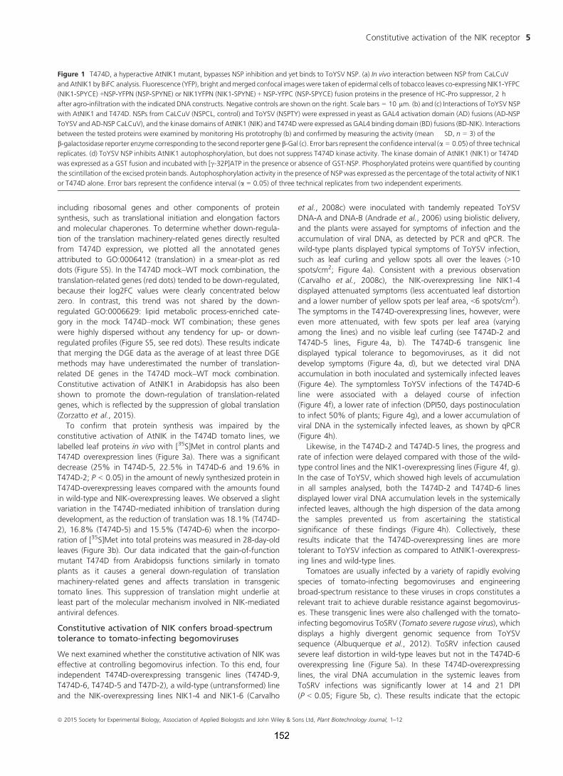
including ribosomal genes and other components of protein
synthesis, such as translational initiation and elongation factors
and molecular chaperones. To determine whether down-regula-
tion of the translation machinery-related genes directly resulted
from T474D expression, we plotted all the annotated genes
attributed to GO:0006412 (translation) in a smear-plot as red
dots (Figure S5). In the T474D mock–WT mock combination, the
translation-related genes (red dots) tended to be down-regulated,
because their log2FC values were clearly concentrated below
zero. In contrast, this trend was not shared by the down-
regulated GO:0006629: lipid metabolic process-enriched cate-
gory in the mock T474D–mock WT combination; these genes
were highly dispersed without any tendency for up- or down-
regulated profiles (Figure S5, see red dots). These results indicate
that merging the DGE data as the average of at least three DGE
methods may have underestimated the number of translation-
related DE genes in the T474D mock–WT mock combination.
Constitutive activation of AtNIK1 in Arabidopsis has also been
shown to promote the down-regulation of translation-related
genes, which is reflected by the suppression of global translation
(Zorzatto et al., 2015).
To confirm that protein synthesis was impaired by the
constitutive activation of AtNIK in the T474D tomato lines, we
labelled leaf proteins in vivo with [35S]Met in control plants and
T474D overexpression lines (Figure 3a). There was a significant
decrease (25% in T474D-5, 22.5% in T474D-6 and 19.6% in
T474D-2; P < 0.05) in the amount of newly synthesized protein in
T474D-overexpressing leaves compared with the amounts found
in wild-type and NIK-overexpressing leaves. We observed a slight
variation in the T474D-mediated inhibition of translation during
development, as the reduction of translation was 18.1% (T474D-
2), 16.8% (T474D-5) and 15.5% (T474D-6) when the incorpo-
ration of [35S]Met into total proteins was measured in 28-day-old
leaves (Figure 3b). Our data indicated that the gain-of-function
mutant T474D from Arabidopsis functions similarly in tomato
plants as it causes a general down-regulation of translation
machinery-related genes and affects translation in transgenic
tomato lines. This suppression of translation might underlie at
least part of the molecular mechanism involved in NIK-mediated
antiviral defences.
Constitutive activation of NIK confers broad-spectrumtolerance to tomato-infecting begomoviruses
We next examined whether the constitutive activation of NIK was
effective at controlling begomovirus infection. To this end, four
independent T474D-overexpressing transgenic lines (T474D-9,
T474D-6, T474D-5 and T47D-2), a wild-type (untransformed) line
and the NIK-overexpressing lines NIK1-4 and NIK1-6 (Carvalho
et al., 2008c) were inoculated with tandemly repeated ToYSV
DNA-A and DNA-B (Andrade et al., 2006) using biolistic delivery,
and the plants were assayed for symptoms of infection and the
accumulation of viral DNA, as detected by PCR and qPCR. The
wild-type plants displayed typical symptoms of ToYSV infection,
such as leaf curling and yellow spots all over the leaves (>10spots/cm2; Figure 4a). Consistent with a previous observation
(Carvalho et al., 2008c), the NIK-overexpressing line NIK1-4
displayed attenuated symptoms (less accentuated leaf distortion
and a lower number of yellow spots per leaf area, <6 spots/cm2).
The symptoms in the T474D-overexpressing lines, however, were
even more attenuated, with few spots per leaf area (varying
among the lines) and no visible leaf curling (see T474D-2 and
T474D-5 lines, Figure 4a, b). The T474D-6 transgenic line
displayed typical tolerance to begomoviruses, as it did not
develop symptoms (Figure 4a, d), but we detected viral DNA
accumulation in both inoculated and systemically infected leaves
(Figure 4e). The symptomless ToYSV infections of the T474D-6
line were associated with a delayed course of infection
(Figure 4f), a lower rate of infection (DPI50, days postinoculation
to infect 50% of plants; Figure 4g), and a lower accumulation of
viral DNA in the systemically infected leaves, as shown by qPCR
(Figure 4h).
Likewise, in the T474D-2 and T474D-5 lines, the progress and
rate of infection were delayed compared with those of the wild-
type control lines and the NIK1-overexpressing lines (Figure 4f, g).
In the case of ToYSV, which showed high levels of accumulation
in all samples analysed, both the T474D-2 and T474D-6 lines
displayed lower viral DNA accumulation levels in the systemically
infected leaves, although the high dispersion of the data among
the samples prevented us from ascertaining the statistical
significance of these findings (Figure 4h). Collectively, these
results indicate that the T474D-overexpressing lines are more
tolerant to ToYSV infection as compared to AtNIK1-overexpress-
ing lines and wild-type lines.
Tomatoes are usually infected by a variety of rapidly evolving
species of tomato-infecting begomoviruses and engineering
broad-spectrum resistance to these viruses in crops constitutes a
relevant trait to achieve durable resistance against begomovirus-
es. These transgenic lines were also challenged with the tomato-
infecting begomovirus ToSRV (Tomato severe rugose virus), which
displays a highly divergent genomic sequence from ToYSV
sequence (Albuquerque et al., 2012). ToSRV infection caused
severe leaf distortion in wild-type leaves but not in the T474D-6
overexpressing line (Figure 5a). In these T474D-overexpressing
lines, the viral DNA accumulation in the systemic leaves from
ToSRV infections was significantly lower at 14 and 21 DPI
(P < 0.05; Figure 5b, c). These results indicate that the ectopic
Figure 1 T474D, a hyperactive AtNIK1 mutant, bypasses NSP inhibition and yet binds to ToYSV NSP. (a) In vivo interaction between NSP from CaLCuV
and AtNIK1 by BiFC analysis. Fluorescence (YFP), bright andmerged confocal images were taken of epidermal cells of tobacco leaves co-expressing NIK1-YFPC
(NIK1-SPYCE) +NSP-YFPN (NSP-SPYNE) or NIK1YFPN (NIK1-SPYNE) + NSP-YFPC (NSP-SPYCE) fusion proteins in the presence of HC-Pro suppressor, 2 h
after agro-infiltration with the indicated DNA constructs. Negative controls are shown on the right. Scale bars = 10 lm. (b) and (c) Interactions of ToYSV NSP
with AtNIK1 and T474D. NSPs from CaLCuV (NSPCL, control) and ToYSV (NSPTY) were expressed in yeast as GAL4 activation domain (AD) fusions (AD-NSP
ToYSV and AD-NSP CaLCuV), and the kinase domains of AtNIK1 (NIK) and T474Dwere expressed as GAL4 binding domain (BD) fusions (BD-NIK). Interactions
between the tested proteins were examined by monitoring His prototrophy (b) and confirmed by measuring the activity (mean � SD, n = 3) of the
b-galactosidase reporter enzyme corresponding to the second reporter gene b-Gal (c). Error bars represent the confidence interval (a = 0.05) of three technical
replicates. (d) ToYSV NSP inhibits AtNIK1 autophosphorylation, but does not suppress T474D kinase activity. The kinase domain of AtNIK1 (NIK1) or T474D
was expressed as a GST fusion and incubated with [c-32P]ATP in the presence or absence of GST-NSP. Phosphorylated proteins were quantified by counting
the scintillation of the excised protein bands. Autophosphorylation activity in the presence of NSPwas expressed as the percentage of the total activity of NIK1
or T474D alone. Error bars represent the confidence interval (a = 0.05) of three technical replicates from two independent experiments.
ª 2015 Society for Experimental Biology, Association of Applied Biologists and John Wiley & Sons Ltd, Plant Biotechnology Journal, 1–12
Constitutive activation of the NIK receptor 5

expression of T474D in tomato confers tolerance to heterologous
species of tomato-infecting begomoviruses, which are phyloge-
netically separated within the two major groups of begomovi-
ruses found in Brazil (Albuquerque et al., 2012). The performance
of the T474D-overexpressing lines upon begomovirus infection
further confirmed that the T474D mutant protein could mount a
sustained NIK-mediated antiviral defence in the absence of viral
infection. The constitutive activation of T474D and its ability to
bypass viral NSP inhibition likely account for the tolerance to
begomovirus infection displayed by the transgenic lines.
Begomovirus infection does not affect resistance-relatedproteins and defence-related genes in the T474D-overexpressing lines
NIK belongs to the LRRII-RLK subfamily and shares high sequence
conservation and a similar receptor configuration with the
(a)
(c)
(d)
(b)
Figure 2 Transcriptomic analyses of mock-inoculated and infected (inf) T474D, NIK-OX and wild-type lines (a) Mock-inoculated T474D-overexpressing
lines display a constitutive infected wild-type (WT) transcriptome. Clustering analysis was performed using the R package pvclust (hierarchical
clustering with P-values via multiscale bootstrap resampling) using the TMM normalization method and the Ward agglomerative clustering method. The
dendrogram provides two types of P-values: AU (approximately unbiased – red, from multiscale bootstrap resampling) and BP (bootstrap probability –
green, normal bootstrap resampling). The AU P-value comes from multiscale bootstrap resampling, and the BP value represents normal bootstrap
resampling. These P-values were calculated by Multiscale Bootstrap Resampling using the R-cran package pvclust with a cut-off of 0.05. These P-values
show the significance of the proximity of each GE experiment profile. NIK-OX indicates NIK-overexpressing lines. (b) Elimination of the mock T474D–mock
WT DE genes from the raw data minimizes the gene expression variation as a result of virus infection. The hierarchical clustering of the gene expression
(GE) data was performed as described in Figure 2, except that the DE genes of mock T474D–mock WT were removed from the raw data from all
treatments prior to the clustering analysis. The red numbers indicate unbiased P-values (au), and the green ones indicate bootstrap probabilities. (c) and (d)
Bar graphs of the enriched down- (c) and up-regulated (d) categories based on biological process from the gene ontology (GO) database. Using at least
four DGE methods, the enriched GO categories were determined by at least three DGE methods and included more than three genes (Table S1).
ª 2015 Society for Experimental Biology, Association of Applied Biologists and John Wiley & Sons Ltd, Plant Biotechnology Journal, 1–12
Ot�avio J.B. Brustolini et al.6

well-characterized PAMP recognition co-receptor BRI1-associated
receptor kinase 1 (BAK1; Sakamoto et al., 2012; Shiu and
Bleecker, 2001). To understand whether the pre-activation of the
NIK-mediated antiviral response in the T474D-expressing lines
would prime typical BAK1-mediated defence responses, such as
PTI or SA signalling marker genes, in response to geminivirus
infection, we compared the transcriptome changes in infected
WT–mock WT vs infected T474D–mock T474D. We detected
eight resistance protein-related genes (R genes) that were up-
regulated by ToYSV infection in the T474D lines, but not in the
WT lines (Table S2). However, significant gene enrichment was
not detected for the R gene category, gene silencing category, or
abiotic stress response gene category, which includes the immune
system category and the response to SA marker genes, using the
gene set enrichment analysis (GSEA) method (Table S3). We also
compared the ToYSV infection-induced transcriptome of T474D
lines with that recently described for the tolerant TYLCV-resistant
tomato breeding line CLN2777A (Chen et al., 2013). None of the
40 defence-related genes up-regulated in CLN2777A in response
to TYLCV infection were T474D up-regulated genes, although we
found some similarities when functional terms were used as the
basis for comparison (Table S4). Collectively, these results indicate
that typical viral defences, such as activation of SA signalling,
gene silencing or the immune system, may not account for the
resistant mechanism of the T474D lines. These lines may invoke
an alternative mechanism for T474D protection against begomo-
virus infection.
The enhanced tolerance to begomovirus displayed bythe T474D-expressing lines may be associated with thetranslational control branch of the NIK-mediatedantiviral responses
The mock T474D–mock WT combination identified genes
affected by T474D expression in the absence of virus infection.
These data clearly showed that expression of the T474D mutant
in tomato led to a significant down-regulation of translation-
related genes and impaired translation, a phenotype associated
with the translational control branch of the NIK-mediated antiviral
signalling pathway, which has recently been described in
Arabidopsis (Zorzatto et al., 2015). Because T474D expression
caused a global down-regulation of protein synthesis, we
determined whether viral RNA translation was impaired in the
T474D-expressing lines. We investigated whether we could
detect viral RNA transcripts in actively translating polysome
fractions that had been separated from nonpolysomal fractions
on sucrose gradients (Figure S6). The polysomal fractions were
prepared from infected leaves at 10 DPI, when the accumulation
of viral mRNA in transgenic and wild-type leaves was similar
(Figure 6b). We observed a significant reduction in the polysome
loading of viral mRNA (coat protein mRNA) in systemically
infected leaves of the T474D-6-overexpresing line compared to
infected wild-type and NIK1-overexpressing leaves (Figure 6a, b).
The accumulation of total virus transcripts in all infected lines was
confirmed in our RNA sequencing data. These results indicated
that the begomovirus was not capable of sustaining high levels of
viral mRNA translation in the T474D-6-expressing lines, indicating
that suppression of global protein synthesis may effectively
protect plant cells against DNA viruses.
Discussion
Begomoviruses are one of the largest and most successful groups
of plant viruses and cause severe diseases in major crops
worldwide, inflicting significant economic losses in many dicot-
yledonous crops. The tomato-infecting begomoviruses have
become an even greater threat to tomato cultivation due to the
emergence of new species along with the recent introduction into
South America of a new biotype of the whitefly vector Bemisia
tabaci, which colonizes tomato plants with high efficiency
(Albuquerque et al., 2012; Castillo-Urquiza et al., 2008; Galv~ao
et al., 2003). Current climate changes are expected to further
alter the whitefly distribution across the globe, posing a serious
threat to agriculture worldwide. Here, we described a novel
strategy to control begomovirus infection. By constitutively
activating NIK-mediated antiviral signalling, we succeeded in
developing a tolerant tomato crop. Tomatoes are usually inflicted
by diverse begomoviruses, making engineered tolerant/resistant
lines even more difficult to develop. Importantly, the T474D-
overexpressing tomato transgenic lines were tolerant to ToYSV
and ToSRV, which display highly divergent genomic sequences
and hence are phylogenetically separated within the two major
groups of begomoviruses found in Brazil (Albuquerque et al.,
2012). The NIK-mediated strategy was also effective in conferring
tolerance to CaLCuV in the model plant Arabidopsis (Zorzatto
et al., 2015). These observations indicate the potential of a
sustained NIK-mediated defence pathway to confer broad-spec-
trum tolerance to begomoviruses in distinct plant species.
(a)
(b)
Figure 3 Ectopic expression of the T474 mutant receptor down-regulates
global protein synthesis in leaves of 10 days (a) old and 28 days old (b) tomato
plants. Equal fresh weights of tomato leaves (300 mg) were incubated with
50 lg/mL chloramphenicol and 20 lCi of [35S]methionine for 3 h at room
temperature. Incorporation of [35S]Met into protein was measured in the
TCA-precipitated total protein (mean � SD, n = 3, P < 0.05) from wild-type
and T474D transgenic lines. Asterisks indicate significant differences from the
wild-type control (P < 0.05, Student’s t-test).
ª 2015 Society for Experimental Biology, Association of Applied Biologists and John Wiley & Sons Ltd, Plant Biotechnology Journal, 1–12
Constitutive activation of the NIK receptor 7

The current mechanistic model for the activation of the
NIK-mediated antiviral signalling pathway holds that upon an
unknown stimulus, the NIK LRR extracellular domain undergoes
oligomerization with itself or another receptor, allowing the
intracellular kinase domains to transphosphorylate and activate
one another. The activation of NIK by phosphorylation on the
(a)
(b) (c)
(d)
(f) (g)(h)
(e)
ª 2015 Society for Experimental Biology, Association of Applied Biologists and John Wiley & Sons Ltd, Plant Biotechnology Journal, 1–12
Ot�avio J.B. Brustolini et al.8

crucial threonine residue at position 474 leads to the regulated
relocation of RPL10 to the nucleus where it interacts to LIMYB to
fully down-regulate translation-related genes (Zorzatto et al.,
2015). Our current data add a number of relevant insights to the
understanding of this layer of defence. First, a comparison
between the transcriptomes induced by virus infection in wild-
type lines and by ectopic expression of the T474D gain-of-function
mutant in transgenic lines indicated that activation of the NIK-
mediated signalling pathway triggers a typical response to virus
infection. This interpretation is supported by the observation that
mock-inoculated T474D-overexpressing lines showed a constitu-
tively infected wild-type transcriptome (Figure 2a). Furthermore,
the elimination of the mock T474D DE genes from the raw data of
all treatments further indicates that the expression profile induced
(a)
(b) (c)
Figure 5 The T474D-6 overexpressing line is also tolerant to ToSRV infection. (a) Symptoms associated with ToSRV infection in the 35S-T474D-6 line. The
T474D-6 line was infected with ToSRV, and photographs were taken at 21 DPI. (b) and (c) Viral DNA accumulation in the T474D-6- and T474-5-
overexpressing lines at 14 DPI (b) and 28 DPI (c). Prior to performing real-time PCR, infected leaves were diagnosed by standard PCR. Subsequently, total
DNA extracted from systemically infected leaves at 14 DPI (b) or 28 DPI (c) was used as a template for quantitative PCR using ToYSV DNA-A-specific primers.
The fold variation (�SD, n = 3 biological replicates) is shown as copy units of the viral genome. The asterisks indicate significant differences with P < 0.05
according to a Student’s t-test.
Figure 4 Ectopic expression of T474D in tomato confers tolerance to ToYSV infection. (a) Ectopic expression of T474D in tomato plants attenuates the
development of symptoms upon ToYSV infection. Tandemly repeated ToYSV DNA-A and DNA-B sequences were introduced into the indicated lines by
biolistic inoculation. Photographs were taken at 21 days postinoculation (DPI). (b) Symptoms associated with ToYSV infection in the 35S-T474D-5 line.
Photographs were taken at 21 DPI. (c) Viral DNA accumulation in the infected leaves of the T474-5 line. Total DNA was isolated from infected plants at 21
DPI, and PCR was performed with viral DNA-B-specific primers and actin-specific primers (as an internal control) in the same reaction. Lo indicates
inoculated and Sys denotes systemic leaves. ‘Un’ indicates mock-inoculated leaves. The gel shows representative samples of WT and the 35S::T474D-5
transgenic line. The upper band (1.2 kb, arrow) is the amplified genomic fragment from actin, and the lower band (0.5 kb, arrowhead) is the viral DNA
fragment. (d) and (e) The line 35S-T474D-6 displayed tolerance to ToYSV infection. The T474D-6 line was infected with ToYSV, and photographs were
taken at 21 DPI. Viral DNA accumulation was detected by PCR in inoculated (‘Lo’) and systemic (‘Sys’) leaves. (f) The course of infection was delayed in
T474D lines. The indicated lines were infected with ToYSV by the biolistic method, and the course of infection was monitored by PCR amplification of viral
DNA. In each experiment (three biological replicates), 20 plants of each line were inoculated. Values represent the percentage of systemically infected
plants at different DPI. (g) Infection efficiency in T474D-overexpressing lines. The infection efficiency is expressed as the DPI required to infect 50% of the
plants (mean � SD of three replicates). Asterisks indicate significantly different means (P < 0.05, Student’s t-test). (h) Viral DNA accumulation in T474D-
overexpressing lines, as determined by quantitative PCR at 28 DPI. The fold variation (�SD, n = 3 biological replicates) is shown as log2-scaled copy units of
the viral genome. Viral DNA accumulation was determined in systemic leaves at 28 DPI.
ª 2015 Society for Experimental Biology, Association of Applied Biologists and John Wiley & Sons Ltd, Plant Biotechnology Journal, 1–12
Constitutive activation of the NIK receptor 9

by the T474D mutant greatly mimics the response to viral
infection, as the mock- and infection-induced transcriptomes
from each genotype clustered together with high significance
(Figure 2b). Second, our data are consistent with the notion that
the gain-of-function mutant T474D can sustain an activated NIK-
mediated antiviral response in the absence of the virus, further
confirming that phosphorylation on Thr-474 is the crucial event
that leads to the activation of the kinase. In fact, expression of the
T474D mutant potentiated the NIK-mediated response, as it
would be expected from expression of a constitutively activated
defence receptor NIK. Accordingly, the ectopic expression of the
T474D gain-of-function mutant was more effective against
begomovirus infection than overexpression of the NIK defence
receptor in the tomato transgenic lines.
The experiments presented here shed light on the response
underlying NIK-mediated antiviral defences. We showed that
constitutive activation of NIK in the T474D lines impaired global
translation, and such activation might constitute an excellent
strategy for fighting begomovirus infection in host cells. We have
also demonstrated recently that constitutive activation of NIK1
and overexpression of LIMYB in Arabidopsis down-regulate
translation machinery-related genes, suppress translation and
confer tolerance to begomovirus infection (Zorzatto et al., 2015).
Because begomoviruses rely completely on the plant translation
machinery and cannot circumvent host translational regulation, a
global repression of translation is expected to significantly affect
virus infection, as observed in the T474D-overexpressing lines. In
fact, by directly assessing viral transcripts, we showed that the
loading of coat protein mRNA into actively translating polysomes
was significantly reduced in systemic infected leaves of T474D-
overexpressing plants compared to WT and NIK1-overexpressing
lines (Figure 6). This result indicates that the suppression of global
protein synthesis may effectively protect plant cells against DNA
viruses.
Based on our data, it is clear that the level of translational
inhibition mediated by constitutive activation of NIK1 did not
impact development in tomato under greenhouse conditions. In
the first generation, however, some transgenic lines displayed
shorter roots that regained normal biomass and growth in the
subsequent generations. As a possible explanation for this
phenotype, the T474D-mediated translation inhibition would
have maintained the transgenic lines under a constant perception
of stress, which, in turn, promoted acclimation to maintain
normal growth under greenhouse conditions. We also found that
T474D-mediated translation inhibition persisted in different
developmental stages of the transgenic lines but to different
extents. At later stages of development (28-day-old leaves), we
observed a 7% reduction in the level of translation inhibition
compared to that of seedlings. This difference in the efficiency of
global translation inhibition mediated by ectopic expression of the
T474D mutant receptor may reflect an adjustment of transgenic
lines towards adaptation at later stages. However, it is very
intriguing that ectopic expression of T474D did not impact
tomato development despite 19–25% suppression of global
translation at earlier stages of development. Therefore, the
intrinsic capacity to withstand the deleterious effect from the
suppression of global translation must be considered in attempts
to transfer the T474D-mediated defence strategy to other
agronomically relevant crops.
Experimental procedures (a detailed descriptionof experimental procedures is provided assupporting information)
Tomato transformation and RNA sequencingexperiment
The clone pK7F-NIK1T474D has been previously described
(Santos et al., 2009). It harbours a GFP gene fused in-frame after
the last codon of the mutant cDNA T474D under the control of
the CaMV 35S promoter. In the mutant cDNA T474D, the
threonine residue at position 474 within the activation loop of
NIK1 was mutated to an aspartate residue. Leaf discs from in
vitro-grown tomato plants (Solanum lycopersicum, cultivar
Moneymaker) were transformed with pK7F-NIK1T474D via
(a)
(b)
Figure 6 Polysome loading of viral mRNA is reduced in systemically
infected leaves in the T47D-6-overexpressing lines. (a) Polysome loading of
coat protein (CP) mRNA from ToYSV DNA-A in systemically infected leaves
of WT, NIK-overexpressing and T47D-6-overexpressing lines. Polysomes
from infected WT, NIK1-4-overexpressing and T474D-6-overexpressing
lines were isolated from systemic leaves at 10 days postinoculation with
tandem copies of DNA-A and DNA-B of ToYSV, as shown in Fig. S6. RNPs
refer to a 40S-enriched fraction. Polysome-bound RNA from pooled
fractions was extracted with phenol/chloroform/isoamyl alcohol,
precipitated with isopropanol, blotted and probed with the coat protein
DNA (CP) and 18S rDNA. The identity of the polysome-pooled fraction was
confirmed by treatment with 25 mM EDTA prior to the sucrose gradient
(data not shown), which releases the mRNA from the polysomes.
(b) Quantitation of polysome-associated coat protein viral transcripts in
T474D-overexpressing lines by qRT-PCR. Polysomes from infected WT,
NIK1-4-overexpressing and T474D-6-overexpressing lines were isolated
10 days postinoculation with infectious ToYSV clones. Polysome-bound
RNA from pooled fractions was extracted with phenol/chloroform/isoamyl
alcohol, precipitated with isopropanol and quantified by qRT-PCR. Values
were normalized to the expression of actin. Error bars represent SD from
three measurements.
ª 2015 Society for Experimental Biology, Association of Applied Biologists and John Wiley & Sons Ltd, Plant Biotechnology Journal, 1–12
Ot�avio J.B. Brustolini et al.10

Agrobacterium-mediated plant transformation (strain LBA4404),
as previously described (Carvalho et al., 2008c). The transgenic
lines T474D-2, T474D-5 and T474D-6 were selected for further
analysis. We also used the previously described NIK1-overex-
pressing lines NIK1-4 and NIK1-6 (Carvalho et al., 2008c) for
infection assays and RNA-seq analysis. NIK1-4 and NIK1-6 lines
harbour the Arabidopsis NIK cDNA fused to a GFP cDNA under
the control of the CaMV 35S promoter. The transgenic and wild-
type lines were infected at the six-leaf stage with ToYSV-[MG-
Bi2], as described below in the infectivity assay. At 10 days
postinoculation (dpi), total RNA was isolated using TRIzol for the
RNA sequencing experiments. The Illumina RNA sequencing data
were obtained using a Genome Analyzer in the Fasteris facilities.
A systematic comparison in our tomato data set of five
representative normalization methods with and without the
correction factors for CG-content and gene length was per-
formed using the Bioconductor packages edgeR (Robinson et al.,
2010), DESeq (Yang et al., 2013), and EDASeq (Risso et al.,
2011). For differential gene expression (DGE) analysis, we
subjected the normalized data provided by the counting table
to the most common negative binomial methods present in
R/Bioconductor software, such as edgeR (Robinson et al., 2010),
DESeq (Anders and Huber, 2010) and baySeq (Hardcastle and
Krystyna, 2010). The RNA-seq data were submitted to NCBI-GEO,
http://www.ncbi.nlm.nih.gov/geo/info/linking.html, accession
number GSM932558.
Infectivity assays
The transgenic and wild-type lines were infected at the six-leaf
stage with either ToYSV-[MG-Bi2] or ToSRV infectious clones by
biolistic delivery. In each experiment, 20 plants of each line were
inoculated with 2 lg of tandemly repeated DNA-A plus DNA-B
per plant and grown in a greenhouse under natural light, 70%
relative humidity and approximately equal day and night lengths.
Viral DNA was quantified by qRT-PCR, and coat protein viral
mRNA was quantified by qRT-PCR from polysome-associated RNA
prepared from T474D, NIK1 and wild-type infected leaves at 10
dpi.
Acknowledgements
This research was financially supported through the following
grants from Brazilian Government Agencies: CNPq grants
483659/2012-6, 573600/2008-2, 447578/2014-6 (to E.P.B.F.),
FAPEMIG grant CBB-APQ-00070-09 (to E.P.B.F.). O.J.B.B. and
D.C were supported by CAPES graduate fellowships; J.P.B.M. was
supported by a CNPq graduate fellowship; M.D. and W.A.P. were
supported by postdoctoral fellowships from CNPq; A.A.S. was the
recipient of a postdoctoral fellowship from CAPES.
Conflict of interest
The authors declare no conflict of interest.
References
Albuquerque, L.C., Varsani, A., Fernandes, F.R., Pinheiro, B., Martin, D.P., de
Tarso, O.F.P., Lemos, T.O. and Inoue-Nagata, A.K. (2012) Further
characterization of tomato-infecting begomoviruses in Brazil. Arch. Virol.
157, 747–752.
Anders, S. and Huber, W. (2010) Differential expression analysis for sequence
count data. Genome Biol. 11, R106.
Andrade, E.C., Manhani, G.G., Alfenas, P.F., Calegario, R.F., Fontes, E.P.B. and
Zerbini, F.M. (2006) Tomato yellow spot virus, a tomato-infecting
begomovirus from Brazil with a closer relationship to viruses from Sida sp.,
forms pseudorecombinants with begomoviruses from tomato but not from
Sida. J. Gen. Virol. 87, 3687–3696.
Arag~ao, F.J. and Faria, J.C. (2009) First transgenic geminivirus-resistant plant in
the field. Nat. Biotechnol. 27, 1086–1088.
Carvalho, C.M., Machado, J.P.B., Zerbini, F.M. and Fontes, E.B.P. (2008a) NSP-
Interacting GTPase: a cytosolic protein as cofactor for nuclear shuttle
proteins. Plant Signal. Behav. 3, 752–754.
Carvalho, C.M., Fontenelle, M.R., Florentino, L.H., Santos, A.A., Zerbini, F.M.
and Fontes, E.P.B. (2008b) A novel nucleocytoplasmic traffic GTPase
identified as a functional target of the bipartite geminivirus nuclear shuttle
protein. Plant J. 55, 869–880.
Carvalho, C.M., Santos, A.A., Pires, S.R., Rocha, C.S., Saraiva, D.I., Machado,
J.P., Mattos, E.C., Fietto, L.G. and Fontes, E.P.B. (2008c) Regulated nuclear
trafficking of rpL10A mediated by NIK1 represents a defense strategy of plant
cells against virus. PLoS Pathog. 4, e1000247.
Castillo-Urquiza, G.P., Beserra, J.E. Jr, Bruckner, F.P., Lima, A.T., Varsani, A.,
Alfenas-Zerbini, P. and Zerbini, F.M. (2008) Six novel begomoviruses
infecting tomato and associated weeds in Southeastern Brazil. Arch. Virol.
153, 1985–1989.
Chen, T., Lv, Y., Zhao, T., Li, N., Yang, Y., Yu, W., He, X., Liu, T. and Zhang, B.
(2013) Comparative transcriptome profiling of a resistant vs. susceptible
tomato (Solanum lycopersicum) cultivar in response to infection by tomato
yellow leaf curl virus. PLoS ONE, 8, e80816.
Day, A.G., Bejarano, E.R., Buck, K.W., Burrell, M. and Lichtenstein, C.P. (1991)
Expression of an antisense viral gene in transgenic tobacco confers resistance
to the DNA virus tomato golden mosaic virus. Proc. Natl Acad. Sci. USA, 88,
6721–6725.
Florentino, L.H., Santos, A.A., Fontenelle, M.R., Pinheiro, G.L., Zerbini, F.M.,
Baracat-Pereira, M.C. and Fontes, E.P.B. (2006) A PERK-like receptor kinase
interacts with the geminivirus nuclear shuttle protein and potentiates viral
infection. J. Virol. 80, 6648–6656.
Fontes, E.P.B., Santos, A.A., Luz, D.F., Waclawovsky, A.J. and Chory, J. (2004)
The geminivirus NSP acts as virulence factor to suppress an innate
transmembrane receptor kinase-mediated defense signaling. Gene Dev. 18,
2545–2556.
Gafni, Y. and Epel, B.L. (2002) The role of host and viral proteins in intra- and
intercellular trafficking of geminiviruses. Physiol. Mol. Plant Pathol. 60,
231–241.
Galv~ao, R.M., Mariano, A.C., Luz, D.F., Alfenas, P.F., Andrade, E.C., Zerbini,
F.M., Almeida, M.R. and Fontes, E.P.B. (2003) A naturally occurring
recombinant DNA-A of a typical bipartite begomovirus does not require the
cognate DNA-B to infect Nicotiana benthamiana systemically. J. Gen. Virol.
84, 715–726.
Hardcastle, T.J. and Krystyna, A.K. (2010) baySeq: empirical Bayesian methods
for identifying differential expression in sequence count data. BMC
Bioinformatics, 11, 422.
Hashmi, J.A., Zafar, Y., Arshad, M., Mansoor, S. and Asad, S. (2011) Engineering
cotton (Gossypium hirsutum L.) for resistance to cotton leaf curl disease using
viral truncated AC1 DNA sequences. Virus Genes, 42, 286–296.
Hong, Y. and Stanley, J. (1996) Virus resistance in Nicotiana benthamiana
conferred by African cassava mosaic virus replication-associated protein
(AC1) transgene. Mol. Plant Microbe Interact. 9, 219–225.
Kunik, T., Salomon, R., Zamir, D., Navot, N., Zeidan, M., Michelson, I., Gafni, Y.
and Czosnek, H. (1994) Transgenic tomato plants expressing the tomato
yellow leaf curl virus capsid protein are resistant to the virus. Biotechnology,
12, 500–504.
Lin, C.Y., Tsai, W.S., Ku, H.M. and Jan, F.J. (2012) Evaluation of DNA fragments
covering the entire genome of a monopartite begomovirus for induction of
viral resistance in transgenic plants via gene silencing. Transgenic Res. 21,
231–241.
Lucioli, A., Sallustio, D.E., Barboni, D., Berardi, A., Papacchioli, V., Tavazza, R.
and Tavazza, M. (2008) A cautionary note on pathogen-derived sequences.
Nat. Biotechnol. 26, 617–619.
Mariano, A.C., Andrade, M.O., Santos, A.A., Carolino, S.M.B., Oliveira, M.L.,
Baracat-Pereira, M.C., Brommonshenkel, S.H. and Fontes, E.P.B. (2004)
ª 2015 Society for Experimental Biology, Association of Applied Biologists and John Wiley & Sons Ltd, Plant Biotechnology Journal, 1–12
Constitutive activation of the NIK receptor 11

Identification of a novel receptor- like protein kinase that interacts with a
geminivirus nuclear shuttle protein. Virology, 318, 24–31.
McGarry, R.C., Barron, Y.D., Carvalho,M.F., Hill, J.E., Gold, D., Cheung, E., Kraus,
W.L. and Lazarowitz, S.G. (2003) A novel Arabidopsis acetyltransferase
interacts with the geminivirus movement protein NSP. Plant Cell, 15, 1605–
1618.
Noris, E., Accotto, G.P., Tavazza, R., Brunetti, A., Crespi, S. and Tavazza, M.
(1996) Resistance to tomato yellow leaf curl geminivirus in Nicotiana
benthamiana plants transformed with a truncated viral C1 gene. Virology,
224, 130–138.
Reyes, M.I., Nash, T.E., Dallas, M.M., Ascencio-Ib�a~nez, J.T. and Hanley-
Bowdoin, L. (2013) Peptide aptamers that bind to geminivirus replication
proteins confer a resistance phenotype to TYLCV and ToMoV infection in
tomato. J. Virol. 87, 9691–9706.
Risso, D., Schwartz, K., Sherlock, G. and Dudoit, S. (2011) GC-content
normalization for RNA-Seq data. BMC Bioinformatics, 12, 480.
Robinson, M.D., McCarthy, D.J. and Smyth, G.K. (2010) edgeR: a Bioconductor
package for differential expression analysis of digital gene expression data.
Bioinformatics, 26, 139–140.
Rocha, C.S., Santos, A.A., Machado, J.P.B. and Fontes, E.P.B. (2008) The
ribosomal protein L10/QM-like protein is a component of the NIK-mediated
antiviral signaling. Virology, 380, 165–169.
Rojas, M.R., Hagen, C., Lucas, W.J. and Gilbertson, R.L. (2005) Exploiting chinks
in the plant’s armor: evolution and emergence of geminiviruses. Annu. Rev.
Phytopathol., 43, 361–394.
Sakamoto, T., Deguchi, M., Brustolini, O.J.B., Santos, A.A., Silva, F.F. and
Fontes, E.P.B. (2012) The tomato RLK superfamily: phylogeny and functional
predictions about the role of the LRRII-RLK subfamily in antiviral defense.
BMC Plant Biol. 12, 229.
Sanderfoot, A.A. and Lazarowitz, S.G. (1995) Cooperation in viral movement:
the geminivirus BL1 movement protein interacts with BR1 and redirects it
from the nucleus to the cell periphery. Plant Cell, 7, 1185–1194.
Sanderfoot, A.A. and Lazarowitz, S.G. (1996) Getting it together in plant virus
movement: cooperative interactions between bipartite geminivirus
movement proteins. Trends Cell Biol. 6, 353–358.
Sanderfoot, A.A., Ingham, D.J. and Lazarowitz, S.G. (1996) A viral movement
protein as a nuclear shuttle: the geminivirus BR1 movement protein contains
domains essential for interaction with BL1 and nuclear localization. Plant
Physiol. 110, 23–33.
Santos, A.A., Carvalho, C.M., Florentino, L.H., Ramos, H.J. and Fontes, E.P.B.
(2009) Conserved threonine residues within the A-Loop of the receptor NIK
differentially regulate the Kinase function required for antiviral signaling.
PLoS ONE, 4, e5781.
Santos, A.A., Lopes, K.V.G., Apfta, J.A.C. and Fontes, E.P.B. (2010) NSP-
interacting kinase, NIK: a transducer of plant defence signalling. J. Exp. Bot.
61, 3839–3845.
Shiu, S.H. and Bleecker, A.B. (2001) Receptor-like kinases from Arabidopsis
form a monophyletic gene family related to animal receptor kinases. Proc.
Natl Acad. Sci. USA, 98, 10763–10768.
Stanley, J., Frischmuth, T. and Ellwood, S. (1991) Defective viral DNA
ameliorates symptoms of geminivirus infection in transgenic plants. Proc.
Natl Acad. Sci. USA, 87, 6291–6295.
Sunitha, S., Marian, D., Hohn, B. and Veluthambi, K. (2011) Antibegomoviral
activity of the agrobacterial virulence protein VirE2. Virus Genes, 43, 445–453.
Ward, B.M. and Lazarowitz, S.G. (1999) Nuclear export in plants: use of
geminivirus movement proteins for a cell-based export assay. Plant Cell, 11,
1267–1276.
Yang, E.-W., Girke, T. and Jiang, T. (2013) Differential gene expression analysis
using coexpression and RNA-Seq data. Bioinformatics, 29, 2153–2161.
Zhang, S.C., Wege, C. and Jeske, H. (2001) Movement proteins (BC1 and BV1)
of Abutilon mosaic geminivirus are cotransported in and between cells of sink
but not of source leaves as detected by green fluorescent protein tagging.
Virology, 290, 249–260.
Zorzatto, C., Machado, J.P.B., Lopes, K.V.G., Nascimento, K.J.T., Pereira, W.A.,
Brustolini, O.J.B., Reis, P.A.B., Calil, I.P., Deguchi, M., Martins, G.S., Gouveia,
B.C., Loriato, V.A.P., Silva, M.A.C., Silva, F.F., Santos, A.A., Chory, J. and
Fontes, E.P.B. (2015) NIK-mediated translation suppression functions as a
plant antiviral immunity mechanism. Nature, doi: 10.1038/nature14171.
Supporting information
Additional Supporting information may be found in the online
version of this article:
Data S1 Supporting Experimental Procedures – detailed descrip-
tion of experimental procedures.
Figure S1 NSP from CaLCuV and ToYSV concentrates in the
nucleus when ectopically expressed in N. benthamiana leaves.
Figure S2 Overexpression of T474D mutant receptor in tomato
leaves.
Figure S3 Characterization of the T474D-overexpressing lines
during the vegetative phase.
Figure S4 Fruit quality and yield of the T474D-overexpressing
lines.
Figure S5 Representation of the translational machinery-related
genes in the down-regulated changes.
Figure S6 Isolation of polysomal fractions from tomato seedlings.
Table S1 Enriched biological process categories from the GO
database using the GSEA method.
Table S2 Resistance protein-related genes differentially expressed
in infected T474D (infected T474D-mock T474D).
Table S3 Gene Enrichment Analysis.
Table S4 Functional overlap of DE defence-related genes up-
regulated in CLN2777 (resistant) by 3, 5, and 7 dpi and in T474D
infected by 10 dpi.
ª 2015 Society for Experimental Biology, Association of Applied Biologists and John Wiley & Sons Ltd, Plant Biotechnology Journal, 1–12
Ot�avio J.B. Brustolini et al.12

Supporting information- Experimental Procedures
Sustained NIK-mediated antiviral signaling confers broad-spectrum tolerance
to begomoviruses in cultivated plants
Otávio J.B.Brustolini a,b*
, Joao Paulo B. Machadoa,b*
, Jorge A. Condori-Apfata b
, Daniela
Coco a,b
, Michihito Deguchia,b
, Virgílio A. P. Loriatoa,b
, Welison A. Pereira, Poliana
Alfenas-Zerbini b
, Francisco M. Zerbini b
, Alice K. Inoue-Nagatab,d
, Anesia A. Santos a,b
,
Joanne Chorye, Fabyano F. Silva
c, Elizabeth P.B. Fontes
a,b,&
aDepartamento de Bioquímica e Biologia Molecular
bNational Institute of Science and
Technology in Plant-Pest Interactions, Bioagro, cDepartamento de Zootecnia, Universidade
Federal de Viçosa, 36570.000, Viçosa, MG, Brazil. dEmbrapa Vegetables, Brasília, DF 70359-
970, Brazil. eHoward Hughes Medical Institute and Plant Biology Laboratory, The Salk Institute
for Biological Studies, La Jolla, California 92037, USA
*These authors contributed equally to this work
&For correspondence:
Elizabeth B. P. Fontes, Departamento de Bioquímica e Biologia Molecular/Bioagro, National
Institute of Science and Technology in Plant-Pest Interactions, Universidade Federal de Viçosa,
36570.000, Viçosa, MG, Brazil, pone +55 31 3899 2948 &
email [email protected]

Yeast strain and two-hybrid assays. The NSP ORF was amplified from ToYSV DNA-B using PCR
with the appropriate linkers and introduced through recombination into the entry vector pDONR201 to
generate pDON-NSP-ToYSV, also designated as pUFV1323; the NSP ORF was subsequently transferred
to the pDEST22 vector. The resulting clone pAD-NSP-ToYSV, also designated pUFV1370 or AD-
NSPTY (Figure 1B), contained the GAL4 DNA-activating domain fused to the NSP-ToYSV sequence.
Sequences encoding the mutant (T474D) kinase domain of NIK were transferred from pDON-T474D
(Santos et al., 2009) to pDEST32 to yield pBD-T474D or BD-T474D (Figure 1B). The clones pAD-NSP-
CaLCuV (AD-NSPCL, in Figure 1B) and pBD-KDNIK (BD-NIK in Figure 1B) have been previously
described (Fontes et al., 2004).
The yeast two-hybrid assays were performed as previously described (Florentino et al., 2006). The yeast
reporter strain AH109 (MATa, Trp1-901, leu2-3, 112, ura3-52, his3-200, gal4Δ, LYS2::GAL1UAS-
GAL1TATA-HIS3, MEL1 GAL2UAS-GALTATA::MELUAS-MEL1TATA-lacZ,) was cotransformed with AD-NSP
fusions and pBD-NIK1 or pBD-T474D. The interactions were monitored by the ability of the reporter
strain to grow on media lacking leucine, tryptophan, uracil and histidine, but supplemented with 25 mM
3-aminotriazole for 7 days at 30 0C. The interactions were further confirmed by measuring β-
galactosidase activity in yeast extracts using o-nitrophenyl β-D-galactopyranoside substrate, as described
previously (Uhrig et al., 1999).
Phosphorylation assay. To create plasmids for E. coli expression, the mutant NIK1 C-terminal kinase
domain or intact NIK (KD, encoding amino acids 298–638) were amplified from the appropriate mutated
clone, introduced by recombination into the entry vector pDONR201 and then transferred to the bacterial
expression vector pDEST15, resulting in GST fused to mutant kinase domains, designated pGST-
KDNIK1T474A or pGST-KDNIK1. The E. coli-expressed and purified KDNIK1T474D and
KDNIK1recombinant proteins were incubated alone or with NSP from CaLCuV, ToYSV or ToSRV at
25C for 45 min in 30 L of kinase buffer containing 18 mM HEPES pH 7.4, 10 mM MgCl2, 10 mM
MnSO4, 1 mM DTT, 10 M ATP and 5 Ci [-32
P]ATP. Phosphoproteins were resolved by SDS-PAGE.

The gel was stained with Coomassie brilliant blue to verify protein loading, dried, and subjected to
autoradiography. Incorporated radioactivity in protein bands was quantified by phosphoimaging and
protein loading by densitometry using the Multi Gauge V3.0 software (Fujifilm).
Bimolecular fluorescence complementation (BiFC) analysis. For biochemical complementation of
fluorescent fragments of yellow fluorescent protein (YFP), NSP and NIK were fused to the N-terminus
(N) or C-terminus (C) of the reporter gene. Then, constructs expressing NSP-YFPC, NSP-YFPN, NIK-
YFPC and NIK-YFPN in different combinations were co-agroinfiltrated in tobacco leaves in the presence
of the suppressor of silencing HC-Pro according to a previously described protocol (Carvalho et al.,
2008c). YFP fluorescence was observed by confocal microscopy.
Tomato transformation. The clone pK7F-NIK1T474D has been previously described (Santos et al.,
2009). It harbors a GFP gene fused in-frame after the last codon of the mutant cDNA T474D under the
control of the CaMV 35S promoter. In the mutant cDNA T474D, the threonine residue at position 474
within the activation loop of NIK1 was mutated to an aspartate residue. Leaf discs from in vitro-grown
tomato plants (Solanum lycopersicum, cultivar Moneymaker) were transformed with pK7F-NIK1T474D
via Agrobacterium-mediated plant transformation (strain LBA4404). The transformed shoots were
selected on MS medium supplemented with 6-benzylaminopurine (500 mg.L-1
), cefotaxime (300 mg.L-1
),
and kanamycin sulfate (50 mg.L-1
). The regenerated shoots were rooted, transferred into soil and grown
under standardized greenhouse conditions to generate seeds. The transgenic lines were confirmed using
PCR. The analysis of transgene expression was performed by RT-PCR and real-time RT-PCR using
transgene-specific primers, and actin was used as an endogenous control to normalize all values. The
transgenic lines 35S:NIK1-4 and 35S:NIK1-6 have been previously described (Carvalho et al., 2008c).
They harbor the Arabidopsis NIK cDNA fused to a GFP cDNA under the control of the CaMV 35S
promoter.

RT-PCR and real-time RT-PCR analyses. Total RNA was extracted from tomato leaves using TRIzol
(Invitrogen). The reverse transcription (RT)-PCR assays were performed with 2 µg of total RNA, 0.5 mM
of poly-dT and 1 U of M-MLV reverse transcriptase (Invitrogen Life Technologies, Inc.) as previously
described (Delu-Filho et al., 2000). The PCR reaction was performed using virus-specific primers. The
PCR comprised 30 cycles of 45 s at 94 °C, 30 s at 55 °C and 2 min at 72 °C. The real-time RT-PCR
reactions were performed on an ABI7500 instrument (Applied Biosystems, Foster City, CA) using the
SYBR Green PCR Master Mix (Applied Biosystems) as previously described (Costa et al., 2008). The
amplification reactions were performed as follows: 2 min at 50°C, 10 min at 95°C and 40 cycles of 94°C
for 15 sec and 60°C for 1 min. To confirm the quality and primer specificity, we verified the size of
amplification products using 1.5% agarose gel electrophoresis and analyzed the Tm (melting temperature)
of the amplification products in a dissociation curve performed on the ABI7500 instrument. Actin was
used as an endogenous control to normalize all values in the real-time RT-PCR assays. Gene expression
was quantified using the 2∆C
T method. The fold variation of gene expression was quantified using the
comparative Ct method: 2-(ΔCtTreatment - ΔCtControl)
.
Immunoblot analysis. Total protein was extracted from the leaves of wild-type and T474D-
overexpressing tomato transgenic seedlings as previously described (Cascardo et al., 2000). Following
SDS-PAGE, the proteins were transferred from 10% SDS-polyacrylamide gels to nitrocellulose
membranes by electroblotting. The membranes were blocked in NaCl/Tris containing 0.05% v/v Tween-
20 and 5% w/v non-fat dry milk, and they were subsequently incubated with rabbit anti-GFP at a 1:10,000
dilution for 2 h at room temperature. The bound antibody was detected using alkaline-phosphatase-
conjugated goat anti-rabbit IgG serum in conjunction with nitroblue tetrazolium/5-bromo-4-chloro-3-
indolyl phosphate detection reagents (Bio-Rad).
RNA sequencing method and data analysis. The transgenic and wild-type lines were infected at the six-
leaf stage with ToYSV-[MG-Bi2], as described below in infectivity assay. After 10 days post-inoculation,

total RNA from systemically infected leaves, as diagnosed by PCR, and mock-inoculated leaves from
wild-type, 35S::NIK1-4 and 35S::T474D lines was isolated using TRIzol (Invitrogen). For the RNA
sequencing experiments we used two biological replicates of a pool of 10 plants at 10 days after
inoculation when we detected high levels of viral DNA in systemic leaves but symptoms were not visible
as yet. The experimental design was 6 treatments as follow: mock-inoculated (mock WT, mock NIK1-
OX, mock T474D) lines and infected (inf WT, inf NIK1-OX, inf T474D) lines with 2 repetitions. The
RNA sequencing Illumina data was obtained using a Genome Analyzer in the Fasteris facilities. The
GEX-NIaIII protocol was used with the following quality filter parameters: maximum of 1 base below a
quality of 5 in the first 30 bases, a minimum average quality of 10, no “N” calls allowed and not more
than 35 identical bases (low information reads). The data were stored in a comma-separated values (csv)
spreadsheet file.
A systematic comparison in our tomato data set of five representative normalization methods with
and without the correction factors for CG-content and gene length was performed using the Bioconductor
packages edgeR (Robinson et al., 2010), DESeq (Yang et al., 2013), and EDASeq (Risso et al., 2011). We
have also adopted as a comparative experimental design a pairwise comparison and a false discover rate
(FDR) p-value adjustment with the cutoff < 0.05. Four of those normalization methods were implemented
on the edgeR, such as total count (TC), upper quartile (UQ) (Bullard et al., 2010), relative log expression
(RLE) (Anders and Huber, 2010) and trimmed mean of M values (TMM) (Robinson and Oshlack, 2010);
the fifth was the normalization method implemented direct in DESeq package. The GC-content and gene
length correction factors were performed by the Bioconductor package EDASeq (Risso et al., 2011). For
differential gene expression (DGE) analysis, we employed the normalized data provided by the counting
table to the most common negative binomial methods present in R/Bioconductor software, such as edgeR
(Robinson et al., 2010), DESeq (Anders and Huber, 2010) and baySeq (Hardcastle et al., 2010). The
parameter of dispersion was estimated by the tagwise program. Differential expression was determined
using the FDR adjusted cutoff p-value of 0.05. The read mapping process was executed using the Bowtie
program (Langmead et al., 2009) with the cDNA data set retrieved from the International Tomato

Annotation Group (ITAG - http://solgenomics.net/organism/Solanum_lycopersicum/genome), second
release. Gene ontology classification was performed using the R/Bioconductor packages GSEABase and
GOstats. The entire annotation data set from ITAG/Phytozome (http://www.phytozome.net) was stored in
the relational database PostgreSQL 9.3 (http://www.postgresql.org). To detect gene set enrichment from
our RNA-seq DE data, we used the GSEA method provided by the R/Bioconductor GSEABase package
based on the Gene Ontology (GO) database (Ashburner et al., 2000). Clustering analysis was performed
using the R package pvclust (Hierarchical Clustering with P-Values via Multiscale Bootstrap
Resampling) using Ward's method (Ward, 1963), and heatmaps were generated using gplots. The results
were stored in a relational database created in PostgreSQL, and a web interface was created using PHP to
allow the database to be accessed and navigated (http://tomatodb.inctipp.ufv.br). The RNA-seq data of
T474D lines were submitted to NCBI-GEO, http://www.ncbi.nlm.nih.gov/geo/info/linking.html,
accession number GSM932558.
In vivo labeling of leaf proteins. Tomato seedlings (300 mg) were incubated with 1 mL of nutrient
solution containing 50 µg/ml chloramphenicol and 20 µCi of [35S]methionine (EasyTag Protein Labeling
Mix, [35
S]-, 2mCi (74MBq), Perkin Elmer) for 3 h at room temperature. To quantitate incorporation of
[35
S]methionine into protein, aliquots of protein extracts were placed in 10% (w/v) TCA and incubated on
ice for 30 min. The samples were filtered onto glass microfiber filters and the filters were washed three
times with 5 ml of cold 5% (w/v) TCA and two times with 5 ml of 95% ethanol. After drying, the filters
were counted with a scintillation counter.
Polysome fractionation. Polysomes were fractionated over sucrose gradients as described (Wang et al.,
2003). Briefly, 500 mg of 15-day-old tomato seedlings were ground in liquid nitrogen and 1 mL of
extraction buffer (0.2 M Tris-HCl, pH 8.0, 50 mM KCl, 25 mM MgCl2, 1% Triton X-100, 400 units/mL
of RNasin and 50 mg/mL of cycloheximide). After centrifuging for 10 min, the supernatant was loaded
onto a 10-mL 15% to 50% sucrose gradient and spun in a Beckman SW41Ti rotor at 135,000 g for 3.5 h.

Fractions were collected manually from the bottom, and total RNA was extracted with
phenol/chloroform/isoamyl alcohol, precipitated with isopropanol, and treated with DNase I. The specific
transcripts were amplified from RNA of T474D, NIK1 and wilt-type infected lines using qRT-PCR.
Infectivity assays. For the infectivity assays, we used T2 transgenic plants harboring the T474D mutant
gene construct, which were derived from four independently regenerated kanamycin-resistant plants
(35S::T474D-2, 35S::T474D-5, 35S::T474D-6 and 35S::T474D-9). We also used the previously
described transgenic lines expressing AtNIK1 under the control of the CaMV 35S promoter, 35S::NIK1-4
and 35S::NIK1-6 (Carvalho et al., 2008c). The transgenic and wild-type lines were infected at the six-leaf
stage with either ToYSV-[MG-Bi2] or ToSRV by biolistic delivery using tandemly repeated viral DNA-
A and DNA-B and a microprojectile bombardment model PDS-1000/He accelerator (BIORAD) at 900
psi. In each experiment, 20 plants of each line were inoculated with 2 μg of tandemly repeated DNA-A
plus DNA-B per plant and grown in a greenhouse under natural conditions of light, 70% relative humidity
and approximately equal day and night lengths. Total nucleic acid was extracted from the systemically
infected leaves (young leaves), and viral DNA was detected by PCR using DNA-A and DNA-B
begomovirus-specific primers (PBL1v 2040, GCCTCTGCAGCARTGRTCKATCTTCATACA, and
PCRC1, CTAGCTGCAGCATATTTACRARWATGCCA, or PAL1v1978,
GCATCTGCAGGCCCACATYGTCTTYCCNGT, and PAR1c496,
AATACTGCAGGGCTTYCTRTACATRGG) at 10 days post-infection.
Quantitation of viral DNA in infected plants. Viral DNA accumulation was measured by quantitative
PCR (qPCR). The reactions were prepared in a final volume of 10 µl using the Fast SYBR Green Master
Mix (Applied Biosystems) according to the manufacturer’s instructions and analyzed on a 7500 Real
Time PCR System (Applied Biosystems). Virus-specific primers were designed using Primer Express 3.0
(Applied Biosystems) and tested by conventional PCR using plasmids containing the complete DNA-A of
each virus (106 copies per reaction). The following primer sequences were used: ToSRVFwd,

CACGTGCCCACATCGTCTT, and ToSRVRev, GGCCGGAACGACCTATTA-3’, or ToYSVFwd,
CCACGATTTTAAAGCTGCATTCT, and ToYSVRev, CAATCCTGGTGAGGGAGTCAGT. For viral
DNA quantitation, standard curves were prepared using serial dilutions of these clones (100 to 10
6 copies
of viral genome per reaction). The genomic unit refers to one copy of the DNA-A of ToYSV or
ToSRV. Standard curves were obtained by regression analysis of the Ct values of each of the three
replicates of a given dilution in relation to the log of the amount of DNA in each dilution. For the absolute
quantitation of the number of viral DNA molecules in the different treatments, 100 ng of total DNA from
the infected plants was used in the qPCR reactions containing virus-specific primers. Each sample was
analyzed in triplicate from at least two biological replicates.
Physiological measurements of tomato transgenic lines. Photosynthetic CO2 assimilation (A),
transpiration rate (E), and stomatal conductance (gs) measurements were performed with a portable open-
flow gas exchange system (LICOR 6400, Li-COR, Lincoln, Nebraska, USA) under ambient CO2
concentrations (370 ± 10 µmol mol-1) and temperature conditions under artificial, saturating PAR (1,000
umol photons m-2
s-1 at the leaf level).
Estimation of total carotenoids, lycopene, and β-carotene. Total carotenoids from fully ripened tomato
fruits were extracted with cold acetone and petroleum ether, as described by Rodriguez-Amaya et al.
(1976). The extract (30 µL) was separated by HPLC using reversed phase C18 column (Phenomenex
Gemini, 250 x 4,6 mm, 5 μm) and a C18 guard column (Phenomenex ODS, 4 mm x 3 mm) on a
Shimadzu, SCL 10AT VP HPLC system coupled to a DAD detector (Shimadzu, SPD-M10A) and
operating at a flow rate of 1.7 mL/min. The mobile phase buffer used was methanol:ethyl
acetate:acetonitrile (70:20:10, v/v/v). The chromatograms were obtained at 450 nm and integrated using
the software Multi System Class Vp 6.12. Lycopene and β-carotene were quantified from HPLC profile

by using a purified lycopene standard (Sigma Chemical Co.) and a β-carotene standard purified from
carrots.
References
Anders, S. and Huber ,W. (2010) Differential Expression Analysis for Sequence Count Data. Genome
Biol., 11, R106.
Ashburner, M. C., Ball, J., Blake, J.A., Botstein, D., Butler, H., Cherry, J.M., Davis, A.P. Dolinski, K.,
Dwight, S.S., Eppig, J.T., Harris, M.A., Hill, D.P., Issel-Tarver, L., Kasarskis, A., Lewis ,S., Matese,
J.C., Richardson, J.E., Ringwald, M., Rubin, G.M. and Sherlock, G. (2000) Gene Ontology: Tool for
the Unification of Biology. The Gene Ontology Consortium. Nat. Genet., 25, 25–29.
Bullard, J.H., Purdom, E., Hansen, K.D. and Dudoit, S. (2010). Evaluation of Statistical Methods for
Normalization and Differential Expression in mRNA-Seq Experiments. BMC Bioinformatics, 11, 94.
Cascardo JCM, Almeida RS, Buzeli RAA, Carolino SMB, Otoni WC and Fontes E.P.B. (2000) The
phosphorylation state and expression of soybean BiP isoforms are differentially regulated following
abiotic stresses. J. Biol. Chem., 275, 14494–14500.
Costa, M.D.L., Reis, P.A.B., Valente, M.A.S., Irsigler, A.S.T., Carvalho, C.M., Loureiro, M.E., Aragão,
F.J., Boston, R.S., Fietto, L.G. and Fontes, E.P.B. (2008) A new branch of endoplasmic reticulum-
stress signaling and the osmotic signal converge on plant specific asparagine-rich proteins to promote
cell death. J. Biol. Chem., 283, 20209–20219.
Delú-Filho, N., Pirovani, C.P., Pedra, J.H.F., Matrangolo, F.S.V., Macêdo, J.N.A., Otoni, W.C. and
Fontes, E.P.B. (2000) A sucrose binding protein homologue from soybean affects sucrose uptake in
transgenic tobacco suspension-cultured cells. Plant Physiol. Biochem., 38, 353–361.
Florentino, L.H., Santos, A.A., Fontenelle, M.R., Pinheiro, G.L., Zerbini, F.M., Baracat-Pereira, M.C. and
Fontes, E.P.B. (2006) A PERK-like receptor kinase interacts with the geminivirus nuclear shuttle
protein and potentiates viral infection. J. Virol., 80, 6648–6656.

Hardcastle, T.J. and Krystyna, A.K. (2010) baySeq: Empirical Bayesian Methods for Identifying
Differential Expression in Sequence Count Data. BMC Bioinformatics, 11, 422.
Langmead, B., Trapnell, C., Pop, M. and Salzberg, S.L. (2009) Ultrafast and memory-efficient alignment
of short DNA sequences to the human genome. Genome Biol., 10, R25.
Risso, D., Schwartz, K., Sherlock, G. and Dudoit, S. (2011) GC-Content Normalization for RNA-Seq
Data. BMC Bioinformatics, 12, 480.
Robinson, M.D., McCarthy, D.J. and Smyth, G.K. (2010) edgeR: a Bioconductor package for differential
expression analysis of digital gene expression data. Bioinformatics 26, 139–140.
Robinson, M.D. and Oshlack, A. (2010) A scaling normalization method for differential expression
analysis of RNA-seq data. Genome Biol., 11, R25.
Uhrig, J.F., Soellick, T.-R., Minke, C.J., Philipp, C., Kellmann, J.-W. and Schreier, P.H. (1999)
Homotypic interaction and multimerization of nucleocapsid protein of tomato spotted wilt topovirus:
identification and characterization of two interacting domains. Proc. Natl Acad. Sci. USA 96, 55–60.
Wang, W., Scali, M., Vignani, R., Spadafora, A., Sensi, E., Mazzuca, S. and Cresti, M. (2003) Protein
extraction for two-dimensional electrophoresis from olive leaf, a plant tissue containing high levels of
interfering compounds. Electrophoresis 24, 2369–2375.
Ward, JH. (1963). Hierarchical Grouping to Optimize an Objective Function. J. Am. Stat. Assoc., 58,
236–244.
Yang, E.-W., Girke T., and Jiang T. (2013). Differential Gene Expression Analysis Using Coexpression
and RNA-Seq Data. Bioinformatics, 29, 2153-2161.

Supporting information - Figures S1-S6
Sustained NIK-mediated antiviral signaling confers broad-spectrum
tolerance to begomoviruses in cultivated plants
Otávio J.B.Brustolini a,b*
, Joao Paulo B. Machadoa,b*
, Jorge A. Condori-Apfata b
,
Daniela Coco a,b
, Michihito Deguchia,b
, Virgílio A. P. Loriatoa,b
, Welison A. Pereira,
Poliana Alfenas-Zerbini b
, Francisco M. Zerbini b
, Alice K. Inoue-Nagatab,d
, Anesia
A. Santos a,b
, Joanne Chorye, Fabyano F. Silva
c, Elizabeth P.B. Fontes
a,b,&
aDepartamento de Bioquímica e Biologia Molecular
bNational Institute of Science and
Technology in Plant-Pest Interactions, Bioagro, cDepartamento de Zootecnia,
Universidade Federal de Viçosa, 36570.000, Viçosa, MG, Brazil. dEmbrapa Vegetables,
Brasília, DF 70359-970, Brazil. eHoward Hughes Medical Institute and Plant Biology
Laboratory, The Salk Institute for Biological Studies, La Jolla, California 92037, USA
*These authors contributed equally to this work
&For correspondence:
Elizabeth B. P. Fontes, Departamento de Bioquímica e Biologia Molecular/Bioagro,
National Institute of Science and Technology in Plant-Pest Interactions, Universidade
Federal de Viçosa, 36570.000, Viçosa, MG, Brazil, pone +55 31 3899 2948 &

Figure S1. NSP from CaLCuV and ToYSV concentrates in the nucleus when
ectopically expressed in N. benthamiana leaves. N. benthamiana leaves were co-
infiltrated with A. tumefaciens carrying a 35S::NSP-GFP construct from CaLCuV or
ToYSV together with a 35S::AtWWP1-mCherry construct. After 48 h post-infiltration,
the subcellular localizations of the fluorescent fusion proteins were examined by
confocal microscopy.
Figure S2. Overexpression of T474D mutant receptor in tomato leaves.
(a) T474D transcript accumulation in primary transformants. The expression of T474D
in the leaves of several independent transgenic lines was monitored by quantitative RT-
PCR. Values of expression were calculated using the 2-ΔCt
method with actin as an
endogenous control. Values represent the mean ± SD of three replicates. (b)
Accumulation of T474D-GFP in transgenic lines. Total protein was extracted from the
leaves of independent transgenic lines (as indicated), fractionated by SDS-PAGE
(bottom) and immunoblotted with an anti-GFP antiserum.

Figure S3. Characterization of the T474D-overexpressing lines during the vegetative phase.
(a), (b), (c) and (d) Developmental phenotypes associated with overexpression of the T474D gain-of-
function mutant in the R3 generation of tomato transgenic lines. (a) The images are plants of T474D-2,
T474D-5, T474D-6 and wild-type lines grown for 30 days under normal greenhouse conditions. The
transgenic lines are visibly undistinguishable from the wild-type plant. The indicated day correspond to
the period of time after transferring germinated seedlings to the soil. (b). Plant height of wild-type line
and T474D-2, T474D-5, T474D-6 and NIK1-4 transgenic lines grown for 8, 14, 20 or 29 days in the
greenhouse. The indicated days correspond to the period of time after transferring germinated seedlings to
the soil. Values represent the mean ± IC (α=0,05) of 11 biological replicates and did not differ between
wild-type and transgenic lines in each period of the measurement. (c) Shoot fresh and dry weight and (d)
root fresh and dry weight of wild-type and transgenic lines (as indicated) grown for 30 days in
greenhouse. Values represent the mean ± IC (α=0,05) of three biological replicates. (e-h) Physiological
measurements of transgenic lines. The net CO2 assimilation rate (A), transpiration rate (E),stomatal
conductance to water vapor (gs) and internal-to-ambient CO2 concentration ratio (Ci/Ca) of fully expanded
leaves of wild-type, T474D-2, T474D-5 and T474D-6 transgenic lines were measured by the LI-6400
infrared (IR) gas analyzer at growth irradiance. The error bars represent the confidence interval (α = 0.05)
of measurements from five individual plants.


Figure S4. Fruit quality and yield of the T474D-overexpressing lines (a) Morphology and
color of fresh ripe tomato fruits from T474D-overexpressing lines. The ripe fruit of the
transgenic lines were bright red and were classified as small round, varying in size as indicated
in the figure. (b) Fruit weight. Error bars, 95% confidence intervals (n=5) based on bootstrap
resampling replicates from 160 fruits. (c) Content of soluble solids of tomato fruits from
T474D-, NIK1-overexpressing lines and wild-type. Fully ripened tomato fruits were also
analyzed for total soluble solids (TSS), which were not different between the transgenic lines
and wild-type. Total soluble solids were determined using a manual refractometer model
ATC103 (BIOBRIX). The tomato sample was squashed manually and about three or four drops
were transferred to the refractometer. Results were expressed as degrees of Brix. Error bars,
95% confidence intervals (n=3) based on bootstrap resampling replicates from 85 fruits. (d)
Skin color of tomato fruits. Skin coloration was analyzed according to luminosity (L),
chromaticity (C) and hue angle (H) parameters (two readings per fruit), by means of
reflectometry in a colorimeter brand KONICA MINOLTA. Values represent the mean ± IC
(α=0,05) of 53 fruits. (e) Fruit Size. Fully ripened tomato fruits were also analyzed for size.
Values represent the mean ± IC (α=0,05) of 160 fruits. (f) Content of carotenoids. Total
carotenoids, lycopene and β-carotene were determined by HPLC. Values represent the mean ±
IC (α=0,05, n=3) from three biological replicates. (g) Vitamin A content. The vitamin A content
is expressed as recommended by the Institute of Medicine (2001), in which 1 retinol
activity equivalent (RAE) corresponds to 1 μg of retinol, 12 μg of β-carotene and 24 μg
of other pro-vitamin carotenoids. Values represent the mean ± IC (α=0,05, n=3) from three
biological replicates. (h) and (i). Developmental and yield performance of wild type and
transgenic lines. The number of days to flowering (30-31 days) did not differ among transgenic
lines and wild-type control. The number and size of inflorescence as well as fruit yield were
measured. Values represent the mean ± IC (α=0,05) of seven biological replicates.

Figure S5. Representation of the translational machinery-related genes in the down-
regulated changes. The 'MA' plots show the log of the ratio of expression levels against log-
concentration, and each dot represents a gene. These plots are from the contrast mock T474D-
mock WT and the normalization method used is shown on the top of the plots. The smear of
points on the left side indicates genes that were observed in only one group of replicated
samples, and the red points denote the translation related genes (GO:0006412) or lipid
metabolic process-related genes (GO:0006629). For the translation related genes, the density of
the red dots is concentrated below zero, whereas for the lipid metabolic process-related genes,
the red dots are spread to the overall dispersion.

Figure S6. Isolation of polysomal fractions from tomato seedlings. (a) UV absorbance profiles of the sucrose gradient used for RNA fractionation from infected
T474D-overexpressing lines. Polysomes from infected T474D-overexpressing lines at 10 DPI
were fractionated on a sucrose gradient, and the fractions were manually collected. (b)
Distribution of 18S rRNA on the sucrose gradient. Total RNA from the fractions, as indicated in
A by the letters, was extracted with phenol/chloroform/isoamyl alcohol, precipitated with
isopropanol, blotted and probed with 18S rDNA. The result indicates the distribution of the 40S
small subunit. The higher concentration of 18S in fraction A indicates a 40S-enriched fraction.
The heavier fractions are monosomes (RNPs) and polysomes. (c) Levels of rbcS mRNA per
fraction. The levels of the small subunit of rubisco (rbcS) mRNA were examined by northern
blotting. This control was used to ensure the quality and distribution of a specific mRNA. (d)
UV absorbance profiles of the sucrose gradient used for RNA fractionation of the infected wild
type leaves at 10 DPI. Polysomes from wild type leaves at 10 DPI were isolated as in (a). The
pooled fractions were used for RNA extraction and for the amplification of specific resistance-
like gene transcripts. The bars indicate actin and rbcS transcripts.

Table S1. Enriched biological process categories from the GO database using the GSEA* method
Infected WT – mock WT (UP-regulated)
Enriched
Categories
DGE** methods: number of genes / number of genes in the GO group (p-
value) AN***
Average
p-value GO:0006073:
cellular glucan
metabolic process
edgeR/RLE: 4/93 (0.007615) edgeR/upper: 4/93 (0.008759) baySeq: 7/93
(0.006532) 5 0.007635
GO:0009765:
photosynthesis,
light harvesting
edgeR/RLE: 3/33 (0.002581) edgeR/upper: 3/33 (0.002895) baySeq: 4/33
(0.007371) 3 0.004282
Infected WT – mock WT (DOWN-regulated)
Enriched
Categories
DGE methods: number of genes / number of genes in the GO group (p-
value) AN
Average
p-value GO:0006629:
lipid metabolic
process
edgeR/TMM: 19/592 (1.79e-05) edgeR/TC: 22/592 (0.0004238)
edgeR/RLE: 16/592 (8.847e-05) edgeR/upper: 7/343 (0.009761) 16 1.77E-04
GO:0006633:
fatty acid
biosynthetic
process
edgeR/TMM: 5/69 (0.00087) edgeR/TC: 5/69 (0.006144) edgeR/RLE:
5/69 (0.0004101) edgeR/upper: 5/69 (0.0001461) DESeq: 2/69 (0.006187) 5 0.00275
GO:0009611:
response to
wounding
edgeR/TMM: 5/22 (3.054e-06) edgeR/TC: 5/22 (2.732e-05) edgeR/RLE:
5/22 (1.351e-06) edgeR/upper: 5/22 (4.486e-07) DESeq: 3/22 (6.617e-06) 5 7.76E-06
GO:0032787:
monocarboxylic
acid metabolic
process
edgeR/TMM: 5/96 (0.003758) edgeR/RLE: 5/96 (0.001836) edgeR/upper:
5/96 (0.000681) 5 0.002091
GO:0044283:
small molecule
biosynthetic
process
edgeR/TMM: 8/217 (0.002388) edgeR/RLE: 7/217 (0.003716)
edgeR/upper: 7/217 (0.001046) 7 0.002383
GO:0046394:
carboxylic acid
biosynthetic
process
edgeR/TMM: 8/189 (0.0009954) edgeR/RLE: 7/189 (0.001713)
edgeR/upper: 7/189 (0.000462) 7 0.0010568
GO:0055114:
oxidation-
reduction process
edgeR/TMM: 29/1645 (0.004426) edgeR/RLE: 25/1645 (0.00631)
edgeR/upper: 21/1645 (0.006762) 25 0.005833
Mock NIK-OX – mock WT (UP-regulated)
Enriched
Categories
DGE methods: number of genes / number of genes in the GO group (p-
value) AN
GO:0046148:
pigment
biosynthetic
process
edgeR/TMM: 3/28 (0.006671) edgeR/TC: 3/28 (0.004615) edgeR/RLE:
3/28 (0.0075) edgeR/upper: 3/28 (0.003001) 3 0.0038946
Mock NIK-OX – mock WT (DOWN-regulated)
Enriched
Categories
DGE methods: number of genes / number of genes in the GO group (p-
value) AN
Average
p-value GO:0009611:
response to
wounding
edgeR/TMM: 3/22 (0.001621) edgeR/TC: 3/22 (0.003048) edgeR/RLE:
3/22 (0.001937) edgeR/upper: 3/22 (0.002832) 3 0.0023595
GO:0043648:
dicarboxylic acid
metabolic process
edgeR/TMM: 3/27 (0.002961) edgeR/TC: 3/27 (0.005512) edgeR/RLE:
3/27 (0.003528) edgeR/upper: 3/27 (0.005128) 3 0.0042822
GO:0044262:
cellular
carbohydrate
edgeR/TMM: 7/214 (0.00841) edgeR/TC: 8/214 (0.00833) edgeR/upper:
8/214 (0.007165) 8 0.007968

metabolic process
Mock T474D – mock WT (UP-regulated)
Enriched
Categories
DGE methods: number of genes / number of genes in the GO group (p-
value) AN
Average
p-value GO:0006629:
lipid metabolic
process
edgeR/TMM: 47/592 (0.007967) edgeR/RLE: 46/592 (0.003633)
edgeR/upper: 45/592 (0.007517) baySeq: 45/343 (5.446e-06) 46 0.00478
GO:0009765:
photosynthesis,
light harvesting
edgeR/TMM: 6/33 (0.008713) edgeR/upper: 6/33 (0.006678) DESeq: 5/33
(0.005769) baySeq: 8/33 (0.001161) 7 0.00558
GO:0009607:
response to biotic
stimulus
edgeR/TMM: 9/58 (0.009423) edgeR/upper: 8/58 (0.007652) baySeq:
10/58 (0.004553) 9 0.007209
GO:0044283:
small molecule
biosynthetic
process
edgeR/TMM: 21/217 (0.009161) edgeR/RLE: 20/217 (0.008819)
edgeR/upper: 21/217 (0.004946) 21 0.007642
GO:0051247:
positive
regulation of
protein metabolic
process
edgeR/TMM: 3/7 (0.00502) edgeR/RLE: 3/7 (0.004125) edgeR/upper: 3/7
(0.004283) 3 0.004476
Mock T474D – mock WT (DOWN-regulated)
Enriched
Categories
DGE methods: number of genes / number of genes in the GO group (p-
value) AN
Average
p-value GO:0006260:
DNA replication
edgeR/TMM: 12/87 (0.001556) edgeR/RLE: 12/87 (0.003899)
edgeR/upper: 12/87 (0.00235) 12 0.0026
GO:0006270:
DNA replication
initiation
edgeR/TMM: 2/2 (0.002646) edgeR/TC: 2/2 (0.007579) edgeR/RLE: 2/2
(0.003304) edgeR/upper: 2/2 (0.002919) 2 0.004112
GO:0006275:
regulation of
DNA replication
edgeR/TMM: 2/2 (0.002646) edgeR/TC: 2/2 (0.007579) edgeR/RLE: 2/2
(0.003304) edgeR/upper: 2/2 (0.002919) DESeq: 2/2 (0.00171) 2 0.00363
GO:0006323:
DNA packaging
edgeR/TMM: 12/77 (0.0005285) edgeR/RLE: 12/77 (0.001394)
edgeR/upper: 12/77 (0.0008159) 12 0.004509
GO:0006333:
chromatin
assembly or
disassembly
edgeR/TMM: 12/80 (0.0007529) edgeR/TC: 15/80 (0.003447)
edgeR/RLE: 12/80 (0.001953) edgeR/upper: 12/80 (0.001154) 13 0.001826
GO:0006334:
nucleosome
assembly
edgeR/TMM: 12/77 (0.0005285) edgeR/RLE: 12/77 (0.001394)
edgeR/upper: 12/77 (0.0008159) 12 0.000913
GO:0006412:
translation
edgeR/TMM: 53/579 (2.923e-05) edgeR/TC: 110/579 (1.131e-15)
edgeR/RLE: 67/579 (2.465e-08) edgeR/upper: 59/579 (1.62e-06) DESeq:
51/579 (2.342e-07)
68 4.69E-07
GO:0006629:
lipid metabolic
process
edgeR/TMM: 56/592 (6.552e-06) edgeR/TC: 82/592 (1.355e-05)
edgeR/RLE: 61/592 (5.595e-06) edgeR/upper: 58/592 (6.822e-06) DESeq:
43/592 (0.0002239)
60 0.000645
GO:0007018:
microtubule-
based movement
edgeR/TMM: 14/60 (1.519e-06) edgeR/TC: 19/60 (3.965e-07)
edgeR/RLE: 16/60 (1.667e-07) edgeR/upper: 15/60 (4.582e-07) DESeq:
11/60 (2.969e-05)
15 4.07E-04
GO:0009059:
macromolecule
biosynthetic
process
edgeR/TC: 250/2200 (1.178e-06) edgeR/RLE: 156/2200 (0.00198)
edgeR/upper: 145/2200 (0.004376) 184 6.45E-03
GO:0009611:
response to
wounding
edgeR/TMM: 7/22 (8.009e-05) edgeR/TC: 7/22 (0.001975) edgeR/RLE:
7/22 (0.0001609) edgeR/upper: 7/22 (0.0001092) DESeq: 7/22 (1.977e-05) 7 0.00251
GO:0009765:
photosynthesis,
edgeR/TMM: 9/33 (3.03e-05) edgeR/TC: 9/33 (0.001575) edgeR/RLE:
9/33 (7.225e-05) edgeR/upper: 9/33 (4.459e-05) DESeq: 9/33 (5.25e-06) 9 0.00237

light harvesting
GO:0015995:
chlorophyll
biosynthetic
process
edgeR/TMM: 4/13 (0.003425) edgeR/RLE: 4/13 (0.005109) edgeR/upper:
4/13 (0.00409) 4 0.000421
GO:0022607:
cellular
component
assembly
edgeR/TMM: 17/153 (0.002249) edgeR/RLE: 17/153 (0.006862)
edgeR/upper: 17/153 (0.003725) 17 0.00428
GO:0034622:
cellular
macromolecular
complex
assembly
edgeR/TMM: 17/131 (0.0003895) edgeR/TC: 21/131 (0.004471)
edgeR/RLE: 17/131 (0.001345) edgeR/upper: 17/131 (0.0006802) 18 0.001721
GO:0044249:
cellular
biosynthetic
process
edgeR/TMM: 163/2634 (0.004014) edgeR/TC: 296/2634 (2.09e-07)
edgeR/RLE: 187/2634 (0.0005531) edgeR/upper: 172/2634 (0.002566) 205 0.00178
GO:0071824:
protein-DNA
complex subunit
organization
edgeR/TMM: 12/77 (0.0005285) edgeR/RLE: 12/77 (0.001394)
edgeR/upper: 12/77 (0.0008159) 12 9,13E-04
GO:0071840:
cellular
component
organization or
biogenesis
edgeR/TMM: 39/488 (0.004133) edgeR/TC: 62/488 (0.001511)
edgeR/RLE: 42/488 (0.005659) edgeR/upper: 40/488 (0.005529) 46 0.00421
GO:1901576:
organic substance
biosynthetic
process
edgeR/TMM: 173/2717 (0.0007624) edgeR/TC: 60/517 (0.002539)
edgeR/RLE: 43/517 (0.003325) DESeq: 139/2717 (0.002492) 104 0.00228
*GSEA - Gene set enrichment analysis
**DGE – Differential gene expression
***AN – Average number

Table S2. Resistance protein-related genes differentially expressed in infected T474D (infected T474D-mock T474D)
Up-regulated resistance protein-related genes
ITAG LOG2FC Q-VALUE GO DESCRIPTION
Solyc02g032050.1.1 1,620578758 0,040756495 Disease resistance-responsive (dirigent-like protein) family protein
Solyc05g042090.1.1 4,907459511 5,24E-06 GO:0043531,GO:0006915,GO:0005524 NB-ARC domain-containing disease resistance protein
Solyc07g008590.1.1 1,572643749 0,005472267 GO:0005515 disease resistance family protein / LRR family protein Solyc07g008620.1.1 2,208276596 2,04E-06 GO:0005515 disease resistance family protein / LRR family protein
Solyc08g081780.1.1 1,470947445 0,002867545 Disease resistance-responsive (dirigent-like protein) family protein
Solyc12g044190.1.1 3,942547883 1,63E-12 GO:0043531,GO:0006915,GO:0005524
LRR and NB-ARC domains-containing disease resistance protein
Solyc12g097010.1.1 1,982314928 0,001087781 GO:0005515 disease resistance protein (TIR-NBS-LRR class), putative Down-regulated resistance protein-related genes
ITAG LOG2FC Q-VALUE GO DESCRIPTION
Solyc05g054340.2.1 -1,752069826 0,000112318 GO:0043531,GO:0006915,GO:0005524 NB-ARC domain-containing disease resistance protein
Solyc07g061940.2.1 -1,119795309 4,23E-03 GO:0000287,GO:0030976,GO:0003824 chlorsulfuron/imidazolinone resistant 1

Table S3. Gene Enrichment Analysis*
(edgeR TMM T474D infected – T474D mock)
Biological Process Total Downregulated genes P-value Upregulated genes P-value2
ITAG 2.3 (Number of genes) 34727 758 1115
Response to Salicylic Acid (GO:0009751) 404 6 0,87703 9 0,903966
Defense response to pathogen (GO:0042742 + GO:0050832) 1038 14 0,981838 36 0,34073
Defense response to virus (GO:0051607) 115 1 0,921305 2 0,887503
Virus induced gene silencing (GO:0009616) 198 1 0,987503 6 0,613786
Resistance like proteins 450 2 9,99E-01 7 0,990446
* Data were retrieved from http://amigo.geneontology.org/amigo/term/GO:0009607 (Reponse to biotic stress GO:0009607). These groups are presented at: Source ITAG 2.3
** Statistical test based on hypergeometric distribution probability with significance of < 0.01

Response to Salicylic Acid (GO:0009751) Up-regulated genes: 9 ITAG log2FC q-value Functional Description
Solyc10g008930.1.1 5,415037799 1,99E-09 Thioredoxin superfamily protein Solyc09g006010.2.1 1,606710467 0,0161833 pathogenesis-related gene 1 Solyc03g044710.2.1 3,15063541 0,0003967 phospholipase A 2A Solyc12g009770.1.1 1,685177219 0,0496567 receptor like protein 33 Solyc09g074270.2.1 2,0162989 0,0409224 alpha/beta-Hydrolases superfamily protein Solyc03g095770.2.1 1,483341855 2,15E-05 WRKY DNA-binding protein 70 Solyc08g062930.1.1 1,371314257 0,0409224 calmodulin-binding family protein Solyc12g008960.1.1 2,706815715 0,0132704 calmodulin-binding family protein Solyc06g073080.2.1 4,773135868 0,0138776 2-oxoglutarate (2OG) and Fe(II)-dependent oxygenase superfamily protein
Down-regulated genes: 6 ITAG log2FC q-value Functional Description
Solyc07g044860.2.1 -1,77385935 0,0228224 photosystem II subunit P-1 Solyc04g076280.2.1 -1,48304437 0,0001254 K-box region and MADS-box transcription factor family protein Solyc06g066370.2.1 -1,15176408 0,0030149 WRKY DNA-binding protein 33 Solyc04g079240.2.1 -2,21696865 0,0155383 phospholipase A 2A Solyc02g065400.2.1 -1,3369428 0,0231291 photosystem II subunit O-2 Solyc11g072540.1.1 -1,23909901 0,0056528 Transducin/WD40 repeat-like superfamily protein

Defense response to pathogen (GO:0042742 + GO:0050832) Up-regulated genes: 36 ITAG log2FC q-value Functional Description
Solyc01g088400.2.1 2,557050285 2,19E-08 Fatty acid hydroxylase superfamily Solyc10g008930.1.1 5,415037799 1,99E-09 Thioredoxin superfamily protein Solyc08g082850.2.1 1,30014893 0,001843 general control non-repressible 3 Solyc11g022590.1.1 4,206634007 3,18E-30 kunitz trypsin inhibitor 1 Solyc09g006010.2.1 1,606710467 0,0161833 pathogenesis-related gene 1 Solyc03g044710.2.1 3,15063541 0,0003967 phospholipase A 2A Solyc03g122140.2.1 2,157982473 0,0248087 Aldolase-type TIM barrel family protein
Solyc01g006320.2.1 2,558595875 0,0147839 Late embryogenesis abundant (LEA) hydroxyproline-rich glycoprotein family Solyc06g074030.1.1 2,074839924 7,25E-05 Polynucleotidyl transferase, ribonuclease H-like superfamily protein Solyc06g084090.2.1 4,159211076 0,0020422 histone H2A 11 Solyc03g095770.2.1 1,483341855 2,15E-05 WRKY DNA-binding protein 70 Solyc09g092580.2.1 6,390790185 7,31E-05 cytochrome P450, family 83, subfamily B, polypeptide 1 Solyc12g096520.1.1 2,734468692 0,0130991 HOPW1-1-interacting 2 Solyc08g062930.1.1 1,371314257 0,0409224 calmodulin-binding family protein Solyc12g008960.1.1 2,706815715 0,0132704 calmodulin-binding family protein Solyc07g066470.2.1 2,115154845 0,0007068 hydroxymethylbilane synthase Solyc06g073080.2.1 4,773135868 0,0138776 2-oxoglutarate (2OG) and Fe(II)-dependent oxygenase superfamily protein Solyc04g014900.2.1 2,261184884 0,0002135 Leucine-rich receptor-like protein kinase family protein Solyc01g088400.2.1 2,557050285 2,19E-08 Fatty acid hydroxylase superfamily Solyc10g008930.1.1 5,415037799 1,99E-09 Thioredoxin superfamily protein Solyc09g006010.2.1 1,606710467 0,0161833 pathogenesis-related gene 1 Solyc03g044710.2.1 3,15063541 0,0003967 phospholipase A 2A

Solyc07g008590.1.1 1,572643749 0,0054723 disease resistance family protein / LRR family protein Solyc07g008620.1.1 2,208276596 2,04E-06 disease resistance family protein / LRR family protein Solyc09g083440.2.1 1,655610795 0,0032739 Serine protease inhibitor, potato inhibitor I-type family protein Solyc09g084480.2.1 3,754166988 2,17E-28 Serine protease inhibitor, potato inhibitor I-type family protein Solyc05g055540.1.1 1,917007619 0,0497139 Major facilitator superfamily protein Solyc10g055810.1.1 1,398374292 1,58E-08 basic chitinase Solyc10g068350.1.1 1,594933688 0,0028495 basic chitinase Solyc10g074460.1.1 2,699915542 0,0002217 basic chitinase Solyc06g035960.2.1 1,79310431 0,0107374 AMP-dependent synthetase and ligase family protein Solyc03g095770.2.1 1,483341855 2,15E-05 WRKY DNA-binding protein 70 Solyc09g074270.2.1 2,0162989 0,0409224 alpha/beta-Hydrolases superfamily protein Solyc08g062930.1.1 1,371314257 0,0409224 calmodulin-binding family protein Solyc12g008960.1.1 2,706815715 0,0132704 calmodulin-binding family protein Solyc06g073080.2.1 4,773135868 0,0138776 2-oxoglutarate (2OG) and Fe(II)-dependent oxygenase superfamily protein
Down-regulated: 14 ITAG log2FC q-value Functional Description
Solyc07g044860.2.1 -1,77385935 0,0228224 photosystem II subunit P-1 Solyc12g011330.2.1 -1,27585231 0,0467073 Signal transduction histidine kinase, hybrid-type, ethylene sensor Solyc04g079240.2.1 -2,21696865 0,0155383 phospholipase A 2A Solyc06g066370.2.1 -1,15176408 0,0030149 WRKY DNA-binding protein 33 Solyc01g099780.2.1 -2,06643518 0,0001809 translationally controlled tumor protein Solyc02g065400.2.1 -1,3369428 0,0231291 photosystem II subunit O-2 Solyc04g012160.2.1 -1,3460686 0,0008015 Protein kinase superfamily protein Solyc04g080960.2.1 -1,6180677 1,01E-08 Papain family cysteine protease Solyc04g078140.2.1 -1,16116624 0,0130819 cytochrome B5 isoform D

Solyc03g115820.2.1 -1,34281829 1,17E-05 D-ribulose-5-phosphate-3-epimerase Solyc07g044860.2.1 -1,77385935 0,0228224 photosystem II subunit P-1 Solyc04g079240.2.1 -2,21696865 0,0155383 phospholipase A 2A Solyc06g066370.2.1 -1,15176408 0,0030149 WRKY DNA-binding protein 33
Solyc08g079230.1.1 -2,8867094 0,0017567 Bifunctional inhibitor/lipid-transfer protein/seed storage 2S albumin superfamily protein
Defense response to virus (GO:0051607) Up-regulated genes: 2 ITAG log2FC q-value Functional Description
Solyc10g006200.2.1 1,587426078 0,029986 dicer-like 1 Solyc03g044710.2.1 3,15063541 0,0003967 phospholipase A 2A Total number of Up-regulated genes: 1115 Total number of the group: 115 Hipergeometric probabilist for gene enrichment: 0.887503
Down-regulated genes: 1 ITAG log2FC q-value Functional Description
Solyc04g079240.2.1 -2,21696865 0,0155383 phospholipase A 2A Total number of Up-regulated genes: 758 Total number of the group: 115 Hipergeometric probabilist for gene enrichment: 0.921305

Virus induced gene silencing (GO:0009616) Up-regulated: 6 ITAG log2FC q-value Functional Description
Solyc10g006200.2.1 1,587426078 0,029986 dicer-like 1 Solyc06g065450.2.1 2,753475097 3,31E-06 forkhead-associated (FHA) domain-containing protein Solyc08g082040.2.1 2,067544001 0,0267454 Protein of unknown function (DUF3133) Solyc04g015030.2.1 3,168099422 3,03E-18 farnesylated protein 3 Solyc04g076850.2.1 0,591435042 0,00391 indole-3-acetic acid inducible 9 Solyc04g076850.2.1 0,591435042 0,00391 indole-3-acetic acid inducible 9
Down-regulated: 1 ITAG log2FC q-value Functional Description
Solyc07g062300.2.1 -2,53317561 0,0247331 NHL domain-containing protein
Resistance protein-related genes Up-regulated: 7 ITAG log2FC q-value Functional Description
Solyc02g032050.1.1 1,620578758 0,0407565 Disease resistance-responsive (dirigent-like protein) family protein Solyc05g042090.1.1 4,907459511 5,24E-06 NB-ARC domain-containing disease resistance protein Solyc07g008590.1.1 1,572643749 0,0054723 disease resistance family protein / LRR family protein Solyc07g008620.1.1 2,208276596 2,04E-06 disease resistance family protein / LRR family protein Solyc08g081780.1.1 1,470947445 0,0028675 Disease resistance-responsive (dirigent-like protein) family protein Solyc12g044190.1.1 3,942547883 1,63E-12 LRR and NB-ARC domains-containing disease resistance protein Solyc12g097010.1.1 1,982314928 0,0010878 disease resistance protein (TIR-NBS-LRR class), putative Total number of Up-regulated genes: 1115

Total number of the group: 450 Hipergeometric probabilist for gene enrichment: 0.990446
Down-regulated: 2 ITAG log2FC q-value Functional Description
Solyc05g054340.2.1 -1,75206983 0,0001123 NB-ARC domain-containing disease resistance protein Solyc07g061940.2.1 -1,11979531 0,0042344 chlorsulfuron/imidazolinone resistant 1 Total number of Up-regulated genes: 758 Total number of the group: 450 Hipergeometric probabilist for gene enrichment: 0.999491

Table S4. Functional overlap DE defense-related genes up-regulated in CLN2777 (resistant) by 3, 5, and 7 dpi and in T474D infected by 10 dpi
ITAG LOG2FC Q-VALUE GO DESCRIPTION FUNCTIONAL TERM
Solyc12g006910.1.1 1,437488357 0,000256891 cadmium tolerance 1 cadmium tolerance
Solyc06g065570.2.1 1,099197794 0,003977388 GO:0008080,GO:0016747,GO:0008152
Acyl-CoA N-acyltransferases (NAT) superfamily protein acyltransferase
Solyc08g078120.1.1 1,119007505 0,012176808 Acyl-CoA N-acyltransferase with RING/FYVE/PHD-type zinc finger protein
acyltransferase
Solyc09g008520.2.1 1,292201253 3,46E-02 GO:0005515 Acyl-CoA N-acyltransferase with RING/FYVE/PHD-type zinc finger domain
acyltransferase
Solyc11g017250.1.1 1,751107855 0,002179764 GO:0016746,GO:0008152,GO:0008415
2-oxoacid dehydrogenases acyltransferase family protein acyltransferase
Solyc06g074030.1.1 2,074839924 7,25E-05 GO:0005634 Polynucleotidyl transferase, ribonuclease H-like superfamily protein
ribonuclease
Solyc04g011430.2.1 2,132076671 0,034795837 GO:0016881 ubiquitin carrier protein 7 ubiquitin Solyc09g017980.1.1 2,641368759 0,038145499 GO:0004221,GO:0006511 ubiquitin-specific protease 20 ubiquitin
Solyc10g081500.1.1 1,288849101 9,39E-05 GO:0005515,GO:0005198 protein kinases;ubiquitin-protein ligases ubiquitin
Solyc12g099030.1.1 0,891122674 0,010381067 GO:0005515,GO:0003735,GO:0006412,GO:0005622,GO:0005840
ubiquitin 6 ubiquitin
Solyc11g013110.1.1 1,451913705 0,001383335 GO:0016491,GO:0016706,GO:0055114 flavonol synthase 1 flavonol synthase 1
Solyc02g088830.2.1 1,546356738 0,022099189 hydroxyproline-rich glycoprotein family protein
hydroxyproline-rich glycoprotein

Solyc10g078160.1.1 1,187143132 0,048966799 hydroxyproline-rich glycoprotein family protein
hydroxyproline-rich glycoprotein
Solyc08g080620.1.1 3,108395737 1,23201E-09 osmotin 34 osmotin
Solyc05g006730.2.1 2,355051524 0,018046248 GO:0005515 Glutathione S-transferase family protein
Glutathione S-transferase
Solyc03g115120.1.1 2,264334786 1,91E-02 GO:0031072 DNAJ heat shock N-terminal domain-containing protein heat shock
c04g005900.2.1 1,560832185 0,040388262 GO:0005515 F-box/RNI-like superfamily protein F-box