jbc, final draft of manu submitted june 21,2000
TRANSCRIPT

Grb3-3 is Up-Regulated in HIV-1 Infected T-Cells
and Can Potentiate Cell Activation through NFATc
June 21, 2000
Xuguang Li*, Marie-Christine Multon#, Yvette Henin#, Fabien Schweighoffer**, Corinne
Venot#, Juliana Josef*, Changhong Zhou*, Joyce LaVecchio*, Patricia Stuckert*, Monika
Raab*, Abner Mhashilkar*, Bruno Tocqué** and Wayne A. Marasco*o +
Department of Cancer Immunology and AIDS* and Department of Medicineo, Dana-
Farber Cancer Institute, Harvard Medical School, Boston, MA 02115, USA;
#Aventis Pharma, Inc., 13 quai Jules Guesde, BP14, 94403 Vitry sur seine Cedex,
France and **Exonhit Therapeutics, 75013 Paris, France
Corresponding author:
+ Wayne A. Marasco, M.D., Ph.D.Department of Cancer Immunology & AIDSDana-Farber Cancer Institute44 Binney Street, JFB 824Boston, MA 02115Telephone: (617) 632-2153FAX: (617) 632-3889
e-mail: [email protected]
Running title: Grb3-3 potentiates NFAT activation
Copyright 2000 by The American Society for Biochemistry and Molecular Biology, Inc.
JBC Papers in Press. Published on July 20, 2000 as Manuscript M005535200 by guest on February 4, 2018
http://ww
w.jbc.org/
Dow
nloaded from

2
SUMMARY
The MAP kinase pathway is required for T cell activation however, its role in
modulating T-cell function following HIV-1 infection is poorly understood. In this report,
we investigated whether Grb3-3, an isoform of the growth factor receptor bound protein-
2 (Grb2) adaptor molecule that is associated with the MAP kinase pathway, could be
involved. We find that Grb3-3 but not its isoform Grb2, is markedly up-regulated in
CD4+peripheral blood mononuclear cells (PBMCs) derived from either in vitro HIV-1
infected-cultures or HIV-1-infected human subjects. Analysis of HIV-1 gene products
indicates that Tat and Nef, both of which have been implicated in modulating T-cell
function, can independently induce expression of Grb3-3. By using NFAT/AP1, AP1 or
NFAT reporter assays, we find that Grb3-3 can potentiate NFAT but not AP1 promoter
activities in Jurkat T-cells upon engagement of the T-cell receptor (TCR) and CD28 co-
receptor. In addition, potentiation of NFAT by Grb3-3 is substantially suppressed by
MEKK1, a kinase that may play an important role in retaining NFAT in the cytoplasm,
and by cyclosporin A. Finally, we also find that Grb3-3 potentiates HIV-1 LTR promoter
activity following TCR stimulation, an effect that can be largely suppressed by
cyclosporin A. Taken together, this study indicates that Grb3-3 is a cellular factor that
can be upregulated by HIV-1. In addition, Grb3-3 can also function as a positive factor
for T-cell activation and in doing so, may aid in establishing an intracellular environment
that can optimally support HIV-1 replication.
by guest on February 4, 2018http://w
ww
.jbc.org/D
ownloaded from

3
INTRODUCTION
Productive HIV-1 replication of primary CD4+ T-cells requires that these cells be
in an active state. For example, in tissue culture quiescent peripheral blood
mononuclear cells (PBMCs) can not be infected efficiently unless pre-stimulated with
activation agents such as PHA or anti-CD3, while treatment of latently infected-cells
with a variety of activation agents can markedly enhance viral particle production (1-6).
These observations indeed point to a close relationship between viral replication and T-
cell activation.
T-cell activation initiates upon engagement of the T-cell antigen receptor (TCR or
CD3) and additional co-receptors such as CD28 by extracellular molecules presented
by antigen presenting cells (7-9). Following receptor engagement, multiple intracellular
signals are activated, leading to increased transcription of a variety of activation-
associated genes encoding immunoresponsive cytokines (10-13). Previous studies
indicate that transcription of these cytokine genes can be drastically inhibited by
immunosuppressive drugs such as cyclosporin A (CsA) and FK506 because these
drugs suppress functional activity of calcineurin (14). Calcineurin is a calcium
dependent phosphatase that dephosphorylates the nuclear factor of activated T cells
(NFAT), thereby enabling the latter to translocate into the nucleus and bind to the
enhancer region of the cytokine gene (13-16). Importantly, the fact that blocking nuclear
import of NFAT can shut off transcription of these genes strongly indicates that NFAT is
one of the essential transcription factors for activation of these cytokine genes (15-20).
by guest on February 4, 2018http://w
ww
.jbc.org/D
ownloaded from

4
Several groups have investigated potential roles of NFAT in HIV-1 replication
and/or pathogenesis, especially interaction of the viral protein Tat with NFAT (21-24). It
is likely that different members of the NFAT family may have distinct effects on HIV-1
replication (22-24) and in the case of NFATc may influence viral replication at multiple
levels. Because signaling molecules in the MAPK pathway are known to be involved in
NFAT/T-cell activation and to be activated in HIV-1 infection (25-29), we investigated
whether an isoform of growth factor receptor bound protein (Grb2) (30), named Grb3-3
(31), could be involved in this process. Grb2 is an adapter protein that is known to
participate in a variety of signal transduction pathways including MAPK pathway and T-
cell activation (30-37). Structural analysis indicates that Grb2 consists of a SH2 domain
flanked by two SH3 domains, and Grb3-3 is structurally similar to Grb2 except that it
has a truncated SH2 domain (deleted in exon III, amino acid residues 60-100 of Grb2)
but retains two intact SH3 domains (30,31). As compared to Grb2, functional activities
of Grb3-3 remains largely unknown except that over-expression can cause apoptosis in
NIH-3T3 cells (31).
In this report, we find that Grb3-3 but not its isoform Grb2, is markedly up-
regulated in CD4+ peripheral blood mononuclear cells (PBMCs) derived from either in
vitro HIV-1 infected-cultures or HIV-1-infected patients. Analysis of HIV-1 gene
products indicates expression of Grb3-3 can be independently induced by Tat and Nef,
two early viral proteins that are known to modulate T-cell function (38-45). By using
NFAT/AP1, AP1 or NFAT reporter assays, we find that Grb3-3 can potentiate NFAT but
not AP1 promoter activities in Jurkat T-cells upon engagement of the T-cell receptor
by guest on February 4, 2018http://w
ww
.jbc.org/D
ownloaded from
5
(TCR) and CD28 co-receptor. In addition, potentiation of NFAT by Grb3-3 is
substantially suppressed by MEKK1, a kinase that may play a critical role in opposing
NFAT translocation into the nucleus (46) and by CsA. Finally, Grb3-3 stimulates HIV-
LTR promotor activity upon engagement of CD3 and CD28 receptors, an effect that can
also be inhibited by CsA. Our findings demonstrate that Grb3-3 is a cellular factor that is
markedly upregulated by HIV-1 infection and may act as a positive regulator for both T-
cell activation and HIV-1 replication through its actions on NFATc.
by guest on February 4, 2018http://w
ww
.jbc.org/D
ownloaded from

6
EXPERIMENTAL PROCEDURES
Patients. The patient blood samples were obtained at Rothschild Hospital (Dr. W.
Rozenbaum) from seropositive persons from November 1993 to October 1994.
Heparinized blood samples were collected after informed consent of HIV-1 seropositive
(n=31) and HIV seronegative (n=10) individuals. For 25 of the HIV-1 seropositive
patients, clinical data was available. Using the Centers for Disease Control (CDC)
classification, the clinical disease status of the 25 patients was as follows: Group II in 2
cases, Group III in 3 cases and Group IV in 20 cases. Twenty-three patients were
under treatment at the time of the study however, because of the years that these
samples were obtained, none of these patients were receiving highly active anti-
retroviral therapies (HAART). Six patients were seropositive for HIV-p24 antigen. The
HIV-1 seronegative individuals were laboratory personnel.
Generation of MAbs Against Grb2/Grb3-3 and Western Blot Assay. GSTGrb3-3 or
a 13mer peptide, EMKPHPFGNDVQ, spanning the junction between the first SH3
domain and the truncated SH2 domain of the Grb3-3 gene (31) and covalently coupled
to keyhole limpet hemocyanin (KLH), were used separately to immunize female Balb/c
mice. Hybridoma 3B5 recovered from the GSTGrb3-3 immunized mice bound Grb2 and
Grb3-3 by ELISA, Western blot and immunoprecipitation, whereas hybridoma 4F2
recovered from the KLH-13mer immunized mice bound specifically to Grb3-3 alone in
all three assays.
by guest on February 4, 2018http://w
ww
.jbc.org/D
ownloaded from

7
Western blot assays were performed as follows: equal amounts of whole cellular
proteins were fractionated on 12.5% SDS-PAGE, followed by transferring the samples
to nitrocellulose filter. MAb 3B5 against Grb2/Grb3-3 was used as primary antibody.
Finally, peroxidase-conjugated goat IgG anti-mouse IgG (Sigma, St. Louis, MO) was
added for further incubation of 1 hour at room temperature. The results were analyzed
using an Amersham ECL kit.
Detection of HIV-1 viral proteins in cells transfected with various expression
vectors was also performed in Western blot assays as described above. Human sera
from HIV-1 positive individuals was used to detect envelope protein. Rabbit anti-Rev
was kindly provided by Dr. Alan Cochrane (University of Toronto). Anti-Tat was a gift
from Dr. Bryan Cullen (Duke University). Anti-Nef was obtained from Dr. Ron
Swanstrom (University of North Carolina). Anti-Vif was a gift from Dr. Dana Gabuzda
(Dana-Farber Cancer Institute). HA-tagged Vpr (from Dr. Nat Landau, Salk Institute)
was detected using anti-HA monoclonal antibody purchased from Babco (Berkeley,
CA).
Analysis of Grb3-3 and Grb2 mRNA and Protein in Patient PBMCs. Selection of
PCR primers and optimization of reaction conditions such as β-actin-controls and
titration curves have been described previously (31).
Plasmids and Proviral DNA. pSVEnv, and pSVRev were obtained from Dr. Joseph
Sodroski (Dana-Farber Cancer Institute). pcDNA-Vif and pSVNef were obtained from
by guest on February 4, 2018http://w
ww
.jbc.org/D
ownloaded from

8
Dr. Dana Gabuzda (Dana-Farber Cancer Institute). pcDNA-Vpr (with HA-tag) was a gift
from Dr. Nat Landau (Salk Institute) (47). pNL4-3 (an infectious HIV-1 proviral clone)
was obtained from NIH AIDS Reagents and Resource Repository. pC-Tat was obtained
from Dr. Bryan Cullen (Duke University). The cDNA clones encoding Grb2 and Grb3-3
were amplified by PCR from template pGSTGrb2 and pGSTGrb3-3 using pfu
polymerase (31) and cloned into pSVL, (Pharmacia, Piscataway, NJ), respectively.
3xNFAT/AP-1-LUC reporter with three binding sites for NFAT/AP-1 was obtained from
Dr. Steven Burakoff (Dana-Farber Cancer Institute). 3xAP-1-LUC and 3xNFAT-Luc
reporter with three binding sites for AP-1 and NFAT respectively were obtained from Dr.
Chris Rudd (Dana-Farber Cancer Institute). pNFATc eukaryotic expression vector was
a generous gift from Dr. Gerald Crabtree at Stanford University (48). Eukaryotic
expression vectors, pSV-Grb2 and Grb3-3, were described in (31). Constitutively active
MEKK-1 (the C-terminal catalytic domain of MEKK1(301-672)) was kindly provided by Dr.
Jacques Pousségur (UMR 6543, CNRS, Université de Nice Nice, France). It was cloned
into pcDNA3 eucaryotic vector (49).
Cell transfection, Promotor Luciferase Assay and Detection of NFAT in the
Nucleus. 5 X 10⁶ Jurkat cells were transfected with DNA constructs using
Lipofectamine (Life Technology) as transfection mediator following the manufacturer’s
instruction. Typically, in each transfection 2 µg of luciferase reporter, 4 µg of DNA
constructs expressing exogenous proteins (see Results section). In all transfection
experiments, 1 µg of β-gal expression vector, pcDNA-LacZ (Invitrogen) was co-
transfected and triplicate transfections were used to standardize transfection efficiency.
by guest on February 4, 2018http://w
ww
.jbc.org/D
ownloaded from
9
Empty vectors were used to bring the total amount of DNA to 10 µg. Twenty-fours hours
after transfection, the cells were activated for 5-8 hours with various stimuli. Usually,
PMA (Sigma, St. Louis, MO) and ionomycin (Calbiochem, La Jolla, CA) were used at 10
nM and 0.5 µM respectively. Mouse monoclonal antibody against SP1 (sc-420) was
purchased from Santa Cruz Biotechnology (Santa Cruz, CA). Monoclonal antibody
against CD3 and CD28 were purchased from Pharmingen (San Diego, CA) and used at
1 µg/ml. Rabbit anti-mouse IgG was obtained from Sigma (St. Louis, MO) and used at 2
µg/ml to cross-link anti-CD3 and anti-CD28 two hours before luciferase assays
(Promega kit) were performed. Data were represented as fold increase in luciferase
activity compared to the empty vector control.
For detection of NFATc in the nucleus, we carried out nuclear protein extraction
essentially as described (50). Twenty micrograms of total nuclear extracts were
subjected to Western blot analysis using mouse monoclonal antibody against NFATc
(sc-7294, Santa Cruz Biotechnology). As an internal control, transcription factor Sp1
was also probed using ant-Sp1 antibody purchased from Santa Cruz Biotechnology (sc-
420).
by guest on February 4, 2018http://w
ww
.jbc.org/D
ownloaded from

10
RESULTS AND DISCUSSION
Several lines of evidences prompted us to investigate possible relationships
between Grb3-3 and HIV-1 infection: i) signaling molecules of the Ras/MAPK pathways
are known to be activated following HIV-1 infection (25-29); ii) Grb2 and its isoform
Grb3-3 are molecules associated with the Ras/MAPK pathways (30,31); and iii) T-cell
activation is required for productive HIV-1 replication and Grb2 is known to play
important role in T-cell activation (2-6, 32-37). We initiated our studies by analyzing
Grb3-3 expression in CD4+ PBMCs following HIV-1 infection.
Upregulation of Grb3-3 Expression in CD4+ Peripheral Blood Lymphocytes
Infected with HIV-1.
CD4+ peripheral blood mononuclear cells (PBMCs) were stimulated by PHA (1
µg/ml) for three days and subsequently infected with HIV-1 (NL4-3 strain, 5 ng p24/107
cells) (51). As expected, productive infection by HIV-1 was observed in CD4+ cells, with
a peak of virus production roughly one week after infection (Figure 1, panel A). We next
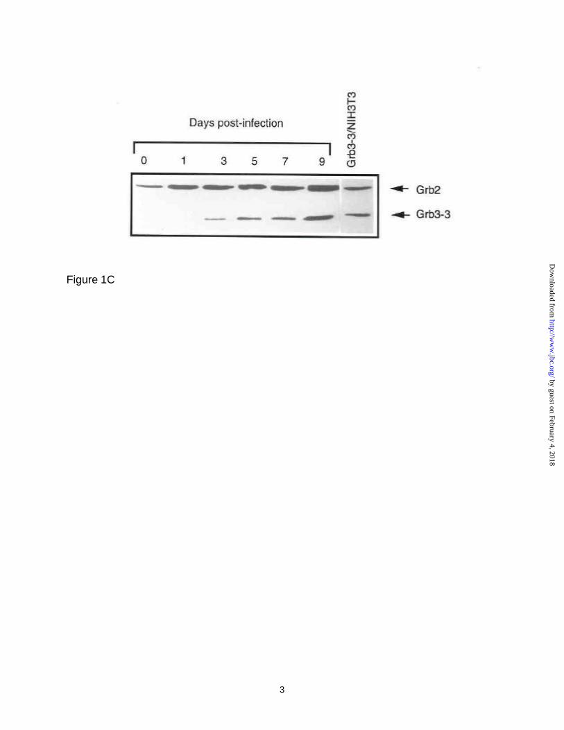
analyzed Grb3-3 protein expression from these same cultures by Western blot. As
shown in Figure 1, panel B, Grb3-3 was not detected and there was little change in
Grb2 in uninfected cells during the entire 9-day culture period (Figure 1, panel B).
Infection of CD4+ PBMCs with HIV-1 caused a circa two-fold increase in Grb2 levels,
however in striking contrast, there is a steady increase in the level of Grb3-3 (Figure 1,
panel C), and densitometry scanning of these protein bands revealed a 80-fold increase
in levels of Grb3-3 expression as compared to only two fold increase in Grb2 level in a
period of 9-day infection (Figure 1, panel D). Similar results were obtained in Jurkat T-
by guest on February 4, 2018http://w
ww
.jbc.org/D
ownloaded from

11
cells infected by HIV-1 (data not shown). These results indicate that Grb3-3 is up-
regulated by HIV-1 infection in vitro.
Detection of Over-Expressed Grb3-3 mRNA and Proteins in HIV-1 Infected Human
Subjects.
To extend our investigation on the relationship between HIV-1 infection and
Grb3-3 upregulation, we evaluated the expression of Grb2 and Grb3-3 mRNAs in
PBMCs obtained from 31 HIV-1 seropositive individuals and compared them with 10
HIV-1 negative health donors using RT-PCR assay. Selection of PCR primers and
optimization of reaction conditions have been described previously (31). As a control to
ensure that the same amounts of cDNA were used in all experiments, β-actin-encoding
cDNA was amplified by PCR with specific oligonucleotides (31). A titration curve was
established to quantitate the ratios of Grb3-3 to Grb2. PCR products were analyzed in
1.5% agarose gels stained with 0.5 mg/ml ethidium bromide and visualized under UV
light. As is shown in Table 1, in PBMCs from all seronegative donors, Grb3-3 mRNA
was barely detectable whereas Grb2 mRNA levels were constant. In contrast, PBMCs
from all seropositive individuals displayed an increased level of Grb3-3. In two cases,
the same amount of Grb3-3 and Grb2 mRNA was detected (patients 13 and 15), in 29
cases a ratio of Grb3-3 to Grb2 was found to be greater than one. Of the 29 cases, 7
patients had undetectable Grb2 mRNA but elevated Grb3-3 mRNA levels (patients 16
through 22), 7 patients had lower Grb2 mRNA levels than those from the control
subjects but increased levels of Grb3-3 (patients 23 through 29). 9 patients had higher
Grb3-3 levels but normal (control subjects) levels of Grb2 mRNA (patients 7 through 12,
by guest on February 4, 2018http://w
ww
.jbc.org/D
ownloaded from

12
14, 30, 31) whereas 4 patients had Grb3-3 mRNA levels greater than control Grb2
mRNA levels (patients 1, 4 through 6). Only 2 seropositive patients had a ratio of Grb3-
3 to Grb2 mRNA of less than 1. Taken together, despite variations in the Grb3-3 to
Grb2 mRNA ratios, elevated Grb3-3 mRNA levels were consistently detected in the
HIV-1-infected subjects although the available data do not allow one to establish a clear
relationship between the clinical stages of disease and the value of the Grb3-3/Grb2
mRNA ratios. A representative sample of these experiments is shown in Figure 2,
panel A. It is noteworthy that Grb2 was observed in all samples from health donors,
while in approximately 24% of the patient samples we failed to detect Grb2 for unclear
reasons (Table 1). A correlation between HIV-1 RNA copy number and Grb3-3/Grb2
ratios was not performed because at the time when these studies were initiated,
analyses of viral loads were not routinely performed (see methods).
Grb3-3 protein expression was also analyzed by Western blot for protein
samples available from a limited number of HIV-1 seropositive individuals. These
results are described in Figure 2, panel B. Lane 1 shows a negative control (NIH3T3
cells transfected with empty vector) and lane 2 shows a positive control (NIH3T3 cells
transfected with Grb3-3 expression construct) whereas lane 3 represents a
seronegative healthy donor. Clearly, Grb3-3 protein was detected in the samples from
all five patients (lanes 4 through 8). Taken together, these data indicate that in
seronegative individuals, Grb3-3 protein expression is very low or non-detectable
whereas HIV-1-infection causes upregulation of Grb3-3 in vivo, an observation that is in
general agreement with the in vitro HIV-1 challenge data.
by guest on February 4, 2018http://w
ww
.jbc.org/D
ownloaded from

13
The Viral Proteins Tat and Nef Can Independently Induce Grb3-3 Expression.
We next determined which viral component(s) was responsible for upregulation
of Grb3-3 using constructs expressing various viral proteins. To this end, Jurkat T-cells
were transfected with various plasmids expressing either HIV-1 Tat, Rev, Vif, Vpr, Env
or Nef. Cells were harvested 72 hours post-transfection and lyzed for extraction of total
cellular proteins for Western blot analyses. We first confirmed, in a Western blot assay,
that these viral protein expression constructs indeed produced corresponding viral
proteins in the transfected Jurkat cells (Figure 2, panel C). We next investigated which
viral protein(s) was responsible for upregulation of Grb3-3. As is shown in Figure 2,
panel D, transfection of the tat and nef genes resulted in an increased expression of
Grb3-3 (lanes 7 & 8), but no upregulation of Grb3-3 was detected in cells transfected
with either empty vector, pSV-Env, pSV-Rev, pcDNA-Vif or pcVpr (lanes 3 through 6,
respectively). The level of expression of Grb3-3 was dose-dependent on the amount of
transfected Tat and Nef expression plasmid (data not shown). Also shown in Figure 2,
panel D, is the positive control (lane 1) in which Jurkat T-cells were transfected with a
Grb3-3 expression plasmid. These results indicate that two of the earliest viral proteins
Tat and Nef can independently induce Grb3-3 expression in Jurkat T-cells. Although
Tat and Nef are known to participate in a variety of cellular activities including
interaction with MAPK pathways and modulation of T-cell functional activities (38-45),
our studies show that they share some unknown but common cellular activation
pathway(s) that can lead to alternative splicing of Grb2. Since Tat and Nef can also act
as positive factors for T-cell activation and viral replication through activation of cell
by guest on February 4, 2018http://w
ww
.jbc.org/D
ownloaded from

14
proliferation pathway(s) and/or by lowering the T- cell activation threshold (27-40, 42-45,
52-57), it was of particular interest to evaluate cellular factors that may contribute to the
virus-induced cell activation. Since Grb2 is known to be involved in T-cell activation, a
role of Grb3-3 in T-cell activation was next investigated (see below).
Grb3-3 Potentiates NFAT and HIV-1 LTR But Not AP-1 Promoter Activity in Jurkat
T-Cells.
Among the transcription factors, NFAT is indispensable for T-cell activation (15,
16). A critical step in this activation pathway is that cytoplasmic phosphorylated NFAT
must be dephosphorylated by calcineurin. The de-phosphorylated NFAT is then able to
translocate into the nucleus (11, 13, 16). To analyze the role of Grb3-3 in T-cell
activation, Jurkat T-cells were cotransfected with 3xNFAT/AP-1-LUC reporter (three
tandem repeats of the NF-AT/AP-1 binding site) with either Grb3-3 or the empty vector,
followed by treatment with either anti-CD3/anti-CD28 or chemical stimuli such as PMA
and/or ionomycin. As is shown in Figure 3, panel A, in the absence of stimulation with
exogenous stimuli, Grb3-3 was not able to affect NFAT/AP-1 activity. However, Grb3-3
caused an approximately 10-fold increase in luciferase activity as compared to vector
control following treatment of the transfected Jurkat T-cells with anti-CD3/anti-CD28.
Similar magnitude of NFAT/AP-1 activation by Grb3-3 (circa 7-fold) was also observed
after treatment with PMA plus ionomycin, a condition that is generally believed to mimic
TCR stimulation but with maximal efficiency (24). These results suggest that Grb3-3 is
capable of potentiating NFAT/AP-1 activation in Jurkat T-cells following TCR
stimulation.
by guest on February 4, 2018http://w
ww
.jbc.org/D
ownloaded from

15
Because the luciferase reporter construct used in the above experiments of
Figure 3, panel A, consists of three tandem repeats of the composite NF-AT/AP-1
binding site, we set out to determine which components were primarily involved. To this
end, we performed the same experiments described above by replacing 3xNFAT/AP-1-
LUC reporter with either 3xAP-1-LUC or 3xNFAT-LUC reporters. As is shown Figure 3,
panel B, no difference in activation of AP-1 was observed between Grb3-3 and vector
control upon stimulation with either anti-CD3/anti-CD28 or PMA/ionomycin. In contrast,
a 2-3 fold increase in activation of NFAT was detected with Grb3-3 when the 3xNFAT-
LUC reporter was used (Figure 3, panel C). The magnitude of stimulation (2-3 fold)
using 3xNFAT-LUC (Figure 3, panel C) is not as high as that using 3xNFAT/AP-1-LUC
reporter (approximately 10 fold) (Figure 3, panel A), since the latter consists of
composite binding elements for additional and cooperative binding between NFAT and
AP-1 transcription factors. In addition, we failed to observe any difference in stimulation
of NFκB-driven reporter between Grb3-3 and the vector (data not shown). Interestingly,
we did not observe significant effects of exogenous Grb2, through transfection of Grb2
expression construct, on NFAT activity under similar experimental conditions (data not
shown). Presumably, the endogenous Grb2 level is already so high that additional
effects could not been detected under our experimental conditions. Collectively, these
results suggest that Grb3-3 is capable of potentiating NFAT activity.
HIV-1 Tat and Nef Potentiation of NFAT Promoter Activity and Grb3-3 Potentiation
of HIV-1 LTR Promoter Activity are Both Sensitive to Cyclosporin A (CsA).
by guest on February 4, 2018http://w
ww
.jbc.org/D
ownloaded from

16
Since Tat and Nef could induce Grb3-3 expression and Grb3-3 in turn can
potentiate NFAT activities, we investigated whether Tat and Nef could potentiate NFAT
activities. As is shown in Figure 3 (left side of panel D), both Tat and Nef caused
approximately three-fold increase in luciferase activity. In addition, cyclosporin A (CsA),
a drug that is known to oppose the function of calcineurin (15, 16), thereby blocking
nuclear-targeting of NFAT, substantially suppressed this activation of Tat, Nef and
Grb3-3 (Figure 3, right side of panel D). These results are in agreement with previous
studies that Tat and Nef can activate several transcription factors including NFAT (38-
45, 52-57).
NFATc has been reported to activate HIV-1 LTR promoter (22). We next
reasoned that Grb3-3 should be able to activate HIV-1 LTR promoter because Grb3-3
was able to potentiate NFAT activity as shown in Figure 3, panels A. & C. To test this
possibility, HIV-1 LTR driven-luciferase reporter was co-transfected with Grb3-3
expression vector into Jurkat T-cells. As expected, transfection of Grb3-3 expression
vector resulted in greater activation of HIV-1 LTR promoter as compared to the vector
control (Figure 3, left side of panel E) and CsA substantially suppressed potentiation of
HIV-1 LTR activity by Grb3-3 (Figure 3, right side of panel E). These results suggest
that Grb3-3, upregulated by HIV-1 infection, can potentiate NFAT activity, and as a
result increase HIV-1 LTR responsiveness following T cell receptor/co-receptor
stimulation.
by guest on February 4, 2018http://w
ww
.jbc.org/D
ownloaded from

17
Blocking Grb3-3 Induced-NFAT nuclear localization by Constitutively Activated
MEKK-1 and Cyclosporin A.
In addition to the immunosuppressive drug CsA, cellular kinases such as MEKK-
1 have been implicated to oppose the function of calcineurin (15, 16), thereby blocking
nuclear-targeting of NFAT. Recently, Grb3-3 has also been demonstrated to bind
MEKK-1 in vivo (49). To extend our study, we analyzed the effects of MEKK-1 on Grb3-
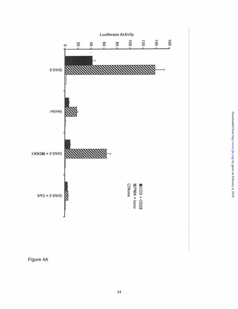
3 potentiated-NFAT nuclear localization. Figure 4, panel A, shows that constitutively
active MEKK-1* can reduce the Grb3-3 potentiated NFAT activity that is seen following
anti-CD3/anti-CD28 and PMA/ionomycin stimulation by circa 85 and 53%, respectively.
As a control, CsA reduced the Grb3-3 potentiation of NFAT activity following anti-
CD3/anti-CD28 and PMA/ionomycin stimulation by circa 93-97%.
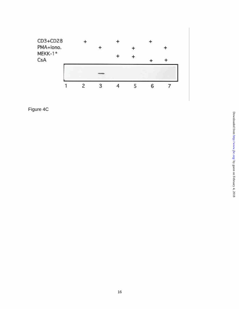
The above observation was further strengthened by examination of NFAT
proteins in the nuclear extracts in immunoblot experiments using antibodies against
NFATc following transfection of Jurkat T-cells with the Grb3-3 expressor (Figure 4,
panel B) or vector (Figure 4, panel C) plasmids. Clearly, in the absence of any stimuli
(Figure 4, panel B), NFAT was not detected in the nuclear extracts in the absence of
any stimuli (lane 1). As expected, treatment of Grb3-3 transfected cells with anti-
CD3/anti-CD28 resulted in nuclear targeting of NFATc (lane 2), while PMA and
ionomycin treatment were found to maximally stimulate NFATc translocation into the
nucleus (lane 3). However, additional treatment of these cells by co-transfection with
constitutive activated MEKK-1 (lane 4 & 5) or with cyclosporin (CsA) (lanes 6 &7) were
found to substantially suppress NFATc translocation into the nucleus. Figure 4, panel C
by guest on February 4, 2018http://w
ww
.jbc.org/D
ownloaded from

18
shows the results of similar Western blot experiments except that control vector was
used in place of Grb3-3 expressor plasmid. In these studies, nuclear NFAT was barely
detectable in the same Western blots except in lane 3, where the strongest stimulation
(PMA plus Ionomycin) was applied (lane 3) however, NFATc protein band disappeared
following additional treatment with either constitutively activate MEKK-1* or CsA (lanes
5 & 7). It should be mentioned that the Western blot assay is not as sensitive as the
luciferase reporter assay, thereby the NFATc band in nuclear extracts following
treatment of anti-CD3/anti-CD28 (lane 2 in Figure 4, panel C) was non-detectable
although increase in luciferase activity could be detected (Figure 4, panel A). However,
the results obtained in Figure 4, panels B & C are consistent with those of Figure 4,
panel A, i.e., Grb3-3 potentiated NFATc activity, and such effects were inhibited by
either treatment with CsA or transfection with constitutively activate MEKK-1*. As an
internal control, transcription factor SP-1 was probed in nuclear extracts under similar
conditions. As is shown in Figure 4, panel D, Grb3-3 exerts no detectable effect on Sp-1
level in nucleus upon treatment with either PMA/Ionomycin or CD3/CD28 stimulation.
These results indicate that Grb3-3 can facilitate NFAT nucleus translocation upon TCR
stimulation.
The mechanism(s) involving facilitation of NFAT nuclear translocation by Grb3-3
is currently unclear. CsA blocks NFAT activation primarily by inhibiting the phosphatase
activity of calcineurin (15-20). However, the role of MEKK-1 in inhibition of NFAT
activation is less understood. MEKK-1 may act through intermediates such as SEK1
and JNKs in retaining NFAT in the cytoplasm, as JNKs has been shown to directly
by guest on February 4, 2018http://w
ww
.jbc.org/D
ownloaded from

19
phosphorylate NFAT (55). Alternatively MEKK-1 may coordinate with other kinases
such as CKIα in masking of nuclear import signal of NFAT (46). Although Grb3-3 has
been demonstrated to bind to MEKK-1 (49), additional work will be needed to determine
whether Grb3-3 exerts its effects through interaction with MEKK-1 and/or to reveal the
identities of other intermediates that might be involved in this process.
Grb3-3 has been reported to inhibit EGF-induced transactivation of a Ras-
responsive element, while Grb2 was found to overcome Grb3-3 inhibition in this
pathway (31), suggesting that Grb3-3 may act as a suppressor over Grb2 in inhibiting
the EGF proliferative signals. However, the positive effects of Grb3-3 on NFAT
potentiation in this report imply that Grb3-3 may not be restricted to work in the same
pathway as Grb2, or to necessarily counteract the functional activity of Grb2 especially
since Grb2 also plays positive role in T cell activation (32). Indeed, it is likely that signal
pathways associated with Ras-responsive element triggered by EGF (31) may not
significantly contribute to NFAT activation because Grb3-3 works as a negative factor in
that case.
In summary, our findings define that Grb3-3 is a cellular factor that can be up-
regulated by HIV-1 and may act as a positive regulator for T cell activation and HIV-1
replication through NFATc. Establishing the pathway(s) by which Tat and Nef induce
expression of this Grb2 isoform, and further clarifying its positive role in T-cell
acdtivation will aid in our understanding of how HIV-1 modulated expression of cellular
by guest on February 4, 2018http://w
ww
.jbc.org/D
ownloaded from

20
signaling molecules to establish an intracellular environment that can favorably support
its replication.
by guest on February 4, 2018http://w
ww
.jbc.org/D
ownloaded from

21
ACKNOWLEDGEMENTS
We thank Drs. D. Gabuzda, X. Yang, C. Zhang, J. He, J. Sodroski for reagents
and helpful discussions and Drs. N. Landau, G. Crabtree, A. Cochrane, S. Burakoff, A.
Rao, C. Rudd, B. Cullen, Jacques Pousségur for generous gifts of reagents. X.L. is
supported by a Medical Research Council of Canada Fellowship. This work was
supported by National Institutes of Health grants CA06516, AI28785 and AI41954.
by guest on February 4, 2018http://w
ww
.jbc.org/D
ownloaded from

22
REFERENCES
1. Pantaleo, G., and Fauci, A. (1995) Annu. Rev. Immunol. 248, 487-512
2. Folks, T., Kelly, J., Benn, S., Kinter, A., Justement, J., Gold , J., Redfield, R., Sell,
K. W., and Fauci, A. S. (1986) J. Immunol. 136, 4049-4053
3. Spina, C. A., Guatilli, J. C., and Richman, D. D. (1995) J. Virol. 69, 2997-2998
4. Stevenson, M., Stanwick, T. L., Dempsey, M. A., and Lamonica, C. A. (1990)
EMBO. J. 9,1551-1560
5. Zack, J. A., Cann, A. J, Lugo, J. P., and Chen, I. S. (1988) Science 240, 1026-
1029
6. Zagury, D., Bernard, J., Leonard, R., Cheynier, R., Feldman, M., Sarin, P. S.,
and Gallo, R. C. (1986) Science 231, 850-853
7. Crabtree, G. R. (1989) Science 243, 355-361
8. Linsley, P. S., and Ledbetter, J. A. (1993) Ann. Rev. Immunol, 11, 191-212
9. June, C. H., Bluestone, J. A., Nadler, L. M., and Thompson, C. B. (1994)
Immunol. Today 15, 321-332
by guest on February 4, 2018http://w
ww
.jbc.org/D
ownloaded from
23
10. Fraser, J. D., Straus, D., and Weiss, A. (1993) Immunol. Today 14, 375-
362
11. Rao, A. (1991) Crit. Rev. Immunol. 10, 495-519
12. Ullman, K. S., Northrop, J. P., Verweij, C. L., and Crabtree, G. R. (1990) Annu.
Rev. Immunol. 8, 421-452
13. Schreiber, S. L. and Crabtree, G. R. (1992) Immunol. Today 13, 136-142
14. Rao, A. (1994) Immunol. Today 15, 274-281
15. Rao, A. (1995) J. Leukocyte Biol. 57, 536-542
16. Crabtree, G. (1999) Cell 96, 611-614
17. Weiss, A., and Littman, D. R. (1994) Cell 76, 263-274
18. Jain, J., McCaffrey, P. G., Miner, Z., Kerppola, T. K., Lamber, J. N., Verdine, G.
L., Curran, T., and Rao. A. (1993) Nature 365, 352-355
by guest on February 4, 2018http://w
ww
.jbc.org/D
ownloaded from

24
19. McCaffrey, P. G., Luo, C., Kerppola, T. K., Jain, J., Badalian, T. M., Ho, A. M.,
Burgeon, E., Lane, W. S., Lamber, J. N., Curran, T., Verdine, G. L., Rao, A., and
Hogan, P. G. (1993) Science 262, 750-754
20. Shaw, K. T. Y., Ho, A. M., Raghavan, J. A., Kim, J., Jain, J., Park, J., Sharma,
S., Rao, A., and Hogan, P. G. (1995) Proc. Natl. Acad. Sci. USA 92,11205-11209
21. Vacca, A., Farina, M., Maroder, M., Alesse, E., Screpanti, I., Frati, L., and Gulino,
A. (1994) Biochem. Biophys. Res. Commun. 205, 467-474
22. Kinoshita, S., Su, M., Amano, M., Timmerman, L. A., Kaneshima, H., and Nolan,
G. P. (1998) Immunity 6, 235-244
23. Kinoshita, S., Chen, B. K., Kaneshima, H., and Nolan, G. P. (1998) Cell 5, 595-
604
24. Macian, F., and Rao, A. (1999). Mol. Cell. Biol. 19, 3645-3653
25. Folgueira, L., Algeciras, A., MacMorran, W. S., Bren, G. D., and Paya, C. V.
(1996) J. Virol. 70, 2332-2338
26. Yang, X., Chen, Y., and Gabuzda, D. (1999) J. Biol. Chem. 274, 27981-27988
by guest on February 4, 2018http://w
ww
.jbc.org/D
ownloaded from

25
27. Popik, W, Hesselgesser, J. E., and Pitha, P. M. (1998) J. Virol. 72, 6406-6413
28. Jacque, J. M., Mann, A., Enslen, H., Sharova, N., Brichacek, B., Davis, R. J.,
Stevenson, M. (1998) EMBO. J. 17, 2607-2618
29. Briant, L., Robert-Hebmann, V., Sivan, V., Brunet, A., Pouyssegur, J., and
Devaux, C. (1998) J. Immunol. 160, 1875-1885
30. Lowenstein, E. J., Daly, R. J., Batzer, A. G., Li, W., Margolis, B., Lammers, R.,
Ullrich, A., Skolnik, E. Y., Bar-Sagi, D., and Schlessinger, J. (1992) Cell 70, 431-
442
31. Fath, I., Schweighoffer, F., Rey, I., Multon, M. C., Boiziau, J., Duchesne, M., and
Tocque, B. (1994) Science 264, 971-974
32. Downward, J. (1994) FEBS Lett. 338, 113-117
33. Holsinger, L. J., Spencer, D. M., Austin, D. J., Schreiber, S. L., and Crabtree, G.
R. (1995) Proc. Natl. Acad. Sci. USA 92, 9810-9814
34. Wange, R. L., and Samelson, L. E. (1996) Immunity 5, 197-205
35. Zhang, W., Trible, R. P., and Samelson, L. E. (1998) Immunity 9, 1-20
by guest on February 4, 2018http://w
ww
.jbc.org/D
ownloaded from

26
36. Zhang, W., Sloan-Lancaster, J., Kitchen, J., Trible, R. P., and Samelson, L. E.
(1998) Cell 92, 83-92
37. Rudd, C. E. (1999) Cell 96, 5-8
38. Li, C. J., Ueda, Y., Shi, B., Borodyansky, L., Huang, L., Li, Y. Z., and Pardee, A.
B. (1997) Proc. Natl. Acad. Sci. USA 94, 8116-8120
39. Ott, M., Emiliani, S., Van Lint, C., Herbein, G., Lovett, J., Chirmule, N.,
McCloskey, T., Pahwa, S., Verdin, E. (1997) Science 275,1481-1485
40. Kumar, A., Manna, S., Dhawan, S., and Aggarwal, B. B. (1998) J. Immunol. 161,
776-781
41. Ganju, R. K., Munshi, N., Nair, B. C., Liu, Z. Y., Gill, P., and Groopman, J. E.
(1998) J. Virol. 72, 6131-6137
42. Biggs, T. E., Cooke, S. J., Barton, C. H., Harris, M. P., Saksela, K., Mann, D. A.
(1999). J. Mol. Biol. 290, 21-35
43. Greenway, A., Azad, A., Mills, J., and McPhee, D. (1996). J. Virol. 70, 6701-6708
by guest on February 4, 2018http://w
ww
.jbc.org/D
ownloaded from

27
44. Zauli, G., Gibellini, D., Celeghini, C., Mischiati, C., Bassini, A., La Placa, M.,
Capitani, S. (1996) J. Immunol. 157, 2216-2224
45. Schrager, J. A., and Marsh, J. W. (1999) Proc. Natl. Acad. Sci. USA 96, 8167-
8172
46. Zhu, J., Shibasaki, F., Price, R., Guillemot, J. C., Yano, T., Dotsch, V., Wagner,
G., Ferrara, P., and McKeon, F. (1998) Cell 93, 851-861
47. Paxton, W., Conor, R. I., and Landau, N. R. (1993) J. Virol. 67, 7229-7237
48. Northrop, J. P., Ho, S. N., Chen, L., Thomas, D. J., Timmerman, L. A., Nolan, G.
P., Admon, A., and Crabtree, G. R. (1994) Nature 369, 497-502
49. Pomerance, M., Multon, M.-C., Parker, F., Venot, C., Blondeau, J.-P., Tocque,
B., and Scheweighoffer, F. (1998) J. Biol. Chem. 273, 24301-24304
50. Flanagan, W. M., Corthesy, B., Bram, R. J., and Crabtree, J. R. (1991) Nature
352, 803-807
51. Li, X., Liang, C., Quan, Y., Chandok, R., Laughrea, M., Parniak, M. A., Kleiman,
L., and Wainberg, M. A. (1997) J. Virol. 71, 6003-6010
by guest on February 4, 2018http://w
ww
.jbc.org/D
ownloaded from
28
52. Luo, W., Peterlin, B. M. (1997) J. Virol. 71, 9531-9537
53. Lafrate A. J., Bronson, S., and Skowronski, J. (1997) EMBO J. 16, 673-684
54. Jeang, K.-T., Xiao, H., and. Rich, E. A. (1999) J. Biol. Chem. 274, 28837-28840
55. Chow, C. W., Rincon, M., Cavanagh, J., Dickens, M., and Davis, R. J. (1997)
Science 278, 1638-1641.
56. Wang, J.K., Kiyokawa, E., Verdin, E., and Trono, D. (2000). Proc. Natl. Sci.
U.S.A. 97: 394-399.
57. Manninen A, Herma Renkema G, Saksela K (2000) J. Biol. Chem. 275,16513-
16517.
by guest on February 4, 2018http://w
ww
.jbc.org/D
ownloaded from
29
FIGURE LEGENDS
Figure 1. Upregulation of Grb3-3 in CD4+ peripheral blood mononuclear cells
(PBMCs) following HIV-1 infection. Panel A, Infection of CD4+ PBMCs. Viral stocks
were prepared by transfection of CD4+ Jurkat cells with pNL4-3 proviral DNA as
described in experimental procedures (51). CD4+ PBMCs were isolated from healthy
donors using Ficoll-Hypaque gradient centrifugation (Pharmacia), followed by selection
on OKT4 coated flasks according to instructions from the manufacturer (Applied
Immune Sciences, Santa Clara, CA). The selected CD4+ mononuclear cells were
stimulated with PHA at a concentration of 1 µg/ml for 3 days. Cells (107) were infected
with 5 ng p24 of HIV-1 for 3 hours, followed by washing three times with PBS. The cells
were then resuspended in fresh RPMI-1640/10% FCS. Supernatants were collected
periodically for reverse transcriptase (RT) assays (51). Symbols – triangles, HIV-1-
infected cells; squares, mock-infected cells. Panel B - D, Determination of Grb2 and
Grb3-3 protein expression. Cells were harvested for isolation of total cellular protein.
Equal amounts of protein were fractionated in 12.5% PAGE in the presence of SDS,
followed by transferring to nitrocellulose filter. Detection of the protein was achieved by
Western blot using MAb 3B5 which binds to both Grb2 and Grb3-3. Panel B. Grb2 and
Grb3-3 protein expression in uninfected cells (mock infection). Panel C. Grb2 and
Grb3-3 protein expression in HIV-1-infected cells. CD4+ PBMCs infected by HIV-1 (NL-
4.3 strain). Panel D. Quantitative densitometry of Grb2 and Grb3-3 protein levels in
CD4+ mononuclear cells during HIV-1-infection (for Figure 1, panel C). The Western
blot shown in panel C was subjected to quantitative densitometry measurements. The
by guest on February 4, 2018http://w
ww
.jbc.org/D
ownloaded from
30
values shown represent relative density (RD) measurements over the nine-day
experiment and are expressed as RD units.
Figure 2. Detection of Grb3-3 and Grb2 mRNA and protein expression. Panel A,
RT-PCR of Grb2 and Grb3-3 was performed as previously described using total RNA
isolated from PBMCs of HIV-1-infected and uninfected (control) individuals (31) (see
Experimental Procedures for details). The data presented are representative samples
of randomly chosen individuals in each group. Panel B, Western blot Grb2 and Grb3-3
from PBMCs of HIV-1-infected and uninfected (control) individuals. Lane 3, PBMC
lysate from seronegative individual; Lane 4 through 8, PBMC lysate from randomly
chosen HIV-1-infected individuals; Lane 1 NIH3T3 cells transfected with empty vector;
Lane 2, NIH 3T3 cells transfected with Grb3-3 expression construct. The Western blot
was developed using an anti-Grb2 MAb (monoclonal UBI Grb2 (1-68)) which recognized
Grb2 and Grb3-3 and revealed by ECL. Panel C, Detection of HIV-1 proteins
expressed in Jurkat T-cells transfected with various viral protein expression vectors.
Note that the difference in intensity amongst these bands is most probably due to
difference in the sources of and/or the affinity of the antibodies used in the blot, and
should not be interpreted as a result of unequal transfection efficiency because
transfection efficiency was routinely monitored by co-transfection with β-gal expression
vector (Invitrogen, San Diego, California). Panel D, Detection of Grb2 and Grb3-3
protein in Jurkat T-cells transfected with various constructs. Jurkat T-cells were
transfected with various constructs (5 µg DNA for 5x10⁶ cells) using Lipofectamine (Life
Technology). 72 hours post-transfection, cells were isolated for isolation of cellular
by guest on February 4, 2018http://w
ww
.jbc.org/D
ownloaded from

31
proteins, which were subsequently analyzed in Western bot using MAb 3B5. Lane 1,
cells transfected with pSV-Grb3-3 (positive control); lane 2, transfection with empty
vector (negative control); Lane 3-8, cells transfected with various expression vectors for
HIV-1 viral proteins. The transfection efficiency and expression level of these individual
viral expression constructs in transfected Jurkat T-cells were found to be quite similar
(data not shown).
Figure 3. Grb3-3 potentiates NFAT activation upon engagement of TCR and CD28
co-receptor. Panel A. Effects of Grb3-3 on NFAT/AP-1 activation. Jurkat T-cells
were transfected with 2 µg of luciferase reporter (3xNFAT/Ap-1-LUC), 0.5 µg of NFATc
expression vector (48) in addition to either Grb3-3 or empty vector (4 µg). Total amount
of DNA was also brought to 10 µg using the empty vector. Cells were treated with either
anti-CD3/CD-28, or PMA, ionophore or a combination of PMA and ionophore to facilitate
dephosphorylation of NFAT (24). Luciferase activities were determined 72 hr post-
transfection (for details see Methods). Note that duplicate tests are usually performed
simultaneously and the experiments are repeated three times. Differences between
each group falls between 5-10%. Error bars represent standard deviations. Panel B.
Effects of Grb3-3 on AP-1 activation. Jurkat T-cells were transfected with 3xAP-1-
LUC plasmid together with either Grb3-3 or empty vector. 72 hr later, luciferase
activities were analyzed. Panel C. Effects of Grb3-3 on NFAT activation. Jurkat T-
cells were transfected with 3xNFAT-LUC plasmid together with either Grb3-3 or empty
vector. 72 hr later, luciferase activities were analyzed. Panel D. Effects of Tat and Nef
on NFAT activation. Jurkat T-cells transfected with 3xNFAT-LUC plasmid together with
by guest on February 4, 2018http://w
ww
.jbc.org/D
ownloaded from
32
Grb3-3, Tat, Nef, or empty vector. In some cases, CsA (µg/ml) was used at the same
time as the stimuli (PMA, Ionomycin, anti-CD3, or anti-CD28) were applied. Panel E.
Effects of Grb3-3 on HIV-1 LTR promotor activity. Jurkat T-cells were transfected
with HIV-LTR driven luciferase construct together with either Grb3-3 or the empty
vector. Treatment of the cells with stimuli and/or CsA and analysis of luciferase activity
was the same as described above.
Figure 4. Effects of Grb3-3 on NFAT nucleus localization. Panel A. Effects of
MEKK-1 and CsA on Grb3-3 induced-NFAT activation. Experiments were performed
as described in Figure 3 with additional treatment with either co-transfection of
constitutively activated MEKK-1* (4 µg) or treatment with CsA (1 µg/ml). Values are
mean +/- S.D. Detection of NFATc in the nuclear extracts of Jurkat cells
transfected with either Grb3-3 (Panel B) or the empty vector control (Panel C).
Lane 1, no treatment; lane 2, treatment with anti-CD3/anti-CD28; lane 3: treatment with
PMA and ionomycin; lane 4, treatment with anti-CD3/anti-CD28 plus co-transfection
with constitutively activate MEKK-1*; lane 5, treatment with PMA/Ionimycin plus co-
transfection with constitutively activated MEKK1*; lane 6, treatment with anti-CD3/CD28
in addition to CSA; lane 7, treatment with PMA/Ionomycin in addition to CsA. Panel D,
as an internal control, anti-Sp1 antibody was used to detect Sp1 in nuclear extracts
following various conditions.
by guest on February 4, 2018http://w
ww
.jbc.org/D
ownloaded from

33
Table 1
Detection of Grb3-3 mRNA by RT-PCR from PBMCs from HIV-1 seropositive
and seronegative individuals using RT-PCR
PatientIdentification Mortality* Sex Age
CDCStaging
p24 Agpg/ml Grb3-3/Grb2 mRNA~
1. 1.90% >12. 0% <13. 0% <14. 0% >15. 5.80% >16. 2.80% >17. 0.60% M 34 III 0 >>18. 0% M 39 IVC2 0 >>19. 0.02% M 51 IVC1, IVD 0 >>110. 0% M 55 IVD 0 >>111. 0% F 31 IVC1 0 >>112. 0% F 26 IVC1 80 >>113. 0.05% M 46 IVC2 0 = 114. 0% M 55 IVC2 0 >>115. 0% M 56 IVD 7 = 116. 0% M 37 III 0 >>1 Grb2=017. 0% F 30 IVC1 87.4 >>1 Grb2=018. 0% M 31 IVD 0 >>1 Grb2=019. 0% M 40 IVC2 0 >>1 Grb2=020. 0% M 31 II 0 >>1 Grb2=021. 0% M 33 IVC2 0 >>1 Grb2=022. 0% M 44 IVC1 9.7 >>1 Grb2=023. 5.20% M 30 III 21.5 >>1 Grb2 low24. 10% F 63 IVC2, IVE 0 >>1 Grb2 low25. 0% M 47 IVC2 7 >>1 Grb2 low26. <1% M 31 IVC2 0 >>1 Grb2 low27. 3.50% M 49 IVC2 40.6 >>1 Grb2 low28. 1% M 37 IVD, IVC1 0 >>1 Grb2 low29. 12% F 30 IVC2 0 >>1 Grb2 low30. 3% F 31 IVC1 0 >>131. 1.90% F 40 II 0 >>1
* Mortality as determined by Trypan blue assay after cell collection.~ See Figure 2A for details of RT-PCR.
by guest on February 4, 2018http://w
ww
.jbc.org/D
ownloaded from


Mhashilkar, Bruno Tocque and Wayne A. MarascoJuliana Josef, Changhong Zhou, Joyce LaVecchio, Patricia Stuckert, Monika Raab, Abner
Xuguang Li, Marie-Christine Multon, Yvette Henin, Fabien Schweighoffer, Corinne Venot,through NFATc
Grb3-3 is Up-Regulated in HIV-1 Infected T-Cells and Can Potentiate Cell Activation
published online July 20, 2000J. Biol. Chem.
10.1074/jbc.M005535200Access the most updated version of this article at doi:
Alerts:
When a correction for this article is posted•
When this article is cited•
to choose from all of JBC's e-mail alertsClick here
by guest on February 4, 2018http://w
ww
.jbc.org/D
ownloaded from