jassin m. jouria, md
TRANSCRIPT

ce4less.com ce4less.com ce4less.com ce4less.com ce4less.com ce4less.com 1
EPILEPSY
Jassin M. Jouria, MD
ABSTRACT
Epilepsy is a seizure disorder of varied etiology and symptomology and its
treatment depends on multiple factors, including age of onset and type of
seizure. Sometimes the seizure is absent or mild enough to go untreated by
medication and resolves over time. Most often, epilepsy is a life long
condition that requires close medical management. Anti-epileptic drug
therapy often requires serum monitoring for dose adjustment and drug
interaction surveillance. Screening for comorbid medical and psychiatric
conditions, especially depression, anxiety, and feelings of social stigma and
isolation is needed. Educating patients and families to increase awareness of
epilepsy and treatment options in their unique circumstance will assist them
to overcome stereotypes and help them obtain a higher quality of life.
Introduction
Epilepsy is a complex brain disorder that is characterized by seizures, which
are caused by disturbances in the brain’s electrical functions. The term
epilepsy encompasses a variety of different neurological syndromes, each
ranging in its symptoms, severity, and duration. The characteristic seizures
are present in all types of epilepsy, but they differ in clinical presentation
and symptom severity depending on the type of epilepsy.
Epilepsy is most common in young children and the elderly, but it can affect
individuals of all ages. In many cases, the cause of epilepsy is unknown. In
those instances when a cause is identified, we find that the cause varies

ce4less.com ce4less.com ce4less.com ce4less.com ce4less.com ce4less.com 2
between environmental or genetic factors, or as part of traumatic injury.
Some epileptic syndromes will only last a short time, especially those caused
by trauma; however, some other epileptic syndromes will be lifelong
conditions that cannot be cured.
While many individuals will experience a single, unprovoked seizure at some
point in their lives, epilepsy is not considered as a diagnosis until the patient
has had two or more unprovoked seizures. Once this occurs, the patient will
begin the process for assessing and diagnosing the type of epilepsy.
Overview Of Epilepsy
Epilepsy affects the central nervous system, thereby causing disruptions in
the nerve cell activity in the brain. When this activity is disrupted, seizures
occur (1). These seizures will cause the patient to experience abnormal
behavior, symptoms, and sensations. In some instances, patients will lose
consciousness. The presentation of seizures will vary. Some patients will
stare blankly for a brief period of time, typically a few seconds. Other
patients may experience twitching and jerking of their bodies (2). The type
of seizure experienced by the patient depends upon the etiology and the
severity of the condition.
Regardless of the severity of the seizures, most patients will require
treatment, as seizures can pose a significant risk to the patient. Seizures
can occur when the patient is engaging in activities such as driving,
operating machinery, or swimming, When this occurs, the patient is at an
increased risk of experiencing significant injuries (3).
Specific symptoms and features typically define epileptic syndromes. The
categories include: (4)

ce4less.com ce4less.com ce4less.com ce4less.com ce4less.com ce4less.com 3
Seizure types
Age when seizures begin
Electroencephalogram (EEG) findings
Brain structure (usually assessed with a brain MRI scan)
Family history of epilepsy or genetic disorder
Prognosis (future outlook)
Approximately fifty percent of epilepsy cases are caused by unknown
factors. In the remaining cases, the causes are typically genetic,
environmental, or trauma related (5).
The following table provides an explanation of the potential cause in cases
where the cause of epilepsy may be identified (6):
Genetic
Influence
Some types of epilepsy, which are categorized by the type of seizure
the individual experiences, run in families. In these cases, it's likely
that there's a genetic influence.
Researchers have linked some types of epilepsy to specific genes;
though it's estimated that up to 500 genes could be tied to the
condition. For most people, genes are only part of the cause of
epilepsy. Certain genes may make a person more sensitive to
environmental conditions that trigger seizures. Generalized epilepsy
seizure types appear to be more related to genetic influences than
partial seizure epilepsies.
Head Trauma Head trauma that occurs due to a car accident or other traumatic
injury can cause epilepsy. Head injuries can cause epilepsy in both
adults and children, with the risk highest in severe head trauma. A

ce4less.com ce4less.com ce4less.com ce4less.com ce4less.com ce4less.com 4
first seizure related to the injury can occur years later, but only very
rarely. People with mild head injuries that involve loss of
consciousness for fewer than 30 minutes have only a slight risk that
lasts up to 5 years after the injury.
Brain conditions - Brain conditions that result in damage to the brain,
such as brain tumors or strokes, also can cause epilepsy. Stroke is a
leading cause of epilepsy in adults older than age 35.
Infectious
Diseases
Infectious diseases, such as meningitis, AIDS and viral encephalitis,
can cause epilepsy.
Prenatal injury Before birth, babies are sensitive to brain damage that could be
caused by several factors, such as an infection in the mother, poor
nutrition or oxygen deficiencies. This brain damage can result in
epilepsy or cerebral palsy.
Developmental
Disorders
Epilepsy can sometimes be associated with developmental disorders,
such as autism and neurofibromatosis.
Brain
Chemistry
Factors
Ion Channels - sodium, potassium, and calcium - act as ions in the
brain. They produce electric charges that must fire regularly in order
for a steady current to pass from one nerve cell in the brain to
another. If the ion channels that carry them are genetically damaged,
a chemical imbalance occurs. This can cause nerve signals to misfire,
leading to seizures. Abnormalities in the ion channels are believed to
be responsible for absence and many other generalized seizures.
Neurotransmitters - Abnormalities may occur in neurotransmitters, the
chemicals that act as messengers between nerve cells. Three
neurotransmitters are of particular interest:
Gamma aminobutyric acid (GABA), which helps prevent nerve
cells from over-firing.
Serotonin's role in epilepsy is also being studied. Serotonin is a
brain chemical that is important for well-being and associated

ce4less.com ce4less.com ce4less.com ce4less.com ce4less.com ce4less.com 5
behaviors (such as eating, relaxation, and sleep). Imbalances
in serotonin are also associated with depression.
Acetylcholine is a neurotransmitter that is important for
learning and memory.
Risk Factors
Epilepsy and seizure disorders affect nearly 3 million Americans and more
than 45 million people worldwide. While anyone can develop epilepsy, there
are a number of factors (outlined below) that will increase an individual’s
risk of developing epilepsy and seizure disorders (7).
Age factor:
Epilepsy affects all age groups. The risk is highest in children under the age
of 2 and older adults over age 65. In infants and toddlers, prenatal factors
and birth delivery problems are associated with epilepsy risk. In children age
10 and younger, generalized seizures are more common. In older children,
partial seizures are more common.
Gender factors:
Men have a slightly higher risk than
women of developing epilepsy.
Family History:
People who have a family history of
epilepsy are at increased risk of
developing the condition.

ce4less.com ce4less.com ce4less.com ce4less.com ce4less.com ce4less.com 6
While there are numerous factors that may cause epilepsy, as well as a
variety of epileptic syndromes, all types share one common feature: all
forms of epilepsy are characterized by recurrent seizures (1). These seizures
are caused by uncontrolled electrical discharges in the nerve cells in the
cerebral cortex. Many individuals will experience a single seizure at some
point in their lifetime. This is not considered epilepsy (3).
Very few initial seizures will recur. In fact, only approximately twenty five
percent of initial seizures will recur (8). Once a patient experiences two or
more recurring seizures, he or she has a 70 % chance of experiencing
recurring seizures. This will result in
a diagnosis of epilepsy.
Epilepsy is generally classified into
two main categories based on seizure type, and these are described in the
table below (9):
PARTIAL SEIZURES
These seizures are more common than generalized seizures and occur in one or
more specific locations in the brain. In some cases, partial seizures can spread
to wide regions of the brain. They are likely to develop from specific injuries, but
in most cases the exact origins are unknown (idiopathic).
Simple Partial
Seizures
A person with a simple partial seizure (sometimes known as
Jacksonian epilepsy) does not lose consciousness, but may
experience confusion, jerking movements, tingling, or odd
mental and emotional events. Such events may include déjà
vu, mild hallucinations, or extreme responses to smell and
taste. After the seizure, the patient usually has temporary
weakness in certain muscles. These seizures typically last
about 90 seconds.
Complex Partial
Seizures
Slightly over half of seizures in adults are complex partial
type. About 80% of these seizures originate in the temporal
(Photo Courtesy of:
http://myqigong.blogspot.com/2011/01/epilepsy-
seizure.html)

ce4less.com ce4less.com ce4less.com ce4less.com ce4less.com ce4less.com 7
lobe, the part of the brain located close to the ear.
Disturbances there can result in loss of judgment, involuntary
or uncontrolled behavior, or even loss of consciousness.
Patients may lose consciousness briefly and appear to others
as motionless with a vacant stare.
Emotions can be exaggerated; some patients even appear to
be drunk. After a few seconds, a patient may begin to perform
repetitive movements, such as chewing or smacking of lips.
Episodes usually last no more than 2 minutes. They may occur
infrequently, or as often as every day. A throbbing headache
may follow a complex partial seizure. In some cases, simple or
complex partial seizures evolve into what are known as
secondarily generalized seizures. The progression may be so
rapid that the initial partial seizure is not even noticed.
GENERALIZED SEIZURES
Generalized seizures are caused by nerve cell disturbances that occur in more
widespread areas of the brain than partial seizures. Therefore, they have a more
serious effect on the patient. They are further subcategorized as tonic-clonic (or
grand mal), absence (petit mal), myoclonic, or atonic seizures.
Tonic-Clonic
(Grand Mal)
Seizures.
The first stage of a grand mal seizure is called the tonic phase,
in which the muscles suddenly contract, causing the patient to
fall and lie stiffly for about 10 - 30 seconds. Some people
experience a premonition or aura before a grand mal seizure;
most, however, lose consciousness without warning. If the
throat or larynx is affected, there may be a high-pitched
musical sound (stridor) when the patient inhales. Spasms
occur for about 30 seconds to 1 minute. Then the seizure
enters the second phase, called the clonic phase. The muscles
begin to alternate between relaxation and rigidity. After this
phase, the patient may lose bowel or urinary control. The
seizure usually lasts a total of 2 - 3 minutes, after which the
patient remains unconscious for a while and then awakens to
ce4less.com ce4less.com ce4less.com ce4less.com ce4less.com ce4less.com 8
confusion and extreme fatigue. A severe throbbing headache
similar to migraine may also follow the tonic-clonic phases.
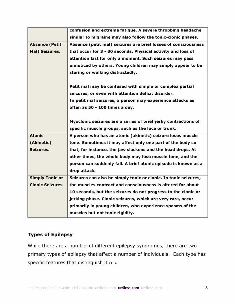
Absence (Petit
Mal) Seizures.
Absence (petit mal) seizures are brief losses of consciousness
that occur for 3 - 30 seconds. Physical activity and loss of
attention last for only a moment. Such seizures may pass
unnoticed by others. Young children may simply appear to be
staring or walking distractedly.
Petit mal may be confused with simple or complex partial
seizures, or even with attention deficit disorder.
In petit mal seizures, a person may experience attacks as
often as 50 - 100 times a day.
Myoclonic seizures are a series of brief jerky contractions of
specific muscle groups, such as the face or trunk.
Atonic
(Akinetic)
Seizures.
A person who has an atonic (akinetic) seizure loses muscle
tone. Sometimes it may affect only one part of the body so
that, for instance, the jaw slackens and the head drops. At
other times, the whole body may lose muscle tone, and the
person can suddenly fall. A brief atonic episode is known as a
drop attack.
Simply Tonic or
Clonic Seizures
Seizures can also be simply tonic or clonic. In tonic seizures,
the muscles contract and consciousness is altered for about
10 seconds, but the seizures do not progress to the clonic or
jerking phase. Clonic seizures, which are very rare, occur
primarily in young children, who experience spasms of the
muscles but not tonic rigidity.
Types of Epilepsy
While there are a number of different epilepsy syndromes, there are two
primary types of epilepsy that affect a number of individuals. Each type has
specific features that distinguish it (10).

ce4less.com ce4less.com ce4less.com ce4less.com ce4less.com ce4less.com 9
Idiopathic
Epilepsy
In idiopathic generalized epilepsy, there is often, but not always, a
family history of epilepsy. Idiopathic generalized epilepsy tends to
appear during childhood or adolescence, although it may not be
diagnosed until adulthood. In this type of epilepsy, no nervous
system (brain or spinal cord) abnormalities, other than the seizures,
can be identified on either an EEG or magnetic resonance imaging
(MRI) studies. The brain is structurally normal on a brain (MRI)
scan, although special studies may show a scar or subtle change in
the brain that may have been present since birth.
People with idiopathic generalized epilepsy have normal intelligence
and the results of the neurological exam and MRI are usually
normal. The results of the EEG may show epileptic discharges
affecting a single area or multiple areas in the brain (so called
generalized discharges).
The types of seizures affecting patients with idiopathic generalized
epilepsy may include:
Myoclonic seizures (sudden and very short duration jerking of
the extremities)
Absence seizures (staring spells)
Generalized tonic-clonic seizures (grand mal seizures)
Idiopathic generalized epilepsy is usually treated with medications.
Some people outgrow this condition and stop having seizures, as is
the case with childhood absence epilepsy and a large number of
patients with juvenile myoclonic epilepsy.
Idiopathic partial epilepsy begins in childhood (between ages 5 and
8) and may be part of a family history. Also known as benign focal
epilepsy of childhood (BFEC), this is considered one of the mildest
types of epilepsy. It is almost always outgrown by puberty and is
never diagnosed in adults. Seizures tend to occur during sleep and
are most often simple partial motor seizures that involve the face
and secondarily generalized (grand mal) seizures. This type of
epilepsy is usually diagnosed with an EEG.
Symptomatic Symptomatic generalized epilepsy (SGE) encompasses a group of

ce4less.com ce4less.com ce4less.com ce4less.com ce4less.com ce4less.com 10
Generalized
Epilepsy
challenging epilepsy syndromes. As a group, SGE has 3 main
features: (1) multiple seizure types, especially generalized tonic and
atonic seizures; (2) brain dysfunction other than the seizures, in the
intellectual domain (mental retardation or developmental delay) and
in the motor domain (cerebral palsy); and (3) EEG evidence of
diffuse brain abnormality. The following are examples of epilepsy
syndromes that are included in the category of SGE:
Early myoclonic encephalopathy
Early infantine epileptic encephalopathy with suppression
bursts or Ohtahara syndrome
West syndrome
Epilepsy with myoclonic atonic seizures
Epilepsy with myoclonic absence
Lennox-Gastaut syndrome
Progressive myoclonic epilepsies
Epilepsy Syndromes
There are a number of different syndromes that fall under the umbrella of
epilepsy. These syndromes are defined based upon the type and severity of
seizures, as well as the area of the brain that is affected.

ce4less.com ce4less.com ce4less.com ce4less.com ce4less.com ce4less.com 11
(Photo Courtesy of: http://healthsciencedegree.info/seizure-brain-activity/)
To further distinguish these syndromes, factors such as age, cause, and
outcome are also included in the defining characteristics. The following
section provides a thorough overview of the various epilepsy syndromes
(9,11–16).
Temporal Lobe Epilepsy
Temporal Lobe Epilepsy (TLE) means that the seizures arise in the temporal
lobe of the brain. Experiences during temporal lobe seizures vary in intensity
and quality. Sometimes the seizures are so mild that the person barely
notices. In other cases, the person may be consumed with feelings of fear,
pleasure, or unreality. A patient may also report an odd smell, an abdominal
sensation that rises up through the chest into the throat, an old memory or
familiar feeling, or a feeling that is impossible to describe.
Types of seizures in TLE
The most common seizure type in TLE is a complex partial seizure. During
complex partial seizures, people with TLE tend to perform repetitive,
automatic movements (called automatisms), such as lip smacking and
rubbing their hands together. Three-quarters of people with TLE also have
simple partial seizures, and about half have tonic-clonic seizures at some
time. Some people with TLE experience only simple partial seizures.
Temporal lobe seizures usually begin in the deeper portions of the temporal
lobe. This area is part of the limbic system, which controls emotions and
memory. This is why the seizures can include a feeling of déjà vu, fear, or
anxiety, and why some people with TLE may have problems with memory
and depression.

ce4less.com ce4less.com ce4less.com ce4less.com ce4less.com ce4less.com 12
In most cases, the seizures associated with TLE can be fully controlled with
medications used for partial seizures. If drugs are ineffective, brain surgery
is often an option for patients with TLE. Temporal lobectomy is the most
common and successful form of epilepsy surgery. Vagus nerve stimulation
can also be beneficial in cases where temporal lobectomy is not
recommended or has failed.
Frontal lobe epilepsy is the next most common form of epilepsy after
temporal lobe epilepsy (TLE), and involves the frontal lobes of the brain. As
in temporal lobe epilepsy, seizures in frontal lobe epilepsy are partial,
though seizure symptoms differ depending on the frontal lobe area involved.
Frontal Lobe Epilepsy
Since the frontal lobes are responsible for a wide array of functions including
motor function, language, impulse control, memory, judgment, problem
solving, and social behavior, seizure symptoms in the frontal lobes vary
widely. Also, the frontal lobes are large and include many areas that do not
have a precisely known function. Therefore, when a seizure begins in these
areas, there may be no symptoms until it spreads to other or most areas of
the brain, causing a tonic-clonic seizure. When motor areas controlling motor
movement are affected, abnormal movements occur on the opposite side of
the body. Seizures beginning in frontal lobe motor areas can result in
weakness or the inability to use certain muscles, such as the muscles that
allow someone to speak.
Complex partial seizures of frontal lobe origin are usually quite different from
temporal lobe seizures. Frontal lobe seizures tend to be short (less than 1
minute), and occur in clusters and during sleep. They include strange
automatisms such as bicycling movements, screaming, or even sexual

ce4less.com ce4less.com ce4less.com ce4less.com ce4less.com ce4less.com 13
activity, followed by confusion or tiredness. Sometimes a person will remain
fully aware during a frontal lobe seizure, while at the same time having wild
movements of the arms and legs. In fact, a seizure from the frontal lobe
may even involve laughing or crying as the only symptom, though both
laughing (gelastic) and crying (dacrystic) seizures could come from the
temporal lobe as well. The EEG might be the only way to determine which
lobe is involved in these cases.
In many cases, frontal lobe seizures can be well controlled with medications
for partial seizures. If antiepileptic drugs are not effective, surgery to
remove the seizure focus may be an option in selected cases. Those patients
with abnormalities on the brain MRI or CT scans limited to one frontal lobe
are the best candidates, but even those with normal imaging studies may be
successfully treated with surgery. Vagus nerve stimulation can also be
beneficial in cases where brain surgery is not recommended or fails.
Parietal Lobe Epilepsy
Parietal lobe epilepsy is a relatively rare form of epilepsy, comprising about
5% of all epilepsy, in which seizures arise from the parietal lobe of the brain.
Parietal lobe epilepsy can start at any age and occurs in both males and
females equally. It may be a result of head trauma, birth difficulties, stroke,
or tumor, though the cause is unknown in 20% of patients.
The parietal lobe is located just behind the frontal lobe and it plays
important roles in touch perception, the integration of sensory information
and in visual perception of spatial relationships among objects (visuospatial
processing). In the language dominant side of the brain (the left side for
most right-handed individuals), the parietal lobe is also involved with

ce4less.com ce4less.com ce4less.com ce4less.com ce4less.com ce4less.com 14
language, planned movements such as writing, as well as mathematical
skills.
Since the parietal lobe involves the processing and integration of sensory
and visual perception, seizures originating from the parietal lobe can involve
both sensory and visual sensations. Seizure duration varies, from a few
seconds in some patients to a few minutes in others. The following are the
different types of symptoms associated with parietal lobe seizures.
Somatosensory seizures
These are the most common type of seizures in parietal epilepsies. Patients
with these types of seizures describe feeling physical sensations of
numbness and tingling, heat, pressure, electricity and/or pain. Pain, though
a rare symptom in seizures overall, is quite common in parietal seizures,
occurring in up to one quarter of patients. Some patients describe a typical
“Jacksonian march”, in which the sensation marches in a predictable pattern
from the face to the hand up the arm and down the leg. Rarely, a patient will
describe a sensation in the genitalia, occasionally leading to orgasm.
Somatic illusions
During a somatic illusion, another common symptom of parietal seizures,
patients may experience a feeling like their posture is distorted, that their
arms or legs are in a weird position or are in motion when they are not, or
that a part of their body is missing or feels like it does not belong.
Patients with parietal seizures may also experience vertigo, a sensation of
movement or spinning of the environment, or of their body within the
environment.

ce4less.com ce4less.com ce4less.com ce4less.com ce4less.com ce4less.com 15
Visual illusions and hallucinations
Patients with visual illusions report a distortion of visual perception. Objects
seem too close, too far, too large, too small, slanted, moving or otherwise
not right. A patient with hallucinations describes seeing objects that seem
very real, though in fact they do not exist. Rarely, a patient with a parietal
seizure will report difficulty understanding spoken words or language,
difficulty reading or performing simple math.
Treatment with antiepileptic medication is usually effective in controlling
seizures in parietal lobe epilepsy. In severe cases, surgery may be an
option.
Occipital Lobe Epilepsy
In occipital lobe epilepsy, seizures arise from the occipital lobe of the brain,
which sits at the back of the brain, just below the parietal lobe and just
behind the temporal lobe. The occipital lobe is the main center of the visual
system. Occipital lobe epilepsy accounts for about 5-10% of all epilepsy
syndromes. This kind of epilepsy can be either idiopathic (of unknown,
presumed genetic, cause) or symptomatic (associated with a known or
suspected underlying lesion). Benign occipital epilepsies usually begin in
childhood and are discussed elsewhere.
Occipital seizures usually begin with visual hallucinations like flickering or
colored lights, rapid blinking, or other symptoms related to the eyes and
vision. They may occur spontaneously but can often be triggered by
particular visual stimuli, such as seeing flashing lights or a repeating pattern.
Occipital seizures are often mistaken for migraine headache because they

ce4less.com ce4less.com ce4less.com ce4less.com ce4less.com ce4less.com 16
share similar symptoms including visual disturbances, partial blindness,
nausea and vomiting, and headache. The following are the different types of
seizure symptoms associated with occipital lobe seizures:
Visual hallucinations and/or illusions
Blindness or decreased vision
Pallinopsia or image repetition (image replayed again and again) can occur:
Sensation of eye movements
Eye pain
Involuntary eye movement to one or other side
Nystagmus or eye jerking to one or other side (rapid involuntary
rhythmic eye movement, with the eyes moving quickly in one direction
(quick phase), and then slowly in the other (slow phase),
Eyelid fluttering
As with any epilepsy syndrome, detailed patient history, neurological
examination, and EEG are very important. In occipital lobe epilepsy, the EEG
may provide information that is very helpful in making the correct diagnosis.
An abnormal response in the EEG to intermittent photic stimulation (rapidly
flashing strobe light) often occurs in occipital lobe epilepsy; however, this
response can occur in other epilepsy syndromes as well.
Treatment with a drug used for partial epilepsy, often carbamazepine, is
usually effective. In intractable cases (those that do not respond to
medication), surgical options may be considered.
Primary Generalized Epilepsy
Primary Generalized Epilepsy (PGE), also called Idiopathic Generalized
Epilepsy (IGE), refers to an epilepsy syndrome of idiopathic or unknown
cause. An idiopathic disease is a primary or intrinsic disorder that cannot be

ce4less.com ce4less.com ce4less.com ce4less.com ce4less.com ce4less.com 17
attributed to a known underlying condition. So, while other types of epilepsy
may be caused by a brain tumor, stroke, or other neurological disorder,
idiopathic epilepsy is a primary brain disorder of unknown cause. In fact,
most idiopathic epilepsy syndromes are presumed to be due to a genetic
cause, but in most cases the specific genetic defect is not known and a
family history of epilepsy may not be present. There are a number of
different PGE syndromes. Each syndrome has its own characteristic seizure
type(s), typical age of onset, and specific EEG patterns. Some of these
syndromes are:
Childhood absence epilepsy
Juvenile myoclonic epilepsy
Juvenile absence epilepsy
Epilepsy with generalized tonic-clonic seizures on awakening
Generalized epilepsies with febrile seizures
PGE is a generalized type of epilepsy, which means there is no single part of
the brain where seizures originate. In fact, EEG results may show epileptic
discharges affecting the entire brain. The types of seizures patients with PGE
exhibit may include myoclonic seizures and absence seizures.
Generalized tonic-clonic seizures
The seizures in PGE usually respond well to medication. Some of the more
commonly prescribed medications for these syndromes include: valproate,
lamotrigine, topiramate, levetiracetam; and, in Childhood Absence Epilepsy,
ethosuximide.
Nearly all patients with PGE begin having seizures in childhood or
adolescence. Most patients with childhood absence epilepsy (CAE) start
having seizures before age 10, and “outgrow” their seizures within a few
years, meaning that they no longer need medication to control their

ce4less.com ce4less.com ce4less.com ce4less.com ce4less.com ce4less.com 18
seizures. On the other hand, juvenile myoclonic epilepsy (JME) is generally
considered a life-long disease. Once seizures start, usually in adolescence,
most patients need medication treatment for life to prevent seizure
recurrence. Individuals with PGE syndromes usually have normal
development and intelligence.
Idiopathic Partial Epilepsy
Just as there are generalized epilepsies of unidentifiable, presumably
genetic, cause, there are also partial epilepsy syndromes of unknown or
idiopathic cause, or Idiopathic Partial Epilepsies. An idiopathic disease is a
disorder that cannot be attributed to a known underlying condition. So, while
other types of epilepsy may be caused by a brain tumor, stroke, or other
neurological disorder, idiopathic epilepsy is a primary brain disorder of
unknown cause. In fact, most idiopathic epilepsy syndromes are presumed
to be due to a genetic cause, but in most cases the specific genetic defect is
not known and a family history of epilepsy may not be present.
Benign rolandic epilepsy
There are a few idiopathic partial epilepsy syndromes. Each individual
syndrome generally has its own characteristic seizure type(s), typical age of
onset, and specific EEG patterns. Some of these syndromes are known as:
benign rolandic epilepsy, is also known as benign epilepsy of childhood with
centrotemporal spikes, early onset benign childhood occipital epilepsy, and,
late onset benign childhood occipital epilepsy.
The seizures in idiopathic partial epilepsy typically respond well to
medications used for other partial epilepsy syndromes. However, depending
on the seizure type, time of day, and frequency, some providers and parents
choose not to treat the individual with medication at all. For example, a

ce4less.com ce4less.com ce4less.com ce4less.com ce4less.com ce4less.com 19
patient with benign rolandic epilepsy who experiences rare nocturnal
seizures consisting of only brief face and arm twitching may do well without
any medication treatment.
Though the prognosis of these syndromes varies by syndrome type, it is
usually quite good. Younger patients with these syndromes most often
“outgrow” their seizures by teenage years or young adulthood, and also
have normal intelligence and motor skills.
Symptomatic Generalized Epilepsy
Symptomatic Generalized Epilepsy (SGE) refers to epilepsy syndromes in
which the majority of seizures are generalized, but partial onset seizures can
also occur. The types of generalized seizures that occur in SGE include
myoclonic, tonic, atonic, atypical absence, and generalized tonic-clonic.
Virtually any type of partial onset seizure can also occur, depending on the
underlying brain pathology. Usually (but not always) there is a known
underlying brain disorder or injury, which is often severe. These syndromes
may occur in the setting of certain neurological diseases, such as Tuberous
Sclerosis (a rare genetic mutation that affects several organ systems), or
may be due to lack of oxygen at birth, trauma, infection, developmental
malformations, chromosomal abnormalities or other causes. SGE syndromes
typically begin in early life.
The following is a list of some symptomatic generalized epilepsy syndromes:
West Syndrome
Lennox-Gastaut Syndrome
Epilepsy with myoclonic-astatic seizures
Epilepsy with myoclonic absences
Early myoclonic encephalopathy

ce4less.com ce4less.com ce4less.com ce4less.com ce4less.com ce4less.com 20
Early infantile epileptic encephalopathy with suppression burst
Progressive myoclonic epilepsies
Antiepileptic medications are the mainstay of treatment in SGE, though
certain syndromes may require additional treatments including
adrenocorticotropic hormone (ACTH) or Immunoglobulin. The ketogenic diet
may be helpful in some patients. The vagus nerve stimulator has been
studied extensively in patients with SGE. In some patients it has been very
helpful, while others have experienced no benefit. In patients with atonic, or
drop seizures, a surgical procedure called corpus callosotomy may help
reduce the falls that may result from seizures.
There are, however, some SGE syndromes in which other surgical options
may be considered. In Tuberous Sclerosis, for example, where the epilepsy
is often considered a SGE syndrome, certain tubers may be more
epileptogenic than others. If such a tuber is found to be the cause of the
most disabling seizures, removal of it could reduce the frequency of
seizures.
The prognosis of SGE depends largely on the underlying cause of the
seizures. For example, up to 15-30% of patients with West Syndrome,
affecting infants, without known cause become seizure free and have normal
or near normal intelligence. However, patients with Lennox-Gastaut
Syndrome or progressive myoclonic epilepsy tend to have seizures
throughout life, and some level of cognitive impairment.
Progressive Myoclonic Epilepsy
Progressive myoclonic epilepsies are rare and frequently result from
hereditary metabolic disorders. They feature a combination of myoclonic and

ce4less.com ce4less.com ce4less.com ce4less.com ce4less.com ce4less.com 21
tonic-clonic seizures. Unsteadiness, muscle rigidity, and mental deterioration
are often also present.
Progressive myoclonic epilepsies are treated with medication, which usually
proves to be successful for a short period of time (months to years).
However, as the disorder progresses, drugs become less effective and
adverse effects may be more severe as more drugs are used at higher
doses. Valproate and zonisamide are most commonly used. Other commonly
prescribed drugs include clonazepam, lamotrigine, topiramate, phenobarbital
and carbamazepine. Types of Progressive Myoclonic Epilepsies include:
Mitchondrial Disorders, involving mutation of genes, and;
Unverricht-Lundborg Syndrome, a myoclonic disorder.
Reflex Epilepsy
In reflex epilepsies, seizures are triggered by specific stimuli in the
environment. In the most common type of reflex epilepsy, flashing lights
trigger absence, myoclonic or tonic-clonic seizures. This is called
photosensitive epilepsy, which usually begins in childhood and is often
outgrown by adulthood. Other environmental triggers in reflex epilepsy
include sounds such as church bells, a certain type of music or song, or a
person’s voice. For some people, activities such as arithmetic, reading,
writing, and even thinking about specific topics can provoke seizures. These
non-visual stimuli may trigger generalized or partial-onset seizures. Some
patients with reflex epilepsy can have spontaneous seizures that occur
without exposure to their specific trigger.
A two-pronged approach is usually best in treating reflex epilepsy: avoiding
the triggering stimulus as much as possible, and treatment with antiepileptic

ce4less.com ce4less.com ce4less.com ce4less.com ce4less.com ce4less.com 22
drugs. Valproate, carbamazepine and clonazepam have been most
commonly prescribed to control reflex seizures, though lamotrigine,
levetiracetam and other newer antiepileptic medication are promising.
Epilepsy Syndromes In Children
Febrile Seizures
Children aged 6 months to 5 - 6 years may have tonic-clonic seizures when
they have a high fever. These are called febrile seizures and occur in 2% to
5% of children. There is a slight familial (hereditary) tendency toward febrile
seizures. In other words, the chances are slightly increased that a child will
have febrile seizures if their parents, brothers or sisters, or other close
relatives have had them.
The peak age of febrile seizures is about 18 months. The usual situation is a
healthy child with normal development, who has a viral illness with high
fever. As the child's temperature rapidly rises, he or she has a tonic-clonic
seizure. The seizure usually involves muscles on both sides of the body.
Febrile seizures can be as short as a minute or two, or as long as 30 minutes
or more. They also can be repetitive. In most instances, hospitalization is
not necessary, although a prompt medical consultation is essential after the
first seizure.
Most children with recurrent febrile seizures do not require daily antiepileptic
drug therapy. Children who have had more than three febrile seizures or
prolonged febrile seizures, or who have seizures when they have no fever,
are usually treated with antiepileptic drugs including phenobarbital and/or
valproate. Diazepam (Valium), if given by mouth or rectum at the time of
fever, has been used effectively to both treat and prevent recurrent febrile

ce4less.com ce4less.com ce4less.com ce4less.com ce4less.com ce4less.com 23
seizures. However, the dose that is effective when given by mouth can cause
irritability, insomnia, or other troublesome side effects that last for days.
The prognosis for febrile seizures is excellent. There is no reason for a child
who has had a single febrile seizure to receive antiepileptic drugs unless the
seizure was unusually long or other medical conditions warrant it.
Recurrence rates (the chances of having another seizure) vary from 50% if
the seizure occurred before age one year to 25% if the seizure occurred
after that age. In addition, 25% to 50% of recurrent febrile seizures are not
preceded by a fever. In some cases, the seizure is the first sign of an illness
(usually viral) and the fever comes later.
The vast majority of children with febrile seizures do not have seizures
without fever after age five. Risk factors for later epilepsy include:
Abnormal development before the febrile seizure
Complex febrile seizures (seizures lasting longer than 15 minutes,
more than one seizure in 24 hours, or body movements during the
seizure restricted to one side)
A history of seizures without fever in a parent or a brother or sister.
If none of these risk factors is present, the chances of later epilepsy are the
same or nearly the same as in the general population; if one risk factor is
present, the chances of later epilepsy are 2.5%; if two or more risk factors
are present, the chances of later epilepsy range from 5% to over 10%.
Rarely, febrile seizures that last more than 30 minutes may cause scar
tissue in the temporal lobe and chronic epilepsy that can be effectively
treated with medication or a temporal lobectomy.
Benign Rolandic Epilepsy
ce4less.com ce4less.com ce4less.com ce4less.com ce4less.com ce4less.com 24
Benign rolandic (sylvian) epilepsy (BRE, also called BECTS (benign epilepsy
of childhood with centrotemporal spikes), is a common childhood seizure
syndrome, with seizures beginning between 2 and 13 years of age. A
hereditary factor is often present. The seizures most commonly observed in
BRE are partial motor seizures (twitching) or a sensory seizure (numbness or
tingling sensation) involving the face or tongue and which may cause
garbled speech. In addition, tonic-clonic seizures may occur, especially
during sleep. Although the seizures are often infrequent, or may occur in
infrequent clusters, some patients need medication. These include children,
in addition to the typical seizure disorder, that have daytime seizures, a
learning disorder, a mild mental handicap, or multiple seizures at night,
which leave the child lethargic in the morning.
The EEG shows a characteristic pattern of abnormal spikes over the central
and temporal regions of the brain, especially during sleep. Despite the
abundant abnormal spike activity, the child may have only one or a few
seizures. This illustrates that the amount or frequency of abnormal spike
activity in the EEG is not necessarily related to the severity of the epileptic
disorder. Siblings or close relatives may have the same EEG pattern during
childhood without ever having seizures.
The seizures are usually easily controlled with low to moderate doses of
carbamazepine, oxcarbazepine, or gabapentin (or, outside the United States,
clobazam). Medication is usually continued until age 15, when the seizures
spontaneously stop in almost all patients.
Juvenile Myoclonic Epilepsy
Juvenile myoclonic epilepsy (JME) accounts for about 7% of the cases of
epilepsy, making it one of the most common epilepsy syndromes. The
ce4less.com ce4less.com ce4less.com ce4less.com ce4less.com ce4less.com 25
syndrome is defined by myoclonic seizures (jerks) with or without tonic-
clonic or absence seizures. The EEG usually shows a pattern of intermittent
spike-and-wave or polyspike-and-wave, even in between seizures. CT and
MRI scans of the brain are normal and typically are not needed.
Seizures usually begin shortly before or after puberty, or sometimes in early
adulthood. They usually occur in the early morning, within a couple hours of
awakening. Persons with JME often have photosensitive myoclonic seizures
in addition to spontaneous seizures. The intellectual functions of persons
with JME are the same as those in the general population.
Juvenile myoclonic epilepsy often has a genetic basis. In some families,
genes associated with an increased risk of JME are located on chromosomes
6, 8, or 15. The chance that a child born to a parent with JME will also have
JME is about 15%. In most cases, the seizures are well controlled with
medication, but the disorder is lifelong. Valproate is the treatment of choice.
Other options include lamotrigine, levetiracetam, or topiramate.
Carbamazepine may actually worsen the myoclonic jerks.
Infantile Spasms
Infantile spasms (West's syndrome), a very uncommon form of epilepsy,
begins between 3 and 12 months of age. The seizures, or spasms, consist of
a sudden jerk followed by stiffening. With some spells, the arms are flung
out as the body bends forward (also called jackknife seizures). Other spells
have more subtle movements limited to the neck or other body parts. A
brain disorder or brain injury, such as birth trauma with oxygen deprivation,
precedes the seizures in 60% of these infants, but in the other 40% no
cause can be determined, and development is normal prior to the onset of
seizures.

ce4less.com ce4less.com ce4less.com ce4less.com ce4less.com ce4less.com 26
Several antiepileptic drugs and hormonal therapy can be used to treat
infantile spasms. Some experts recommend a trial of an antiepileptic drug
(e.g., vigabatrin, valproate, topiramate) before hormonal therapy, but
others use hormonal therapy as the first treatment. In countries where it is
available, vigabatrin (Sabril) is often used as the initial therapy because it is
relatively safe (especially for short-term use) and effective. Vigabatrin is
especially effective in children with infantile spasms due to tuberous
sclerosis (a disorder associated with abnormalities involving the brain, skin,
heart, and other parts of the body).
If vigabatrin does not control the seizures in 3 or 4 days, adrenocorticotropic
hormone (ACTH) is usually used next. ACTH is a hormone made by the
pituitary gland. It stimulates the adrenal glands to make and release
additional cortisol, which acts much like prednisone. ACTH has been proven
to be slightly more effective than prednisone, but it must be given as an
injection, once a day for the first several weeks, then every other day.
Steroid hormones such as prednisone, on the other hand, can be given by
mouth. ACTH stops seizures in more than half of children with infantile
spasms.
In the United States, ACTH is often used as the first therapy and is typically
given for 1 month. The dosage is highest during the first 2 weeks and then
usually lowered gradually. The adverse effects of ACTH depend on the dose
used, the duration of therapy, and the baby’s sensitivity to the drug.
Although rare allergic reactions may occur, all other adverse effects occur
because ACTH stimulates the infant’s body to produce cortisol, a steroid
hormone. Excessive cortisol can cause the following:
Irritability

ce4less.com ce4less.com ce4less.com ce4less.com ce4less.com ce4less.com 27
Increased appetite
High blood pressure
Kidney problems
Redistribution of body fat to make the face and trunk fatter and the
arms and legs thinner
Increased risk of infection or gastrointestinal bleeding
Metabolic changes that alter the concentrations of glucose (sugar),
sodium, and potassium in the blood.
For most babies with infantile spasms, the adverse effects of ACTH can be
safely managed. Often the baby will be given another anti-epileptic drug
after the spasms have stopped and the ACTH therapy has been completed.
The future course of the disorder and of the child's development is related to
the cause of the seizures, the child's intellectual and neurological
development before the seizures began (the better the condition at that
time, the better the outlook), and whether they are controlled quickly. The
sooner therapy is begun, the better the results.
When the spasms stop, some children will later develop other types of
seizure. Untreated children often have frequent spasms for many years, and
later develop partial and generalized seizures. Approximately one-fifth of the
cases of West’s syndrome will evolve into Lennox-Gastaut syndrome.
Lennox-Gastaut Syndrome
Lennox-Gastaut syndrome is serious but uncommon. Three things define it:
Difficult-to-control generalized seizures
Mental handicap
Slow spike-and-wave pattern on the EEG

ce4less.com ce4less.com ce4less.com ce4less.com ce4less.com ce4less.com 28
The seizures usually begin between 1 and 6 years of age, but can begin
later. The syndrome involves some combination of tonic, atonic, atypical
absence, myoclonic, and tonic-clonic seizures that are usually resistant to
medications. Useful medications for controlling the seizures of patients with
Lennox-Gastaut syndrome include valproate, carbamazepine, clobazam (not
available in the US), lamotrigine, and topiramate. Felbamate is also an
effective drug and can often improve behavior and quality of life, but it
carries a risk of life-threatening blood or liver disorders and must be used
carefully.
In children or adults with frequent, poorly controlled seizures, it is often wise
to avoid high doses of antiepileptic drugs because they may intensify the
behavioral, social, and intellectual problems, especially when two or more
drugs are used together. It may be better to tolerate slightly more frequent
seizures in order to have a more alert and attentive family member.
In those patients whose seizures are not controlled with medication, there
are other options. These include the vagus nerve stimulator, the ketogenic
diet or corpus callosotomy (a palliative surgical procedure). Vagus nerve
stimulation or corpus callosotomy can be helpful treatments for some
patients. However, experts typically recommend vagus nerve stimulation
before consideration of corpus callosotomy because of lower risks.
Most children with Lennox-Gastaut syndrome have intellectual impairment
ranging from mild to severe. Behavioral problems are also common and
probably relate to a combination of the brain dysfunction, seizures, and
antiepileptic drugs. The course of the seizures varies greatly. Some children

ce4less.com ce4less.com ce4less.com ce4less.com ce4less.com ce4less.com 29
will later have fairly good seizure control. Others will continue to have
multiple types of poorly controlled seizures throughout life.
The intellectual and behavioral development of children whose seizures come
under fair to good control may be almost normal, but the development of
those who have frequent seizures and are given high doses of more than
one drug may be severely delayed. This syndrome usually persists into
adulthood and affected persons often need to live in a residential (adult
foster care) group home when their parents are no longer able to care for
them.
Childhood Absence Epilepsy
Absence seizures are generalized seizures that occur in school-aged children
usually between the ages of 5 and 9. Sometimes childhood absence epilepsy
(CAE) can be inherited, but it can also occur as a sporadic event. Typical
absence seizures consist of sudden cessation of movement, staring, and
sometimes blinking. Sometimes, there may be a mild loss of body tone,
causing the child to lean forwards or backwards slightly. Unlike other types
of seizures, absence seizures occur without an aura or warning. When
diagnosing CAE, typical absence seizures need to be differentiated from
atypical absence seizures, which can occur at an earlier age. An EEG of a
child with CAE will show a typical pattern known as 3-Hz generalized spike
and wave complexes.
Many children with CAE have normal neurological examinations and
intellectual abilities. However, some children may have developmental and
intellectual impairments and may have other types of seizures including, but
not limited to, tonic clonic seizures. The medications that are usually used to
treat CAE include ethosuximide and valproic acid, but other medications can

ce4less.com ce4less.com ce4less.com ce4less.com ce4less.com ce4less.com 30
also be used successfully. Usually children are treated for a minimum of 2
years.
The prognosis for CAE is excellent. Remission can be achieved in
approximately 80% of patients. Close attention must be paid to seizure
control to avoid academic or social difficulties.
Benign Occipital Epilepsy
In this epilepsy syndrome, seizures usually begin between the ages of 5 and
7, and originate in the occipital lobe. Seizure symptoms often include the
following:
visual hallucinations
loss of vision, or forced deviation of the eyes
vomiting
The hallucinations can take any form, but tend to be of brightly colored
shapes of all sizes. Children may then complain of intense headache and
may have extended periods of nausea and/or vomiting. Benign occipital
epilepsy (BOE) can sometimes be mistaken for migraines due to the visual
changes and headaches associated with this type of epilepsy. In addition to
hallucinations and visual disturbances children may also experience jerking
movements on one side of their body.
The EEG of a child with BOE shows spikes in the occipital region of the head
during sleep, or when the eyes are closed during wakefulness. An MRI scan
of the brain will be normal. By definition, BOE is not caused by a structural
lesion or abnormality. Since the seizures are of partial origin, medications
such as carbamazepine and oxcarbazepine are good treatment options.
Children with BOE are usually neurologically normal and complete seizure
control can be attained in 60% of patients.

ce4less.com ce4less.com ce4less.com ce4less.com ce4less.com ce4less.com 31
Mitochondrial Disorders
Mitochondria are the energy factories of the cell. Abnormalities in
mitochondrial DNA or genes produce metabolic disorders that affect different
parts of the body, including muscle and brain. Mitochondrial disorders can be
inherited or sporadic. When inherited, the abnormal genes always come
from the mother, since all mitochondria are of maternal origin. Two
mitochondrial disorders can be associated with epileptic seizures:
one is MELAS (which stands for mitochondrial encephalopathy), lactic
acidosis (too much lactic acid in the blood), and stroke-like episodes. MELAS
can lead to stroke-like episodes at a young age (usually before 40), seizures,
dementia, headaches, vomiting, unsteadiness, and ill effects from exercise.
Persons with MELAS can have both generalized (including myoclonic and
tonic-clonic) and partial seizures.
The other mitochondrial disorder with epileptic seizures is MERRF, which
stands for myoclonic epilepsy with ragged red muscle fibers. MERRF is one of
the progressive myoclonic epilepsies. It can also be associated with hearing
loss, unsteadiness, dementia, and ill effects from exercise. In addition to
myoclonic seizures, patients with MERRF often have generalized tonic-clonic
seizures. There are other mitochondrial disorders that do not fit clearly into
the MELAS or MERRF syndromes but which can cause epilepsy and additional
neurological problems.
There is no specific cure yet for mitochondrial disorders. Treatment is geared
towards controlling symptoms and slowing the progression of the disease. A
medical provider may prescribe a combination of supplements such as Co-
enzyme Q 10 or L-Carnitine in addition to other supplements. For patients
who have isolated deafness, evaluation for a cochlear implant may be
possible. For patients with seizures, standard antiepileptic medications are
ce4less.com ce4less.com ce4less.com ce4less.com ce4less.com ce4less.com 32
used, such as those mentioned below in the section on Anti-Epileptic
Medications.
Landau-Kleffner Syndrome
The Landau-Kleffner syndrome (acquired epileptic aphasia) is another rare
disorder. Acquired aphasia means the loss of language abilities that had
been present. In the typical case, a child between 3 and 7 years of age
experiences progressive language problems, with or without seizures. The
language disorder may start suddenly or slowly. It usually affects auditory
comprehension (understanding spoken language) the most, but it may affect
both understanding speech and speaking ability, or it may affect speaking
only. Seizures are usually rare and often occur during sleep. Simple partial
motor seizures are most common, but tonic-clonic seizures can also occur.
Seizure control is rarely a problem.
The EEG is often the key to the diagnosis. A normal EEG, especially one
done when the child is awake, does not rule out this disorder. Sleep
activates the abnormal spike activity, and therefore sleep recordings are
extremely important.
The boundaries of the Landau-Kleffner syndrome are imprecise. Some
children may first have a delay in language development followed by a loss
of speech abilities. Landau-Kleffner syndrome (or a variant of it) may also
occur in some children in whom language function never develops, or in
others whose language skills move backward but who very seldom have
spike-wave discharges on the EEG. The exact relationship between the EEG
findings and the language disorder is imprecise, although in some cases the
epilepsy activity may contribute to the language problems.

ce4less.com ce4less.com ce4less.com ce4less.com ce4less.com ce4less.com 33
Standard antiepileptic drugs may help the seizures but are ineffective in
treating the language disorder. Steroids are effective in some children,
improving both the EEG abnormalities and the language problems. A form of
epilepsy surgery, multiple subpial transections, may improve both the EEG
abnormalities and the language disorder in a small number of children, but
results to confirm this finding are still coming in from various epilepsy
centers. In some cases, intravenous immunoglobulin (IVIG) has proven to
be helpful.
Rasmussen Syndrome
Rasmussen syndrome usually begins between 14 months and 14 years of
age and is associated with slowly progressive neurologic deterioration and
seizures. Seizures are often the first problem to appear. Simple partial motor
seizures are the most common type, but in one-fifth of these children, the
first seizure is an episode of partial or tonic-clonic status epilepticus.
Although Rasmussen syndrome is rarely fatal, its effects are devastating.
Progressive weakness on one side (hemiparesis) and mental handicap are
common, and language disorder (aphasia) often occurs if the disorder affects
the side of the brain that controls most language functions, which is usually
the left side. Mild weakness of an arm or leg is the most common initial
symptom besides seizures. The weakness and other neurologic problems
often begin 1 to 3 years after the seizures start. CT and MRI scans of the
brain show evidence of a slow loss (atrophy) of brain substance. Recent
studies suggest that the cause of Rasmussen’s syndrome is an autoimmune
disorder (antibodies are produced against the body’s own tissues) directed
against receptors on the brain cells. The process may be triggered by a viral
infection.

ce4less.com ce4less.com ce4less.com ce4less.com ce4less.com ce4less.com 34
Treatment of this disease with antiepileptic drugs has been disappointing.
Steroids may be effective, but additional studies are needed. Immunologic
therapies (gamma globulin, plasmapheresis, prednisone) may be helpful in
some cases. In children with severe weakness and loss of touch sensation
and vision on the side of the body opposite to the involved hemisphere of
the brain, a surgical procedure called a functional hemispherectomy may be
successful.
Hypothalamic Hamartoma & Epilepsy
Small tumors in the base of the brain that affect the hypothalamus can
cause a syndrome consisting of abnormally early puberty, partial seizures
with laughing as a frequent feature, and increased irritability and aggression
between the seizures. The partial seizures may be simple or complex and
there may be secondary generalized tonic-clonic seizures.
Affected individuals are often short and have mild abnormalities in their
physical features (dysmorphisms). A high-quality MRI brain scan is
necessary for diagnosis. If the tumor extends beyond the hypothalamus and
below the brain, treatment with surgery may be an option. Antiepileptic
drugs can also be beneficial, as well as drugs aimed at hormonal and
behavioral problems, if needed.
Treatment
Treatment is typically required to control the seizures associated with
epilepsy. However, some patients may not require treatment. The initiation
and continuation of treatment will depend on a number of factors, including
the severity of the condition, the extent and duration of seizures, the
presence of other physical conditions, and the patient’s individual needs.

ce4less.com ce4less.com ce4less.com ce4less.com ce4less.com ce4less.com 35
Therefore, it is important for providers to work with each patient to
determine what type of treatment will best meet the needs of the patient.
In addition, regular monitoring is crucial once treatment is initiated, as the
patient may require adjustments depending on how he or she responds to
the therapy. This is especially crucial when treating the patient
pharmacologically (17).
Some patients will require lifelong treatment to manage their seizures, while
others will only require short term, intermittent treatment to manage their
symptoms. In many instances, patients will only experience seizures during
specific periods in their lifetime. In fact, a number of cases of epilepsy will
include seizures that present in childhood and diminish over time (18). In
these instances, treatment will only be required during the time that the
patient is experiencing seizures. The following guidelines are typically used
when determining if treatment is required: (15)
Usually, Anti-Epileptic Drug (AED) treatment will not begin until after an individual has
had a second seizure. This is because a single seizure is not a reliable indicator that an
individual has epilepsy. In some cases, treatment will begin after a first seizure if:
An electroencephalogram (EEG) test shows brain activity associated with epilepsy.
A magnetic resonance imaging (MRI) scan shows damage to the brain.
The patient has a condition that has damaged the brain, such as a stroke.
For some people, surgery may be an option. However, this is only the case if removing
the area of the brain where epileptic activity starts would not cause damage or disability.
If successful, there is a chance the epilepsy will be cured.
If surgery is not an option, an alternative may be to implant a small device under the skin
of the chest. The device sends electrical messages to the brain. This is called vagus nerve
stimulation.

ce4less.com ce4less.com ce4less.com ce4less.com ce4less.com ce4less.com 36
A variety of treatment options are available to patients experiencing epileptic
seizures. Most patients will attempt to manage their symptoms through
non-pharmacologic therapies. If these treatments are not successful, the
patient will begin pharmacologic treatment (19).
Diet
Some patients will attempt to manage the symptoms of epilepsy through a
change in diet. The ketogenic diet is a high fat, low carbohydrate diet that
has been shown to reduce symptoms of epilepsy, especially in children (20).
While the diet is effective, it is also very difficult to manage and can be quite
limiting for the patient. The success of the ketogenic diet relies on strict
adherence to carbohydrate restriction. Therefore, patients cannot allow any
flexibility in their daily eating patterns (21).
When excess amounts of carbohydrates are consumed, the patient will
“reset” ketone metabolism for up to two weeks, which will minimize the
efficacy of the diet in managing seizure activity (22). Many patients find the
diet too restrictive and are unable to fully adhere to it. In fact, less than ten
percent of patients are able to commit to the diet for more than a year (23).
Ketogenic, and in some instances,
modified Atkins diets have been
shown to reduce epileptic seizures
by approximately fifty percent (24).
The most significant results occur in
patients who reduce daily
carbohydrate levels to ten grams or
(Photo courtesy of: http://www.ketogenic-
diet-resource.com/images/ketoratios.jpg)

ce4less.com ce4less.com ce4less.com ce4less.com ce4less.com ce4less.com 37
less per day. However, some patients will still experience a reduction in
seizures while allowing for a higher number of carbohydrates each day. In
these patients, twenty to thirty grams of carbohydrates appears to be an
appropriate number (25). The diet is especially successful in children, but
does appear to be helpful in adults experiencing epileptic seizures (26).
In most cases, patients will require a period of adjustment to determine if
the diet will reduce symptoms. Often, physicians will require patients to
adhere to the diet for three months before making a determination regarding
its effectiveness (23). In the early stages of the diet, the patient will continue
medication. However, once the patient has had time to adjust to the diet,
medication will be tapered. The eventual goal is complete discontinuation,
but, in some instances, the patient will still require low doses of medication
(27).
While the ketogenic diet is quite effective, there are some potential side
effects (28). Reported side effects include dehydration, constipation, and,
sometimes, complications from kidney stones or gall stones. Adult women
on the diet may have menstrual irregularities. Pancreatitis (inflammation
of the pancreas), decreased bone density and certain eye problems have
also been reported. Again, this is why the medical team closely follows
children or adults who are on the diet.
The diet lacks several important vitamins, which have to be added
through supplements. Sometimes high levels of fat build up in the blood,
especially if a child has an inborn defect in his ability to process fat. This
possibility can lead to serious effects, which is another reason for careful
monitoring.

ce4less.com ce4less.com ce4less.com ce4less.com ce4less.com ce4less.com 38
The ketogenic diet is very effective, but it is not the right treatment for all
patients. If a patient will be unable to adhere to the strict guidelines
required of the diet, it is not considered an appropriate method of treatment.
Therefore, the treating provider must work with the patient to determine of
if he or she is a viable candidate for diet therapy. If it is determined that the
patient is not suited for this type of treatment, other methods must be
considered.
Electroencephalography Biofeedback
Electroencephalography (EEG) biofeedback has been used to treat many
forms of epilepsy since the early 1970’s. It is especially helpful in treating
petit mal, grand mal, and complex partial seizures (29). In earlier years, the
technique was used infrequently, as it was quite expensive. In addition,
training for the procedure required a long term commitment and was not
easily accessible (30). However, recent advances in technology and
methodology have made the procedure more affordable, while also reducing
the cost and length of training. Therefore, biofeedback is utilized more
frequently as a treatment for
epilepsy (31).
Although access to the procedure
has increased the number of
individuals who revive biofeedback
treatment, there are still
discrepancies in the outcomes
experienced. Some patients will
respond to treatment quickly,
requiring only a few sessions to
experience a reduction in seizures.
(Photo courtesy of:
http://heartzine.com/diagrams/eeg-system.jpg)

ce4less.com ce4less.com ce4less.com ce4less.com ce4less.com ce4less.com 39
Other patients may require a more extensive treatment period, often
requiring 80 – 100 treatment sessions before experiencing any reduction in
seizures (32). Therefore, the procedure is still not a viable option for some
patients. In addition, many patients will require complementary treatment
with other therapies in conjunction with biofeedback (33).
In most instances, biofeedback is used as part of a comprehensive treatment
program that includes other therapies such as dietary management, lifestyle
changes, and pharmacologic intervention. This multi-faceted approach to
treatment typically produces the greatest results in patients who have more
severe cases of epilepsy. In patients with less severe cases, a single
treatment such as biofeedback is often adequate for reducing seizures (30).
Biofeedback can help regulate behavioral disturbances in patients with
epilepsy, even when it does not eliminate seizures. In addition, it can help
reduce the dose of medication the patient requires to achieve seizure
elimination (34).
The neurons in the brain are divided into bands, some slow, some moderate
and some fast, measured by cycles per second (30).
Delta (.05-3 hertz)
Produced in deep, dreamless sleep
Theta (4-7 hertz)
Drowsiness, inattention, deep meditation. A person with epilepsy will often

ce4less.com ce4less.com ce4less.com ce4less.com ce4less.com ce4less.com 40
produce bursts of theta.
Alpha (8-12 hertz)
General relaxation and meditation
SMR (sensorimotor rhythm) (12-15 hertz)
Relaxed concentration. Often used for seizure control.
Beta (15-18 hertz)
Focused attention

ce4less.com ce4less.com ce4less.com ce4less.com ce4less.com ce4less.com 41
Gamma (24 hertz and above)
Intense concentration or anxiety
EEGs of people with epilepsy appear as follows:
Spike-and-slow-wave
3-second spike-and-wave (Absence or Petit Mal)
During Tonic Clonic seizure
ce4less.com ce4less.com ce4less.com ce4less.com ce4less.com ce4less.com 42
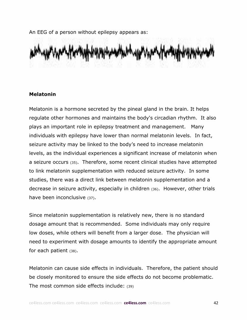
An EEG of a person without epilepsy appears as:
Melatonin
Melatonin is a hormone secreted by the pineal gland in the brain. It helps
regulate other hormones and maintains the body's circadian rhythm. It also
plays an important role in epilepsy treatment and management. Many
individuals with epilepsy have lower than normal melatonin levels. In fact,
seizure activity may be linked to the body’s need to increase melatonin
levels, as the individual experiences a significant increase of melatonin when
a seizure occurs (35). Therefore, some recent clinical studies have attempted
to link melatonin supplementation with reduced seizure activity. In some
studies, there was a direct link between melatonin supplementation and a
decrease in seizure activity, especially in children (36). However, other trials
have been inconclusive (37).
Since melatonin supplementation is relatively new, there is no standard
dosage amount that is recommended. Some individuals may only require
low doses, while others will benefit from a larger dose. The physician will
need to experiment with dosage amounts to identify the appropriate amount
for each patient (38).
Melatonin can cause side effects in individuals. Therefore, the patient should
be closely monitored to ensure the side effects do not become problematic.
The most common side effects include: (39)

ce4less.com ce4less.com ce4less.com ce4less.com ce4less.com ce4less.com 43
Some people may have vivid dreams or nightmares when they take
melatonin. Taking too much melatonin may disrupt circadian rhythms
(“body clock”).
Melatonin can cause drowsiness if taken during the day. If an
individual is drowsy the morning after taking melatonin, a lower dose
may be necessary.
Additional side effects include stomach cramps, dizziness, headache,
irritability, decreased libido, breast enlargement in men (called
gynecomastia), and decreased sperm count.
Pregnant or nursing women should not take melatonin because it could
interfere with fertility.
Some studies show that melatonin supplements worsened symptoms
of depression. For this reason, people with depression should consult
their doctor before using melatonin supplements.
Melatonin may interact with various medications. The following table
provides an overview of the drugs that have the highest risk of interacting
with melatonin: (40)
Antidepressant
medications
In an animal study, melatonin supplements reduced the
antidepressant effects of desipramine and fluoxetine (Prozac).
More research is needed to know if the same thing would happen
in people. In addition, fluoxetine (a member of a class of drugs
called selective serotonin reuptake inhibitors, or SSRIs) can
cause low levels of melatonin in people.
Antipsychotic
medications
A common side effect of antipsychotic medications used to treat
schizophrenia is a condition called tardive dyskinesia, which
causes involuntary movements. In a study of 22 people with
schizophrenia and tardive dyskinesia caused by antipsychotic
medications, those who took melatonin supplements had fewer

ce4less.com ce4less.com ce4less.com ce4less.com ce4less.com ce4less.com 44
symptoms compared to those who did not take the supplements.
Benzodiazepines The combination of melatonin and triazolam (Halcion) improved
sleep quality in one study. In addition, a few reports have
suggested that melatonin supplements may help people stop
using long-term benzodiazepine therapy, which is habit-forming.
Blood pressure
medications
Melatonin may make blood pressure medications like
methoxamine (Vasoxyl) and clonidine (Catapress) less effective.
In addition, medications in a class called calcium channel
blockers may lower melatonin levels. Calcium channel blockers
include:
Nifedipine (Procardia)
Amlodipine (Norvasc)
Verapamil (Calan, Isoptin)
Diltiazem (Cardizem)
Felodipine (Plendil)
Nisoldipine (Sular)
Bepridil (Vascor)
Beta-blockers Use of beta-blockers may lower melatonin levels in the body.
Beta-blockers include:
Acebutolol (Sectral)
Atenolol (Tenormin)
Bisoprolol (Zebeta)
Carteolol (Cartrol)
Metoprolol (Lopressor, Toprol XL)
Nadolol (Corgard)
Propranolol (Inderal)
Anticoagulant
medications
Melatonin may increase the risk of bleeding from anticoagulant
medications such as warfarin (Coumadin).

ce4less.com ce4less.com ce4less.com ce4less.com ce4less.com ce4less.com 45
Interleukin-2 In one study of 80 cancer patients, use of melatonin along with
interleukin-2 led to more tumor regression and better survival
rates than treatment with interleukin-2 alone.
Nonsteroidal anti-
inflammatory drugs
(NSAIDs)
NSAIDs such as ibuprofen (Advil, Motrin) may lower levels of
melatonin in the blood.
Steroids and
immunosuppressant
medications
Melatonin may cause these medications to lose their
effectiveness. Do not take melatonin with corticosteroids or
other medications used to suppress the immune system.
Tamoxifen Preliminary research suggests that the combination of tamoxifen
(a chemotherapy drug) and melatonin may benefit some people
with breast and other cancers. More research is needed to
confirm these results.
Other Caffeine, tobacco, and alcohol can all lower levels of melatonin in
the body.
Vitamins
Many epileptic patients will benefit from supplementation with vitamins. In
many instances, epileptic seizures and other symptoms increase if the
patient is deficient in a specific vitamin (41). In other instances, patients may
benefit from an increase in nutritional supplementation as it will improve
basic body composition and increase the patient’s ability to withstand the
negative effects of epilepsy (42). The following section provides a thorough
overview of the vitamins most beneficial in epilepsy treatment: (41,43–51)
Folic acid

ce4less.com ce4less.com ce4less.com ce4less.com ce4less.com ce4less.com 46
Supplementation with folic acid on a daily basis is important for both women
as well as men. The vitamin named folic acid (also known as folate) is an
important part of the production of blood cells, of the function of some
nerves and to help prevent heart disease. Low levels (deficiency) of folic acid
can be the cause of intrauterine growth delay, inherited malformations,
miscarriages and neural tube defects in women, and heart disease in both
men and women.
For patients who have epilepsy, this is especially important since some
seizure medicines can cause low levels of folic acid by changing the way it is
absorbed in the body. Patients who take more than one seizure medicine
may be advised to take higher doses of folic acid. Babies born to women
who did not get enough folic acid early in their pregnancies are more likely
to have birth defects, especially a type called neural tube defects, which
affect the brain and spinal cord. The most well-known of these is spina
bifida, in which the spinal column is not completely closed. By the time a
woman knows for sure that she is pregnant, it may be too late to prevent
these defects.
Folic acid should be added to a person’s daily diet, either as food or as a
supplement, starting in the teenage years for women, and young adulthood
for men with epilepsy. Some providers recommend up to 4 mg per day for
patients who have been taking daily anti-seizure medications for many
years.
Epileptologists are now concerned that folic acid may be too low in persons
with epilepsy taking some antiepileptic drugs. Low serum and red blood cell
levels of folic acid in women of childbearing potential increase the risk of
fetal birth defects. For men and women, low levels of folic acid are

ce4less.com ce4less.com ce4less.com ce4less.com ce4less.com ce4less.com 47
associated with elevated homocysteine and an increased risk for
cardiovascular disease. A convincing argument now develops that routine
folic acid supplementation is important for women and men receiving
antiepileptic drugs.
Folic acid (vitamin B9) is a water-soluble B vitamin that is essential for DNA
repair, cell division, and normal cellular growth. Low folic acid levels during
pregnancy in women with epilepsy have been associated with fetal
malformation, and older enzyme-inducing antiepileptic drugs are known to
reduce serum folate levels. As mentioned earlier, profound deficiency of folic
acid during pregnancy has been associated with neural tube defects such as
spina bifida. Deficiency in adults has been associated with megaloblastic
anemia and peripheral neuropathy. In both men and women, low serum
levels of folate can increase homocysteine levels, which are correlated with
elevated cardiovascular risk.
Certain antiepileptic drugs, but not all agents, can potentially decrease folate
levels, either via hepatic enzyme induction and/or decreased absorption.
Addressing the question of which patients on AEDs need folic acid
supplementation is challenging because it depends on whether the patient is
pregnant or has a history of epilepsy. For example, the risk of having a
pregnancy complicated by a major congenital malformation (e.g., neural
tube defect) is doubled in epileptic women taking AEDs compared with those
women with a history of epilepsy not taking these agents. In fact, that risk is
tripled with AED polypharmacy, especially when valproic acid is included.
Additionally, many AEDs are used for conditions other than epilepsy, such as
chronic pain and mood disorders, but the effect of AEDs on folate has not
been adequately assessed in this population.

ce4less.com ce4less.com ce4less.com ce4less.com ce4less.com ce4less.com 48
There are some general guidelines about folic acid supplementation.
Consensus statements recommend 0.4-0.8 mg of folic acid per day in all
women planning a pregnancy. Ideally, this should be started at least 1
month prior to pregnancy if possible. These guidelines recommend higher
daily folic acid doses (4 mg/day) in women with a history of neural tube
defects. In addition, enzyme-inducing anticonvulsants, such as phenytoin,
carbamazepine, primidone, and phenobarbital, are known to decrease folate
levels, and valproic acid may interfere with folate metabolism. Other AEDs,
such as oxcarbazepine, lamotrigine, and zonisamide, do not appear to alter
folate levels.
Unfortunately, the effectiveness of folic acid supplementation for the
prevention of AED-induced teratogenicity and the appropriate dose of folic
acid for specific AEDs has not been determined. Not all studies designed to
determine effects of fetal AED exposure consistently demonstrate a
protective effect against congenital malformations with folic acid
supplementation. However, this may be due in part to inadequate dosage.
Because many pregnancies are unplanned, most authorities recommend that
folic acid supplementation be given routinely to all women of childbearing
potential at 0.4 mg/day. Women who have already had a child with a neural
tube defect are encouraged to consult with their clinician regarding
appropriate dosage, and those on AEDs should receive 0.4 - 4 mg/day.
Current data are inconclusive to support high-dose folic acid use in women
without epilepsy on AEDs for other indications, though supplemental folic
acid should not be regarded as harmful. For men and women on AEDs that
reduce folate levels, such as phenytoin, carbamazepine, primidone, and
phenobarbital, it seems prudent to monitor homocysteine and folate levels
and monitor for the development of megaloblastic anemia.
ce4less.com ce4less.com ce4less.com ce4less.com ce4less.com ce4less.com 49
The Comprehensive Epilepsy Center has developed guidelines for folate
supplementation for women of childbearing years. These guidelines were
established to enhance patient education and awareness of the potential
vitamin deficiencies that can occur when taking antiepilepsy medications
(AED's). They help to promote the general health of women, and minimize
potential birth defects associated with folate deficiency.
Folate (or folic acid) deficiency and medications used to treat epilepsy are
associated with an increased risk of birth defects. Specifically, they are
associated with spina bifida and anencephaly, two of the most common and
severe neurologic birth defects. Clinical studies have shown that
supplementing a woman's diet with folate can reduce this risk by 50-75%.
In order to reduce the risk of neural tube defects, the Center for Disease
Control and Prevention (CDC) recommends that all women who are capable
of becoming pregnant should take 0.4 mg of folate each day. Neural tube
defects occur early in the pregnancy, often before a woman is aware that
she is pregnant. In additional, about one-half of pregnancies in the United
States are unplanned. Therefore, supplementation with folate should
continue throughout a woman's reproductive years. A woman who has a
family history of neural tube defects or has a previous child born with neural
tube defects should receive folate supplementation of 4.0 mg per day.
The Comprehensive Epilepsy Center Guidelines for Folate supplementation
are as follows:
All women should supplement their diet by taking 1 prenatal
multivitamin each day. Prenatal multivitamins are available over-the-
counter (OTC) or by prescription. OTC prenatal multivitamins contain

ce4less.com ce4less.com ce4less.com ce4less.com ce4less.com ce4less.com 50
0.8 mg of folate while prescription prenatal multivitamins contain 1.0
mg of folate. Generic multivitamins are generally the least expensive,
followed in order of expense by brand name over-the-counter
vitamins, and finally prescription tablets.
A woman who is planning on becoming pregnant or who is pregnant,
and has a family history of neural tube defects, or has had a previous
child born with neural tube defects, or is on either Tegretol or
Depakote, should receive 3.0 mg of folate in addition to a prenatal
multivitamin.
All other women who are planning to become pregnant or are pregnant and
taking an antiepilepsy medication other than Tegretol or Depakote should
receive 1.0 mg of folate in addition to a prenatal multivitamin.
Calcium
Calcium is an important element in the body, and so important that an
individual has more calcium in his or her body than any other mineral.
Calcium is a necessary part of bone formation, development and repair. The
majority of calcium in the body is stored within bones, while the rest is in
the blood and the body’s other fluids. Abnormal calcium levels may result
in major health problems. Both hypocalcemia (low calcium levels), and
hypercalcemia (high calcium levels) can cause seizures. The main sources of
calcium are dairy products, such as milk, yogurt and ice cream. Green leafy
vegetables, such as broccoli and kale, canned sardines and shellfish are also
good sources of calcium.
Initially, low calcium levels may not give any warning signs. However, as the
level decreases, a person may feel confused and have hallucinations,
memory loss and depression. Because of calcium’s importance in muscle

ce4less.com ce4less.com ce4less.com ce4less.com ce4less.com ce4less.com 51
movement and the function of the nervous system, hypocalcemia can cause
muscle aches, spasms, stiffening of the muscles, and tingling sensations in
the face, mouth, lips, fingers and toes.
Low calcium levels can also cause several types of seizures, including the
following: tonic-clonic seizures, categorized by whole body shaking and loss
of consciousness; focal muscle seizures, during which a set of muscles
contract against a person’s will; and absence seizures, during which a
person appears to be staring off into space. Certain anti-seizure medications
can contribute to lowering calcium levels, especially when taken daily for a
long time period. This happens when the medication makes the liver work
harder than usual, and it causes the elimination of the calcium deposits from
the bone, leading to what is known as brittle bones, bone loss or
osteoporosis.
From a physiological perspective, it is logical that calcium supplementation
may be indicated when myoclonic seizures are encountered. For “when the
calcium ion concentration falls below about one half of normal, a person is
likely to experience tetanic contraction of muscles throughout the body
because of spontaneous nerve impulses in the peripheral nerves” (42). Since
calcitonin and the parathyroid hormone affect serum calcium concentrations,
it is possible that problems in the production of either can lead to limited
tetanic contractions.
Significant changes in important body chemicals such as calcium and
magnesium can cause seizures; so can a lack of certain vitamins. These
chemical changes may provoke a disturbance in the brain, or a single
seizure, by influencing the thresholds for firing. Calcium is a very important
mineral for the normal functioning of brain cells, and low levels of calcium
ce4less.com ce4less.com ce4less.com ce4less.com ce4less.com ce4less.com 52
(hypocalcemia) can cause seizures. Hypocalcemia can be a consequence of
severe kidney disease when too much calcium escapes from the kidney into
the urine. It may also, but rarely, be caused by a hormonal problem that has
the same effects. A deficiency of magnesium, a mineral that interacts with
calcium, may cause low blood calcium and, thus, seizures. With a ketogenic
diet, a calcium supplement must be taken every day to be nutritionally
complete.
There is growing evidence that elevated extra-cellular calcium levels and
homeostatic calcium control mechanisms may be factors in developing
acquired epilepsy (epilepsy that occurred after an injury). It is important to
evaluate the possible functional consequences of altered CA 2+ dynamics in
epileptogenesis. The ability of the neuron to restore CA 2+ loads to resting
[CA 2+] is regulated by CA 2+ homeostatic mechanisms. Increased or
prolonged entry of extracellular CA 2+ could contribute to the altered CA 2+
homeostatic mechanisms in epilepsy. It is important to note that cellular
calcium levels tend to be inversely correlated with extra-cellular calcium
levels. Thus, it does not seem unreasonable to conclude that those without
injury could have seizures caused by calcium problems.
Those that were on long-term anticonvulsant medications had higher levels
of calcium than non-medicated controls. This might suggest that one of the
reasons that some of these medications are continued long-term is that for
some people, they somehow increase the retention of calcium, which may
account for some of their anticonvulsant effects.
Some forms of juvenile myoclonic epilepsy can result from mutations of a Ca
2+ channel. This line of evidence suggests the involvement of channels
expressed in the brain in the pathogenesis of certain types of epilepsy. Ca

ce4less.com ce4less.com ce4less.com ce4less.com ce4less.com ce4less.com 53
2+ influx into excitable cells is a prerequisite for neurotransmitter release
and regulated exocytosis. Within the group of ten-cloned voltage-gated Ca
2+ channels, the Ca(v) 2.3-containing E-type Ca 2+ channels are involved in
various physiological processes, such as neurotransmitter release and
exocytosis together with other voltage-gated Ca 2+ channels of the Ca(v)1,
Ca(v)2 and Ca(v)3 subfamily. The interaction of Ca(v) 2.3 with the EF-hand
motif containing protein EFHC1 is involved in the etiology and pathogenesis
of juvenile myoclonic epilepsy. However, E-type Ca 2+ channels also exhibit
several subunit-specific features, most of which still remain poorly
understood. While they are not fully understood, it seems apparent that
calcium control mechanisms play some role in myoclonic seizures.
Mutations in the calcium-sensing receptor gene (CaSR) may result in
disorders of calcium homeostasis manifesting as familial benign hypocalciuric
hypercalcemia (FBHH), neonatal severe hyperparathyroidism (NSHPT) or
autosomal dominant hypocalcemia with hypercalciuria (ADHH). The ADHH
condition may result in asymptomatic hypocalcemia and a minority
experience seizures in infancy, which can recur into adulthood.
Even in generalized seizures, epileptics are generally mildly hypocalcemic,
especially in the period before the seizure. Stress, which releases
epinephrine and corticotropin, results in high serum citrate concentration,
which probably contributes to decreased serum [Ca2+] just before a seizure.
Long-term treatment of epileptic children with various anticonvulsant
medications was found to raise the TSH and diminished T3 and T4.
Calcitonin levels were lowered as well. Long-term use of certain
anticonvulsant medications tended to impair at least a portion of thyroid
function.

ce4less.com ce4less.com ce4less.com ce4less.com ce4less.com ce4less.com 54
Myoclonic seizures tend to be resistant to drug therapies. Since many anti-
epileptic medications impair thyroid function and/or somehow result in
increased calcium levels, perhaps a partial reason for their occasional
success with myoclonic seizures is the partial suppression of the thyroid
hormone calcitonin, which results in an increase of serum calcium levels.
There are scattered reports that the anticonvulsant medications
phenobarbital, carbamazepine, valproate, lamotrigine, gabapentin, and
vigabatrin can cause or induce myoclonic seizures in epileptics who had not
been experiencing those types of seizures. It is possible this occurs because
some anticonvulsant medications can reduce vitamin D levels. Other
researchers have thus suggested supplemental vitamin D when taking
certain anticonvulsant medications.
Myoclonic seizures can have an appearance of a limited tetanic contraction
associated with insufficient calcium levels. It is important to note that
others, while not specifically discussing myoclonic seizures, have also
suggested that somehow increasing calcium levels should be looked at for
the treatment of epileptics. Hence, it may be wise to consider nutritional
interventions that affect calcium levels as a first-line treatment
Currently, this is rarely the case. Even though some antiepileptic drugs could
also worsen some types of seizures, it is known that other therapies can be
efficient in refractory epilepsies; steroids, vague nerve stimulation, ketogenic
diet and surgery, nutritional therapies (especially outside the ketogenic diet)
seem to be often overlooked. It should be noted that it is theoretically
possible that, for some types of seizures, calcium could be contraindicated.

ce4less.com ce4less.com ce4less.com ce4less.com ce4less.com ce4less.com 55
Yet it is not unheard of that nutrition, including calcium supplementation,
should be considered as a first-line treatment for intractable forms of
epilepsy, as others have sometimes advocated it (though this investigator
appears to be the first advocating supplemental calcium, vitamin D, etc., as
first-line nutrients, as well as first to advise nutrients specifically for
myoclonic seizures). One of the reasons that nutrition should be considered
as a front-line therapy is that it is low risk.
Consumption of calcium-containing foods and/or calcium-containing
supplements is so safe that, although calcium can react with some
medications, over dosage has not been reported with calcium supplements.
Forms other than calcium carbonate are preferred, as calcium carbonate
may cause gastrointestinal side reactions such as constipation, bloating, gas
and flatulence. Prolonged use of large doses of calcium carbonate — greater
than 12 grams daily (about 5 grams of elemental calcium) — may lead to
milk-alkali syndrome, nephrocalcinosis and renal insufficiency.
There is no specific quantitative recommendation for each possible
substance that could affect calcium levels, as the amount needed appears to
vary by individual (as well as size in the case of children). But irrespective of
the quantities, it does appear reasonable to conclude that calcium control
mechanisms can play a causal role in myoclonic seizures and that calcium
and other nutrients should be considered as possible front-line therapies for
these hard to treat myoclonic seizures.
Vitamin D
Vitamin D is a necessary part in the process of proper breakdown and use of
calcium. Because of this, vitamin D deficiency caused or worsened by daily
use of anti-seizure medications for a long time can make the bones very soft

ce4less.com ce4less.com ce4less.com ce4less.com ce4less.com ce4less.com 56
and brittle, causing them to break more easily. Adding vitamin D to the daily
diet can prevent this. In addition, patients taking anti-seizure medications
should increase their calcium intake every day. Exposure to sunlight is a
natural way to speed up the body’s ability to produce vitamin D.
Nearly half of people with epilepsy are also vitamin D deficient, but despite
this well-known association, only a single study has been published on the
effect of vitamin D for seizure control in the last 40 years. That study
revealed that treating epileptic patients with vitamin D2 – the far inferior
type of synthetic vitamin D – reduced the number of seizures, and in 1974
researchers concluded “the results may support the concept that epileptics
should be treated prophylactically with vitamin D (46).“
Now, nearly four decades later researchers have again revealed that “the
normalization of serum vitamin 25(OH)D [vitamin D] level has an
anticonvulsant [anti-seizure] effect (50).” The findings are even more
important given that people with epilepsy face an even greater risk of
vitamin D deficiency than the general population (and even the general
population is vastly vitamin D deficient). The reasons are two-fold, with the
first being that having frequent seizures may interfere with a person’s ability
to get outdoors and stay active.
If an individual spends most of his or her time inside, they will miss out on
regular sun exposure, which is key for the natural production of vitamin D.
Even exposing the skin to sunlight through a windowpane will prevent the
entry of the UVB rays, which are the specific wavelength that produces
vitamin D in the skin. It is crucial for epilepsy patients to get outside and
experience direct skin contact with the sunlight instead of sunning in a
sunroom, for instance. Second, anti-epileptic drugs that are often given to
epilepsy patients can interfere with vitamin D metabolism, leading to

ce4less.com ce4less.com ce4less.com ce4less.com ce4less.com ce4less.com 57
deficiency. If these drugs are taken, it is especially crucial that vitamin D
levels are actively monitored to avoid this side effect.
Vitamin D has a significant impact on epileptic seizures because epilepsy is a
disorder of the central nervous system, particularly of the brain. Vitamin D is
a vitamin that is also a neuroregulatory steroidal hormone that influences
nearly 3,000 different genes in the body. Vitamin D receptors can be found
in the brain, spinal cord, and central nervous system, and may enhance the
amount of important chemicals in the brain needed to protect brain cells.
Surgical Options
A number of individuals with epilepsy may benefit from surgical intervention.
There are a variety of surgical procedures that can help with various aspects
of the disorder. While medication is effective at controlling most seizure
activity, approximately thirty percent of individuals will not respond to
pharmacologic treatment and will require more advanced therapy (52). These
individuals often benefit from surgery.
There are three primary forms of surgery that are used to treat individuals
with epilepsy: (53)
Surgery to remove the area of the brain producing seizures
Surgery to interrupt the nerve pathways through which seizure
impulses spread within the brain
Surgery to implant a device used to treat epilepsy
Surgery is an invasive procedure and should only be considered if the
section of the brain where the seizures originate can be clearly identified (54).
In addition, the physician must ensure that surgery will not negatively affect

ce4less.com ce4less.com ce4less.com ce4less.com ce4less.com ce4less.com 58
any areas that are responsible for critical functions (55). A thorough
assessment is required before determining if surgery is a viable option.
There are a number of different surgical procedures that can be used to treat
epilepsy. The specific type of surgery performed on a patient will be
determined based upon the type of seizures the patient is experiencing and
the area of the brain where seizure activity originates (52). The following
section provides an overview of the risks and benefits of various surgical
procedures (14,56–83).
(Photo courtesy of: http://www.epilepsy.net.in/images/epilepsy_surgery.jpg)
ce4less.com ce4less.com ce4less.com ce4less.com ce4less.com ce4less.com 59
Procedure Description and Benefits/Risks
Vagus Nerve
Stimulation
(VNS)
VNS is a palliative technique that involves surgical implantation of a
stimulating device. VNS is FDA approved to treat medically refractory
focal-onset epilepsy in patients older than 12 years. Some studies
demonstrate its efficacy in focal-onset seizures and in a small number
of patients with primary generalized epilepsy. Randomized studies
showed modest efficacy at 3 months, but post marketing experience
showed delayed improvement in another group of patients.
In August 2013, the American Academy of Neurology issued an
update to its 1999 guidelines on the use of VNS for epilepsy. VNS is
currently indicated for patients older than 12 years with medically
intractable partial seizures who are not candidates for potentially
curative surgical resections, as well as for the adjunctive long-term
treatment of chronic or recurrent depression in patients older than 18
years with a major depressive episode not adequately relieved by 4
or more antidepressant treatments. Recent reports also indicate long-
term efficacy and successful VNS use in pediatric epilepsy and other
seizure types and syndromes.
Key recommendations of the updated guidelines include the
following:
VNS may be considered for (1) the adjunctive treatment of
partial or generalized epilepsy in children, (2) seizures
associated with Lennox-Gastaut syndrome, and (3) improving
mood in adults with epilepsy
VNS may have improved efficacy over time
Children should be carefully monitored for site infection after
VNS implantation
According to the manufacturer's registry, efficacy of the stimulating
device at 18 months is 40-50%, where efficacy is defined as a seizure

ce4less.com ce4less.com ce4less.com ce4less.com ce4less.com ce4less.com 60
reduction of 50% or more. Many patients report improvement in
seizure intensity and general mood. However, seizure-free rates for
pharmacologically intractable focal-onset epilepsy are less than 10%.
A meta-analysis of VNS clinical studies reported an average reduction
in seizures of at least 50% in approximately 50% of patients at last
follow-up. Although VNS was not initially FDA approved for children
or patients with generalized epilepsy, the authors also found that
these groups benefitted significantly from VNS.
Positive predictors of a favorable outcome with VNS therapy include
posttraumatic epilepsy and tuberous sclerosis. Few patients achieve
complete seizure freedom with VNS, and about a quarter of patients
receive no benefit in their seizure frequency. Some patients have
clinical improvement in terms of milder and shorter seizures.
Multiple Subpial
Transection
Multiple subpial transection was pioneered as an alternative to
removal of brain tissue. It is used to control partial seizures
originating in areas that cannot be safely removed. For example, if
the seizure focus involves the dominant temporal-lobe language area
(Wernicke’s area), which is critical for comprehension, the removal of
this area to control seizures would cause a devastating complication:
the inability to understand spoken or written language. Similarly, if
the primary motor area is part of the seizure focus, its removal would
cause permanent weakness on the opposite side of the body.
The operation involves a series of shallow cuts (transections) into the
cerebral cortex. The transections are made only as deep as the gray
matter, approximately a quarter of an inch deep. Because of the
complex way in which the brain is organized, these cuts are thought
to interrupt some fibers that connect neighboring parts of the brain,
but they do not appear to cause long-lasting impairment in the
critical functions served by these areas. Examination of brain tissue
after multiple subpial transections reveals that some nerve cells are
destroyed.

ce4less.com ce4less.com ce4less.com ce4less.com ce4less.com ce4less.com 61
Sometimes, brain seizures begin in a vital area of the brain -- for
example, in areas that control movement, feeling, language, or
memory. When this is the case, a relatively new epilepsy treatment
called multiple subpial transection (MST) may be an option. MST
stops the seizure impulses by cutting nerve fibers in the outer layers
of the brain (gray matter), sparing the vital functions concentrated in
the deeper layers of brain tissue (white matter).
Most people with epilepsy can control their seizures with medication.
However, about 20% of people with epilepsy do not improve with
drugs. In some cases, surgery to remove the part of the brain
causing the seizures may be recommended.
MST may be an option for people who do not respond to medication
and whose seizures begin in areas of the brain that cannot be safely
removed. In addition, there must be a reasonable chance that the
person will benefit from surgery. MST may be done alone or with the
removal of a section of brain tissue (resection). MST also may be
used as a treatment for children with Landau-Kleffner syndrome
(LKS), a rare childhood brain disorder which causes seizures and
affects the parts of the brain that control speech and comprehension.
Candidates for MST undergo an extensive pre-surgery evaluation -
including seizure monitoring, electroencephalography (EEG),
magnetic resonance imaging (MRI), and positron emission
tomography (PET). These tests help to pinpoint the area in the brain
where the seizures occur and determine if surgery is feasible.
Another test to assess electrical activity in the brain is EEG-video
monitoring, in which video cameras are used to record seizures as
they occur, while the EEG monitors the brain's activity. In some
cases, invasive monitoring - in which electrodes are placed inside the
skull over a specific area of the brain - is also used to further identify
the tissue responsible for seizures.

ce4less.com ce4less.com ce4less.com ce4less.com ce4less.com ce4less.com 62
MST requires exposing an area of the brain using a procedure called a
craniotomy. ("Crani" refers to the skull and "otomy" means "to cut
into.") After the patient is put to sleep with anesthesia, the surgeon
makes an incision (cut) in the scalp, removes a piece of bone and
pulls back a section of the dura, the tough membrane that covers the
brain. This creates a "window" in which the surgeon inserts his or her
surgical instruments. The surgeon utilizes information gathered
during pre-surgical brain imaging to help identify the area of
abnormal brain tissue and avoid areas of the brain responsible for
vital functions.
Using a surgical microscope to produce a magnified view of the brain,
the surgeon makes a series of parallel, shallow cuts (transections) in
gray matter, just below the pia mater (subpial), the delicate
membrane that surrounds the brain (it lies beneath the dura). The
cuts are made over the entire area identified as the source of the
seizures. After the transactions are made, the dura and bone are
fixed back into place, and the scalp is closed using stitches or staples.
There may be bleeding at the site of the transection, but the
procedure is generally well tolerated. Major complications appear to
be rare. Transections in language areas may cause mild impairments
in the language function served by that area. The risks and benefits
of multiple subpial transections need to be better defined.
Multiple subpial transections can help reduce or eliminate seizures
arising from vital functional cortical areas. Transections have been
used successfully in Landau-Kleffner syndrome, a disorder in which
language problems appear in a child whose language was previously
developing normally. One concern is that the epileptic activity may
recur after a period of 2 to 20 months. It is uncertain whether this
procedure can achieve long-term seizure control
Temporal Lobe
Resection
The most common surgical procedure performed for epilepsy is the
removal of a portion of the temporal lobe, or temporal lobectomy.
These brain structures play an important role in the generation or

ce4less.com ce4less.com ce4less.com ce4less.com ce4less.com ce4less.com 63
propagation of the majority of temporal lobe seizures. In most cases,
a modest portion of the brain measuring approximately 2 inches long
is removed. The temporal lobes are important in memory, emotion
and language comprehension. However, extensive preoperative
assessments (MRI, Wada tests, PET scans) ensure that removal of
the area causing seizures will not disrupt any of these critical
functions.
The largest part of the brain, the cerebrum, is divided into four paired
sections - the frontal, parietal, occipital, and temporal lobes. Each
lobe controls a specific group of activities. The temporal lobe, located
on either side of the brain just above the ear, plays an important role
in hearing, language, and memory. The most common type of
epilepsy in teens and adults originates in the temporal lobe, the
seizure focus.
Temporal Lobe Resection
A temporal lobe resection is a surgery performed on the brain to
control seizures. In this procedure, brain tissue in the temporal lobe
is resected, or cut away, to remove the seizure focus. The anterior
(front) and mesial (deep middle) portions of the temporal lobe are
the areas most often involved.
Temporal lobe resection may be an option for people with epilepsy
whose seizures are disabling and/or not controlled by medication, or
when the side effects of medication are severe and significantly affect
the person's quality of life. In addition, it must be possible to remove
the brain tissue that contains the seizure focus without causing
damage to areas of the brain responsible for vital functions, such as
movement, sensation, language, and memory.
Candidates for temporal lobe resection undergo an extensive pre-
surgery evaluation - including seizure monitoring,
electroencephalography (EEG), magnetic resonance imaging (MRI),
and positron emission tomography (PET). These tests help to pinpoint
ce4less.com ce4less.com ce4less.com ce4less.com ce4less.com ce4less.com 64
the seizure focus within the temporal lobe and to determine if surgery
is possible.
A temporal lobe resection requires exposing an area of the brain
using a procedure called a craniotomy. After the patient is put to
sleep with anesthesia, the surgeon makes an incision in the scalp,
removes a piece of bone and pulls back a section of the dura, the
tough membrane that covers the brain. This creates a "window" in
which the surgeon inserts special instruments for removing the brain
tissue. Surgical microscopes also are used to give the surgeon a
magnified view of the area of the brain involved. The surgeon utilizes
information gathered during the pre-operative evaluation - as well as
during surgery - to define, or map out, the route to the correct area
of the temporal lobe.
In some cases, a portion of the surgery is performed while the
patient is in a ''twilight state'' - awake but under sedation - so that
the patient can help the surgeon find and avoid areas of the brain
responsible for vital functions. While the patient is awake, the doctor
uses special probes to stimulate different areas of the brain. At the
same time, the patient is asked to count, identify pictures, or perform
other tasks. The surgeon can then determine the area of the brain
associated with each task. After the brain tissue is removed, the dura
and bone are fixed back into place, and the scalp is closed using
stitches or staples.
Permanent complications associated with temporal lobe resection
surgery are very low. Mortality is less than 0.1% and permanent
unexpected morbidity less than 1%. In dominant hemisphere
resections, temporary language difficulties are seen in 10% of the
cases although these usually resolve. An upper quadrantanopsia
(partial upper peripheral vision loss) is expected in large temporal
resections, but seen in less than 25% of the patients. Memory
impairment rarely occurs from temporal lobectomies because of
extensive preoperative testing of language and memory functions.
ce4less.com ce4less.com ce4less.com ce4less.com ce4less.com ce4less.com 65
The success rate for seizure control in temporal lobectomy varies:
60%-70% of patients are free of seizures that impair
consciousness or cause abnormal movements, but some still
experience auras
20%-25% of patients have some seizures but are significantly
improved (greater than 85% reduction of complex partial and
tonic-clonic seizures)
10%-15% of patients have no worthwhile improvement
Therefore, over 85% of patients enjoy a marked improvement in
seizure control. Most of them need less medication after surgery.
Approximately 25% of those who are seizure-free eventually can
discontinue antiepileptic drugs
Lesionectomy
Twenty five percent of patients with epilepsy will have lesions
identified by MRI as the cause of recurrent seizures. Abnormalities
such as low-grade astrocytomas, cortical dysplasias, cavernous
angiomas, and areas of focal atrophy are the common causes of
refractory seizures. Since surgical removal of these lesions can result
in complete seizure control in many patients, the patient is
considered an excellent candidate for epilepsy surgery depending on
the location of the lesion and its relationship to eloquent cortex. If
the seizures have been present for many years then invasive
monitoring is often recommended to further identify the involvement
of the adjacent cortical rim around the lesion. When lesions are
within the non-dominant temporal lobe, removal of the lesion along
with a temporal lobectomy yields excellent results in over 80% of
cases.
A lesionectomy is the surgical removal of lesions. MRI identifies small
lesions as the cause of recurrent seizures in up to 25% of patients.
Common types of lesions include low grade astrocytomas, cortical
dysplasias, cavernous angiomas, and areas of focal atrophy.
Functional The largest part of the brain, the cerebrum, can be divided down the

ce4less.com ce4less.com ce4less.com ce4less.com ce4less.com ce4less.com 66
Hemispherectomy
middle lengthwise into two halves, called hemispheres. A deep
groove splits the left and right hemispheres, which communicate
through a thick band of nerve fibers called the corpus callosum. Each
hemisphere is further divided into four paired sections, called lobes -
the frontal, parietal, occipital, and temporal lobes.
The two different sides or hemispheres are responsible for different
types of activities. The left side of the brain controls the right side of
the body and vice versa. For most people, the ability to speak and
understand the spoken word is a function of the left side of the brain.
A functional hemispherectomy is a procedure in which portions of one
hemisphere - which are causing the seizures - are removed, and the
corpus callosum, which connects the two sides of the brain, is cut.
This disconnects communication between the two hemispheres,
preventing the spread of electrical seizures from one side of the brain
to the other. As a result, the person usually has a marked reduction
in physical seizures.
This procedure generally is used only for people with epilepsy who do
not experience improvement in their condition after taking many
different medications and who have severe, uncontrollable seizures.
This type of epilepsy is more likely to be seen in young children who
have an underlying disease, such as Rasmussen's encephalitis or
Sturge-Weber syndrome, which has damaged the hemisphere.
Candidates for functional hemispherectomy undergo an extensive
pre-surgery evaluation - including seizure monitoring,
electroencephalography (EEG), and magnetic resonance imaging
(MRI). These tests help the doctor identify the damaged parts of the
brain and confirm that it is the source of the seizures. An intracarotid
amobarbital test, also called a WADA test, is done to determine which
hemisphere is dominant for critical functions such as speech and
memory. During this test, each hemisphere is alternately injected
with a medication to put it to sleep. While one side is asleep, the
awake side is tested for memory, speech, and ability to understand

ce4less.com ce4less.com ce4less.com ce4less.com ce4less.com ce4less.com 67
speech.
A functional hemispherectomy requires exposing the brain using a
procedure called a craniotomy. Surgical microscopes are utilized to
give the surgeon a magnified view of the brain structures. During the
procedure, the surgeon removes portions of the affected hemisphere,
often taking all of the temporal lobe but leaving the frontal and
parietal lobes. The surgeon also gently separates the hemispheres to
access and cut the corpus callosum. After the tissue is removed, the
dura and bone are fixed back into place, and the scalp is closed using
stitches or staples.
The patient generally stays in an intensive care unit for 24 to 48
hours and then stays in a regular hospital room for three to four
days. Most people who have a functional hemispherectomy will be
able to return to their normal activities, including work or school in
six to eight weeks after surgery. Most patients will need to continue
taking anti-seizure medication, although some may eventually be
able to stop taking medication or have their dosages reduced.
About 85% of people who have a functional hemispherectomy will
experience significant improvement in their seizures, and about 60%
will become seizure-free. In many cases, especially in children, the
remaining side of the brain takes over the tasks that were controlled
by the section that was removed. This often improves a child's
functioning and quality of life, as well as reduces or eliminates the
seizures.
The following symptoms may occur after a functional
hemispherectomy, although they generally go away over time and
with therapy:
Scalp numbness.
Nausea.
Muscle weakness on the affected side of the body.
Puffy eyes.

ce4less.com ce4less.com ce4less.com ce4less.com ce4less.com ce4less.com 68
Feeling tired or depressed.
Difficulty speaking, remembering, or finding words.
Headaches.
The risks associated with a functional hemispherectomy include the
following:
Risks associated with surgery, including infection, bleeding,
and an allergic reaction to anesthesia.
Loss of movement or sensation on the opposite side of the
body.
Swelling in the brain.
Delayed development.
Loss of peripheral (side) vision.
Failure to control seizures.
Corpus
Callosotomy
The corpus callosum is a band of nerve fibers located deep in the
brain that connects the two halves (hemispheres) of the brain. It
helps the hemispheres share information, but it also contributes to
the spread of seizure impulses from one side of the brain to the
other. A corpus callosotomy is an operation that severs (cuts) the
corpus callosum, interrupting the spread of seizures from hemisphere
to hemisphere. Seizures generally do not completely stop after this
procedure (they continue on the side of the brain in which they
originate). However, the seizures usually become less severe, as they
cannot spread to the opposite side of the brain.
A corpus callosotomy, sometimes called split-brain surgery, may be
performed in people with the most extreme and uncontrollable forms
of epilepsy, when frequent seizures affect both sides of the brain.
People considered for corpus callosotomy are typically those who do
not respond to treatment with antiseizure medications.
Candidates for corpus callosotomy undergo an extensive pre-surgery
evaluation - including seizure monitoring, electroencephalography
(EEG), magnetic resonance imaging (MRI), and positron emission
tomography (PET). These tests help the doctor pinpoint where the
seizures begin and how they spread in the brain. It also helps the

ce4less.com ce4less.com ce4less.com ce4less.com ce4less.com ce4less.com 69
doctor determine if a corpus callosotomy is an appropriate treatment.
A corpus callosotomy requires exposing the brain using a procedure
called a craniotomy. After the patient is put to sleep with anesthesia,
the surgeon makes an incision in the scalp, removes a piece of bone
and pulls back a section of the dura, the tough membrane that covers
the brain. The surgeon inserts special instruments for disconnecting
the corpus callosum, gently separates the hemispheres to access the
corpus callosum. Surgical microscopes are used to give the surgeon a
magnified view of brain structures.
In some cases, a corpus callosotomy is done in two stages. In the
first operation, the front two-thirds of the structure is cut, but the
back section is preserved. This allows the hemispheres to continue
sharing visual information. If this does not control the serious
seizures, the remainder of the corpus callosum can be cut in a second
operation. After the corpus callosum is cut, the dura and bone are
fixed back into place, and the scalp is closed using stitches or staples.
The patient generally stays in the hospital for two to four days. Most
people having a corpus callosotomy will be able to return to their
normal activities, including work or school, in six to eight weeks after
surgery. The hair over the incision will grow back and hide the
surgical scar. The person will continue taking antiseizure drugs.
Complications of corpus callosotomy are greater than with frontal or
temporal lobe surgery. Behavioral, language, and other problems
may affect function and the quality of life, but serious problems are
temporary or uncommon. The potential risks of callosotomy must be
weighed against its possible benefits, such as a reduction in the
frequency of seizures that cause injury and other problems. The
persons most susceptible to behavioral problems after callosotomy
are those in whom language and motor dominance are controlled by
different hemispheres. In left-handed persons, for example, the left
side of the brain controls language, but the right side of the brain

ce4less.com ce4less.com ce4less.com ce4less.com ce4less.com ce4less.com 70
controls movement. Some of the problems resulting from callosotomy
are caused by injury to the frontal lobes during the operation. Since
the corpus callosum is buried deep between the frontal lobes, the
middle portions of these lobes must be separated, which poses some
risk. Surgical advances may help to minimize this risk.
Seizure frequency is reduced by an average of 70% to 80% after
partial callosotomy and 80% to 90% after complete callosotomy.
Partial seizures are often unchanged, but they may be improved or
worsened. In many cases, especially after partial callosotomy,
seizures are less frequent but persist.
Extratemporal
Cortical Resection
Extra-temporal seizure surgery constitutes about a quarter of the
surgical procedures for epilepsy and includes resection of the frontal
lobes, parietal lobes or occipital lobes. These resections are guided by
localization from invasive subdural electrodes and, if necessary,
detailed cortical functional mapping. Extra-temporal resections are
individualized to the seizure onset focus, the type of seizure or
syndrome, and the functional mapping, which defines a safe resection
boundary. Motor and sensory cortex and language cortex localization
is performed and greatly minimizes neurological deficits from
surgery.
The largest part of the brain, the cerebrum, is divided into four paired
sections, called lobes - the frontal, parietal, occipital, and temporal
lobes. Each lobe controls a specific group of activities. The temporal
lobe is the most common ''seizure focus,'' the area where most
seizures start, in teens and adults.
However, epileptic seizures can be ''extratemporal,'' or outside of the
temporal lobe, originating in the frontal, parietal or occipital lobes, or
even more than one lobe. If this is the case, extratemporal cortical
resection surgery may be warranted in some cases. An extratemporal
cortical resection is an operation to resect, or cut away, brain tissue
that contains a seizure focus. Extratemporal means the tissue is
located in an area of the brain other than the temporal lobe. The

ce4less.com ce4less.com ce4less.com ce4less.com ce4less.com ce4less.com 71
frontal lobe is the most common extratemporal site for seizures. In
some cases, tissue may be removed from more than one area/lobe of
the brain.
Extratemporal cortical resection may be an option for people with
epilepsy whose seizures are disabling and/or not controlled by
medications, or when the side effects of the medication are severe
and significantly affect the person's quality of life. In addition, it must
be possible to remove the brain tissue that contains the seizure focus
without causing damage to areas of the brain responsible for vital
functions, such as movement, sensation, language, and memory.
Candidates for extratemporal cortical resection undergo an extensive
pre-surgery evaluation including video electroencephalographic (EEG)
seizure monitoring, magnetic resonance imaging (MRI), and positron
emission tomography (PET). Other tests include neuropsychological
memory testing, the Wada test (to determine which side of the brain
controls language function), Single Photon Emission Computed
Tomography (SPECT), and magnetic resonance spectroscopy. These
tests help to pinpoint the seizure focus and determine if surgery is
possible.
An extratemporal cortical resection requires exposing an area of the
brain using a procedure called a craniotomy. After the patient is put
to sleep (general anesthesia), the surgeon makes an incision in the
scalp, removes a piece of bone and pulls back a section of the dura,
the tough membrane that covers the brain. The surgeon inserts
special instruments to remove brain tissue. Surgical microscopes are
used to give the surgeon a magnified view of the area of the brain
involved. The surgeon utilizes the information gathered during the
pre-operative evaluation - as well as during surgery - to define, or
map out, the route to the correct area of the brain.
In some cases, a portion of the surgery is performed while the
patient is awake, using medication to keep the person relaxed and
pain-free. This is done so that the patient can help the surgeon find
ce4less.com ce4less.com ce4less.com ce4less.com ce4less.com ce4less.com 72
and avoid areas in the brain responsible for vital functions such as
brain regions of language and motor control. While the patient is
awake, the doctor uses special probes to stimulate various areas of
the brain. At the same time, the patient may be asked to count,
identify pictures, or perform other tasks. The surgeon can then
identify the area of the brain associated with each task. After the
brain tissue is removed, the dura and bone are fixed back into place,
and the scalp is closed using stitches or staples.
The risk of a major complication, such as a stroke, is about 1% in
these types of surgery. The risk of behavioral changes is higher than
with temporal lobectomy although these are often difficult to measure
and define. Personality, motivation, ability to plan and to follow up on
a multistep process, ability to organize actions over time, social
graces, and demeanor are among the behaviors that the frontal lobes
help to serve. In parietal and occipital lobectomies, there may be a
risk of losing touch sensation or vision.
Results of surgical management for extratemporal epilepsy vary
depending upon seizure types, invasive mapping, and epilepsy
syndrome. Overall:
50%-60% of patients are free of seizures that impair
consciousness or cause abnormal movements.
20%-40% of patients are markedly improved (more than 90%
reduction of complex partial and tonic-clonic seizures)
20%-30% of patients have no worthwhile improvement.
Although extratemporal surgical cure rates are not as high as
temporal surgery rates, patients with well-defined epileptic
zones limited to smaller areas of the brain which can be
resected do better than in cases of widespread seizure areas.
It is in the area of extratemporal seizures that our institution
has improving success rates as these more difficult problems
are managed with the latest techniques, imaging modalities
and our greater understanding.
Implantable The NeuroPace RNS System, a device that is implanted into the

ce4less.com ce4less.com ce4less.com ce4less.com ce4less.com ce4less.com 73
neurostimulator
cranium, senses and records electrocorticographic patterns and
delivers short trains of current pulses to interrupt ictal discharges in
the brain. The Neurological Devices panel of the FDA concluded that
this device was safe and effective in patients with partial-onset
epilepsy in whom other antiepileptic treatment approaches have
failed and that the benefits outweigh the risks.
In November 2013, the FDA approved the NeuroPace RNS System for
the reduction of seizures in patients with drug-resistant epilepsy.
Approval was based on a clinical trial involving 191 subjects with
drug-resistant epilepsy. The neurostimulator was implanted in all of
these patients but activated in only half of them. After 3 months, the
average number of seizures per month in patients with the activated
device fell by a median of 34%, compared with an approximately
19% median reduction in patients with an unactivated device.
Anti-Epileptic Medication
Anti-epileptic medication is often a necessary component of treatment.
Many patients will require pharmacologic therapy to control seizures. In
most instances, medication will be used in conjunction with other non-
pharmacologic therapies to provide a comprehensive approach to treatment
(17). Utilizing numerous options together provides the best means of seizure
control, especially in patients who experience severe or frequent seizures.
The following table provides an overview of the various types of anti-
epileptic medication: (12,55,84–101)

ce4less.com ce4less.com ce4less.com ce4less.com ce4less.com ce4less.com 74
Valproate
Sodium
Carbamazepine
Valproate sodium (Depacon, generic), valproic acid (Depakene,
generic), and divalproex sodium (Depakote, generic) are
anticonvulsants that are chemically very similar to each other. (In this
report, they are referred to together as valproate.) Valproate products
are the most widely prescribed anti-epileptic drugs worldwide. They are
the first choice for patients with generalized seizures and are used to
prevent nearly all other major seizures as well.
Side Effects: These drugs have a number of side effects that vary
depending on dosage and duration. Most side effects occur early in
therapy and then subside. The most common side effects are upset
stomach and weight gain. Less common side effects include dizziness,
hair thinning and loss, and difficulty concentrating.
Serious side effects include a higher risk for serious birth defects than
other AEDs especially if taken during the first trimester of pregnancy.
In particular, these drugs are associated with facial cleft deformities
(cleft lip or palate) and cognitive impairment.
Liver damage or failure is a rare but extremely dangerous side effect
that usually affects children under 2 years of age who have birth
defects and are taking more than one antiseizure drug. Pancreatitis
(inflammation of the pancreas) and kidney problems are also rare but
serious side effects.
Carbamazepine (Tegretol, Equetro, Carbatrol, generic) is used for many
types of epilepsy. It is taken alone or in combination with other drugs.
In addition to controlling seizures, it may help relieve depression and
improve alertness. A chewable form is available for children.
Side Effects: Different side effects may develop or resolve at different
points during treatment. Initial side effects may include the following:
ce4less.com ce4less.com ce4less.com ce4less.com ce4less.com ce4less.com 75
Double vision, headache, sleepiness, dizziness, and stomach upset.
These usually subside after a week and can be greatly reduced by
starting with a small dose and building up gradually.
Some people experience visual disturbances, ringing in the ears,
agitation, or odd movements when drug levels are at their peak. The
extended-release form of carbamazepine (Carbatrol) may help reduce
these symptoms.
Serious side effects are less common but can include: skin reactions,
including toxic epidermal necrolysis and Stevens-Johnson syndrome, so
severe the drug has to be discontinued develop in about 6% of
patients. These skin reactions cause skin lesions, blisters, fever,
itching, and other symptoms. People of Asian ancestry have a 10 times
greater risk for skin reactions than other ethnicities.
A decrease in white blood cells occurs in about 10% of those taking the
drug. This is generally not serious unless infection accompanies it.
Other blood conditions can arise that are also potentially serious.
Patients should be sure to inform their doctors if they have any sign of
irregular heartbeats, sore throat, fever, easy bruising, or unusual
bleeding.
Long-term therapy can cause bone density loss (osteoporosis) in
women, who should take preventive calcium and vitamin D
supplements to improve bone mass.
Children are at higher risk for behavioral problems.
Note: Grapefruit, Seville oranges, and tangelos can increase
carbamazepine's blood levels and risk of adverse effects.

ce4less.com ce4less.com ce4less.com ce4less.com ce4less.com ce4less.com 76
Phenytoin
Phenytoin (Dilantin, generic) is effective for adults who have the
following seizures or conditions:
Grand mal seizures
Partial seizures
Status epilepticus
Can be effective for people with head injuries who are at high risk for
seizures. This drug is not useful for the following seizures:
Petit mal seizures
Myoclonic seizures
Atonic seizures
Side effects are sometimes difficult to control. Some people may
develop a toxic response to normal doses, while others may require
higher doses to achieve benefits. As with any drug, side effects
generally depend on dosage and duration.
Side effects may include the following:
Excess body hair, eruptions and coarsening of the skin, and
weight loss
Gum disease
Staggering, lethargy, nausea, depression, eye-muscle problems,
anemia, and an increase in seizures can occur as a result of
excessive doses.
Liver damage may develop in rare cases.
Bone density loss from long-term therapy. Patients should take
preventive calcium and vitamin D supplements and exercise
regularly to improve bone mass.
Severe and even rare life-threatening skin reactions (Stevens-
Johnson syndrome, toxic epidermal necrosis)
A possible increased risk for birth defects (cleft palate, poor
thinking skills)

ce4less.com ce4less.com ce4less.com ce4less.com ce4less.com ce4less.com 77
Barbiturates
(Phenobarbital
and Primidone)
Phenobarbital (Luminal, generic), also called phenobaritone, is a
barbiturate anticonvulsant. Primidone (Mysoline, generic) is converted
in the body to phenobarbital, and has the same benefits and adverse
effects.
Barbiturates may be used to prevent grand mal (tonic-clonic) seizures
or partial seizures. They are no longer typically used as first-line drugs,
although they may be the initial drugs prescribed for newborns and
young children.
Side Effects: Phenobarbital has fewer toxic effects on other parts of the
body than most anti-epileptic drugs, and drug dependence is rare,
given the low doses used for treating epilepsy. Nevertheless, many
patients experience difficulty with side effects.
Patients sometimes describe their state as "zombie-like." The most
common and troublesome side effects are:
Drowsiness
Memory problems
Problems with tasks requiring sustained performance
Problems with motor skills
Hyperactivity in some patients, particularly in children and the
elderly
Depression in some adults
When taken during pregnancy, phenobarbital, like phenytoin and
valproate, may lead to impaired cognitive function in the child. There
has been some evidence that phenobarbitol may cause heart problems
in the fetus.
Ethosuximide
and Similar
Drugs
Ethosuximide (Zarontin, generic) is used for petit mal (absence)
seizures in children and adults when the patient has experienced no
other type of seizures. Methsuximide (Celontin), a drug similar to
ethosuximide, may be suitable as an add-on treatment for intractable
epilepsy in children.

ce4less.com ce4less.com ce4less.com ce4less.com ce4less.com ce4less.com 78
Side Effects: This drug can cause stomach problems, dizziness, loss of
coordination, and lethargy. In rare cases, it may cause severe and even
fatal blood abnormalities. Periodic blood counts are recommended for
patients taking this drug.
Clonazepam
Clonazepam (Klonopin, generic) is recommended for myoclonic and
atonic seizures that cannot be controlled by other drugs and for
Lennox-Gastaut epilepsy syndrome. Although clonazepam can prevent
generalized or partial seizures, patients generally develop tolerance to
the drug, which causes seizures to recur.
Side Effects: People who have had liver disease or acute angle
glaucoma should not take clonazepam, and people with lung problems
should use the drug with caution. Clonazepam can be addictive, and
abrupt withdrawal may trigger status epilepticus. Side effects include
drowsiness, imbalance and staggering, irritability, aggression,
hyperactivity in children, weight gain, eye muscle problems, slurred
speech, tremors, skin problems, and stomach problems.
Lamotrigine
Lamotrigine (Lamictal, generic) is approved as add-on (adjunctive)
therapy for partial seizures, and generalized seizures associated with
Lennox-Gastaut syndrome, in children aged 2 years and older and in
adults. Lamotrigine is also approved as add-on therapy for treatment of
primary generalized tonic-clonic (PGTC) seizures, also known as “grand
mal” seizures, in children aged 2 years and older and adults.
Lamotrigine can be used as a single drug treatment (monotherapy) for
adults with partial seizures. Birth control pills lower blood levels of
lamotrigine.
Side Effects: Common side effects include dizziness, headache, blurred
or double vision, lack of coordination, sleepiness, nausea, vomiting,
insomnia, and rash. Although most cases of rash are mild, in rare cases
the rash can become very severe. The risk of rash increases if the drug
is started at too high a dose or if the patient is also taking valproate.

ce4less.com ce4less.com ce4less.com ce4less.com ce4less.com ce4less.com 79
(Serious rash is more common in young children who take the drug
than it is in adults.) Rash is most likely to develop within the first 8
weeks of treatment. The medical provider should be immediately
notified for development of a rash, even if it is mild.
Lamotrigine may cause aseptic meningitis. Symptoms of meningitis
may include headache, fever, stiff neck, nausea, vomiting, rash, and
sensitivity to light. Patients who take lamotrigine should immediately
contact their doctors if they experience any of these symptoms.
Gabapentin
Gabapentin (Neurontin, generic) is an add-on drug for controlling
complex partial seizures and generalized partial seizures in both adults
and children.
Side Effects: Side effects include sleepiness, headache, fatigue, and
dizziness. Some weight gain may occur. Children may experience
hyperactivity or aggressive behavior. Long-term adverse effects are still
unknown.
Pregabalin
Pregabalin (Lyrica) is similar to gabapentin. It is approved as add-on
therapy to treat partial-onset seizures in adults with epilepsy.
Side Effects: Dizziness, sleepiness, dry mouth, swelling in hands and
feet, blurred vision, weight gain, and trouble concentrating may occur.
Topiramate
Topiramate (Topamax, generic) is similar to phenytoin and
carbamazepine and is used to treat a wide variety of seizures in adults
and children. It is approved as add-on therapy for patients 2 years and
older with generalized tonic-clonic seizures, partial-onset seizures, or
seizures associated with Lennox-Gastaut syndrome. It is also approved
as single drug therapy.
Side Effects: Most side effects are mild to moderate and can be reduced
or prevented by beginning at low doses and increasing dosage
gradually. Common side effects may include numbness and tingling,

ce4less.com ce4less.com ce4less.com ce4less.com ce4less.com ce4less.com 80
fatigue, abnormalities of taste, difficulty concentrating, and weight loss.
Serious side effects may include glaucoma and other eye problems.
A medical provider should be notified right away for blurred vision or
eye pain. If used during pregnancy, topiramate can increase the risk for
cleft lip or palate birth defects.
Oxcarbazepine
Oxcarbazepine (Trileptal, generic) is similar to phenytoin and
carbamazepine but generally has fewer side effects. It is approved as
single or add-on therapy for partial seizures in adults and for children
ages 4 years and older.
Side Effects: Serious side effects, while rare, include Stevens-Johnson
syndrome and toxic epidermal necrolysis. These skin reactions cause a
severe rash that can be life threatening. Rash and fever may also be a
sign of multi-organ hypersensitivity, another serious side effect
associated with this drug. Oxcarbazepine can reduce sodium levels
(hyponatremia). Serum sodium levels should be monitored. This drug
can reduce the effectiveness of birth control pills. Women who take
oxcarbazepine may need to use a different type of contraceptive.
Zonisamide
Zonisamide (Zonegran, generic) is approved as add-on therapy for
adults with partial seizures.
Side Effects: Zonisamide increases the risk for kidney stones. It may
reduce sweating and cause a sudden rise in body temperature,
especially in hot weather. Other side effects tend to decrease over time
and may include dizziness, forgetfulness, headache, weight loss, and
nausea.
Levetiracetam
Levetiracetam (Keppra, generic) is approved both in oral and
intravenous forms as add-on therapy for treating many types of
seizures in both children and adults.

ce4less.com ce4less.com ce4less.com ce4less.com ce4less.com ce4less.com 81
Side Effects: These tend to occur mostly in the first month. They
include sleepiness, dizziness, and fatigue.
More serious side effects may include muscle weakness and
coordination difficulties, behavioral changes, and increased risk of
infections.
Tiagabine
Tiagabine (Gabitril) has properties similar to phenytoin and
carbamazepine.
Side Effects: Tiagabine may cause significant side effects including
dizziness, fatigue, agitation, and tremor. The FDA has warned that
tiagabine may cause seizures in patients without epilepsy. Tiagabine is
only approved for use with other anti-epilepsy medicines to treat partial
seizures in adults and children 12 years and older.
Ezogabine
Ezogabine (Potiga), a potassium channel opener, was approved in 2011
for treatment of partial seizures in adults. Ezogabine is used as an add-
on (adjunctive) medication.
Its most serious side effect is urinary retention. Patients should be
monitored for symptoms such as difficulty initiating urination, weak
urine stream, or painful urination. Other side effects may include
coordination problems, memory problems, fatigue, dizziness, and
double vision.
Perampanel
Perampanel (Fycompa) was approved in 2012 as add-on treatment for
partial onset seizures in patients age 12 years and older. It is the first
in a new class of AEDs for uncontrolled partial epilepsy. Perampanel
targets the AMPA glutamate receptor, which is involved in seizure
activity. Perampanel is taken as a once-daily tablet.
Common side effects may include dizziness, drowsiness, and fatigue.
Peramanel also has a boxed warning to alert about potential risks of
serious mood changes and mental disturbances including irritability,
aggression, anxiety, and violent thoughts or behaviors.

ce4less.com ce4less.com ce4less.com ce4less.com ce4less.com ce4less.com 82
Less Commonly
Used AEDs
Felbamate (Felbatol) is an effective antiseizure drug. However, due to
reports of deaths from liver failure and from a serious blood condition
known as aplastic anemia, felbamate is recommended only under
certain circumstances. They include severe epilepsy, such as Lennox-
Gastaut syndrome, or as monotherapy for partial seizures in adults
when other drugs fail.
Vigabatrin (Sabril) has serious side effects, such as vision disturbances,
and is generally prescribed only in specific cases. It is sometimes given
in low doses for patients with Lennox-Gastaut syndrome. Vigabatrin is
also prescribed as a low-dose oral solution to treat infantile spasms in
children ages 1 month to 2 years.
Emotional Impact And Support
Individuals with epilepsy are more prone to behavioral and emotional
problems than their peers. In fact, mental health and behavioral problems
occur at a rate of approximately thirty to fifty percent in those with epilepsy,
while only affecting 8.5 percent of individuals who do not have epilepsy (102).
Children with epilepsy are especially prone to behavioral and emotional
problems as a result of the condition (103). These problems typically fall into
two categories: internal and external factors.
Internal factors are a direct result of complications in the affected area of
the brain. They are typically caused by structural or functional problems and
are biologically based (104). External factors are not biologically based and
occur as a result of the social response to the individual’s epilepsy. External
factors include feelings of anxiety and depression (105). In most instances,
patients will experience a combination of internal and external factors (106).

ce4less.com ce4less.com ce4less.com ce4less.com ce4less.com ce4less.com 83
The following section provides an overview of the main factors involved in
the development of emotional and behavioral problems.
For the person with epilepsy, a range of factors can combine to produce a
heightened sense of anxiety, depression, low self-esteem, and feelings of
isolation. While most people with the condition learn how to deal with these
feelings, some may respond to such pressures by reacting in an overly
aggressive, asocial, irritable, or introverted manner.
It is often the possibility of having a seizure, rather than the seizure itself,
which may be handicapping to the person with epilepsy. Afraid of having a
seizure in public and the very real possibility of injury, the person with
epilepsy may seclude her- or himself. As a result a person may become very
isolated. As well, the person with seizures may be anxious about other
people's reactions to a seizure. Many people who witness a seizure may
react by being afraid and embarrassed by the situation. Since the individual
who has seizures has no control over other people's reactions during a
seizure, he or she may prefer to stay alone and in isolation.
One of the greatest concerns for the person who has recurring seizures is
the perceived loss of control, which goes along with having seizures.
Contemporary western culture has glorified the image of the controlled and
independent adult. The unpredictability of having a seizure, as well as the
very obvious loss of control during seizures clearly does not reflect this
image. By thus "failing" to meet the basic standards of our culture, a
person's sense of self-worth may well be affected. This sense of not being in
control may also extend to include other aspects of a person's life.
Being stigmatized for having epilepsy is also an important aspect. Popular

ce4less.com ce4less.com ce4less.com ce4less.com ce4less.com ce4less.com 84
misconceptions about epilepsy are still widespread. Again, other people's
negative responses may considerably add to the stress of the person with
epilepsy and may lead them to choose isolation over social interaction.
Sometimes, if the condition is well controlled, and a person has only a few
seizures, he or she may not feel compelled to deal with the condition. Then,
the denial of the condition can compound feelings of anxiety. In a sense, the
person does not get "used" to having seizures, and each seizure becomes
yet another traumatic experience. A person's own attitudes towards having
seizures can also very much influence their emotional state. By not
accepting the reality of having seizures, some persons may go through some
length to hide it from the people around them. The anxieties of possibly
being found out may reinforce the desire to socially isolate themselves.
Another important factor for the person with epilepsy that can greatly
increase stress and thereby emotional strain is economic hardship. High
rates of unemployment and underemployment - more than 50% for persons
with seizures - severely restricts the income of many people with epilepsy.
Thus they may have difficulty sustaining a household, not to mention the
added expenses of anticonvulsant medication.
Most persons who take anticonvulsant medication to control their seizures do
not experience serious and intolerable side effects from it. In some cases,
however, the side effects from taking medication may affect an individual's
behavior and/or emotional state. Such changes may include an impairment
of drive, mood, sociability, alertness, or concentration. People who
experience side effects in response to taking one single drug will generally
find that these effects will disappear over the first few months. However,
side effects may become a problem when the person is taking more than
one kind of anticonvulsant medication to control different types of seizures.
ce4less.com ce4less.com ce4less.com ce4less.com ce4less.com ce4less.com 85
It may be that the side effects of one medication are compounded by the
side effects of another. If these effects are not well tolerated, changes in
behavior and mood can occur. However, it has been found that, if the
amount of medication an individual receives is reduced, these changes are
reversed. While it is important to be aware of the possible effects of
medication, it should be recognized that they do not usually present a
serious problem to adults with epilepsy as long as they are administered in
the appropriate dosage.
The place in the brain where seizures originate may also have an effect on a
person's emotions and on her or his behavior. Seizures with temporal lobe
involvement, complex partial seizures (formerly known as psychomotor or
temporal lobe epilepsy) are most commonly associated with behavioral
changes. Such changes can include rapid fluctuations in mood, or over-
attention to detail (107–111).
The type of seizure will often impact the severity, and type, of emotional and
behavioral problems experienced by patients. The seizure type can impact
the basic functions of the brain, thereby causing internal factors that will
affect the emotional and behavioral health of the patient (12). In addition,
the severity and type of the seizure can lead to the development of external
factors such as depression and anxiety. When a patient feels impacted by
potential seizures, he or she is more apt to develop anxiety. In addition, the
limitations caused by epilepsy can cause patients to experience depression
and anger (102).
Patients will also experience stress, anxiety, and depression as a result of
treatment from others. Many patients will feel stigmatized and will allow
these feelings to affect how they perceive their situation. For some, the
ce4less.com ce4less.com ce4less.com ce4less.com ce4less.com ce4less.com 86
impact will cause high levels of stress and depression (112). This is especially
common in patients who are already prone to mental health issues (104).
Self-esteem issues are quite prevalent in patients who feel stigmatized, as
well as in those who are concerned with the attention they will receive when
a seizure occurs (113). Patients who are prone to frequent seizures in public
places will see an increase in self-esteem issues as a result (110).
Some patients will experience emotional and behavioral issues as a result of
the anti-epilepsy medication they take to control their seizures. Since
anticonvulsants primary function involves the inhibition of electrical activity
in the brain, they can also impact behavioral and cognitive function. This
can lead to the development of emotional and behavioral issues in some
patients, especially children (114). Some emotional and behavioral problems
are more common in those with epilepsy. The following table provides an
overview of the most common behavioral and emotional conditions in
individuals with epilepsy: (102,115–120)
Depression
Depression is the mood disorder most commonly associated with
epilepsy. However, it can often go unrecognized and untreated in
people with the disorder, especially in children. Epilepsy-related
depression can occur before, during, or after seizures, but is most
often associated with periods between seizures.
The symptoms of depression vary widely from one individual to
another. Those most often seen in children with epilepsy are sleep
disturbances, fatigue or listlessness, lack of enthusiasm, and frequent
emotional outbursts. Other behavioral issues, such as anxiety,
agitation, frustration, or impulsive behaviors, often accompany
depression.
Although the cause of depression in people with epilepsy is unknown,
it is thought to result from both internal and external factors.

ce4less.com ce4less.com ce4less.com ce4less.com ce4less.com ce4less.com 87
Attention
Deficit
Disorder
Attention deficit disorder with or without hyperactivity is considered a
common behavioral problem in children with epilepsy. It is estimated
that nearly 8 percent of children with epilepsy have problems with
attention. In general, attention deficit/hyperactivity disorder (ADHD) is
a neurobehavioral disorder that causes individuals to be easily
distracted, frustrated, fidgety, impulsive, and forgetful. The disorder
makes learning and social interactions difficult, regardless of an
individual's cognitive abilities. While ADHD is a clinical diagnosis made
on the basis of observation and medical history, mental health experts
and scientists agree that there are identifiable characteristics of the
disorder. Measures such as rating scales and reports from teachers
and parents can be helpful in making the diagnosis.
Anxiety
Disorders
Anxiety disorders associated with epilepsy may take the form of
chronic, generalized worrying; acute, overwhelming panic attacks; or
obsessive-compulsive tendencies. The disorders often arise in
response to the unpredictability and lack of control associated with
seizures. For some people with epilepsy, anxiety may cause them to
overestimate the threat posed by future seizures, or underestimate
their ability to cope. Such thoughts can cause physical symptoms that
accentuate the feeling of a lack of control.
Aggression
Impulse-control problems are common among children with epilepsy.
One of the most common forms of impulsivity is aggression. Although
the cause of aggression in people with epilepsy varies, the
unpredictability of seizures and the individual's lack of control over
them may contribute to frustration and irritability. In addition, children
who are more severely affected and lack good communication skills
may act out their frustration with aggressive or even violent
outbursts.
In general, aggressive behaviors tend to become less frequent and
decrease in severity as a person grows older. However, aggressive
tendencies may then be replaced by depression and anxiety.

ce4less.com ce4less.com ce4less.com ce4less.com ce4less.com ce4less.com 88
Autism
Autism is a spectrum disorder, or combination of symptoms,
characterized by deficits in verbal and nonverbal communication skills,
severe social dysfunction, and repetitive behaviors. Such behavioral
problems are sometimes seen in children with Lennox-Gastaut
syndrome, tuberous sclerosis complex, Angelman syndrome, and
other genetic disorders. Despite decades of research attempting to
link autism to a wide variety of potential causes, there still is no
consensus, and effective medical treatments have yet to be found.
However, there are behavioral and educational interventions that have
been developed specifically for individuals with autism.
Individuals with epilepsy will often require support in coping with the
emotional and behavioral problems associated with their condition. At the
most basic level, patients can benefit from having a support team that will
help manage the various aspects of the illness (121). Patients who have such
a network will be more involved in the care and will feel less stigmatized by
their peers. In addition, providing access to knowledge will enable patients
to make informed decisions and feel empowered and confident (122).
Beyond the basic level of support, patients will often require a combination
of medication and cognitive and behavioral interventions. This
comprehensive approach will combine pharmacological support with therapy
and peer support. Patients who receive this level of treatment often report
reduced feelings of stress, anxiety, and depression (102). In most instances,
the patient will be prescribed antianxiety medication and/or antidepressants.
If the patient is experiencing other forms of emotional or behavioral distress,
additional medications may be prescribed.
ce4less.com ce4less.com ce4less.com ce4less.com ce4less.com ce4less.com 89
Stigma
Many individuals experience problems as a result of the stigma attached to
epilepsy. Stigma causes social avoidance of all age groups and challenges in
an employment setting. There are various levels of stigma associated with
epilepsy: (124–127)
Internalized stigma is felt within the person with the condition and
reflects their feelings, thoughts, beliefs and fears about being
different.
Interpersonal stigma occurs in interactions with others both within and
external to the family system; and in these interactions the person
with the illness is treated differently and negatively because of the
health condition.
Institutionalized stigma reflects indirect expressions of different
treatment of persons with an illness as a group in the larger society,
e.g., discrimination of persons with epilepsy by policies of an insurance
company.
Most of the stigma associated with epilepsy is caused by a long history of
misinformation and misrepresentations of the impact of the illness. Fear of
sudden seizures and the physical impact they can have on an individual
often causes anxiety in those who have contact with them (128). Poor
portrayal of the illness in the media further enhances the stigma associated
with it (129). These misperceptions have existed for centuries. As a result,
people with epilepsy have experienced prejudice and discrimination. They
have felt stigmatized and ostracized because of their medical condition and,
as a result, have limited their social engagement and involvement in the
workforce (130). In addition, the stigma associated with epilepsy can often
lead to increased feelings of depression and anxiety in the patient (113).
ce4less.com ce4less.com ce4less.com ce4less.com ce4less.com ce4less.com 90
Due to the level of stigma associated with epilepsy, many individuals have
been hesitant to disclose their status due to feelings of shame and fear (131).
This can have detrimental effect on the individuals as they struggle to obtain
care and treatment without bringing attention to their medical disorder. In
fact, some patients may feel so stigmatized that they refuse to admit that
they are afflicted with epilepsy. These individuals often refrain from
receiving, medical treatment in favor of maintaining anonymity (132).
To combat the stigma associated with epilepsy, a number of education and
awareness programs have been developed. In addition, to education and
awareness programs, support networks can help patients cope with the
repercussions of the stigma (133). The Epilepsy Foundation has conducted
public campaigns since the 1970s, including efforts to reduce stigma, but
their long-term impact on attitudes is unknown (134). Advocacy campaigns for
other health conditions provide a variety of lessons and best practices for
the epilepsy community; some efforts have effectively used carefully
selected spokespeople and have achieved important policy changes. Actions
needed to improve public awareness and knowledge include informing
journalists as well as writers and producers in the entertainment industry;
engaging people with epilepsy and their families in public awareness efforts;
coordinating public awareness efforts and developing shared messaging; and
ensuring that all campaigns include rigorous formative research,
considerations for health literacy and audience demographics, and
mechanisms for evaluation and sustainability (135).
In recent years, the education and awareness campaigns aimed at reducing
the stigma associated with epilepsy have been somewhat successful. Recent
studies have shown that there has been a reduction in negative attitudes
toward the illness, especially in the social sphere. However, some negative

ce4less.com ce4less.com ce4less.com ce4less.com ce4less.com ce4less.com 91
attitudes still exist. They are most prevalent in the employment sector,
where individuals still experience discrimination based upon misinformation
and misrepresentations. Many individuals with epilepsy still experience
barriers to employment (136). The following Epilepsy Foundation fact sheet
provides an overview of the employment barriers individuals may experience
as a result of their epilepsy: (137)
According to the Epilepsy Foundation’s 2008 Needs Assessment Survey, 38% of adult
respondents were unemployed (as compared to the overall rate of 9.6% at the time).
Furthermore, the mean annual personal income of full-time, year-round workers with
epilepsy was $39,690, as compared to the U.S. average of $52,703 (American
Community Survey, 2007). Unemployment and underemployment among adults with
epilepsy have a significant impact on financial security and quality of life.
Perhaps the greatest barrier to employment for people with epilepsy is the inability to
reliably get to and from work because of driving restrictions and a lack of other
transportation options. Unless a person has been seizure-free for six months, he or she is
not allowed to drive and, therefore, must rely upon family members, co-workers, or
public transportation to get to work. Unfortunately, in most areas of the state, public
transportation is only an option if you work in the community in which you live, and you
live in a community that has good public transportation. In addition, it’s not always
feasible to get a ride from friends, family members, or co-workers.
The symptoms of epilepsy (e.g., seizures, medication side effects, memory problems,
depression, etc.) can also be major barriers to employment. Seizures can limit one’s
ability to safely perform certain job duties and disrupt one’s work schedule, especially if
the individual has a prolonged recovery period after seizures. Drowsiness, poor
coordination, and cognitive problems can make it difficult to perform at the level
expected by employers and can also pose safety risks. If you develop epilepsy as a
working adult, it can be difficult to adjust to new restrictions and limitations. In some
cases, you may need to consider switching the field in which you work, and this may
require additional education or training.
Despite these challenges, though, most people with epilepsy can work effectively and are

ce4less.com ce4less.com ce4less.com ce4less.com ce4less.com ce4less.com 92
not at significantly higher risk of injury on the job. In most cases, simple
accommodations can help people with epilepsy get around these barriers to employment;
however, this is dependent on having an employer that understands epilepsy and
employment rights.
Unfortunately, many employers have fears about epilepsy that are largely unfounded.
These fears can ultimately result in discrimination in the form of dismissal from
employment or failure to get hired in the first place. Therefore, it is important for an
individual to know when to disclose epilepsy to an employer, how to anticipate and
address employer concerns, and what your rights are.
SUMMARY
Epilepsy is a complex brain disorder that is characterized by seizures, which
are caused by disturbances in the brain’s electrical functions. The term
epilepsy encompasses a variety of different syndromes, each ranging in its
symptoms, severity, and duration. The characteristic seizures are present in
all types of epilepsy, but they differ in presentation and severity depending
on the type of epilepsy.
Epilepsy is most common in young children and the elderly, but it can affect
individuals of all ages. In 50 % of the cases, the cause of epilepsy is
unknown. In those instances when a cause is identified, we find that the
cause varies between environmental or genetic factors, or as part of
traumatic injury. Some epileptic syndromes will only last a short time,
especially those caused by trauma; however, some other epileptic
syndromes will be lifelong conditions that cannot be cured.
While many individuals will experience a single unprovoked seizure at some
point in their lives, epilepsy is not considered until the patient has had two

ce4less.com ce4less.com ce4less.com ce4less.com ce4less.com ce4less.com 93
or more unprovoked seizures. Once this occurs, the patient will begin the
process for assessing and diagnosing the type of epilepsy.
Epilepsy can be a frustrating and scary condition, but recent advances in
medication and surgical options have made it easier to control. Even though
the cause of this disorder is still not understood, great strides have been
made in the effort to improve care of the epileptic patient. Understanding
current trends in epilepsy care will assist medical providers and nurses to
provide best practice care to patients and ensure that they have the best
quality of life possible.
ce4less.com ce4less.com ce4less.com ce4less.com ce4less.com ce4less.com 94
References:
1. Scharfman HE. The neurobiology of epilepsy. Current Neurology and
Neuroscience Reports. 2007. p. 348–54.
2. Fisher RS, Van Emde Boas W, Blume W, Elger C, Genton P, Lee P, et al.
Epileptic seizures and epilepsy: Definitions proposed by the
International League Against Epilepsy (ILAE) and the International
Bureau for Epilepsy (IBE). Epilepsia. 2005. p. 470–2.
3. Ono T, Galanopoulou AS. Epilepsy and epileptic syndrome. Adv Exp
Med Biol. 2012;724:99–113.
4. Sander JW. The epidemiology of epilepsy revisited. Curr Opin Neurol.
2003;16:165–70.
5. Shorvon SD. The causes of epilepsy: Changing concepts of etiology of
epilepsy over the past 150 years. Epilepsia. 2011. p. 1033–44.
6. Berg AT, Millichap JJ. The 2010 revised classification of seizures and
epilepsy. CONTINUUM Lifelong Learning in Neurology. 2013. p. 571–97.
7. Parsons LM, Angus-Leppan H. Epilepsy management. Medicine. 2008.
p. 579–85.
8. Dulac O, Nabbout R, Plouin P, Chiron C, Scheffer IE. Early seizures:
causal events or predisposition to adult epilepsy? Lancet Neurology.
2007. p. 643–51.
9. Epilepsy | University of Maryland Medical Center [Internet]. [cited 2014
Aug 16]. Available from:
http://umm.edu/health/medical/reports/articles/epilepsy
10. Banerjee PN, Filippi D, Allen Hauser W. The descriptive epidemiology of
epilepsy-A review. Epilepsy Research. 2009. p. 31–45.
11. Types of Epilepsy [Internet]. [cited 2014 Aug 16]. Available from:
http://epilepsy.med.nyu.edu/epilepsy/types-
epilepsy#sthash.GXR004kE.IwfwMlfd.dpbs

ce4less.com ce4less.com ce4less.com ce4less.com ce4less.com ce4less.com 95
12. Elger CE, Schmidt D. Modern management of epilepsy: A practical
approach. Epilepsy & Behavior. 2008. p. 501–39.
13. Gardiner M. Genetics of idiopathic generalized epilepsies. Epilepsia.
2005. p. 15–20.
14. Tellez-Zenteno JF, Wiebe S, Lopez-Mendez Y. Extratemporal epilepsy.
Its clinical, diagnostic and therapeutic aspects. Rev Neurol.
2010;51:85–94.
15. Shorvon S. Handbook of Epilepsy Treatment. Progress in Neurology and
Psychiatry. 2011;15:4–4.
16. Duncan JS, Sander JW, Sisodiya SM, Walker MC. Adult epilepsy.
Lancet. 2006. p. 1087–100.
17. Schmidt D, Krämer G. Drug treatment of epilepsy. Der Nervenarzt.
2012. p. 213–9.
18. French J, Friedman D. Epilepsy: From newly diagnosed to treatment-
resistant disease. The Lancet Neurology. 2011. p. 9–11.
19. Tamber MS, Mountz JM. Advances in the diagnosis and treatment of
epilepsy. Semin Nucl Med. 2012;42:371–86.
20. Levy RG, Cooper PN, Giri P. Ketogenic diet and other dietary
treatments for epilepsy. Cochrane database Syst Rev.
2012;3:CD001903.
21. Hartman AL, Vining EPG. Clinical aspects of the ketogenic diet.
Epilepsia. 2007. p. 31–42.
22. Kim DY, Rho JM. The ketogenic diet and epilepsy. Curr Opin Clin Nutr
Metab Care. 2008;11:113–20.
23. Nangia S, Caraballo RH, Kang HC, Nordli DR, Scheffer IE. Is the
ketogenic diet effective in specific epilepsy syndromes? Epilepsy Res.
2012;100:252–7.
ce4less.com ce4less.com ce4less.com ce4less.com ce4less.com ce4less.com 96
24. Barañano KW, Hartman AL. The ketogenic diet: Uses in epilepsy and
other neurologic illnesses. Current Treatment Options in Neurology.
2008. p. 410–9.
25. Kim YM, Vaidya VV, Khusainov T, Kim HD, Kim SH, Lee EJ, et al.
Various indications for a modified Atkins diet in intractable childhood
epilepsy. Brain Dev. 2012;34:570–5.
26. Neal EG, Chaffe H, Schwartz RH, Lawson MS, Edwards N, Fitzsimmons
G, et al. The ketogenic diet for the treatment of childhood epilepsy: a
randomised controlled trial. Lancet Neurol. 2008;7:500–6.
27. Dhamija R, Eckert S, Wirrell E. Ketogenic diet. Can J Neurol Sci.
2013;40:158–67.
28. Nei M, Ngo L, Sirven JI, Sperling MR. Ketogenic diet in adolescents and
adults with epilepsy. Seizure. 2014;23:439–42.
29. Sterman MB. Biofeedback in the treatment of epilepsy. Cleve Clin J
Med. 2010;77 Suppl 3:S60–S67.
30. Biofeedback for Epileptic Seizures; EEG Neurofeedback for Epilepsy
[Internet]. [cited 2014 Aug 21]. Available from:
http://www.epilepsyhealth.com/biofeedback.html
31. Tan G, Thornby J, Hammond DC, Strehl U, Canady B, Arnemann K, et
al. Meta-analysis of EEG biofeedback in treating epilepsy. Clin EEG
Neurosci. 2009 Jul;40(3):173–9.
32. Walker JE, Kozlowski GP. Neurofeedback treatment of epilepsy. Child
and Adolescent Psychiatric Clinics of North America. 2005. p. 163–76.
33. McElroy-Cox C. Alternative approaches to epilepsy treatment. Current
Neurology and Neuroscience Reports. 2009. p. 313–8.
34. Uhlmann C, Fröscher W. Biofeedback treatment in patients with
refractory epilepsy: Changes in depression and control orientation.
Seizure. 2001;10:34–8.

ce4less.com ce4less.com ce4less.com ce4less.com ce4less.com ce4less.com 97
35. Ardura J, Andres J, Garmendia JR, Ardura F. Melatonin in epilepsy and
febrile seizures. J Child Neurol. 2010;25:888–91.
36. Jain S, Besag FMC. Does melatonin affect epileptic seizures? Drug Saf.
2013 Apr;36(4):207–15.
37. Rufo-Campos M. Melatonin and epilepsy. Rev Neurol. 2002;35 Suppl
1:S51–S58.
38. Brigo F, Del Felice A. Melatonin as add-on treatment for epilepsy.
Cochrane database Syst Rev. 2012;6:CD006967.
39. Sánchez-Barceló EJ, Mediavilla MD, Reiter RJ. Clinical uses of melatonin
in pediatrics. Int J Pediatr. 2011;2011:892624.
40. Melatonin | University of Maryland Medical Center [Internet]. [cited
2014 Aug 21]. Available from:
https://umm.edu/health/medical/altmed/supplement/melatonin
41. Ranganathan LN, Ramaratnam S. Vitamins for epilepsy. Cochrane
Database of Systematic Reviews. 2009.
42. Kaplan BJ, Crawford SG, Field CJ, Simpson JSA. Vitamins, minerals,
and mood. Psychol Bull. 2007;133:747–60.
43. Folic Acid | Epilepsy Action [Internet]. [cited 2014 Aug 21]. Available
from: https://www.epilepsy.org.uk/about/positionstatements/folicacid
44. Morrell MJ. Folic Acid and Epilepsy. Epilepsy Curr. 2002;2:31–4.
45. Moore JL. The significance of folic acid for epilepsy patients. Epilepsy
and Behavior. 2005. p. 172–81.
46. Thacher TD, Clarke BL. Vitamin D insufficiency. Mayo Clin Proc.
2011;86:50–60.
47. Holló A, Clemens Z, Kamondi A, Lakatos P, Szucs A. Correction of
vitamin D deficiency improves seizure control in epilepsy: A pilot study.
Epilepsy Behav. 2012;24:131–3.
48. Holló A, Clemens Z, Lakatos P. Epilepsy and vitamin D. Int J Neurosci.
2014 Jun;124(6):387–93.

ce4less.com ce4less.com ce4less.com ce4less.com ce4less.com ce4less.com 98
49. Adams JS, Hewison M. Update in vitamin D. Journal of Clinical
Endocrinology and Metabolism. 2010. p. 471–8.
50. Steinlein OK. Calcium signaling and epilepsy. Cell and Tissue Research.
2014. p. 385–93.
51. Might Calcium Disorders Cause or Contribute to Myoclonic Seizures in
Epileptics? [Internet]. [cited 2014 Aug 22]. Available from:
http://www.healthresearch.com/myoclonic.htm
52. Miller JW, Hakimian S. Surgical treatment of epilepsy. Continuum
(Minneap Minn). 2013;19:730–42.
53. Noachtar S, Rémi J. Epilepsy surgery. Der Nervenarzt. 2012. p. 209–
12.
54. Wiebe S, Jetté N. Epilepsy surgery utilization. Current Opinion in
Neurology. 2012. p. 187–93.
55. Janszky J, Kovacs N, Gyimesi C, Fogarasi A, Doczi T, Wiebe S. Epilepsy
surgery, antiepileptic drug trials, and the role of evidence. Epilepsia.
2010;51:1004–9.
56. Lulic D, Ahmadian A, Baaj AA, Benbadis SR, Vale FL. Vagus nerve
stimulation. Neurosurg Focus. 2009;27:E5.
57. Elliott RE, Rodgers SD, Bassani L, Morsi A, Geller EB, Carlson C, et al.
Vagus nerve stimulation for children with treatment-resistant epilepsy:
a consecutive series of 141 cases. J Neurosurg Pediatr. 2011;7:491–
500.
58. Tecoma ES, Iragui VJ. Vagus nerve stimulation use and effect in
epilepsy: What have we learned? Epilepsy Behav. 2006;8:127–36.
59. Krahl S, Clark K. Vagus nerve stimulation for epilepsy: A review of
central mechanisms. Surgical Neurology International. 2012. p. 255.
60. Multiple Subpial Transections [Internet]. [cited 2014 Sep 11]. Available
from: http://epilepsy.med.nyu.edu/epilepsy-surgery/surgery-

ce4less.com ce4less.com ce4less.com ce4less.com ce4less.com ce4less.com 99
treatment-options/multiple-subpial-
transections#sthash.4SOuRSmh.3aQYSCBa.dpbs
61. Benifla M, Otsubo H, Ochi A, Snead OC, Rutka JT. Multiple subpial
transections in pediatric epilepsy: Indications and outcomes. Child’s
Nervous System. 2006. p. 992–8.
62. Ntsambi-Eba G, Vaz G, Docquier MA, Van Rijckevorsel K, Raftopoulos
C. Patients with refractory epilepsy treated using a modified multiple
subpial transection technique. Neurosurgery. 2013;72:890–7.
63. Blumenfeld H, Westerveld M, Ostroff RB, Vanderhill SD, Freeman J,
Necochea A, et al. Selective frontal, parietal, and temporal networks in
generalized seizures. Neuroimage. 2003;19:1556–66.
64. Al Sufiani F, Ang LC. Neuropathology of Temporal Lobe Epilepsy.
Epilepsy Research and Treatment. 2012. p. 1–13.
65. Bonilha L, Yasuda CL, Rorden C, Li LM, Tedeschi H, de Oliveira E, et al.
Does resection of the medial temporal lobe improve the outcome of
temporal lobe epilepsy surgery? Epilepsia. 2007;48:571–8.
66. Hwang S-K, Hirose S. Genetics of temporal lobe epilepsy. Brain Dev.
2012;34:609–16.
67. Tandon N, Esquenazi Y. Resection strategies in tumoral epilepsy: Is a
lesionectomy enough’. Epilepsia. 2013;54:72–8.
68. Bourgeois M, Di Rocco F, Sainte-Rose C. Lesionectomy in the pediatric
age. Child’s Nerv Syst. 2006;22:931–5.
69. Lee JY, Phi JH, Wang KC, Cho BK, Kim SK. Transsylvian-transcisternal
selective lesionectomy for pediatric lesional mesial temporal lobe
epilepsy. Neurosurgery. 2011;68:582–7.
70. Steinhoff BJ, Staack AM, Bilic S, Kraus U, Schulze-Bonhage A, Zentner
J. Functional hemispherectomy in adults with intractable epilepsy
syndromes: A report of 4 cases. Epileptic Disord. 2009;11:251–7.

ce4less.com ce4less.com ce4less.com ce4less.com ce4less.com ce4less.com 100
71. Vadera S, Moosa AN V, Jehi L, Gupta A, Kotagal P, Lachhwani D, et al.
Reoperative hemispherectomy for intractable epilepsy: A report of 36
patients. Neurosurgery. 2012;71:388–92.
72. Schramm J, Delev D, Wagner J, Elger CE, Von Lehe M. Seizure
outcome, functional outcome, and quality of life after hemispherectomy
in adults. Acta Neurochir (Wien). 2012;154:1603–12.
73. Guillamón E, Miró J, Gutiérrez A, Conde R, Falip M, Jaraba S, et al.
Combination of corpus callosotomy and vagus nerve stimulation in the
treatment of refractory epilepsy. Eur Neurol. 2014;71:65–74.
74. Kasasbeh AS, Smyth MD, Steger-May K, Jalilian L, Bertrand M, Limbrick
DD. Outcomes after anterior or complete corpus callosotomy in
children. Neurosurgery. 2014;74:17–28.
75. Iwasaki M, Uematsu M, Nakayama T, Hino-Fukuyo N, Sato Y,
Kobayashi T, et al. Parental satisfaction and seizure outcome after
corpus callosotomy in patients with infantile or early childhood onset
epilepsy. Seizure. 2013;22:303–5.
76. Asadi-Pooya AA, Sharan A, Nei M, Sperling MR. Corpus callosotomy.
Epilepsy and Behavior. 2008. p. 271–8.
77. Park MS, Nakagawa E, Schoenberg MR, Benbadis SR, Vale FL. Outcome
of corpus callosotomy in adults. Epilepsy Behav. 2013;28:181–4.
78. Iwasaki M, Uematsu M, Sato Y, Nakayama T, Haginoya K, Osawa S, et
al. Complete remission of seizures after corpus callosotomy. Journal of
Neurosurgery: Pediatrics. 2012. p. 7–13.
79. Sunaga S, Shimizu H, Sugano H. Long-term follow-up of seizure
outcomes after corpus callosotomy. Seizure. 2009;18:124–8.
80. See SJ, Jehi LE, Vadera S, Bulacio J, Najm I, Bingaman W. Surgical
outcomes in patients with extratemporal epilepsy and subtle or normal
magnetic resonance imaging findings. Neurosurgery. 2013;73:68–76.

ce4less.com ce4less.com ce4less.com ce4less.com ce4less.com ce4less.com 101
81. Hakimian S, Kershenovich A, Miller JW, Ojemann JG, Hebb AO,
D’Ambrosio R, et al. Long-term outcome of extratemporal resection in
posttraumatic epilepsy. Neurosurgical Focus. 2012. p. E10.
82. Sinclair DB, Aronyk K, Snyder T, McKean JDS, Wheatley M, Gross D, et
al. Extratemporal resection for childhood epilepsy. Pediatric Neurology.
2004. p. 177–85.
83. Mani J, Gupta A, Mascha E, Lachhwani D, Prakash K, Bingaman W, et
al. Postoperative seizures after extratemporal resections and
hemispherectomy in pediatric epilepsy. Neurology. 2006;66:1038–43.
84. Epilepsy | University of Maryland Medical Center [Internet]. [cited 2014
Sep 11]. Available from:
http://umm.edu/health/medical/reports/articles/epilepsy#ixzz3AbbWry
lX
85. Andersohn F, Schade R, Willich SN, Garbe E. Use of antiepileptic drugs
in epilepsy and the risk of self-harm or suicidal behavior. Neurology.
2010;75:335–40.
86. Belcastro V, Striano P. Antiepileptic drugs, hyperhomocysteinemia and
B-vitamins supplementation in patients with epilepsy. Epilepsy
Research. 2012. p. 1–7.
87. Badawy RAB, Macdonell RAL, Berkovic SF, Newton MR, Jackson GD.
Predicting seizure control: Cortical excitability and antiepileptic
medication. Ann Neurol. 2010;67:64–73.
88. Schmidt D, Schachter SC. Drug treatment of epilepsy in adults. BMJ.
2014;348:g254.
89. Schmidt D. Drug treatment of epilepsy: Options and limitations.
Epilepsy Behav. 2009;15:56–65.
90. Muller M, Marson AG, Williamson PR. Oxcarbazepine versus phenytoin
monotherapy for epilepsy. Cochrane Database of Systematic Reviews.
2009.

ce4less.com ce4less.com ce4less.com ce4less.com ce4less.com ce4less.com 102
91. Sørensen AT, Kokaia M. Novel approaches to epilepsy treatment.
Epilepsia. 2013. p. 1–10.
92. Depondt C. The potential of pharmacogenetics in the treatment of
epilepsy. European Journal of Paediatric Neurology. 2006. p. 57–65.
93. Krasowski MD. Therapeutic drug monitoring of the newer anti-epilepsy
medications. Pharmaceuticals. 2010. p. 1909–35.
94. Tenney JR, Jain S V. Absence Epilepsy: Older vs Newer AEDs. Current
Treatment Options in Neurology. 2014;
95. Nakken KO, Rytter EM, Brockmeier F. Benzodiazepines in the treatment
of epilepsy. Tidsskr Nor Laegeforen. 2010;130:842–4.
96. Skjei KL, Dlugos DJ. The Evaluation of Treatment-Resistant Epilepsy.
Seminars in Pediatric Neurology. 2011. p. 150–70.
97. Perucca E, Tomson T. The pharmacological treatment of epilepsy in
adults. The Lancet Neurology. 2011. p. 446–56.
98. Johnston CA, Crawford PM. Anti-epileptic drugs and hormonal
treatments. Curr Treat Options Neurol. 2014;16.
99. Lagae L. Cognitive side effects of anti-epileptic drugs. The relevance in
childhood epilepsy. Seizure. 2006;15:235–41.
100. Aneja S, Sharma S. Newer anti-epileptic drugs. Indian Pediatrics. 2013.
p. 1033–40.
101. McTague A, Appleton R. Treatment of difficult epilepsy. Arch Dis Child.
2011;96:200–4.
102. Kanner AM. Psychiatric issues in epilepsy: The complex relation of
mood, anxiety disorders, and epilepsy. Epilepsy Behav. 2009;15:83–7.
103. Freilinger M, Reisel B, Reiter E, Zelenko M, Hauser E, Seidl R.
Behavioral and emotional problems in children with epilepsy. J Child
Neurol. 2006;21:939–45.
104. Pauli E, Stefan H. Emotional and affective disorders in epilepsy.
Nervenarzt. 2009;80:729–44.

ce4less.com ce4less.com ce4less.com ce4less.com ce4less.com ce4less.com 103
105. Malhi P, Singhi P. Correlates of quality of life with epilepsy. Indian J
Pediatr. 2005;72:131–5.
106. Kanner AM. The Behavioral Aspects of Epilepsy: An Overview of
Controversial Issues. Epilepsy Behav. 2001;2:8–12.
107. Levenson JL. Psychiatric Issues in Neurology , Part 3 : Epilepsy. Prim
Psychiatry. 2008;15:21–5.
108. Lu B, Elliott JO. Beyond seizures and medications: Normal activity
limitations, social support, and mental health in epilepsy. Epilepsia.
2012;53.
109. Austin JK, Dunn DW, Johnson CS, Perkins SM. Behavioral issues
involving children and adolescents with epilepsy and the impact of their
families: Recent research data. Epilepsy Behav. 2004;5.
110. Maroufi A, Khomand P, Ahmadiani S, Alizadeh NS, Gharibi F.
Prevalence and quality of anxiety in patients with epilepsy. Epilepsy
Behav. 2014;32:34–7.
111. Kanner AM. Depression and epilepsy: A bidirectional relation? Epilepsia.
2011;52:21–7.
112. Sawyer NT, Escayg A. Stress and epilepsy: multiple models, multiple
outcomes. J Clin Neurophysiol. 2010;27:445–52.
113. De Souza EAP, Salgado PCB. A psychosocial view of anxiety and
depression in epilepsy. Epilepsy Behav. 2006;8:232–8.
114. Faught E. Epilepsy Case Studies. Neurologic Clinics. 2006. p. 291–307.
115. Tse E, Hamiwka L, Sherman EMS, Wirrell E. Social skills problems in
children with epilepsy: Prevalence, nature and predictors. Epilepsy
Behav. 2007;11:499–505.
116. Tuchman R, Cuccaro M, Alessandri M. Autism and epilepsy: Historical
perspective. Brain and Development. 2010. p. 709–18.

ce4less.com ce4less.com ce4less.com ce4less.com ce4less.com ce4less.com 104
117. Hesdorffer DC, Ludvigsson P, Olafsson E, Gudmundsson G, Kjartansson
O, Hauser WA. ADHD as a risk factor for incident unprovoked seizures
and epilepsy in children. Arch Gen Psychiatry. 2004;61:731–6.
118. Helmstaedter C, Aldenkamp AP, Baker GA, Mazarati A, Ryvlin P, Sankar
R. Disentangling the relationship between epilepsy and its behavioral
comorbidities - The need for prospective studies in new-onset
epilepsies. Epilepsy and Behavior. 2014. p. 43–7.
119. Schmitz B. Depression and mania in patients with epilepsy. Epilepsia.
2005. p. 45–9.
120. Davis SM, Katusic SK, Barbaresi WJ, Killian J, Weaver AL, Ottman R, et
al. Epilepsy in Children With Attention-Deficit/Hyperactivity Disorder.
Pediatr Neurol. 2010;42:325–30.
121. Elafros MA, Mulenga J, Mbewe E, Haworth A, Chomba E, Atadzhanov M,
et al. Peer support groups as an intervention to decrease epilepsy-
associated stigma. Epilepsy Behav. 2013;27:188–92.
122. Elger CE, Schmidt D. Modern management of epilepsy: a practical
approach. Epilepsy Behav. 2008;12:501–39.
123. Fraser RT, Johnson EK, Miller JW, Temkin N, Barber J, Caylor L, et al.
Managing epilepsy well: Self-management needs assessment. Epilepsy
Behav. 2011;20:291–8.
124. De Boer HM, Mula M, Sander JW. The global burden and stigma of
epilepsy. Epilepsy and Behavior. 2008. p. 540–6.
125. Fernandes PT, Snape DA, Beran RG, Jacoby A. Epilepsy stigma: What
do we know and where next? Epilepsy and Behavior. 2011. p. 55–62.
126. Fiest KM, Birbeck GL, Jacoby A, Jette N. Stigma in epilepsy. Curr Neurol
Neurosci Rep. 2014;14.
127. Guo W, Wu J, Wang W, Guan B, Snape D, Baker GA, et al. The stigma
of people with epilepsy is demonstrated at the internalized,
interpersonal and institutional levels in a specific sociocultural context:

ce4less.com ce4less.com ce4less.com ce4less.com ce4less.com ce4less.com 105
Findings from an ethnographic study in rural China. Epilepsy Behav.
2012;25:282–8.
128. Jacoby A. Stigma, epilepsy, and quality of life. Epilepsy Behav.
2002;3:10–20.
129. Lo AS, Esser MJ, Gordon KE. YouTube: A gauge of public perception
and awareness surrounding epilepsy. Epilepsy Behav. 2010;17:541–5.
130. Paschal AM, Hawley SR, Romain TS, Liow K, Molgaard CA, Sly J, et al.
Epilepsy patients’ perceptions about stigma, education, and awareness:
Preliminary responses based on a community participatory approach.
Epilepsy Behav. 2007;11:329–37.
131. Jacoby A, Snape D, Baker GA. Epilepsy and social identity: The stigma
of a chronic neurological disorder. Lancet Neurology. 2005. p. 171–8.
132. Jacoby A, Wang W, Vu TD, Wu J, Snape D, Aydemir N, et al. Meanings
of epilepsy in its sociocultural context and implications for stigma:
Findings from ethnographic studies in local communities in China and
Vietnam. Epilepsy Behav. 2008;12:286–97.
133. Thomas S, Nair A. Confronting the stigma of epilepsy. Annals of Indian
Academy of Neurology. 2011. p. 158.
134. Amoroso C, Zwi A, Somerville E, Grove N. Epilepsy and stigma. Lancet.
2006. p. 1143–4.
135. Birbeck G. Interventions to reduce epilepsy-associated stigma. Psychol
Health Med. 2006;11:364–6.
136. Harden CL, Kossoy A, Vera S, Nikolov B. Reaction to epilepsy in the
workplace. Epilepsia. 2004 Sep;45(9):1134–40.
137. Parfene C, Stewart TL, King TZ. Epilepsy stigma and stigma by
association in the workplace. Epilepsy Behav. 2009;15:461–6.

ce4less.com ce4less.com ce4less.com ce4less.com ce4less.com ce4less.com 106