jaeger hhs public access , and luc barbara, ca 93106 … chem. 2010 september ; 2(9): 772–779....
TRANSCRIPT

A polyhedron made of tRNAs
Isil Severcan1,2, Cody Geary1, Arkadiusz Chworos1,3, Neil Voss4, Erica Jacovetty4, and Luc Jaeger1,†
1Department of Chemistry and Biochemistry, University of California at Santa Barbara, Santa Barbara, CA 93106-9510
4Automated Molecular Imaging Group, Dept. of Cell Biology, The Scripps Research Institute, MC CB129, 10550 North Torrey Pines Road, La Jolla, CA 92037
Abstract
Supra-molecular assembly is a powerful strategy used by nature for building nano-scale
architectures with predefined sizes and shapes. Numerous challenges remain however to be solved
in order to demonstrate precise control over the synthesis, folding and assembly of rationally
designed three-dimensional (3D) nano-objects made of RNA. Using the transfer RNA molecule as
a structural building block, we report the design, efficient synthesis and structural characterization
of stable, modular 3D particles adopting the polyhedral geometry of a non-uniform square
antiprism. The spatial control within the final architecture allows precise positioning and
encapsulation of proteins. This work demonstrates that a remarkable degree of structural control
can be achieved with RNA structural motifs to build thermostable 3D nano-architectures that do
not rely on helix bundles or tensegrity. RNA 3D particles can potentially be used as carriers or
scaffolds in nano-medicine and synthetic biology.
Classification
SELF-ASSEMBLY; SUPRA-MOLECULAR CHEMISTRY; BIOCHEMISTRY; BIOPOLYMERS
Users may view, print, copy, download and text and data- mine the content in such documents, for the purposes of academic research, subject always to the full Conditions of use: http://www.nature.com/authors/editorial_policies/license.html#terms †To whom correspondence should be addressed: Phone: 805-8933628; Fax: 805-8934120; [email protected] address: Center for Biotechnology and Interdisciplinary Studies, Rensselaer Polytechnic Institute, 110 8th St., Troy, New York 12180.3Present address: Department of Physics, UCSB, Santa Barbara, CA 93106.
AUTHOR CONTRIBUTIONSI.S. and C.G. contributed equally to this work; I.S., C.G. and L.J. conceived and designed experiments; I.S., C.G., and L.J analyzed data; C.G. and L.J. designed the RNA particle and 3D model; I.S. performed PAGE experiments; A.C. performed AFM characterization; N.V. and E.J. performed the Cryo-EM characterization and reconstruction; L.J., C.G and I.S. co-wrote the paper; All authors discussed the results and commented on the manuscript.
ADDITIONAL INFORMATIONSupplementary information file with an extended Materials and Methods section, two supporting tables (S1 and S2) and six additional Figures (S1–S6).
HHS Public AccessAuthor manuscriptNat Chem. Author manuscript; available in PMC 2011 March 01.
Published in final edited form as:Nat Chem. 2010 September ; 2(9): 772–779. doi:10.1038/nchem.733.
Author M
anuscriptA
uthor Manuscript
Author M
anuscriptA
uthor Manuscript

INTRODUCTION
RNA molecules are extremely versatile functional biopolymers that participate in key
informational pathways that characterize living organisms on Earth1. Far from being simply
coding for proteins as mRNA, a plethora of non-coding RNAs are actively contributing to
proteins translation as ribosomal RNAs and transfer RNAs, and various catalytic and
regulatory functions as ribozymes, riboswitches and regulatory elements. The remarkable
functions of RNA stem from the ability of RNA to fold and assemble into exquisite modular
and hierarchical three-dimensional (3D) shapes2–4 that, in a cell, are only surpassed by
proteins. Understanding how RNA folding and assembly can be controlled in order to create
any desired functional shapes made of RNA is therefore an important and challenging
problem, with numerous implications in the developments of therapeutics and tools for
nano-medicine and synthetic biology applications5–7.
By taking advantage of RNA architectonics, an approach for rationally designing 3D RNA
architectures6, we previously showed that the structural information encoding specific
conformation of natural RNAs could be implemented within artificial RNA sequences to
control both 3D shape and self-assembling interfaces. For instance, several modular RNA
units were designed to construct particles8–14, filaments8,15–17 and a variety of self-
assembling programmable planar nano-structures12,14. Recently, the potential of RNA
assemblies for nanobiotechnology was best exemplified by functionalized dimeric and
trimeric nanoparticles based from natural Phi29 RNA molecules able to deliver siRNA to
induce apoptosis in cancer cells13,18,19. Herein, we report a versatile strategy to synthesize
thermally stable, self-assembling RNA particles with non-uniform antiprism polyhedral
shape. A series of 11 tRNA squares (TS1–TS11) were prepared from folded tRNA units
bearing programmed kissing loop and tail connectors, as detailed in the Supplementary
Information (Table S1). These were assembled into tRNA octamers (TO1–2 being
constructed from TS1 and TS2, for example) whose structures were characterized by atomic
force microscopy (AFM) and cryogenic electron microscopy (cryo-EM) with single particle
image reconstruction. Fully addressable RNA antiprisms were further functionalized to
immobilize molecules inside or outside the cage. This study incorporates complex RNA
structural motifs and spatial addressability to generate well-defined 3D polyhedral RNA
nanoparticles with full control over composition and stoichiometry. As such, it paves the
way towards RNA-based delivery systems of therapeutic molecules for nano-medicine and
in vivo multifunctional complexes for synthetic biology.
RESULTS AND DISCUSSION
Rational design of spatially addressable 3D tRNA particles
tRNAs are classified primarily according to the length of the variable arm region, where
Class I tRNAs correspond to the canonical tRNA shape such as tRNA(Phe), and Class II
tRNAs differ by exhibiting an elongated variable arm that branches from the vertex of the
tRNA core. Class II tRNAs are particularly attractive as vertices for designing artificial 3D
polyhedron architectures. Their five-way helix junction motifs fold into stable 3D junctions,
with three arms extending in the x, y and z dimensions. The helical axes of the amino
acceptor (aa) and anticodon (ac) arms are perpendicular to one another and can be
Severcan et al. Page 2
Nat Chem. Author manuscript; available in PMC 2011 March 01.
Author M
anuscriptA
uthor Manuscript
Author M
anuscriptA
uthor Manuscript

superimposed to the x and y axes (Figure 1a). According to the crystallographic structure of
the class II tRNA(Ser)20, the variable arm is oriented in the z direction, out of the xy plane
formed by the aa and ac arms, by making angles of 120°, 70°, and 25° with the x, y and z
axes, respectively (Figure 1a). However, as the variable arm is not as structurally
constrained as the two other arms, its orientation in relation to the xy plane can eventually be
changed by ± 15° without affecting the structural integrity of the motif21.
Using an inverse folding design strategy6,7, we previously used the class II tRNA(Ser)
motif as a building block for constructing tetrameric particles with square shape14. Its fold
allows relocation of the 5’/3’ termini from the aa arm to the variable arm, and insertion of
kissing-loop motifs (KL) at the ends of extended aa and ac arms (Figure 1b). By taking
advantage of different, selective kissing-loop interactions (Figure 1c), four addressable
tRNA units can be programmed to assemble into stable, tRNA squares with 14 nm edges. As
demonstrated for other tetrameric RNA particles12, more complex architectures based on
tRNA squares can potentially be generated via assembly through programmed tail-tail
connectors located at the level of their variable arms (Figure 1c)14. For instance, possible
control over the final supramolecular architecture can be achieved by varying the base pair
length of tail-tail edges in order to position two adjacent tRNA squares in cis (21bp) or trans
(26bp) configurations, leading to either 3D particles or planar arrays, respectively (Figure
1d). Based on this approach, we designed an octameric tRNA particle with the shape of a
non-uniform square antiprism by combining two tRNA squares through four tail-tail edges
(Figure 1e). Square antiprisms are symmetrical objects with d4 symmetry. They typically
consist of eight vertices that define two parallel copies of square connected by an alternating
band of eight triangles (Figure S1). By contrast to a perfectly uniform square antiprism with
16 identical edges defining two squares and eight equilateral triangles, the RNA antiprism is
non-uniform as its eight triangles are non-equilateral (Figure S1). Rather than taking
advantage of pure tensegrity, the RNA octamer relies on structural constraints imposed by
the tRNA motif for twisting the two squares by 30° relative to each other. This is achieved
through only four tail-tail edges of 8.5 nm, the other four triangle edges remaining virtual
(Figure S1). While keeping this distance constant, the positioning of the 3' and 5' ends with
respect to the RNA antiprism can be modulated based on the length of the interacting
variable arms. Additionally, the presence of two extra nucleotides at the tail 3' end can also
enhance the stability of the tail-tail connector by formation of local triple base pairs with the
first two base pairs of the variable arm (see Figure 1c).
Self-assembly and stability of tRNA nano-particles
Several sets of tRNA units were designed and synthesized by run-off transcription from
PCR generated templates to make a variety of finite size tRNA octamer (TO) particles.
First, we designed eight different tRNA units able to assemble through selective kissing-
loops and four tail-tail connectors of different base pair sequences into a fully addressable
octamer (TO1–2), with the spatial position of each unit being known into the final
architecture. When all mixed in one-pot (Materials and Methods and Supplementary
Information), these tRNA units lead to octamers with ~90% yields as estimated by native
polyacrylamide gel electrophoresis (PAGE) assays (Figure 2a). Because kissing-loop
connectors efficiently assemble at ten-fold lower Mg2+ concentrations than tail-tail
Severcan et al. Page 3
Nat Chem. Author manuscript; available in PMC 2011 March 01.
Author M
anuscriptA
uthor Manuscript
Author M
anuscriptA
uthor Manuscript

connectors, a stepwise assembly strategy at different Mg2+ concentration can be used to
generate octamers with fewer distinct kissing-loop or tail-tail connectors (Materials and
Methods and Supplementary Information). While tRNA squares (TS1 to TS11 in
Supplementary Information) are typically obtained with ~80% yield at RNA concentrations
ranging from 10 nM to 2 µM14, stepwise assembly of non-fully programmable octamers
lead to 60% to 80% yields, slightly lower than those for fully programmable octamers. In the
range of conditions tested, we do not observe any exchange of tRNA units between
assembled squares or octamers with identical kissing-loop connectors (data not shown).
The thermal stability of tRNA octamers (TO) versus tRNA squares (TS) was studied at
various Mg2+ concentrations by temperature gradient gel electrophoresis (TGGE), an
efficient method to separate different supra-molecular assemblies based on temperature
dependent conformational changes12,14 (Figure 2b). Initial thermal stability measurements
indicate that the transition of disassembly of tRNA squares into monomers, tr(TS-TM), is a
cooperative process with the 90° tRNA motif contributing significantly to the overall
stability of the tRNA square14 (Figure 1b). Transition tr(TS-TM) is Mg2+ concentration
dependent, with observed melting temperatures (Tm) increasing by 20°C from 0.2 to 15 mM
Mg2+ (Figure 2).
The dissociation of the TO3–4 octamer, which is formed of two squares (TS3+TS4) with
identical kissing-loop edges and connected through four identical tail-tail edges, was also
shown to be highly magnesium dependent (Figure 2). At 0.2 mM Mg2+, it occurs in two
transitions where the octamer disassembles first into two tetramers through a transition
tr(TO-TS) at 34°C, followed by the tr(TS-TM) transition of disassembly of tetramers into
monomers at 46°C. This is expected as the kissing-loop connectors are known to be more
stable than tail-tail connectors at low Mg2+ concentrations. However, at Mg2+
concentrations equal or above 2 mM, the TO3–4 octamer is more stable than its constitutive
squares (Figure 2b–d) and directly disassembles in one unique transition into monomers
(transition tr(TO-TM)). At 15 mM Mg2+, while tr(TS-TM) occurs at ~65°C, the Tm for
tr(TO-TM) is above 70°C, the upper temperature limit for TGGE (Figure 2 c–d, Table 1).
At 2 mM Mg2+, tr(TO-TM) for TO3–4 is 62°C. By contrast, the removal of one and two
tail-tail edges results in octamers TO3–5 and TO3–6 that disassemble through biphasic
processes with tr(TO-TS) occurring at 54°C and 43°C, respectively, below the same
transition tr(TS-TM) of 58°C. Octamer TO3–7 that lacks three tail-tail edges is only
observed at temperature below 15°C. The degree of compactness (and rigidity) of octamers
seem to be directly proportional to the number of connectors as suggested by the decreasing
electrophoretic mobility of octamers with three (TO3–5), two (TO3–6) and one (TO3–7)
tail-tail edges versus octamers (TO3–4). The remarkable thermal stability (and rigidity) of
the TO3–4 with four tail-tail edges results from the cooperative structural stabilization of
kissing-loop and tail-tail connectors that enhance the stability and rigidity of the square
structure upon formation of tail-tail connectors at Mg2+ concentrations equal or above 2
mM. In these conditions, the penalty for bringing two adjacent tRNA squares in close
proximity, which likely results from electro-negative repulsions, can be reduced by counter
ions.
Severcan et al. Page 4
Nat Chem. Author manuscript; available in PMC 2011 March 01.
Author M
anuscriptA
uthor Manuscript
Author M
anuscriptA
uthor Manuscript

Like protein complexes that are typically more resistant to protease degradation than
monomeric proteins, RNA units within octamers are more resistant to nuclease degradation
than monomers or those within tetramers (Figure S2). By comparing the extent of
ribonuclease T1 degradation of monomeric radio-labeled tRNA unit to those of similar units
in both TO3–4 octamer and TS3 tetramer contexts (Materials and Methods), we show that a
significant amount of RNA units in TO3–4 (25%) and to a lesser extent in TS3 (5%) are still
intact while the monomer is completely degraded after 1 hour incubation (Figure S2). This
correlates with the higher thermal stability of RNA octamers versus tetramers and
monomers.
Characterization of the tRNA anti-prism architecture
To provide direct evidence for the self-assembly of tRNA units into square antiprism shapes,
the octamer samples were imaged by atomic force microscopy (AFM)12,14,22 and
cryogenic transmission electron microscopy (cryo-EM)23–25 (Materials and Methods).
AFM images reveal a rather uniform and mono-dispersed population of TO3–4 particles
(Figure 3). More than 70% of them have an averaged size of 25.2 ± 3.6 nm (at mid height)
and height of 3.3 ± 0.3 nm (Figure 3g). The remaining 30% particles essentially correspond
to squares with height of 1.5 nm and average width of ~14 nm (Figure 3). These data are in
good agreement with the ~80–90% yield of octamer formation estimated by native PAGE
analysis. Most of the particles visualized by AFM correspond to flattened octamers likely
caused by strong electrostatic interactions with the substrate and dehydration24. Their
observed height corresponds to two superimposed squares and their width corresponds to the
measured diagonals of 2D objects resulting from two squares sliding on top of each other
(Figure S3). Interestingly, the squares lying on the surface are essentially square dimers
resulting from the "opening" of octamers onto the surface. Visualization of the TO3–6 octamer, which lacks two adjacent tail-tail edges, revealed an interesting rearrangement into
small RNA nano-grids (Figure 3d). This pattern is similar to the one adopted by tRNA
squares (TS10 +TS11) that are programmed with tail-tail edges in the trans configuration to
assemble into 2D arrays (Figure 3e). The strong electrostatic interactions with the substrate
can significantly alter the preferred orientation of tail-tail connectors, forcing the "open"
tRNA octamer, and to a lesser extent the tRNA antiprism, to open or rearrange on the mica
surface. While the tail-tail connectors offer good geometrical control over the assembly of
particular set of tRNA squares, destabilization of some of these connectors can therefore
lead to shape-shifting transitions, especially when RNA assemblies interact with a surface.
Overall, the AFM data strongly suggest that the various structural components of the tRNA
octamer favor formation of a closed, compact molecular complex. However, the most
convincing structural evidence for TO1–2 antiprism formation comes from cryo-EM
imaging with single particle reconstruction23–25. The cryo-EM images show that most
particles have the expected size (Figure 4a). The yield of correctly assembled antiprism is
~84%, in good agreement with AFM and native PAGE analysis. However, since particles
apparently adopt a preferred orientation, particles were classified to obtain an approximately
equal distribution of particles in all orientations. A reconstruction of the RNA particle 3D
structure was obtained at 24.5 Å resolution from 1,570 particles using the EMAN
reconstruction packages26 (Figure S4, Materials and Methods and Supplementary
Severcan et al. Page 5
Nat Chem. Author manuscript; available in PMC 2011 March 01.
Author M
anuscriptA
uthor Manuscript
Author M
anuscriptA
uthor Manuscript

Information). The computed projections from this 3D reconstruction match well with the
class averages of observed particles with similar views (Figure 4b–c). The antiprism
reconstruction is in excellent agreement with the predicted 3D model displayed in Figure 1.
As predicted, the length for the kissing-loop edges and side tail-tail edges are 14 nm and 8.5
nm, respectively. Most remarkably, the arms of the tRNA ser motifs at the vertices of the
tRNA octamer particle adopt similar orientations as those from the tRNA(Ser)
crystallographic structure20 used for building the model. All the DNA particles that have
been synthesized take advantage of helix bundles or tensegrity to fold. By contrast, the
tRNA octamer relies on the structural constraints imposed by the tRNA motif for twisting
the two squares by ~30° relative to each other. As the orientation of the variable arms varies
within class II tRNAs21, it is anticipated that different geometrical objects could be obtained
from other tRNA motifs.
Spatial control of the tRNA antiprism
To illustrate the spatial control of the tRNA antiprism as well as its potential as a carrier for
proteins, some of its tRNA units were biotinylated at their 5' end in order to be conjugated to
streptavidin, a globular tetrameric protein complex of 5 nm in diameter27,28. By keeping
the overall length of a tail-tail edge constant (21 bp) but varying the length of its two
variable arm components, the precise positioning of the 5' end of each constituting tRNA
unit can be controlled due to the helicity of RNA duplexes. Therefore, with both a "short
variable arm" tRNA unit and "long variable arm" tRNA unit, the 5' end can be oriented
either toward the inside or the outside of the antiprism (TO8–9), respectively (Figure S1d).
Streptavidin is able to simultaneously bind up to four biotin molecules. Therefore, the
stoichiometry of streptavidin versus functionalized antiprism and the number and location of
5'-biotinylated tectoRNAs within the antiprism are variables allowing various distinct
supramolecular architectures to be obtained (Figures 5a and S5). As the central cavity of the
antiprism particle can potentially accommodate globular proteins up to 8–9 nm in diameter,
inward "encapsulation" of two streptavidins is possible after biotinylation of two "short"
tRNA units localized on two opposite corners of the antiprism (Figure S1e). At saturating
concentration of streptavidin versus antiprism, up to 50% of antriprisms are conjugated to
encapsulated streptavidins and blocked from aggregating. By contrast, outward positioning
of "long" 5' biotinylated tRNA units within the antiprism (Figures S1d) leads to the
preferential formation of supra-molecular filaments and higher order structures when mixed
with streptavidin.
As demonstrated by AFM imaging, at streptavidin vs antiprism ratio of 1:1, "outward"
antiprism-streptavidin conjugates assemble mostly into filaments with an average of 7 ± 3
antiprisms and a periodicity of ~26 nm (Figure 5b–c–f). Small circular arrangements and
larger aggregates corresponding to branched structures can also be occasionally observed.
By contrast, in similar experimental conditions, ~17% of "inward" antiprisms-streptavidin
conjugates do not form any aggregate while the remaining ~80% assemble in small
aggregates of mostly two to three antiprisms (Figure 5d–e–f). The observed height of
antiprisms with inward streptavidin is significantly higher than the one of uncoupled
antiprisms (4.1 nm with streptavidin versus 3.3 nm) suggesting that streptavidin slightly
contribute to rigidifying the antiprism. These results clearly demonstrate that the tRNA
Severcan et al. Page 6
Nat Chem. Author manuscript; available in PMC 2011 March 01.
Author M
anuscriptA
uthor Manuscript
Author M
anuscriptA
uthor Manuscript

antiprisms offer spatial control in the positioning of streptavidin. That two inward antiprisms
can be bridged by streptavidin is expected considering that each biotin is attached to the
RNA through a flexible linker of 2.7 nm that allows a significant degree of freedom of
movement for the bound protein. Rather than blocking multimerization through streptavidin
bridges, the inward orientation of biotin restrains the movement of the protein at the level of
the RNA cage so that it cannot form in-line interactions as easily as when biotins are
positioned outwards. By contrast, the outward positioning of biotin on the antiprism
facilitates the assembly into controlled chains of RNA-protein filaments.
CONCLUSION
The current study clearly demonstrates the application of RNA architectonics to the
construction of 3D polyhedral objects. The versatile tRNA fold can be used as a structural
building block for efficient synthesis of stable, programmable all-RNA NPs with antiprism
polyhedral shape defining an inside and outside. By contrast to DNA design strategies that
essentially rely on classic Watson-Crick base pairings29–31, tensegrity32–35 or helical
bundles35–39, our RNA architectonics design strategy takes advantage of natural structural
motifs to build rigid and stable self-assembling RNA nanostructures6,7. For instance, the
non-uniform RNA antiprism relies on the particular structural constraints of the tRNA(Ser)
motif for twisting the two squares by ~30° relative to each other. As the orientation of the
variable arms varies within class II tRNAs21, other geometrical 3D objects could potentially
be obtained from other tRNA motifs (Figure S6). RNA antiprism particles offer spatial
control for the positioning of other molecules such a proteins, have the possibility to protect
encapsulated molecules against the surrounding, and can be functionalized to comprise
novel properties or exhibit specific targeting against different molecules. For instance, the
information coding for therapeutic siRNAs, functional aptamers or ribozymes can directly
be embedded within the sequences of these all-RNA NPs for acting as all-RNA cargos,
scaffoldings for delivery vehicles of other molecules or multifunctional nano-factories
within the cell (Figure S6). While these RNA NPs could be chemically modify to increase
their stability towards ribonucleases, they could also be synthesized in vivo. Indeed, the in
vivo availability of RNA molecules that can be directly incorporated into nanostructures
makes RNA self-assembly particularly appealing for in vivo nanoconstruction. While the
RNA 3D antiprism is thermostable at magnesium concentrations above 2 mM, its ability to
open up on a surface and forms planar arrays also illustrates its potential as controllable
shape-shifting nano-structures. As being constituted of tRNAs, its assembly could possibly
be modulated by tRNA modifying enzymes within the cell.
Because of the rich treasure trove of small RNA motifs available in nature, RNA
architectonics offers the possibility to generate any arbitrary shapes with sizes ranging
between 5 and 25 nm, complementing well 2D and 3D DNA origami30,35,37,39–42, which
are better suited for the design of larger nanostructures. Considering the strategic role of
RNA in multiple cellular functions, synthetic approaches for generating stable 3D RNA NPs
with full control over the stoichiometry of their multiple components have a broad range of
potential applications in synthetic biology and nano-medicine5,7,43,44.
Severcan et al. Page 7
Nat Chem. Author manuscript; available in PMC 2011 March 01.
Author M
anuscriptA
uthor Manuscript
Author M
anuscriptA
uthor Manuscript

MATERIALS AND METHODS
RNA 3D Modeling and Sequence Design
The antiprism atomic model was manually constructed “in silico” with the software Swiss
Pdbviewer by combining RNA fragments extracted from known x-ray crystallographic
structures. Four Thermus thermophilus class II tRNA(Ser) (1SER) were arranged into a
square-shaped tetramer and connected through four kissing-loop edges built after the DIS
HIV-1 kissing complex structure (1JJM). The antiprism is formed by combining two square
models through tail-tail edges resulting from the formation of 6 bps duplex between
complementary tails localized at the 3' end of the variable arms of the tRNA(ser) motif. The
optimal length for the tail-tail edge is 21bp. By choosing variable arms of length 5bp and
10bp, the arm 5' position can be directed inwards or outwards relative to the tRNA motif,
respectively. According to consensus secondary structures specifying invariant nucleotide
positions, sequences were optimized with the mFold program45 to maximize
thermodynamic stability, minimize the occurrence of alternative secondary structure folds
and minimize non-cognate cross-pairings through loop and tail connectors (Table S1).
RNA synthesis
All RNA molecules were prepared by in vitro T7 RNA polymerase run-off transcription
from PCR generated templates as previously described9,14. RNA assembly was typically
monitored by PAGE using either 32P-alpha-ATP labeled RNA or 3’-[32P]pCp labeled RNA
as described previously (see Supplementary Information). RNA-5’-phosphorothioates
(GMP(S)-RNA) transcripts were synthesized by T7 RNA run-off transcription in presence
of guanosine mono-phosphorothioate (GMP(S)) as previously described46. GMP(S)-RNA
(Bs2, Ds4, Bs4’, Ds2’) were coupled to biotin using EZ-Link PEO-Iodoacetyl Biotin (PIAB)
(Pierce Biotechnology) according to the manufacturer protocol and purified by denaturing
PAGE followed by HPLC (see Supplementary Information).
RNA self-assembly, native PAGE and TGGE assays
RNA antiprisms were assembled from tRNA units (Tables S1 and S2) either using one-pot
(TO1–2) or stepwise (TO3–4 to TO8–9 and arrays) assembly protocols. When all mixed in
one-pot at equimolar concentration [100 nM – 200 nM], heated at 90°C for 3 mins, and
slowly cooled from 60°C to 10°C after addition of magnesium buffer to a final concentration
(10 mM Tris-borate pH 8.2 (TB), 50 mM KCl, and 15 mM Mg(OAc)2,), tRNA units lead to
octamers with ~90% yields as estimated by native PAGE assays. For stepwise assembly,
tRNA squares (TS1 to TS11 in Supplementary Information) were first separately assembled
at low Mg2+ concentration (10 mM Tris-borate pH 8.2 (TB), 50 mM KCl, and 0.2 mM
Mg(OAc)2) according to a procedure similar to the "one pot" assembly. Sets of two squares
were then mixed at higher Mg2+ concentration (2–15 mM) to assemble further into octamers
by slow annealing from 50°C to 10°C (Figure 2a and Supplementary Information). Native
PAGE and TGGE experiments were performed as described9,12,14. For TGGE analysis, a
temperature gradient typically set up from 25 to 65°C was applied perpendicular to the
electric field. Mg(OAc)2 was present at the specified concentration in both the gel and
running buffer (TB).
Severcan et al. Page 8
Nat Chem. Author manuscript; available in PMC 2011 March 01.
Author M
anuscriptA
uthor Manuscript
Author M
anuscriptA
uthor Manuscript

RNase T1 probing
RNase T1 degradation was performed on 3'-32P labeled tRNA units as monomers (800 nM)
or as parts of octamer (TO3–4, 100 nM) and tetramer (TS3, 200 nM) assemblies at various
enzyme concentrations (0.01 u/µl to 5 u/µl) and times in presence of 15 mM Mg2+ and 25°C
(Figure S2). T1 nuclease cuts single stranded RNA regions at the level of unpaired Gs. RNA
degradation products were visualized on 15% / 8 M urea PAGE.
AFM characterization
RNA architectures were assembled in solution, deposited on a mica surface in 15mM Mg2+
buffer and visualized either under solution (TO3–6 and TS10–TS11 arrays) or dried under
nitrogen before AFM imaging in air (TO3–4 and TO8–9/streptavidin complexes). AFM
images were acquired at room temperature in tapping mode using a Multimode microscope
equipped with a Nanoscope IIIa controller (Veeco, Santa Barbara) (see Supplementary
Information).
Cryo-EM and single particle reconstruction
TO1–2 samples for cryo-EM were prepared as described above. Micrographs were acquired
using a Tecnai F20 Twin transmission electron microscope operating at 120 kV, a nominal
magnification of 80,000X, and a dose of ~30 e−/Å2. 764 images were automatically
collected by the Leginon system47. All images were recorded with a Tietz F415 4k × 4k
pixel CCD camera. Experimental data were processed using the Appion software
package48. 23,387 particles were automatically selected taking special care to pick oblique
views of the particles. After classification, the number of particles was reduced to 1,570 to
ensure an approximately equal distribution of particles in all orientations (Figure S4d). The
3D reconstruction was carried out using the EMAN reconstruction package26. A resolution
of 24.5 Å was determined by Fourier Shell Correlation (FSC) at a cutoff of 0.5 (see
Supplementary Information).
Streptavidin-antiprism conjugates
Biotinylated antiprisms were obtained by incorporating two biotinylated-tRNA units in two
opposite corners (Bs2 and Ds4 of TS9 for inward orientation and Bs4’ and Ds2’ of TS8 for
outward orientation) (see Supplementary Information). "Outward" or "inward" biotinylated-
antiprisms (50 nM of TO8–9) and streptavidin (strep) were mixed in varying molar ratios
and incubated for 2 hrs at 25 °C before being analyzed by AFM or PAGE. Best
discrimination between inward and outward antiprism-streptavidin conjugates is obtained at
strep: TO8–9 molar ratios of 1:1. At higher ratio, AFM and native PAGE indicate that the
number of antiprisms involved in chains decreases (data not shown).
Supplementary Material
Refer to Web version on PubMed Central for supplementary material.
Severcan et al. Page 9
Nat Chem. Author manuscript; available in PMC 2011 March 01.
Author M
anuscriptA
uthor Manuscript
Author M
anuscriptA
uthor Manuscript

ACKNOWLEDGEMENTS
L.J wishes to dedicate this paper to St. Luke and St. Marguerite Bourgeoys. Research funding to LJ was provided by the National Institutes of Health (R01 GM079604). Cryo-EM experiments were conducted at the National Resource for Automated Molecular Microscopy which is supported by the National Institutes of Health though the "National Center for Research Resources" P41 program (RR17573). We thank Dr. Helen Hansma for the generous use of her AFM and laboratory facilities. We also thank Dr. Clint Potter and Dr. Bridget Carragher for their scientific input regarding Cryo-EM and reconstruction.
REFERENCES
1. Gesteland, RF.; Cech, TR.; Atkins, JF. The RNA world. Third Edition. Cold Spring Harbor, New York: Cold Spring Harbor Laboratory Press; 2005.
2. Leontis NB, Lescoute A, Westhof E. The building blocks and motifs of RNA architecture. Curr. Opin. Struct. Biol. 2006; 16:279–287. [PubMed: 16713707]
3. Holbrook SR. Structural principles from large RNAs. Annu Rev Biophys. 2008; 37:445–464. [PubMed: 18573090]
4. Jaeger L, Verzemnieks EJ, Geary C. The UA_handle: a versatile submotif in stable RNA architectures. Nucleic Acids Res. 2009; 37:215–230. [PubMed: 19036788]
5. Guo P. RNA nanotechnology: engineering, assembly and applications in detection, gene delivery and therapy. J. Nanosci. Nanotechnol. 2005; 5:1964–1982. [PubMed: 16430131]
6. Jaeger L, Chworos A. The architectonics of programmable RNA and DNA nanostructures. Curr. Opin. Struct. Biol. 2006; 16:531–543. [PubMed: 16843653]
7. Severcan, I., et al. Computational and Experimental RNA Nanoparticle Design. In: Alterovitz, G.; Ramoni, M.; Mary Benson, R., editors. Automation in genomics and proteomics: An engineering case based approach. New York: Wiley; 2009. p. 193-220.
8. Jaeger L, Leontis NB. Tecto-RNA: One-dimensional Self-assembly through Tertiary Interactions. Angew. Chemie. Int. Ed. 2000; 14:2521–2524.
9. Jaeger L, Westhof E, Leontis NB. TectoRNA: modular assembly units for the construction of RNA nano-objects. Nucleic Acids Res. 2001; 29:455–463. [PubMed: 11139616]
10. Shu D, Huang LP, Hoeprich S, Guo P. Construction of phi29 DNA-packaging RNA monomers, dimers, and trimers with variable sizes and shapes as potential parts for nanodevices. J. Nanosci. Nanotechnol. 2003; 3:295–302. [PubMed: 14598442]
11. Horiya S, et al. RNA LEGO: magnesium-dependent formation of specific RNA assemblies through kissing interactions. Chem. Biol. 2003; 10:645–654. [PubMed: 12890538]
12. Chworos A, et al. Building programmable jigsaw puzzles with RNA. Science. 2004; 306:2068–2072. [PubMed: 15604402]
13. Khaled A, Guo S, Li F, Guo P. Controllable self-assembly of nanoparticles for specific delivery of multiple therapeutic molecules to cancer cells using RNA nanotechnology. Nano Lett. 2005; 5:1797–1808. [PubMed: 16159227]
14. Severcan I, Geary C, Verzemnieks E, Chworos A, Jaeger L. Square-shaped RNA particles from different RNA folds. Nano Lett. 2009; 9:1270–1277. [PubMed: 19239258]
15. Shu D, Moll WD, Deng Z, Mao C, Guo P. Bottom-up Assembly of RNA Arrays and Superstructures as Potential Parts in Nanotechnology. Nano Lett. 2004; 4:1717–1723. [PubMed: 21171616]
16. Koyfman AY, et al. Controlled spacing of cationic gold nanoparticles by nanocrown RNA. J. Am. Chem. Soc. 2005; 127:11886–11887. [PubMed: 16117496]
17. Nasalean L, Baudrey S, Leontis NB, Jaeger L. Controlling RNA self-assembly to form filaments. Nucleic Acids Res. 2006; 34:1381–1392. [PubMed: 16522648]
18. Guo S, Tschammer N, Mohammed S, Guo P. Specific delivery of therapeutic RNAs to cancer cells via the dimerization mechanism of phi29 motor pRNA. Hum. Gene. Ther. 2005; 16:1097–1109. [PubMed: 16149908]
19. Li L, et al. Evaluation of specific delivery of chimeric phi29 pRNA/siRNA nanoparticles to multiple tumor cells. Mol. Biosyst. 2009; 5:1361–1368. [PubMed: 19823753]
Severcan et al. Page 10
Nat Chem. Author manuscript; available in PMC 2011 March 01.
Author M
anuscriptA
uthor Manuscript
Author M
anuscriptA
uthor Manuscript

20. Biou V, Yaremchuk A, Tukalo M, Cusack S. The 2.9 A crystal structure of T. thermophilus seryl-tRNA synthetase complexed with tRNA(Ser). Science. 1994; 263:1404–1410. [PubMed: 8128220]
21. Perona JJ, Hou YM. Indirect readout of tRNA for aminoacylation. Biochemistry. 2007; 46:10419–10432. [PubMed: 17718520]
22. Hansma HG, Oroudjev E, Baudrey S, Jaeger L. TectoRNA and 'kissing-loop' RNA: atomic force microscopy of self-assembling RNA structures. J. Microsc. 2003; 212:273–279. [PubMed: 14629553]
23. Shih WM, Quispe JD, Joyce GF. A 1.7-kilobase single-stranded DNA that folds into a nanoscale octahedron. Nature. 2004; 427:618–621. [PubMed: 14961116]
24. He Y, et al. Hierarchical self-assembly of DNA into symmetric supramolecular polyhedra. Nature. 2008; 452:198–201. [PubMed: 18337818]
25. Kato T, Goodman RP, Erben CM, Turberfield AJ, Namba K. High-resolution structural analysis of a DNA nanostructure by cryoEM. Nano Lett. 2009; 9:2747–2750. [PubMed: 19492821]
26. Ludtke SJ, Baldwin PR, Chiu W. EMAN: semiautomated software for high-resolution single-particle reconstructions. J. Struct. Biol. 1999; 128:82–97. [PubMed: 10600563]
27. Weber PC, Ohlendorf DH, Wendoloski JJ, Salemme FR. Structural origins of high-affinity biotin binding to streptavidin. Science. 1989; 243:85–88. [PubMed: 2911722]
28. Le Trong I, Aubert DG, Thomas NR, Stenkamp RE. The high-resolution structure of (+)-epi-biotin bound to streptavidin. Acta Crystallogr. D Biol. Crystallogr. 2006; 62:576–581. [PubMed: 16699183]
29. Seeman NC. DNA enables nanoscale control of the structure of matter. Q. Rev. Biophys. 2006:1–9. [PubMed: 16772049]
30. Seeman NC. An overview of structural DNA nanotechnology. Mol. Biotechnol. 2007; 37:246–257. [PubMed: 17952671]
31. Aldaye FA, Sleiman HF. Modular access to structurally switchable 3D discrete DNA assemblies. J. Am. Chem. Soc. 2007; 129:13376–13377. [PubMed: 17939666]
32. Liu D, Wang M, Deng Z, Walulu R, Mao C. Tensegrity: construction of rigid DNA triangles with flexible four-arm DNA junctions. J. Am. Chem. Soc. 2004; 126:2324–2325. [PubMed: 14982434]
33. Goodman RP, et al. Rapid chiral assembly of rigid DNA building blocks for molecular nanofabrication. Science. 2005; 310:1661–1665. [PubMed: 16339440]
34. Zheng J, et al. From molecular to macroscopic via the rational design of a self-assembled 3D DNA crystal. Nature. 2009; 461:74–77. [PubMed: 19727196]
35. Dietz H, Douglas SM, Shih WM. Folding DNA into twisted and curved nanoscale shapes. Science. 2009; 325:725–730. [PubMed: 19661424]
36. Mathieu F, et al. Six-Helix Bundles Designed from DNA. Nano Lett. 2005; 5:661–665. [PubMed: 15826105]
37. Rothemund PW. Folding DNA to create nanoscale shapes and patterns. Nature. 2006; 440:297–302. [PubMed: 16541064]
38. Wang R, Liu W, Seeman NC. Prototyping nanorod control: A DNA double helix sheathed within a DNA six-helix bundle. Chem. Biol. 2009; 16:862–867. [PubMed: 19716476]
39. Douglas SM, et al. Self-assembly of DNA into nanoscale three-dimensional shapes. Nature. 2009; 459:414–418. [PubMed: 19458720]
40. Andersen ES, et al. Self-assembly of a nanoscale DNA box with a controllable lid. Nature. 2009; 459:73–76. [PubMed: 19424153]
41. Ke Y, et al. Scaffolded DNA Origami of a DNA Tetrahedron Molecular Container. Nano Lett. 2009; 9:2445–2447. [PubMed: 19419184]
42. Kuzuya A, Komiyama M. Design and construction of a box-shaped 3D-DNA origami. Chem. Commun. (Camb). 2009:4182–4184. [PubMed: 19585014]
43. Saito H, Inoue T. RNA and RNP as new molecular parts in synthetic biology. J. Biotechnol. 2007; 132:1–7. [PubMed: 17875338]
44. Saito H, Inoue T. Synthetic biology with RNA motifs. Int. J. Biochem. Cell Biol. 2009; 41:398–404.
Severcan et al. Page 11
Nat Chem. Author manuscript; available in PMC 2011 March 01.
Author M
anuscriptA
uthor Manuscript
Author M
anuscriptA
uthor Manuscript

45. Zuker M. Mfold web server for nucleic acid folding and hybridization prediction. Nucleic Acids Res. 2003; 31:3406–3415. [PubMed: 12824337]
46. Liu B, Baudrey S, Jaeger L, Bazan GC. Characterization of tectoRNA assembly with cationic conjugated polymers. J. Am. Chem. Soc. 2004; 126:4076–4077. [PubMed: 15053575]
47. Suloway C, et al. Automated molecular microscopy: the new Leginon system. J. Struct. Biol. 2005; 151:41–60. [PubMed: 15890530]
48. Lander GC, et al. Appion: an integrated, database-driven pipeline to facilitate EM image processing. J. Struct. Biol. 2009; 166:95–102. [PubMed: 19263523]
Severcan et al. Page 12
Nat Chem. Author manuscript; available in PMC 2011 March 01.
Author M
anuscriptA
uthor Manuscript
Author M
anuscriptA
uthor Manuscript
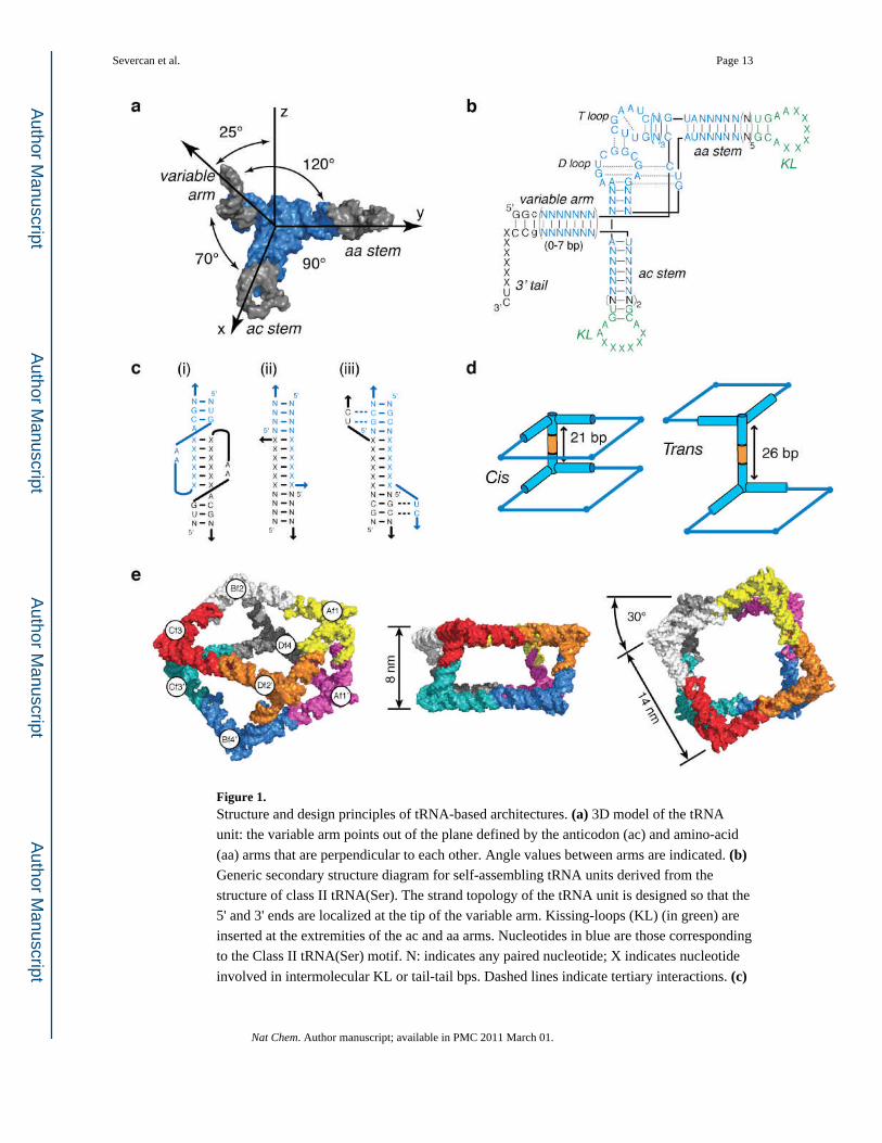
Figure 1. Structure and design principles of tRNA-based architectures. (a) 3D model of the tRNA
unit: the variable arm points out of the plane defined by the anticodon (ac) and amino-acid
(aa) arms that are perpendicular to each other. Angle values between arms are indicated. (b) Generic secondary structure diagram for self-assembling tRNA units derived from the
structure of class II tRNA(Ser). The strand topology of the tRNA unit is designed so that the
5' and 3' ends are localized at the tip of the variable arm. Kissing-loops (KL) (in green) are
inserted at the extremities of the ac and aa arms. Nucleotides in blue are those corresponding
to the Class II tRNA(Ser) motif. N: indicates any paired nucleotide; X indicates nucleotide
involved in intermolecular KL or tail-tail bps. Dashed lines indicate tertiary interactions. (c)
Severcan et al. Page 13
Nat Chem. Author manuscript; available in PMC 2011 March 01.
Author M
anuscriptA
uthor Manuscript
Author M
anuscriptA
uthor Manuscript

Schematic indicating the various intermolecular connectors. KL complex (i) and tail-tail
connectors without (ii) and with (iii) major triple base pairs. Blue and black colors indicate
the two different interacting RNAs. (d) Cis and trans orientations of tRNA squares resulting
from tail-tail edges connectors of 21bp or 26 bp, respectively. (e) 3D model of the non-
uniform square antiprism. Three-different views are shown. The tRNA units entering into
the composition of the fully programmable octamer (TO1–2) are indicated (see also Tables
S1 and S2 in Supplementary Information for other TO combinations).
Severcan et al. Page 14
Nat Chem. Author manuscript; available in PMC 2011 March 01.
Author M
anuscriptA
uthor Manuscript
Author M
anuscriptA
uthor Manuscript

Figure 2. tRNA octamer self-assembly and thermal stability. (a) Characterization of tRNA
supramolecular assembly of various tRNA octamers (TO) by non-denaturing PAGE at 2
mM Mg(OAc)2: lane M is for the monomer tRNA unit control (400 nM), lane TS1 are for a
control tRNA square (TS1, 100 nM), lanes TO1–2 show octamer products (50 nM) after one
pot or stepwise assembly. Lanes TO3–4 to TO3–7 (50 nM) correspond to octamers with a
varying number of tail connectors. (b) Schematic of the three major transitions of TO
disassembly observed by TGGE analysis (Materials and Methods and Supplementary
Information). (c) Example of TGGE gel showing the cooperative biphasic dissociation of
TO3–4 (20 nM) into squares and monomers at 0.2 mM Mg(OAc)2 (see also Table 1). (d) Comparison of melting curves for TS3 (grey squares) and TO3–4 (black circles) obtained
from TGGE gels at 0.2 mM (empty symbols) and 15 mM (solid symbols) Mg(OAc)2.
Severcan et al. Page 15
Nat Chem. Author manuscript; available in PMC 2011 March 01.
Author M
anuscriptA
uthor Manuscript
Author M
anuscriptA
uthor Manuscript

Figure 3. Structural characterization of tRNA architectures by AFM. (a) Top-to-bottom: Hierarchical
stepwise assembly schemes for tRNA squares designed to assemble into (b–c) the antiprism
TO3–4, (d) the "open" octamer TO3–6 and (e) the planar TS10–11 array (TS10 + TS11).
TO3–4 and TS10–TS11 have tail-tail connectors in cis and trans configuration,
respectively. TO3–6 has two tail-tail connectors missing with respect to TO3–4.
Corresponding AFM images were obtained in air. (f) 3D rendering of AFM image data to
compare the relative height of "closed" octamer versus square. (g) Height distribution of
octamers obtained from AFM analysis indicates that more than 70% of supra-molecular
assemblies correspond to two superimposed squares.
Severcan et al. Page 16
Nat Chem. Author manuscript; available in PMC 2011 March 01.
Author M
anuscriptA
uthor Manuscript
Author M
anuscriptA
uthor Manuscript

Figure 4. Structural characterization of tRNA architectures by Cryo-EM with single image particle
reconstruction. (a) Typical cryo-EM image of antiprism particles. More than 85% of the
objects seen are well-formed antiprisms. (b) Reconstructed 3D model of the non-uniform
square antiprism TO1–2 at 24.5 Å resolution. The view orientations are similar to those in
Figure 1e. (c) Class averages of particles with similar views observed by cryo-EM (EM)
with the corresponding projections of the RNA antiprism 3D structure reconstructed from
the cryo-EM images (recon) and the corresponding view of the initial antiprism model
(model). These particle views are selected from different image frames to represent views at
different orientations (Figure S3).
Severcan et al. Page 17
Nat Chem. Author manuscript; available in PMC 2011 March 01.
Author M
anuscriptA
uthor Manuscript
Author M
anuscriptA
uthor Manuscript

Figure 5. Coupling streptavidin to spatially addressable antiprims (TO8–9). (a) The location of the 3’
tail-tail connector in the variable arm determines the orientation of the iodoacetyl-biotin
linker, and thus can be used to control the positioning of streptavidin with respect to the
antiprism. (b–c) Antiprisms with biotin oriented inward form small antiprism-streptavidin
complexes. (d–e) Antiprisms with biotin oriented outward form antiprism-streptavidin
filaments. Corresponding AFM images of the streptavidin-antiprism conjugates, formed at
ratio of streptavidin vs antiprism is 1:1 (50 nM), are shown (see Figure S5, Supplementary
Severcan et al. Page 18
Nat Chem. Author manuscript; available in PMC 2011 March 01.
Author M
anuscriptA
uthor Manuscript
Author M
anuscriptA
uthor Manuscript

Information and Materials and Methods). (f) Distribution of the population of antiprisms in
function of antiprism-streptavidin chain lengths and inward or outward orientation of
conjugated biotin. The frequency (in percent) stands for the total occurrence of antiprisms in
an antiprism-streptavidin complex of specific length (indicated in number of constituent
antiprisms). These experiments were reproduced at least twice.
Severcan et al. Page 19
Nat Chem. Author manuscript; available in PMC 2011 March 01.
Author M
anuscriptA
uthor Manuscript
Author M
anuscriptA
uthor Manuscript

Author M
anuscriptA
uthor Manuscript
Author M
anuscriptA
uthor Manuscript
Severcan et al. Page 20
Table 1
Melting temperature (Tm) values measured by TGGE for various transitions between tRNA squares (TS),
tRNA octamers (TO) and tRNA monomers (TM) architectures at 0.2, 2 and 15 mM Mg(OAc)2 (see Materials
and Methods and Supplementary Information).
Mg2+(mM) 0.2 2 15
transition TO-TS TS-TM TO-TS TS-TM TO-TM TS-TM TO-TM
TS3 - 46 - 58 - 65 -
TO3-4(4 tails) 34 46 - - 62 - >70
TO3-5(3 tails) n.d. n.d. 54 58 - n.d. n.d.
TO3-6(2 tails) n.d. n.d. 43 58 - n.d. n.d.
Nat Chem. Author manuscript; available in PMC 2011 March 01.