j. virol.-2014-pitts-5462-73
TRANSCRIPT
Crystal Structure of the Herpesvirus Inner Tegument Protein UL37Supports Its Essential Role in Control of Viral Trafficking
Jared D. Pitts,a Jenifer Klabis,b Alexsia L. Richards,b Gregory A. Smith,b Ekaterina E. Heldweina
Department of Molecular Biology and Microbiology and Graduate Program in Molecular Microbiology, Sackler School of Graduate Studies, Tufts University School ofMedicine, Boston, Massachusetts, USAa; Department of Microbiology-Immunology, Northwestern University, Feinberg School of Medicine, Chicago, Illinois, USAb
ABSTRACT
In cells infected with herpesviruses, two capsid-associated, or inner tegument, proteins, UL37 and UL36, control cytosolic traf-ficking of capsids by as yet poorly understood mechanisms. Here, we report the crystal structure of the N-terminal half of UL37from pseudorabies virus, an alphaherpesvirus closely related to herpes simplex viruses and varicella-zoster virus. The struc-ture—the first for any alphaherpesvirus inner tegument protein—reveals an elongated molecule of a complex architecture richin helical bundles. To explore the function of the UL37 N terminus, we used the three-dimensional framework provided by thestructure in combination with evolutionary trace analysis to pinpoint several surface-exposed regions of potential functionalimportance and test their importance using mutagenesis. This approach identified a novel functional region important for cell-cell spread. These results suggest a novel role for UL37 in intracellular virus trafficking that promotes spread of viral infection, afinding that expands the repertoire of UL37 functions. Supporting this, the N terminus of UL37 shares structural similarity withcellular multisubunit tethering complexes (MTCs), which control vesicular trafficking in eukaryotic cells by tethering transportvesicles to their destination membranes. Our results suggest that UL37 could be the first viral MTC mimic and provide a struc-tural rationale for the importance of UL37 for viral trafficking. We propose that herpesviruses may have co-opted the MTC func-tionality of UL37 to bring capsids to cytoplasmic budding destinations and further on to cell junctions for spread to nearby cells.
IMPORTANCE
To move within an infected cell, viruses encode genes for proteins that interact with host trafficking machinery. In cells infectedwith herpesviruses, two capsid-associated proteins control the cytosolic movement of capsids by as yet poorly understood mech-anisms. Here, we report the crystal structure for the N-terminal half of one of these proteins, UL37. Structure-based mutagenesisrevealed a novel function for UL37 in virus trafficking to cell junctions for cell-cell spread. The unexpected structural similarityto components of cellular multisubunit tethering complexes, which control vesicular traffic, suggests that UL37 could be the firstviral MTC mimic and provides a structural basis for the importance of UL37 for virus trafficking.
The ability to undergo directional movement within a host cy-toplasm is essential for replication of many viruses. During
viral entry, genomes must be delivered to the replication compart-ment, while later, newly assembled viral particles need to escapethe cell to initiate new rounds of infection. To move within theinfected cell, viruses typically encode genes for proteins that inter-act with the host trafficking machinery.
Herpesviruses are large enveloped viruses that cause lifelong,latent infections from which viruses can reactivate, causing dis-eases ranging from mucocutaneous eruptions to encephalitis,cancers, and disseminated disease. All herpesviruses share a mor-phology consisting of four components: a double-stranded DNAgenome, an icosahedral capsid, a lipid envelope, and the tegu-ment, a protein layer between the capsid and the envelope. Her-pesvirus replication occurs in the nucleus, requiring incomingparticles to traverse the cytoplasm following entry into a cell andprogeny virions to traffic back to the cell surface during the egressphase of infection (1). Following genome replication and encap-sidation, nascent capsids first bud into the inner nuclear mem-brane. The resulting perinuclear viral particles fuse their enve-lopes with the outer nuclear membrane, releasing unenvelopedcapsids into the cytosol. Cytosolic capsids next travel to tubularvesicles thought to originate from intracellular membranes (trans-Golgi network [TGN] or early endosomes) (1, 2), where the finalround of budding—secondary envelopment—takes place. Cyto-
solic capsids bud into these vesicles and acquire lipid envelopesand outer tegument proteins. The resulting mature virions aresubsequently released into the extracellular milieu by exocytosisor directed to cell junctions for spread into uninfected cells.
The movement of capsids within the cytosol is controlled by asubset of tegument proteins that are capsid associated during boththe entry and egress phases of infection. These tegument proteinsinclude the herpesvirus protein UL37, which is a highly conservedcomponent of the capsid-proximal, or inner, tegument (3–5).UL37 interacts with capsids indirectly by binding another con-served tegument protein, UL36 (6–13), which in turn binds cap-sids through a direct interaction with the capsid proteins UL25(14–16), UL19, and UL17 (17). During transport to the nuclearpore, UL37 remains attached to the incoming capsids (18–21),and although UL37 is not required for capsid trafficking to thenucleus (13), in its absence the incoming transport is delayed (22).
Received 20 January 2014 Accepted 20 February 2014
Published ahead of print 5 March 2014
Editor: L. Hutt-Fletcher
Address correspondence to Ekaterina E. Heldwein, [email protected].
Copyright © 2014, American Society for Microbiology. All Rights Reserved.
doi:10.1128/JVI.00163-14
5462 jvi.asm.org Journal of Virology p. 5462–5473 May 2014 Volume 88 Number 10
on Decem
ber 7, 2015 by UN
IV O
F C
ALIF
SA
N D
IEG
Ohttp://jvi.asm
.org/D
ownloaded from
The UL37 protein is also necessary for herpesvirus egress. InUL37-null viruses, unenveloped capsids accumulate in the cytosolof infected cells (4, 6, 13, 23–25) and the release of infectiousvirions is either completely blocked, as seen in herpes simplexvirus 1 (HSV-1) (23, 24), or is strongly impaired, as observed inpseudorabies virus (PRV), a herpesvirus closely related to HSVand varicella-zoster virus (4, 23). The directed movement ofHSV-1 and PRV UL37-null capsids is substantially impeded,which supports the idea that efficient capsid transport to the or-ganelle of secondary envelopment requires assembly of UL37 ontocapsids (6, 26). In contrast, the role of UL37 in cytoplasmic envel-opment is less clear. UL37 is not required for the formation of theso-called light, or L, particles, which lack capsids and contain onlyenveloped tegument (4, 13), and, therefore, is not required for theassembly of the outer tegument or for membrane deformationduring cytoplasmic budding. However, UL37 may be necessaryfor the correct addition of the outer tegument proteins to thecapsid, which is essential for secondary envelopment (4).
The C-terminal half of HSV-1 UL37 interacts with UL36 (27,28) and dystonin/BPAG1 (29). While the N-terminal half of UL37has been proposed to have a nuclear export signal (30) and a smallself-association domain (27), the role of the N-terminal half ofUL37 in replication remains largely unknown. To gain insight intothe mechanism by which UL37 enables capsid trafficking and cy-toplasmic capsid envelopment, we determined the crystal struc-ture of the N-terminal half of UL37 (UL37N), residues 1 to 479,from PRV. UL37N is an elongated molecule composed of multiplehelical bundles. The structure revealed an unanticipated similarityto the structures of several components of the complexes associ-ated with tethering containing helical rods (CATCHR) family ofeukaryotic multisubunit tethering complexes (MTCs), which di-rect vesicular trafficking in cells by tethering vesicles to their targetorganelles. Furthermore, mutagenesis of a surface cluster identi-fied in the UL37N structure using evolutionary trace analysis(ETA) reduced the spread of PRV without impeding the rate ofviral production. This finding suggests an additional, novel rolefor UL37 in intracellular trafficking that promotes the subsequentspread of virus between cells. Collectively, these observations offernew insights into the function of UL37 and provide a structuralbasis for its essential role in intracellular virus trafficking. We pro-pose that herpesviruses may utilize the tethering function of UL37to bring capsids to their cytoplasmic budding sites perhaps usingUL37 to specify their budding destination and to promote traf-ficking of mature virions inside transport vesicles to cell junctionsfor spread to nearby cells.
MATERIALS AND METHODSCloning. Plasmid pGS3610 encodes the PRV Becker UL37 gene fused toan N-terminal His6-SUMO tandem tag. This was made by cutting thepETDuet-SUMO vector (a derivative of pETDuet-1 and a gift fromThomas Schwartz) and the pGS1740 subclone of UL37 with BamHI andHindIII. The pJP4 plasmid, which contains a His6-SUMO-PreScission tagin frame with the BamHI restriction site of the multiple-cloning site in apET24b vector, was made through PCR of the His6-SUMO-PreScissiontag from pETDuet-SUMO using the primers 5=-GGGAATTCCATATGGGCAGCAGCCATCACCATCA and 3=-CTAGGGATCCGGGCCCCTGGAACAGAACTT (restriction sites are underlined). The PCR product wassubcloned into pET24b using NdeI and BamHI restriction sites. The PRVUL37 gene for Escherichia coli expression was synthesized by GeneArt. TheN-terminal half (residues 1 to 496) of codon-optimized PRV UL37 (re-ferred to as UL37N) was amplified by PCR from the full-length PRV
codon-optimized UL37 gene using the primers 5=-CTAGGGATCCATGGAAGCACTGGTTCGTGC and 3=-CTAGAAGCTTCTAGGCTGCGCTGGTCGGTG (restriction sites are underlined). The PCR product wassubcloned into pJP4 using the BamHI and HindIII restriction sites to yieldplasmid pJP23.
Virus construction. All recombinant PRV (strain Becker) isolateswere derived from a variant of the pBecker3 infectious clone, pGS4284,that encodes the mCherry red fluorescent protein fused in frame to theUL25 capsid protein (31). Viruses were produced by electroporation ofinfectious clones into the pig kidney epithelial cell line PK15, as previouslydescribed (20). PK15 cells were maintained in Dulbecco modified Eaglemedium (DMEM; Invitrogen) supplemented with 10% bovine growthsupplement (BGS; HyClone), which was reduced to 2% during transfec-tion and infection. The harvested virus was passaged once to produce ahigh-titer stock by infecting a 10-cm dish of PK15 cells with 1 �l virus.Transfection of pGS4284 resulted in PRV-GS4284, which upon passagepropagated to titers of �5 � 108 PFU/ml.
To make PRV encoding the pentuple mutations D79A/D81A/E82A/D382A/D383A in the calcium-binding region (Ca) of UL37, codonchanges were introduced through two rounds of en passant mutagenesisof pGS4284 (32). The first set of primers, 5=-CTCGCCGAGAACCTGGCCGGCCTGGCGCTGTGGCGCCTGCGCCACGCCTGGGCCGCGGGCACGGCCCCGCTGAGGATGACGACGATAAGTAGGG and 5=-GTCGCCGTTGACGACCCCCAGGAGCTCCAGCAGCGGGGCCGTGCCCGCGGCCCAGGCGTGGCGCAGGCGCCACAACCAATTAACCAATTCTGATTAG, was used to generate the D382A/D383A mutations(mutated bases are in bold, the sequence from template plasmidpEPkan-S is underlined) and produced pGS5456. The second set of prim-ers, 5=-GTCGGCTGCACGGCGGTCGTCGGCGGCGTCGTGCACCGCCTCCTCGCCGCCTACGGGCCCGGGCTGAGGATGACGACGATAAGTAGGG and 5=-CGCGACGTCCGTGTAGGCGCGCACGTAGTCCAGCCCGGGCCCGTAGGCGGCGAGGAGGCGGTGCACCAACCAATTAACCAATTCTGATTAG, was used to generate the D79A/D81A/E82Amutations, which were introduced into pGS5456 to produce the finalmutant, pGS5476. PRV-GS5476 typically propagated to a titer of �5 �108 PFU/ml.
The region 2 (R2) and region 3 (R3) mutant viruses were produced inthe same manner as the Ca mutant just described. For R2, mutations wereintroduced into pGS4284 in three sequential rounds using primers 5=-CTCGACCACACGCAGGTGGACGCCACGGGCGTGTGGGAGGCGGTGGCGGCCAGCGCCTCGCCGAGGATGACGACGATAAGTAGGG and 5=-CGCGGTCACGAGCGCCTCCACGACCTGCAGCGGCGAGGCGCTGGCCG CCACCGCCTCCCACACCAACCAATTAACCAATTCTGATTAG (encoding Q324A), 5=-GACCTCCTCGAGCGCGCCGTGCTGGACCGCGCGCCCCGCCTGACGGCCGCGCAGGCTGCCGTCGGCTGCACGAGGATGACGACGATAAGTAGGG and 5=-GAGGCGGTGCACGACGCCGCCGACGACCGCCGTGCAGCCGACGGCAGCCTGCGCGGCCGTCAGGCGGGGCGCCAACCAATTAACCAATTCTGATTAG (encoding D362A/R365A), and5=-GGGGACGTGACGGCGGCGCTGGGGCTCCCCGAGAAGGGCGTGGAGGCCGTGGTGCGCGCTTGCATGGCGCCGCGCAGGATGACGACGATAAGTAGGG and 5=-GCGCGCCGCGCCCACGTGCTCCGTGGGCGGGCGCGGCGCCATGCAAGCGCGCACCACGGCCTCCACGCCCTTCTCCAACCAATTAACCAATTCTGATTAG (encoding H421A/H425A). The first PCR product was recombined into pGS4284, resultingin pGS5483. The second PCR product was then recombined intopGS5483, resulting in pGS5558. The final recombination was made intopGS5558, resulting in pGS5604. PRV-GS5604 typically propagated to atiter of �5 � 108 PFU/ml. The R3 mutations were introduced in tworounds using primers 5=-CTGCCGCTGGCGTTGGCGGTGCGCCAGATGCAGAACGAGGGCCTGGCGCAGCTGACGCGCGCGCTCAGGATGACGACGATAAGTAGGG and 5=-GAAGAACTCGTCGGCGATCGTGAGGGCAAAGAGCGCGCGCGTCAGCTGCGCCAGGCCCTCGTTCTGCAACCAATTAACCAATTCTGATTAG (encoding D239A/E240A) andprimers 5=-AACCCGACGCTGCGCGAGCAGTTCGCCGAGGCGGCG
Structure of Herpesvirus Traffic Controller UL37
May 2014 Volume 88 Number 10 jvi.asm.org 5463
on Decem
ber 7, 2015 by UN
IV O
F C
ALIF
SA
N D
IEG
Ohttp://jvi.asm
.org/D
ownloaded from
CGGGCCGTGGCCGCGGCGGCGCTGGTGCCCAGGATGACGACGATAAGTAGGG and 5=-CGTGCGCGGCGTGGCGTTGACCTCGCCCACGGGCACCAGCGCCGCCGCGGCCACGGCCCGCGCCGCCAACCAATTAACCAATTCTGATTAG (encoding K203A/P204Q). The first PCRproduct was recombined into pGS4284, resulting in pGS5242. The secondPCR product was then recombined into pGS5242, resulting in pGS5350.PRV-GS5350 typically propagated to a titer of �5 � 108 PFU/ml.
The region 1 (R1) mutant virus (V249R/R254A/R285A/D287A/H311A) was generated using a modified two-step recombination. Theregion of the UL37 gene encoding amino acids 249 to 311 was first re-placed with the kanamycin resistance cassette of pEPkan-S using primers5=-CCGAGGCGGCGCGGGCCGTGGACGAGGCGGCGCTGGTGCCCGTGGGCGAGACGCAGGTGGACGCCACGGGAGGATGACGACGATAAGTAGGG and 5=-GAGGCGCTGGCCTGCACCGCCTCCCACACGCCCGTGGCGTCCACCTGCGTCTCGCCCACGGGCACCAGCGCAACCAATTAACCAATTCTGATTAG. The PCR product was recombinedinto pGS4284, resulting in the intermediate construct pGS5313. The de-letion in pGS5313 was then repaired using a 489-bp synthetic DNA en-coding the missing UL37 sequence with the five codon changes and 150 bpof flanking homologous sequence to each side (pGS5267; Integrated DNATechnologies). The synthetic DNA was released from a pIDTSmart vectorusing flanking HindIII sites and recombined into pGS5313. Recombina-tion was carried out by growing E. coli strain GS1783 harboring pGS5313in 30 ml of Luria Broth (LB) supplemented with 20 �g/ml chloramphen-icol to an optical density at 600 nm (OD600) of 0.6 at 32°C in a baffled flask.At this point, 20 ml of LB supplemented with 20 �g/ml chloramphenicoland 2% L-arabinose was added, and the culture was incubated with shak-ing at 32°C for 70 min. The culture was then transferred to a 42°C shakingwater bath for 15 min, and the contents were then transferred to a 50-mlconical tube and chilled on ice. The chilled bacteria were washed threetimes, and the final pellet was suspended in 300 ml double-distilled H2O,of which 48 �l was used in an electroporation with 2 �l of the pGS5267synthetic fragment. After recovery, the reaction mixture was plated on LBagar plates supplemented with 20 �g/ml chloramphenicol and 2% L-ara-binose. The resulting isolate was saved as pGS5321. PRV-GS5321 typicallypropagated to a titer of �5 � 108 PFU/ml. The sequences of all geneticmodifications in the infectious clones were confirmed.
Viral propagation kinetics, viral titers, and plaque size analysis.Quantitation of viral propagation kinetics was assessed by single-stepgrowth in PK15 cells infected at a multiplicity of infection (MOI) of 10 foreach viral stain. Viral titers from cells or medium supernatants harvestedat 2, 5, 8, 12, or 24 h postinfection (hpi) were determined in duplicate byplaque assay, as previously described (33).
Measurements of plaque diameters were obtained by infection ofPK15 cells in 6-well trays with serial 10-fold dilutions for each virus. At 4days postinfection, images were captured with a �4 objective on a NikonTE2000 inverted fluorescence microscopy (Nikon Instruments) fittedwith a CoolSnap HQ2 camera (Photometrics). Two orthogonal diametermeasurements of each fluorescent plaque were obtained using the Meta-morph software package (Molecular Devices) and averaged. The reportedplaque diameters represented an average of more than 50 plaques pervirus. Measurements of the plaque diameters of mutant viruses were al-ways conducted side by side with measurement of the plaque diameter ofPRV-GS4284 (the virus encoding wild-type [WT] UL37), and the diam-eters of the mutant viruses were normalized to that diameter. Single-stepgrowth and plaque diameters were plotted using the Prism software pack-age (GraphPad Software).
Virion protein incorporation. PK15 cells were infected with eitherPRV-GS4284 (WT) or PRV-GS604 (R2) at an MOI of 3. Infections werecarried out in 15-cm dishes of confluent cells. Infected cells and extracel-lular media were harvested once all cells displayed a cytopathic effect,which was typically at 18 hpi. Cellular debris was removed by centrifuga-tion at 5,000 � g, and virions were concentrated from the supernatant bypelleting through a 10% Nycodenz cushion at 13,000 rpm in an SW28rotor (Beckman). The resulting pellet was resuspended in 100 �l of TNE
buffer (150 mM NaCl, 50 mM Tris [pH 7.4], 10 mM EDTA). Viral parti-cles were dispersed by 10 1-s pulses of sonication in a cup horn ultrasonicprocessor (VCX-500; Sonics and Materials, Newtown, CT). The samplewas loaded onto a 12 to 32% dextran gradient and centrifuged at 20,000rpm for 1 h at 4°C. The heavy viral band was collected and spun at 25,000rpm in a Beckman SW50.1 rotor at 4°C for 30 min. The final pellet wasresuspended in final sample buffer (10 mM Tris [pH 7.4], 150 mM NaCl,1% Triton X-100) containing 10% �-mercaptoethanol, and the sampleswere boiled for 5 min prior to electrophoresis of 5 �l of each samplethrough an 8% sodium dodecyl sulfate (SDS)-polyacrylamide gel. Pro-teins were subsequently transferred onto an Immobilon polyvinylidenedifluoride membrane (Millipore), and VP5 was detected using the 3C10mouse monoclonal antibody (a gift of Lynn Enquist) at a 1:1,000 dilution.UL37 was detected using D1789, a rabbit antiserum raised against a pep-tide derived from the PRV UL37 sequence (REAADRVLGDYHE), at a1:2,500 dilution. The secondary goat antimouse and antirabbit dye-la-beled antibodies (LiCor) were used at 1:5,000 dilutions. Proteins werevisualized and quantitated using an Odyssey Fc imager and ImageStudiosoftware (LiCor). The ratio of UL37 to VP5 was quantified for four inde-pendent experiments and normalized to the average value obtained forthe UL37-to-VP5 ratio for WT virus. Data were plotted using the Prismsoftware package (GraphPad Software), and significance was determinedusing an unpaired Student’s t test.
Protein expression and purification. Both UL37 and UL37N con-structs were expressed as N-terminal His6-SUMO fusions in T7 Express E.coli (New England BioLabs). Freshly transformed cells were incubated at37°C overnight in 5 ml LB starter culture supplemented with 50 �g/mlkanamycin. The starter culture was diluted into 1 liter LB supplementedwith 50 �g/ml kanamycin and grown at 37°C until the OD600 reached 0.8to 1.0. At this point, the temperature was shifted to 16°C and the cells wereinduced with 0.5 mM isopropyl-�-D-thiogalactopyranoside (IPTG).
For production of UL37N, expression was induced for 16 to 20 h. Cellswere harvested by centrifugation at 12,000 � g for 40 min, resuspended in25 ml 20 mM piperazine-N,N=-bis(2-ethanesulfonic acid) (PIPES), pH7.0, 50 mM NaCl, 0.1% Igepal CA-630 (Sigma), 5% glycerol, 10 mMimidazole, 0.1 mM tris(2-carboxyethyl)phosphine (TCEP), and 1 EDTA-free cOmplete protease inhibitor cocktail tablet (Roche), and lysed by useof a French press. The insoluble fraction was removed by centrifugation ofthe whole-cell lysate at 14,000 � g for 30 min at 4°C. Soluble lysate wasloaded onto a 5-ml Ni-Sepharose 6B FF column (GE Healthcare). Thecolumn was subsequently washed with 10 column volumes (CVs) of 20mM PIPES, pH 7.0, 50 mM NaCl, 0.1 mM TCEP (buffer A) containingincreasing amounts of imidazole at 10 mM or 25 mM. Protein was elutedin buffer A containing 100 mM imidazole. The eluate was immediatelyconcentrated, and the imidazole was removed by buffer exchange intobuffer A using an Ultra-15 50-kDa-cutoff concentrator (Millipore). Theprotein concentration was determined from the absorbance at 280 nmusing a calculated extinction coefficient. Glutathione S-transferase(GST)-tagged PreScission protease was added to the protein solution at a1:50 protease-to-protein ratio, and the protein was cleaved overnight at4°C to remove the His6-SUMO tag. The protease-protein solution wassequentially applied to glutathione-Sepharose 4B (GE Healthcare) andNi-Sepharose 6B to remove the GST-tagged PreScission protease and theHis6-SUMO tag, respectively. Cleaved protein was present in the un-bound and wash fractions. UL37N was further purified by size exclusionchromatography using a Superdex 200 column (GE Healthcare) and con-centrated to 3.5 to 4.0 mg/ml using an Ultra-15 30-kDa-cutoff concentra-tor (Millipore). Protein purity was assessed by SDS-polyacrylamide gelelectrophoresis (PAGE) and Coomassie G-250 staining. The final yieldwas 18 mg of pure protein per 1 liter of E. coli culture. All UL37N proteinsamples used for crystallization and biochemical studies were stored in 20mM PIPES, pH 7.0, 50 mM NaCl, and 0.5 mM TCEP.
A BL21 E. coli strain expressing GST-tagged PreScission protease was agift from Peter Cherepanov (London Research Institute, London, UnitedKingdom). Protein expression was induced with 0.5 mM IPTG at 30°C for
Pitts et al.
5464 jvi.asm.org Journal of Virology
on Decem
ber 7, 2015 by UN
IV O
F C
ALIF
SA
N D
IEG
Ohttp://jvi.asm
.org/D
ownloaded from
4 h before the cells were harvested and lysed. The PreScission protease waspurified over glutathione-Sepharose in a buffer containing 20 mM Tris,pH 8.0, 200 mM NaCl, and 1 mM TCEP. The column was washed 3 timeswith 10 CVs of the binding buffer, and protein was eluted from the col-umn in binding buffer containing 5 mM reduced glutathione. The elutedprotein was concentrated in a 30-kDa-cutoff concentrator (Millipore)and further purified over a Superdex 200 size exclusion column equili-brated with the binding buffer. The protein was concentrated to 1 mg/ml,flash frozen, and stored at �80°C.
Thermofluor assay. The optimal buffer composition and the optimalNaCl concentration for the stability of the UL37N protein (PIPES, pH 7.0,and 50 mM NaCl) were determined using the Thermofluor method (34).Protein was diluted to 0.15 mg/ml in the storage buffer, and a fluorescentdye, SYPRO orange (Invitrogen), was added at a 1:1,000 dilution. Tenmicroliters of the protein-dye solution was pipetted into each well of a96-well PCR microplate. Next, 10 �l of buffer (from a custom-madescreen containing buffers at pH 4.5 to 10.5 and NaCl concentrationsranging from 0 to 500 mM) was added to wells containing the protein-dye solution. The plate was sealed and centrifuged for 1 min at 500 �g and 25°C. Samples were analyzed on a Roche LightCycler 480 quan-titative PCR machine using an excitation wavelength of 465 nm anddetection of emission at 610 nm. The emission signal was analyzed from25°C to 95°C at a continuous acquisition rate of 3 measurements per °C.Data were analyzed using the ThermoQ software program (http://jshare.johnshopkins.edu/aherna19/thermoq/). Conditions that stabilizedUL37N further increased its solubility.
Mass spectrometry. For mass spectrometry analysis, the UL37 proteinwas analyzed using sinapinic acid (Agilent Technologies) as the matrix.Mass spectrometry measurements were performed on a Voyager DE-Promatrix-assisted laser desorption ionization–time of flight mass spectrom-eter (Applied Biosystems).
Crystallization and structure determination. Crystals of UL37Nwere grown by vapor diffusion at room temperature in hanging dropsusing 1 �l protein and 1 �l well solution containing 24 to 26% polyethyl-ene glycol 1000, 0.3 M Ca(CH3COO)2, and 0.1 M imidazole, pH 8.0. Largeplates formed in 3 to 8 days and were harvested 2 to 4 weeks later. For datacollection, crystals were incubated in a solution identical to the well solu-tion plus 10% glycerol for 30 s to 2 min prior to flash freezing in liquid N2.Heavy atom derivative crystals were obtained by soaking native crystals inwell solution containing 5 mM thimerosal (Na salt of ethylmercurithio-salicylic acid or C9H9HgNaO2S) for 12 to 16 h. Derivative crystals wereharvested and frozen using the protocol developed for the native crystals.
X-ray diffraction data were collected at 100 K at the X25 beam line atthe National Synchrotron Light Source. The data were processed usingHKL2000 (35) and indexed in space group P21 (Table 1). The native dataset was processed up to a 2.0-Å resolution, and the single-wavelengthanomalous dispersion (SAD) Hg data set was processed to a 2.3-Å reso-lution (Table 1). All 12 heavy atom sites were found using the phenix.au-tosol program, and the experimental density allowed the tracing of �70%of the residues in the phenix.autobuild program. Additional residues weremanually built using the Coot program (36). There are two UL37N mol-ecules in the asymmetric unit.
Before refinement of the heavy atom model, 10% of the data was setaside for cross-validation. The model was refined against the SAD Hg dataset to 2.3-Å resolution using the phenix.refine program. Next, test set flagswere transferred to the native data set; additionally, 10% of the native databetween 2.3 and 2.0 Å was set aside for cross-validation. After severalcycles of refinement in the phenix.refine program (37) and rebuilding inCoot (36), Rwork was 17.3% and Rfree was 22.0%. The final model con-tained all amino acids from residues 1 to 479, including 3 of the 4 linkerresidues left after protease cleavage of the N-terminal tag. The final modelis missing residues 480 to 496 in both chains. The MolProbity server (38)was used to assess the stereochemical quality of all models. According toMolProbity, 99.0% of the residues lie in the most favored regions of the
Ramachandran plot and 1% lie in the additionally allowed regions of theRamachandran plot. Final statistics are listed in Table 1.
Structure analysis. The sequence alignment was generated and an-alyzed using the ClustalW (39) and ESPRIPT (40) programs. Interfaceswere analyzed using the PISA program (41). Structural homologysearches were performed using the Dali server (42), and the top hitswere superposed onto the UL37N protein using the Dalilite pairwisecomparison tool. The Evolutionary Trace server was used for evolu-tionary trace analysis (http://www-cryst.bioc.cam.ac.uk/�jiye/evoltrace/evoltrace.html). All structure figures were made in the PyMOL program(http://www.pymol.org).
Protein structure accession number. Atomic coordinates and struc-ture factors for the UL37N structure have been deposited in the RCSBProtein Data Bank under accession number 4K70.
RESULTSCharacterization of UL37N. Initially, we expressed full-lengthPRV UL37 with an N-terminal His6-SUMO tag in E. coli (Fig. 1A).During expression, this protein underwent spontaneous proteol-ysis, which generated a fragment containing the His6-SUMO tagand the N terminus of UL37 (Fig. 1B). Using mass spectrometry,the proteolytic site was localized around residue 498, which isapproximately in the middle of the UL37 sequence. The difficultyin separating full-length UL37 from the truncated UL37 resultedin a very low yield of the purified full-length UL37, �200 �g/litercell culture. Unlike full-length UL37, which was prone to aggre-gation, the N-terminal product of proteolytic cleavage was readilysoluble and was pursued further. We expressed a fragment con-taining residues 1 to 496 of UL37 (UL37N) plus an N-terminal
TABLE 1 Data collection and refinement statistics
Parameter
Value fora:
Native crystalNative crystal soakedin thimerosal
Data collectionSpace group P21 P21
Unit cell dimensionsa, b, c (Å) 51.67, 156.59, 67.38 51.53, 156.30, 66.34�, �, (°) 90, 91.33, 90 90, 91.78, 90
Resolution (Å) 43.12–2.00 (2.07–2.00) 48.91–2.05 (2.12–2.05)Rsym or Rmerge 0.086 (0.516) 0.097 (0.280)I/I 20.32 (2.74) 13.87 (2.18)Completeness (%) 89.4 (49.5) 85.1 (35.31)Redundancy 6.3 (4.4) 3.9 (2.1)
Refinement statisticsResolution range (Å) 43.12–2.00No. of reflections (free) 64,342 (2,347)Rwork/Rfree 17.30/22.01No. of atoms 7,983
Protein 7,342Ligand/ion 45Water 596
B-factorsb 35.05Protein 35.03Ligand/ion 42.8Water 37.5
RMSDBond length (Å) 0.007Bond angle (°) 0.96
a Values in parentheses are for the highest-resolution shell.b B-factor, isotropic displacement parameter.
Structure of Herpesvirus Traffic Controller UL37
May 2014 Volume 88 Number 10 jvi.asm.org 5465
on Decem
ber 7, 2015 by UN
IV O
F C
ALIF
SA
N D
IEG
Ohttp://jvi.asm
.org/D
ownloaded from
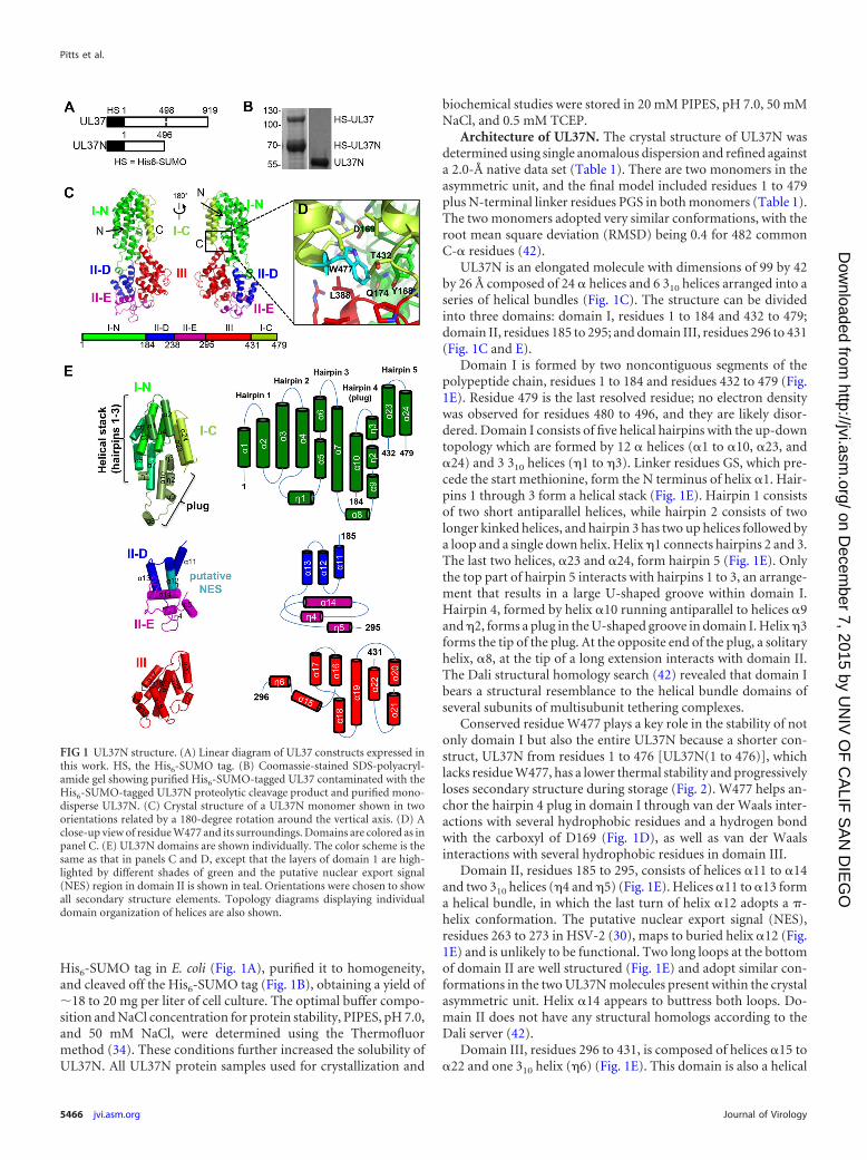
His6-SUMO tag in E. coli (Fig. 1A), purified it to homogeneity,and cleaved off the His6-SUMO tag (Fig. 1B), obtaining a yield of�18 to 20 mg per liter of cell culture. The optimal buffer compo-sition and NaCl concentration for protein stability, PIPES, pH 7.0,and 50 mM NaCl, were determined using the Thermofluormethod (34). These conditions further increased the solubility ofUL37N. All UL37N protein samples used for crystallization and
biochemical studies were stored in 20 mM PIPES, pH 7.0, 50 mMNaCl, and 0.5 mM TCEP.
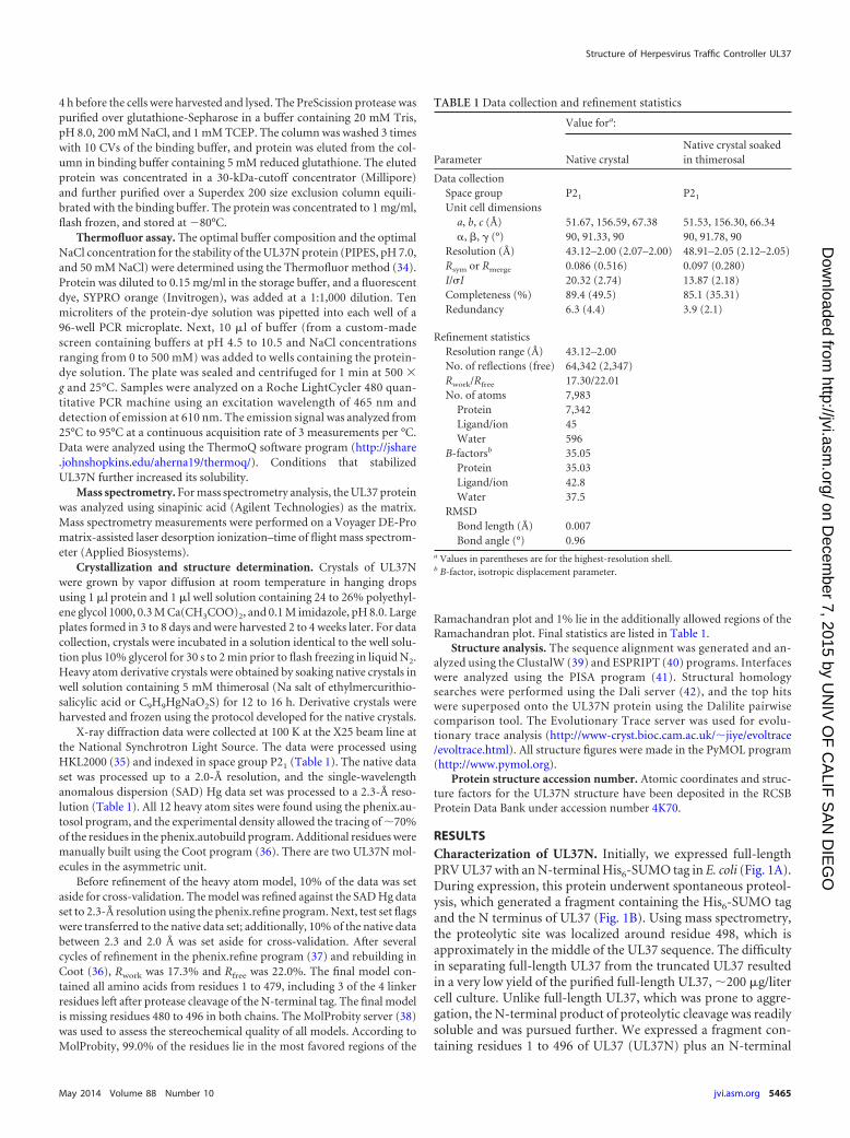
Architecture of UL37N. The crystal structure of UL37N wasdetermined using single anomalous dispersion and refined againsta 2.0-Å native data set (Table 1). There are two monomers in theasymmetric unit, and the final model included residues 1 to 479plus N-terminal linker residues PGS in both monomers (Table 1).The two monomers adopted very similar conformations, with theroot mean square deviation (RMSD) being 0.4 for 482 commonC-� residues (42).
UL37N is an elongated molecule with dimensions of 99 by 42by 26 Å composed of 24 � helices and 6 310 helices arranged into aseries of helical bundles (Fig. 1C). The structure can be dividedinto three domains: domain I, residues 1 to 184 and 432 to 479;domain II, residues 185 to 295; and domain III, residues 296 to 431(Fig. 1C and E).
Domain I is formed by two noncontiguous segments of thepolypeptide chain, residues 1 to 184 and residues 432 to 479 (Fig.1E). Residue 479 is the last resolved residue; no electron densitywas observed for residues 480 to 496, and they are likely disor-dered. Domain I consists of five helical hairpins with the up-downtopology which are formed by 12 � helices (�1 to �10, �23, and�24) and 3 310 helices (�1 to �3). Linker residues GS, which pre-cede the start methionine, form the N terminus of helix �1. Hair-pins 1 through 3 form a helical stack (Fig. 1E). Hairpin 1 consistsof two short antiparallel helices, while hairpin 2 consists of twolonger kinked helices, and hairpin 3 has two up helices followed bya loop and a single down helix. Helix �1 connects hairpins 2 and 3.The last two helices, �23 and �24, form hairpin 5 (Fig. 1E). Onlythe top part of hairpin 5 interacts with hairpins 1 to 3, an arrange-ment that results in a large U-shaped groove within domain I.Hairpin 4, formed by helix �10 running antiparallel to helices �9and �2, forms a plug in the U-shaped groove in domain I. Helix �3forms the tip of the plug. At the opposite end of the plug, a solitaryhelix, �8, at the tip of a long extension interacts with domain II.The Dali structural homology search (42) revealed that domain Ibears a structural resemblance to the helical bundle domains ofseveral subunits of multisubunit tethering complexes.
Conserved residue W477 plays a key role in the stability of notonly domain I but also the entire UL37N because a shorter con-struct, UL37N from residues 1 to 476 [UL37N(1 to 476)], whichlacks residue W477, has a lower thermal stability and progressivelyloses secondary structure during storage (Fig. 2). W477 helps an-chor the hairpin 4 plug in domain I through van der Waals inter-actions with several hydrophobic residues and a hydrogen bondwith the carboxyl of D169 (Fig. 1D), as well as van der Waalsinteractions with several hydrophobic residues in domain III.
Domain II, residues 185 to 295, consists of helices �11 to �14and two 310 helices (�4 and �5) (Fig. 1E). Helices �11 to �13 forma helical bundle, in which the last turn of helix �12 adopts a �-helix conformation. The putative nuclear export signal (NES),residues 263 to 273 in HSV-2 (30), maps to buried helix �12 (Fig.1E) and is unlikely to be functional. Two long loops at the bottomof domain II are well structured (Fig. 1E) and adopt similar con-formations in the two UL37N molecules present within the crystalasymmetric unit. Helix �14 appears to buttress both loops. Do-main II does not have any structural homologs according to theDali server (42).
Domain III, residues 296 to 431, is composed of helices �15 to�22 and one 310 helix (�6) (Fig. 1E). This domain is also a helical
FIG 1 UL37N structure. (A) Linear diagram of UL37 constructs expressed inthis work. HS, the His6-SUMO tag. (B) Coomassie-stained SDS-polyacryl-amide gel showing purified His6-SUMO-tagged UL37 contaminated with theHis6-SUMO-tagged UL37N proteolytic cleavage product and purified mono-disperse UL37N. (C) Crystal structure of a UL37N monomer shown in twoorientations related by a 180-degree rotation around the vertical axis. (D) Aclose-up view of residue W477 and its surroundings. Domains are colored as inpanel C. (E) UL37N domains are shown individually. The color scheme is thesame as that in panels C and D, except that the layers of domain 1 are high-lighted by different shades of green and the putative nuclear export signal(NES) region in domain II is shown in teal. Orientations were chosen to showall secondary structure elements. Topology diagrams displaying individualdomain organization of helices are also shown.
Pitts et al.
5466 jvi.asm.org Journal of Virology
on Decem
ber 7, 2015 by UN
IV O
F C
ALIF
SA
N D
IEG
Ohttp://jvi.asm
.org/D
ownloaded from
bundle, with the �19 central helix surrounded by the other sixhelices. This central helix maintains the structural integrity of do-main III and is highly conserved.
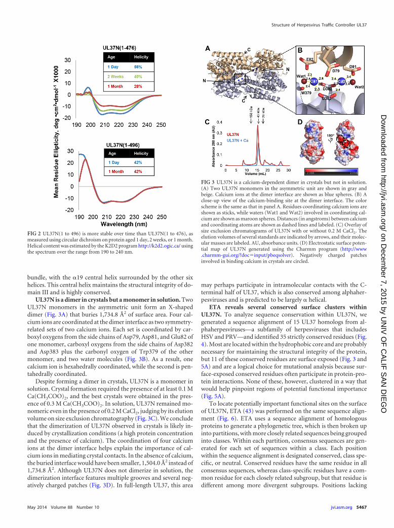
UL37N is a dimer in crystals but a monomer in solution. TwoUL37N monomers in the asymmetric unit form an X-shapeddimer (Fig. 3A) that buries 1,734.8 Å2 of surface area. Four cal-cium ions are coordinated at the dimer interface as two symmetry-related sets of two calcium ions. Each set is coordinated by car-boxyl oxygens from the side chains of Asp79, Asp81, and Glu82 ofone monomer, carboxyl oxygens from the side chains of Asp382and Asp383 plus the carbonyl oxygen of Trp379 of the othermonomer, and two water molecules (Fig. 3B). As a result, onecalcium ion is hexahedrally coordinated, while the second is pen-tahedrally coordinated.
Despite forming a dimer in crystals, UL37N is a monomer insolution. Crystal formation required the presence of at least 0.1 MCa(CH3COO)2, and the best crystals were obtained in the pres-ence of 0.3 M Ca(CH3COO)2. In solution, UL37N remained mo-nomeric even in the presence of 0.2 M CaCl2, judging by its elutionvolume on size exclusion chromatography (Fig. 3C). We concludethat the dimerization of UL37N observed in crystals is likely in-duced by crystallization conditions (a high protein concentrationand the presence of calcium). The coordination of four calciumions at the dimer interface helps explain the importance of cal-cium ions in mediating crystal contacts. In the absence of calcium,the buried interface would have been smaller, 1,504.0 Å2 instead of1,734.8 Å2. Although UL37N does not dimerize in solution, thedimerization interface features multiple grooves and several neg-atively charged patches (Fig. 3D). In full-length UL37, this area
may perhaps participate in intramolecular contacts with the C-terminal half of UL37, which is also conserved among alphaher-pesviruses and is predicted to be largely � helical.
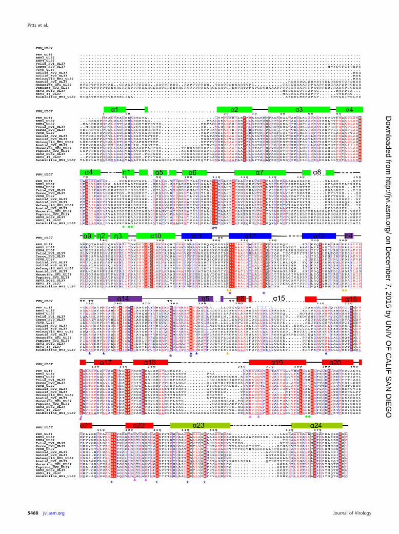
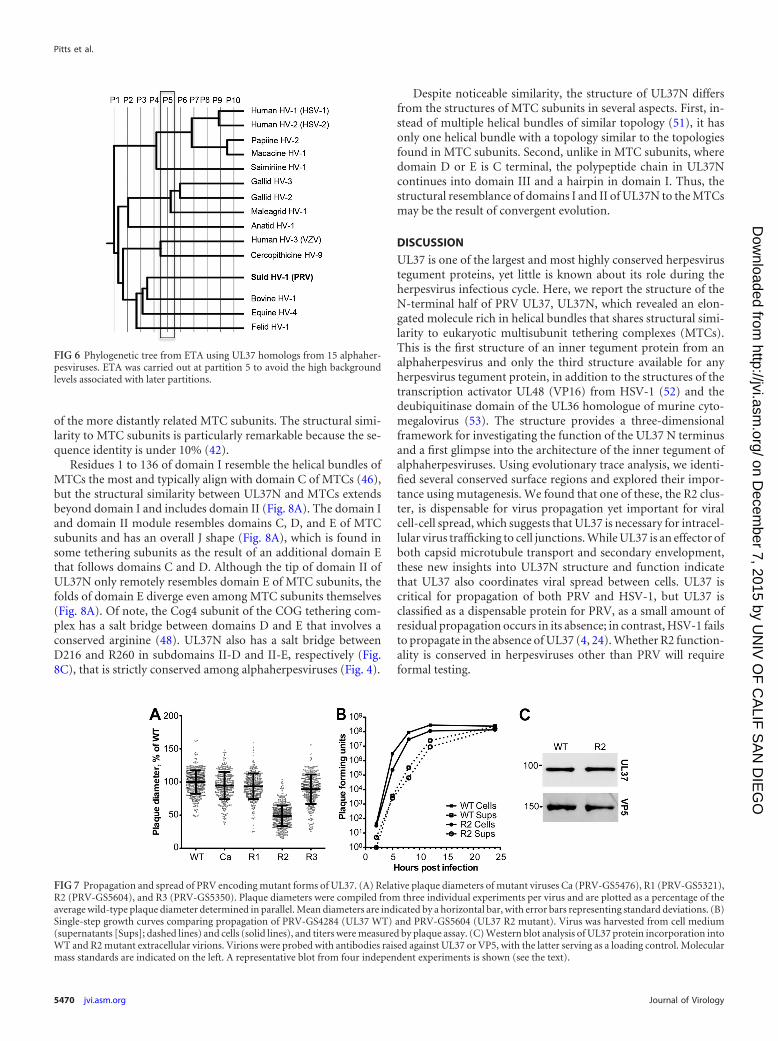
ETA reveals several conserved surface clusters withinUL37N. To analyze sequence conservation within UL37N, wegenerated a sequence alignment of 15 UL37 homologs from al-phaherpesviruses—a subfamily of herpesviruses that includesHSV and PRV—and identified 35 strictly conserved residues (Fig.4). Most are located within the hydrophobic core and are probablynecessary for maintaining the structural integrity of the protein,but 11 of these conserved residues are surface exposed (Fig. 3 and5A) and are a logical choice for mutational analysis because sur-face-exposed conserved residues often participate in protein-pro-tein interactions. None of these, however, clustered in a way thatwould help pinpoint regions of potential functional importance(Fig. 5A).
To locate potentially important functional sites on the surfaceof UL37N, ETA (43) was performed on the same sequence align-ment (Fig. 6). ETA uses a sequence alignment of homologousproteins to generate a phylogenetic tree, which is then broken upinto partitions, with more closely related sequences being groupedinto classes. Within each partition, consensus sequences are gen-erated for each set of sequences within a class. Each positionwithin the sequence alignment is designated conserved, class spe-cific, or neutral. Conserved residues have the same residue in allconsensus sequences, whereas class-specific residues have a com-mon residue for each closely related subgroup, but that residue isdifferent among more divergent subgroups. Positions lacking
FIG 2 UL37N(1 to 496) is more stable over time than UL37N(1 to 476), asmeasured using circular dichroism on protein aged 1 day, 2 weeks, or 1 month.Helical content was estimated by the K2D2 program http://k2d2.ogic.ca/ usingthe spectrum over the range from 190 to 240 nm.
FIG 3 UL37N is a calcium-dependent dimer in crystals but not in solution.(A) Two UL37N monomers in the asymmetric unit are shown in gray andbeige. Calcium ions at the dimer interface are shown as blue spheres. (B) Aclose-up view of the calcium-binding site at the dimer interface. The colorscheme is the same as that in panel A. Residues coordinating calcium ions areshown as sticks, while waters (Wat1 and Wat2) involved in coordinating cal-cium are shown as maroon spheres. Distances (in angstroms) between calciumand coordinating atoms are shown as dashed lines and labeled. (C) Overlay ofsize exclusion chromatograms of UL37N with or without 0.2 M CaCl2. Theelution volumes of several standards are indicated by arrows, and their molec-ular masses are labeled. AU, absorbance units. (D) Electrostatic surface poten-tial map of UL37N generated using the Charmm program (http://www.charmm-gui.org/?doc input/pbeqsolver). Negatively charged patchesinvolved in binding calcium in crystals are circled.
Structure of Herpesvirus Traffic Controller UL37
May 2014 Volume 88 Number 10 jvi.asm.org 5467
on Decem
ber 7, 2015 by UN
IV O
F C
ALIF
SA
N D
IEG
Ohttp://jvi.asm
.org/D
ownloaded from
Pitts et al.
5468 jvi.asm.org Journal of Virology
on Decem
ber 7, 2015 by UN
IV O
F C
ALIF
SA
N D
IEG
Ohttp://jvi.asm
.org/D
ownloaded from
consensus among the members of at least one subgroup are con-sidered neutral. Clustering of conserved and class-specific resi-dues on the protein surface may indicate regions of potential func-tional importance (43). This method has been used to detectfunctional sites in a number of proteins (44, 45).
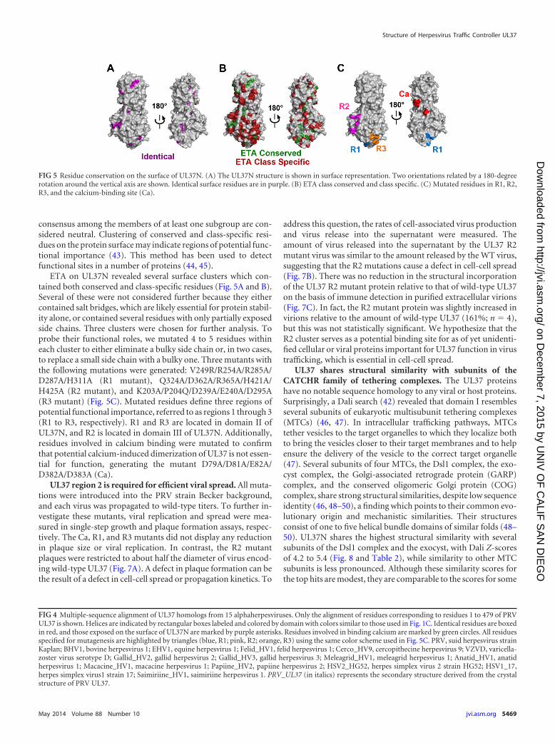
ETA on UL37N revealed several surface clusters which con-tained both conserved and class-specific residues (Fig. 5A and B).Several of these were not considered further because they eithercontained salt bridges, which are likely essential for protein stabil-ity alone, or contained several residues with only partially exposedside chains. Three clusters were chosen for further analysis. Toprobe their functional roles, we mutated 4 to 5 residues withineach cluster to either eliminate a bulky side chain or, in two cases,to replace a small side chain with a bulky one. Three mutants withthe following mutations were generated: V249R/R254A/R285A/D287A/H311A (R1 mutant), Q324A/D362A/R365A/H421A/H425A (R2 mutant), and K203A/P204Q/D239A/E240A/D295A(R3 mutant) (Fig. 5C). Mutated residues define three regions ofpotential functional importance, referred to as regions 1 through 3(R1 to R3, respectively). R1 and R3 are located in domain II ofUL37N, and R2 is located in domain III of UL37N. Additionally,residues involved in calcium binding were mutated to confirmthat potential calcium-induced dimerization of UL37 is not essen-tial for function, generating the mutant D79A/D81A/E82A/D382A/D383A (Ca).
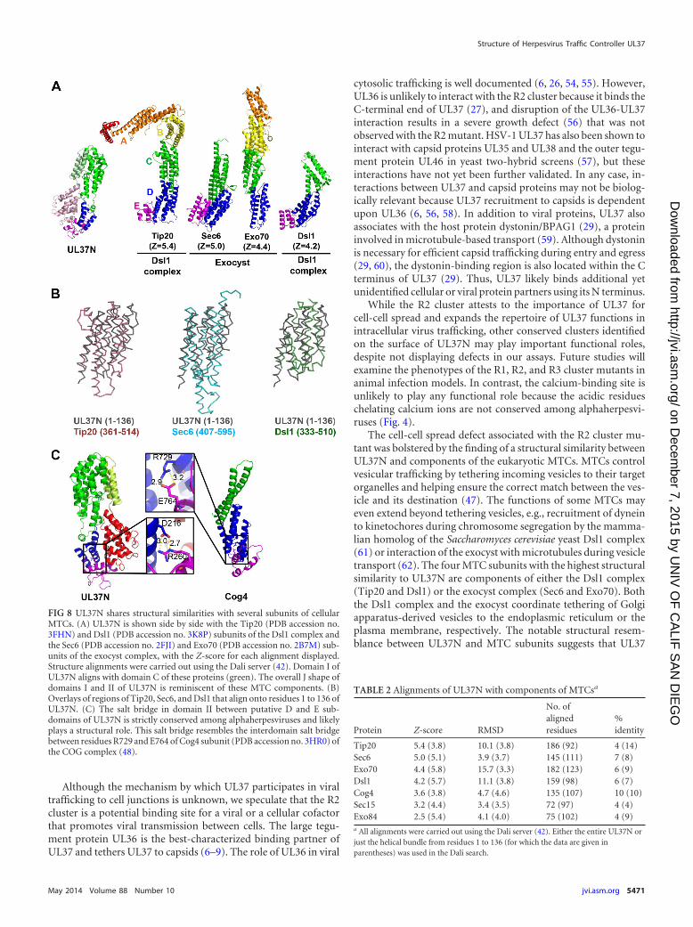
UL37 region 2 is required for efficient viral spread. All muta-tions were introduced into the PRV strain Becker background,and each virus was propagated to wild-type titers. To further in-vestigate these mutants, viral replication and spread were mea-sured in single-step growth and plaque formation assays, respec-tively. The Ca, R1, and R3 mutants did not display any reductionin plaque size or viral replication. In contrast, the R2 mutantplaques were restricted to about half the diameter of virus encod-ing wild-type UL37 (Fig. 7A). A defect in plaque formation can bethe result of a defect in cell-cell spread or propagation kinetics. To
address this question, the rates of cell-associated virus productionand virus release into the supernatant were measured. Theamount of virus released into the supernatant by the UL37 R2mutant virus was similar to the amount released by the WT virus,suggesting that the R2 mutations cause a defect in cell-cell spread(Fig. 7B). There was no reduction in the structural incorporationof the UL37 R2 mutant protein relative to that of wild-type UL37on the basis of immune detection in purified extracellular virions(Fig. 7C). In fact, the R2 mutant protein was slightly increased invirions relative to the amount of wild-type UL37 (161%; n 4),but this was not statistically significant. We hypothesize that theR2 cluster serves as a potential binding site for as of yet unidenti-fied cellular or viral proteins important for UL37 function in virustrafficking, which is essential in cell-cell spread.
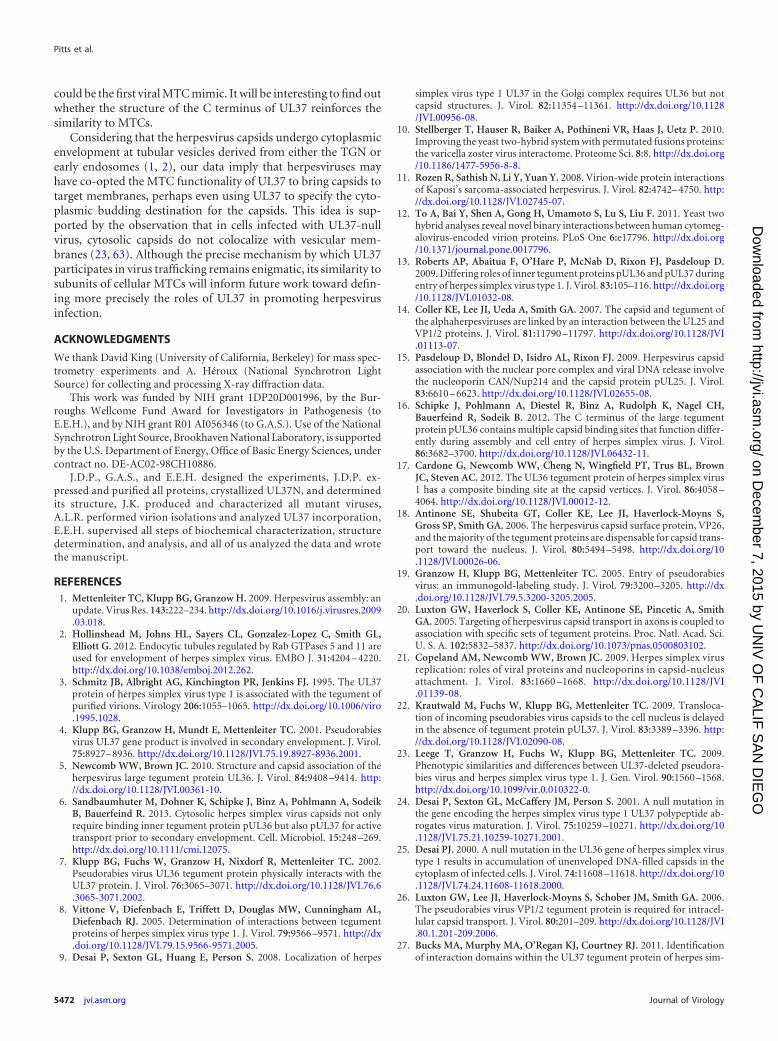
UL37 shares structural similarity with subunits of theCATCHR family of tethering complexes. The UL37 proteinshave no notable sequence homology to any viral or host proteins.Surprisingly, a Dali search (42) revealed that domain I resemblesseveral subunits of eukaryotic multisubunit tethering complexes(MTCs) (46, 47). In intracellular trafficking pathways, MTCstether vesicles to the target organelles to which they localize bothto bring the vesicles closer to their target membranes and to helpensure the delivery of the vesicle to the correct target organelle(47). Several subunits of four MTCs, the Dsl1 complex, the exo-cyst complex, the Golgi-associated retrograde protein (GARP)complex, and the conserved oligomeric Golgi protein (COG)complex, share strong structural similarities, despite low sequenceidentity (46, 48–50), a finding which points to their common evo-lutionary origin and mechanistic similarities. Their structuresconsist of one to five helical bundle domains of similar folds (48–50). UL37N shares the highest structural similarity with severalsubunits of the Dsl1 complex and the exocyst, with Dali Z-scoresof 4.2 to 5.4 (Fig. 8 and Table 2), while similarity to other MTCsubunits is less pronounced. Although these similarity scores forthe top hits are modest, they are comparable to the scores for some
FIG 4 Multiple-sequence alignment of UL37 homologs from 15 alphaherpesviruses. Only the alignment of residues corresponding to residues 1 to 479 of PRVUL37 is shown. Helices are indicated by rectangular boxes labeled and colored by domain with colors similar to those used in Fig. 1C. Identical residues are boxedin red, and those exposed on the surface of UL37N are marked by purple asterisks. Residues involved in binding calcium are marked by green circles. All residuesspecified for mutagenesis are highlighted by triangles (blue, R1; pink, R2; orange, R3) using the same color scheme used in Fig. 5C. PRV, suid herpesvirus strainKaplan; BHV1, bovine herpesvirus 1; EHV1, equine herpesvirus 1; Felid_HV1, felid herpesvirus 1; Cerco_HV9, cercopithecine herpesvirus 9; VZVD, varicella-zoster virus serotype D; Gallid_HV2, gallid herpesvirus 2; Gallid_HV3, gallid herpesvirus 3; Meleagrid_HV1, meleagrid herpesvirus 1; Anatid_HV1, anatidherpesvirus 1; Macacine_HV1, macacine herpesvirus 1; Papiine_HV2, papiine herpesvirus 2; HSV2_HG52, herpes simplex virus 2 strain HG52; HSV1_17,herpes simplex virus1 strain 17; Saimiriine_HV1, saimiriine herpesvirus 1. PRV_UL37 (in italics) represents the secondary structure derived from the crystalstructure of PRV UL37.
FIG 5 Residue conservation on the surface of UL37N. (A) The UL37N structure is shown in surface representation. Two orientations related by a 180-degreerotation around the vertical axis are shown. Identical surface residues are in purple. (B) ETA class conserved and class specific. (C) Mutated residues in R1, R2,R3, and the calcium-binding site (Ca).
Structure of Herpesvirus Traffic Controller UL37
May 2014 Volume 88 Number 10 jvi.asm.org 5469
on Decem
ber 7, 2015 by UN
IV O
F C
ALIF
SA
N D
IEG
Ohttp://jvi.asm
.org/D
ownloaded from
of the more distantly related MTC subunits. The structural simi-larity to MTC subunits is particularly remarkable because the se-quence identity is under 10% (42).
Residues 1 to 136 of domain I resemble the helical bundles ofMTCs the most and typically align with domain C of MTCs (46),but the structural similarity between UL37N and MTCs extendsbeyond domain I and includes domain II (Fig. 8A). The domain Iand domain II module resembles domains C, D, and E of MTCsubunits and has an overall J shape (Fig. 8A), which is found insome tethering subunits as the result of an additional domain Ethat follows domains C and D. Although the tip of domain II ofUL37N only remotely resembles domain E of MTC subunits, thefolds of domain E diverge even among MTC subunits themselves(Fig. 8A). Of note, the Cog4 subunit of the COG tethering com-plex has a salt bridge between domains D and E that involves aconserved arginine (48). UL37N also has a salt bridge betweenD216 and R260 in subdomains II-D and II-E, respectively (Fig.8C), that is strictly conserved among alphaherpesviruses (Fig. 4).
Despite noticeable similarity, the structure of UL37N differsfrom the structures of MTC subunits in several aspects. First, in-stead of multiple helical bundles of similar topology (51), it hasonly one helical bundle with a topology similar to the topologiesfound in MTC subunits. Second, unlike in MTC subunits, wheredomain D or E is C terminal, the polypeptide chain in UL37Ncontinues into domain III and a hairpin in domain I. Thus, thestructural resemblance of domains I and II of UL37N to the MTCsmay be the result of convergent evolution.
DISCUSSION
UL37 is one of the largest and most highly conserved herpesvirustegument proteins, yet little is known about its role during theherpesvirus infectious cycle. Here, we report the structure of theN-terminal half of PRV UL37, UL37N, which revealed an elon-gated molecule rich in helical bundles that shares structural simi-larity to eukaryotic multisubunit tethering complexes (MTCs).This is the first structure of an inner tegument protein from analphaherpesvirus and only the third structure available for anyherpesvirus tegument protein, in addition to the structures of thetranscription activator UL48 (VP16) from HSV-1 (52) and thedeubiquitinase domain of the UL36 homologue of murine cyto-megalovirus (53). The structure provides a three-dimensionalframework for investigating the function of the UL37 N terminusand a first glimpse into the architecture of the inner tegument ofalphaherpesviruses. Using evolutionary trace analysis, we identi-fied several conserved surface regions and explored their impor-tance using mutagenesis. We found that one of these, the R2 clus-ter, is dispensable for virus propagation yet important for viralcell-cell spread, which suggests that UL37 is necessary for intracel-lular virus trafficking to cell junctions. While UL37 is an effector ofboth capsid microtubule transport and secondary envelopment,these new insights into UL37N structure and function indicatethat UL37 also coordinates viral spread between cells. UL37 iscritical for propagation of both PRV and HSV-1, but UL37 isclassified as a dispensable protein for PRV, as a small amount ofresidual propagation occurs in its absence; in contrast, HSV-1 failsto propagate in the absence of UL37 (4, 24). Whether R2 function-ality is conserved in herpesviruses other than PRV will requireformal testing.
FIG 6 Phylogenetic tree from ETA using UL37 homologs from 15 alphaher-pesviruses. ETA was carried out at partition 5 to avoid the high backgroundlevels associated with later partitions.
FIG 7 Propagation and spread of PRV encoding mutant forms of UL37. (A) Relative plaque diameters of mutant viruses Ca (PRV-GS5476), R1 (PRV-GS5321),R2 (PRV-GS5604), and R3 (PRV-GS5350). Plaque diameters were compiled from three individual experiments per virus and are plotted as a percentage of theaverage wild-type plaque diameter determined in parallel. Mean diameters are indicated by a horizontal bar, with error bars representing standard deviations. (B)Single-step growth curves comparing propagation of PRV-GS4284 (UL37 WT) and PRV-GS5604 (UL37 R2 mutant). Virus was harvested from cell medium(supernatants [Sups]; dashed lines) and cells (solid lines), and titers were measured by plaque assay. (C) Western blot analysis of UL37 protein incorporation intoWT and R2 mutant extracellular virions. Virions were probed with antibodies raised against UL37 or VP5, with the latter serving as a loading control. Molecularmass standards are indicated on the left. A representative blot from four independent experiments is shown (see the text).
Pitts et al.
5470 jvi.asm.org Journal of Virology
on Decem
ber 7, 2015 by UN
IV O
F C
ALIF
SA
N D
IEG
Ohttp://jvi.asm
.org/D
ownloaded from
Although the mechanism by which UL37 participates in viraltrafficking to cell junctions is unknown, we speculate that the R2cluster is a potential binding site for a viral or a cellular cofactorthat promotes viral transmission between cells. The large tegu-ment protein UL36 is the best-characterized binding partner ofUL37 and tethers UL37 to capsids (6–9). The role of UL36 in viral
cytosolic trafficking is well documented (6, 26, 54, 55). However,UL36 is unlikely to interact with the R2 cluster because it binds theC-terminal end of UL37 (27), and disruption of the UL36-UL37interaction results in a severe growth defect (56) that was notobserved with the R2 mutant. HSV-1 UL37 has also been shown tointeract with capsid proteins UL35 and UL38 and the outer tegu-ment protein UL46 in yeast two-hybrid screens (57), but theseinteractions have not yet been further validated. In any case, in-teractions between UL37 and capsid proteins may not be biolog-ically relevant because UL37 recruitment to capsids is dependentupon UL36 (6, 56, 58). In addition to viral proteins, UL37 alsoassociates with the host protein dystonin/BPAG1 (29), a proteininvolved in microtubule-based transport (59). Although dystoninis necessary for efficient capsid trafficking during entry and egress(29, 60), the dystonin-binding region is also located within the Cterminus of UL37 (29). Thus, UL37 likely binds additional yetunidentified cellular or viral protein partners using its N terminus.
While the R2 cluster attests to the importance of UL37 forcell-cell spread and expands the repertoire of UL37 functions inintracellular virus trafficking, other conserved clusters identifiedon the surface of UL37N may play important functional roles,despite not displaying defects in our assays. Future studies willexamine the phenotypes of the R1, R2, and R3 cluster mutants inanimal infection models. In contrast, the calcium-binding site isunlikely to play any functional role because the acidic residueschelating calcium ions are not conserved among alphaherpesvi-ruses (Fig. 4).
The cell-cell spread defect associated with the R2 cluster mu-tant was bolstered by the finding of a structural similarity betweenUL37N and components of the eukaryotic MTCs. MTCs controlvesicular trafficking by tethering incoming vesicles to their targetorganelles and helping ensure the correct match between the ves-icle and its destination (47). The functions of some MTCs mayeven extend beyond tethering vesicles, e.g., recruitment of dyneinto kinetochores during chromosome segregation by the mamma-lian homolog of the Saccharomyces cerevisiae yeast Dsl1 complex(61) or interaction of the exocyst with microtubules during vesicletransport (62). The four MTC subunits with the highest structuralsimilarity to UL37N are components of either the Dsl1 complex(Tip20 and Dsl1) or the exocyst complex (Sec6 and Exo70). Boththe Dsl1 complex and the exocyst coordinate tethering of Golgiapparatus-derived vesicles to the endoplasmic reticulum or theplasma membrane, respectively. The notable structural resem-blance between UL37N and MTC subunits suggests that UL37
TABLE 2 Alignments of UL37N with components of MTCsa
Protein Z-score RMSD
No. ofalignedresidues
%identity
Tip20 5.4 (3.8) 10.1 (3.8) 186 (92) 4 (14)Sec6 5.0 (5.1) 3.9 (3.7) 145 (111) 7 (8)Exo70 4.4 (5.8) 15.7 (3.3) 182 (123) 6 (9)Dsl1 4.2 (5.7) 11.1 (3.8) 159 (98) 6 (7)Cog4 3.6 (3.8) 4.7 (4.6) 135 (107) 10 (10)Sec15 3.2 (4.4) 3.4 (3.5) 72 (97) 4 (4)Exo84 2.5 (5.4) 4.1 (4.0) 75 (102) 4 (9)a All alignments were carried out using the Dali server (42). Either the entire UL37N orjust the helical bundle from residues 1 to 136 (for which the data are given inparentheses) was used in the Dali search.
FIG 8 UL37N shares structural similarities with several subunits of cellularMTCs. (A) UL37N is shown side by side with the Tip20 (PDB accession no.3FHN) and Dsl1 (PDB accession no. 3K8P) subunits of the Dsl1 complex andthe Sec6 (PDB accession no. 2FJI) and Exo70 (PDB accession no. 2B7M) sub-units of the exocyst complex, with the Z-score for each alignment displayed.Structure alignments were carried out using the Dali server (42). Domain I ofUL37N aligns with domain C of these proteins (green). The overall J shape ofdomains I and II of UL37N is reminiscent of these MTC components. (B)Overlays of regions of Tip20, Sec6, and Dsl1 that align onto residues 1 to 136 ofUL37N. (C) The salt bridge in domain II between putative D and E sub-domains of UL37N is strictly conserved among alphaherpesviruses and likelyplays a structural role. This salt bridge resembles the interdomain salt bridgebetween residues R729 and E764 of Cog4 subunit (PDB accession no. 3HR0) ofthe COG complex (48).
Structure of Herpesvirus Traffic Controller UL37
May 2014 Volume 88 Number 10 jvi.asm.org 5471
on Decem
ber 7, 2015 by UN
IV O
F C
ALIF
SA
N D
IEG
Ohttp://jvi.asm
.org/D
ownloaded from
could be the first viral MTC mimic. It will be interesting to find outwhether the structure of the C terminus of UL37 reinforces thesimilarity to MTCs.
Considering that the herpesvirus capsids undergo cytoplasmicenvelopment at tubular vesicles derived from either the TGN orearly endosomes (1, 2), our data imply that herpesviruses mayhave co-opted the MTC functionality of UL37 to bring capsids totarget membranes, perhaps even using UL37 to specify the cyto-plasmic budding destination for the capsids. This idea is sup-ported by the observation that in cells infected with UL37-nullvirus, cytosolic capsids do not colocalize with vesicular mem-branes (23, 63). Although the precise mechanism by which UL37participates in virus trafficking remains enigmatic, its similarity tosubunits of cellular MTCs will inform future work toward defin-ing more precisely the roles of UL37 in promoting herpesvirusinfection.
ACKNOWLEDGMENTS
We thank David King (University of California, Berkeley) for mass spec-trometry experiments and A. Héroux (National Synchrotron LightSource) for collecting and processing X-ray diffraction data.
This work was funded by NIH grant 1DP20D001996, by the Bur-roughs Wellcome Fund Award for Investigators in Pathogenesis (toE.E.H.), and by NIH grant R01 AI056346 (to G.A.S.). Use of the NationalSynchrotron Light Source, Brookhaven National Laboratory, is supportedby the U.S. Department of Energy, Office of Basic Energy Sciences, undercontract no. DE-AC02-98CH10886.
J.D.P., G.A.S., and E.E.H. designed the experiments, J.D.P. ex-pressed and purified all proteins, crystallized UL37N, and determinedits structure, J.K. produced and characterized all mutant viruses,A.L.R. performed virion isolations and analyzed UL37 incorporation,E.E.H. supervised all steps of biochemical characterization, structuredetermination, and analysis, and all of us analyzed the data and wrotethe manuscript.
REFERENCES1. Mettenleiter TC, Klupp BG, Granzow H. 2009. Herpesvirus assembly: an
update. Virus Res. 143:222–234. http://dx.doi.org/10.1016/j.virusres.2009.03.018.
2. Hollinshead M, Johns HL, Sayers CL, Gonzalez-Lopez C, Smith GL,Elliott G. 2012. Endocytic tubules regulated by Rab GTPases 5 and 11 areused for envelopment of herpes simplex virus. EMBO J. 31:4204 – 4220.http://dx.doi.org/10.1038/emboj.2012.262.
3. Schmitz JB, Albright AG, Kinchington PR, Jenkins FJ. 1995. The UL37protein of herpes simplex virus type 1 is associated with the tegument ofpurified virions. Virology 206:1055–1065. http://dx.doi.org/10.1006/viro.1995.1028.
4. Klupp BG, Granzow H, Mundt E, Mettenleiter TC. 2001. Pseudorabiesvirus UL37 gene product is involved in secondary envelopment. J. Virol.75:8927– 8936. http://dx.doi.org/10.1128/JVI.75.19.8927-8936.2001.
5. Newcomb WW, Brown JC. 2010. Structure and capsid association of theherpesvirus large tegument protein UL36. J. Virol. 84:9408 –9414. http://dx.doi.org/10.1128/JVI.00361-10.
6. Sandbaumhuter M, Dohner K, Schipke J, Binz A, Pohlmann A, SodeikB, Bauerfeind R. 2013. Cytosolic herpes simplex virus capsids not onlyrequire binding inner tegument protein pUL36 but also pUL37 for activetransport prior to secondary envelopment. Cell. Microbiol. 15:248 –269.http://dx.doi.org/10.1111/cmi.12075.
7. Klupp BG, Fuchs W, Granzow H, Nixdorf R, Mettenleiter TC. 2002.Pseudorabies virus UL36 tegument protein physically interacts with theUL37 protein. J. Virol. 76:3065–3071. http://dx.doi.org/10.1128/JVI.76.6.3065-3071.2002.
8. Vittone V, Diefenbach E, Triffett D, Douglas MW, Cunningham AL,Diefenbach RJ. 2005. Determination of interactions between tegumentproteins of herpes simplex virus type 1. J. Virol. 79:9566 –9571. http://dx.doi.org/10.1128/JVI.79.15.9566-9571.2005.
9. Desai P, Sexton GL, Huang E, Person S. 2008. Localization of herpes
simplex virus type 1 UL37 in the Golgi complex requires UL36 but notcapsid structures. J. Virol. 82:11354 –11361. http://dx.doi.org/10.1128/JVI.00956-08.
10. Stellberger T, Hauser R, Baiker A, Pothineni VR, Haas J, Uetz P. 2010.Improving the yeast two-hybrid system with permutated fusions proteins:the varicella zoster virus interactome. Proteome Sci. 8:8. http://dx.doi.org/10.1186/1477-5956-8-8.
11. Rozen R, Sathish N, Li Y, Yuan Y. 2008. Virion-wide protein interactionsof Kaposi’s sarcoma-associated herpesvirus. J. Virol. 82:4742– 4750. http://dx.doi.org/10.1128/JVI.02745-07.
12. To A, Bai Y, Shen A, Gong H, Umamoto S, Lu S, Liu F. 2011. Yeast twohybrid analyses reveal novel binary interactions between human cytomeg-alovirus-encoded virion proteins. PLoS One 6:e17796. http://dx.doi.org/10.1371/journal.pone.0017796.
13. Roberts AP, Abaitua F, O’Hare P, McNab D, Rixon FJ, Pasdeloup D.2009. Differing roles of inner tegument proteins pUL36 and pUL37 duringentry of herpes simplex virus type 1. J. Virol. 83:105–116. http://dx.doi.org/10.1128/JVI.01032-08.
14. Coller KE, Lee JI, Ueda A, Smith GA. 2007. The capsid and tegument ofthe alphaherpesviruses are linked by an interaction between the UL25 andVP1/2 proteins. J. Virol. 81:11790 –11797. http://dx.doi.org/10.1128/JVI.01113-07.
15. Pasdeloup D, Blondel D, Isidro AL, Rixon FJ. 2009. Herpesvirus capsidassociation with the nuclear pore complex and viral DNA release involvethe nucleoporin CAN/Nup214 and the capsid protein pUL25. J. Virol.83:6610 – 6623. http://dx.doi.org/10.1128/JVI.02655-08.
16. Schipke J, Pohlmann A, Diestel R, Binz A, Rudolph K, Nagel CH,Bauerfeind R, Sodeik B. 2012. The C terminus of the large tegumentprotein pUL36 contains multiple capsid binding sites that function differ-ently during assembly and cell entry of herpes simplex virus. J. Virol.86:3682–3700. http://dx.doi.org/10.1128/JVI.06432-11.
17. Cardone G, Newcomb WW, Cheng N, Wingfield PT, Trus BL, BrownJC, Steven AC. 2012. The UL36 tegument protein of herpes simplex virus1 has a composite binding site at the capsid vertices. J. Virol. 86:4058 –4064. http://dx.doi.org/10.1128/JVI.00012-12.
18. Antinone SE, Shubeita GT, Coller KE, Lee JI, Haverlock-Moyns S,Gross SP, Smith GA. 2006. The herpesvirus capsid surface protein, VP26,and the majority of the tegument proteins are dispensable for capsid trans-port toward the nucleus. J. Virol. 80:5494 –5498. http://dx.doi.org/10.1128/JVI.00026-06.
19. Granzow H, Klupp BG, Mettenleiter TC. 2005. Entry of pseudorabiesvirus: an immunogold-labeling study. J. Virol. 79:3200 –3205. http://dx.doi.org/10.1128/JVI.79.5.3200-3205.2005.
20. Luxton GW, Haverlock S, Coller KE, Antinone SE, Pincetic A, SmithGA. 2005. Targeting of herpesvirus capsid transport in axons is coupled toassociation with specific sets of tegument proteins. Proc. Natl. Acad. Sci.U. S. A. 102:5832–5837. http://dx.doi.org/10.1073/pnas.0500803102.
21. Copeland AM, Newcomb WW, Brown JC. 2009. Herpes simplex virusreplication: roles of viral proteins and nucleoporins in capsid-nucleusattachment. J. Virol. 83:1660 –1668. http://dx.doi.org/10.1128/JVI.01139-08.
22. Krautwald M, Fuchs W, Klupp BG, Mettenleiter TC. 2009. Transloca-tion of incoming pseudorabies virus capsids to the cell nucleus is delayedin the absence of tegument protein pUL37. J. Virol. 83:3389 –3396. http://dx.doi.org/10.1128/JVI.02090-08.
23. Leege T, Granzow H, Fuchs W, Klupp BG, Mettenleiter TC. 2009.Phenotypic similarities and differences between UL37-deleted pseudora-bies virus and herpes simplex virus type 1. J. Gen. Virol. 90:1560 –1568.http://dx.doi.org/10.1099/vir.0.010322-0.
24. Desai P, Sexton GL, McCaffery JM, Person S. 2001. A null mutation inthe gene encoding the herpes simplex virus type 1 UL37 polypeptide ab-rogates virus maturation. J. Virol. 75:10259 –10271. http://dx.doi.org/10.1128/JVI.75.21.10259-10271.2001.
25. Desai PJ. 2000. A null mutation in the UL36 gene of herpes simplex virustype 1 results in accumulation of unenveloped DNA-filled capsids in thecytoplasm of infected cells. J. Virol. 74:11608 –11618. http://dx.doi.org/10.1128/JVI.74.24.11608-11618.2000.
26. Luxton GW, Lee JI, Haverlock-Moyns S, Schober JM, Smith GA. 2006.The pseudorabies virus VP1/2 tegument protein is required for intracel-lular capsid transport. J. Virol. 80:201–209. http://dx.doi.org/10.1128/JVI.80.1.201-209.2006.
27. Bucks MA, Murphy MA, O’Regan KJ, Courtney RJ. 2011. Identificationof interaction domains within the UL37 tegument protein of herpes sim-
Pitts et al.
5472 jvi.asm.org Journal of Virology
on Decem
ber 7, 2015 by UN
IV O
F C
ALIF
SA
N D
IEG
Ohttp://jvi.asm
.org/D
ownloaded from
plex virus type 1. Virology 416:42–53. http://dx.doi.org/10.1016/j.virol.2011.04.018.
28. Kelly BJ, Mijatov B, Fraefel C, Cunningham AL, Diefenbach RJ. 2012.Identification of a single amino acid residue which is critical for the inter-action between HSV-1 inner tegument proteins pUL36 and pUL37. Virol-ogy 422:308 –316. http://dx.doi.org/10.1016/j.virol.2011.11.002.
29. Pasdeloup D, McElwee M, Beilstein F, Labetoulle M, Rixon FJ. 2013.Herpesvirus tegument protein pUL37 interacts with dystonin/BPAG1 topromote capsid transport on microtubules during egress. J. Virol. 87:2857–2867. http://dx.doi.org/10.1128/JVI.02676-12.
30. Watanabe D, Ushijima Y, Goshima F, Takakuwa H, Tomita Y,Nishiyama Y. 2000. Identification of nuclear export signal in UL37 pro-tein of herpes simplex virus type 2. Biochem. Biophys. Res. Commun.276:1248 –1254. http://dx.doi.org/10.1006/bbrc.2000.3600.
31. Bohannon KP, Sollars PJ, Pickard GE, Smith GA. 2012. Fusion of afluorescent protein to the pUL25 minor capsid protein of pseudorabiesvirus allows live-cell capsid imaging with negligible impact on infection. J.Gen. Virol. 93:124 –129. http://dx.doi.org/10.1099/vir.0.036145-0.
32. Tischer BK, Smith GA, Osterrieder N. 2010. En passant mutagenesis: atwo step markerless red recombination system. Methods Mol. Biol. 634:421– 430. http://dx.doi.org/10.1007/978-1-60761-652-8_30.
33. Smith GA, Enquist LW. 1999. Construction and transposon mutagenesisin Escherichia coli of a full-length infectious clone of pseudorabies virus,an alphaherpesvirus. J. Virol. 73:6405– 6414.
34. Phillips K, de la Pena AH. 2011. The combined use of the Thermofluorassay and ThermoQ analytical software for the determination of proteinstability and buffer optimization as an aid in protein crystallization. Curr.Protoc. Mol. Biol. Chapter 10:Unit 10.28. http://dx.doi.org/10.1002/0471142727.mb1028s94.
35. Otwinowski Z, Minor W. 1997. Processing of X-ray diffraction datacollected in oscillation mode. Methods Enzymol. 276:307–326. http://dx.doi.org/10.1016/S0076-6879(97)76066-X.
36. Emsley P, Cowtan K. 2004. Coot: model-building tools for moleculargraphics. Acta Crystallogr. D Biol. Crystallogr. 60:2126 –2132. http://dx.doi.org/10.1107/S0907444904019158.
37. Adams PD, Grosse-Kunstleve RW, Hung LW, Ioerger TR, McCoy AJ,Moriarty NW, Read RJ, Sacchettini JC, Sauter NK, Terwilliger TC.2002. PHENIX: building new software for automated crystallographicstructure determination. Acta Crystallogr. D Biol. Crystallogr. 58:1948 –1954. http://dx.doi.org/10.1107/S0907444902016657.
38. Davis IW, Leaver-Fay A, Chen VB, Block JN, Kapral GJ, Wang X,Murray LW, Arendall WB, III, Snoeyink J, Richardson JS, RichardsonDC. 2007. MolProbity: all-atom contacts and structure validation for pro-teins and nucleic acids. Nucleic Acids Res. 35:W375–W383. http://dx.doi.org/10.1093/nar/gkm216.
39. Larkin MA, Blackshields G, Brown NP, Chenna R, McGettigan PA,McWilliam H, Valentin F, Wallace IM, Wilm A, Lopez R, ThompsonJD, Gibson TJ, Higgins DG. 2007. Clustal W and Clustal X version 2.0.Bioinformatics 23:2947–2948. http://dx.doi.org/10.1093/bioinformatics/btm404.
40. Gouet P, Courcelle E, Stuart DI, Metoz F. 1999. ESPript: analysis ofmultiple sequence alignments in PostScript. Bioinformatics 15:305–308.http://dx.doi.org/10.1093/bioinformatics/15.4.305.
41. Krissinel E, Henrick K. 2007. Inference of macromolecular assembliesfrom crystalline state. J. Mol. Biol. 372:774 –797. http://dx.doi.org/10.1016/j.jmb.2007.05.022.
42. Holm L, Rosenstrom P. 2010. Dali server: conservation mapping in 3D.Nucleic Acids Res. 38:W545–W549. http://dx.doi.org/10.1093/nar/gkq366.
43. Lichtarge O, Bourne HR, Cohen FE. 1996. An evolutionary trace methoddefines binding surfaces common to protein families. J. Mol. Biol. 257:342–358. http://dx.doi.org/10.1006/jmbi.1996.0167.
44. Sowa ME, He W, Slep KC, Kercher MA, Lichtarge O, Wensel TG. 2001.Prediction and confirmation of a site critical for effector regulation of RGSdomain activity. Nat. Struct. Biol. 8:234 –237. http://dx.doi.org/10.1038/84974.
45. Chakravarty S, Hutson AM, Estes MK, Prasad BV. 2005. Evolutionarytrace residues in noroviruses: importance in receptor binding, antigenic-ity, virion assembly, and strain diversity. J. Virol. 79:554 –568. http://dx.doi.org/10.1128/JVI.79.1.554-568.2005.
46. Jackson LP, Kummel D, Reinisch KM, Owen DJ. 2012. Structures andmechanisms of vesicle coat components and multisubunit tethering com-plexes. Curr. Opin. Cell Biol. 24:475– 483. http://dx.doi.org/10.1016/j.ceb.2012.05.013.
47. Brocker C, Engelbrecht-Vandre S, Ungermann C. 2010. Multisubunittethering complexes and their role in membrane fusion. Curr. Biol. 20:R943–R952. http://dx.doi.org/10.1016/j.cub.2010.09.015.
48. Richardson BC, Smith RD, Ungar D, Nakamura A, Jeffrey PD, Lupa-shin VV, Hughson FM. 2009. Structural basis for a human glycosylationdisorder caused by mutation of the COG4 gene. Proc. Natl. Acad. Sci.U. S. A. 106:13329 –13334. http://dx.doi.org/10.1073/pnas.0901966106.
49. Tripathi A, Ren Y, Jeffrey PD, Hughson FM. 2009. Structural charac-terization of Tip20p and Dsl1p, subunits of the Dsl1p vesicle tetheringcomplex. Nat. Struct. Mol. Biol. 16:114 –123. http://dx.doi.org/10.1038/nsmb.1548.
50. Dong G, Hutagalung AH, Fu C, Novick P, Reinisch KM. 2005. Thestructures of exocyst subunit Exo70p and the Exo84p C-terminal domainsreveal a common motif. Nat. Struct. Mol. Biol. 12:1094 –1100. http://dx.doi.org/10.1038/nsmb1017.
51. Sivaram MV, Furgason ML, Brewer DN, Munson M. 2006. The struc-ture of the exocyst subunit Sec6p defines a conserved architecture withdiverse roles. Nat. Struct. Mol. Biol. 13:555–556. http://dx.doi.org/10.1038/nsmb1096.
52. Liu Y, Gong W, Huang CC, Herr W, Cheng X. 1999. Crystal structureof the conserved core of the herpes simplex virus transcriptional regula-tory protein VP16. Genes Dev. 13:1692–1703. http://dx.doi.org/10.1101/gad.13.13.1692.
53. Schlieker C, Weihofen WA, Frijns E, Kattenhorn LM, Gaudet R, PloeghHL. 2007. Structure of a herpesvirus-encoded cysteine protease reveals aunique class of deubiquitinating enzymes. Mol. Cell 25:677– 687. http://dx.doi.org/10.1016/j.molcel.2007.01.033.
54. Shanda SK, Wilson DW. 2008. UL36p is required for efficient transportof membrane-associated herpes simplex virus type 1 along microtubules.J. Virol. 82:7388 –7394. http://dx.doi.org/10.1128/JVI.00225-08.
55. Zaichick SV, Bohannon KP, Hughes A, Sollars PJ, Pickard GE, SmithGA. 2013. The herpesvirus VP1/2 protein is an effector of dynein-mediated capsid transport and neuroinvasion. Cell Host Microbe 13:193–203. http://dx.doi.org/10.1016/j.chom.2013.01.009.
56. Fuchs W, Klupp BG, Granzow H, Mettenleiter TC. 2004. Essentialfunction of the pseudorabies virus UL36 gene product is independent ofits interaction with the UL37 protein. J. Virol. 78:11879 –11889. http://dx.doi.org/10.1128/JVI.78.21.11879-11889.2004.
57. Lee JH, Vittone V, Diefenbach E, Cunningham AL, Diefenbach RJ.2008. Identification of structural protein-protein interactions of herpessimplex virus type 1. Virology 378:347–354. http://dx.doi.org/10.1016/j.virol.2008.05.035.
58. Ko DH, Cunningham AL, Diefenbach RJ. 2010. The major determinantfor addition of tegument protein pUL48 (VP16) to capsids in herpes sim-plex virus type 1 is the presence of the major tegument protein pUL36(VP1/2). J. Virol. 84:1397–1405. http://dx.doi.org/10.1128/JVI.01721-09.
59. Ryan SD, Bhanot K, Ferrier A, De Repentigny Y, Chu A, Blais A,Kothary R. 2012. Microtubule stability, Golgi organization, and transportflux require dystonin-a2-MAP1B interaction. J. Cell Biol. 196:727–742.http://dx.doi.org/10.1083/jcb.201107096.
60. McElwee M, Beilstein F, Labetoulle M, Rixon FJ, Pasdeloup D. 2013.Dystonin/BPAG1 promotes plus-end-directed transport of herpes sim-plex virus 1 capsids on microtubules during entry. J. Virol. 87:11008 –11018. http://dx.doi.org/10.1128/JVI.01633-13.
61. Schmitt HD. 2010. Dsl1p/Zw10: common mechanisms behind tetheringvesicles and microtubules. Trends Cell Biol. 20:257–268. http://dx.doi.org/10.1016/j.tcb.2010.02.001.
62. Heider MR, Munson M. 2012. Exorcising the exocyst complex. Traffic13:898 –907. http://dx.doi.org/10.1111/j.1600-0854.2012.01353.x.
63. Pasdeloup D, Beilstein F, Roberts AP, McElwee M, McNab D, Rixon FJ.2010. Inner tegument protein pUL37 of herpes simplex virus type 1 isinvolved in directing capsids to the trans-Golgi network for envelopment.J. Gen. Virol. 91:2145–2151. http://dx.doi.org/10.1099/vir.0.022053-0.
Structure of Herpesvirus Traffic Controller UL37
May 2014 Volume 88 Number 10 jvi.asm.org 5473
on Decem
ber 7, 2015 by UN
IV O
F C
ALIF
SA
N D
IEG
Ohttp://jvi.asm
.org/D
ownloaded from