j. biol. chem. 2014-selwa-jbc.m113.530410
TRANSCRIPT

Daniel Ladant and Thérèse E. MalliavinChenal, Ana-Cristina Sotomayor-Perez, Edithe Selwa, Marilyne Davi, Alexandre StudiesMolecular Dynamics and MutagenesisAdenylyl Cyclase by Calmodulin:
Bordetella pertussisAllosteric Activation of Molecular Biophysics:
published online June 6, 2014J. Biol. Chem.
10.1074/jbc.M113.530410Access the most updated version of this article at doi:
.JBC Affinity SitesFind articles, minireviews, Reflections and Classics on similar topics on the
Alerts:
When a correction for this article is posted•
When this article is cited•
to choose from all of JBC's e-mail alertsClick here
http://www.jbc.org/content/early/2014/06/06/jbc.M113.530410.full.html#ref-list-1
This article cites 0 references, 0 of which can be accessed free at by guest on June 8, 2014http://w
ww
.jbc.org/D
ownloaded from
by guest on June 8, 2014
http://ww
w.jbc.org/
Dow
nloaded from

Mutants of AC and AC/CaM interaction 1
June 5, 2014
Allosteric activation of Bordetella pertussis adenylyl
cyclase by calmodulin: molecular dynamics and
mutagenesis studies
Edithe Selwa1,3, Marilyne Davi2, Alexandre Chenal2, Ana-Cristina Sotomayor-Perez2,Daniel Ladant*2 and Therese E Malliavin*1
1 Institut Pasteur and CNRS UMR 3528, rue du Dr Roux, Unite de Bioinformatique Struc-turale, 75015 Paris, France2 Institut Pasteur and CNRS UMR 3528, rue du Dr Roux, Unite de Biochimie des InteractionsMacromoleculaires, 75015 Paris, France3 Universite Pierre et Marie Curie, Cellule Pasteur UPMC, rue du Dr Roux, 75015 Paris, France
Corresponding authors:Therese E Malliavin, Unite de Bioinformatique Structurale, UMR CNRS 3528, rue du DrRoux, 75015 Paris, France; mail: [email protected], tel.: 00 33 1 45 68 88 54
Daniel Ladant, Unite de Biochimie des Interactions Macromoleculaires, UMR CNRS 3528,rue du Dr Roux, 75015 Paris, France; mail: [email protected], tel.: 00 33 1 45 6884 00
Keywords: adenylyl cyclase,Bordetella pertussis, calcium, calmodulin affin-ity, allostery, molecular dynamics
June 5, 2014Running Title: AC mutants and AC/CaM interaction
Background: Adenylyl cyclase (AC)from Bordetella pertussis is activated whenit interacts with calmodulin (CaM).
Results: A triple mutant of AC, whichwas predicted by molecular modeling, exhib-ited a highly reduced affinity for CaM.
Conclusion: This study suggests thata long-range connection between CaM andthe AC catalytic loop is crucial for AC acti-vation.
Significance: Molecular modelingidentified critical molecular determinants forthe allosteric activation of AC.
ABSTRACT
Adenylyl cyclase (AC) toxin is an es-sential toxin that allows Bordetella per-tussis to invade eukaryotic cells, whereit is activated after binding to calmod-ulin (CaM). Based on the crystalstructure of the AC catalytic domainin complex with the C-terminal halfof CaM (C-CaM), our previous molec-ular dynamics simulations (Selwa etal., Proteins 80, 1028–1040, 2012) sug-gested that three residues, i.e., Arg338,
http://www.jbc.org/cgi/doi/10.1074/jbc.M113.530410The latest version is at JBC Papers in Press. Published on June 6, 2014 as Manuscript M113.530410
Copyright 2014 by The American Society for Biochemistry and Molecular Biology, Inc.
by guest on June 8, 2014http://w
ww
.jbc.org/D
ownloaded from

Mutants of AC and AC/CaM interaction 2
Asn347, and Asp360, might be impor-tant for stabilizing the AC/CaM in-teraction. These residues belong to aloop-helix-loop motif (LHL) at the C-terminal end of AC, which is locatedat the interface between CaM and theAC catalytic loop. In the presentstudy, we conducted the in silico andin vitro characterization of three ACvariants, where one (Asn347; ACm1A),two (Arg338 and Asp360; ACm2A), orthree residues (Arg338, Asn347, andAsp360; ACm3A) were substitutedwith Ala. Biochemical studies showedthat the affinities of ACm1A andACm2A for CaM were not affectedsignificantly, whereas that of ACm3Awas reduced dramatically. To under-stand the effects of these modifica-tions, molecular dynamics (MD) sim-ulations were performed based on themodified proteins. The MD trajecto-ries recorded for the ACm3A/C-CaMcomplex showed that the calcium-binding loops of C-CaM exhibitedlarge fluctuations, which could berelated to the weakened interactionbetween ACm3A and its activator.Overall, our results suggest that theLHL motif at the C-terminal end ofAC is crucial during CaM-binding forstabilizing the AC catalytic loop in anactive configuration.
INTRODUCTION
The adenylyl cyclase toxin (CyaA) fromBordetella pertussis, the causative agent ofwhooping cough, plays an essential rolein host invasion [1–3]. CyaA is able toinvade eukaryotic target cells where it isactivated by interacting with calmodulin(CaM), thereby overproducing cAMP, whichdisorganizes cellular signaling processes andtriggers host cell death [4–12]. Several crys-tallographic structures have been reportedof the CyaA catalytic domain (AC) in com-plex with the C terminal moiety of CaM
(C-CaM) [13] (Figure 1). These structuresare physiologically relevant because C-CaMhas been shown [14] to be as potent in acti-vating the enzymatic activity of AC as thefull-length CaM. In the AC/C-CaM com-plex (Figure 1a), the AC domain includesthree main regions, CA (residues 7–61, 187–197, 261–299, and 313–345; colored in green)in the middle of the structure, and CB(residues 62–186, colored in orange) andSA (residues 192–254, colored in purple) atthe two extremities. The C-terminal tail(residues 346–364, colored in cyan) and theC-loop of the catalytic site (residues 300–312, colored in yellow) are found in the CAregion. At the C-terminal end of AC, aloop-helix-loop motif (LHL) that includesresidues from Arg338 to Asp360 (Figure 1c)is present at the interface between C-CaMand a major AC catalytic loop that spansresidues 300–312. In C-CaM, the calciumions are bound to EF-hands 3 and 4, whereEF-hand 3 is close to the interaction inter-face with AC.
In a previous study [15], we analyzedthe interaction between the catalytic do-main of AC from B. pertussis and C-CaMbased on the molecular dynamics (MD) tra-jectories using maps of the energetic influ-ences [16, 17]. Three residues in the LHLmotif, i.e., Arg338, Asn347, and Asp360 (Fig-ure 1b), were predicted to be important forthe stability of the AC/CaM interaction,where Arg338 and Asp360 make contact withC-CaM, whereas Asn347 is in direct contactwith the AC catalytic loop. In the presentstudy, to further characterize the potentialroles of these residues in AC/CaM interac-tions, we performed in silico MD simulationsand in vitro biochemical and biophysicalstudies of the three modified AC proteins,where one (Asn347; ACm1A), two (Arg338
and Asp360; ACm2A), or three residues(Arg338, Asn347, and Asp360; ACm3A) weresubstituted with Ala. We found that theACm3A variant exhibited a strongly re-duced affinity for CaM, whereas the affini-ties of the ACm1A and ACm2A variantswere similar to that of the wild-type enzyme.
by guest on June 8, 2014http://w
ww
.jbc.org/D
ownloaded from

Mutants of AC and AC/CaM interaction 3
This demonstrated the strong synergistic ef-fects of the three mutations. The MD sim-ulations showed that the differences in thebehavior of the modified and wild-type com-plexes were related to differences in the in-ternal mobility of the C-CaM calcium loops.These differences in mobility may induce di-rect or indirect weakening of the C-CaM/ACinteraction observed in the ACm3A modi-fied protein. These results, as well as ananalysis of the geometrical strain, suggestthat the LHL motif at the C-terminal endof AC is important for establishing a long-range connection between CaM binding andthe stabilization of the AC catalytic loop inan active configuration, thus it is a criti-cal module during the allosteric activationof AC by CaM.
1 EXPERIMENTAL PRO-CEDURES
MD simulations
The wild-type AC/C-CaM complexes inthe presence (ACwt 2Ca) and absence(ACwt 0Ca) of calcium were prepared as de-scribed previously [15] and the three mod-ified systems, i.e., ACm1A, ACm2A, andACm3A, were generated in the followingmanner. The last snapshot of the simula-tion WT2Ca T1 (see Table I for the defi-nition of this trajectory) was extracted andthe sidechains of the modified residues werereplaced by Ala sidechains using the LEaPmodule from AMBER 10 [18] and the forcefield FF99SB [19]. Na+ counterions wereadded to neutralize the system.
The five systems, i.e., ACwt 2Ca,ACwt 0Ca, ACm1A, ACm2A, and ACm3A(Table I) were then hydrated in a box ofTIP3P [20] water molecules using cutoff val-ues of 10 or 12.5 A and periodic boundaryconditions. The calcium Lennard-Jones pa-rameters of the Ca2+ ions comprised a vander Waals radius R of 1.7131 A, and a welldepth ε of 0.459789 kcal/mol [21].
The MD trajectories were initiated andran as described previously 15. For each sys-
tem, two simulations were recorded, whichwere labeled using the system name and thestrings “T1” and “T2” (Table I).
The formation of hydrogen bonds be-tween protein acceptor and donor groupswas monitored by selecting donor and ac-ceptor pairs with a proximity of less than 3Aevery ten recorded frames. This selectionwas implemented using the Biskit python li-brary [22]. In the following, the AC andC-CaM residues are labeled based on theirnumbers in the crystallographic structure1YRT [13] and the C-CaM residue numbersare followed by the letter a.
Along the MD trajectories, the geomet-ric strain [23] of a residue i was calculatedusing the following equation:
p(i) =∑j
(dij−〈dij〉)2[1− tanh(〈dij〉−7)]/2
(1)where (dij − 〈dij〉)2 is the intantaneous de-viation from the average of the distance dij
between the α carbons of residues i andj. It was shown previously [23] that hingeresidues are characterized by large p(i) val-ues.
Plasmids
Plasmid pTRAC384GK was used to expressthe wild-type AC (corresponding to the first384 codons of CyaA, followed by the tworesidues Gly and Lys), as described pre-viously 24. Plasmids pKTRACm2A andpKTRACm3A, which were used to expressACm2A and ACm3A, respectively, wereconstructed from plasmid pKTRAC, an ex-pression vector for the CyaC and CyaA pro-teins of B. pertussis. This plasmid was ob-tained from plasmid pTRACG [25] by re-placing the ampicillin (Amp) resistance genewith a kanamycin (Kan) resistant gene fromplasmid pKT25 [26]. In pKTRAC, both thecyaC and cyaA genes are expressed underthe control of the λ phage Pr promoter. Theplasmid has a ColE1 replication origin and italso expresses a thermosensitive λ repressorcI857, which strongly represses gene tran-scription at the λ Pr promoter at tempera-
by guest on June 8, 2014http://w
ww
.jbc.org/D
ownloaded from

Mutants of AC and AC/CaM interaction 4
tures below 32 ◦C. Thus, expression of theCyaC and CyaA proteins can be triggeredby shifting the cells to 37–42 ◦C.
Plasmid pKTRACm3A was constructedby subcloning between the unique AgeI andBamHI sites of pKTRAC, a synthetic gene(synthesized by GeneArt, Life TechnologiesSAS, France) that encodes CyaA residues320–384, followed by the two residues Glyand Lys, and where the three codons,Arg338, Asn347, and Asp360, were changedto Ala codons (the sequence of the ACm3Agene is available upon request).
Plasmid pKTRACm2A was constructedby subcloning between the unique SnaBIand BsiWI sites of plasmid pKTRACm3Ausing a double-stranded synthetic oligonu-cleotide produced via the hybridizationof the following two oligonucleotides: 5’-GTTTTCTACGAAAACCGCGC-3’ and 5’-GTACGCGCGGTTTTCGTAGAAAAC-3’(Eurofins MWG GmbH, Ebersberg, Ger-many), thereby restoring the wild-typeAsn347 codon (the SnaBI site was abol-ished after oligonucleotide insertion; seeFigure S1). The DNA sequences of themodified cyaA’ genes in pKTRACm2A andpKTRACm3A were confirmed by DNA se-quencing (Eurofins MWG GmbH, Ebers-berg, Germany).
Plasmid pKT7AC contained a syntheticAC gene (synthesized by GeneArt, LifeTechnologies SAS, France) that encoded thefirst 384 codons of CyaA, which was op-timized for high expression in Escherichiacoli, followed by the two residues Gly andLys (the full DNA sequence is availableupon request). This synthetic AC gene wascloned under the control of a T7 promoterand a synthetic RBS sequence in the vectorpMK-RQ (GeneArt, Life Technologies SAS,France), a plasmid with a ColE1 replicationorigin and a Kan resistance gene.
Plasmid pKT7ACm1A was usedto express ACm1A and it was con-structed by subcloning between theunique SnaBI and BsiWI sites of plas-mid pKT7AC, a double-stranded syn-thetic oligonucleotide, which was obtained
by hybridizing two oligonucleotides: 5’-GTGTTCTACGAAGCTCGCGC-3’ and 5’-GTACGCGCGAGCTTCGTAGAACAC-3’(Eurofins MWG GmbH, Ebersberg, Ger-many), thereby replacing the wild-typeAsn347 codon with an Ala codon (sequenceavailable upon request). The DNA sequenceof the modified cyaA’ gene in pKT7ACm1Awas confirmed by DNA sequencing (EurofinsMWG GmbH, Ebersberg, Germany).
Purification of AC proteins
The wild-type AC, ACm2A, and ACm3Awere expressed in the E. coli BLRstrain (Novagen, Darmstadt, Germany)by transformation with the plasmidspTRAC384GK, pKTRACm2A, and pK-TRACm3A, respectively. The transfor-mants were grown at 30 ◦C in LB mediumthat contained either 100 µg/mL Amp(for plasmid pTRAC384GK) or 50 µg/mLKan (for plasmids pKTRACm2A or pK-TRACm3A). When the culture reached anoptical density of 0.6–0.8 at 600 nm, expres-sion of the proteins was triggered by shiftingthe growth temperature to 42 ◦C. After 150min of additional growth at 42 ◦C, the cellswere collected by centrifugation (20 min,10,000 × g, 4 ◦C) and the cell pellets werefrozen at -20 ◦C.
ACm1A was expressed in the E. coliKRX strain (Promega, Madison, USA),which was transformed with the plas-mid pKT7ACm1A. The transformants weregrown at 37 ◦C in LB medium containing50 µg/mL Kan to an optical density of 0.6–0.8. Next, expression of the proteins wastriggered by the addition of 0.1% rhamnose(to induce the expression of the chromoso-mally encoded T7 RNA polymerase). After180 min of additional growth at 37 ◦C, thecells were collected by centrifugation, as de-scribed above.
The cell pellets were resuspended in 20mM HEPES-Na, pH 7.5, and disrupted bysonication at 4 ◦C. The sonicated suspen-sion was centrifuged for 20 min at 13,000 ×g and 4 ◦C. The supernatant was discarded
by guest on June 8, 2014http://w
ww
.jbc.org/D
ownloaded from

Mutants of AC and AC/CaM interaction 5
and the pellet was resuspended in 8 M ureawith 20 mM HEPES-Na (pH 7.5), and ag-itated overnight at 4 ◦C. After 20 min ofcentrifugation at 13,000 × g at 4 ◦C, the su-pernatant (“urea extract”) that containedthe solubilized AC proteins was collected.
The AC proteins were purified accord-ing to a previously described protocol us-ing two sequential chromatographic treat-ments with DEAE-Sepharose [24]. Briefly,the urea extract was first loaded onto aDEAE-Sepharose column (20 mL packedresin) equilibrated in 8 M urea with 20mM HEPES-Na (pH 7.5). In these con-ditions, the AC protein did not bind tothe resin and it was recovered in the flow-through (FT) fractions. The collected FTfractions were then diluted five times with20 mM HEPES-Na (pH 7.5) and applied toa second DEAE-Sepharose column (20 mLpacked resin), which had been equilibratedin 20 mM HEPES-Na (pH 7.5). In theseconditions, the AC proteins were retainedon the resin and, after extensive washingwith 20 mM HEPES-Na (pH 7.5), the pro-teins were eluted in a soluble form using 20mM HEPES-Na (pH 7.5) containing 100–200 mM NaCl. All of the AC protein prepa-rations exceeded a purity of 95% accordingto SDS-PAGE analysis (Figure 6A). The ACprotein content was determined based on theabsorption spectra using an extinction coef-ficient at 278 nm of 28,000 M−1/cm−−1.
AC enzymatic activity assays
The activity of AC was measured using asensitive colorimetric assay, which was re-ported recently [27]. AC converts ATP intocAMP and pyrophosphate (PPi), and thelatter can be hydrolyzed further by an ex-ogenously added pyrophosphatase into twophosphate molecules (Pi), which can bequantified using a standard colorimetric as-say based on the change in the absorbance ofmalachite green dye in the presence of phos-phomolybdate complexes. The amount ofPi produced was determined using the Picolor Lock TM ALS colorimetric assay from
Innova Biosciences (Cambridge, UK).
The AC enzymatic assays were per-formed using a 96-well microtiter plate at30◦C in a final volume of 50 µL, which con-tained 50 mM Tris-HCl (pH 8.0), 10 mMMgCl2, 0.1 mM CaCl2, 0.5 mg/mL bovineserum albumin, 2 Unit/mL of E. coli inor-ganic pyrophosphatase (Sigma-Aldrich; theamount of pyrophosphatase added to the re-action medium was found to be sufficientto ensure the immediate and complete con-version of the released PPi into Pi), 0–1µM of CaM, and 0.1–1 nM (depending onthe specific activity) of AC proteins (di-luted from stock solutions in 10 mM Tris-HCl with 0.2% Tween 20, pH 8.0). The mix-tures were pre-incubated for 10 min at 30◦C.Then, the enzymatic reactions were initiatedby adding 2 mM ATP (final concentration)and the microplate was incubated at 30◦Cwith agitation. At various incubation times(typically 2–10 min), 10 µL samples weretaken from each well and transferred into asecond 96-well microtiter plate, where eachwell contained 100 µL of a Pi-ALS mixturemade of 2 volumes of H2O plus 8 volumesof Pi ColorLock ALS reagent (provided inthe Pi-ALS kit from Innova Biosciences).The enzymatic reaction was stopped imme-diately by the acidic conditions of the Pi-ALS mixture. After 10–13 min incubationat room temperature, 10 µL of stabilizer so-lution (from the Pi-ALS kit) was added toprevent further nonenzymatic breakdown ofthe phosphorylated substrate in acidic con-ditions (according to the kit instructions).After further incubation for 30–60 min atroom temperature, the optical density at595 nm (OD595) was recorded using a mi-croplate reader (Tecan, Lyon, France). Astandard curve was obtained in parallel byadding known concentrations of Pi to thePi-ALS mixture, which was used to convertthe OD595values into moles of PPi produced.The enzymatic activity was calculated basedon the initial velocity of PPi synthesis (inthe conditions described above, the accumu-lation of PPi was linear with time).
by guest on June 8, 2014http://w
ww
.jbc.org/D
ownloaded from

Mutants of AC and AC/CaM interaction 6
Biophysical characterization of ACproteins
Size exclusion chromatography (SEC) wasperformed using a Superdex 200 column(GE Healthcare), which was controlled bya GPCmax module that was connected on-line to a triple detector array (TDA) model302 (Viscotek Ltd, Houston, Basingstoke,U.K.), as described previously [28]. Therunning buffer was buffer A (20 mM Hepes,100 mM NaCl, pH 7.4) and the proteinconcentrations were 10–20 µM. Synchrotronradiation circular dichroism (SR-CD) ex-periments were conducted at the SOLEILsynchrotron facility (DISCO beamline, Gif-sur-Yvette, France), as described previ-ously [29]. Briefly, the SR-CD spectra wererecorded at 25◦C using an integration timeof 1.2 s and a bandwidth of 1 nm, with a1-nm resolution step. Each far-UV spec-trum represented the average of at leastthree individual scans. Optical cells witha 26 µm path-length and CaF2 windows(Hellma) were used for recording CD sig-nals in the far-UV region (180–260 nm).The protein concentrations were 25–50 µMfor AC proteins and 250 µM for CaM inbuffer A. Equimolar mixtures of enzyme andactivator were used to obtain the AC-CaMand ACm3A-CaM spectra. The thermo-dynamic stability of the AC proteins wasinvestigated by following their urea-induceddenaturation (at 25◦C, in buffer A), whichwas monitored by tryptophan fluorescencespectroscopy as described previously [30]using an FP-6200 spectrofluorimeter (Jasco,Japan) in a Peltier-thermostated cell holder,with a 1-cm path length quartz cell (101.QS,Hellma). The thermodynamic parameters(urea concentration required to unfold halfthe population of native proteins, [urea]1/2,the cooperativity, m, and free energy, ∆G)were deduced from the fluorescence data, asdescribed previously 30.
RESULTS
C-CaM/AC interface in the crystal-lographic structures
To explore the potential role of AC residuesArg338, Asn347, and Asp360 in CaM-inducedactivation, as suggested previously [15], wefirst checked the conservation of the atomiccontacts established by these residues inthe crystallographic structures of the AC/C-CaM complex [13]. The analysis of thehydrogen bonds and water-mediated con-tacts established between C-CaM and ACresidues showed that the residues Arg338
and Asp360 were often involved in hydrogenbonds or in water-mediated contacts withC-CaM (data not shown, but available onrequest). Furthermore, most of the contactsand hydrogen bonds between the LHL mo-tif (residues 338–360) (Figure 1c) and otherAC residues were present in more than threecrystallographic structures (data not shown,but available on request), thereby suggest-ing that the LHL motif plays an importantrole in the stabilization of the AC structure.
C-CaM/AC interaction along MDtrajectories
Six independent MD simulations wererecorded using the AC/C-CaM complex,where AC was modified in three dif-ferent ways. Two simulations, i.e.,”T1” and ”T2,” were performed for eachAC variant, ACm1A (N347A), ACm2A(R338A, D360A), and ACm3A(R338A,N347A, D360A) (Figure 1b). The aim ofthese simulations was to analyze the rela-tive stability of the different ACm/C-CaMcomplexes and to relate these to the obser-vations described previously based on the X-ray crystallographic structures.
The conformational drift of the AC/C-CaM complex was monitored duringtrajectories ACm1A T1, ACm2A T1,ACm3A T1, ACm1A T2, ACm2A T2, andACm3A T2 (Figure 2). The global RMSDcalculated for the different AC/C-CaM com-plexes (Figures 2a and 2e) were similar
by guest on June 8, 2014http://w
ww
.jbc.org/D
ownloaded from

Mutants of AC and AC/CaM interaction 7
for all variants and they rapidly reacheda plateau around 3 A. The RMSD plateauwas similar to that obtained based on theMD trajectories of the wild-type AC/C-CaM complex (Figure 2a in Ref [15]). Thus,these modifications did not destabilize theoverall AC structure at least during thetimescales considered by the MD trajecto-ries.
The conformational drifts (RMSD) ofthe different protein regions in the two setsof simulations (Figures 2b-d and 2f-h) werevery similar for CB (orange curves), the Cloop (yellow curves), the F, G, H, and H’α helices, and the loop at the extremity ofSA, which all had RMSD plateaus that weresimilar to or less than 2 A. The CA region(green curves), SA region (violet curves),and C-terminal tail (cyan curves) had moresignificant drifts, where the RMSD valueswere larger than 4 A. The CA and C ter-minal drifts are not surprising because themodified residues were located in these re-gions. Interestingly, the increased internalmobility of the C terminal tail is analogousto that observed in a wild-type AC/C-CaMcomplex (Figure 3a, b of Ref [15]) when theCa2+ ions were removed (Figure 3a, b ofRef [15]). The latter had a lower affinityin vitro, thus the modifications of residuesArg338, Asn347, and Asp360 may have de-creased the affinity of C-CaM for AC.
The water-mediated connections be-tween AC and C-CaM were also ana-lyzed along the MD trajectories (data notshown). The connections that involved theC-terminal tail of AC were present mainlyin one or two trajectories, which shows thatthe interaction at this interface underwentreorganizations.
C-CaM and AC fluctuations in theMD trajectories
The behavior of C-CaM was monitoredalong the two series of mutant trajectoriesand compared with previous observations[15] obtained using the wild type AC/C-CaM complex.
In the simulation of the wild-type AC/C-CaM complex in the absence of calcium ions(WT0Ca T1) (Figure 3a, solid red curve),the calcium loop of EF-hand 3 (i.e., betweenα helices V and VI) and the C-terminal partof α helix V exhibited highly increased fluc-tuations (RMSF). A similar increase was ob-served in ACm3A T1 (Figure 3b, solid redcurve) but in ACm2A T2 and ACm3A T2(Figure 3b, dashed green and red curves),increased fluctuations were found in the cal-cium loop of EF-hand 4 (i.e., between αhelices VII and VIII). Interestingly, the in-creased fluctuations in the C-terminal partof α helix V in ACm3A T1 was associatedwith the small number of hydrogen bondsthat connect C-CaM and C tail, i.e., only asingle hydrogen bond is present for over 80%of the trajectory length between Arg90a andGlu346.
Analysis of the internal fluctuations ofAC along the various trajectories (data notshown) detected no major variations be-tween the different modified proteins, whichagreed with the similar global RMSD thatwas observed for all trajectories (Figure 2).The fluctuations measured in the wild-typesystems were similar to those observed inthe modified proteins, where the largest dif-ferences were observed for the 226–232 loopand the C-terminal tail of the protein. Itshould be noted that the loop 226–232 is notvisible in the X-ray crystallographic struc-tures, thereby indicating that it is probablya highly flexible region.
The coordination of calcium ions by theC-CaM residues was stable in the EF-hand3 along the trajectories of the modified pro-teins (data available on request), but therewere some variations in the EF-hand 4. In-deed, the calcium coordination by Asp133
in the EF-hand 4 was perturbed in thetwo series of simulations. This perturba-tion agrees with the increased fluctuationobserved in the calcium loop of EF-hand4 for ACm2A T2 and ACm3A T2 (Figure3b), and with the increase in the EF-hand 4angle up to about 100◦.
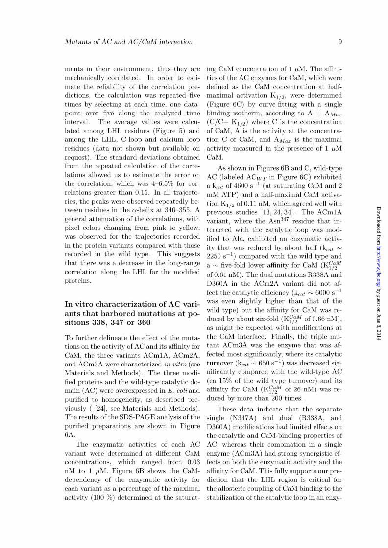
The C-CaM conformations were ex-
by guest on June 8, 2014http://w
ww
.jbc.org/D
ownloaded from

Mutants of AC and AC/CaM interaction 8
tracted from ACm2A T2 and ACm3A T2(Figure 4) at times when different distanceswere observed between Asp133-Oδ1/Oδ2 andthe calcium ion. In both simulations, thesidechain of Asp133 moved apart and atthe end of the trajectory, the calcium ionwas coordinated by the backbone carbonyl,instead of the sidechain. This conforma-tional tendency of Asp133 was increased inACm3A T2 compared with ACm2A T2. In-terestingly, in a steered MD study of the cal-cium dissociation in CaM [31], it was foundthat Asp133 was among the latest residues tolose calcium coordination. The destabiliza-tion of the calcium/Asp133 interaction ob-served in the present study corresponds tothe destabilization of one of the strongestinteractions that defines the calcium coor-dination in EF-hand 4. Thus, this desta-bilization indicates that the AC mutationsdecreased the calcium binding to CaM.
Overall, the perturbations of the EF-hand angles and calcium coordination areprecursory indicators of the decrease in theC-CaM affinity for calcium ions [32,33]. Onlonger time scales, this decrease could leadto the dissociation of calcium ions from C-CaM and to a decrease in the affinity of ACfor C-CaM, as shown previously [14].
Network of interactions involvingLHL along the MD trajectories
The LHL motif, which contains the threemodified residues, lies at the interface be-tween C-CaM and the AC catalytic loop.The hydrogen bonds established by theresidues in the LHL motif were analyzedin the different AC/C-CaM complexes (dataavailable on request). Only long-range hy-drogen bonds (that connected residues sep-arated by more than ten residues in theprotein sequence) were considered in orderto exclude interactions related to local sec-ondary structures. Most of these hydrogenbonds were conserved along the MD tra-jectories, except for one hydrogen bond be-tween Asn35 and Tyr342, which was presentonly in the modified complexes, and two hy-
drogen bonds between Glu346 and Leu357,and between Tyr350 and Glu308, which wereobserved in less than two simulations. Alower number of stable hydrogen bonds thatinvolved modified residues were observedin the ACm3A T1 and ACm3A T2 simula-tions. Indeed, Asn347 established hydrogenbonds with the residues Glu301, Gln302, andAsn304, which are located in the catalyticloop. These hydrogen bonds were disruptedby the change from Asn347 to Ala, except forthe hydrogen bonds that involved the Asn347
backbone hydrogen. Similarly, the hydro-gen bonds between Asp360 and Arg338 dis-appeared when Asp360 or Arg338 were mod-ified to Ala. These observations show thatthe overall structure of the LHL motif is con-served in all systems, and that the LHL con-nection to C-CaM on one side, as well as tothe C-loop on the other side, were signifi-cantly weakened in the presence of the triplemodification.
The hydrogen bonds that involved LHLresidues, as well as the protein fluctua-tions and the correlations in the fluctua-tion, indicated that there was limited vari-ation among the protein variants (data notshown). Thus, the possibility of long-rangecommunication through LHL was investi-gated using a parameter that was poten-tially more sensitive to small variations ingeometry. The geometric strain, as definedby Chiappori et al. [23] (see Materials andMethods), exhibits sufficient sensitivity be-cause it depends on the geometry of the en-vironment of the analyzed residue within asphere of 7 A while it also follows the dis-tance variations with respect to the averagedistance values.
The variations in the geometric strainalong the MD trajectories exhibited corre-lated transitions between the given residues,which were due to simultaneous displace-ments of the environments of these residues.To summarize these variations, the timecorrelation matrices of the geometric strainwere analyzed. The strain correlation peaks(in pink/yellow) correspond to residue pairsthat are undergoing correlated displace-
by guest on June 8, 2014http://w
ww
.jbc.org/D
ownloaded from
Mutants of AC and AC/CaM interaction 9
ments in their environment, thus they aremechanically correlated. In order to esti-mate the reliability of the correlation pre-dictions, the calculation was repeated fivetimes by selecting at each time, one data-point over five along the analyzed timeinterval. The average values were calcu-lated among LHL residues (Figure 5) andamong the LHL, C-loop and calcium loopresidues (data not shown but available onrequest). The standard deviations obtainedfrom the repeated calculation of the corre-lations allowed us to estimate the error onthe correlation, which was 4–6.5% for cor-relations greater than 0.15. In all trajecto-ries, the peaks were observed repeatedly be-tween residues in the α-helix at 346–355. Ageneral attenuation of the correlations, withpixel colors changing from pink to yellow,was observed for the trajectories recordedin the protein variants compared with thoserecorded in the wild type. This suggeststhat there was a decrease in the long-rangecorrelation along the LHL for the modifiedproteins.
In vitro characterization of AC vari-ants that harbored mutations at po-sitions 338, 347 or 360
To further delineate the effect of the muta-tions on the activity of AC and its affinity forCaM, the three variants ACm1A, ACm2A,and ACm3A were characterized in vitro (seeMaterials and Methods). The three modi-fied proteins and the wild-type catalytic do-main (AC) were overexpressed in E. coli andpurified to homogeneity, as described pre-viously ( [24], see Materials and Methods).The results of the SDS-PAGE analysis of thepurified preparations are shown in Figure6A.
The enzymatic activities of each ACvariant were determined at different CaMconcentrations, which ranged from 0.03nM to 1 µM. Figure 6B shows the CaM-dependency of the enzymatic activity foreach variant as a percentage of the maximalactivity (100 %) determined at the saturat-
ing CaM concentration of 1 µM. The affini-ties of the AC enzymes for CaM, which weredefined as the CaM concentration at half-maximal activation K1/2, were determined(Figure 6C) by curve-fitting with a singlebinding isotherm, according to A = AMax
(C/C+ K1/2) where C is the concentrationof CaM, A is the activity at the concentra-tion C of CaM, and AMax is the maximalactivity measured in the presence of 1 µMCaM.
As shown in Figures 6B and C, wild-typeAC (labeled ACWT in Figure 6C) exhibiteda kcat of 4600 s−1 (at saturating CaM and 2mM ATP) and a half-maximal CaM activa-tion K1/2 of 0.11 nM, which agreed well withprevious studies [13, 24, 34]. The ACm1Avariant, where the Asn347 residue that in-teracted with the catalytic loop was mod-ified to Ala, exhibited an enzymatic activ-ity that was reduced by about half (kcat ∼2250 s−1) compared with the wild type anda ∼ five-fold lower affinity for CaM (KCaM
1/2
of 0.61 nM). The dual mutations R338A andD360A in the ACm2A variant did not af-fect the catalytic efficiency (kcat ∼ 6000 s−1
was even slightly higher than that of thewild type) but the affinity for CaM was re-duced by about six-fold (KCaM
1/2 of 0.66 nM),as might be expected with modifications atthe CaM interface. Finally, the triple mu-tant ACm3A was the enzyme that was af-fected most significantly, where its catalyticturnover (kcat ∼ 650 s−1) was decreased sig-nificantly compared with the wild-type AC(ca 15% of the wild type turnover) and itsaffinity for CaM (KCaM
1/2 of 26 nM) was re-duced by more than 200 times.
These data indicate that the separatesingle (N347A) and dual (R338A, andD360A) modifications had limited effects onthe catalytic and CaM-binding properties ofAC, whereas their combination in a singleenzyme (ACm3A) had strong synergistic ef-fects on both the enzymatic activity and theaffinity for CaM. This fully supports our pre-diction that the LHL region is critical forthe allosteric coupling of CaM binding to thestabilization of the catalytic loop in an enzy-
by guest on June 8, 2014http://w
ww
.jbc.org/D
ownloaded from

Mutants of AC and AC/CaM interaction 10
matically active configuration. The two- tofive-fold higher KATP
M levels of all the mod-ified AC enzymes compared with the wild-type AC also demonstrate that the LHL mo-tif is important for maintaining the optimalconformation of the active site.
To verify that the triple modification ofR338A, N347A, and D360A in ACm3A didnot affect the structural integrity of the pro-tein, we performed the following biophysi-cal analyses. First, the proteins were ana-lyzed by SEC followed by triple detector ar-ray (SEC-TDA) to determine whether themutations affected their oligomeric status.SEC-TDA provides the molecular massesof the eluted species by combining staticright angle light scattering with UV ab-sorbance. The results showed that boththe wild-type and ACm3A proteins weremonomeric species in solution (Figure 7A).The secondary structure contents of theCaM and AC proteins, either isolated or incomplexes, were analyzed by SRCD in thefar-UV range. The shapes of the far-UVCD spectra of wild-type AC and ACm3Awere similar (Figure 7B). The addition ofCaM induced similar changes in the over-all secondary structure content of both ACproteins. These results clearly indicate thatthe secondary structure content of ACm3Awas not affected by the mutations. Fi-nally, the thermodynamic stability of bothAC proteins was characterized. The urea-induced denaturation of the AC proteinswas monitored by tryptophan fluorescence.The denaturation profiles (Figure 7C). andthe thermodynamic parameters (Figure 7D)obtained for both proteins were very similar.Overall, these experiments indicate that thethree modifications in ACm3A did not af-fect the structural properties or the overallstability of the protein.
DISCUSSION
Maps of the energetic influences [16, 17]that were calculated [15] previously usingthe AC/C-CaM complex allowed us to iden-tify three residues, i.e., Arg338, Asn347, and
Asp360, that might affect the interaction be-tween AC and C-CaM. In the present study,we characterized three modified proteins,i.e., ACm1A (N347A), ACm2A (R338A,D360A), and ACm3A (R338A, N347A andD360A), using in silico MD simulations andbased on in vitro biochemical and biophysi-cal studies.
Our in vitro studies showed that theaffinity of the triple mutant ACm3A forCaM was greatly reduced whereas thatof the single (ACm1A) or dual mutant(ACm2A) was not affected significantlycompared with that of the wild-type en-zyme. These results indicate that thesethree mutations had a strong synergistic ef-fect on the CaM-binding affinity. A similarsynergistic effect was noted for the catalyticefficiency of the enzyme because the enzy-matic activity of ACm3A was also reducedsignificantly (about 15% of the wild-type en-zyme activity), whereas that of ACm1A wasabout 50% of the wild-type activity. Thelower activity of the ACm1A and ACm3Avariants might be expected because theAsn347 residue interacts directly with thecatalytic loop. Thus, any perturbation ofthe interaction between Asn347 and the cat-alytic loop might directly affect the catalyticefficiency of AC.
The dynamical behaviors of the threemodified proteins in complex with C-CaMwere also analyzed in silico based on twoseries of MD simulations. High variabil-ity in the establishment of hydrogen bondsin the complex was observed in the dif-ferent modified proteins, as well as amongrepeated trajectories. However, this vari-ability could not be assigned to simula-tion artifacts because similar variability wasobserved among the four crystallographicstructures of the complex AC/C-CaM [13].The analysis of the trajectories recordedusing modified and wild-type AC/C-CaMcomplexes, which were conducted in paral-lel with the experimental characterizationof the AC/CaM interaction, identified twodifferent routes that allowed the affinity ofAC for C-CaM to decrease. In the first
by guest on June 8, 2014http://w
ww
.jbc.org/D
ownloaded from

Mutants of AC and AC/CaM interaction 11
simulation of the triple modified protein(ACm3A T1), a destabilization of the cal-cium loop of EF-hand 3 was observed, aswell as a large decrease in the interactionsbetween C-CaM and the CA region. In thesecond trajectory recorded using ACm3A(ACm3A T2), a destabilization of the cal-cium coordination in the EF-hand 4 of C-CaM led to the disruption of the hydrogenbond between the Asp133 sidechain and cal-cium. This could be the first step in the pos-sible dissociation of calcium from C-CaM. Itis known that calcium-free CaM has a loweraffinity for AC [14], thus the destabilizationof calcium coordination would result in a re-duction of the CaM affinity for AC.
Guo et al. [13] described the crys-tallographic structure of the complexAC/C-CaM and characterized one ACmodification: the double modificationE346A/R348A in the C terminal tail. Themodified protein exhibited a reduced enzy-matic activity as well as a reduced affinityfor CaM. The two amino acids changedin the E346A/R348A mutant flanked theAsn347 residue that was modified in thepresent study. The hydrogen bonds that in-volve Glu346 and Arg348 residues varied inthe wild-type AC when calcium ions or C-CaM were removed from the system, as wellas in the trajectories of the modified AC pro-tein, but no systematic variations were de-tected. However, the observations of Guoet al. [13] based on the E346A/R348A ACvariant agree well with our present study,as well as supporting the hypothesis thatthe LHL motif is critical for linking CaMbinding with the stabilization of the AC cat-alytic loop in a configuration that is favor-able for catalysis. Indeed, the LHL mo-tif is in direct contact with C-CaM on oneside via the Arg338 and Asp360 residues,as well as the catalytic loop on the otherside via the Asn347 residue. Thus, it isideally positioned to relay information re-lated to CaM-binding on the distant cat-alytic site of the AC enzyme via a densenetwork of atomic contacts, which primar-ily involve the Arg338, Asn347, and Asp360
residues, as demonstrated in our MD simu-lations. Long-range communications withinthe LHL motif were further suggested by ouranalysis of the geometric strain along theMD trajectories of the different ACs. There-fore, the LHL motif appears to be a crit-ical module for the allosteric activation ofAC by CaM. The analysis performed in thepresent study was more qualitative than themethod proposed recently by Weinkam etal. [35,36], but we should note that the avail-ability of only one structure of AC in com-plex with Ca2+-loaded C-CaM, as well asthe conformational equilibrium that is prob-ably adopted by AC in the unbound state,prevent a more straightforward applicationof this method.
From a more general perspective, the de-tection of the residue networks that are re-sponsible for long-range communication in-side a given protein has been attractingmuch interest during the last decade [37–41].These networks are thought to be involvedin biomolecular interactions [42] with fun-damental roles in many biological processes[43]. Homologous protein sequence align-ments [44–46] or analysis of the covariance ofthe NMR chemical shifts [47] have predictedlong-range protein communication networksthat agree with experimental observations.Some of these analyses have facilitated pro-tein engineering [48]. In the present study,we combined MD and mutagenesis to iden-tify a long-range communication network re-lated to the activation of a key virulencefactor derived from a bacterial pathogen.This investigation may facilitate new meth-ods for interfering with the AC-CaM inter-action, thereby allowing a novel drug discov-ery approach [49].
ACKNOWLEDGMENTS
We acknowledge SOLEIL for providing thesynchrotron radiation facilities (ProposalIDs 20110586 and 20120444) and we thankFrank Wien for assistance with using theDISCO beamline. Edithe Selwa was sup-ported by an allocation doctorale from
by guest on June 8, 2014http://w
ww
.jbc.org/D
ownloaded from

Mutants of AC and AC/CaM interaction 12
MENRT, which was distributed by Uni-versite Pierre et Marie Curie (Paris VI).Ana Cristina Sotomayor Perez was sup-ported by an Institut Pasteur PTR grant(PTR#374). This project was supported bythe Institut Pasteur and the Centre Nationalde la Recherche Scientifique (CNRS UMR
3528, Biologie Structurale et Agents Infec-tieux). Data not shown in the manuscriptmay be obtained from the authors (DanielLadant: [email protected]; ThereseMalliavin: [email protected]) onrequest.
References
[1] Ladant, D. and Ullmann, A. (1999) Bordetella pertussis adenylate cyclase: a toxinwith multiple talents. Trends Microbiol 7, 172–176
[2] Vojtova, J., Kamanova, J., and Sebo, P. (2006) Bordetella adenylate cyclase toxin: aswift saboteur of host defense. Curr Opin Microbiol 9, 69–75
[3] Carbonetti, N. (2010) Pertussis toxin and adenylate cyclase toxin: key virulencefactors of Bordetella pertussis and cell biology tools. Future Microbiol 5, 455–469
[4] Wolff, J., Cook, G., Goldhammer, A., and Berkowitz, S. (1980) Calmodulin activatesprokaryotic adenylate cyclase. Proc Natl Acad Sci USA 77, 3841–3844
[5] Confer, D. and Eaton, J. (1982) Phagocyte impotence caused by an invasive bacterialadenylate cyclase. Science 217, 948–950
[6] Rogel, A., Schultz, J., Brownlie, R., Coote, J., Parton, R., and Hanski, E. (1989)Bordetella pertussis adenylate cyclase: purification and characterization of the toxicform of the enzyme. EMBO J 8, 2755–2760
[7] Goodwin, M. and Weiss, A. (1990) Adenylate cyclase toxin is critical for colonizationand pertussis toxin is critical for lethal infection by Bordetella pertussis in infantmice. Infect Immun 58, 3445–3447
[8] Khelef, N., Zychlinsky, A., and Guiso, N. (1993) Bordetella pertussis induces apoptosisin macrophages: role of adenylate cyclase-hemolysin. Infect Immun 61, 4064–4071
[9] Harvill, E., Cotter, P., Yuk, M., and Miller, J. (1999) Probing the function of Bor-detella bronchiseptica adenylate cyclase toxin by manipulating host immunity. InfectImmun 67, 1493–1500
[10] Hewlett, E., Donato, G., and Gray, M. (2006) Macrophage cytotoxicity produced byadenylate cyclase toxin from Bordetella pertussis: more than just making cyclic AMP!Mol Mic 59, 447–459
[11] Cheung, G., Dickinson, P., Sing, G., Craigon, M., Ghazal, P., Parton, R., and Coote,J. (2008) Transcriptional responses of murine macrophages to the adenylate cyclasetoxin of Bordetella pertussis. Microb Pathog 44, 61–70
[12] Kamanova, J., Kofronova, O., Masin, J., Genth, H., Vojtova, J., Linhartova, I., Be-nada, O., Just, I., and Sebo, P. (2008) Adenylate cyclase toxin subverts phagocytefunction by RhoA inhibition and unproductive ruffling. J Immunol 181, 5587–5597
by guest on June 8, 2014http://w
ww
.jbc.org/D
ownloaded from

Mutants of AC and AC/CaM interaction 13
[13] Guo, Q., Shen, Y., Lee, Y., Gibbs, C., Mrksich, M., and Tang, W. (2005) Structuralbasis for the interaction of Bordetella pertussis adenylyl cyclase toxin with calmodulin.EMBO J. 24, 3190–3201
[14] Shen, Y., Lee, Y., Soelaiman, S., Bergson, P., Lu, D., Chen, A., Beckingham, K.,Grabarek, Z., Mrksich, M., and Tang, W. (2002) Physiological calcium concentrationsregulate calmodulin binding and catalysis of adenylyl cyclase exotoxins. EMBO J 21,6721–6732
[15] Selwa, E., Laine, E., and Malliavin, T. (2012) Differential role of Calmodulin andCalcium ions in the stabilization of the catalytic domain of adenyl cyclase CyaA fromBordetella pertussis. Proteins 80, 1028–1040
[16] Hamacher, K., Trylska, J., and McCammon, J. (2006) Dependency map of proteinsin the small ribosomal subunit. PloS Comput. Biol. 2, e10
[17] Laine, E., Blondel, A., and Malliavin, T. (2009) Dynamics and Energetics: A Con-sensus Analysis of the Impact of Calcium on EF-CaM Protein Complex. BiophysicalJournal 96, 1–15
[18] Case, D., Cheatham, T., Darden, T., Gohlke, H., Luo, R., Merz, K., Onufriev, A.,Simmerling, C., Wang, B., and Woods, R. (2005) The AMBER biomolecular simula-tion programs. J Computat Chem 26, 1668–1688
[19] Hornak, V., Abel, R., Okur, A., Strockbine, B., Roitberg, A., and Simmerling, C.(2006) Comparison of multiple AMBER force fields and development of improvedprotein backbone parameters. Proteins 65, 712–725
[20] Jorgensen, W. (1981) Transferable intermolecular potential functions for water, alco-hols and ethers. application to liquid water. J. Am. Chem. Soc. 103, 335–340
[21] Aqvist, J. (1990) Ion-water interaction potentials derived from free energy perturba-tion simulations. J Phys Chem 94, 8021–8024
[22] Grunberg, R., Nilges, M., and Leckner, J. (2007) Biskit–a software platform for struc-tural bioinformatics. Bioinformatics 23, 769–770
[23] Chiappori, F., Merelli, I., Colombo, G., Milanesi, L., and Morra, G. (2012) Molecularmechanism of allosteric communication in Hsp70 revealed by molecular dynamicssimulations. PLoS Comput Biol 8, e1002844
[24] Vougier, S., Mary, J., Dautin, N., Vinh, J., Friguet, B., and Ladant, D. (2004) Essen-tial role of methionine residues in calmodulin binding to Bordetella pertussis adeny-late cyclase, as probed by selective oxidation and repair by the peptide methioninesulfoxide reductases. J Biol Chem 279, 30210–30218
[25] Gmira, S., Karimova, G., and Ladant, D. (2001) Characterization of recombinantBordetella pertussis adenylate cyclase toxins carrying passenger proteins. Researchin microbiology 152, 889–900
[26] Karimova, G., Ullmann, A., and Ladant, D. (2001) Protein-protein interaction be-tween Bacillus stearothermophilus tyrosyl-tRNA synthetase subdomains revealed bya bacterial two-hybrid system. Journal of molecular microbiology and biotechnology3, 73–82
by guest on June 8, 2014http://w
ww
.jbc.org/D
ownloaded from

Mutants of AC and AC/CaM interaction 14
[27] Laine, E., Goncalves, C., Karst, J., Lesnard, A., Rault, S., Tang, W., Malliavin,T., Ladant, D., and Blondel, A. (2010) Use of allostery to identify inhibitors ofcalmodulin- induced activation of Bacillus anthracis Edema Factor. Proc. Natl. Acad.Sci. U.S.A. 107, 11277–11282
[28] Karst, J., Sotomayor-Perez, A., Guijarro, J., Raynal, B., Chenal, A., and Ladant,D. (2010) Calmodulin-induced conformational and hydrodynamic changes in the cat-alytic domain of Bordetella pertussis adenylate cyclase toxin. Biochemistry 49, 318–328
[29] Sotomayor-Perez, A., Subrini, O., Hessel, A., Ladant, D., and Chenal, A. (2013)Molecular Crowding Stabilizes Both the Intrinsically Disordered Calcium-Free Stateand the Folded Calcium-Bound State of a Repeat in Toxin (RTX) Protein. J AmChem Soc 135, 11929–11934
[30] Chenal, A., Karst, J., Sotomayor-Perez, A., Wozniak, A., Baron, B., England, P.,and Ladant, D. (2010) Calcium-induced folding and stabilization of the intrinsicallydisordered RTX domain of the CyaA toxin. Biophys J 99, 3744–3753
[31] Zhang, Y., Tan, H., Lu, Y., Jia, Z., and Cheng, G. (2008) Ca(2+) dissociation fromthe C-terminal EF-hand pair in calmodulin: a steered molecular dynamics study.FEBS Lett 582, 1355–1361
[32] Zhang, M., Tanaka, T., and Ikura, M. (1995) Calcium-induced conformational transi-tion revealed by the solution structure of apo calmodulin. Nat Struct Biol 2, 758–767
[33] Finn, B., Evenas, J., Drakenberg, T., Waltho, J., Thulin, E., and Forsen, S. (1995)Calcium-induced structural changes and domain autonomy in calmodulin. Nat StructBiol 2, 777–783
[34] Glaser, P., Elmaoglou-Lazaridou, A., Krin, E., Ladant, D., Barzu, O., and Danchin,A. (1989) Identification of residues essential for catalysis and binding of calmodulinin Bordetella pertussis adenylate cyclase by site-directed mutagenesis. EMBO J 8,967–972
[35] Weinkam, P., Pons, J., and Sali, A. (2012) Structure-based model of allostery predictscoupling between distant sites. PNAS 109, 4875–4880
[36] Weinkam, P., Chen, Y., Pons, J., and Sali, A. (2013) Impact of mutations on theallosteric conformational equilibrium. J Mol Biol 425, 647–661
[37] Chennubhotla, C. and Bahar, I. (2007) Signal propagation in proteins and relation toequilibrium fluctuations. PLoS Comput Biol 3, 1716–1726
[38] del Sol, A., Tsai, C., Ma, B., and Nussinov, R. (2009) The origin of allosteric func-tional modulation: multiple pre-existing pathways. Structure 17, 1042–1050
[39] Dubay, K., Bothma, J., and Geissler, P. (2011) Long-range intra-protein communi-cation can be transmitted by correlated side-chain fluctuations alone. PLoS ComputBiol 7, e1002168
[40] Kuzu, G., Keskin, O., Gursoy, A., and Nussinov, R. (2012) Constructing structuralnetworks of signaling pathways on the proteome scale. Curr Opin Struct Biol 22,367–377
by guest on June 8, 2014http://w
ww
.jbc.org/D
ownloaded from

Mutants of AC and AC/CaM interaction 15
[41] Cilia, E., Vuister, G., and Lenaerts, T. (2012) Accurate prediction of the dynamicalchanges within the second PDZ domain of PTP1e. PLoS Comput Biol 8, e1002794
[42] Bakan, A. and Bahar, I. (2009) The intrinsic dynamics of enzymes plays a dominantrole in determining the structural changes induced upon inhibitor binding. Proc NatlAcad Sci USA 106, 14349–14354
[43] Nussinov, R., Ma, B., Tsai, C., and Csermely, P. (2013) Allosteric conformationalbarcodes direct signaling in the cell. Structure 21, 1509–1521
[44] Lockless, S. and Ranganathan, R. (1999) Evolutionarily conserved pathways of ener-getic connectivity in protein families. Science 286, 295–299
[45] Dickson, R., Wahl, L., Fernandes, A., and Gloor, G. (2010) Identifying and seeingbeyond multiple sequence alignment errors using intra-molecular protein covariation.PLoS One 5, e11082
[46] Reynolds, K., McLaughlin, R., and Ranganathan, R. (2011) Hot spots for allostericregulation on protein surfaces. Cell 147, 1564–1575
[47] Selvaratnam, R., Chowdhury, S., VanSchouwen, B., and Melacini, G. (2011) Mappingallostery through the covariance analysis of NMR chemical shifts. Proc Natl Acad SciU S A 108, 6133–6138
[48] Lee, J., Natarajan, M., Nashine, V., Socolich, M., Vo, T., Russ, W., Benkovic, S.,and Ranganathan, R. (2008) Surface sites for engineering allosteric control in proteins.Science 322, 438–442
[49] Seifert, R. and Dove, S. (2012) Towards selective inhibitors of adenylyl cyclase toxinfrom Bordetella pertussis. Trends Microbiol 20, 343–351
[50] Eswar, N., Eramian, D., Webb, B., Shen, M., and Sali, A. (2008) Protein structuremodeling with MODELLER. Methods Mol Biol 426, 145–159
by guest on June 8, 2014http://w
ww
.jbc.org/D
ownloaded from

Mutants of AC and AC/CaM interaction 16
FIGURE LEGENDS
Figure 1a) X-ray crystallographic structure of the catalytic domain (AC) of adenyl cyclase
(CyaA, PDB entry 1YRT) drawn in cartoons. Helices F, G, and H colored in purple formthe SA region (residues 192–254). The loop 226–232 (Hom loop) colored in salmon atthe left extremity of SA colored in purple was missing from the X-ray crystallographicstructure and was modeled using Modeller9v4 [50]. The other protein regions are coloredin green (CA: residues 7–61, 187–197, 261–299, and 313–345), orange (CB: residues 62–186), cyan (C-terminal tail/C tail: residues 346–364), and yellow (catalytic loop/C loop:residues 300–312). The C-CaM lobe, which is colored in red, interacts with SA and theC-terminal tail. Calcium ions are represented by silver beads. b) The modified residues,i.e., Arg338, Asn347, and Asp360, located in CA and the C terminal tail, are indicated bysticks. c) The LHL motif localized between the C-CaM/AC interface and the catalyticloop is colored in brown.
Figure 2Conformational drifts estimated based on the α carbon coordinates RMSD from the
starting conformations in the T1 (a-d) and T2 (e-h) series of simulations. (a, e) Drift cal-culated based on the Cα carbons in the AC domain for ACm1A (black), ACm2A (green),and ACm3A (red). (b–d,f–h) Drift calculated based on different regions of the AC domain,which were analyzed for ACm1A (b, f), ACm2A (c, g), and ACm3A (d, h). The colorcodes of the different protein regions are shown in panel (d). The AC regions are definedas follows. 1) CA: residues 7–61, 187–197, 261–299, and 313–345. 2) Catalytic loop (CLoop): residues 300–312. 3) C-terminal tail (C tail): residues 346–364. 4) CB: residues62–186. 5) SA: residues 198–260. Helices H, F, G, and H’ correspond to residues 234–253,198-210, 214-223, 256-260.
Figure 3Fluctuations of the residues (RMSF: A) observed in C-CaM based on the trajectories
recorded for: (a) wild-type and (b) modified C-CaM/AC complexes. The trajectory namesare shown in the legends. The WT2Ca trajectories were run on two Ca2+-loaded AC/C-CaM complexes, whereas the WT0Ca trajectories were run on the AC/C-CaM complexin the absence of calcium ions. The fluctuations were calculated based on the trajectoriesafter removing their first 10 ns.
Figure 4C-CaM conformation extracted from ACm2A T2 and ACm3A T2, which are colored
according to the simulation times indicated. The N- and C-terminal ends are indicated,as well as Asp-133, which is shown in black. The calcium ions are shown as gray spheres,and the carboxyl oxygens of the Asp-133 sidechain are colored in red.
Figure 5Correlation between geometric strains among the LHL residues, displayed for each
recorded trajectory. The time-correlation matrices were calculated between the variationsof the geometric strains of LHL residues and the first ten nanoseconds were discarded fromthe analysis. In order to estimate the reliability of the prediction of correlation, the cal-culation was repeated five times by selecting at each time, one data-point over five alongthe analyzed time interval. The average values calculated for the LHL residues are shown
by guest on June 8, 2014http://w
ww
.jbc.org/D
ownloaded from

Mutants of AC and AC/CaM interaction 17
in the figure. The intensity scale varies from 1.0 (pink) to –0.2 (dark green).
Figure 6A. SDS-PAGE analysis of the purified AC variants, wild type AC, and CaM. The pro-
teins were separated by electrophoresis on a 4–12% SDS-polyacrylamide gel (Life Tech-nologies). After migration, the gel was stained with PageBlue protein staining solution(Thermo Fisher Scientific). B. Activation of modified ACs by CaM. The activity of thepurified wild-type AC and/or the variants were measured (as described in the Materialsand Methods) in the presence of the indicated CaM concentrations, and the results areexpressed as the percentage of the maximal activity (measured in the presence of 1µMCaM). C. Catalytic properties of AC variants. The parameters were determined withinan error of ± 10%. ∆G is the calculated energy of association between CaM and AC.
Figure 7A. SEC-TDA analysis of the oligomerization state of ACwt and ACm3A. Red curve:
right angle light scattering; blue curve: UV absorbance; green curve: molecular masses ofthe eluted species. See the Materials and Methods for details. B. Synchrotron radiationcircular dichroism spectra of CaM and AC proteins in isolation or in a 1:1 complex. MRE:mean residual ellipticity. See the Materials and Methods for details. C. Thermodynamicstability of AC proteins following urea-induced denaturation. The ratio of the fluorescenceintensity at 360 and 320 nm (excitation wavelength = 295 nm) for the proteins as a func-tion of the urea concentration was measured as described in the Materials and Methods.D. Thermodynamics parameters of the urea-induced denaturation of AC proteins.
by guest on June 8, 2014http://w
ww
.jbc.org/D
ownloaded from

Mutants of AC and AC/CaM interaction 18
Table I Details of the molecular systems simulated in molecular dynamics(MD) trajectories.
Systems ACwt 2Ca ACwt 0CaTrajectories WT2Ca T1 WT0Ca T1
WT2Ca T2 WT0Ca T2Number of Na+
counter-ions 14 18Water box 92.6 x 106.9 91.3 x 106.9dimensions x 103.3 A3 x 106.4 A3
Number ofwater molecules 26455 27056Total number
of atoms 85825 87630Length (ns) 30 30
Table I
Systems ACm1A ACm2A ACm3ATrajectories ACm1A T1 ACm2A T1 ACm3A T1
ACm1A T2 ACm2A T2 ACm3A T2Number of Na+
counter-ions 14 18 14Water box 109.6 x 82.8 108.1 x 82.8 109.6 x 82.8dimensions x 111.7 A3 x 111.7 A3 x 111.7 A3
Number ofwater molecules 29003 29008 29008Total number
of atoms 93465 93468 93464Length (ns) 50 50 50
by guest on June 8, 2014http://w
ww
.jbc.org/D
ownloaded from

Mutants of AC and AC/CaM interaction 19
Figure 1
a) C-CaM
C-tail
C-Loop
CA
CB
SAHomloop
GF
H
b) c)
L16ASN-347
ARG-338
ASP-360
Figure 1
by guest on June 8, 2014http://w
ww
.jbc.org/D
ownloaded from

Mutants of AC and AC/CaM interaction 20
Figure 2
Figure 2
by guest on June 8, 2014http://w
ww
.jbc.org/D
ownloaded from

Mutants of AC and AC/CaM interaction 21
Figure 3R
MS
F (A
° )
79 84 89 94 99 105 111 117 123 129 135 141 147
01
23 a)
Helix V Helix VI Helix VII Helix VIII
WT2Ca_T1WT2Ca_T2
WT0Ca_T1WT0Ca_T2
RM
SF
(A° )
79 84 89 94 99 104 110 116 122 128 134 140 146
01
23
b)
Helix V Helix VI Helix VII Helix VIII
ACm1A_T1ACm1A_T2
ACm2A_T1ACm2A_T2
ACm3A_T1ACm3A_T2
Residue number
RM
SF
(A° )
RM
SF
(A° )
Figure 3
by guest on June 8, 2014http://w
ww
.jbc.org/D
ownloaded from
Mutants of AC and AC/CaM interaction 22
Figure 4
ACm2A_T2
Asp 133 COO-
C N ACm3A_T2 CN
Asp 133 COO-
10 ns
20 ns
25 ns
start
Figure 4
by guest on June 8, 2014http://w
ww
.jbc.org/D
ownloaded from

Mutants of AC and AC/CaM interaction 23
Figure 5
Res
idue
num
ber
345
350
355
360
ACm1A_T1 ACm1A_T2R
esid
ue n
umbe
r
345
350
355
360
ACm2A_T1 ACm2A_T2
Res
idue
num
ber
345
350
355
360
ACm3A_T1 ACm3A_T2
Res
idue
num
ber
345
350
355
360
WT2Ca_T1 WT2Ca_T2
Residue number
Res
idue
num
ber
340 345 350 355 360
345
350
355
360
WT0Ca_T1
Residue numberResidue number
340 345 350 355 360
WT0Ca_T2
Residue number
−0.2 0.0 0.2 0.4 0.6 0.8 1.0 1.2
Res
idue
num
ber
Res
idue
num
ber
Res
idue
num
ber
Res
idue
num
ber
Res
idue
num
ber
Figure 5
by guest on June 8, 2014http://w
ww
.jbc.org/D
ownloaded from

Mutants of AC and AC/CaM interaction 24
Figure 6
A
170 -130 -
95 -72 -
55 -43 -
34 -
26 -
17 -
10 -
AC ACm3A
ACm2A
ACm1A
CaM B
C kcat KATPM KCaM
1/2 ∆G kcat/ KM
(s−1) (mM) (nM) (kcal.mol−1) 106 M−1s−1
ACwt 4600 0.60 0.11 13.8 7.6ACm1A 2250 2.2 0.61 12.8 1.0ACm2A 6000 1.5 0.66 12.7 4.0ACm3A 650 3.0 26.7 10.5 0.2
Figure 6
by guest on June 8, 2014http://w
ww
.jbc.org/D
ownloaded from

Mutants of AC and AC/CaM interaction 25
Figure 7
050
100
150 AC
A
uu
mm
[247
2:31
01]
010
2030
4050
050
100
150
uv
ACm3A
uu
mm
[247
2:31
01]
10 11 12 13 14 15 16 17 180
1020
3040
50
UV wavelength
Sig
nal i
nten
sity
(A
U)
Sig
nal i
nten
sity
(A
U)
Mol
ecul
ar m
ass
(kD
a)M
olec
ular
mas
s (k
Da)
−20
−10
010
2030
180 200 220 240
B
wavelength (nm)
MR
E,K
deg*
cm2/
dmol
ACm3AACm3A:CaMACWTACWT:CaMCaM
● ●
●●
●●
● ● ● ● ● ● ●
0.6
0.8
1.0
1.2
1.4
1.6
1.8
2.0
● ●●
●● ● ●
● ●●
●
●
●
● ●●
●
●
●● ● ● ● ● ● ●
● ● ●●
●
●●
●●
●
●
●●
0 0.5 1 1.5 2 2.5 3 3.5 4 5 6 7 8
C
urea (M)
ratio
of f
luor
esce
nce
inte
nsity
360
/320
ACm3AACm3A:CaMACWTACWT:CaM
D
proteins AC WT ACm3A[urea]1/2
(M) 1.8 1.7∆G,
kcal.mol−1 2.4 2.6m
parameter 1335 1490
Figure 7
by guest on June 8, 2014http://w
ww
.jbc.org/D
ownloaded from