it's all in your head: new insights into craniofacial
TRANSCRIPT
J. Anat. (2005) 207, pp461–477
© 2005 The Authors Journal compilation © 2005 Anatomical Society of Great Britain and Ireland
Blackwell Publishing, Ltd.REVIEW
It’s all in your head: new insights into craniofacial development and deformationMinal D. Tapadia,1 Dwight R. Cordero2 and Jill A. Helms1
1Department of Plastic and Reconstructive Surgery, Stanford University, Stanford, California, USA 2Department of Obstetrics and Gynecology, Brigham and Women‘s Hospital, Harvard Medical School, Boston, Massachusetts, USA
Introduction
‘Our ignorance of the laws of variation is profound. Not
in one case out of a hundred can we pretend to assign
any reason why this or that part has varied. But when-
ever we have the means of instituting a comparison,
the same laws appear to have acted in producing the
lesser differences between varieties of the same
species, and the greater differences between species
of the same genus.’ (Darwin, 1859).
Charles Darwin did not gild the truth when he candidly
pointed out just how imperfect is our knowledge of
how morphological diversity is generated. Over the
intervening decades we have scrutinized the process
of embryonic development and, in doing so, gained a
deeper appreciation for the tissue interactions that lie
at the basis of morphogenesis. More recently, some of
the molecules involved in mediating these tissue inter-
actions have been identified and, in a few rare cases,
we even have gleaned insights into how species-
specific development is controlled. These instances, how-
ever, are few and far between and much remains to be
accomplished.
In this review we will present some of the most
recent advances that lie at the heart of understanding
the mechanisms controlling normal craniofacial devel-
opment, the consequences of when these normal path-
ways are disrupted and how this information sheds
light on the basis for evolutionary diversity among the
species. We choose to focus on morphogenesis of the
craniofacial complex for two reasons. First, faces show
tremendous phenotypic variation, first evident during
the later stages of fetal development. Yet despite these
differences in the facial appearance of embryos, they
all look remarkably similar during earlier stages of
embryogenesis. This suggests that whatever factors
influence craniofacial diversity primarily act during a
discrete period of embryonic development; and know-
ing when diversity first arises is an important first step
towards understanding how diversity is generated.
The second reason we use craniofacial development
as a model is that variations in the craniofacial complex
are tightly associated with adaptive radiations into
ecological niches. If the relationship between craniofa-
cial morphology and speciation is causal (and not
merely correlative) then perhaps we can understand
how modifying the spatial and temporal patterns of
gene expression create diversity within a species.
One final justification for using craniofacial mor-
phology as a model system is the prodigious amount
of documentation that has accrued on head and neck
anomalies. As the noted embryologist William Harvey
so eloquently wrote, ‘Nature is nowhere more accus-
tomed openly to display her secret mysteries than in
cases where she shows traces of her working apart
from the beaten path; nor is there any better way to
advance the proper practice of medicine than to give
our minds to the discovery of the usual law of Nature
by careful investigation of cases of rarer forms of
disease’ (Harvey, 1657). Thus, thoughtful inspection
of the malformed face may offer valuable clues into
the mechanisms governing normal craniofacial
morphogenesis.
Components of the craniofacial complex
In its postnatal form the vertebrate head has an intri-
cate and highly varied morphology, but during early
stages of embryonic development it exhibits a much
more simple geometry (Fig. 1). There are seven promi-
nences that comprise the vertebrate face: the midline
Correspondence
Jill A. Helms, Stanford University, 257 Campus Drive, Stanford, CA 94305, USA. T.: +1 650 736 0919; F: +1 650 736 4374; E: [email protected]
Accepted for publication 2 September 2005

Craniofacial development and deformation, M. D. Tapadia et al.462
© 2005 The AuthorsJournal compilation © 2005 Anatomical Society of Great Britain and Ireland
frontonasal prominence, and three paired structures,
the lateral nasal, maxillary and mandibular promi-
nences (Fig. 1D–F). These maxillary and mandibular
prominences are derived from the first pharyngeal
(branchial) arch, whereas the frontonasal prominence
is derived from a midline primordium that forms on top
of the forebrain. The frontonasal prominence contri-
butes to the forehead, middle of the nose, philtrum of
the upper lip and primary palate (Fig. 1). The lateral
nasal prominence forms the sides (ala) of the nose; the
maxillary prominences contribute to the sides of the
face and lips, and the secondary palate; and the man-
dibular prominences produce the lower jaw (Fig. 1).
Disruptions in the rate, the timing or the extent of
outgrowth of any of these prominences will adversely
affect the fusion process. Consequently, one can appre-
ciate the wide variety of facial clefts that occur (Tessier,
1976) and also why facial clefting is the most frequently
occurring head and neck birth defect (Perrotin et al.
2001).
Craniofacial patterning and the neural crest
Each of the pharyngeal arches, and the frontonasal
prominence, is composed of mesenchyme surrounded
by epithelia. In the case of the frontonasal prominence,
the mesenchyme is derived from the cranial neural
crest, and the epithelia encasing the prominence
include the neuroectoderm of the forebrain, and facial
(surface) ectoderm (Fig. 2E,F). In the maxillary and
mandibular prominences, the encasing epithelia are
derived from facial ectoderm and pharyngeal endo-
Fig. 1 Development of the craniofacial primordia. (A–D) Representations of frontal views of mouse embryos showing the prominences that give rise to the main structures of the face. The frontonasal (or median nasal) prominence (pink) gives rise to the forehead (A), the middle of the nose (B), the philtrum of the upper lip (C) and the primary palate (D), whereas the lateral nasal prominence (blue) forms the sides of the nose (B,D). The maxillomandibular prominences (green) give rise to the lower jaw (specifically from the mandibular prominences), to the sides of the middle and lower face, to the lateral borders of the lips, and to the secondary palate (from the maxillary prominences). (E) Frontal view of a chick embryo, also showing which prominences give rise to different facial structures. (F) Frontal view of a human child, with different facial structures colour-coded to indicate the prominences from which each structure developed.

Craniofacial development and deformation, M. D. Tapadia et al. 463
© 2005 The Authors Journal compilation © 2005 Anatomical Society of Great Britain and Ireland
derm, whereas the mesenchyme is derived from both
cranial neural crest and mesoderm.
The cranial neural crest cells populating each arch
arise from distinct anterioposterior positions along the
neural axis (Kontges & Lumsden, 1996), and once resi-
dent in the arches, proliferate in a highly regimented
fashion. In some cases, the instructions for this prolifer-
ative activity are inherent within the neural crest cells
themselves; in other cases, the surrounding epithelia
provide the directives. For example, neural crest cells in
which Homeobox transcription factor (Hox) genes such
as Hoxa2 are expressed are constrained, to a certain
degree, in their ability to respond to local cues from
overlying or underlying epithelia (reviewed in Le
Fig. 2 Neural crest induction and migration in the developing embryo. (A) The neural plate consists of a unified layer of ectoderm, beneath which lies the endoderm. The neural folds arise as the ectoderm begins to fold upwards. Interactions between signalling molecules cause the medial portion of ectoderm to begin to assume a neural character (green) while lateral portions of ectoderm begin to take on a non-neural character (blue). The prechordal plate mesendoderm (pcp) and the buccopharyngeal membrane (bpm) are indicated. (B) As the neural folds begin to fuse, the neural tube takes shape, giving rise to distinct tissue layers of neuroectoderm (green) and surface ectoderm (blue). Neural crest cells start to delaminate from the border region between the neuroectoderm and surface ectoderm. (C) Once the neural tube has closed, neural crest cells lie interposed between the facial (surface) ectoderm (fe) and the neuroectoderm (ne). (D–F) As the central nervous system begins to form from the neural tube, the neural crest starts to migrate anteriorly from rhombomeres (r1–r3) into different areas of the face, and into the pharyngeal arches. Abbreviations: C, caudal; is, isthmus; mes, mesencephalon; mn, mandible; PA, pharyngeal arch; pe, pharyngeal endoderm; rp, Rathke’s pouch; R, rostral; tel ne, telencephalic neuroectoderm.

Craniofacial development and deformation, M. D. Tapadia et al.464
© 2005 The AuthorsJournal compilation © 2005 Anatomical Society of Great Britain and Ireland
Douarin et al. 2004). Cells devoid of Hox gene expres-
sion appear much more responsive to signals emanat-
ing from the epithelial environment, even into very
late stages of morphogenesis (reviewed in Helms et al.
2005). Some of these epithelial cues have been identi-
fied and in the next sections we will review these
molecular pathways and what is known about their
roles in craniofacial morphogenesis.
Not all neural crest cells play by the same rules
The presence or absence of a particular Hox code in a
neural crest cell determines the extent to which neural
crest cells are able to respond to cues from the sur-
rounding milieu (reviewed in Trainor & Krumlauf,
2000; Le Douarin et al. 2004). Other neural crest cells,
such as those destined for the frontonasal prominence,
do not express Hox genes, and therefore our labora-
tory set out to determine the extent to which these
cells were restricted in their developmental potential;
or, phrased another way, the extent to which these
cells exhibited plasticity and thus were able to respond
to local cues from the facial ectoderm (Schneider &
Helms, 2003).
To test whether neural crest cells inherently possess
directions for facial patterning, we began by exchang-
ing neural crest cells between quail and duck embryos
(Schneider & Helms, 2003) (Fig. 3). We specifically
targeted neural crest cells destined for the upper beak
for two reasons. First, the transplanted neural crest cells
were Hox negative and therefore we would be assess-
ing the plasticity vs. pre-patterned status of neural
crest cells separate from the function of Hoxa2. Second,
the short, narrow quail beak and the long, broad duck
bill meant that we could use differences in the mor-
phology of duck and quail faces (Fig. 3A) as a readout
of whether neural crest cells contained patterning
information.
After neural crest cells were exchanged between
duck and quail embryos the chimeras were allowed to
develop to the stage where morphology of their beaks
was evident (Schneider & Helms, 2003). Duck embryos
that received grafts of quail frontonasal neural crest
cells exhibited short quail-like beaks (‘qucks’) (Fig. 3B)
whereas quail embryos that had received transplants
of duck neural crest cells had duck-like bills (‘duails’)
(Schneider & Helms, 2003) (Fig. 3C). These findings
suggested that neural crest cells direct their own
morphogenesis according to instructions inherent in
the donor population (Schneider & Helms, 2003). To
understand the molecular basis for these morphologi-
cal transformations, we performed the same types of
grafting experiments and then used molecular and cel-
lular analyses to determine how the grafted cells acted
in their new environment and, in turn, how the envi-
ronment influenced the behaviour of the transplanted
neural crest cells. We found that transplanted neural
crest cells maintained the temporal gene expression
patterns of their original environment despite being
transplanted into a new site. In addition, the trans-
planted neural crest cells altered the temporal pattern
of gene expression to reflect the donor, and not the
host, environment (Schneider & Helms, 2003).
The same conclusion was reached by Abigail Tucker
and Andrew Lumsden, who independently performed
similar types of interspecies transplants (Tucker &
Lumsden, 2004). They, too, found that the capacity to
form species-specific skeletal elements in the head was
an inherent property of the neural crest, and con-
cluded that this characteristic is articulated is response
to signals from epithelia (Tucker & Lumsden, 2004). In
fact, other new experiments directly show that most
neural crest cells acquire at least some patterning infor-
mation from nearby epithelia (reviewed in Helms et al.
2005).
The extent to which facial features were transformed
was directly proportional to the number of trans-
planted neural crest cells that made their way into the
chimeric tissue. In other words, the transformation was
a ‘population-dependent’ effect, quite analogous to
previous transplantation studies (reviewed in Helms
et al. 2005). So it seems that when the contingency is
large enough, neural crest cells follow molecular cues
that are generated and maintained by the assemblage
itself, and disregard signals emanating from the local
environment. Just what these population-dependent
cues are, and how many cells are required to maintain
them, is completely unknown.
The nature of prespecification
Hox genes are probable mediators of the population-
based behaviour exhibited by neural crest cells (Capec-
chi, 1997; Barrow & Capecchi, 1999). In the craniofacial
region, Hox genes are expressed by neural crest before
and after migration to the arches (Hunt et al. 1991b,c;
Wilkinson, 1993; Favier & Dolle, 1997; Couly et al. 1998;
Rijli et al. 1998; Trainor & Krumlauf, 2000, 2001;

Craniofacial development and deformation, M. D. Tapadia et al. 465
© 2005 The Authors Journal compilation © 2005 Anatomical Society of Great Britain and Ireland
Trainor, 2003). These domains are preserved even after
neural crest cells take up residence in the branchial
arches (Hunt et al. 1991a,b).
After migration, Hoxa2-positive neural crest cells
occupy the second and more posterior arch mesen-
chyme, which gives rise to the hyoid bone and other
structures (Creuzet et al. 2002) (Fig. 4A). Hoxa2 is
not expressed in crest cells of the first arch in most
vertebrates (Creuzet et al. 2002) (Fig. 4A). This distinct
expression boundary of Hoxa2 has led to speculation
Fig. 3 Transplantation experiments provide evidence that the neural crest inherently contains species-specific patterning information. (A) Quail and duck embryos exhibit distinct anatomical features. For example, quails exhibit a shorter, narrower beak compared with the longer, broader duck bill. (B) When quail neural crest cells from forebrain (fb), midbrain (mb) and rhombomeres 1 and 2 (r1, r2) are transplanted into a duck host, a quail-like beak develops in lieu of a duck’s bill. (C) When duck neural crest cells are transplanted into a quail host, the quail develops a duck-like bill. (Figure reproduced courtesy of Nature.)

Craniofacial development and deformation, M. D. Tapadia et al.466
© 2005 The AuthorsJournal compilation © 2005 Anatomical Society of Great Britain and Ireland
that neural crest cells may be prespecified by virtue of
the expression of this Hox gene.
Gain- and loss-of-function studies bolster this hypo-
thesis. For example, loss of Hoxa2 from the second arch
allowed the second arch to take on first arch character,
eventually resulting in duplication of maxillary and
mandibular structures (Gendron-Maguire et al. 1993;
Rijli et al. 1993) (Fig. 4B). By contrast, over-expression
of Hoxa2 in all tissues of the first arch caused the
first arch to take on a second arch character (Gram-
matopoulos et al. 2000; Pasqualetti et al. 2000) (Fig. 4C).
Additionally, if Hoxa2 is transfected into the rostral
domain of the cephalic neural crest, these cells lose
their ability to differentiate into skeletal structures
(Creuzet et al. 2002). Transplantation and ablation
experiments lend further confirmation that Hoxa2
expression confers upon neural crest cells an anterio-
posterior identity (Couly et al. 1998; Ruhin et al. 2003).
Craniofacial epithelia as sources of instructive signals
Given that the Hox transplantation and ablation experi-
ments demonstrated that some neural crest cells are
capable of responding to local cues from the surround-
ing environment, the question then becomes which
tissues in the environment are ‘talking’ to the neural
crest. Once the neural crest delaminates from the
surface ectoderm during neurulation (Fig. 2B), it lies
sandwiched between several epithelia: the surface
ectoderm, the neuroectoderm and the pharyngeal
endoderm (Fig. 2C–F). Its close contact with these
epithelia during development (Fig. 2D–F) allows these
tissues to provide instructive signals that help to pattern
the neural crest. Recent studies have shed light on the
role of the surface ectoderm in initiating outgrowth of
the frontonasal prominence, and studies on pharyngeal
ectoderm have revealed the importance of endoderm
in patterning of the pharyngeal arches. The neuroecto-
derm also has a significant influence on patterning the
neural crest into the craniofacial skeleton, as blocking
molecular signals from the neuroectoderm leads to
craniofacial syndromes such as holoprosencephaly (HPE).
Surface ectoderm as a source of craniofacial patterning
information
Are neural crest cell fates dictated by patterning infor-
mation inherent in this population of cells, or do neural
Fig. 4 Manipulation of Hox expression. (A) In jawed animals, Hoxa2 is expressed up to pharyngeal arch 2 (PA2). (B) Loss of Hoxa2 expression in pharyngeal arch 1 (PA1) gives neural crest cells in PA1 increased plasticity, and they undergo transformation to a first arch fate. (C) When Hoxa2 is ectopically expressed in PA1, neural crest cells in this arch take on second arch fates, giving rise to a duplication of the hyoid arch.

Craniofacial development and deformation, M. D. Tapadia et al. 467
© 2005 The Authors Journal compilation © 2005 Anatomical Society of Great Britain and Ireland
crest cells respond to signals from their local environ-
ment? Studies from our laboratory also suggest that in
the frontonasal prominence, the surface ectoderm
provides instructive signals that influence neural crest
cell fate, even late in development. We identified a
region of facial ectoderm that was delineated by the
gene expression boundaries of Fibroblast Growth
Factor 8 (Fgf8) and Sonic Hedgehog (Shh) (Fig. 5). The
junction of the dorsal, Fgf8-positive domain and the
ventral, Shh-positive domain coincided with the tip
of the upper beak, such that the dorsal (top) surface
was derived from Fgf8-expressing epithelium and
the ventral (inside) surface arose from Shh-expressing
epithelium (Hu et al. 2003). We termed this junction
Fig. 5 Transplantation experiments reveal that the facial ectodermal zone (FEZ) is crucial for patterning of the frontonasal primordia. (A) In situ hybridization on a sagittal section of a stage-20 chick embryo; red corresponds to an Shh-expressing domain and green corresponds to an Fgf8-expressing domain. (B,D) Representations of similar lateral sections of donor stage-20 (B) and host stage-25 (D) chick embryos, respectively. (C,E) Trichrome-stained sections of stage-36 control and transplanted embryos, respectively. When the FEZ is transplanted (yellow arrows) from a stage-20 chick donor (B) to a stage-25 chick host (D), an ectopic beak forms by stage 36 (E, black arrowhead). Donor FEZ tissue is indicated by darker red and green colouring, while host FEZ is indicated by lighter red and green colouring.

Craniofacial development and deformation, M. D. Tapadia et al.468
© 2005 The AuthorsJournal compilation © 2005 Anatomical Society of Great Britain and Ireland
between Fgf8 and Shh the frontonasal ectodermal
zone (FEZ).
Not only does the FEZ demarcate the initial site of
frontonasal outgrowth, it also is responsible for setting
up the dorsoventral axis of the upper beak. We showed
this by ectopically transplanting the FEZ to a more
dorsal position of the frontonasal prominence (Fig. 5B–E).
In this new site, the FEZ induced a molecular cascade
that re-programmed the fate of neural crest cells at
the transplant site (Hu et al. 2003). The net result was
duplications of upper beak structures (Hu et al. 2003)
(Fig. 5D–E). We found that transplantation of the FEZ
to the mandible also induced the duplication of lower
beak structures, indicating that the neural crest cells in
both locations remained highly responsive to signals
emanating from the facial ectoderm (Hu et al. 2003).
The dorsoventral polarity of the upper beak is also
controlled by the FEZ. When the FEZ graft was inverted,
the polarity of the ectopic beak structures was also inverted
(Hu et al. 2003). Clearly, some neural crest cell fates can
be modulated by external cues from surrounding tissues.
When the FEZ graft was transplanted to the second
(Hoxa2-expressing) arch, however, the transplant failed
to induce a molecular cascade as we had observed
earlier, nor was it able to re-pattern the neural crest
cells to duplicate any skeletal structures (Hu et al. 2003).
Seemingly, these Hoxa2-positive cells disregarded the
patterning signals being sent by the transplanted FEZ
(Hu et al. 2003). This location-dependent response
suggested that the plasticity of a neural crest cell, and
its ability to respond to environmental signals, is context
dependent (Hu et al. 2003). By extension, these data
suggest that removing the influence of Hox genes con-
ferred greater plasticity upon neural crest cells.
Pharyngeal endoderm as a source of craniofacial
patterning information
Some of the first experimental evidence demonstrating
that the pharyngeal endoderm is a source of pattern-
ing information came from a study that was originally
designed to test the role of neural crest cells on pat-
terning of the pharyngeal skeleton. In this study, the
neural tube was ablated prior to neural crest migration
(Veitch et al. 1999). Despite the absence of neural crest
cells post-ablation, the pharyngeal arches were pro-
perly organized (Veitch et al. 1999). These data indi-
cated that neither the formation nor the patterning of
the pharyngeal arches was absolutely dependent upon
neural crest cells. The most likely candidate tissue that
controlled patterning in this region of the craniofacial
complex was the pharyngeal endoderm.
From an evolutionary point of view, pharyngeal
‘perforations’, the predecessors of the pharyngeal clefts,
preceded the development of the neural crest as a cell
population (Gans & Northcutt, 1983; Northcutt & Gans,
1983). This type of correlation has been interpreted as
indicating that the pharyngeal endoderm was proba-
bly the initial source of patterning information. Studies
from zebrafish and quail/chick chimeras now offer
direct experimental evidence that the pharyngeal
endoderm can influence that pattern of the lower face.
In zebrafish, the van gogh (vgo) mutant shows an
absence of pharyngeal segmentation and a failure of
the surrounding mesoderm to pattern correctly (Pio-
trowski & Nusslein-Volhard, 2000). Although hindbrain
segmentation proceeds normally, the vgo pharyngeal
clefts do not form. Consequently, neural crest cells exit-
ing from the rhombencephalon fuse in the ventral
surface because of the lack of pharyngeal pouches; the
end result is a lack of skeletal elements in the pharyn-
geal region (Piotrowski & Nusslein-Volhard, 2000). This
phenotype suggests that the segmentation and differ-
entiation of crest cells to form the pharyneal skeleton
is primarily determined by endodermal signalling.
Using the quail/chick chimeric system, Couly, Le
Douarin and their colleagues showed that if regions of
pharyngeal endoderm were removed then correspond-
ing regions of the facial skeleton were affrected (Couly
et al. 2002). For example, removing one strip of pha-
ryngeal endoderm resulted in the reduction or absence
of the nasal capsule and upper beak; when another
strip of tissue was removed then Meckel’s cartilage was
affected, and when a third strip of tissue was removed
then the articular, quadrate and proximal portions of
Meckel’s cartilage were perturbed (Couly et al. 2002).
Transplantation of quail endodermal strips of tissue to
an ectopic location resulted in supernumerary lower
jaws that were malpositioned immediately above the
host jaw, and as we had found for facial ectoderm,
the rostrocaudal inversion of these grafts resulted in the
ectopic lower jaw developing in an inverted position
(Couly et al. 2002). Taken together, these data demon-
strate that patterning and orientation of the pharyn-
geal arch skeleton is dependent upon the endoderm. In
turn, the pharyngeal endoderm is able to instruct the
Hox-expressing neural crest as to the size, morphology
and orientation of the pharyngeal skeletal elements.

Craniofacial development and deformation, M. D. Tapadia et al. 469
© 2005 The Authors Journal compilation © 2005 Anatomical Society of Great Britain and Ireland
At least some of the patterning information in the
pharyngeal endoderm is mediated via Fgf signaling. In
the zebrafish acerebellar (ace) mutant the loss of Fgf8
results in deformed pharyngeal pouches and the reduc-
tion of the hyoid cartilage (Reifers et al. 1998; Draper
et al. 2001; Roehl & Nusslein-Volhard, 2001). More
recently, Chuck Kimmel and colleagues showed that
disrupting Fg8 signalling results in a complete failure
of pouch formation (Crump et al. 2004). Although
the pharyngeal endoderm was present in their Fgf
mutants, the lateral migration of the endoderm
was disorganized and consequently the hyoid and
branchial cartilages were truncated, similar to the
ace phenotype (Crump et al. 2004). In mice, Fgf8
compound heterozygous (Fgf8neo/–) mutant embryos
exhibit hypoplasia of the first and second pharyngeal
arches and their associated clefts (Abu-Issa et al. 2002),
and although neural crest cells migrate appropriately
into the arches, once they arrive they undergo prema-
ture programmed cell death (Abu-Issa et al. 2002).
Fgfs are not the only molecular signal in the pharyngeal
endoderm; endothelin-1 (Edn1) is an intercellular sig-
nalling molecule that is expressed in the mesoderm of
the pharyngeal arches, as well as in the epithelia of the
arches. Both mammalian and teleost data indicate that
Edn1 is involved in dorsovental patterning of the arches
(Schilling et al. 1996; Miller et al. 2000; Remuzzi et al. 2002;
Ozeki et al. 2004), perhaps by establishing a morphogen
gradient. Downstream targets of Edn1, such as the bHLH
transcription factor Hand2 and the homeobox tran-
scription factor Bapx1, are also involved in dorsoventral
patterning in the anterior pharyngeal arches (Miller
et al. 2003). Specifically, Hand2 plays a role in specifying
the ventral pharyngeal cartilages of the lower jaw and
Bapx1 in specifying the jaw joint (Tucker et al. 2004b).
Sonic Hedgehog and craniofacial patterning
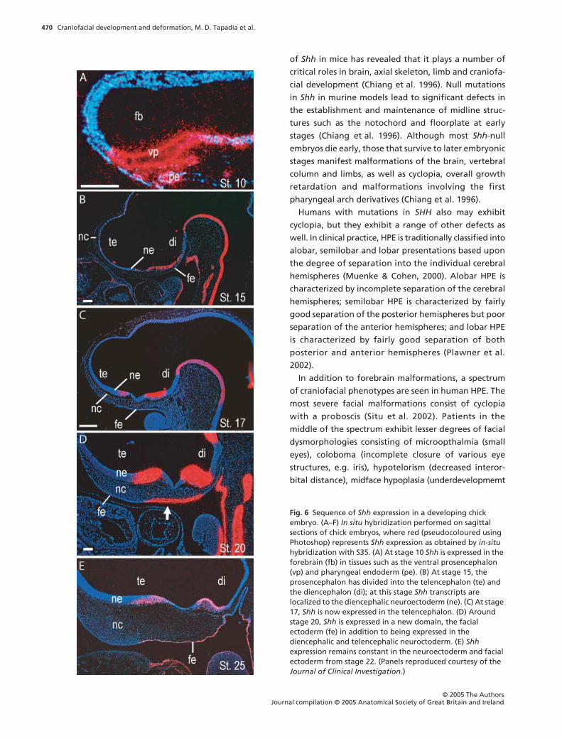
Over the past decade, studies have begun to delve into the
precise nature of the environmental cues that influence
the neural crest. Many of these studies have implicated
Shh as a critical factor in regulating craniofacial mor-
phogenesis. Indeed, Shh seems almost omnipresent,
with expression domains in several epithelia presenting
themselves at various stages of development. The dynamic
expression of Shh in the craniofacial tissues is one indi-
cator of the multiple roles this growth factor plays in
modulating normal, as well as abnormal, craniofacial
development. Its role in all of these tissues has been
examined in mammals (humans and mice), birds, and most
recently, zebrafish. Despite the species differences in
facial forms, the results have been remarkably consistent.
Shh expression is dynamic during embryonic
development
Shh is expressed in the facial ectoderm, the neuroecto-
derm and the pharyngeal endoderm at various stages
during development. In birds, Shh is sequentially
induced in the brain and subsequently in the face
(Fig. 6). At stage 15 (Hamburger & Hamilton, 1951), Shh
expression in the brain is initially restricted to the
ventral diencephalon (Cordero et al. 2004) (Fig. 6B). At
stage 17, a new domain of Shh is induced in the ventral
telencephalon, which is separated from the dien-
cephalic domain by an Shh-negative optic recess (Cord-
ero et al. 2004) (Fig. 6C). Subsequently, around stage
20, Shh expression is induced in the facial ectoderm
(Cordero et al. 2004) (Fig. 6D). These same expression
patterns for Shh are appreciated in zebrafish as well
(Wada et al. 2005).
Removal of avian Shh expression reveals its integral
role in craniofacial development
Some of the first hints that Shh is capable of directing
craniofacial development came from ablation studies
in avian beaks. Akin to the jaws of fish, upper beaks are
derived from the frontonasal prominence and the max-
illary and mandibular prominences. When we removed
frontonasal ectoderm that normally expressed Shh
we saw an arrest of maxillary prominence outgrowth,
whereas ablation of frontonasal ectoderm that did not
express Shh had no discernible effect on facial morpho-
genesis (Hu & Helms, 1999). The importance of ectodermal
Shh in the development of the midface was confirmed
by removing Shh itself from the frontonasal process,
via a function-blocking blocking antibody (Hu & Helms,
1999). Although skeletal elements formed, their dors-
oventral patterning and fusion were disrupted, result-
ing in facial clefting and midfacial hypoplasia (Hu &
Helms, 1999; Rallu et al. 2002; Jeong et al. 2004).
Perturbations in Shh signalling in mice and humans
result in a wide range of holoprosencephalic defects
Mice with null mutations in Shh bear even more drastic
defects (Chiang et al. 1996). Targeted gene disruption
Craniofacial development and deformation, M. D. Tapadia et al.470
© 2005 The AuthorsJournal compilation © 2005 Anatomical Society of Great Britain and Ireland
of Shh in mice has revealed that it plays a number of
critical roles in brain, axial skeleton, limb and craniofa-
cial development (Chiang et al. 1996). Null mutations
in Shh in murine models lead to significant defects in
the establishment and maintenance of midline struc-
tures such as the notochord and floorplate at early
stages (Chiang et al. 1996). Although most Shh-null
embryos die early, those that survive to later embryonic
stages manifest malformations of the brain, vertebral
column and limbs, as well as cyclopia, overall growth
retardation and malformations involving the first
pharyngeal arch derivatives (Chiang et al. 1996).
Humans with mutations in SHH also may exhibit
cyclopia, but they exhibit a range of other defects as
well. In clinical practice, HPE is traditionally classified into
alobar, semilobar and lobar presentations based upon
the degree of separation into the individual cerebral
hemispheres (Muenke & Cohen, 2000). Alobar HPE is
characterized by incomplete separation of the cerebral
hemispheres; semilobar HPE is characterized by fairly
good separation of the posterior hemispheres but poor
separation of the anterior hemispheres; and lobar HPE
is characterized by fairly good separation of both
posterior and anterior hemispheres (Plawner et al.
2002).
In addition to forebrain malformations, a spectrum
of craniofacial phenotypes are seen in human HPE. The
most severe facial malformations consist of cyclopia
with a proboscis (Situ et al. 2002). Patients in the
middle of the spectrum exhibit lesser degrees of facial
dysmorphologies consisting of microopthalmia (small
eyes), coloboma (incomplete closure of various eye
structures, e.g. iris), hypotelorism (decreased interor-
bital distance), midface hypoplasia (underdevelopmemt
Fig. 6 Sequence of Shh expression in a developing chick embryo. (A–F) In situ hybridization performed on sagittal sections of chick embryos, where red (pseudocoloured using Photoshop) represents Shh expression as obtained by in-situ hybridization with S35. (A) At stage 10 Shh is expressed in the forebrain (fb) in tissues such as the ventral prosencephalon (vp) and pharyngeal endoderm (pe). (B) At stage 15, the prosencephalon has divided into the telencephalon (te) and the diencephalon (di); at this stage Shh transcripts are localized to the diencephalic neuroectoderm (ne). (C) At stage 17, Shh is now expressed in the telencephalon. (D) Around stage 20, Shh is expressed in a new domain, the facial ectoderm (fe) in addition to being expressed in the diencephalic and telencephalic neuroctoderm. (E) Shh expression remains constant in the neuroectoderm and facial ectoderm from stage 22. (Panels reproduced courtesy of the Journal of Clinical Investigation.)

Craniofacial development and deformation, M. D. Tapadia et al. 471
© 2005 The Authors Journal compilation © 2005 Anatomical Society of Great Britain and Ireland
or undergrowth of the midface), cleft lip and palate,
and a single incisor (Belloni et al. 1996; Schimmenti
et al. 2003). Individuals at the mild end of the spectrum
can have normal or near-normal appearing faces.
For a number of years it was assumed that the sever-
ity of the craniofacial malformations directly corre-
lated with the severity of the brain malformation
(Demyer et al. 1964). Neuroimaging studies and the
ability to detect a number of mutations in HPE genes
have shown that this relationship does not always exist,
and has also led to a genetic classification of HPE. For
example, patients with the most severe, or alobar, HPE
brain malformations do not always exhibit cyclopia
but may have facial malformations of intermediate
severity. Conversely, patients with a mutation in an
HPE gene such as SHH may reveal normal brain imaging
results yet still exhibit facial malformations of inter-
mediate severity. In addition, patients from the same
family with identical mutations in SHH can exhibit
differences both in brain and in facial manifestations,
and yet other individuals with identical mutations may
fail to meet the traditional or anatomical criteria for
HPE.
Temporal perturbations in Shh signalling in avian
models may help to explain the range of phenotypes
associated with HPE
Given this absence of a clear genotype–phenotype
correlation, it remains unclear how genetic alterations
in SHH can lead to the various craniofacial phenotypes
seen in HPE. It has been suggested that the production
of an HPE phenotype requires multiple hits: in other
words, not only an underlying genetic mutation, but
also subsequent environmental influences such as
teratogens must act simultaneously to produce HPE-
related malformations (Ming & Muenke, 2002). Based
on this idea, we hypothesized that the spectrum of HPE
phenotypes may be related to the developmental stage
at which subsequent teratogenic insults occur (Cordero
et al. 2004). We chose the chick as a model system
and Shh as our signalling molecule of interest to
explore this possibility.
We used the steroidal alkaloid cyclopamine to block
Shh signal transduction by exposing chick embryos
to this agent at select timepoints governed by the
dynamic Shh induction pattern (Cordero et al. 2004).
Cyclopamine is a steroidal alkaloid extracted from the
plant Veratrum californicum (Keeler, 1970, 1978; Incar-
dona et al. 1998) that has the ability to alter the pro-
tein conformation of a downstream component of the
Shh signalling pathway, Smo, by binding to it (Chen
et al. 2002a,b). In doing so, cyclopamine inhibits a
series of intracellular processes that normally culmin-
ate in Shh signalling (Chen et al. 2002a,b).
Variable facial phenotypes recapitulating those
observed in the human HPE spectrum were observed
following administration of cyclopamine at different
developmental timepoints (Cordero et al. 2004). In-
hibition of Shh signalling in gastrulation-stage chick
embryos produced forebrain malformations and cyclo-
pia with a proboscis, as observed by other investi-
gators (Incardona et al. 1998) and as seen in Shh-null
mice (Chiang et al. 1996). When stage-15 embryos were
treated with cyclopamine, prior to initiation of Shh
expression in the telencephalon, as well as abnormal
forebrain development craniofacial malformations
consisting of microcephaly, microopthalmia, hypo-
telorism and hypoplasia of the maxillary primordia
were noted (Cordero et al. 2004). Cyclopamine admin-
istration at stage 17, after the induction of Shh in the
telencephalon but prior to its expression in the facial
ectoderm, failed to produce gross brain anomalies
(Cordero et al. 2004). In addition, the facial malforma-
tions resulting from treatment at stage 17 were less
severe than those resulting from treatment at earlier
stages (Cordero et al. 2004). The facial dysmophology
consisted of mild hypotelorism and truncation of the
distal upper beak (Cordero et al. 2004) (Fig. 7C), which
is analogous to cleft lip and palate in humans. Evalua-
tion of the skeletal elements revealed that truncation
of the distal upper beak was secondary to a small
premaxilla, which was aberrantly positioned ventral to
the nasal capsule (Cordero et al. 2004) (Fig. 7D).
Molecular analyses revealed that the facial malfor-
mations were not secondary to neural crest apoptosis,
but instead were due to molecular mispatterning in
the facial ectoderm (Cordero et al. 2004). As men-
tioned, blocking Shh signalling at stage 15 led to the
failure of Shh induction in the telencephalon and facial
ectoderm, whereas treatment at stage 17 led to the
failure of induction of Shh in the facial ectoderm
domain (Cordero et al. 2004). The loss of the Shh
expression domain in the facial ectoderm after treat-
ment at these stages was accompanied by an ectopic
proximal expression of the Fgf8 domain from the FEZ
(Cordero et al. 2004). As a result of this shift, the organ-
izing properties of the FEZ (Hu et al. 2003) are lost.

Craniofacial development and deformation, M. D. Tapadia et al.472
© 2005 The AuthorsJournal compilation © 2005 Anatomical Society of Great Britain and Ireland
One drawback of cyclopamine is that it inhibits Shh
signal transduction in a number of tissues. We there-
fore sought to determine the effects on craniofacial
morphology by specifically blocking Shh emanating
from the forebrain. Hybridoma cells producing anti-
body to Shh (5E1) were injected into the brains of
stage-9.5 chick embryos (Marcucio et al. 2005). Block-
ing Shh from the forebrain via this approach resulted in
Fig. 7 Consequences of inhibition of Shh signalling. (A,C,E) Oblique views of stage-41 chick embryos; (B,D,F) alcian blue- and alizarin red-stained skeletal structures of these embryos. (A,B) Control embryos exhibit normal morphology and skeletal development. (C,D) When stage-17 embryos are exposed to cyclopamine, they exhibit mild microcephaly and hypotelorism, along with malpositioning and truncation of the distal upper beak [e.g. the body of the premaxillary bone (pm) is shifted ventrally and shortened]. Proximal elements such as the dorsal component of the premaxillary bone remain unaffected [e.g. the nasal process of the premaxilla (pn) is intact]. The mandible (mn) is unaltered in cyclopamine-treated embryos. (E,F) Injection of 5E1 cells, which produce antibodies to Shh at stage 9.5, results in microcephaly, abnormal eye development and abnormalities of the distal upper beak. The gross phenotype resulting from 5E1 injection (which blocks Shh emanating from the neuroectoderm) is reminiscent of the morphological changes resulting from cyclopamine treatment; compare with C and D. (Panels A–D reproduced courtesy of the Journal of Clinical Investigation; panels E and F reproduced courtesy of Developmental Biology.)

Craniofacial development and deformation, M. D. Tapadia et al. 473
© 2005 The Authors Journal compilation © 2005 Anatomical Society of Great Britain and Ireland
facial phenotypes seen following cyclopamine admin-
istration at stage 17 (Fig. 7E), and like cyclopamine was
not secondary to neural crest cell apoptosis but from
a molecular alteration in the FEZ leading to truncation
of the upper beak, ventralization of the premaxillary
bone and decreased expansion of the medial–lateral
axes (Marcucio et al. 2005) (Fig. 7F). The resultant less
severe phenotype in the 5E1 embryos treated at stage
9.5 as compared with the more severe phenotypes seen
with cyclopamine at earlier stages reflects the ability of
cyclopamine to diffuse into multiple tissues and pan-
inhibit Shh in those tissues.
These data suggest that Shh plays an important role
in ‘linking’ the brain and face during certain earlier
developmental time points, and Shh signalling thereby
provides evidence that the brain–face relationship is
more than just a structural relationship based on phys-
ical proximity between these two important structures.
However, at later stages after Shh has been established
in the telencephalon the brain and face appear to
develop independently. This temporal dependence
and subsequent apparent independence between the
brain and face during embryonic development may
account for the incongruous presentations of HPE seen
in clinical practice (e.g. some patients present with
both brain and facial malformations, whereas other
patients exhibit normal brain morphology despite
facial malformations). However, it remains unclear
whether SHH exhibits this dynamic pattern of expres-
sion during human development.
Shh from the epithelia is required for neural crest
survival
Clearly, a definite correlation exists between proper
timing of Shh expression in epithelial tissues and
normal craniofacial development. But what cellular
processes does Shh affect at these crucial developmental
timepoints in order to ensure proper craniofacial devel-
opment? As already discussed, Shh signalling clearly
impacts cell proliferation and outgrowth of the fronto-
nasal process. But moulding the craniofacial primordia
into facial structures requires not only cell proliferation
but also the induction of cell death in appropriate places,
and perhaps more importantly, the survival of cells in
other key places. Two seminal studies indicate that Shh
signalling may be of importance in this regard as well.
Sara Ahlgren and colleagues noticed that the pheno-
type (e.g. Fetal Alcohol Syndrome) resulting from
maternal alcohol consumption during pregnancy is
incredibly similar to the phenotype that results from
blocking Shh signalling (Ahlgren et al. 2002). Embryos
in which Shh is blocked or which have been exposed to
ethanol both display reduction of the frontonasal proc-
ess, hypoplastic branchial arches and apoptosis of neural
crest cells (Ahlgren et al. 2002). The similarity between
phenotypes suggested to them that Shh may be medi-
ating cell survival during craniofacial development. To
that end, they examined the amount of Shh expression
after ethanol exposure, and found that expression of a
number of genes in the Shh signalling pathway, includ-
ing the Shh receptor Ptc and its downstream effector
Gli, are reduced after ethanol treatment (Ahlgren et al.
2002). Retroviral expression of Shh resulted in a dra-
matic decrease in the amount of cell death, along with
proper formation of the frontonasal process, thereby
rescuing the phenotype and confirming the role of Shh
as a cell survival factor (Ahlgren et al. 2002). This find-
ing is in accord with more studies that demonstrate
that Shh is also required for the survival of neuroepi-
thelial cells early in development (Thibert et al. 2003).
Ahlgren’s conclusions that Shh is an important medi-
ator of cell survival were reinforced by studies from
Andrew McMahon’s laboratory. McMahon removed
Shh responsiveness from neural crest cells specifically
by generating Wnt-1-Cre; conditionally null Smoothened
(Smo) mice (Wnt-1-Cre; Smo n/c) (Jeong et al. 2004).
Embryos carrying the conditional knockout of Smo,
a downstream component of the Shh pathway, exhibit
extensive loss of many neural-crest-derived craniofacial
structures, as well as a down-regulation of Hedgehog
targets [such as certain Forkhead (Fox) transcription
factors] in the neural crest of these structures (Jeong
et al. 2004). Perhaps more significantly, removal of Shh
signalling from neural crest cells resulted in increased
apoptosis and decreased cell proliferation in the
arches, which manifests as a reduction of the frontona-
sal process in embryos that survive (Jeong et al. 2004).
These results echoed Ahlgren’s conclusion that Shh is
necessary for cell survival, and also accords with new
studies that demonstrate the role of Shh in skull develop-
ment, as discussed later.
Shh is also required for chondrogenesis in the skull and
in the lower face
Sculpting the craniofacial complex is a complex process.
It requires the neural crest cells that form skeletal

Craniofacial development and deformation, M. D. Tapadia et al.474
© 2005 The AuthorsJournal compilation © 2005 Anatomical Society of Great Britain and Ireland
elements not only to survive and proliferate, but also
then to differentiate appropriately. As recent studies
reveal, the omnipresent Shh is implicated even in the
later differentiation of neural crest cells.
Thomas Schilling made these discoveries via elegant
experiments in zebrafish (Wada et al. 2005). By creat-
ing a sox10:egfp transgenic zebrafish, he and col-
leagues were able to follow neural crest cells as they
migrated and differentiated into craniofacial struc-
tures such as the anterior neurocranium, which consists
of a medial ethimoid plate flanked by trabeculae on
each side (Wada et al. 2005). Then, by mating this
sox10:egfp fish with the sonic you (syu) fish in which
shh signalling is disrupted, Schilling was able to deter-
mine what happens to these neural crest cells when
Shh signalling is disrupted (Wada et al. 2005).
In normal embryos, the neural crest cells destined to
become anterior neurocranium lie widely separated
from one another across the ventral midline (Wada et al.
2005). These crest cells form condensations at later
stages, and then differentiate into the cartilages
(trabeculae and ethimoid) that comprise the anterior
neurocranium (Wada et al. 2005). The transgenic
sox10-egfp;syu mutant exhibited significant defects in
formation of the anterior neurocranium, including a
failure of the presumptive trabecular neural crest cells
to separate (Wada et al. 2005). This lack of separation
was traced to fusion (as opposed to apoptosis) of
neural crest cells across the ventral midline, which led
to a midline condensation that formed a single rod of
cartilage (e.g. a fused trabeculae) (Wada et al. 2005).
Schilling suspected that Hedgehog signalling might
have different actions on neural crest cells depending
on the stage at which it exerts its effects (Wada et al.
2005). To inhibit Shh signalling at discrete timepoints,
he and colleagues turned to cyclopamine (Wada et al.
2005). Similar to Cordero et al., Schilling found that
the severity of the phenotype was dependent on the
stage at which Shh was interrupted (Wada et al. 2005).
Disruption of Shh with cyclopamine at earlier stages
caused a complete loss of the anterior neurocranium,
whereas disruption of Shh at later stages eliminated
the medial ethmoid plate, while sparing the trabecular
cartilages (Wada et al. 2005). Thus, these studies offered
unequivocal confirmation that the range of HPE-associated
phenotypes may reflect stage-dependent require-
ments for Hedgehog signalling (Wada et al. 2005).
What cellular processes does Shh signalling affect
in order to produce these phenotypes? The answer
was elucidated by applying exogenous Shh via mRNA
misexpression at early stages in wild-type embryos, and
via bead implantation at later stages in both wild-type
embyos and mutants (Wada et al. 2005). Application of
exogenous Shh after migration promoted chondrogen-
esis in the anterior neurocranium in normal embryos
(Wada et al. 2005). Application of exogenous Shh
in sox9-egfp;syu mutants rescued the phenotype and
restored separation of the presumptive trabecular
neural crest (Wada et al. 2005). Taken together, these
data supported a role for Hedgehog signalling at later
stages to promote chondrogenesis in the elements of
the anterior neurocranium.
Other recent papers have also found that Shh plays
an integral role in promoting chondrogenesis from
mesenchymal condensations in other areas of the face.
For instance, differentiation of condensations into
Meckel’s cartilage in the mandible has been shown to
be Hedgehog dependent (Melnick et al. 2005). Noting
the absence of mandibular structures in Shh-null mutants,
Tina Jaskoll and colleagues undertook in vitro culture
studies of stage E11–12 mandibular explants (Melnick
et al. 2005). They found that application of cyclopamine
to these cultures exhibit a stage-dependent inhibition
of Meckel’s cartilage chondroblast differentiation to
mature chondrocytes (Melnick et al. 2005). Other stud-
ies by Cliff Tabin, however, have applied exogenous
Shh alone to culture preparations and in vivo without
finding evidence of increased chondrogenesis in the
frontonasal prominence (Abzhanov & Tabin, 2004).
Rather, they find that while Shh alone may not promote
chondrogenesis in the midface, the FEZ domain of Shh
juxtaposed with Fgf8 serves to promote chondro-
genesis and chondrogenic outgrowth during cranial
development (Abzhanov & Tabin, 2004). These results
suggest that Shh must act syngergistically with other
molecules, such as Fgf8, to produce chondrocyte differ-
entiation and proliferation (Abzhanov & Tabin, 2004).
Future directions
Although Shh has gained prominence in recent years
as one of the major mediators of craniofacial morpho-
genesis, this molecule is by no means the only player
involved. As mentioned, Shh acts in conjunction with
Fgf8 to initiate outgrowth and chondrogenesis of
the frontonasal process. Other studies investigating
retinoic acid signalling in the face also highlight Fgf8
as a molecule of interest.

Craniofacial development and deformation, M. D. Tapadia et al. 475
© 2005 The Authors Journal compilation © 2005 Anatomical Society of Great Britain and Ireland
More recently, several papers have highlighted the
role of bone morphogenetic proteins (Bmps) in produc-
ing outgrowth of the frontonasal prominence, and
have suggested that differences in Bmp levels may be
responsible for species-specific differences the appear-
ance of the frontonasal prominence (Abzhanov et al.
2004; Wu et al. 2004). Yet other data are beginning to
hint that Wnts may also mediate craniofacial develop-
ment (Niemann et al. 2004; S. Brugmann, unpublished
observations). The role of these molecular pathways in
craniofacial development has yet to be elucidated in
detail; additionally, how these molecules interact with
Shh and Fgf8 has yet to be determined. Given the com-
plexity and individuality inherent in faces, both within
and among species, it is certain that craniofacial devel-
opment requires a complex interplay between all of
these molecular pathways.
Now that it is known that molecular signalling in the
face might involve a number of molecular pathways,
an equally intriguing question emerges: namely, how
are all these molecular signals coordinated between
tissues? For example, hypotelorism can be elicited by
disrupting signals emanating from the neuroectoderm,
but it can also be caused by disrupting signals from the
facial ectoderm (Cordero et al. 2004). These findings
suggest that both the brain and the face are sources of
patterning signals. But how are these signals from the
brain and face coordinated? How much crosstalk actu-
ally occurs between the brain and the face, and at what
developmental timepoints does it occur? Is the neural
crest involved in mediating crosstalk between the neu-
roectoderm and facial ectoderm, or does it merely pas-
sively respond to signals from these tissues? If the crest
actually participates in the signalling, what molecules
in the neural crest relay signals between these tissues?
Finding the answers to these questions will be crucial to
gaining a complete picture of craniofacial develop-
ment, and in learning how to repair congenital cranio-
facial defects.
Perhaps an equally pressing line of inquiry concerns
Hox genes. While understanding molecular signalling
is crucial to preventing birth defects, further studies on
Hox genes may help facilitate regenerative processes
of craniofacial healing. As mentioned, Hox genes imbue
neural crest cells with a positional code that directs
them to take residence in particular arches. Evidence
exists that depending on the particular Hox code
present, the neural crest cells adopt different degrees
of fates, and possess different degrees of plasticity
that enable them to form different facial structures. It
remains to be determined whether these Hox codes are
retained into adulthood, and if so, whether that infor-
mation can be used to give cells the plasticity necessary
for craniofacial repair and regeneration after injury.
All of these questions will provide intriguing areas of
exploration.
Conclusion
Although it has been resolved that the neural crest
plays an enormous role in the production of facial
structures, some neural crest populations require
instructions from their local environment. In instances
where a Hox code dictates plasticity, the complex
signals come from the local milieu of the superficial
ectoderm, the neuroepithelium and the pharyngeal
endoderm. Much of the crosstalk that occurs between
these tissues during early embryonic development
involves common signalling pathways such as the
Shh, Fgf, Wnt and Bmp pathways. These molecular
dialogues appear to impact upon several cellular
processes, including neural crest survival, proliferation
and differentiation. An equally important aspect of this
crosstalk is that it occurs at very specific developmental
timepoints, and outside these timepoints there appear
to be tissue-specific developmental mechanisms that
are independent of other tissues. The dependence and
independence shared by the brain and face at different
timepoints may explain why brain and facial malforma-
tions are often seen together, but also how abnormal
facial development can occur in the presence of an
apparently normal brain.
References
Abu-Issa R, Smyth G, Smoak I, et al. (2002) Fgf8 is requiredfor pharyngeal arch and cardiovascular development in themouse. Development 129, 4613–4625.
Abzhanov A, Protas M, Grant RB, et al. (2004) Bmp4 andmorphological variation of beaks in Darwin’s finches. Science305, 1462–1465.
Abzhanov A, Tabin CJ (2004) Shh and Fgf8 act synergisticallyto drive cartilage outgrowth during cranial development.Dev Biol 273, 134–148.
Ahlgren SC, Thakur V, Bronner-Fraser M (2002) Sonic hedge-hog rescues cranial neural crest from cell death induced byethanol exposure. Proc Natl Acad Sci USA 99, 10476–10481.
Barrow JR, Capecchi MR (1999) Compensatory defects associ-ated with mutations in Hoxa1 restore normal palatogenesisto Hoxa2 mutants. Development 126, 5011–5026.

Craniofacial development and deformation, M. D. Tapadia et al.476
© 2005 The AuthorsJournal compilation © 2005 Anatomical Society of Great Britain and Ireland
Belloni E, Muenke M, Roessler E, et al. (1996) Identificationof Sonic hedgehog as a candidate gene responsible forholoprosencephaly. Nat Genet 14, 353–356.
Capecchi MR (1997) Hox genes and mammalian development.Cold Spring Harb Symp Quant Biol 62, 273–281.
Chen JK, Taipale J, Cooper MK, et al. (2002a) Inhibition ofHedgehog signaling by direct binding of cyclopamine toSmoothened. Genes Dev 16, 2743–2748.
Chen JK, Taipale J, Young KE, et al. (2002b) Small moleculemodulation of Smoothened activity. Proc Natl Acad Sci USA21, 21.
Chiang C, Litingtung Y, Lee E, et al. (1996) Cyclopia and defec-tive axial patterning in mice lacking Sonic hedgehog genefunction. Nature 383, 407–413.
Cordero D, Marcucio R, Hu D, et al. (2004) Temporal perturba-tions in sonic hedgehog signaling elicit the spectrum ofholoprosencephaly phenotypes. J Clin Invest 114, 485–494.
Couly G, Grapin-Botton A, Coltey P, et al. (1998) Determina-tion of the identity of the derivatives of the cephalic neuralcrest: incompatibility between Hox gene expression andlower jaw development. Development 125, 3445–3459.
Couly G, Creuzet S, Bennaceur S, et al. (2002) Interactionsbetween Hox-negative cephalic neural crest cells and theforegut endoderm in patterning the facial skeleton in thevertebrate head. Development 129, 1061–1073.
Creuzet S, Couly G, Vincent C, et al. (2002) Negative effectof Hox gene expression on the development of the neuralcrest-derived facial skeleton. Development 129, 4301–4313.
Crump JG, Maves L, Lawson ND, et al. (2004) An essential rolefor Fgfs in endodermal pouch formation influences latercraniofacial skeletal patterning. Development 131, 5703–5716.
Darwin C (1859) The Origin of Species. New York: The Crowell-Collier Publishing Co.
Demyer W, Zeman W, Palmer CG (1964) The face predicts thebrain: diagnostic significance of median facial anomaliesfor holoprosencephaly (arhinencephaly). Pediatrics 34, 256–263.
Draper BW, Morcos PA, Kimmel CB (2001) Inhibition ofzebrafish fgf8 pre-mRNA splicing with morpholino oligos:a quantifiable method for gene knockdown. Genesis 30,154–156.
Favier B, Dolle P (1997) Developmental functions of mamma-lian Hox genes. Mol Hum Reprod 3, 115–131.
Gans C, Northcutt RG (1983) Neural crest and the origin ofvertebrates: a new head. Science 220, 268–274.
Gendron-Maguire M, Mallo M, Zhang M, et al. (1993) Hoxa-2mutant mice exhibit homeotic transformation of skeletalelements derived from cranial neural crest. Cell 75, 1317–1331.
Grammatopoulos GA, Bell E, Toole L, et al. (2000) Homeotictransformation of branchial arch identity after Hoxa2 over-expression. Development 127, 5355–5365.
Hamburger V, Hamilton HL (1951) A series of normal stages inthe development of the chick embryo. J Morph 88, 49–92.
Harvey W (1657) Letter, written six weeks before his death, toJan Vlakfeld of Haarlem, published in The Works of WilliamHarvey: Translated from the Latin with a Life of the Authorby R. Willis. London: Sydenham Society, 1847.
Helms JA, Cordero D, Tapadia MD (2005) New insights intocraniofacial morphogenesis. Development 132, 851–861.
Hu D, Helms JA (1999) The role of sonic hedgehog in normaland abnormal craniofacial morphogenesis. Development126, 4873–4884.
Hu D, Marcucio RS, Helms JA (2003) A zone of frontonasalectoderm regulates patterning and growth in the face.Development 130, 1749–1758.
Hunt P, Gulisano M, Cook M, et al. (1991a) A distinct Hox codefor the branchial region of the vertebrate head. Nature 353,861–864.
Hunt P, Whiting J, Muchamore I, et al. (1991b) Homeoboxgenes and models for patterning the hindbrain andbranchial arches. Dev Suppl. 1, 187–196.
Hunt P, Whiting J, Nonchev S, et al. (1991c) The branchialHox code and its implications for gene regulation, pattern-ing of the nervous system and head evolution. Dev Suppl. 2,63–77.
Incardona JP, Gaffield W, Kapur RP, et al. (1998) The teratogenicVeratrum alkaloid cyclopamine inhibits sonic hedgehogsignal transduction. Development 125, 3553–3562.
Jeong J, Mao J, Tenzen T, et al. (2004) Hedgehog signaling inthe neural crest cells regulates the patterning and growthof facial primordia. Genes Dev 18, 937–951.
Keeler RF (1970) Teratogenic compounds of Veratrum californicum(Durand) X. Cyclopia in rabbits produced by cyclopamine.Teratology 3, 175–180.
Keeler RF (1978) Cyclopamine and related steroidal alkaloidteratogens: their occurrence, structural relationship, andbiologic effects. Lipids 13, 708–715.
Kontges G, Lumsden A (1996) Rhombencephalic neural crestsegmentation is preserved throughout craniofacial ontog-eny. Development 122, 3229–3242.
Le Douarin NM, Creuzet S, Couly G, et al. (2004) Neural crestcell plasticity and its limits. Development 131, 4637–4650.
Marcucio RS, Cordero DR, Hu D, et al. (2005) Molecular inter-actions coordinating the development of the forebrain andface. Dev Biol 284, 48–61.
Melnick M, Witcher D, Bringas P Jr, et al. (2005) Meckel’scartilage differentiation is dependent on hedgehog signaling.Cells Tissues Organs 179, 146–157.
Miller CT, Schilling TF, Lee K, et al. (2000) sucker encodes azebrafish Endothelin-1 required for ventral pharyngeal archdevelopment. Development 127, 3815–3828.
Miller CT, Yelon D, Stainier DY, et al. (2003) Two endothelin 1effectors, hand2 and bapx1, pattern ventral pharyngealcartilage and the jaw joint. Development 130, 1353–1365.
Ming JE, Muenke M (2002) Multiple hits during early embry-onic development: digenic diseases and holoprosencephaly.Am J Hum Genet 71, 1017–1032.
Muenke M, Cohen MM Jr (2000) Genetic approaches tounderstanding brain development: holoprosencephaly as amodel. Ment Retard Dev Disabil Res Rev 6, 15–21.
Niemann S, Zhao C, Pascu F, et al. (2004) Homozygous WNT3mutation causes tetra-amelia in a large consanguineousfamily. Am J Hum Genet 74, 558–563.
Northcutt RG, Gans C (1983) The genesis of neural crest andepidermal placodes. Q Rev Biol 58, 1–27.
Ozeki H, Kurihara Y, Tonami K, et al. (2004) Endothelin-1regulates the dorsoventral branchial arch patterning in mice.Mech Dev 121, 387–395.
Pasqualetti M, Ori M, Nardi I, et al. (2000) Ectopic Hoxa2 induction
Craniofacial development and deformation, M. D. Tapadia et al. 477
© 2005 The Authors Journal compilation © 2005 Anatomical Society of Great Britain and Ireland
after neural crest migration results in homeosis of jawelements in Xenopus. Development 127, 5367–5378.
Perrotin F, de Poncheville LM, Marret H, et al. (2001) Chromo-somal defects and associated malformations in fetal cleft lipwith or without cleft palate. Eur J Obstet Gynecol ReprodBiol 99, 19–24.
Piotrowski T, Nusslein-Volhard C (2000) The endoderm playsan important role in patterning the segmented pharyngealregion in zebrafish (Danio rerio). Dev Biol 225, 339–356.
Plawner LL, Delgado MR, Miller VS, et al. (2002) Neuroanat-omy of holoprosencephaly as predictor of function: beyondthe face predicting the brain. Neurology 59, 1058–1066.
Rallu M, Machold R, Gaiano N, et al. (2002) Dorsoventralpatterning is established in the telencephalon of mutantslacking both Gli3 and Hedgehog signaling. Development129, 4963–4974.
Reifers F, Bohli H, Walsh EC, et al. (1998) Fgf8 is mutated inzebrafish acerebellar (ace) mutants and is required formaintenance of midbrain–hindbrain boundary develop-ment and somitogenesis. Development 125, 2381–2395.
Remuzzi G, Perico N, Benigni A (2002) New therapeutics thatantagonize endothelin: promises and frustrations. Nat RevDrug Discov 1, 986–1001.
Rijli FM, Mark M, Lakkaraju S, et al. (1993) A homeotic trans-formation is generated in the rostral branchial region of thehead by disruption of Hoxa-2, which acts as a selector gene.Cell 75, 1333–1349.
Rijli FM, Gavalas A, Chambon P (1998) Segmentation andspecification in the branchial region of the head: the roleof the Hox selector genes. Int J Dev Biol 42, 393–401.
Roehl H, Nusslein-Volhard C (2001) Zebrafish pea3 and ermare general targets of FGF8 signaling. Curr Biol 11, 503–507.
Ruhin B, Creuzet S, Vincent C, et al. (2003) Patterning ofthe hyoid cartilage depends upon signals arising from theventral foregut endoderm. Dev Dyn 228, 239–246.
Schilling TF, Piotrowski T, Grandel H, et al. (1996) Jaw andbranchial arch mutants in zebrafish I: branchial arches.Development 123, 329–344.
Schimmenti LA, de la Cruz J, Lewis RA, et al. (2003) Novel
mutation in sonic hedgehog in non-syndromic coloboma-tous microphthalmia. Am J Med Genet A 116, 215–221.
Schneider RA, Helms JA (2003) The cellular and molecularorigins of beak morphology. Science 299, 565–568.
Situ D, Reifel CW, Smith R, et al. (2002) Investigation of acyclopic, human, term fetus by use of magnetic resonanceimaging (MRI). J Anat 200, 431–438.
Tessier P (1976) Anatomical classification facial, cranio-facialand latero-facial clefts. J Maxillofac Surg 4, 69–92.
Thibert C, Teillet MA, Lapointe F, et al. (2003) Inhibition ofneuroepithelial patched-induced apoptosis by sonic hedge-hog. Science 301, 843–846.
Trainor PA, Krumlauf R (2000) Patterning the cranial neuralcrest: hindbrain segmentation and Hox gene plasticity. NatRev Neurosci 1, 116–124.
Trainor PA, Krumlauf R (2001) Hox genes, neural crest cells andbranchial arch patterning. Curr Opin Cell Biol 13, 698–705.
Trainor PA (2003) Making headway: the roles of Hox genesand neural crest cells in craniofacial development. Scien-tificWorldJournal 3, 240–264.
Tucker AS, Lumsden A (2004) Neural crest cells providespecies-specific patterning information in the developingbranchial skeleton. Evol Dev 6, 32–40.
Tucker AS, Watson RP, Lettice LA, et al. (2004) Bapx1 regulatespatterning in the middle ear: altered regulatory role in thetransition from the proximal jaw during vertebrate evolu-tion. Development 131, 1235–1245.
Veitch E, Begbie J, Schilling TF, et al. (1999) Pharyngeal archpatterning in the absence of neural crest. Curr Biol 9, 1481–1484.
Wada N, Javidan Y, Nelson S, et al. (2005) Hedgehog signalingis required for cranial neural crest morphogenesis and chon-drogenesis at the midline in the zebrafish skull. Develop-ment 132, 3977–3988.
Wilkinson DG (1993) Molecular mechanisms of segmentalpatterning in the vertebrate hindbrain and neural crest.Bioessays 15, 499–505.
Wu P, Jiang TX, Suksaweang S, et al. (2004) Molecular shapingof the beak. Science 305, 1465–1466.