issn 2222-9442 mjem - new health concept emergency 16.pdf(uae), wassim raffoul (switzerland), ......
TRANSCRIPT

September 2013-N°16Endorsed by
Trim
estri
el
Vasopressor use in prehospital hemorrhagic shock management
surVey of patients who left the eD without Being seen
how to optimize ccr stanDarDs 2013
pertinence De la transfusion De cgr auX urgences
aVc et ait: tout est Dans la Vascularisation
intoXication aigue a la colchicine
ISSN 2222-9442
M J E M

Informations et inscriptions :T + 961 373 373F + 961 373 374C [email protected]
LEADERS DE DEMAIN
MASTER MANAGEMENT DE L'HÔPITAL ET DE LA SANTÉ :POUR FAIRE FACE AUX NOUVEAUX DÉFIS DE L'ENVIRONNEMENT DE LA SANTÉ ET DES ÉTABLISSEMENTS HOSPITALIERS DU LIBAN ET DU MOYEN-ORIENT
Formation en 16 mois
Un emploi du temps adapté à votre carrière professionnelle
2 Diplômes :• Le Master Management de l'Hôpital et de la Santé de l'ESA• Le Master 2 «AMES» délivré par Paris Diderot - Paris 7 et l'École des Hautes Études en Santé Publique
(EHESP) Période d’inscription : de Janvier à Mars 2014Entretiens de sélection : Avril 2014Début des cours : Septembre 2014

1 Med Emergency, MJEM – 2013, No 16
E D I T O R ’ s N O T E
MED Emergency, MJEM Mediterranean Journal of Emergency Medicine
Publication of the Lebanese Resuscitation Council By New Health Concept
P.O.Box 90.815 Jdeideh - LebanonTel: 00961.1.888921 Fax: 00.961.1.888922
Email: [email protected]: www.newhealthconcept.net
EDitoRiaL boaRD
EDitoR in ChiEfNagi SOUAIBY
REsEaRCh Abdo KHOURY (France)Steve PHOTIOU (Italy)
Jean-Cyrille PITTELOUD (Switzerland)
Continuous EDuCationElvis CORDIER (France)
Daryl MACIAS (USA),Karim BEN MILOUD (Switzerland)
innovation, EDiting anD tRansLationGuillaume ALINIER (Qatar / UK)
Karim FARAH (Lebanon)Hugues LEFORT (France)
onLinE PubLiCation anD DEsignIsmaël HSSAIN (France)
Alec KAZANDJIANMireille SROUR
aDMinistRation anD MaRkEtingGeorges KHALIL
stuDEnts’ foRuMs anD ConfEREnCEsZiad KHOUEIRY (France)
nuRsing Lina AOUN CHOUEIRY
Chantal SAADEH KHALIL
PaRaMEDiCs anD aMbuLanCEsFrédéric HOEPPLI (Switzerland)
Juerg LINIGER (Switzerland)
aLLianCEsFire Brigade of Paris – France
Global Network Association of Emergency MedicineGlobal Emergency Medicine Literature ReviewLebanese Society for Quality and Patient Safety
aDvisoRy CoMMittEE
Pierre ABI HANNA, Georges ABI SAAD, Arthur ATCHABAHIAN (USA), Omar AYACH, Abdelouahab BELLOU (France), Maria Paula GOMEZ
(Spain), Thierry GROS (France), Maurice HADDAD, Berthe HACHEM, Mohamed HACHELAF (France), Jamil HALABI, Khalil HELOU, Aziz
KOLEILAT, Bruno MEGARBANE (France), Hicham NEJMI (Morocco), Ahmad OSMAN (Egypt), Alissar RADY (WHO), Hussain AL RAHMA
(UAE), Wassim RAFFOUL (Switzerland), Sami RICHA, Abdul Mohsen AL SAAWI (KSA), Karim TAZAROURTE (France)
special thanks: Dr Jean Claude DESLANDES
nagi souaiby, MD, MPh, MhMChief Editor
A two-speed world
More than ever we are living in a two-speed world. The publication of the world’s largest wealth comes as a striking contrast with the terrible humanitarian crisis resulting from poverty and armed conflict. Scientific and technological advance that is supposed to improve our daily life is not meeting its objective. The so-called “once superpowers” are struggling with their geopoliticalstrategic interests. Likewise for our specialty and we feel it very strongly when we meet in congresses; because whilst our colleagues in some countries are testing the best of the best of technology to improve the quality of care, others unfortunately who are practicing in conflict zones are forced to “run for their lives” as the saying goes. To those colleagues we pay a vibrant tribute.
Med Emergency Journal that stretches way more than the Mediterranean aims at becoming a forum of exchange for Emergency Medicine professionals. The published articles reflect indeed this objective. We express our deepest thanks to Professor Domres for his editorial that reminds us in very simple words how to detect the use of chemical weapons that is a burning topic nowadays. The original articles and those of continuous training tackle issue from our daily life. As we have said before, our publication is distinguished by its high ethical level. All authors and articles are scrutinized before being published. Once again I extend all my thanks to the editorial board members for their tremendous work.
When there is a will, there is a way ..

Editorial Chemical warfare weaponsProf. Bernd domres
Original Articles vasopressor use in prehospital hemorrhagic shock management: Evolution of french professional practices over the past decade. Foudi L, Gauthier a, Pacchioni F, checinski a, Foudi h, haddad Z, Gammoura k, cesareo e, saPir d, travers s, tourtier JP, atchaBahian a, taZarourte k
telephonic survey of patients who left the Emergency Department without being seen: Data from tertiary care hospital, Pakistan.kursheed m, FaYYaZ J, umer mir m, Zia n
Original Articles (French)Pertinence de la transfusion de concentrés de globules rouges aux urgences. Pertinence of red blood cell (RbC) transfusion in emergency.maBit h, domanski L, roche B
Forum for Emergency Developmenthow to optimize cardiocerebral resuscitation standards in 2013.hoePPLi F, BanerJee P, Garcia W, hssain i
Continuous Education (French) avC et ait : tableau clinique stroke and tia: Clinical presentationkemPF n, thiBaud e, dussau L, Patarin c, Ben hammouda k, savineau J r, GottWaLLes Y
testez vos connaissances en toxicology: intoxication aigue à la Colchicinetest your knowledge in toxicology: Management of acute colchicine poisoningmeGarBane B
General informationsRecommendations for authorsMembership
. . . . . . . . . . . . . . . . . . . . . . . . . . . . . . . . . . . . . . . . . . . . . . . . . . . . . . . . . . . . . . . . . . . . . . . . . . . . . . . . . . . . . . . . . . . . . . . . . . . . . . . . . . . . . . . . . . . . . . . . . . . . . . . . . . . . . . . . . . . . . . . . . . . . . . . . . . . . . . . . . . . . . . . . . . . . . . . . . . . . . . . . . . . . . . . . . . . . . . . . . . . . . . . . . . . . . . . . p. 3
. . . . . . . . . . . . . . . . . . . . . . . . . . . . . . . . . . . . . . . . . . . . . . . . . . . . . . . . . . . . . . . . . . . . . . . . . . . . . . . . . . . . . . . . . . . . . . . . . . . . . . . . . . . . . . . . . . . . . . . . p. 5
. . . . . . . . . . . . . . . . . . . . . . . . . . . . . . . . . . . . . . . . . . . . . . . . . . . . . . . . . . . . . . . . . . . p. 11
. . . . . . . . . . . . . . . . . . . . . . . . . . . . . . . . . . . . . . . . . . . . . . . . . . . . . . . . . . . . . . . . . . . . . . . . . . . . . . . . . . . . . . . . . . . p. 26
. . . . . . . . . . . . . . . . . . . . . . . . . . . . . . . . . . . . . . . . . . . . . . . . . . . . . . . . . . . . . . . . . . . . . . . . . . . . . . . . . . . . . . . . . . . . . . . . . . . . . . . . . . . . . . . . . . . . . . . . . . . . . p. 20
. . . . . . . . . . . . . . . . . . . . . . . . . . . . . . . . . . . . . . . . . . . . . . . . . . . . . . . . . . . . . . . . . . . . . . . . . . . . . . . . . . . . . . . . . . . . . . . . . . . . . . . . . . . . . . . . . . . . . . . . . . . . . . . . . . . . . . . . . . . . . . . . . . . . . . . . . . . . . . . . . . . . . . . . . . . . . . . . . . . . . . . . . . . . . . . . . . . . . . . . . . . . . . . . . . . p. 32
. . . . . . . . . . . . . . . . . . . . . . . . . . . . . . . . . . . . . . . . . . . . . . . . . . . . . . . . . . . . . . . . . . . . . . . . . . . . . . . . . . . . . . . . . . . . . . . . . . p. 37
. . . . . . . . . . . . . . . . . . . . . . . . . . . . . . . . . . . . . . . . . . . . . . . . . . . . . . . . . . . . . . . . . . . . . . . . . . . . . . . . . . . . . . . . . . . . . . . . . . . . . . . . . . . . . . . . . . . . . . . . . . . . . . . . . . . . . . . . . . . . . . . . . . . . . . . . . . . . . . . . . . . . . . . . . . . . . . . . . . . . . . . . . . . . . . . . . . . . . . . . . . . . . . . . . . p. 43. . . . . . . . . . . . . . . . . . . . . . . . . . . . . . . . . . . . . . . . . . . . . . . . . . . . . . . . . . . . . . . . . . . . . . . . . . . . . . . . . . . . . . . . . . . . . . . . . . . . . . . . . . . . . . . . . . . . . . . . . . . . . . . . . . . . . . . . . . . . . . . . . . . . . . . . . . . . . . . . . . . . . . . . . . . . . . . . . . . . . . . . . . . . . . . . . . . . . . . . . . . . . . . . . . . . . . . . . . . . . . . . . . . . . . . . . . . . . . . . . . . . . . . . . . . . . . . . . p. 48
C O N T E N T s
2 Med Emergency, MJEM – 2013, No 16

3 Med Emergency, MJEM – 2013, No 16
E D I T O R I A L
ChemiCAl wArfAre weApon
Crude chemical warfare weapons have been used since thousands of years. Heracles, for instance, poisoned his arrows with the Hydra Monster venom. But Germany was the first to produce modern chemical warfare agents (CWA) and used them during World War I.
As a result of this, the International Law of Chemical Weapons Treaty was signed on June, 17, 1925 in Geneva, and then followed by the Chemical Weapons Convention (CWC) to outlaw the production, stock piling, dissemination and use of chemical weapons. The treaty was revised on 5 September 2013.
Unfortunately and in spite of this CWC, hundreds of innocent civilians were killed by Organophosphate in the terrible civil war in Syria this year.
From the emergency medical response perspective, special aspects of chemical weapons use have to be known: the EMS has to be prepared for the detection of the chemical agent used in order to proceed with its decontamination.
Several organizations have defined criteria to stratify the likelihood of use of the different chemical agents.
Ranking for Chemical stratification STANAG, CDC, GHSI, BMI
• Toxicity (acute: LD50, LDC50), (chronic: DNA, Carcinogenesis)• Synthesis• Acquisition of Agents and Precursors• Dissemination• Threat Analysis• Detection• Incident Management• Release Environment• Antidotes• Decontamination• Persistence• Risk Perception• Public Preconception
The detection by laboratory procedures is time consuming as chemical poisons act very fast. Therefore it is indispensable for physicians, nurses and paramedics to be trained on the use of toxidromes to identify the chemical agent concerned. Organophosphates like Sarin have a high likelihood to be used. The symptoms of hypersecretion of excretoric glands combined with myosis, bradycardia and seizures allow with over 90% safety the detection of organophosphates.
Prof. Dr. H. C. Bernd Domres


5 Med Emergency, MJEM – 2013, No 16
FOUDI L, GAUTHIER A, PACCHIONI F, CHECINSKI A, FOUDI H, HADDAD Z, GAMMOURA K, CESAREO E, SAPIR D, TRAvERS S, TOUR-TIER JP, ATCHABAHIAN A, TAZAROURTE K - vasopressor use in pre-hospital hemorrhagic shock management: Evolution of French professional practices over the past decade. Med Emergency, MJEM 2013; 16:5-10key words: vasopressor, pre-hospital, hemorrhagic shock
ORIgINAL ARTICLE
VAsopressor use in pre-hospitAl hemorrhAgiC shoCk mAnAgement: eVolution of frenCh professionAl prACtiCes oVer the pAst deCAde. gestion du choc hémorragique et l’usage des catécholamines par 400 médecins smur français.
Authors’ affiliation:Correspondent author: Dr Karim TAZAROURTE, MD, PhDPôle SAMU 77-URgence-Réanimation. Hôpital Marc JacquetRue Freyteay de Pény, 77000, Melun – [email protected]
Lahcène Foudi1, MD, Arnaud Gauthier1, MD, Fabrice Pacchioni1, MD, Anthony Che-cinski1, MD, Hocine Foudi1, MD, Zouhair Haddad1, MD, Karim Gammoura1, MD, Eric Cesareo1, MD, David Sapir2, MD, Stéphane Travers3, MD, Jean-Pierre Tourtier3, MD, Arthur Atchabahian4, MD, Karim Tazarourte1, MD, PhD
1. Pôle SAMU 77-Urgence-Réanimation, Hôpital Marc Jacquet. Melun, France2. SAMU 91. Hôpital Sud francilien. Corbeil, France3. Service médical d’urgence, Brigade de sapeurs-pompiers de Paris. Paris, France4. NYU School of Medicine. New York, USA
ABstrACtintroduction: Over the past decade, experimental studies have provided new insights into the pathophysiologic mechanisms of hemorrhagic shock (HS). These advances have been directly translated into new guidelines on the management of HS, without the support of randomized clinical trials. Those changes are therefore still controversial.
study aim: To evaluate the knowledge of emergency physicians about hemorrhagic shock, especially regarding catecholamine use, at two different time points (2001 and 2010), and to assess the change in professional practices in correlation with the evolution of guidelines.
Material & Methods: Information was collected using a standardized telephone questionnaire administered to a sample of pre-hospital Emergency Medical Team (EMT) consultants, over a 2 months period in 2001, and again in 2010. The sample was established first by randomly picking 230 among the 371 french hospitals endowed with a pre-hospital emergency medical team, then by randomly choosing one consultant on the EMT duty roster. Each consultant was individually contacted by one of the investigators.
Results: 214 and 218 EMT consultants, respectively, among the 230 initially randomly chosen, answered the questionnaire during the 2001 and 2010 study periods (95%). Dopamine, predominantly used in 2001, has been discarded in favor of norepinephrine (NE) as the vasopressor of choice in 2010. NE is started after an average fluid resuscitation of 1200mL. Hydroxyethylstarch (HES) remains the first resuscitation fluid choice for the majority of respondents despite an tenfold increase of the use of normal saline (NS) (3% in 2001 vs. 31% in 2010, p<0.05). More than half of the physicians who took the survey do not follow the blood pressure goals established by European guidelines on HS resuscitation.
Conclusion: Despite the lack of validated clinical data, NE has become the vasopressor of choice in hemorrhagic shock not responsive to moderate fluid resuscitation. Physicians unwilling to modify their practices might complicate future clinical trials.
Article history / info: Category: Original articleReceived: Aug. 17, 2013Revised: Sep. 2, 2013Accepted: Sep. 9, 2013This study has been published as an abstract at the Urgences 2011 congress in Paris.
Conflict of interest statement: The authors declare no conflict of interest.

Med Emergency, MJEM – 2013, No 166
introduCtion
Hemorrhagic shock is the leading cause of death in severe trauma and pregnancy [1]. Survival is directly related to bleeding control (surgical or interventional radiology). However, as many as 9% of the patients, despite the bleeding being controlled, will develop Multi Organ Failure Syndrome (MOFS) and will die [2]. The onset of MOFS in those patients and their death is directly related with the quality of the initial resuscitation, especially during the pre-hospital phase, in particular. Over the last decade, the understanding of the pathophysiology of HS has progressed. Experimental studies have shown that blood pressure (BP) goals during HS resuscitation should be lower than the normal values, in order to decrease bleeding without hampering oxygen delivery to organs, thus bringing about the concept of permissive hypotension [4]. These animal studies have also shown that high volume fluid resuscitation increases blood loss through hemodilution. Finally, it has recently been suggested that early vs. delayed initiation of vasopressors could positively affect the outcome in mice [5].
The clinical application of those results is still controversial because of the lack of well-conducted clinical studies [6]. However, despite the lack of clinical data, the use of vasopressors during the initial resuscitation phase has been integrated into the algorithm proposed as the European guidelines on the management of HS [7]. To our knowledge, catecholamine use in the management of HS in the pre-hospital setting has never been studied clinically. Therefore, the main goal of this study is to describe the use of catecholamines by EMS physicians in the pre-hospital setting during the management of HS in France. The secondary objective is to highlight the evolution of professional practices between the two study periods in 2001 and 2010. Our main hypothesis is that the use of norepinephrine rather than dopamine became widespread despite a low level of proof.
mAteriAl And methods
We conducted a prospective, multicenter, declarative study. Data were collected by standardized telephone questionnaire administered to a sample of consultants involved in pre-hospital care. Interviews were conducted during two distinct periods in May and June 2001 and September and October 2010, using the same questionnaire and the same methodology.
The French emergency medical system is characterized by the physician-led management of patients in life-threatening condition in out-of-hospital settings. It relies on the dispatch of mobile medical teams. Those teams belong to a hospital department, named Mobile Emergency and Intensive Care Department (Service Mobile d’Urgence et de Réanimation-SMUR). All 371 hospitals in France endowed with a SMUR that were referenced in the SMUR Directory version 1999 and the updated 2009 version (SFEM, Paris, France) were selected. 230 SMUR were randomly selected for study purposes. As a rule, pediatric SMURs and SMURs located in overseas French territories were not included.
Each of the SMUR was contacted by phone, and the investigator asked to interview one of the doctors present that day. Requests for an appointment or to answer the questionnaire in writing were refused. In case there was no available doctor, or if the doctor refused the interview, the SMUR was contacted again 24 hours later. This procedure was repeated 3 times in a row at the most. All the SMURs who accepted to answer the interview on the phone were included. The SMURs with no available doctor or with doctors refusing to take the interview on the phone 3 days in a row were excluded.
dAtA ColleCtion
The study required a telephone interview of about ten minutes and was based on responses to a pre-established questionnaire. The questions were open-ended. They focused on the treatment of hemorrhagic shock without associated severe head trauma. variables collected were: diploma and number of years of experience, physician’s knowledge of the objectives of HS resuscitation, equipment available, and management of catecholamines and volume expanders during HS resuscitation. Data for the two periods (2001 and
ORIgINAL ARTICLE
Table 1: Professional characteristics of the physicians included during the 2001 and 2010 study periods. Data is given as n (%), except for years of experience, where it is given as median [IQR]. Ns: non-significant.
Certification (%) 2001(n=214)
2010 (n=218)
p
Emergency Medicine 122 (57) 202 (93) 0.05
Critical Care 13 (6) 13 (6) ns
Other 79 (37) 3 (1) 0.001
Experience (years) 8 [6-14]

Med Emergency, MJEM – 2013, No 16
2010) were collected in a Microsoft Excel® 2007 spreadsheet anonymously, without any identifier as to the location or the specific physician. In accordance with the French Bioethics laws on clinical research, this declarative study did not require any specific authorization from an ethics committee. Furthermore, since data were recorded anonymously, the study did not require any authorization from the National Commission for Informatics and Liberties (CNIL).
stAtistiCAl AnAlysis
Qualitative data were summarized as percentages, and quantitative data were expressed as medians and interquartile range. Either Student’s t Test or the Mann-Whitney statistical test was used to compare continuous variables, whereas the chi-square test was used for qualitative data. Statistical analysis was performed using SPSS 17 (SPSS Inc., Chicago, IL, USA).
7
ORIgINAL ARTICLE
Figure 1: Evolution of declared systolic blood pressure aims for hemorrhagic shock patients over the last decade (2001-10)
Table 2: First choice of resuscitation fluid in the management of hemorrhagic shock, as stated by the respondents. Data is given as n (%). HES: Hydroxyethyl starch.
Table 3: Catecholamine use by respondents in 2001 and 2010. Data are given as n (%).
Table 3: Catecholamine use by respondents in 2001 and 2010. Data are given as n (%).
year 2001 2010Total (n) 214 218
Crystalloids* 7 (3) 68 (31)*- Normal saline 5 56 (26)
- Hypertonic saline 0 8 (4)
- Lactated Ringer 2 (1) 4 (2)
Colloids 191 (89) 175 (80)
- HES 165 (77) 162 (74)
- Gelatin 31 (7) 13 (6)
* p<0.001
year 2001 2010
Total (n) 214 218
Catecholamine use
Never 0 (0) 3 (1.5)
Systematically 0 (0) 3 (1.5)
Afterfluidresuscitation≤500mL 0 (0) 5 (2.2)
Afterfluidresuscitation≤1000mL 53 (26) 88 (40)*
After fluid resuscitation > 1000 mL 150 (74) 130 (60)
Afterfluidresuscitation≥1500mL 0 (0) 80 (36)
Catecholamine first choiceNorepinephrine 6 (3) 185 (85)**
Epinephrine 78 (36) 19 (9)**Dopamine 90 (44) 6 (3)*
Dobutamine 24 (12) 8 (4)** p<0.05; ** p<0.001

8 Med Emergency, MJEM – 2013, No 16
results
Among the 230 randomized SMURs for the two study periods, 95% were included: 214 physicians in 2001 and 218 in 2010 accepted to take the survey. Characteristics of the physicians are presented in table 1. In 2010, 16% of the included physicians had less than 5 year of field experience of emergency medicine. Physicians experience in 2001 could not be analyzed due to a high number of missing data (>50%).
Automated, non-invasive BP monitoring was available in most SMURs over the past decade (85%). However, invasive BP monitoring was only available in 4% of the SMUR in 2001 and 6% in 2010 (p=0.07).
In 2001, no physician stated taking into account the mean arterial pressure (MAP), whereas in 2010, the MAP was the sole hemodynamic criterion taken into account for therapeutic decisions regarding hemodynamic management for about one third of the physicians. Systolic blood pressure goals ranged from 90 mmHg to 120 mmHg for most physicians. SBP goals are presented in figure 1.
Colloids, and hydroxyethyl starch (HES) in particular, were the first-line resuscitation fluids used by most physicians during both periods. However, over the past decade, the use of crystalloids as a first-line fluid has increased tenfold (3% in 2001 vs. 31% in 2010, p<0.01). Hypertonic saline was quoted by 4% of the physicians in 2010 (table 2).
Catecholamines are used in the pre-hospital setting by nearly all of the interviewed physicians, and always following a fluid resuscitation of more than 500 mL. More than 60% declared infusing more than 1000 mL and 36% more than 1500 mL before initiating catecholamines. Dopamine has been almost completely replaced by norepinephrine over the last decade (table 3). Dobutamine is used by 4% of physicians as the first line catecholamine in HS; however, two-thirds of them declare seeking a vasoconstrictor effect.
disCussion
The main point of interest of this study is to provide a picture of the level of knowledge of HS management among French emergency physicians, and to gauge its evolution over the past decade. However, it is a declarative study and the limits of such studies are well known [8]. Even though our protocol did not allow physicians to check references during the interview, this type of study, based on an auto-evaluation questionnaire, always tends to yield an embellished picture of the reality. However, regarding a disease as uncommon as HS, this type of study allows to rapidly evaluate the level of knowledge of the physicians.
It can thus help to detect possible areas for improvement. Moreover, it is, to our knowledge, the only study that examines how catecholamines are used in the field, during the pre-hospital management of HS.
The first finding of our study is that emergency physicians have integrated the use of catecholamines in the management of HS.
Pathophysiologic arguments for vasopressor initiation in HS are well known. Hypotension in HS results from both hypovolemia and peripheral vasodilatation. vasodilatation is caused by the early activation of the inflammatory cascade, after the onset of HS. Alpha-adrenergic receptors stimulation by catecholamines results in arterial and, to a lesser degree, venous vasoconstriction. Thus it contributes to the restoration of cardiac output, by increasing the preload, and tissue perfusion pressure, especially in the splanchnic territory [9, 10].
In 200, dopamine was the vasopressor of reference. It has now been completely discarded in favor of norepinephrine (NE). The reason is that NE, as opposed to dopamine, has a more powerful and dose-dependent vasoconstrictive effect [9].
However, NE is not completely safe, as has been demonstrated by Poloujadoff and al. in a murine model of HS. In their study, they compared groups of animals treated with increasing doses of NE. The mortality rate of the animals treated with a human dose equivalent of 10 mg/hr of NE, and above, was 100%. A North American multicenter study on the outcome of hemorrhagic shock in blunt trauma patients treated with catecholamines seemed to confirm this hazard.
This study, based on data obtained from nearly 900 patients, suggested that early administration of catecholamines adversely affects the outcome [11]. However, this study’s methodology is confused, and its results might be affected by a selection bias. In any case, fluid resuscitation has to be initiated before vasopressors are started and has to be continued until the hemodynamic goals have been achieved [12].
Dobutamine administration in HS, as a small number of physicians declared doing, does not make sense because this drug is devoid of any vasoconstrictor effect. Its use in HS, outside of an associated heart failure, is deleterious [9]. Despite the lack of clinical studies, scientific societies have considered that benefit/risk ratio was in favor of an early use of catecholamines in HS. A randomized controlled trial might be of interest, in order to demonstrate their positive effect on outcome. However, the acceptance of randomizing patients to a placebo group by physicians who systematically use catecholamines might not be easy.
ORIgINAL ARTICLE

9 Med Emergency, MJEM – 2013, No 16
ORIgINAL ARTICLE
In our study, fluid resuscitation before catecholamine use was systematic. However, fluid type and administered volume varied. One physician out of two prescribed more than 1000 mL of resuscitation fluid and one out of three more than 1500 mL. That is probably too much. The small volume resuscitation concept was developed simultaneously with permissive hypotension [13].
The goal of this strategy is to limit both hemodilution and inflammatory cascade activation by infusing the smallest volume of resuscitation fluid possible before achieving bleeding control. However, there is currently no clinical data that point out exactly how small that volume should be. Bickel et al. showed that patients in HS following chest trauma randomized to small volume rather than liberal volume resuscitation (400 mL vs. 700 mL of crystalloids) during the pre-hospital management have a better outcome in terms of mortality [14]. However, in this study, volumes infused after admission reached several liters in both groups. More recently, Haut et al. have shown a deleterious impact on mortality in the group receiving fluid resuscitation (vs. none) during pre-hospital management (4,8 vs. 4,5 % p<0,01) [15].
However, the authors could not quantify the infused volumes [15]. Thus, while we know that both excessive fluid resuscitation and insufficient compensation of hypovolemia are deleterious, there is no available data on exactly what the optimal fluid resuscitation volume should be [9].
We also noted a change in (declared) physicians’ practices regarding the types of resuscitation fluid used. The use of crystalloids alone, or as a first-line fluid, has increased tenfold in 10 years. In 2010, one physician out of three used normal saline as a first-line resuscitation fluid in HS.
This change probably results from several studies comparing colloids to crystalloids that were unable to demonstrate superiority of the former [16, 17]. HES use, in particular, has become controversial following the publication of a meta-analysis that demonstrated a significant increase in mortality, acute renal failure and need for renal replacement therapy risk [18].
Thus, the lack of any proven benefit in the use of colloids has incited physicians to discard this type of fluids in favor of crystalloids. HES is now seldom used except in clinical trials. Furthermore, the European Society of Intensive Care Medicine has now recommended stopping using HES [19]. Hypertonic saline solute (HSS) did not show superiority versus normal saline either on 28 days mortality in a randomized trial [20]. It has therefore been discarded by most guidelines.
For half of the physicians who took the survey, declared hemodynamic goals in terms of SBP are too low or too high compared to the European guidelines that recommend a target SBP of 80 to 100 mmHg [7].
This observation is disturbing because there is a direct relationship between the quality of hemodynamic management and the outcome. The optimal value in terms of MAP target is still being discussed. The balance between the risk of increasing the bleeding with a MAP that is too high and the risk of altering tissue perfusion with a MAP that is too low remains to be determined.
K Tazarourte ©

10 Med Emergency, MJEM – 2013, No 16
REfEREnCEs
1. Murray CJ, Lopez AD, Alternative projections of mortality and disability by cause 1990-2020 Global Burden of Disease Study. Lancet 1997; 349:1498-504.2. Kauvar DS, Lefering R, Wade CE. Impact of hemorrhage on trauma outcome: an overview of epidemiology, clinical presentations and therapeutic considerations. J Trauma 2006; 60:S3-11.3. Gruen RL, Brohi K, Martin Schreiber M, Balogh ZJ, Pitt v, Narayan M, Maier Rv. Haemorrhage control in severely injured patients. Lancet 2012; 380: 1099–108.4. Mapstone J, Roberts I, Evans P. Fluid resuscitation strategies: a systematic review of animal trials. J Trauma 2003; 55:571-89.5. Poloujadoff MP, Borron SW, Amathieu R, Favret F, vicaut E, Adnet F. Improved survival after resuscitation with norepinephrine in a murine model of uncontrolled hemorrhagic shock. Anesthesiology 2007; 107:591-6.6. Beloncle F, Meziani F, Lerolle N, Radermacher P, Asfar A. Does vasopressor therapy have an indication in hemorrhagic shock? Annals of Intensive Care 2013; 3:13 doi:10.1186/2110-5820-3-13 .7. Rossaint R, Bouillon B, Cerny v, Coats TJ, Jacques Duranteau J, Fernández-Mondéjar E, Hunt BJ, Komadina R, Nardi G, Neugebauer E, Ozier Y, Riddez L, Schultz A, Stahel PF, vincent JL, Spahn DR. Management of bleeding following major trauma: an updated European guideline. Critical Care 2010; 14: R52.8. Adams AS, Soumeraï SB, Lomas J, Ross-Degnan D. Evidence of self-report bias in assessing adherence to guidelines. Int J Qual Health Care 1999; 11:187-92.9. Steel A, Bihari D. Choice of catecholamine: does it matter ? Current opinion in Critical Care 2000; 6:347-53.10. Gelman S, Mushlin PS: Catecholamine-induced changes in the splanchnic circulation affecting systemic hemodynamics. Anesthesiology 2004; 100:434–39.11. Sperry JL, Minei JP, Frankel HL, West MA, Harbrecht BG, Moore EE, Maier Rv, Nirula R: Early use of vasopressors after injury: caution before constriction. J Trauma 2008; 64:9–14.12. Curry N, Davis PW. What’s new in resuscitation strategies for the patient with multiple trauma? Injury 2012; 43:1021–28.13. Bouglé A, Harrois A, Duranteau J. Resuscitative strategies in traumatic hemorrhagic shock. Annals of Intensive Care 2013;3:1.14. Bickell WH, Wall MJ, Pepe PE, Martin RR, Ginger vF, Allen MK, Mattox KL: Immediate versus delayed fluid resuscitation for hypotensive patients with penetrating torso injuries. N Engl J Med 1994; 331:1105–09.15. Haut ER, Kalish BT, Cotton BA, Efron DT, Haider AH, Stevens KA, Kieninger AN, Cornwell EE III, Chang DC: Prehospital intravenous fluid administration is associated with higher mortality in trauma patients. Ann Surg 2011; 253:371–77.16. Perel P, Roberts I. Colloids versus crystalloids for fluid resuscitation in critically ill patients. Cochrane Database Syst Rev 2012; 13:6: CD000567.17. Tazarourte K, Jaber S, vigué B. Recent advances in crystalloid and colloid. Ann Fr Anesth Réanim 2013, in press .18. Zarychanski R, Abou-Setta AM, Turgeon AF, Houston BL, McIntyre L, Marshall JC, Fergusson DA. Association of hydroxyethyl starch administration with mortality and acute kidney injury in critically ill patients requiring volume resuscitation: a systematic review and meta-analysis. JAMA 2013; 309:678-88.19. Reinhart K, Perner A, Sprung CL, Jaeschke R, Schortgen F, Johan Groeneveld AB, Beale R, Hartog CS; European Society of Intensive Care Medicine. Consensus statement of the ESICM task force on colloid volume therapy in critically ill patients. Intensive Care Med 2012; 38:368-83.20. Bulger EM, May S, Kerby JD, Emerson S, Stiell IG, Schreiber MA, Brasel KJ, Tisherman SA, Coimbra R, Rizoli S, et al: Out-of-hospital hypertonic resuscitation after traumatic hypovolemic shock: a randomized, placebo controlled trial. Ann Surg 2011; 253:431–41.21. Morrison CA, Carrick MM, Norman MA, Scott BG, Welsh FJ, Tsai P, Liscum KR, Wall MJ Jr, Mattox KL: Hypotensive resuscitation strategy reduces transfusion requirements and severe postoperative coagulopathy in trauma patients with hemorrhagic shock: preliminary results of a randomized controlled trial. J Trauma 2011; 70:652–63.22. Sende J, Jabre P, Leroux B, Penet C, E Lecarpentier E, Khalid M, Margenet A, Marty J, Combes X. Invasive arterial blood pressure monitoring in an out of- hospital setting: an observational study. Emerg Med J 2009; 26:210–12.
ORIgINAL ARTICLE
Indeed, the study by Morrisson et al. could not demonstrate any difference in terms of survival between two groups of patients randomly assigned to two different levels of MAP (50 mmHg and 60 mmHg). However the average MAP recorded in both arms of the study was 63 mmHg [21].
Furthermore, despite several studies demonstrating the feasibility of invasive monitoring of arterial blood pressure in pre-hospital settings, very few SMURs possess the equipment needed for this type of monitoring [22].
ConClusion
Norepinephrine has become the vasopressor of choice in the pre-hospital management of HS. However, BP goals and vasopressor initiation criteria need to be better defined. The use of crystalloids as first-line resuscitation fluids has increased significantly. Future randomized controlled trials on vasopressor might be impeded by an unwillingness of French physicians to alter their practices.

11 Med Emergency, MJEM – 2013, No 16
telephoniC surVey of pAtients who left the emergenCy depArtment without Being seen: dAtA from tertiAry CAre hospitAl, pAkistAn
KURSHEED M, FAYYAZ J, UMER MIR M, ZIA N. Telephonic Survey of patients who left the Emergency Department without Being Seen: Data from tertiary care hospital, Pakistan. Med Emergency, MJEM 2013; 16: 11-19keywords: LWBS, Telephonic survey, Pakistan, Emergency department
Authors’ affiliation:Munawar Khursheed, Jabeen Fayyaz*, Mohammed Umer Mir, Nukhba Zia:Department of Emergency Medicine, Aga Khan University Hospital, Karachi, Pakistan* Correspondent author. Email Address: [email protected]
Article history / info: Category: Original articleReceived: Aug. 1, 2013Revised: Aug. 19, 2013Accepted: Sep. 2, 2013
Conflict of interest statement: There is no conflict of interest to declare
Jabeen Fayyaz
ABstrACtintroduction: Left without being seen patients (LWBS) due to overcrowding is a well known problem for busy emergency rooms. LWBS is now being taken as one of the quality indicators for measuring ED performance. The aim of our study was to determine the reasons and patient satisfaction associated with the decision of leaving the emergency department (ED) without being seen.
Methods: This study was a telephonic survey of patients who had left the Aga Khan University Hospital ED without being seen by a physician between October to December 2011.Information on age, sex, triage priority level 1 to 5 from Emergency Severity Index-Iv, presenting complaints, shift, time, day of week and waiting times before leaving was collected. Responders were asked questions regarding ED services according to a predesigned questionnaire after informed consent. Basic descriptive analysis was done and the reasons of leaving and wait times were expressed in percentages among the patient subgroups. Chi-square and Fisher Exact test were used to check for significance of differences.
Results: During the study period 129 patients who left without being seen (LWBS) had participated. There were 55% female patients who left. Young adults were 62.8%, while children and elderly were 20.9% and 16.3% respectively. Majority patients belonged to Priority3 category (89.1%). The LWBS patients presented to ED during evening hours in 51.9%.The main presenting complaints were abdominal pain (17.1%), fever (17.1%) and acute gastroenteritis (9.3%).Almost half of patients left within one hour of their presentation and 63.6% patients went home. The main reason for leaving ED was prolong wait time (48%). 74% of patients prefer to utilize the ED services in future.
Conclusion: Young adults, female patients presentation during evening hours, Priority3 category and prolong wait times had been found to be associated with LWBS patients.
ORIgINAL ARTICLE
12 Med Emergency, MJEM – 2013, No 16
introduCtion
Overcrowding in emergency departments (ED) is an emerging problem worldwide. ED overcrowding not only reduces patient satisfaction but it also increases the number of patients that leave without being seen (LWBS) by a physician (an indirect marker of ED performance and overcrowding) [1, 2]. These patients are at higher risks of both morbidity and mortality due to their illness.
A large number of these patients may not find appropriate care elsewhere and may miss out on much needed treatment. Studies from the USA show rates of LWBS patients ranging from 0.84%-15.0% of the total patients presenting to EDs [3-5], while the most common reason for leaving was prolonged waiting-time[6]. Up to 44% of such cases sought further medical care and to 1.7% needed to be hospitalized within a week after leaving [7, 8]. Another study from Australia saw 5.7% of patients leaving the ED without being seen [9] and this proportion was as high as 20.3% in a Canadian study; although the reason in this case was low-income and poorly insured patients [9, 10]. Successful innovations have been instituted widely to reduce the LWBS patient population e.g. the university of California’s “rapid entry process” in the ED has reduced LWBS patients by 3.2% [11].
Similar interventions have the potential to reduce the issue of patients who leave without being seen in Pakistan but they need to be evidence- based and require information such as reasons for patients leaving without being seen and their subsequent outcomes, for them to be effective [1, 12, 13]. Literature review of published research from Pakistan revealed no studies documenting such information.
Our study was thus aimed to determine the reasons behind leaving from the patients’ perspective. We collected baseline information which would help to improve the health care management of these patients in Pakistani hospitals and consequently reducing the morbidity and mortality resulting from this phenomenon.
methods
settingThe study was conducted in the Emergency Medicine Department of the Aga Khan University Hospital (AKUH-ED) Karachi, Pakistan. AKUH is a 600 bedded, private tertiary care teaching hospital with an annual ED census of approximately 50,000 patients and a 37% admission rate. The ED has 46 patients care beds with pediatric, critical and non-critical care areas as well as an 8 bedded observation unit.
ED has well defined triage criteria using Emergency Severity Index-Iv (ESI-Iv) with an electronic patient’s data base system. Five-level triage scale was used for triaging and categorization of the patients. On arrival in the ED triage area, all patients are interviewed by triage nurse and their vital signs are recorded. Patients are asked to wait in the waiting area if bed is not available in the ED and condition of patient is not critical (P1 &
P2). Patients are labeled left without being seen (LWBS) if they are no longer present in the ED waiting area when called thrice by triage nurse for assigning a bed or reassessment (annex 1).
Registration and patient demographic details at hospital desk only occurred when patient gets bed inside the patient care area. Medical record (MR) number of patients was available only if they had visited the AKUH previously as well. No such information is there for patients who are visiting AKUH for the first time. During triage if MR no is available than it is noted.
All patients who were triaged and LWBS from the emergency Department of AKUH during October 1, 2011 to December 31, 2011 were included in the study. Emergency Department Emergency Room Management System (ERMS) was used to collect data on all LWBS patients presenting in this period. Information was collected on age, sex, and triage priority level, presenting complaints, ED diversion status, time, and clinical shift, day of the week and contact numbers of patients. A questionnaire was developed by the authors in consultation with the AKUH marketing department as they conduct regular quality surveys in the hospital. The questionnaire was used to interview LWBS patients whose phone numbers were available from the ERMS system. Telephonic interviews were conducted within one month of the LWBS visit of the patient. Informed verbal consent was taken by the trained interviewers and special emphasis was given to ensuring consistency in the interview language and format. Interviewees eligible to answer were either the patients themselves or a close family member who accompanied the patient in the hospital visit. In case of children, parents were interviewed. Participants were excluded if they refused or could not be contacted due to reasons such as incorrect phone numbers. All efforts were made to maintain the patient confidentiality.
The questionnaire elicited information on the basic demographics of the participants, the reasons for leaving the hospital without being seen, their satisfaction with the hospital visit experience, and what followed after leaving the hospital in terms of treatment and outcomes of the patient. Satisfaction was measured using on a 5-point likert scale with “1” being strongly dissatisfied and “5” meant strong satisfaction. Questions on the disposition included questions on where the patient went from the hospital, did he/she seek further medical care, and what was the outcome.
statistical analysisBasic descriptive analysis was done using the Statistical Package for the Social Sciences version 19. The patients were stratified into three groups (0-14 years, 14-60 years and more than 60 years) in the analysis based on the disposition of the patient after leaving the hospital. These groups were assessed for differences in basic demographic profile, satisfaction levels and outcomes. Chi-square and Fisher Exact test were used to check for significance of differences. Patients who went home were further cross tabulated against triage categories considering that this group could be at more risk of having a medical complication. Frequencies and percentages were reported for all variables.
ORIgINAL ARTICLE

13 Med Emergency, MJEM – 2013, No 16
results
Total visits were 12,821 during the study period with daily average of 120 visits. The patients who left were 976(7.16%).Only 370 (37.90%) patients had MR numbers at the triage desk. Among them 129 (34.86%) patients were able to be contacted on telephone. Of the total 43 (33.33%) patients themselves participated in the telephonic interview while for the rest (n=86, 66.6%) interview was given by a relative. The mean age of the patients was 36.7 years (SD±22.9) with minimum being one month and maximum being 88 years. Most of the patients were young adults (n=81, 62.8%) while children and elderly were 27 (20.9%) and 21 (16.3%) respectively. There were 71 (55%) females and 58 (45%) males. Of the 129 patients majority (n=115, 89.1%) were in the Priority 3 (P3) triage category (p-value=0.022). The rest of the patients were categorized into P2 (n=4, 3.1%), P4 (n=8, 6.2%) and P5 (n=2, 1.6%). There was no P1 patient who left without being seen. The main presenting complains were abdominal pain (n=22, 17.1%), fever (n=22, 17.1%) and acute gastroenteritis (n=12, 9.3%). The patients mostly presented to the ED between 3 hr and 11 hr pm (n=67, 51.9%) (table 1).
Went home Went to OPD Went to another ED Others
n=129 % n % n % n % n % p-value
Age
0.321
Paediatric 27 20.9 17 63 5 18.5 4 14.8 1 3.7
Young adults 81 62.8 32 39.5 27 33.3 17 21 5 6.2
Elderly 21 16.3 6 28.6 8 38.1 6 28.6 1 4.8
Sex
0.292Male 58 45 25 45.5 22 55 9 33.3 2 28.6
Female 71 55 30 54.5 18 45 18 66.7 5 71.4
Triage category
0.022
P1 - - - - - - - - -
P2 4 3.1 3 5.5 - - 1 3.7 - -
P3 115 89.1 48 87.3 37 92.5 26 96.3 4 57.1
P4 8 6.2 2 3.6 3 7.5 - - 3 42.9
P5 2 1.6 2 3.6 - - - - - -
Time of arrival
0.497
7am - 3pm 37 28.7 15 27.3 13 32.5 8 29.6 1 14.3
3pm - 11pm 67 51.9 26 47.3 22 55 13 48.1 6 85.7
11pm - 7am 14 19.4 14 25.5 5 12.5 6 22.2 - -
Figure 1: Wait time before leaving according to Sex of Patient n=129
ORIgINAL ARTICLE
Table 1: Patient demographics

14 Med Emergency, MJEM – 2013, No 16
Went home Went to OPD Went to another ED Others
n=129 % n % n % n % n % p-value
Wait-time before leaving
0.018
< 1 hour 66 51.2 35 63.6 19 47.5 7 25.9 5 71.4
1-2 hours 35 27.1 11 20 10 25 14 51.9 - -
2-3 hours 14 10.9 6 10.9 3 7.5 4 14.8 1 14.3
> 3 hours 10 7.8 2 3.6 6 15 1 3.7 1 14.3
Others 4 3.1 1 1.8 2 5 1 3.7 - -
Informed before leaving
0.03Yes 72 55.8 35 64.8 25 64.1 9 33.3 3 42.9
No 55 42.6 19 35.2 14 35.9 18 66.7 4 57.1
Presenting complaints
0.622
Abdominal pain 22 17.1 7 12.7 7 17.5 6 22.2 2 28.6
Fever 22 17.1 11 20 8 20 3 11.1 - -
Acute Gastro-enteritis 12 9.3 7 12.7 2 5 3 11.1 - -
Respiratory 8 6.2 3 5.5 2 5 3 11.1 - -
Musculoskeletal 8 6.2 2 3.6 5 12.5 1 3.7 - -
Neurological 6 4.7 2 3.6 2 5 1 3.7 1 14.3
Syncope 3 2.3 2 3.6 - - 1 3.7 - -
Obs & Gynae 7 5.4 1 1.8 4 10 2 7.4 - -
Injury 5 3.9 2 3.6 2 5 1 3.7 - -
Chest pain 3 2.3 1 1.8 1 2.5 - - 1 14.3
Nonspecific 29 22.5 16 29.1 6 15 4 14.8 3 42.9
Missing 4 3.1 1 1.8 1 2.5 2 7.4 - -
Outcome
0.463
Received treat-ment later 89 80.9 36 94.7 29 72.5 20 74.1 4 80
Admitted to an-other hospital 13 11.8 1 2.6 6 15.0 6 22.2 - -
Expired 1 0.9 - - 1 2.5 - - - -
Others 7 6.4 1 2.6 4 10 1 3.7 1 20
ORIgINAL ARTICLE

15 Med Emergency, MJEM – 2013, No 16
Went home Went to out-pa-tient clinics Went to another ED Others
n=129 % n % n % n % n % p-value
Reasons for leaving the ED
<0.001
Waited too long 62 48 28 50.9 17 42.5 14 51.9 3 42.9Patient condition worsened 18 14 1 1.8 10 25 7 25.9 - -
Patient not critical, advised to go to clinic 16 12 6 10.9 7 17.5 - - 3 42.9
Given waiting time was too long 16 12 7 12.7 4 10 4 14.8 1 14.3
Patient felt better 13 10 11 20 1 2.5 1 3.7 - -Financial reasons 2 2 1 1.8 1 2.5 - - - -Disruption in ER/ Insecurity reasons 1 1 1 1.8 - - - - - -
Advised by staff to go to another ED due to diversion status
1 48 87.3 37 92.5 26 96.3 4 57.1
Communication by triage staff
<0.001
ED is full and you/your pt. have to wait 90 70 35 63.6 27 67.5 23 85.2 5 71.4
Advised to go to out-patient clinic 14 11 13 5.5 10 25 - - 1 14.3
Take patient to another ED 4 3 - - - - 4 14.8 - -
Others 21 16 17 30.9 3 7.5 - - 1 14.3
How would you feel about your experience in AKU ED?
0.261
Strongly satisfied 19 15 12 22.2 2 5 3 11.1 2 28.6Satisfied 41 32 20 37 11 27.5 7 25.9 2 28.6Neutral 18 14 6 11.1 7 17.5 5 18.5 - -Dissatisfied 37 29 10 18.5 16 40 8 29.6 3 42.9Strongly dissatisfied 14 11 6 11.1 4 10 4 14.8 - -
How do you feel about your decision of leaving the ED?
0.149
Strongly satisfied 19 15 5 9.4 6 15 5 18.5 3 42.9
Satisfied 81 63 32 60.4 28 70 15 55.6 4 57.1
Neutral 11 9 5 9.4 5 12.5 1 3.7 - -
Dissatisfied 10 8 7 13.2 1 2.5 2 7.4 - -
Strongly dissatisfied 8 6 4 7.5 - - 4 14.8 -
Will you still prefer to come to AKU ED?
0.082Yes 95 74 45 83.3 24 60 20 74.1 5 71.4
No 34 26 9 16.7 16 40 7 25.9 2 28.6
Will you recommend this AKU ED to others?
0.318Yes 91 71 42 77.8 24 60 19 70.4 5 71.4
No 38 29 12 22.2 16 40 8 29.6 2 28.6
ORIgINAL ARTICLE
Table 2: Patient perceptions and level of satisfaction
16 Med Emergency, MJEM – 2013, No 16
table 3: ED satisfaction level distribution
Satisfied Neutral Dissatisfied
n % n % n % p-value
Age
0.201
Pediatric 11 40.7 5 18.5 11 40.7
Young adults 44 53.7 8 9.8 30 36.6
Elderly 6 28.6 5 23.8 10 47.6
Triage category
0.684P3 53 45.7 16 13.8 47 40.5
Others 8 57.1 2 14.3 4 28.6
Sex
0.878Male 27 46.6 9 15.5 22 37.9
Female 34 47.2 9 12.5 29 40.3
Time of arrival
0.982
7am - 3pm 16 44.4 5 13.9 15 41.7
3pm - 11pm 33 48.5 10 14.7 25 36.8
11pm - 7am 12 46.2 3 11.5 11 42.3
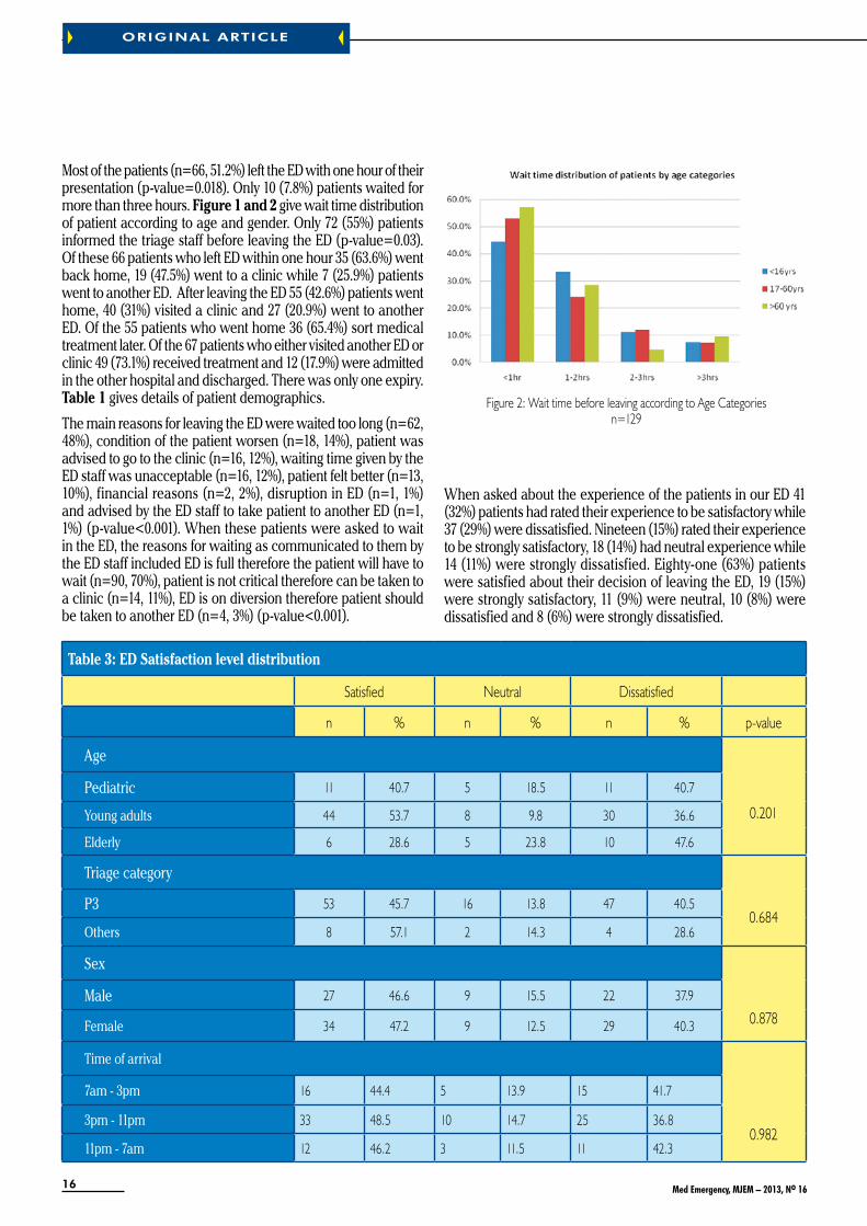
Figure 2: Wait time before leaving according to Age Categoriesn=129
Most of the patients (n=66, 51.2%) left the ED with one hour of their presentation (p-value=0.018). Only 10 (7.8%) patients waited for more than three hours. figure 1 and 2 give wait time distribution of patient according to age and gender. Only 72 (55%) patients informed the triage staff before leaving the ED (p-value=0.03). Of these 66 patients who left ED within one hour 35 (63.6%) went back home, 19 (47.5%) went to a clinic while 7 (25.9%) patients went to another ED. After leaving the ED 55 (42.6%) patients went home, 40 (31%) visited a clinic and 27 (20.9%) went to another ED. Of the 55 patients who went home 36 (65.4%) sort medical treatment later. Of the 67 patients who either visited another ED or clinic 49 (73.1%) received treatment and 12 (17.9%) were admitted in the other hospital and discharged. There was only one expiry. table 1 gives details of patient demographics.
The main reasons for leaving the ED were waited too long (n=62, 48%), condition of the patient worsen (n=18, 14%), patient was advised to go to the clinic (n=16, 12%), waiting time given by the ED staff was unacceptable (n=16, 12%), patient felt better (n=13, 10%), financial reasons (n=2, 2%), disruption in ED (n=1, 1%) and advised by the ED staff to take patient to another ED (n=1, 1%) (p-value<0.001). When these patients were asked to wait in the ED, the reasons for waiting as communicated to them by the ED staff included ED is full therefore the patient will have to wait (n=90, 70%), patient is not critical therefore can be taken to a clinic (n=14, 11%), ED is on diversion therefore patient should be taken to another ED (n=4, 3%) (p-value<0.001).
When asked about the experience of the patients in our ED 41 (32%) patients had rated their experience to be satisfactory while 37 (29%) were dissatisfied. Nineteen (15%) rated their experience to be strongly satisfactory, 18 (14%) had neutral experience while 14 (11%) were strongly dissatisfied. Eighty-one (63%) patients were satisfied about their decision of leaving the ED, 19 (15%) were strongly satisfactory, 11 (9%) were neutral, 10 (8%) were dissatisfied and 8 (6%) were strongly dissatisfied.
ORIgINAL ARTICLE

17 Med Emergency, MJEM – 2013, No 16
Of all the patients 95 (74%) still preferred coming to our ED while 91 (71%) said that they would recommend our ED to others. table 2 gives details of patient perception and level of satisfaction. No significant differences were observed in ED satisfaction among different categories (table 3).
disCussion
Hospital emergency departments have evolved as an integral part of health care services bridging the gap between primary and specialty services by providing care 24/7 [14]. EDs are working mostly beyond their intended working capacity because of increasing patient volumes, high acuity patients presenting to ED requiring prolong stay for evaluation and limited inpatient beds leading to overcrowding [15, 19]. As a consequence considerable number of patients may leave without being seen by a physician [20]. Our study had shown the characteristics and contributing factors of patients who LWBS. The uncompleted visit rate was 13% during the study period which is although higher but not atypical for an ED which is located in highly populated urban area [21].
Most common reasons quoted by patients (61.1%) who left without being seen by physicians is prolong waiting time while 10% quoted improvement in their symptoms as the reason for leaving. A study on 222 LWBS patients from Australia showed that 49% of patients left because of waiting and that 52% of these patients would not left if the waiting time was shorter [22]. Another study by Arendt et al also quoted too long waiting time as a reason of leaving in 67% of patients [23]. Half of the patients left within one hour and two third of patients left within two hours. Females tend to leave earlier possibly reflecting the existing gender iniquity and community norms , where females are mostly responsible for taking care of their household matters and they may have smaller children waiting at home because of that she don’t want to wait longer and left [24, 26]. This fact needs to be further explored.
Most of the patients who left were high acuity needing emergency or urgent medical care while who wait the longest was comparatively walk in stable patients. This is in contrary with other studies showing the dose response relationship between LWBS and triage category [12, 27, 28]. A study from UK had shown 38.1%, 56.1%, 2.7% of patients leaving belong to triage category P3, P4 and P5 respectively [6]. Our study had shown that 89% of LWBS patients were P3 category probably because of increasing numbers of life threatening and critical patients are boarded in ED due to non-availability of high dependency unit (HDU) beds in hospital.
Majority of the patients leaving were young adults (16-60 years) as reported internationally. Interestingly when the wait time before leaving was stratified in three age groups we observed that 85.7% of elderly patients are leaving within two hours although not statistically significant but may be reflecting critical nature of their illness as evident by triage category (95.2% elderly were triaged as emergent or urgent). No adverse events had been reported in our study population. Most of the patients (54%) went home while one third (27%) were admitted to another ED and subsequently got discharged. This observation could not be generalized because we are not able to contact all the patients who LWBS.
One expiry was reported in our study which occurred in another hospital after two weeks of hospitalization. This patient belonged to P3 category. It is our protocols that even during diversion hours critically ill patients with life threatening conditions are continued to be seen and resuscitated in ED irrespective of ED occupancy status. It is mostly P3 patients that are sent to the waiting area after triaging. It could be possible that these patients initially present with nonspecific signs and symptoms of critical illness that was missed on initial assessment at triage.
An encouraging observation was that 75% of patients were satisfied with the healthcare services provided at AKU-ED and more than three quarter not only prefers to come to AKU-ED but also recommend it to others.
The Limitation that we found in our study includes the recall bias associated with survey research in general. Another bias was the non-response bias as we were able to complete survey in 129 (35%) patients only .Among the patients who did not responded 241 (65%), only 6 (2.5%) refused while in 169 (70.1%) cell phone was switched off despite trying several times and 66 (27.4%) the number was incorrect. Therefore study results should be considered cautiously keeping the fact in mind that we are not able to contact all the patients who were LWBS and the answers of the non-contacted LWBS may have been different. But only few in the study refused to participate; therefore the result would not have been significantly different if all of the patients had been interviewed.
ConClusion
Strategies aimed to reduce wait t ime and improving communication regarding estimated waiting time can decrease the patients who left without being seen. This could be improved by having a dedicated triage team including a physician and a trained staff at triage.
ORIgINAL ARTICLE

18 Med Emergency, MJEM – 2013, No 16
ORIgINAL ARTICLE
annEX 1
TRIAGE FLOW CHARTAnnex 2: TRIAGE FLOW CHART
Triage
Assessment
Priority
P1
Immediately
P2
Within 15 min
P3
Within 60
P4
Within 120
Resuscitation Room
Resuscitation room
Critical Care area
Urgent Care Area
Clinical Decision Unit
Treatment Area Waiting Area
If no bed available in patient care area If no bed available in patient care area
Left without being seen
(helped with Emergency Severity Index-Iv)

19 Med Emergency, MJEM – 2013, No 16
REfEREnCEs
1. Polevoi SK, Quinn Jv, Kramer NR. Factors associated with patients who leave without being seen. Academic emergency medicine 2005 ;12(3):232-6.2. Weiss SJ, Ernst AA, Derlet R, King R, Bair A, Nick TG. Relationship between the National ED Overcrowding Scale and the number of patients who leave without being seen in an academic ED. Am J Emerg Med 2005 ;23(3):288-94.3. Ding R, McCarthy ML, Li G, Kirsch TD, Jung JJ, Kelen GD. Patients who leave without being seen: their characteristics and history of emergency department use. Ann Emerg Med 2006 ;48(6):686-93.4. Arendt KW, Sadosty AT, Weaver AL, Brent CR, Boie ET. The left-without-being-seen patients: what would keep them from leaving? Ann Emerg Med 2003; 42(3):317-23.5. Kelen GD, Scheulen JJ, Hill PM. Effect of an emergency department (ED) managed acute care unit on ED overcrowding and emergency medical services diversion. Acad Emerg Med 2001; 8(11):1095-100.6. Goodacre S, Webster A. Who waits longest in the emergency department and who leaves without being seen? Emerg Med J 2005; 22(2):93.7. Baker DW, Stevens CD, Brook RH. Patients who leave a public hospital emergency department without being seen by a physician. JAMA: 1991; 266(8):1085.8. Sainsbury S. Emergency patients who leave without being seen: are urgently ill or injured patients leaving without care? Mil Med 1990; 155(10):460.9. Hsia RY, Asch SM, Weiss RE, Zingmond D, Liang LJ, Han W, et al. Hospital Determinants of Emergency Department Left Without Being Seen Rates. Ann Emerg Med 2011; 58(1):24-32.10. Mohsin M, Young L, Ieraci S, Bauman AE. Factors associated with walkout of patients from New South Wales hospital emergency departments, Australia. Emerg Med Australas 2005; 17(56):434-42.11. Chan TC, Killeen JP, Kelly D, Guss DA. Impact of rapid entry and accelerated care at triage on reducing emergency department patient wait times, lengths of stay, and rate of left without being seen. Ann Emerg Med 2005; 46(6):491-7.12. Mohsin M, Forero R, Ieraci S, Bauman AE, Young L, Santiano N. A population follow-up study of patients who left an emergency department without being seen by a medical officer. Emerg Med J 2007; 24(3):175.13. Johnson M, Myers S, Wineholt J, Pollack M, Kusmiesz AL. Patients who leave the emergency department without being seen. J Emerg Nurs 2009; 35(2):105-8.14. Schneider SM, Hamilton GC, Moyer P, Stapczynski JS. Definition of emergency medicine. Acad Emerg Med 1998; 5(4):348-51.15. Derlet RW, Richards JR, Kravitz RL. Frequent overcrowding in US emergency departments. Acad Emerg Med 2001; 8(2):151-5.16. Derlet RW, Richards JR. Overcrowding in the nation’s emergency departments: complex causes and disturbing effects. Ann Emerg Med 2000; 35(1):63-8.17. Lambe S, Washington DL, Fink A, Herbst K, Liu H, Fosse JS, et al. Trends in the use and capacity of California’s emergency departments, 1990-1999. Ann Emerg Med 2002; 39(4):389-96.18. Derlet RW. Overcrowding in emergency departments: increased demand and decreased capacity. Ann Emerg Med 2002; 39(4):430.19.MeggsWJ,CzaplijskiT,BensonN.TrendsinEmergencyDepartmentUtilization,1988â€�1997.AcadEmergMed1999;6(10):1030-5.20. Kennedy M, MacBean CE, Brand C, Sundararajan v, McD Taylor D. Review article: leaving the emergency department without being seen. Emerg Med Australas 2008; 20(4):306-13.21. Cunningham PJ. What accounts for differences in the use of hospital emergency departments across US communities? Health Affairs 2006; 25(5):w324-w36.22. Fry M, Thompson J, Chan A. Patients regularly leave emergency departments before medical assessment: a study of did not wait patients, medical profile and outcome characteristics. Aust Emerg Nurs J 2004; 6(2):21-6.23. Arendt KW, Sadosty AT, Weaver AL, Brent CR, Boie ET. The left-without-being-seen patients: What would keep them from leaving? Ann Emerg Med 2003; 42(3):317-IN2.24. Agha S. The determinants of infant mortality in Pakistan. Soc Sci Med 2000; 51(2):199-208.25. Khan MM, Reza H. Gender differences in nonfatal suicidal behavior in Pakistan: Significance of sociocultural factors. Suicide Life Threat Behav 1998; 28(1):62-8.26. Kazi S. Gender inequalities and development in Pakistan. Shahrukh Rafi Khan (eds). 1999; 50.27. Clarey A, Cooke M. Patients who leave emergency departments without being seen: literature review and English data analysis. Emerg Med J 2012; 29(8):617-21.28. Ding R, McCarthy ML, Li G, Kirsch TD, Jung JJ, Kelen GD. Patients who leave without being seen: their characteristics and history of emergency department use. Ann Emerg Med 2006; 48(6):686-93.
ORIgINAL ARTICLE

20 Med Emergency, MJEM – 2013, No 16
EmERgENCy DEvELOpmENT
how to optimize CArdioCereBrAl resusCitAtion (CCr) stAndArds in 2013
HOEPPLI F, BANERJEE P, GARCIA W, HSSAIN I. How to optimize cardiocerebral resuscitation (CCR) standards in 2013. Med Emergency, MJEM 2013; 16:20-25keywords: Cardiac arrest, passive oxygenation, intraosseous access, impedance threshold device, double sequence defibrillation, hypothermic treatment.
Authors’ affiliation:
Correspondent author: HOEPPLI FredericAmbulances Sud Fribourgeois, Vaulruz, Switzerland144 Fribourg, Fribourg, [email protected]
Frederic Hoeppli1, Paul Banerjee2, MD, Wenceslao Garcia3, MD, Ismaël Hssain4, MD, MSc1. Swiss Paramedic & Dispatcher AMPDS, Switzerland2. Emergency departments at Citrus Memorial Hospital, USA3. Emergency department at Fribourg Hospital, Fribourg, Switzerland4. Department of Emergency Medicine, Prehospital Care and HEMS, Mulhouse General Hospital, France
Article history / info: Category: Emergency DevelopmentReceived: Jun. 29, 2013Revised: Jul. 26, 2013 Accepted: Aug. 15, 2013
Conflict of interest statement: There is no conflict of interest to declareI would like to clarify that there is no contract, compensation of any kind with the brands mentioned in this article. Other equally effective devices as described below exist on the market.
ABstrACtbackground: Based on the guidelines 2010 and regarding the next version 2015, this article shows the new possibilities to care about the cardiac arrest casualties, is there any increase of return of spontaneous circulation (ROSC), survival rate and neurological outcome? Do we need to adapt our emergency care for the future?
Methods: Using a simulation case, the CCR pathway brings the new techniques and devices on the front of the scene. The association between them was a critical point to get a positive alchemy.
Results: Based on studies, evidence based-medicine and field experiences in different companies around the world, we can note a good outcome between all this new supports for our patients.
Conclusion: This pathway shows, with a good survival chain, an important improvement to the cardiac arrest care and gives a new vision for the reanimation pre and in-hospital.

21 Med Emergency, MJEM – 2013, No 16
EmERgENCy DEvELOpmENT
introduCtion
It has already been three years since the «new» resuscitation guidelines are in force and we are looking forward to 2015 for the new edition. This article will offer you an overview of avant-garde that it can respond to various published studies, conferences and relatively/appearance new technologies.
In cardiac arrest care the “chain of survival” is central to all emergencies requiring resuscitation (figure 1 & 2). The proposed changes that are listed below focuses on the links «professional» and do not in any way challenge the work that is or should be done by the first three links (early access - early CCR - early defibrillation). It will be not discussed in this text the famous question «whether or not to initiate resuscitation for some victims».
This article highlights the changes that you may already apply in your various services / companies / hospitals. These ideas will help improve ROSC, survival to discharge rates and neurological outcomes post-CA in both the pre and hospital setting.
abbREviations:
CA = cardiac arrestROSC = return of spontaneous circulationOPA = oropharyngeal airwayCCR = cardiocerebral resuscitationAED = automated external defibrillatorvF/pulselessvT = ventricular fibrillation/pulseless ventricular tachycardiaLucas2 = Lund University Cardiac Arrest System, generation 2IO = intraosseousIv = intravenousPEA = pulseless electrical activityLEMON = Look Evaluation Mallampati Obstruction NeckETCO2 = End-tidal carbon dioxideITD = impedance threshold deviceHT = hypothermic treatmentPCI = percutaneous coronary intervention
1) Initial impression of the safety, the situation and the scene
2) Quick check based on CAB place on a monitor – 5-10 seconds
3) Start compressions (100-120/min)
4) Single & double sequence defibrillation
5) Passive oxygenation with OPA
6) Lucas2®
7) Iv vs. IO
8) Drugs
9) Advanced airways
10) ResQpod®
11) RhinoChill®
oVerView - ApproACh And mAnAgement of A person in CArdiAC Arrest

22 Med Emergency, MJEM – 2013, No 16
EmERgENCy DEvELOpmENT
Figure 2 : Chain of survival
Figure 4 : Double sequence defibrillation
Figure 3 : Simulation sequence
simulAtion - the perfeCt CliniCAl CAse
Consider the case of a person who had a heart attack in the street, witnesses recognize the cardiac arrest, call 911 or 112 (or any other emergency dispatch center), start compressions and defibrillator is on the way from a pharmacy equipped with a AED (information provided by the dispatch center). Witnesses take turns every two minutes to ensure effective compressions, 5cm deep for a rate between 100/120 per minute, not exceeding 125 because the rate of ROSC would decreased [1-4]. The defibrillator is applied in accordance with its usage. If there is presence of shockable rhythm, the patient is defibrillated. Otherwise, compressions resume immediately. If ROSC does not occur, CCR by witnesses must continue until the arrival of professionals (figure 3).
After the ambulance arrives, rescuers leave their vehicle analysing the scene, it appears to be safe (do not forget that safety is a dynamic process and may change at any time). They go to the patient, and the situation is explained to them. The monitor/defibrillator manual is set up, the AED can remain in place for potential refractory vF (we will see it below).
The professionals perform a quick check to make sure there is CA, breathing and carotid pulse controls are done simultaneously in 5-10secs max [5]. After confirming the CA, the compressions resumes. If there is presence of vF/pulselessvT, shock will be given (200J biphasic) (figure 4). Three ice packs will be applied if a HT will be done, considering environment and other factors that could interfere with HT, one to head and two on carotids [5].
The airway management involving suction and oropharyngeal airway is performed. A high concentration mask at 15L/min is introduced, allowing the supply of passive O2, possibly increasing ROSC and improving neurological outcomes post-CA, specially in witnessed vF patient [5-7].
The mechanical compression device is set up; the complete installation of Lucas2® should interrupt compressions only a very short time [1, 8]. The education and training on this device are critical points; the Lucas® will afford effective compressions without interruption or any kind of alteration throughout the code, that could increase the ROSC, survival rate and neurological outcome [8-10].
Once the mechanical compression system is enabled, secure and verified, one of the rescuers will prepare the drugs and the intubation, leaving the implementation of Iv or IO to his colleague. The Iv and IO are both workforce access on a CA code [4, 5]. The device with the greatest chance of success should be used in the first attempt (figure 5).
The IO access is placed on humeral site, delivering probably a better flow rate than the tibia site and the medications are ready to be injected. If there is presence of vF/pulselessvT, the patient is defibrillated as the first time (200J biphasic). After the first four shocks (including the use of AED like a one shock), the double sequence will be tried for the refractory vF using a second monitor/AED (400J biphasic - one time 200J by AED/first defibrillator and one time 200J by second defibrillator all managed by one rescuer who synchronise and push the two buttons in the exact same time to get the double sequence at 400J), there are promising results on the double sequence defibrillation, high energy 720J (two times 360J biphasic) has shown results [5].

23 Med Emergency, MJEM – 2013, No 16
EmERgENCy DEvELOpmENT
Figure 5 : IO access up
Without presence of ROSC, mechanical compressions resumes, vasopressin (40U) and epinephrine (1mg/3-5min) are administered at the same time (if the rhythm is a PEA, some services are also being tested for a cocktail administration of several drugs at once - HCO3, D50, 500mL NaCl, naloxone, in order to exclude a maximum of 5H-5T). Consider alternatively twice amiodarone (300mg and 150mg). One other recent study is trying a new revolutionary way, focus on hemodynamic pressure and not vasoconstriction using sodium nitroprusside, but it needs more time and tests on human body before to change anything [11].
Rescuers turn to the end of the preparation for intubation equipment and the establishment of the tube, either an endotracheal tube or supraglottic device (king airway®, combi-tube®, i-gel®, etc.). The ET tube still remains the most appropriate way to secure airway and ventilate a patient, but it still requires a regular and proper training to be effective and not become deleterious for patient taking time of the chest for compressions. Alternative means allow to have a probability of successful implementation almost faster than ET intubation while effective. Tests are running to determine if the pressure on the proximal balloon affect on the carotid blood flow for the king tube [5].
The rescuer located at the head of the patient opts for an alternative device, based on the LEMON. Under compressions, a king airway is applied, controlled and secure. The ETCO2
is 18mmHg what is right for one person in CA under good compressions. The sudden rise of CO2, around 30mmHg or more would be a sign of ROSC, allowing the team to anticipate the following intervention. But the patient is still pulseless [5].
Figure 6a : Focus on the connection ITD, filter and ETCO2
Figure 6b : Same connection without the filter
24 Med Emergency, MJEM – 2013, No 16
EmERgENCy DEvELOpmENT
A ResQpod® device (ITD) is placed between the detector ETCO2
and the tube. This device should allow better venous return by negative intrathoracic pressure, increasing the amount of ventricular ejection, improving by about 25% the carotid blood flow and the cerebral perfusion. This process increases, among others, the survival rate. To be an effective tool, the ResQpod® need a leak-free tube in place & optimal compressions-decompressions device [8, 12, 13] (figure 6a & 6b).
The introduction of RhinoChill® device to initiate therapeutic hypothermic by an addition intranasal cold vapor to improve, among other things, the ROSC and the neurological outcomes post-CA [14, 15].
The patient is then set for transport if presence of ROSC occurs and taken to an appropriate hospital for further cares (PCI, continuity of HT). If there is a ROSC, keep the patient in a state of moderate hypothermia (32-34oC) and prevent any shaking or shivering by medication are important points [5].
ConClusion
It is the «dreamed mega-code» for a rescuer who tries to be optimal about the recovering probability for a cardiac arrest. It goes without saying that the implementation of this type of resuscitation is not free, funds, time, special training for rescuers, as well as skills need to be maintained. This is valid whatever is our job (paramedics, physicians, nurses, ambulanciers, firefighters, cops, etc.).
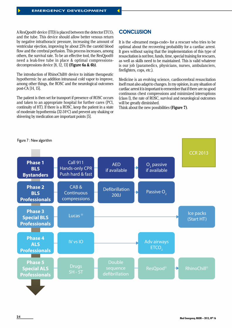
Medicine is an evolving science, cardiocerebral resuscitation itself must also adapt to changes. In my opinion, in any situation of cardiac arrest it is important to remember that if there are no good continuous chest compressions and minimized interruptions (class I), the rate of ROSC, survival and neurological outcomes will be greatly diminished.Think about the new possibilities (figure 7).
Phase 1BLS
Bystanders
Call 911 Hands-only CPRPush hard & fast
AEDif available
O2 passiveif available
Phase 2BLS
Professionals
CAB &Continuous
compressions
Lucas
IV vs IO Adv airwaysETCO2
Drugs5H - 5T
ResQpod RhinoChillDouble
sequencede�brillation
Ice packs(Start HT)
De�brillation200J
Passive O2
Phase 3Special BLS
Professionals
Phase 4ALS
Professionals
Phase 5Special ALS
Professionals
CCR 2013
Figure 7 : New algorithm

25 Med Emergency, MJEM – 2013, No 16
EmERgENCy DEvELOpmENT
REfEREnCEs1. Berg RA, Hemphill R, Abella BS, Aufderheide TP, Cave DM, Hazinski MF, et al. Part 5: adult basic life support: 2010 American Heart Association Guidelines for Cardiopulmonary Resuscitation and Emergency Cardiovascular Care. Circulation 2010; 122(18):S685–705. 2. Nolan JP, Perkins GD, Soar J. Chest compression rate: where is the sweet spot? Circulation 2012; 125(24):2968–70. 3. Idris AH, Guffey D, Aufderheide TP, Brown S, Morrison LJ, Nichols P, et al. Relationship between chest compression rates and outcomes from cardiac arrest. Circulation 2012; 125(24):3004–12. 4. EMS State of the Science 2012 - [Internet]. JEMS.com. [cited 2013 Nov 7]. Available from: http://www.jems.com/special/state-of-the-science-20125. Neumar RW, Otto CW, Link MS, Kronick SL, Shuster M, Callaway CW, et al. Part 8: adult advanced cardiovascular life support: 2010 American Heart Association Guidelines for Cardiopulmonary Resuscitation and Emergency Cardiovascular Care. Circulation 2010; 122(18):S729–767. 6. Bobrow BJ, Ewy GA, Clark L, Chikani v, Berg RA, Sanders AB, et al. Passive oxygen insufflation is superior to bag-valve-mask ventilation for witnessed ventricular fibrillation out-of-hospital cardiac arrest. Ann Emerg Med 2009; 54(5):656–662.e1. 7. Bobrow BJ, Ewy GA. ventilation during resuscitation efforts for out-of-hospital primary cardiac arrest. Curr Opin Crit Care 2009; 15(3):228–33. 8. Cave DM, Gazmuri RJ, Otto CW, Nadkarni vM, Cheng A, Brooks SC, et al. Part 7: CPR techniques and devices: 2010 American Heart Association Guidelines for Cardiopulmonary Resuscitation and Emergency Cardiovascular Care. Circulation 2010; 122(18):S720–728. 9. Dp K. The merits of mechanical CPR: Do mechanical devices improve compression consistency and resuscitation outcomes? Jems J Emerg Med Serv 2012; 37(9):24–9. 10. Adedipe A, Nichol G. To push or not to push: manual or mechanical compressions for cardiac arrest? Crit Care Med 2013; 41(7):1824–6. 11. Yannopoulos D, Matsuura T, Schultz J, Rudser K, Halperin HR, Lurie KG. Sodium nitroprusside enhanced cardiopulmonary resuscitation improves survival with good neurological function in a porcine model of prolonged cardiac arrest. Crit Care Med 2011; 39(6):1269–74. 12. Lurie K, Setum C. An impedance threshold device in out-of-hospital cardiac arrest. N Engl J Med 2012; 366(2):185–86; author reply 186–88. 13. Aufderheide TP, Frascone RJ, Wayne MA, Mahoney BD, Swor RA, Domeier RM, et al. Standard cardiopulmonary resuscitation versus active compression-decompression cardiopulmonary resuscitation with augmentation of negative intrathoracic pressure for out-of-hospital cardiac arrest: a randomised trial. Lancet 2011; 377(9762):301–11. 14. Busch H-J, Eichwede F, Födisch M, Taccone FS, Wöbker G, Schwab T, et al. Safety and feasibility of nasopharyngeal evaporative cooling in the emergency department setting in survivors of cardiac arrest. Resuscitation 2010; 81(8):943–9. 15. Yu T, Barbut D, Ristagno G, Cho JH, Sun S, Li Y, et al. Survival and neurological outcomes after nasopharyngeal cooling or peripheral vein cold saline infusion initiated during cardiopulmonary resuscitation in a porcine model of prolonged cardiac arrest. Crit Care Med 2010; 38(3):916–21.

26 Med Emergency, MJEM – 2013, No 16
MABIT H, DOMANSKI L, ROCHE B. Pertinence de la transfusion de concentrés de globules rouges aux urgences. Med Emergency, MJEM 2013; 16:26-31Mots clés : Transfusion, concentrés de globules rouges, urgenceskey words: Transfusion, blood red cell, emergency
pertinenCe de lA trAnsfusion de ConCentrÉs de gloBules rouges AuX urgenCes pertinence of red blood cell (rBC) transfusion in emergency
Authors’ affiliation:Dr Hélène MABIT : Correspondent authorCentre Médical des Armées de Cazaux.Base aérienne 120. BP 70 413. 33 164 La [email protected]
Laurent Domanski1, MD, Brigitte Roche2, MD1. Service médical d’urgence, Brigade de Sapeur-Pompier de Paris, France2. Laboratoire de biologie, hôpital de Langon, France
Article history / info: Category: Original articleReceived: Apr.1, 2013Accepted: Apr. 30, 2013Published in Urgence Pratique
Conflict of interest statement: There is no conflict of interest to declare
ORIgINAL ARTICLE
ABstrACtobjective: The purpose of the study was to assess appropriateness of red blood cell (RBC) transfusion in Robert Picqué French military hospital emergency, because of the lack of blood cell on the “Etablissement français du sang” (EFS), to improve practices in relation with recommendation of French national guidelines (AFFSAPS nowadays ANSM) published in 2002.
Method: We conducted a retrospective study covering one year, including 67 RBC transfusions in emergencies.
Results: All the transfusions wasn’t useful (80%), kind of blood wasn’t appropriate all the time, emergency degree of the prescription wasn’t suitable. 25% of blood prescription respected all criteria.
Discussion: To improve these practices, we have done a computerization of the prescription to decrease the delay and increase its quality, and a decisional tree for the prescription’s decision.
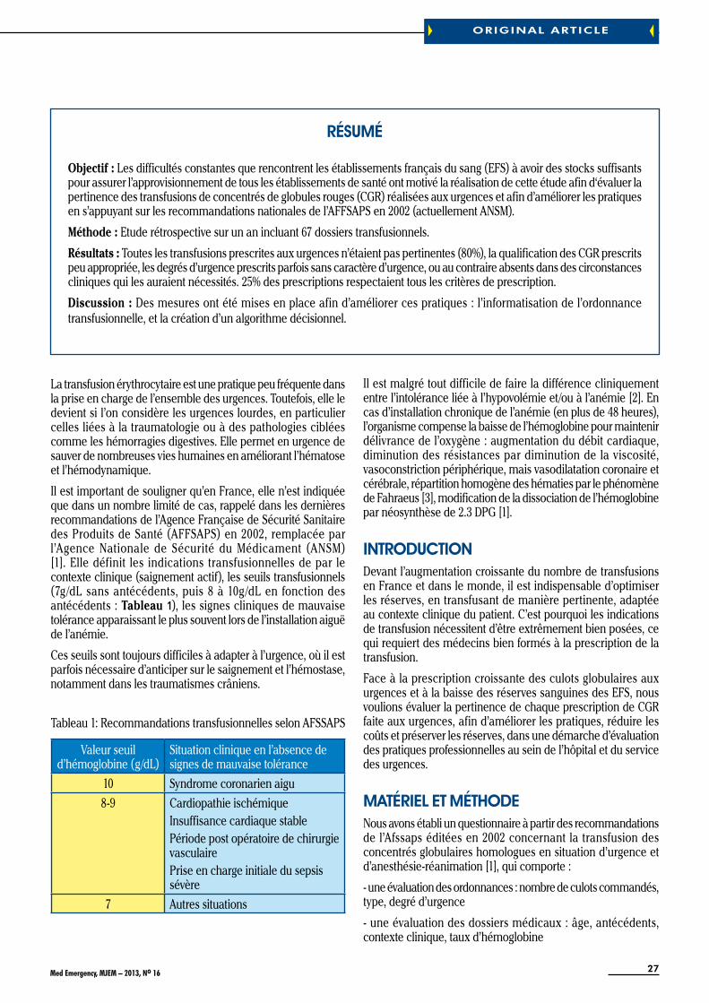
27 Med Emergency, MJEM – 2013, No 16
ORIgINAL ARTICLE
rÉsumÉ
objectif : Les difficultés constantes que rencontrent les établissements français du sang (EFS) à avoir des stocks suffisants pour assurer l’approvisionnement de tous les établissements de santé ont motivé la réalisation de cette étude afin d‘évaluer la pertinence des transfusions de concentrés de globules rouges (CGR) réalisées aux urgences et afin d’améliorer les pratiques en s’appuyant sur les recommandations nationales de l’AFFSAPS en 2002 (actuellement ANSM).
Méthode : Etude rétrospective sur un an incluant 67 dossiers transfusionnels.
Résultats : Toutes les transfusions prescrites aux urgences n’étaient pas pertinentes (80%), la qualification des CGR prescrits peu appropriée, les degrés d’urgence prescrits parfois sans caractère d’urgence, ou au contraire absents dans des circonstances cliniques qui les auraient nécessités. 25% des prescriptions respectaient tous les critères de prescription.
Discussion : Des mesures ont été mises en place afin d’améliorer ces pratiques : l’informatisation de l’ordonnance transfusionnelle, et la création d’un algorithme décisionnel.
La transfusion érythrocytaire est une pratique peu fréquente dans la prise en charge de l’ensemble des urgences. Toutefois, elle le devient si l’on considère les urgences lourdes, en particulier celles liées à la traumatologie ou à des pathologies ciblées comme les hémorragies digestives. Elle permet en urgence de sauver de nombreuses vies humaines en améliorant l’hématose et l’hémodynamique.
Il est important de souligner qu’en France, elle n’est indiquée que dans un nombre limité de cas, rappelé dans les dernières recommandations de l’Agence Française de Sécurité Sanitaire des Produits de Santé (AFFSAPS) en 2002, remplacée par l’Agence Nationale de Sécurité du Médicament (ANSM) [1]. Elle définit les indications transfusionnelles de par le contexte clinique (saignement actif), les seuils transfusionnels (7g/dL sans antécédents, puis 8 à 10g/dL en fonction des antécédents : tableau 1), les signes cliniques de mauvaise tolérance apparaissant le plus souvent lors de l’installation aiguë de l’anémie.
Ces seuils sont toujours difficiles à adapter à l’urgence, où il est parfois nécessaire d’anticiper sur le saignement et l’hémostase, notamment dans les traumatismes crâniens.
Tableau 1: Recommandations transfusionnelles selon AFSSAPS
valeur seuil d’hémoglobine (g/dL)
Situation clinique en l’absence de signes de mauvaise tolérance
10 Syndrome coronarien aigu8-9 Cardiopathie ischémique
Insuffisance cardiaque stablePériode post opératoire de chirurgie vasculairePrise en charge initiale du sepsis sévère
7 Autres situations
Il est malgré tout difficile de faire la différence cliniquement entre l’intolérance liée à l’hypovolémie et/ou à l’anémie [2]. En cas d’installation chronique de l’anémie (en plus de 48 heures), l’organisme compense la baisse de l’hémoglobine pour maintenir délivrance de l’oxygène : augmentation du débit cardiaque, diminution des résistances par diminution de la viscosité, vasoconstriction périphérique, mais vasodilatation coronaire et cérébrale, répartition homogène des hématies par le phénomène de Fahraeus [3], modification de la dissociation de l’hémoglobine par néosynthèse de 2.3 DPG [1].
introduCtionDevant l’augmentation croissante du nombre de transfusions en France et dans le monde, il est indispensable d’optimiser les réserves, en transfusant de manière pertinente, adaptée au contexte clinique du patient. C’est pourquoi les indications de transfusion nécessitent d’être extrêmement bien posées, ce qui requiert des médecins bien formés à la prescription de la transfusion.
Face à la prescription croissante des culots globulaires aux urgences et à la baisse des réserves sanguines des EFS, nous voulions évaluer la pertinence de chaque prescription de CGR faite aux urgences, afin d’améliorer les pratiques, réduire les coûts et préserver les réserves, dans une démarche d’évaluation des pratiques professionnelles au sein de l’hôpital et du service des urgences.
mAtÉriel et mÉthodeNous avons établi un questionnaire à partir des recommandations de l’Afssaps éditées en 2002 concernant la transfusion des concentrés globulaires homologues en situation d’urgence et d’anesthésie-réanimation [1], qui comporte :
- une évaluation des ordonnances : nombre de culots commandés, type, degré d’urgence
- une évaluation des dossiers médicaux : âge, antécédents, contexte clinique, taux d’hémoglobine

28 Med Emergency, MJEM – 2013, No 16
- une évaluation de la pertinence de la prescription, de la quantité, de la qualité des culots globulaires et du degré d’urgence
- une évaluation de la délivrance : en adaptation au degré d’urgence
La population étudiée dans cette étude était âgée de plus de 15 ans et 3 mois, et ne présentait pas de polytraumatisme, ni d’hémorragie d’origine gynécologique (sauf s’il ne s’agissait pas du motif initial de consultation), partant du fait que l’d’Instruction des Armées d’Instruction des Armées (HIA) Robert Picqué est un hôpital militaire qui ne dispose pas de spécialiste en pédiatrie et en gynécologie-obstétrique, et que le plateau technique chirurgical ne permet pas la prise en charge des polytraumatisés graves, habituellement régulés par le centre 15 vers le déchocage du CHU de Bordeaux.
Seules les transfusions de CGR homologues ont été étudiées, les transfusions autologues ne se pratiquent qu’en postopératoire et peu en préhospitalier.
Les résultats de cette étude ne peuvent donc pas être comparés à un autre service d’urgence prenant en charge ces pathologies et/ou pratiquant la transfusion autologue.
Les données ont été analysées grâce au Logiciel Sphinx.
rÉsultAts et AnAlyse
démographieCette étude s’appuie sur l’analyse de 67 prescriptions de CGR, soit 60 dossiers transfusionnels, de patients ayant consulté aux urgences de l’HIA Robert Picqué de mai 2006 à avril 2007 et pour lesquels des CGR ont été prescrits. Plusieurs prescriptions ont été réalisées pour un même patient : 7 patients ont été transfusés deux fois, dont 3 en moins de 24 heures.
Ces 60 dossiers transfusionnels représentaient 0,2 % de la totalité des patients ayant consulté de mai 2006 à avril 2007, soit peu de patients par rapport à la fréquentation globale du service.
Sur l’année, ces 67 prescriptions représentaient 190 CGR, soit le troisième taux de CGR prescrit la même année sur tout l’hôpital, après la réanimation et la médecine interne. Sur ces 67 prescriptions de CGR, seules 66 transfusions ont été réalisées (aux urgences ou ailleurs), une n’a pas été effectuée en raison du transfert du patient vers un autre établissement de santé, soit 2 CGR phénotypés.
Sur la période étudiée, 9,5% des CGR prescrits (n=18/190) n’ont pas été transfusés, seules 82 % des prescriptions ont été transfusées en entier.
La majorité des CGR prescrits et transfusés l’a été aux urgences ; le reste était, soit transfusé dans les services (14%), soit, ne l’était pas (9%).
Le nombre médian de CGR transfusés est de deux, avec pour extrêmes un à six. Cependant, dans certaines situations, la quantité prescrite est souvent trop importante ou prescrite malgré le transfert du patient. Le même constat a été réalisé dans une étude multicentrique de 2010 sur 9 établissements hospitaliers [4]. A l’inverse, deux commandes d’un seul CGR ont été recensées,
correspondant à moins de 3% des prescriptions. Or, il est prouvé, dans les recommandations de l’AFSSAPS, que la transfusion d’un seul CGR n’est pas utile, puisqu’elle n’augmente le taux d’hémoglobine que d’un seul un point ou 1g/dL, ce qui n’est pas suffisant pour permettre la disparition des signes de mauvaise tolérance.
Ces prescriptions n’ont pas bénéficié d’une bonne évaluation bénéfice/risque : l’intérêt d’une transfusion est d’apporter une amélioration à l’état clinique du patient et de faire en sorte qu’elle soit supérieure aux risques qu’il encoure de par ce geste. Or, transfuser un seul CGR n’apporte pas plus de bénéfices qu’il n’existe de risques liés à cette transfusion. Ces deux prescriptions auraient donc pu être évitées ou, le nombre de CGR mieux calculé. Ce phénomène entraîne un gaspillage non négligeable de CGR : en un an, 16 CGR n’ont pas été transfusés, soit 8,5% des CGR prescrits. Même si ces CGR ont pu être réattribués, ce qui n’est possible qu’avec un dépôt de délivrance. Ces prescriptions inadaptées, en quantité de CGR, contribuent à l’utilisation inappropriée de produits rares et chers ( au 1er mars 2010, tarif CGR standard est d’environ 200 euros), mais aussi à la survenue d’incidents de type surcharge volémique, particulièrement dans la population des patients transfusés où la moyenne d’âge est élevée.
Les patients sont le plus souvent des femmes âgées (83,5% de plus de 71 ans, sexe ratio de 0,8).
La majorité des anémies transfusées étaient des anémies chroniques (52,2%). Or celles ci si elles sont bien tolérées, seuls la recherche et le traitement étiologique sont nécessaires [5].
Les hémorragies digestives étaient l’étiologie la plus fréquente retrouvée. Pour une part importante des patients (63% des patients étudiés), l’anémie était liée à la prise d’un traitement anticoagulant (antivitamine K ou AvK), antiagrégant plaquettaire ou anti-inflammatoires non stéroïdiens (AINS). Parmi les patients traités par AvK, la moitié présentaient des surdosages. Ces chiffres semblent être plus élevés que pour d’autres études du même type faites dans d’autres services d’urgences, mais ayant une population plus jeune [6]. Cet élément soulève le problème de la pertinence de ce type de traitement chez des personnes âgées souvent plus fragiles, exposées à des surdosages ou à une toxicité digestive plus importante. On peut facilement comprendre que la meilleure façon de diminuer la fréquence des transfusions chez ces patients, soit une remise en question du rapport bénéfice-risque plus fréquente et une surveillance plus rapprochée de ce traitement chez ces patients. L’HAS a récemment recommandé un suivi des patients sous AvK, par la création de cliniques des AvK. Il est possible d’envisager la mise en place d’une telle structure sur l’hôpital Robert Picqué, qui travaillerait en collaboration avec celle de l’hôpital Haut-Lévêque [7, 8].
pertinence de la prescriptionLa pertinence sur le contexte clinique a été établie en associant la présence de signes cliniques de mauvaise tolérance et/ou d’hémorragie active. 52% des prescriptions étaient pertinentes sur le contexte clinique. Trente sept patients présentaient des anémies chroniques et 22 d’entre eux ne présentaient pas de signes de mauvaise tolérance. Cependant, cette étude a également
ORIgINAL ARTICLE

29 Med Emergency, MJEM – 2013, No 16
pu faire l’objet de biais dans l’interprétation de données. Les signes de mauvaise tolérance, lorsqu’ils étaient présents, ont toujours été rapportés à l’anémie. Or, pour certains patients polypathologiques, ces signes pouvaient être l’expression d’une autre pathologie. Certaines prescriptions ont pu être interprétées abusivement comme pertinentes, devant la présence de signes dus à la décompensation d’une autre affection.
La mesure du taux d’hémoglobine était toujours réalisée avant que la décision et l’acte transfusionnel ne soient réalisés. La pertinence a été établie selon les recommandations de AFFSAPS [1]. La majorité des patients transfusés présentaient un taux d’hémoglobine entre 7 et 10g/dL. Le seuil transfusionnel moyen était de 7,36g/dL± 1,82 (extrêmes : 3,2 – 13,4). 67% des transfusions réalisées aux urgences en un an étaient pertinentes sur le taux d’hémoglobine. La majorité des prescriptions non conformes concernaient des taux d’hémoglobine compris entre 7 et 8g/dL, par excès de transfusion (tableau 2).
Tableau 2: Repartition des prescriptions non conformes selon l’hemoglobinemie pretransfusionnelle.
Hb prétransfusionnelle (g/dL) Nombre (%)moins de 7 0
7 à 8 11 (50)8 à 9 6 (27)9 à 10 2 (9)
plus de 10 3 (14)Total 22
Tableau 3: Pertinence transfusion pour chaque critere transfusionnel.
PERTINENCE Nombre FréquenceTransfusion 54 80,6%
Degré d'urgence 43 65,1%Qualification 29 43,9%
Transfusion Degré d'urgence 32 48,8%Transfusion Qualification 24 36,6%
Transfusion Qualification Degré d'urgence 17 25,7%
La pertinence de la prescription de la transfusion a été établie en croisant la pertinence sur le contexte clinique et/ou sur le taux d’hémoglobine. 80,6% des prescriptions de CGR réalisées aux urgences de mai 2006 à avril 2007 sont pertinentes (tableau 3).
pertinence de la qualificationLa pertinence de la qualification a été établie en fonction du contexte clinique (âge, sexe, antécédents transfusionnels, recherche d’Ant icorps ir réguliers ou R AI) grâce aux recommandations de l’AFFSAPS. La qualification n’était pas adaptée à la situation clinique pour 56,1% des prescriptions (n=37/66), correspondant à 25 prescriptions de sang phénotypé
et 5 de sang compatibilisé (tableau 3). Cette qualification phénotypée ne doit être prescrite uniquement dans certaines circonstances : femme en âge de procréer, patient dont l’espérance de vie est raisonnable (moins de 75 ans), « transfusions itératives». Ce dernier terme est souvent mal interprété : il correspond à des transfusions répétées et chroniques, et non pas à « plus d’une transfusion » ou à « transfusions ultérieures probables ». Or, la catégorie de patients la plus souvent transfusée aux urgences est majoritairement les sujets âgés, pour lesquels le risque d’immunisation est faible, et l’espérance de vie réduite.
La qualification « compatibilisé » n’est jamais adaptée à la situation biologique : aucune des cinq prescriptions de CGR compatibilisé faites en un an n’est justifiée. Lorsque la RAI est positive, seuls des CGR compatibilisés peuvent être prescrits et chaque prescription de sang compatibilisé n’est justifiée que par la positivité de la RAI, ou par des antécédents de RAI positive. L’étude montre que pour chaque prescription de sang compatibilisé, aucune RAI n’est positive ni les antécédents. La prescription d’un tel produit impose que celui-ci soit adressé à l’EFS-AL à Bordeaux, puis rapporté après le test de compatibilisation. Les coûts engagés sont donc plus importants et monopolisent plusieurs intervenants, pour un produit qui n’était pas justifié. En réalité, une requalification a été faite au dépôt de sang, validée avec les prescripteurs pour ces non-conformités.
Ces résultats semblent inférieurs à d’autres études, dont celle de Gouezec et al. [4], qui montrait un taux de conformité de 66%.
pertinence du niveau d’urgence Dans 7 cas, soit 58% des prescriptions urgentes.
Le niveau d’urgence regroupe l’absence d’urgence, l’urgence relative et l’urgence vitale immédiate. Dans 7 cas, soit 58% des prescriptions urgentes, la prescription en urgence était pertinente. Les prescriptions qui ne l’étaient pas, devant l’absence de contexte hémorragique ou de mauvaise tolérance (choc…), comportaient 3 prescriptions vitales immédiates, et 2 relatives. Pour 16 prescriptions non urgentes, soit 29% de celles-ci, la situation clinique aurait mérité que les CGR soient délivrés en urgence, par la présence de signes d’intolérance ou par l’existence d’une hémorragie active.
pertinence de la prescription de transfusion aux urgencesIl est important de rappeler que les prescriptions non justifiées aux urgences incluent la transfusion qui n’a pas eu lieu et la prescription anticipée dans le cadre d’une intervention chirurgicale. 52,2% des prescriptions sont pertinentes dans le service des urgences. 47,8% des prescriptions ne le sont pas, puisque les patients ne présentaient pas de signes d’instabilité.
transfusions prescrites en respectant tous les critères transfusionnelsEnfin, la pertinence des prescriptions respectant tous les critères transfusionnels a été établie. Nous observons en croisant les données que seulement 25,7% des prescriptions pertinentes respectaient tous ces critères (tableau 3).
ORIgINAL ARTICLE

30 Med Emergency, MJEM – 2013, No 16
Ces critères correspondent à des non-conformités majeures et mineures. L’absence de pertinence de la prescription représente une non-conformité majeure. L’inadéquation de la qualification et du degré d’urgence de la prescription constitue des non-conformités mineures. Ces deux derniers critères ne remettent pas en question la prescription, mais lui ajoutent des qualités supplémentaires. 81% des prescriptions réalisées aux urgences sur la période étudiée sont pertinentes, parmi elles 58,5% ne présentent aucune non-conformité mineure. Différents moyens pourraient être développés pour améliorer ce niveau de pertinence : une meilleure orientation du patient, une meilleure indication du traitement anti-thrombotique, une meilleure surveillance de ce traitement, une formation des médecins aux critères et aux indications transfusionnelles, un contrôle moins précoce du taux d’hémoglobine post-transfusionnel. En effet, celui-ci était réalisé de manière trop précoce dans 46,3% des cas (moins de 24 heures), entraînant une sous-estimation du taux d’hémoglobine et une deuxième transfusion potentiellement inutile [9].
typage de l’anémieLe typage complet de l’anémie était prescrit dans 7,5% des cas. Or, 52,5% des anémies transfusées étaient chroniques et auraient nécessité d’être explorées [10]. La recherche d’un syndrome inflammatoire (94,6%), d’une insuffisance rénale (93,3%) et le bilan hépatique (69,6%) était le plus souvent présent.
Les éléments du bilan d’anémie les plus souvent négligés étaient le dosage des réticulocytes (35,7%), la recherche d’une carence martiale (25,0%), le dosage de la vitamine B12 et des folates (19,6 %), la recherche d’une hypothyroïdie (2,2%) et de schizocytes (0%). On remarque que onze anémies microcytaires ont été transfusées, anémie qui ne nécessite pas dans la majorité des cas de transfusion, mais d’un traitement étiologique (carence martiale, syndrome inflammatoire, atteinte de l’hémoglobine), puisqu’il s’agit le plus souvent d’une anémie arégénérative d’installation chronique.
Dans le cadre du bilan étiologique d’une anémie, il est utile de pouvoir déterminer s’il existe une atteinte des autres lignées sanguines (neutrophiles et plaquettes), dont s’était le cas pour 28,3% des anémies. Une origine centrale à cette anémie peut être alors envisagée.
délai de délivrance et de transfusionLes délais de délivrance sont analysés sur les dossiers et adaptés au niveau d’urgence de la prescription. On constate que 81,4% des prescriptions sont délivrées dans les délais correspondants au niveau d’urgence. En revanche, on observe que 11,6% des prescriptions ne sont pas délivrées dans les délais, et parmi elles, la majorité des urgences vitales immédiates prescrites. Les délais de transfusion sont respectés dans 88% des prescriptions. Seul le délai d’urgence vitale immédiate n’est pas respecté.
pertinence de la prescription pour chaque type de prescripteurIl a été montré qu’il existe des inégalités en terme de pertinence de prescription entre les différents prescripteurs (figure 1).
Même si la majorité des prescriptions sont réalisées par les urgentistes (40%), ils ne sont pas forcément les plus pertinents dans leur démarche d’indication transfusionnelle. Au contraire, les médecins des unités opérationnelles prenant des gardes aux urgences n’adaptent pas assez le caractère d’urgence et le type de culots globulaires au contexte clinique.
La mauvaise qualité de la pertinence de la transfusion des CGR s’explique aussi par une mauvaise connaissance des prescripteurs. En 2009, une étude a montré que les médecins urgentistes de la région Nord-Pas-de-Calais connaissaient très mal les indications, les obligations légales, l’ordonnance, ainsi que les modalités transfusionnelles [9].
ConClusionL’analyse de la pertinence des transfusions faite en 2007 par le groupe d’Evaluation des Pratiques Professionnelles (EPP) a retrouvé des lacunes dans la prescription des transfusions sur l’hôpital.
C’est pourquoi cette étude portant sur la pertinence des prescriptions a été menée sur le service des urgences. Ces prescriptions estimées parfois trop nombreuses et peu appropriées pouvaient engendrer des désorganisations dans la prise en charge des patients.
L’étude a montré qu’un nombre non négligeable de transfusions n’est pas pertinent, ni sur le taux d’hémoglobine du patient, et ni sur le contexte clinique. La qualification des CGR prescrits n’est pas non plus toujours appropriée. Les degrés d’urgence sont prescrits parfois sans réel caractère d’urgence, ou au contraire absents dans des circonstances cliniques qui les auraient nécessités.
Au cours de cette étude sont ressortis les problèmes que posent les patients transférés vers les autres structures et pour lesquels des CGR sont commandés à tort, ainsi que ceux posés par la transfusion aux urgences de patients stables, présentant des anémies bien tolérées sans réel caractère d’urgence.
Le typage de l’anémie est également insuffisamment réalisé. Les patients sont transfusés sans que le diagnostic étiologique n’ai été fait. Le traitement étiologique, en dehors de tout caractère
Figure 1 : Pertinence de transfusion, qualification et degré d’urgence en pourcentage des médecins prescripteurs
ORIgINAL ARTICLE

31 Med Emergency, MJEM – 2013, No 16
d’urgence et de mauvaise tolérance du patient, rentre dans le cadre d’alternatives transfusionnelles qui doivent être envisagées, et qui pourraient permettre l’économie d’un nombre non négligeable de CGR.
C’est pourquoi, devant l’existence de lacunes dans la prescription des transfusions aux urgences de l’HIA Robert Picqué, il apparaissait nécessaire que certaines améliorations soient apportées. Elles reposent sur la mise en place de moyens didactiques d’aide à la prescription (algorithme décisionnel), de formations des médecins prescripteurs ainsi que sur l’informatisation de l’ordonnance de PSL développée par le groupe d’EPP. Ces améliorations pourront permettre des économies en termes de coût (d’autant de PSL économisés) et en termes de temps (d’autant de minutes que de patients non transfusés).
RéféREnCEs
1. Transfusion de globules rouges homologues : produits, indications, alternatives. Transfusion de globules rouges en situation d’urgence hémorragique, d’anesthésie, de réanimation. AFSSAPS, Août 2002.2. Riou B. Transfusion érythrocytaire en urgence. 11ème journée normande d’anesthésie-réanimation et médecin d’urgence. Juin 2004.3. Pourriat J-L, André S, Perruche F, Classens Y-E. Anémie et urgences. Médecine d’urgence. EMC.4. Gouezec H, Berger E, Bergoin-Castello v et al. Evaluation multicentrique de la pertinence des prescriptions de concentrés de globules rouges. Transfusion clinique et biologique 2013; 17:318-30.5. Transfusion de globules rouges homologues : produits, indications, alternatives. Transfusion de globules rouges en cas d’anémie chronique. AFSSAPS, Août 2002.6. Alfonsi P, Mendes S, Pisapia M, Galiez F, Attard N, Alazia M. Les pratiques transfusionnelles dans un service d’urgences : étude rétrospective sur 18 mois. JEUR 2007; 20:S172-5.7. Prise en charge d’un surdosage, situations à risque hémorragique et accidents hémorragique chez les patients traités par AvK en ville et en milieu hospitalier. HAS, Avril 2008.8. Evaluation de l’auto-surveillance de l’INR chez les adultes traités par AvK. Rapport d’évaluation de la CEPP. HAS, Décembre 2008.9. Levy JP. Hématologie et transfusion. Abrégés Masson.10. Zyla A, Sandlarz M, Hubert H, Mauriaucourt P, vilhelm C, Taverne D, Wiel E. Evaluation de la connaissance et des pratiques transfusionnelles des médecins urgentistes en région Nord-Pas-de-Calais. JEUR 2009; 225:A148-51
La RéDaCtion
Ce que nous savions
L’approvisionnement difficile et aléatoire en produits sanguins impose une certaine rigueur dans leur prescription. Peu d’études ont évalué la pertinence d’apport de concentrés globulaires aux urgences.
Ce que cet article nous apporte
Une bonne évaluation bénéfice/risque n’est pas toujours conduite. Il existe une certaine hétérogénéité entre les prescripteurs. Ceux issus d’unités opérationnelles adaptent plus spontanément le type de transfusion au contexte clinique.
ORIgINAL ARTICLE
Elles pourront permettre avant tout l’amélioration de la prise en charge des patients, passant par la délivrance plus rapide des CGR lorsqu‘ils sont commandés en quantité raisonnable, avec une qualification adaptée au contexte clinique, et un degré d’urgence correspondant à l’urgence de la situation.
Il est important de souligner que l’objectif principal de cette étude est de préserver la santé des patients : une transfusion peut bien souvent permettre de sauver une vie.
Dans l’avenir peut-être, la prise en charge des anémies reposera sur des solutions de substitutions, telles que les transporteurs de l’hémoglobine, actuellement peu utilisables en urgence, ainsi que les cellules souches de globules rouges, qui rendront ces interrogations obsolètes.
32 Med Emergency, MJEM – 2013, No 16
AVC et Ait : tABleAu Clinique tout est dAns lA VAsCulArisAtion, et dAns les CollAtÉrAlesstoke and tiA : Clinical presentation
KEMPF N, THIBAUD E, DUSSAU L, PATARIN C, BEN HAMMOUDA K, SAvINEAU J R, GOTTWALLES Y. AvC et AIT : Tableau clinique Tout est dans la vascularisation, et dans les collatérales. Med Emergency, MJEM 2013; 16:32-36Mots clés : Circulation cérébrale, accident vasculaire cérébral, zone de pénombrekeywords: Cerebral circulation, ischemic stroke, penumbra.
Authors’ affiliation:Dr KEMPF Nicolas, MD (correspondent author)Pôle Urgences Pasteur – Hôpitaux Civils de Colmar39 av. de la liberté 68000 [email protected]
Nicola Kempf, MD, Eric Thibaud, MD, Leslie Dussau, MD, Christelle Patarin, MD, Kassara Ben Hammouda, MD, Jean-Rémy Savineau, MD, Yannick Gottwalles, MDPôle Urgences Pasteur – Hôpitaux Civils de Colmar
rÉsumÉvéritable problème de santé publique dans les pays occidentaux, les AvC ont connu une révolution de leur traitement à la phase aiguë avec le développement de la thrombolyse intraveineuse, systémique ou in situ, et plus récemment des techniques endovasculaires. Bien que l’organisation des soins en filière spécialisée et les grandes campagnes de sensibilisation du public aient beaucoup réduit les délais de prise en charge, la survie neuronale n’est possible que grâce à une circulation cérébrale de secours assurée par 3 réseaux anastomotiques : le polygone de Willis, les anastomoses piales et les anastomoses avec le système carotidien externe. Cette zone cérébrale de circulation dégradée est appelée pénombre ischémique. Elle finit par évoluer vers la nécrose et l’enjeu de la revascularisation est de la sauvegarder. Les phénomènes hémodynamiques liés à l’établissement de la zone de pénombre ischémique sont générateurs de tableaux cliniques parfois trompeurs. Les symptômes liés à un AvC aigu peuvent régresser totalement et mimer un AIT. La zone cérébrale centrale du territoire de l’artère occluse qui souffre en premier de l’ischémie peut également exprimer un tableau atypique comme le syndrome insulaire. Ce syndrome associe une dysrégulation autonomique, une altération de la gestion des aversions et un trouble de l’intégration des émotions et de la douleur. Enfin la gravité du tableau clinique et le délai depuis l’apparition des symptômes peuvent être dissociés de la gravité de la lésion initiale. L’IRM cérébrale est devenue un outil incontournable de la prise en charge précoce des AvC aigus. Elle permet de poser le diagnostic positif d’AvC ischémique en visualisant indirectement la zone de pénombre ischémique.
ABstrACtStrokes are a major public health issue in occidental countries. The emergence of intravenous and in-situ thrombolysis techniques ignited a revolution in their acute phase. Although specialized treatment pathways and public education campaigns did reduce the time of management of acute strokes, the neural survival is only possible through an emergency brain circulation. This circulation is achieved through three arterial networks: the polygon of Willis, the pial anastomosis and the anastomoses with the external carotidal system. The brain area where emergency circulation occurs is called ischemic penumbra. It ends up progressing to necrosis and the stake of revascularization therapies is to save it. Hemodynamics phenomenons connected with the setup of the ischemic penumbra sometimes result in misleading clinical presentations. The symptoms related to an acute stroke may initially regress and therefore mimic a transient ischemic attack. The central area of the territory of a cerebral artery suffers earlier from ischemia. It can lead to atypical clinical expression such as the insular syndrome in the middle cerebral artery. This syndrome combines autonomic dysregulation, impaired management of aversions and trouble in integration of emotions and pain. Finally, the severity of the initial clinical presentation and the time from onset of the first symptoms might be disconnected from the severity of the initial lesion. Cerebral MRI has become an essential tool in the management of acute ischemic strokes as it is able to confirm the diagnosis of ischemic stroke and to indirectly visualize the ischemic penumbra area.
Article history / info: Category: Continuous Education Received: Jul 30, 2013Revised: Aug 23, 2013Accepted: Aug 29, 2013
Conflict of interest statement: There is no conflict of interest to declare
CONTINuOus EDuCATION

33 Med Emergency, MJEM – 2013, No 16
ConteXteEn France, comme dans l’ensemble du monde occidental, les accidents vasculaires cérébraux (AvC) constituent un problème de santé publique avec de lourdes conséquences tant sur le plan individuel que sur le plan médico-économique: troisième cause de mortalité, première cause de handicap physique acquis de l’adulte et deuxième cause de démence.Si l’AvC ischémique est resté pendant des décennies une fatalité inaccessible à la thérapeutique, ce n’est plus le cas depuis une vingtaine d’années. L’apparition de stratégies de revascularisation, la thrombolyse intraveineuse (TIv) par l’altéplase [1] puis plus récemment les techniques de thromboaspiration et de thrombolyse in situ ont transformé le pronostic de cette pathologie. Ces techniques nécessitant une prise en charge la plus précoce possible, il en a découlé la mise en place et le développement de filières spécifiques ainsi que des campagnes d’information destinées au public.L’urgentiste joue un rôle central dans la prise en charge de cette pathologie à sa phase aiguë, qu’il s’agisse de coordonner les différents intervenants (neurologue, radiologue, neuroradiologue interventionnel) ou d’en détecter les présentations atypiques. Nous verrons dans cet article comment les différents réseaux anastomotiques artériels cérébraux peuvent, lors de l’occlusion d’une artère cérébrale, assurer une circulation de secours potentiellement génératrice de tableaux cliniques atypiques du point de vue de leur évolutivité et de leur symptomatologie.
l’AnAtomie
la circulation cérébrale de secoursLa vascularisation cérébrale est assurée par quatre axes artériels : le système carotidien interne en avant et le système vertébro-basilaire en arrière. Ces 4 axes sont reliés entre eux par plusieurs réseaux anastomotiques décrits par Lazorthes et Zülch depuis les années 60 [2, 3]. Leur fonction est la régulation du débit sanguin cérébral constant et sa répartition dans les différents territoires cérébraux en fonction de leurs besoins métaboliques qui changent d’une seconde à l’autre en fonction des conditions physiologiques [4, 5].La différence de pressions engendrée par l’occlusion pathologique d’une des artères cérébrales entraîne un détournement de ces réseaux anastomotiques et permet ainsi une circulation de secours dans une plus ou moins grande partie du territoire de l’artère concernée. Dans cette zone, le débit sanguin dégradé permet de prolonger la survie des neurones qui ne serait au
Figure 1La circulation de secours définit trois zones cérébrales en fonction du débit sanguin assuré et du temps d’évolution de l’occlusion artérielle
Figure 2Quelques variantes du polygone de WillisBes A, Geraud G. Les accidents vasculaires cérébraux - Physiopathogénie de l’ischémie cérébrale. Circulation Cérébrale - Physiopathologie. Sandoz ed. Paris France; 1974 p. 301.
plus que de quelques minutes en anoxie complète, phénomène appellé la « pénombre ischémique » [6]. Pour autant, cette situation de circulation dégradée n’est pas pérenne. En effet, le débit sanguin de secours est instable et la zone de pénombre finit généralement par évoluer vers un infarctus cérébral constitué [7, 8]. L’enjeu thérapeutique principal dans la prise en charge des AvC à leur phase aiguë est la sauvegarde de cette zone de pénombre par la restauration d’une circulation cérébrale physiologique (figure 1).
les réseaux anastomotiques artériels à l’origine de la pénombre ischémiqueLes boucles anastomotiques extra-intra crâniennesDeux boucles anastomotiques existent entre le système carotidien externe et les artères destinées au cerveau : une première entre l’artère faciale originaire de la carotide externe et l’artère ophtalmique dépendant du système carotidien interne, et une deuxième entre des branches de l’artère occipitale, issue de l’artère carotide externe, et l’artère vertébrale. Elles n’ont pas de fonctions clairement démontrées en physiologie vasculaire du cerveau mais les branches à destination des muscles et des tissus de la face, pourraient jouer un rôle dans la transcription en mots, en mimiques et expressions faciales des idées, souvenirs, sensations et émotions produites par le cerveau. En pathologie, ces anastomoses n’ont qu’une utilité marginale dans l’établissement d’une pénombre ischémique [9].
Le polygone de WillisLe polygone de Willis est un cercle anastomotique entre les systèmes artériels carotidien et vertébro-basilaire. Il assure une communication entre les systèmes carotidiens droit et gauche via l’artère communicante antérieure, qui relie les deux artères cérébrales antérieures (ACA) au niveau de leur segment A1. Les deux artères communicantes postérieures s’étendent des artères carotides internes (ACI) aux deux segments P1 des artères cérébrales postérieures (ACP) connectant ainsi le système carotidien au système vertébrobasilaire. Ce système de suppléance artérielle, théoriquement parfait, n’a d’équivalent nulle part dans le reste du corps humain. Il joue un rôle dans plusieurs situations physiologiques où le flux est réduit dans un des axes artériels du cerveau, comme la rotation forcée ou l’hyper-extension de la tête [10]. Chez la plupart des sujets, un ou plusieurs segments de ce polygone sont atrophiés et un cercle anastomotique complet n’est retrouvé que dans 20 à 50% des cas [11] (figure 2).
CONTINuOus EDuCATION

34 Med Emergency, MJEM – 2013, No 16
Le polygone de Willis n’est pas générateur de pénombre ischémique à proprement parler. En pathologie, il peut assurer une véritable collatéralité potentiellement pérenne en fonction de sa qualité (figure 3). Pour autant, une occlusion artérielle en amont du polygone avec une préservation de la circulation cérébrale quasiment physiologique en aval ne peut être considérée comme une situation stable : un thrombus ou une dissection artérielle à tendance à s’étendre vers l’aval et le risque est ainsi de perdre brutalement la collatéralité de ce système avec une nouvelle aggravation du tableau neurologique. Une occlusion en aval du polygone de Willis fait intervenir la circulation corticale.
La circulation corticaleIl s’agit d’un réseau anastomotique très dense constitué de fines artérioles, de 10 à 100µm de diamètre, situées au niveau de la pie-mère. Au niveau d’un hémisphère, les anastomoses piales relient les territoires des branches superficielles distales des trois artères cérébrales, antérieure, moyenne et postérieure. Des anastomoses piales inter-hémisphériques cheminent sur le corps calleux et relient les territoires des deux artères cérébrales antérieures. Ces artérioles se dilatent ou se contractent pour répartir le débit sanguin cortical en fonction des besoins métaboliques des divers circuits neuronaux [4, 5]. Lors de l’obstruction d’une artère cérébrale en aval du polygone de Willis, elles peuvent être détournées de cette fonction pour revasculariser un territoire ischémique. La suppléance engendrée est instable dans le temps et très variable d’un individu à l’autre. Son histoire naturelle évolue en trois temps:
1. quasiment immédiatement après l’occlusion de l’artère cérébrale, la majeure partie de son territoire se retrouve hypoxique ce qui entraine un déficit neurologique ;2. en quelques minutes les variations rhéologiques font entrer en jeu les anastomoses piales ; lorsque le débit sanguin de secours est suffisant, on assiste à une régression complète des symptômes ; lorsqu’il est insuffisant, une zone centrale du territoire de l’artère occluse continue à souffrir d’un degré plus ou moins important d’ischémie ; plus la zone est éloignée des anastomoses piales, moins le débit sanguin est important [12, 13] (figure 4). Dans ce cas, les symptômes déficitaires sont en rapport soit avec une lésion ischémique constituée ou en voie de l’être, soit avec une zone de pénombre ischémique ;3. dans un troisième temps et après un délai variable, la dégradation de la circulation piale de secours entraîne une aggravation de l’ischémie dans la zone de pénombre avec une évolution vers la constitution d’un infarctus cérébral ; on peut
également assister à une réapparition ou une exacerbation des symptômes.
les pièges à éviter, et à reconnaîtreLe faux accident ischémique transitoire (ait)Comme nous l’avons vu ci-dessus la suppléance corticale entraîne une installation du déficit neurologique en trois temps : apparition d’un déficit neurologique brutal et d’emblée maximal, régression plus ou moins complète des signes puis nouvelle aggravation dans un délai variable. En cas de suppléance parfaite le patient est asymptomatique après un déficit neurologique régressif. Un autre circuit artériel peut être responsable d’un déficit transitoire en rapport avec une obstruction artérielle persistante : la boucle anastomotique impliquant l’artère ophtalmique peut être à l’origine d’un phénomène d’amaurose transitoire potentiellement trompeur. En effet, lors d’une obstruction de l’ACI, il se produit dans ce circuit une inversion du flux sanguin qui entraine une ischémie transitoire au niveau de la rétine [14].Un AvC peut donc se présenter initialement comme un AIT, la différence étant que l’AvC va quasiment inévitablement évoluer vers la constitution d’une lésion ischémique définitive (figure 5). Cette phase de régression du déficit est un stade très précoce de l’histoire naturelle de la maladie cérébrovasculaire ischémique. Elle doit être dépistée par une imagerie angiographique cérébrale dans un délai court chez tout patient se présentant avec un déficit neurologique transitoire afin de pouvoir entreprendre une revascularisation dans les meilleurs délais en cas d’obstruction artérielle avérée.
Les symptômes atypiques: l’exemple de l’insulaCertaines zones cérébrales sont plus touchées par l’ischémie en cas d’obstruction d’une artère cérébrale avec mise en jeu du réseau d’anastomoses piales : plus un territoire se situe en distalité de la suppléance, moins il sera vascularisé [12, 13]. Lors de l’obstruction de l’artère cérébrale moyenne (ACM) dans son 2ème segment, une suppléance par l’ACA et l’ACP entre en jeu via les anastomoses piales. Le territoire le moins protégé car le plus éloigné de ces anastomoses est alors la région insulo-operculaire (figures 4 et 6). L’insula est impliquée dans des circuits neuronaux intervenant pour des fonctions cérébrales complexes et non explorées par un examen neurologique même approfondi. Certaines de ces fonctions apparaissent dans la littérature :
1- Plusieurs travaux sur les AvC insulaires suggèrent des connections entre la régulation nerveuse autonome et le cortex
Figure 3Une obstruction artérielle qui reste en amont d’un polygone de Willis fonction-nel, comme ici l’obstruction de l’ACI droite, permet une circulation cérébrale quasiment physiologique en aval.
Figure 4Obstruction du segment M2 de l’ACM droite (en noir). Le territoire de l’ACM droite bénéficie d’une circulation de secours par les anastomoses piales avec l’ACA (flèches vertes) et l’ACP (flèches rouges). Une zone centrale est moins protégée (zone grisée.)
CONTINuOus EDuCATION

35 Med Emergency, MJEM – 2013, No 16
Figure 5Une occlusion d’une artère cérébrale se présentant avec un déficit totalement régressif finit par évoluer vers une lésion ischémique constituée.
Figure 6En cas d’obstruction de l’ACM dans son segment 2, la région insulo-operculaire est la moins protégée par les anastomoses piales avec l’ACA et l’ACP.FZülch KJ, Hossmann V. Patterns of cerebral infarctions.Vascular Diseases.Elsevier Science Publishers. 1988.
de l’insula, principalement à droite [15–17].2- Une fonction de gestion des attirances et aversions au sein d’un circuit fronto-cingulo-insulaire est également évoquée [18, 19].3- L’insula joue aussi un rôle dans l’intégration des émotions, que ce soit leur reconnaissance chez autrui ou la régulation de son propre affect [20–22].4- Enfin, il semble exister une connexion aux réseaux neuronaux de perception et d’intégration de la douleur [23–25].
Une atteinte insulaire isolée va donc vraisemblablement se manifester par un ensemble de symptômes non moteurs, sensitifs ou sensoriels qu’il est difficile pour le clinicien de rattacher à une lésion neurologique [26]. Un début brutal par une syncope ou un déficit moteur, sensitif ou sensoriel transitoire va orienter le diagnostic vers une origine cérébrovasculaire. Avec la dégradation de la circulation de secours, le tableau finit par évoluer vers le déficit sylvien classique facilement reconnaissable, mais le temps d’une opportunité de prise en charge précoce sera dépassé.
L’âge et gravité des symptômes non corrélés à la lésionL’ancienneté et la gravité du tableau clinique ne sont pas corrélables avec l’étendue de l’infarctus cérébral dans les premières heures d’un AvC ischémique puisque le phénomène de pénombre ischémique peut permettre une survie neuronale sans fonction pendant quelques heures. Ainsi, l’existence d’une zone de pénombre très étendue peut donner lieu à un tableau neurologique gravissime sans infarctus cérébral constitué de même qu’un tableau peu déficitaire peut être en rapport avec lésion artérielle à fort risque d’aggravation. De même la durée de maintien d’une circulation de secours efficace est variable d’un individu à l’autre en fonction des conditions physiologiques et des circuits anastomotiques mis en jeu.Exclure de principe un patient présentant un déficit neurologique récent sur les seuls critères du délai de prise en charge et de la gravité ou de la bénignité du tableau clinique est une perte de chances pour lui. En effet, l’imagerie cérébrale permet aujourd’hui grâce à l’Imagerie par Résonance Magnétique (IRM) de visualiser indirectement la zone de pénombre ischémique par une inadéquation entre la zone de nécrose visible en séquence de diffusion et la zone de flux artériels lents visibles en séquence FLAIR [27].L’IRM cérébrale est donc une horloge permettant une évaluation
individuelle objective de l’âge de l’AvC et de de l’étendue du territoire que l’on peut éventuellement sauvegarder par une restauration de la circulation cérébrale physiologique. Quelques études cliniques basées sur cette évaluation par l’IRM cérébrale ont montré un rapport bénéfice / risque favorable à la TIv jusqu’à 12 heures après l’apparition des symptômes [28–30].
ConClusionLe neurone est une cellule sans réserves énergétiques et de ce fait très sensible à l’ischémie. En cas d’anoxie complète, sa mort survient en quelques minutes. S’il est possible de revasculariser un AvC à sa phase aiguë, c’est grâce à une circulation cérébrale de secours au débit dégradé qui permet de prolonger la survie des neurones pendant quelques heures. Les circuits anastomotiques cérébraux à l’origine de cette circulation de secours sont également responsables de présentation cliniques piégeuses des AvC qu’il est important de dépister afin de les reconnaître le plus précocement possible. Ainsi, devant un déficit neurologique récent même transitoire, gravissime ou minime et quel que soit le délai, une IRM cérébrale est indiquée car c’est le seul moyen diagnostic qui permet de poser le diagnostic positif d’AvC ischémique en faisant le diagnostic différentiel principalement avec l’AIT. Dans le même temps, cet examen radiologique visualise la zone de pénombre et la zone d’infarctus cérébral permettant de poser ou non l’indication de revascularisation. La limite de délai pour la réalisation d’une TIv après l’installation d’un déficit neurologique lié à un AvC aigu est imprécise et au mieux statistique voire arbitraire. Il en est de même pour les contre-indications de la Haute Autorité de Santé (HAS) à la TIv liées à la gravité des symptômes. Ces éléments ne devraient pas être pris en compte pour la décision de TIv du moment qu’un outil d’évaluation objective et individuelle de l’âge et de l’étendue de la lésion définitive de l’AvC est disponible, ce qui est le cas dans de nombreux centres équipés en IRM.L’IRM cérébrale est donc l’outil incontournable du diagnostic de plus en plus précoce des AvC nécessaire à l’amélioration de leur prise en charge à la phase aiguë. Pour autant cet examen radiologique pose souvent encore un problème de disponibilité et l’indication d’une IRM en urgence pour un déficit neurologique récent doit tout de même être modulée par l’âge et les comorbidités du patient qui pourraient, eux, contre-indiquer une TIv.
CONTINuOus EDuCATION

36 Med Emergency, MJEM – 2013, No 16
RéféREnCEs
1. Tissue plasminogen activator for acute ischemic stroke. The National Institute of Neurological Disorders and Stroke rt-PA Stroke Study Group. N Engl J Med 1995; 333(24):1581–7. 2. Lazorthes G. Arterial vascularization of the brain stem. Research technic and new data. Bull Acad Natl Med. 1965; 149(30)s:714–721. 3. Zülch KJ. Some basic patterns of the collateral circuliation of the cerebral arteries. Cerebral circulation and stroke 1971; 106–22. 4. Wahl M, Schilling L. Regulation of cerebral blood flow--a brief review. Acta Neurochir Suppl (Wien) 1993; 59:3–10. 5. Kulik T, Kusano Y, Aronhime S, Sandler AL, Winn HR. Regulation of cerebral vasculature in normal and ischemic brain. Neuropharmacology 2008; 55(3):281–8. 6. Astrup J, Siesjö BK, Symon L. Thresholds in cerebral ischemia - the ischemic penumbra. Stroke 1981; 12(6):723–5. 7. Heiss WD, Graf R, Wienhard K, Löttgen J, Saito R, Fujita T, et al. Dynamic penumbra demonstrated by sequential multitracer PET after middle cerebral artery occlusion in cats. J Cereb Blood Flow Metab 1994; 14(6):892–902. 8. Hata R, Maeda K, Hermann D, Mies G, Hossmann KA. Dynamics of regional brain metabolism and gene expression after middle cerebral artery occlusion in mice. J Cereb Blood Flow Metab 2000; 20(2):306–15. 14. Paillas JE, Sedan R, Pellet W, Lavielle J. Importance of circulatory substitution by maxillo-ophthalmic arterial anastomosis during carotid thrombosis. Presse Med 1966; 74(31):1631–6.10. Toole JF, Tucker SH. Influence of head position upon cerebral circulation. Studies on blood flow in cadavers. Arch Neurol 1960; 2:616–23. 11. Riggs HE, Rupp C. variation in form of circle of Willis. The relation of the variations to collateral circulation: anatomic analysis. Arch Neurol 1963; 8:8–14. 12. Schneider M. Durchblutung und Sauerstoffversorgung des Gehirns. verh Dtsch Ges Kresilaufforsch 1953; 19:3–25. 13. Opitz E, Schneider M. Uber die Sauerstoffversorgung des Gehirns und den Mechanisms von Mangelwirkungen. Ergebn Physiol 1960; 46:126–260. 14. Cherninkova S, Shotekov P, Petrova I. [Ophthalmological symptoms in patients with internal carotid artery pathology]. vestn Oftalmol 1994; 110(4):3–4. 15. Colivicchi F, Bassi A, Santini M, Caltagirone C. Cardiac autonomic derangement and arrhythmias in right-sided stroke with insular involvement. Stroke 2004; 35(9):2094–8. 16. Colivicchi F, Bassi A, Santini M, Caltagirone C. Prognostic implications of right-sided insular damage, cardiac autonomic derangement, and arrhythmias after acute ischemic stroke. Stroke 2005; 36(8):1710–5. 17. Rincon F, Dhamoon M, Moon Y, Paik MC, Boden-Albala B, Homma S, et al. Stroke location and association with fatal cardiac outcomes: Northern Manhattan Study (NOMAS). Stroke 2008; 39(9):2425–31. 18. Chaudhry AM, Parkinson JA, Hinton EC, Owen AM, Roberts AC. Preference judgements involve a network of structures within frontal, cingulate and insula cortices. Eur J Neurosci 2009; 29(5):1047–55. 19. Oliveira-Maia AJ, de Araujo IE, Monteiro C, Workman v, Galhardo v, Nicolelis MAL. The insular cortex controls food preferences independently of taste receptor signaling. Front Syst Neurosci 2012; 6:5. 20. Schienle A, Stark R, Walter B, Blecker C, Ott U, Kirsch P, et al. The insula is not specifically involved in disgust processing: an fMRI study. Neuroreport 2002; 13(16):2023–6. 21. Wicker B, Keysers C, Plailly J, Royet JP, Gallese v, Rizzolatti G. Both of us disgusted in My insula: the common neural basis of seeing and feeling disgust. Neuron 2003; 40(3):655–64. 22. Gu X, Liu X, van Dam NT, Hof PR, Fan J. Cognition-Emotion Integration in the Anterior Insular Cortex. Cerebral Cortex (New York, NY: 1991) [Internet]. 2012 Jan 23 [cited 2012 Jun 5]; Available from: http://www.ncbi.nlm.nih.gov/pubmed/2227547623. Garcia-Larrea L, Perchet C, Creac’h C, Convers P, Peyron R, Laurent B, et al. Operculo-insular pain (parasylvian pain): a distinct central pain syndrome. Brain 2010; 133(9):2528–39. 24. Wiech K, Lin C, Brodersen KH, Bingel U, Ploner M, Tracey I. Anterior insula integrates information about salience into perceptual decisions about pain. J Neurosci 2010; 30(48):16324–31. 25. Isnard J, Magnin M, Jung J, Mauguière F, Garcia-Larrea L. Does the insula tell our brain that we are in pain? Pain 2011; 152(4):946–51. 26. Kempf N. Le syndrome de menace d’AvC ischémique, une urgence diagnostique et ses pièges [Thèse pour le diplôme d’état de Docteur en Médecine]: Faculté de Médecine de Strasbourg; 2012. 27. Heiss W-D. The ischemic penumbra: correlates in imaging and implications for treatment of ischemic stroke. The Johann Jacob Wepfer award 2011. Cerebrovasc Dis 2011; 32(4):307–20. 28. Hacke W, Albers G, Al-Rawi Y, Bogousslavsky J, Davalos A, Eliasziw M, et al. The Desmoteplase in Acute Ischemic Stroke Trial (DIAS): a phase II MRI-based 9-hour window acute stroke thrombolysis trial with intravenous desmoteplase. Stroke 2005; 36(1):66–73. 29. Köhrmann M, Jüttler E, Fiebach JB, Huttner HB, Siebert S, Schwark C, et al. MRI versus CT-based thrombolysis treatment within and beyond the 3 h time window after stroke onset: a cohort study. Lancet Neurol 2006; 5(8):661–7. 30. Li Y-H, Li M-H, Zhao J-G, Wang W. MRI-based ultrafast protocol thrombolysis with rt-PA for acute ischemia stroke in 12-hour time window. J Neuroimaging. 2011; 21(4):332–9.
CONTINuOus EDuCATION
37 Med Emergency, MJEM – 2013, No 16
MéGARBANE B. Intoxication aigue a la colchicine : Quelle prise en charge ? Med Emergency, MJEM 2013; 16:37-42Mots clés : colchicine , intoxication , diarrhée , facteur pronostique , choc cardiogéniquekeywords: colchicine, poisoning, diarrhea, prognostic factor, cardiogenic shock
Authors’ affiliation:Dr Bruno Mégarbane, MD, PhDRéanimation Médicale et Toxicologique, Hôpital Lariboisière, INSERM 1144, Université Paris-Diderot, 2 Rue Ambroise Paré, 75010 ParisCorrespondence: [email protected]
Article history / info: Category: Continuous educationReceived: Aug. 23, 2013Accepted: Aug. 31, 2013
Conflict of interest statement: There is no conflict of interest to declare
ABstrACtAcute colchicine poisoning is rare but with a potential severity. The digestive presentation is constant, while, in the most severe poisoned patients, medullary insufficiency and multiorgan failure may occur, leading to cardiac dysfunction which is generally fatal. Prognostic factors have been identified including the ingested dose, the decrease in prothrombin index, and the initial elevation in the white blood cells count. Treatment is supportive. Physicians in the emergency department should not underestimate the severity of colchicine intoxication despite an initial rather reassuring presentation. Hypovolemia is frequent with a third intestinal sector. Massive fluid repletion is required to preserve renal function. Immunotoxicotherapy represents a hope for the future to reverse injuries in the most severe poisoned patients at risk of death; however, to date, antibodies are still not available.
rÉsumÉL’intoxication aiguë à la colchicine est relativement rare mais toujours préoccupante. Le tableau digestif domine la phase précoce puis apparaissent au cours des formes plus sévères, une insuffisance médullaire voire une atteinte polyviscérale qui peut aller jusqu’à la défaillance cardiaque généralement mortelle. Les facteurs pronostiques sont bien établis : dose ingérée, chute du taux de prothrombine et hyperleucocytose initiale. Le traitement est symptomatique. L’urgentiste ne doit pas sous-estimer la gravité d’une telle intoxication car, au départ, le tableau peut être faussement rassurant. La part d’hypovolémie est toujours importante avec un 3e secteur digestif. Il convient donc d’effectuer une hydratation massive pour maintenir la fonction rénale. L’immunotoxicothérapie constitue l’espoir pour traiter les patients gravement intoxiqués et à risque de décès ; mais à ce jour, les anticorps ne sont pas encore disponibles.
Bruno Mégarbane
testez Vos ConnAissAnCes en toXiCologieintoXiCAtion Aigue A lA ColChiCinequelle prise en ChArge ? management of acute colchicine poisoning.
CONTINuOus EDuCATION

38 Med Emergency, MJEM – 2013, No 16
oBserVAtion
Une jeune femme de 17 ans (60 kg, 1,75 m) est admise au service des urgences, après avoir été amenée par sa mère, qui l’avait retrouvée, prostrée dans sa chambre vers minuit. Cette jeune fille sans antécédents médicaux particuliers, avait appris son redoublement en classe de seconde, il y a 24h. Ses parents lui avaient alors fait des remontrances sur son manque de travail et ses sorties nocturnes tardives avec ses copains. Ils lui avaient également indiqué qu’elle allait rejoindre l’année prochaine un internat, loin de son domicile, afin de lui permettre de se concentrer sur ses études.La jeune, sous le coup de cette annonce, s’était alors enfermée dans la salle de bain puis dans sa chambre, pendant 3 heures, sans en sortir. Inquiète, la mère a fini par entrer dans la chambre et a découvert sa fille, allongée prostrée sur le lit. Rapidement, elle réalisa que sa fille avait ingéré une vingtaine de comprimés d’une boite de colchicine qui se trouvait dans la pharmacie familiale, dans la salle de bain. Il s’agissait du traitement de sa grande sœur suivie pour fièvre méditerranéenne.
QCM1 – Parmi les propositions suivantes concernant les propriétés de la colchicine, lesquelles sont exactes ?
A- La colchicine est un alcaloïde obtenu par extraction à partir d’une plante.B- La colchicine est un poison du fuseau avec action antimitotique.C-La colchicine est un anti-inflammatoire aux propriétés antalgiques.D- La colchicine agit dans la cellule en se fixant sur les microfilaments intracytoplasmiques.E- La colchicine est un cardiotonique qui augmente la contractilité cardiaque.
QCM2 – Parmi les manifestations suivantes, lesquelles sont à rapporter au tableau d’intoxication par la colchicine ?
A- Les manifestations cliniques apparaissent immédiatement après l’ingestion de colchicine.B- Les troubles digestifs sont rares et occasionnent principalement un ballonnement abdominal.C-La colchicine entraîne souvent un trouble de conscience d’installation progressive.D- Les anomalies métaboliques sont exceptionnelles.E- L’hématotoxicité survient plusieurs jours après l’ingestion.
QCM3 – Parmi les facteurs suivants, lesquels ont une valeur pronostique à la suite d’une intoxication par la colchicine?
A- La dose supposée ingérée.B- Les anomalies sur l’ECG.C-L’élévation des transaminases.D- La chute du taux de prothrombine.E- La chute rapide des plaquettes.
QCM4 – Parmi les propositions suivantes concernant les effets circulatoires de la colchicine, lesquelles sont exactes ?
A- Le mécanisme principal de l’hypotension est une hypovolémieB- La vasoplégie doit faire rechercher un processus infectieux.C-L’atteinte cardiogénique est rare mais de bon pronostic.D- La colchicine peut induire des troubles conductifs à tous les étages.E- L’étude hémodynamique n’apporte pas de renseignement supplémentaire par rapport aux informations cliniques.
QCM5 – Parmi les affirmations suivantes concernant les propriétés pharmacocinétiques de la colchicine, lesquelles sont exactes ?
A- Les selles diarrhéiques du sujet intoxiqué sont riches en colchicine. B- La colchicine est un substrat de la P-glycoprotéine.C-Le volume de distribution de la colchicine est limité.D- La demi-vie d’élimination de la colchicine s’allonge dans les intoxications graves.E- L’hémodialyse est utile pour accroitre l’élimination de la colchicine.
QCM6 – La ou lesquelles des mesures suivantes allez-vous mettre en route chez cette patiente ?
A- Lavage gastrique.B- Perfusion de larges volumes de NaCl 0,9%.C-Anti-diarrhéiques pour réduire les pertes hydriques par le tube digestif. D- Antibiothérapie en cas de fièvre.E- Surveillance dans le service porte du département d’urgence.
QCM7 – Parmi les propositions suivantes concernant le développement d’un traitement spécifique par anticorps anti-colchicine, lesquelles sont exactes ?
A- Le principe d’un tel traitement est la redistribution tissulaire de la colchicine dans le plasma loin de ses cibles intracellulairesB-L’affinité de l’anticorps pour la colchicine doit être inférieure à celle de la colchicine pour le monomère de tubuline.C- Après administration, une ascension de la concentration plasmatique de colchicine serait attendue.D- Plusieurs séries de patients supportent le bénéfice d’un tel traitement dans l’intoxication à la colchicine.E- Les anticorps anti-colchicine ne sont toujours pas commercialisés dans le monde.
Solutions :
QCM1 – A,B,C ; QCM 2 – E ; QCM 3 – A,D ; QCM 4 – A,B,D ; QCM 5 – A,B,D ; QCM 6 – B,D ; QCM 7 – A,C,E.
CONTINuOus EDuCATION

39 Med Emergency, MJEM – 2013, No 16
disCussion
Commentaire QCM 1 : La colchicine est un alcaloïde, extrait de la colchique. Elle appartient à la famille des poisons du fuseau. Son effet antimitotique est en rapport avec sa liaison à la tubuline, formée d’un hétérodimère α et β et qui constitue la sous-unité élémentaire des microtubules. Cette liaison empêche la polymérisation des microtubules et bloque les cellules en métaphase.
La colchicine est utilisée, en thérapeutique pour son action anti-inflammatoire dans la crise de goutte, la chondrocalcinose articulaire et la prévention de pathologies inflammatoires de physiopathologie encore peu claire, comme la maladie périodique, la maladie de Behçet, la sclérodermie, ou certaines formes cliniques de sarcoïdose et de maladie sérique.
Deux spécialités disponibles contiennent de la colchicine: la Colchicine Houdé® et le Colchimax®. Ce dernier renferme également du tiémonium (50 mg par comprimé), du phénobarbital (15 mg par comprimé) et de l’opium (12,5 mg par comprimé). La posologie quotidienne préconisée est de l’ordre de 1 mg/jour.
La cardiotoxicité de la colchicine a largement été démontrée in vitroet chez l’homme. Elle participe largement à l’évolution défavorable du patient intoxiqué. Ainsi, dans une étude expérimentale sur le muscle papillaire ventriculaire gauche de rats intoxiqués (par 2 ou 4 mg/kg de colchicine versus contrôle), il a été montré une toxicité fonctionnelle dose-dépendante du myocarde, comprenant: 1)- une dépression de la contractilité myocardique avec une réduction de la vitesse maximale de raccourcissement des fibres (32 et 61% respectivement), une baisse de leur force de contraction isométrique (47 et 65%) et une baisse de leur capacité d’éjection maximale (57 et 69%) ; 2)-
une atteinte de la contraction isotonique et de la relaxation charge-dépendante, suggérant une altération de la fonction du réticulum sarcoplasmique ; 3)- une accélération de la relaxation isométrique, démontrant une baisse de sensibilité des myofilaments au calcium.
Chez l’homme, il convient de distinguer 2 formes d’intoxication par la colchicine : le surdosage qui résulte de la prescription d’une trop forte dose chez un patient insuffisant rénal et l’intoxication aiguë, comme chez notre patiente, à la suite d’une ingestion massive de colchicine. Des cas d’intoxications par le végétal produisant la colchicine ont été décrits. La colchique, ou safran des près contient du colchicoside, de l’alpha-déméthyl-3-colchicine et surtout de la colchicine. On retrouve ces composés dans toute la plante mais surtout dans les graines et le bulbe. L’ingestion ou la succion d’une plante n’engendre pas de signes cliniques graves. Par contre, l’ingestion de 1 à 2 grammes de graines peut être mortel. Le tableau clinique s’apparente à celui de l’intoxication médicamenteuse aiguë par la colchicine.
Commentaire QCM 2 : La toxicité de la colchicine est multiviscérale, provoquant successivement au cours du temps, après un intervalle libre de 6 à 12 heures, l’apparition de troubles digestif, hépatique, cardiovasculaire, hématologique et neurologique (tableau 1). Les symptômes digestifs dominent le tableau à type de diarrhées (parfois sanglantes), de vomissements et de douleurs abdominales. Les anomalies métaboliques sont fréquentes, essentiellement liées aux pertes hydriques et ioniques par le tube digestif : déshydratation extra- et intracellulaire, acidose métabolique par pertes de bicarbonates
tableau 1 - Chronologie des manifestations toxiques survenant à la suite d’une ingestion massive de colchicine
avant la 24e heure:•Douleursabdominales,nausées,vomissements,diarrhée•Hyperleucocytose•Hypovolémie,anomaliesélectrolytiques•Fibrinolyse,CIVD•CytolysehépatiqueDu 2e au 7e jour:•Hypoplasiemédullaire,leucopénie,thrombopénie•Syndromehémorragique,infections•Troublesdurythme,défaillancecardiaqueaiguë•Insuffisancehépatique•Syndromeconfusionnel•Défaillancemultiviscérale•Syndromededétresserespiratoireaiguëa partir du 7e jour:•Alopécie•Sortied’aplasie
CONTINuOus EDuCATION
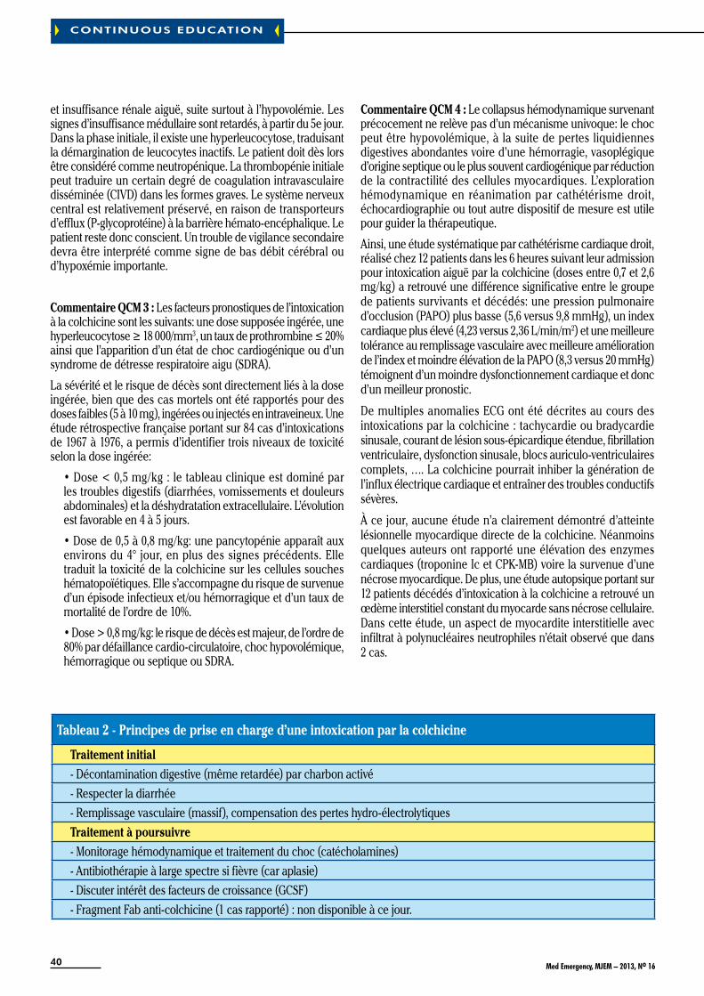
40 Med Emergency, MJEM – 2013, No 16
et insuffisance rénale aiguë, suite surtout à l’hypovolémie. Les signes d’insuffisance médullaire sont retardés, à partir du 5e jour. Dans la phase initiale, il existe une hyperleucocytose, traduisant la démargination de leucocytes inactifs. Le patient doit dès lors être considéré comme neutropénique. La thrombopénie initiale peut traduire un certain degré de coagulation intravasculaire disséminée (CIvD) dans les formes graves. Le système nerveux central est relativement préservé, en raison de transporteurs d’efflux (P-glycoprotéine) à la barrière hémato-encéphalique. Le patient reste donc conscient. Un trouble de vigilance secondaire devra être interprété comme signe de bas débit cérébral ou d’hypoxémie importante.
Commentaire QCM 3 : Les facteurs pronostiques de l’intoxication à la colchicine sont les suivants: une dose supposée ingérée, une hyperleucocytose≥18000/mm3,untauxdeprothrombine≤20%ainsi que l’apparition d’un état de choc cardiogénique ou d’un syndrome de détresse respiratoire aigu (SDRA).
La sévérité et le risque de décès sont directement liés à la dose ingérée, bien que des cas mortels ont été rapportés pour des doses faibles (5 à 10 mg), ingérées ou injectés en intraveineux. Une étude rétrospective française portant sur 84 cas d’intoxications de 1967 à 1976, a permis d’identifier trois niveaux de toxicité selon la dose ingérée:
•Dose<0,5mg/kg:letableaucliniqueestdominéparles troubles digestifs (diarrhées, vomissements et douleurs abdominales) et la déshydratation extracellulaire. L’évolution est favorable en 4 à 5 jours.
•Dosede0,5à0,8mg/kg:unepancytopénieapparaîtauxenvirons du 4° jour, en plus des signes précédents. Elle traduit la toxicité de la colchicine sur les cellules souches hématopoïétiques. Elle s’accompagne du risque de survenue d’un épisode infectieux et/ou hémorragique et d’un taux de mortalité de l’ordre de 10%.
•Dose>0,8mg/kg:lerisquededécèsestmajeur,del’ordrede80% par défaillance cardio-circulatoire, choc hypovolémique, hémorragique ou septique ou SDRA.
Commentaire QCM 4 : Le collapsus hémodynamique survenant précocement ne relève pas d’un mécanisme univoque: le choc peut être hypovolémique, à la suite de pertes liquidiennes digestives abondantes voire d’une hémorragie, vasoplégique d’origine septique ou le plus souvent cardiogénique par réduction de la contractilité des cellules myocardiques. L’exploration hémodynamique en réanimation par cathétérisme droit, échocardiographie ou tout autre dispositif de mesure est utile pour guider la thérapeutique.
Ainsi, une étude systématique par cathétérisme cardiaque droit, réalisé chez 12 patients dans les 6 heures suivant leur admission pour intoxication aiguë par la colchicine (doses entre 0,7 et 2,6 mg/kg) a retrouvé une différence significative entre le groupe de patients survivants et décédés: une pression pulmonaire d’occlusion (PAPO) plus basse (5,6 versus 9,8 mmHg), un index cardiaque plus élevé (4,23 versus 2,36 L/min/m2) et une meilleure tolérance au remplissage vasculaire avec meilleure amélioration de l’index et moindre élévation de la PAPO (8,3 versus 20 mmHg) témoignent d’un moindre dysfonctionnement cardiaque et donc d’un meilleur pronostic.
De multiples anomalies ECG ont été décrites au cours des intoxications par la colchicine : tachycardie ou bradycardie sinusale, courant de lésion sous-épicardique étendue, fibrillation ventriculaire, dysfonction sinusale, blocs auriculo-ventriculaires complets, …. La colchicine pourrait inhiber la génération de l’influx électrique cardiaque et entraîner des troubles conductifs sévères.
À ce jour, aucune étude n’a clairement démontré d’atteinte lésionnelle myocardique directe de la colchicine. Néanmoins quelques auteurs ont rapporté une élévation des enzymes cardiaques (troponine Ic et CPK-MB) voire la survenue d’une nécrose myocardique. De plus, une étude autopsique portant sur 12 patients décédés d’intoxication à la colchicine a retrouvé un œdème interstitiel constant du myocarde sans nécrose cellulaire. Dans cette étude, un aspect de myocardite interstitielle avec infiltrat à polynucléaires neutrophiles n’était observé que dans 2 cas.
tableau 2 - Principes de prise en charge d’une intoxication par la colchicine
traitement initial- Décontamination digestive (même retardée) par charbon activé - Respecter la diarrhée- Remplissage vasculaire (massif), compensation des pertes hydro-électrolytiquestraitement à poursuivre- Monitorage hémodynamique et traitement du choc (catécholamines)- Antibiothérapie à large spectre si fièvre (car aplasie) - Discuter intérêt des facteurs de croissance (GCSF)- Fragment Fab anti-colchicine (1 cas rapporté) : non disponible à ce jour.
CONTINuOus EDuCATION

41 Med Emergency, MJEM – 2013, No 16
Commentaire QCM 5 : Peu de données existent sur le comportement cinétique de la colchicine en situation toxique et beaucoup de considérations doivent se déduire des études pharmacologiques. Le volume de distribution de la colchicine est important. Il n’existe pas de corrélation entre la concentration plasmatique de la colchicine à l’admission et le pronostic final. Par contre, le profil de la cinétique d’élimination de la colchicine du plasma considéré à partir de la date d’ingestion serait lié à l’évolution clinique. Une quantité importante du toxique s’élimine par les urines, surtout pendant les premiers jours. Les selles diarrhéiques contiennent des quantités non négligeables de toxiques. Il est donc important de maintenir une bonne diurèse et de respecter le transit. L’hémodialyse a par contre peu d’intérêt, en raison des faibles quantités de colchicine circulantes et d’une clairance rénale en général conservée. La colchicine est un substrat de la P-glycoprotéine (transporteur d’efflux) et du cytochrome P450 3A4 (métabolisme hépatique) : L’ingestion simultanée d’autres médicaments pourrait modifier les paramètres cinétiques de la colchicine et expliquer la majoration ou la prolongation de ses effets toxiques. Il faut donc être très attentif aux co-ingestions, même s’il pourrait s’agir de substances apparemment anodines.
Commentaire QCM 6 : La décontamination digestive, surtout utile dans les premières heures, est basée sur l’administration de charbon activé en l’absence de contre-indications. Celle-ci peut être plus large que les 2 heures habituelles en raison du pronostic vital corrélé à la quantité absorbée de toxique. Le lavage gastrique n’est pas recommandé. La prise en charge initiale est basée essentiellement sur la correction de l’hypovolémie et la correction des troubles hydro-électrolytique avec la perfusion de quantités importantes de NaCI 0,9% voire de macromolécules et de bicarbonates 1,4% en cas d’acidose par déperdition. La patiente doit être alors transférée en réanimation médicale, pour suite de la prise en charge. L’antibiothérapie probabiliste en cas de fièvre doit être active sur les germes de l’oropharynx (streptocoques) et tube digestif (bacille à gram négatif), en raison du risque de survenue de translocation bactérienne lors des phases d’instabilité hémodynamique. Elle doit tenir compte d’une éventuelle colonisation par des germes nosocomiaux. L’association par exemple d’une β-lactamine à large spectre de type ticarcilline ou pipéracilline +/- tazobactam et d’un aminoside satisfait à ces conditions. Ultérieurement, dès la baisse significative des globules blancs, le GCS-F ou Granulocyte Colony Stimulating Factor (filgrastim) pourrait se révéler utile pour réduire la durée et l’importance de le neutropénie, mais celle-ciesthabituellement≤3jours.
Commentaire QCM 7 : De l’absence d’une thérapeutique spécif ique eff icace est née l’idée de développer une immunothérapie pour les situations d’intoxication grave (> 0,8 mg/kg). Le principe d’un tel traitement est l’extraction, la séquestration et l’élimination du toxique de ses organes cibles, par l’administration intravasculaire de fragments Fab d’anticorps spécifiques. Des anticorps IgG spécifiques anti-colchicine ont pu
être obtenus, après immunisation chez la chèvre. Le mécanisme d’extraction tissulaire repose sur le principe de la loi d’action de masse.
Le succès est fondé sur la réversibilité de l’interaction toxique / récepteur, pour la colchicine et ses récepteurs intracellulaires. La séquestration du toxique dépend de la spécificité et de l’affinité de l’anticorps. La constante d’affinité intrinsèque moyenne des anticorps obtenus (1,5 à 8 x 10⁹ M-1) est supérieure à celle de la colchicine pour son récepteur biologique (106 à 107 M-1), soit environ 1000 fois plus.
La formation de complexes immuns stables dans le sang circulant permet de masquer les sites moléculaires du toxique et d’empêcher l’interaction avec son site d’action. L’anticorps piège la colchicine présente dans le compartiment vasculaire, créant un gradient de concentration des tissus vers le sang, ce qui favorise le passage du toxique du compartiment tissulaire vers le compartiment vasculaire, site de distribution des anticorps. Les anticorps peuvent ainsi réduire la redistribution de molécules liées aux tissus et empêcher leur effet toxique. L’élimination du toxique se fait alors rapidement sous forme complexée dans les urines.
Les facteurs limitant l’utilisation des anticorps en clinique sont la disponibilité des anticorps et le délai écoulé depuis l’ingestion lors de l’admission du patient à l’hôpital. Le problème de la dose de Fab à injecter par rapport à la dose ingérée n’est pas tranché. Il a été démontré que des doses non-stœchiométriques inframolaires de fragments Fab suffisaient pour traiter et neutraliser une intoxication expérimentale potentiellement létale.
Une seule patiente, âgée de 25 ans, a été traitée jusqu’à présent, avec succès, grâce à ces anticorps spécifiques, à la suite de l’ingestion d’une dose mortelle de 1 mg/kg. Le tableau clinique était marqué par l’apparition de troubles digestifs, d’hypotension artérielle, de CIvD, d’oligurie, de SDRA et d’un choc cardiogénique réfractaire à la dobutamine.
La concentration plasmatique de colchicine était à 24 ng/ml. Elle a été traitée par les Fab spécifiques administrés en perfusion intraveineuse, à la dose de 480 mg (dose non stœchiométrique). Il s’en est suivi une ascension de 6 fois de la concentration de colchicine dans le sang, 10 minutes après l’administration des anticorps. La colchicine libre est devenue indétectable, jusqu’à 7 heures après la perfusion et le taux urinaire a été multiplié par 6. Le choc cardiogénique a rapidement régressé et la patiente a survécu mais l’immunothérapie n’a pas pu empêcher la survenue d’une neutropénie profonde, d’une bactériémie, d’une polynévrite transitoire et d’une alopécie.
Les anticorps anti-colchicine sont actuellement au stade du développement clinique (mise en place prochaine d’un essai européen multicentrique). Le développement chez l’homme avait été bloqué en raison des coûts élevés de telles études cliniques et du faible nombre de patients potentiellement susceptible de bénéficier d’une telle thérapie. Aucune entreprise pharmaceutique n’avait accepté à ce jour de développer cette thérapie, la reléguant dans la catégorie des médicaments orphelins.
CONTINuOus EDuCATION

42 Med Emergency, MJEM – 2013, No 16
PouR En savoiR PLus
1. Sackett DL, varma JK. Molecular mechanism of colchicine action: induced local unfolding of ß-tubulin. Biochemistry 1993; 32:13560-5.2. Finkelstein Y, Aks SE, Hutson JR, Juurlink DN, Nguyen P, Dubnov-Raz G, Pollak U, Koren G, Bentur Y.Colchicine poisoning: the dark side of an ancient drug. ClinToxicol (Phila) 2010; 48:407-14.3. Folpini A, Furfori P. Colchicine toxicity - clinical features and treatment. Massive overdose case report. J ToxicolClinToxicol 1995; 33:71-7.4. Bismuth C, Gaultier M, Conso F. Aplasie médullaire après intoxication aiguë par la colchicine. Presse Med 1977; 6:1625-8.5. Sauder P, Kopferschmitt J, Jaeger A, Mantz JM. Hemodynamic studies in eight cases of acute colchicine poisoning. Human Toxicol 1983; 2:169-73. 6. Rochdi M, Sabouraud A, Baud FJ, Bismuth C, Scherrmann JM. Toxicokinetics of colchicine in humans: analysis of tissue, plasma and urine data in ten cases. Hum ExpToxicol 1992; 11:510-6.7. Borron SW, Scherrmann JM, Baud FJ. Markedly altered colchicine kinetics in a fatal intoxication: examination of contributing factors. Hum Exp Toxicol 1996; 15:885-90.8. Bismuth C, Borron SW, Baud FJ, Taboulet P, Scherrmann JM. Immunotoxicotherapy: successes, disapointments and hopes. Hum ExpToxicol 1997; 16:602-608.9. Baud FJ, Sabouraud A, vicaut E, Taboulet P, Lang J, Bismuth C, Rouzioux JM, Schermann JM. Brief report: treatment of severe colchicine overdose with colchicine-specific Fab fragments. N Engl J Med 1995; 332:642-5.
ConClusionEn conclusion, les intoxications par colchicine sont potentiellement sévères. Les effets cardiovasculaires toxiques contribuent de façon importante à l’évolution défavorable des patients les plus gravement intoxiqués. L’atteinte cardiaque fait partie d’une atteinte multiviscérale toxique globale. Le traitement est avant tout symptomatique. Aucun traitement spécifique n’existe à ce jour.
CONTINuOus EDuCATION

43 Med Emergency, MJEM – 2013, No 16
RECOmmENDATIONs FOR AuTHORs
Med Emergency, MJEM The Mediterranean Journal of Emergency Medicine
The Journal publishes articles in English and/or French pertai-ning to Emergency Medicine from its scientific aspect (research, case studies, clinical articles, orientation and practical conduct), administrative (Management and organization of Emergency Me-dicine), medical-legal and social aspects. It also accepts articles that deal with prevention of emergencies. Although it focuses more on practical issues of emergency medicine, the Journal accepts theoretical, methodological and analytical articles. It is also interested in communications, letters, commentaries and critiques of issues related to emergency. Priority is given to articles that pertain to the practice of emergency medicine spe-cifically in the Mediterranean and MENA region. Some editions will be consecrated to specific subjects such as accidents, catas-trophes, cardiac arrest and first aid. We would also welcome any announcements of events (national and international) related to emergency medicine such as workshops, conferences, courses and new books and publications. Authors can submit their original articles and the accompanying references to the editor: New Health Concept B.P. 90.815 Jdeideh- Lebanon or via email. The article should be accompanied by a letter by the author/s that clearly states that joint authors of the article are aware of the application to publish and have agreed to allow free accessing of texts by New Health Concept Edition publication. Please create a separate file (indicating the name of the author) for all the photographs, tables and graphs you would like to be included in the article and send them to the following address: [email protected] All submissions will undergo a preliminary evaluation and an ethical revision by the editorial board to determine whether it will be allowed to appear in the journal. Articles that pass this preliminary evaluation will also be anonymously reviewed by two members of a scientific committee. Once the article has been approved for publication, a biography of 10 lines should be developed.
Manuscript Preparation Articles are to be submitted in a typewritten format. Paragra-phs are double spaced. Font size should be 12. The submitting author should send his contact details with the article such as telephone number or an email address. The original text of the article should be sent without illustrations in its original format (e.g. Microsoft Word). Pages should be numbered. Titles and subtitles of equal importance should be marked identically. Abbreviations should be explained when first encountered in the text. The articles should not exceed 2500 words or not more than 10 pages. Abstracts and Key Words: Each article should include an abstract In English (and in French for French articles) no longer than 300 words. Keywords (not more than 6 words) and the title of the article should also be presented in both languages. Text: The author needs to respect the following formatting procedures when submitting the article: - On the front page- the author’s name, affiliations, complete mai-ling address, telephone number and email address. The names and the affiliations of collaborators should be clearly indicated. Please ensure that this information is only presented on the front page and does not appear on the other pages of the article. - Bibliographic References need to appear in order of appearance in the text. They must be identified in the text by Arabic numbers in brackets. There should be about 10-30 references. They must conform to presentation norms applied in the scientific editing world (E.g. APA). - Photographs, figures, graphs and tables: these should be sent
in separate files and need to be numbered and marked with the author’s name and commentary. They need to be numbered in chronological ordered when they are to be referred to in the text. The term “graph/table/figure/photo number x” should be used in order to avoid confusion with bibliographical references. - End notes should be listed separately at the end of the text and not at the end of each page.
PS: It’s strongly recommended to add photography of the author who can also allow us to communicate his E-mail address.
aDEnDuMConflict-of-interest statement* Conflict of interest exists when an author (or the author’s institution), reviewer, or editor has financial or personal relationships that inappropriately influence (bias) his or her actions (such relationships are also known as dual commitments, competing interests, or competing loyalties). These relationships vary from those with negligible potential to those with great potential to influence judgment, and not all relationships represent true conflict of interest. The potential for conflict of interest can exist whether or not an individual believes that the relationship affects his or her scientific judgment. Finan-cial relationships (such as employment, consultancies, stock ownership, honoraria, paid expert testimony) are the most easily identifiable conflicts of interest and the most likely to undermine the credibility of the journal, the authors, and of science itself. However, conflicts can occur for other reasons, such as personal relationships, academic competition, and intellectual passion.
statement of informed Consent* Patients have a right to privacy that should not be infringed without informed consent. Iden-tifying information, including patients’ names, initials, or hospital numbers, should not be published in written descriptions, pho-tographs, and pedigrees unless the information is essential for scientific purposes and the patient (or parent or guardian) gives written informed consent for publication. Informed consent for this purpose requires that a patient who is identifiable be shown the manuscript to be published. Authors should identify Indivi-duals who provide writing assistance and disclose the funding source for this assistance. Identifying details should be omitted if they are not essential. Complete anonymity is difficult to achieve, however, and informed consent should be obtained if there is any doubt. For example, masking the eye region in photographs of patients is inadequate protection of anonymity. If identifying characteristics are altered to protect anonymity, such as in gene-tic pedigrees, authors should provide assurance that alterations do not distort scientific meaning and editors should so note. statement of human and animal Rights* When reporting expe-riments on human subjects, authors should indicate whether the procedures followed were in accordance with the ethical stan-dards of the responsible committee on human experimentation (institutional and national) and with the Helsinki Declaration of 1975, as revised in 2000 (5). If doubt exists whether the research was conducted in accordance with the Helsinki Declaration, the authors must explain the rationale for their approach, and demonstrate that the institutional review body explicitly approved the doubtful aspects of the study. When reporting experiments on animals, authors should be asked to indicate whether the institutional and national guide for the care and use of laboratory animals was followed.
*international committee of medical Journal editors ("uniform requirements for manuscripts submitted to Biomedical Journals") -- February 2006

44 Med Emergency, MJEM – 2013, No 16
BOOks OF THE IssuE / LIvREs Du NuméRO
In case of a breakdown of regional hospital structures in a major accident or disaster, a civilian, mobile, modular hospital is a indispensable means of ensuring the long term provision of healthcare for the population. Alongside the modules usually present in hospitals (operating theatre, imaging, laboratory, supply and waste management), it should have modules for decontamination and specialized laboratory diagnostics (dirty bomb).
Rédigé par le Comité « Advanced Medical Life Support » de l’Association américaine des techniciens médicaux d’urgence (NAEMT), ce livre est une mine d’infor-mations sur les techniques de prise en charge des urgences médicales en pra-tique préhospitalière et hospitalière.
Partout dans le monde, les formations AMLS sont fondées sur ce livre. véritable référence internationale, il permet aux intervenants de développer leur capacité à évaluer rapidement et précisément la gravité d une urgence médicale, puis de définir la stratégie thérapeutique la plus adaptée au patient, et ceci dans les condi-tions de sécurité maximale. Cet ouvrage privilégie l efficacité par son approche pédagogique moderne origi-nale, basée sur une méthode d’évalua-tion originale et les modalités de prise en charge des patients qui en découlent.
Auteur : Laurent ObertDate de publication: 22 août 2012
Dans le cadre de l’urgence traumatique, les blessures du membre «supérieur», de la main à l’épaule, nécessitent une prise en charge particulière. Elles n’empêchent pas le blessé de se déplacer, et, de façon un peu caricaturale, seules les plaies hémorragiques ou les plaies de la main vont «pousser» le patient aux urgences. Les premières minutes qui entourent l’accueil du patient blessé sont cruciales. C’est en effet lors de ce délai très court que va s’établir la confiance qui permettra de faire le ou les bons diagnostics. Parce que les pièges sont nombreux, les auteurs
Les textes sont étayés par :- une abondante iconographie : tableaux, algorithmes, schémas et photographies.- des scénarios d’entrainement et leurs réponses qui permettent d’appliquer les connaissances théoriques à des situations rencontrées sur le terrain.- des encadrés qui mettent en exergue les notions essentielles, des résumés à la fin des chapitres et des questions d auto-évaluation qui aident à retenir les concepts-clés.
Cet ouvrage s’adresse aux équipes plu-ridisciplinaires en médecine d’urgence qui doivent effectuer l’évaluation initiale, l’orientation, la prise en charge des patho-logies courantes jusqu’à la réanimation des urgences vitales, de l’abord du patient jusqu’à son transfert vers un service de spécialité, un autre professionnel de santé ou jusqu à son traitement définitif.
The CMMH should fulfill the following requirements:• Potential to operate 200 beds, self
sufficiently (water, electricity, heat)• Availability at short notice (72 hours)• Etc.
The book presents a lso va luable information’s of designs of container hospitals of Germany s military and civil concepts.
ont souhaité faire ce petit guide afin d’accompagner les urgentistes dans leur démarche diagnostique et thérapeutique. Recueil d’expériences et de conduites pratiques, cet ouvrage propose également des images de diagnostics faciles ou de pièges à éviter, facilitant le travail de ceux qui côtoient l’urgence traumatique au quotidien.
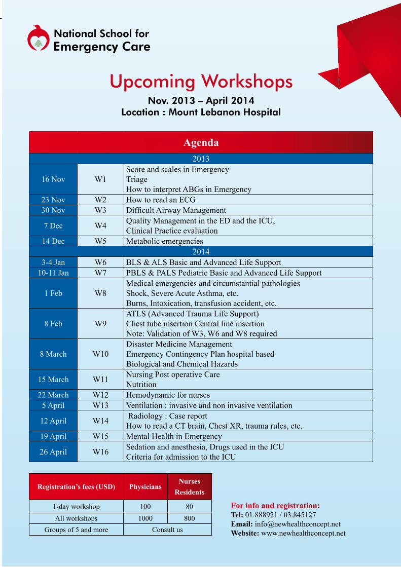
Agenda2013
Score and scales in Emergency Triage How to interpret ABGs in Emergency
W116 Nov
How to read an ECGW223 NovDifficult Airway Management W330 NovQuality Management in the ED and the ICU, Clinical Practice evaluation W47 Dec
Metabolic emergenciesW514 Dec2014
BLS & ALS Basic and Advanced Life Support W63-4 JanPBLS & PALS Pediatric Basic and Advanced Life SupportW710-11 JanMedical emergencies and circumstantial pathologies Shock, Severe Acute Asthma, etc.Burns, Intoxication, transfusion accident, etc.
W81 Feb
ATLS (Advanced Trauma Life Support)Chest tube insertion Central line insertion Note: Validation of W3, W6 and W8 required
W98 Feb
Disaster Medicine ManagementEmergency Contingency Plan hospital basedBiological and Chemical Hazards
W108 March
Nursing Post operative Care Nutrition W1115 March
Hemodynamic for nursesW1222 MarchVentilation : invasive and non invasive ventilation W135 April Radiology : Case reportHow to read a CT brain, Chest XR, trauma rules, etc. W1412 April
Mental Health in EmergencyW1519 AprilSedation and anesthesia, Drugs used in the ICUCriteria for admission to the ICUW1626 April
Upcoming WorkshopsNov. 2013 – April 2014
Location : Mount Lebanon Hospital
For info and registration: Tel: 01.888921 / 03.845127Email: [email protected] Website: www.newhealthconcept.net
Registration’s fees (USD) PhysiciansNurses
Residents
1-day workshop 100 80All workshops 1000 800
Groups of 5 and more Consult us

KARL STORZ GmbH & Co. KG, Mittelstraße 8, 78532 Tuttlingen/Germany Telephone: +49 (0)7461 708-0, Fax: + 49 (0)7461 708-105, E-Mail: [email protected] STORZ Endoskope – East Mediterranean and Gulf S.A.L., Block M, 3rd Floor, Beirut Souks, Weygand Street, 2012 3301 Beirut/Lebanon,
Phone: +961 (1) 999390, Fax: +961 (1) 999391, E-mail: [email protected]
AN
40
08/2
012/
A-L
B
* Image output for S-Video and USB, compatible with Philips IntelliVue MX 800
for Airway Cockpit as well as other, non-medical grade monitors
C-MAC®
Pocket Monitor
FIVE
C-HUB™*
C-MAC® for Airway Management
– a Sophisticated System
©2013 Physio-Control, Inc. Redmond, WA, USA
The responsive emergency team delivers exceptional CPR
Today’s responsive emergency team is always looking to elevate the level of care they deliver, and they rely on evidence and data to get them there. With the CPR Solution from Physio-Control, they have the science-based, Guidelines-consistent tools they need to respond better to patients—and the demands of constant performance improvement.
And they choose Physio-Control to make it happen.
real-time and post-event CPR performance feedback
NEW
uninterrupted hands-free compressions
featuring comprehensive CPR analytics
with capnography and CPR Metronome
Get ready for a more responsive approach to CPR.
Physio-Control Lebanon Telephone +961 4 718 414 § Fax +961 4 718 415 Email [email protected] § www.physio-control.comKARL STORZ GmbH & Co. KG, Mittelstraße 8, 78532 Tuttlingen/Germany Telephone: +49 (0)7461 708-0, Fax: + 49 (0)7461 708-105, E-Mail: [email protected]
KARL STORZ Endoskope – East Mediterranean and Gulf S.A.L., Block M, 3rd Floor, Beirut Souks, Weygand Street, 2012 3301 Beirut/Lebanon, Phone: +961 (1) 999390, Fax: +961 (1) 999391, E-mail: [email protected]
www.karlstorz.com
AN
40
08/2
012/
A-L
B
* Image output for S-Video and USB, compatible with Philips IntelliVue MX 800
for Airway Cockpit as well as other, non-medical grade monitors
C-MAC®
Pocket Monitor
FIVE
C-HUB™*
C-MAC® for Airway Management
– a Sophisticated System
48 Med Emergency, MJEM – 2013, No 16
EDitoR in ChiEf anD DiRECtoR Nagi SOUAIBY
EXECutivE boaRDIsmaël HSSAIN Abdo KHOURY
Hugues LEFORT
CovER PiCtuRE CPR
Cerebral circulationPrehospital Airway management
DEsign
Mireille SROUR
WEbMastERAlec KAZANDJIAN: Web Sart
PRintingMELKI PRINT INTERNATIONAL S.A.L.
Quarterly Journal ISSN No 2222-9442Printed in Lebanon
All rights reserved. Please note Med Emergency Publication copyright in all reprints.
the advertising organizations: ESA – cover page 2. Med Emergency MJEM – page 4. NSEC – page 45. Karl-Storz – page 46. Physio-Control – page 47. Bassoul & Heneine – cover page 3. GNCEM - back cover.
MEMBERSHIP 4 ISSUES/YEAR ($USD)
8 ISSUES/2 YEARS($USD)
Individual o 80 o 140
Student o 60 o 100
Institution o 100 o 180
Outside Lebanon* o Add +20% o Add +20%Name : Surname : Address : P O Box: : City : Country : Email :
Telephone: --------------------------
Profession: -------------------------- Affiliation: -------------------------------------------
o Bank Check (Cheque Bancaire)Please send to: MED EMERGENCY PUBLICATIONS - New Health Concept, Samra Center, Block C 4th floor Fanar, Jdeidet El Metn P O Box 90 815
Kindly fill and return to: MED EMERGENCY Publications P.O. Box 90.815, Jdeideh- Lebanon, Tel: +961-1-888921; Fax: +961-1-888922
* +$10 USD to send outside Lebanon
The MED Emergency, MJEM team hope to see you all in Marrakech for a special Morocco

100% PERSONNALISABLE
RENAULT CAPTURCAPTURE LIFE
BOITE DE VITESSEAUTOMATIQUE A 6 RAPPORTS
5 ÉTOILES
City CarBeirut01 803313/4
Bauchrieh Car Center Beirut01 880213
Elie TabetJounieh09 918402
Bejco SARLJamhour05 768800
Highway Auto, KhaldehHighway05 800149
Pascal 2 SARLZalka Seaside01 889469
North MotorsTripoli06 411293/4
Fouad SrourZahleh08 800403
Youssef TradeCompanyTyr & Nabatieh07 351313
Saida Car Zone, Saida07 72688807 754887
Tabet & Matar SAL, Highway Mastita Jbeil 09 796939
Sed El Bauchrieh: 01 684 684 - Ain El Mreisseh: 01 360 779 /RenaultLebanonwww.renault-liban.comAgents agrées:
$22,500 Hors TVA
Captur Pad A4 fr.indd 1 9/23/13 12:24 PM
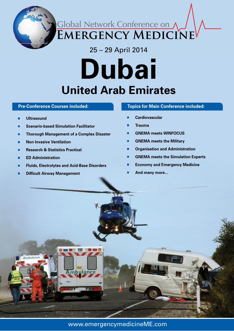
www.emergencymedicineME.com
DubaiUnited Arab Emirates
Global Network Conference onEmergency Medicine
• Ultrasound
• Scenario-based Simulation Facilitator
• Thorough Management of a Complex Disaster
• Non Invasive Ventilation
• Research & Statistics Practical
• ED Administration
• Fluids, Electrolytes and Acid-Base Disorders
• Difficult Airway Management
• Cardiovascular
• Trauma
• GNEMA meets WINFOCUS
• GNEMA meets the Military
• Organisation and Administration
• GNEMA meets the Simulation Experts
• Economy and Emergency Medicine
• And many more...
Pre-Conference Courses included: Topics for Main Conference included:
25 – 29 April 2014