issn: 0975 -766x coden: ijptfi available online through ... · pdf filenovel drug delivery...
TRANSCRIPT

Rabiya Ansari*et al. /International Journal of Pharmacy & Technology
IJPT| June-2017| Vol. 9 | Issue No.2 | 29735-29758 Page 29735
ISSN: 0975-766X
CODEN: IJPTFI
Available Online through Research Article
www.ijptonline.com LIPOSOMES AS A NOVEL DRUG DELIVERY SYSTEM
Rabiya Ansari*, Abdul Mannan
Department of Pharmaceutics, Deccan School of Pharmacy, Hyderabad, Telangana 500001.
Email: [email protected]
Received on: 25-02-2017 Accepted on: 28-03-2017
Abstract
This review article is intended to provide an overview of liposomes as a novel drug delivery system. It has focused on
the structure, components, mechanism of liposome formation, advantages, disadvantages, classification, method of
preparation, characterization, stability and applications of the liposomes Liposomes are spherical shaped bilayered
vesicles in which an aqueous volume is entirely enclosed by a membranous lipid bilayer. The hydration of a dry lipid
film lead to the formation of liposome. Liposomes have a wide range of applications in science, pharmaceutical
industry, as a drug carrier, anti cancer agent and in dermatology and cosmetics. Liposome as novel drug delivery
systems has played a significant role in formulation of potent drug to improve therapeutics.
Keywords: Liposome, Vesicles, Amphiphilic, Hydrophilic, Hydrophobic, Phospholipids, Novel Drug Delivery
System.
Introduction
Novel drug delivery system aims at providing some control, whether this is of time related or dimensional nature, or
both, of drug release in the body. Novel drug delivery attempts to either sustain drug action at a predetermined rate,
or by maintaining a relatively constant, effective drug level in the body associated with minimization of undesirable
side effects [1]
. Different types of pharmaceutical carriers are present. They are – particulate, polymeric, and
macromolecular. Particulate type carrier also known as colloidal carrier system, which includes lipid particles i.e. low
and high density lipoprotein- LDL and HDL respectively, microspheres, nanoparticles, polymeric micelles and the
vesicular like liposomes, niosomespharmacosomes, virosomes, etc[2]
. Liposomes are simple microscopic vesicles.
Structurally, liposomes are concentric bilayered vesicles in which an aqueous volume is entirely enclosed by a
membranous lipid bilayer mainly composed of natural or synthetic phospholipid [3]
. Generally, liposomes are
spherical vesicles with particle sizes ranging from 50-150 nm to several micrometers [4]
.

Rabiya Ansari*et al. /International Journal of Pharmacy & Technology
IJPT| June-2017| Vol. 9 | Issue No.2 | 29735-29758 Page 29736
Bangham and colleagues first discovered liposomes in early 1960’s. They were discovered when Bangham and R. W.
Horne. They were testing the institute's new electron microscope by adding negative stain to dry phospholipids. The
resemblance to the plasma lemma was obvious, and the microscope pictures served as the first real evidence for the
cell membrane being a bilayer lipid structures[5,6]
.
The name liposome is derived from two Greek words ‘Lipid’ meaning fat and ‘Soma’ meaning body and it
subsequently became the new most extensive explore drug delivery system. The hydration of a dry lipid film was
found to lead to the formation of enclosed spherical vesicles or liposomes that resemble miniature cellular organelles
with lipid bilayers [7]
. A free drug injected in bloodstream typically achieves therapeutic level for short duration due
to metabolism and excretion. Drug encapsulated by liposome achieve therapeutic level for long duration as, drug
must first be release from liposome before metabolism and excretion [8]
. Liposome drug delivery is gaining interest
due to its contribution to varied areas like drug delivery, cosmetics, and structure of biological membrane [9]
.
Structure and Components of Liposomes
Liposomes are colloidal particles. There are number of components of liposomes, however phospholipids and
cholesterol are the main components.
Most liposome formulations approved for human use contain phosphotidyl choline with a fraction of cholesterol is
often included in it.
Phospholipids are the major structural components of biological membranes. The most common phospholipid is
phosphatidylcholine (PC) molecule. It is an amphiphatic molecule in which a glycerol bridge links a pair of
hydrophobic acyl hydrocarbon chains with a hydrophilic polar head group phosphocholine[10]
.
Examples of phospholipids are- Phosphatidyl choline (Lecithin) – PC, Phosphatidyl ethanolamine(cephalin) –PE,
Phosphatidyl serine – PS, Phosphatidyl inositol – PI, Phosphatidyl Glycerol – PG.
Figure 1: Some Common Naturally Occuring Phosphatidyl Phospholipids [7]
.

Rabiya Ansari*et al. /International Journal of Pharmacy & Technology
IJPT| June-2017| Vol. 9 | Issue No.2 | 29735-29758 Page 29737
The general chemical structure of phospholipids shows a glyceral backbone. At a position of number 3 of the glyceral
molecule, the hydroxyl group is esterified to phosphoric acid. The hydroxyl groups at positions 1 and 2 of the
glycerol are usually esterified with long chain fatty acids. One of the remaining oxygen groups of the phosphoric acid
may be further esterified to a variety of organic molecules including glycerol, choline, ethanolamine, serine and
inositol [7].
Sphingolipids backbone is sphingosine or a related base. Most common Sphingolipids are Sphingomyelin,
Glycosphingo lipids. These are included in liposomes to provide a layer of surface charged group [2,7].
Cholesterol has a steroid backbone, and its derivatives are included in liposome preparation to improve their bilayer
characteristics, rigidity of the bilayer membrane, reduces the permeability of water-soluble molecules through the
membrane and improves the stability of the bilayer membrane in the presence of biological fluids such as blood and
plasma. The cholesterol molecule orients itself among the phospholipid molecules with its hydroxyl group facing
towards the water phase and the tricyclic ring sandwiched between the first few carbons of fatty acyl chains into the
hydrocarbon core of the bilayer [7,10].
Figure 2: Structure of Liposome [11,12].
Figure 3: Structure of Liposome [11,12].
Advantages
Can be administered through various routes [1].
Provides selective passive targeting to tumor tissues, example, liposomal doxorubicin [4].
Rabiya Ansari*et al. /International Journal of Pharmacy & Technology
IJPT| June-2017| Vol. 9 | Issue No.2 | 29735-29758 Page 29738
Alter pharmacokinetics and pharmacodynamics of drugs.
Suitable for delivery of hydrophobic, amphipathic and hydrophilic drugs [13].
Protect the encapsulated drug from the external environment [14].
Reduced toxicity and increased stability [15].
Reduce exposure of sensitive tissues to toxic drugs [16].
Provide sustained release.
Direct interaction of the drug with cell biodegradable and flexible [17].
Disadvantages
Allergic reactions may occur to liposomal constituents [1].
Low solubility [2].
Liposomes are extensively used as carriers for molecules in cosmetic and pharmaceutical industries.
Short half life [18].
Leakage and fusion [19].
Less stability.
Phospholipids undergoes oxidation, hydrolysis.
High production cost [20].
Liposomes can trap both hydrophobic and hydrophilic compounds, avoid decomposition of the entrapped
combinations, and release the entrapped at designated targets [21].
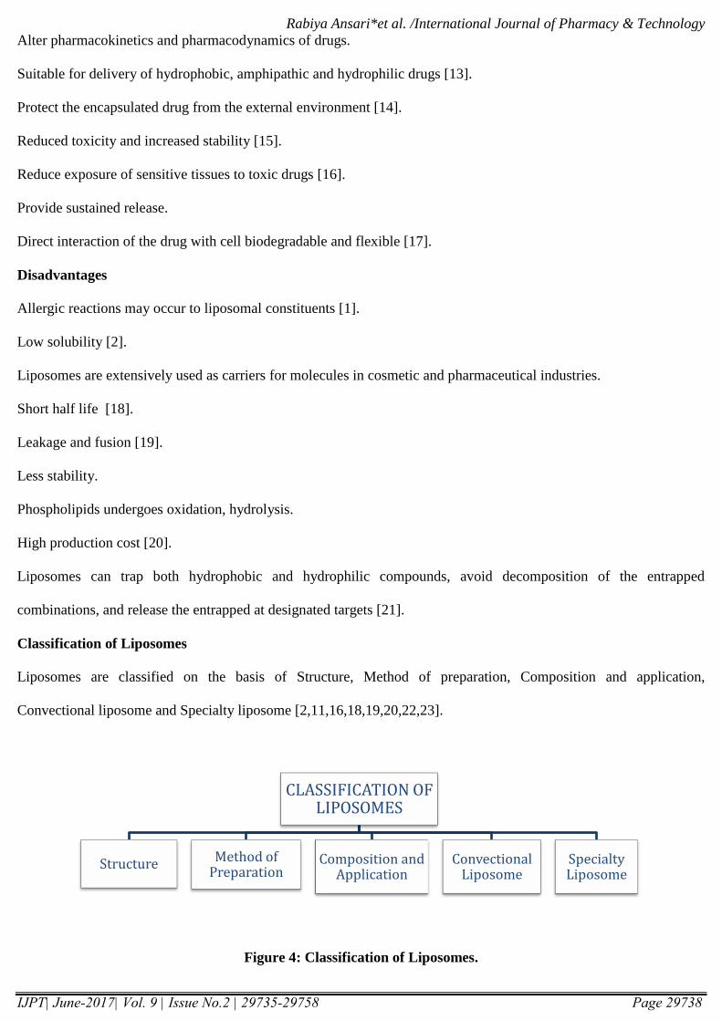
Classification of Liposomes
Liposomes are classified on the basis of Structure, Method of preparation, Composition and application,
Convectional liposome and Specialty liposome [2,11,16,18,19,20,22,23].
Figure 4: Classification of Liposomes.
CLASSIFICATION OF LIPOSOMES
Structure Method of Preparation
Composition and Application
Convectional Liposome
Specialty Liposome

Rabiya Ansari*et al. /International Journal of Pharmacy & Technology
IJPT| June-2017| Vol. 9 | Issue No.2 | 29735-29758 Page 29739
Classification Based on Structure (Table 1)
Table 1- Vesicle Types with their Size and Number of Lipid Layers [23]
Based on Method of Preparation (Table 2)
Table 2- Different Preparation Methods and the Vesicles Formed by these Methods [23]
PREPARATION METHOD VESICLE TYPE
Single or oligo lamellar vesicle made by reverse phase evaporation
method
REV
Multi lamellar vesicle made by reverse phase evaporation method MLV-REV
Stable plurilamellar vesicle SPLV
Frozen and thawed multi lamellar vesicles FATMLV
Vesicle prepared by extrusion technique VET
Dehydration-rehydration method DRV
VESICLE TYPE ABBREVIATION DIAMETER SIZE NUMBER OF LIPID
BILAYER
Unilamellar vesicle UV All size range One
Small unilamellar vesicle SUV 20-100 nm One
Medium unilamellar
vesicle
MUV More than 100nm One
Large unilamellar vesicle LUV More than 100 nm One
Giant unilamellar vesicle GUV More than one micro
meter
One
Oligolamellar vesicle OLV 0.1-1 micrometer Approximately 5
Multilamellar vesicle MLV More than 0.5 5-25
Multi vesicular vesicle MV More than one micro
meter
Multi compartment
structure

Rabiya Ansari*et al. /International Journal of Pharmacy & Technology
IJPT| June-2017| Vol. 9 | Issue No.2 | 29735-29758 Page 29740
Based on Composition and Application (Table 3)
Table 3- Different Liposome with their Compositions [23]
Based Upon Conventional Liposome
Stabilize natural lecithin (PC) mixtures
Synthetic identical, chain phospholipids
Glycolipids containing liposomes
Based Upon Specialty Liposome
Bipolar fatty acid
Antibody directed liposome
Methyl/Methylene x-linked liposome
Lipoprotein coated liposome
Carbohydrate coated liposome
Multiple encapsulated liposome
Mechanism of Formation of Liposomes
The basic part of liposome is formed by phospholipids, which are amphiphilic molecules having a hydrophilic head
and hydrophobic tail. The hydrophilic part is mainly phosphoric acid bound to a water soluble molecule, whereas, the
hydrophobic part consists of two fatty acid chains with 10 – 24 carbon atoms and 0 – 6 double bonds in each chain.
When these phospholipids are dispersed in aqueous medium, they form lamellar sheets by organizing in such a way
TYPE OF
LIPOSOME
ABBREVIATION COMPOSITION
Conventional
liposome
CL Neutral or negatively charge
phospholipid and cholesterol
Fusogenic
liposome
RSVE Reconstituted sendai virus envelops
PH sensitive
liposome
- Phospholipid such as PER or DOPE
with either CHEMS or OA
Cationic liposome - Cationic lipid with DOPE
Long circulatory
liposome
LCL Neutral high temperature, cholesterol,
and 5-10% PEG, DSP
Immunolatory
liposome
IL CL or LCL with attached monoclonal
antibody or recognition sequences

Rabiya Ansari*et al. /International Journal of Pharmacy & Technology
IJPT| June-2017| Vol. 9 | Issue No.2 | 29735-29758 Page 29741
that, the polar head group faces outwards to the aqueous region while the fatty acid groups face each other and finally
form spherical vesicle like structures called as liposomes. The polar portion remains in contact with aqueous region
along with shielding of the non-polar part, which is oriented at an angle to the membrane surface [24]. It is the
hydrophilic/ hydrophobic interactions between lipid – lipid, lipid – water molecules that lead to the formation of
bilayered vesicles in order to achieve a thermodynamic equilibrium in the aqueous phase [25].
Figure 5: Mechanism of Formation of Liposomes [4]
Methods of Liposome Preparation
Liposomes can be prepared by active loading or passive loading techniques [4,26,27,28].
Figure 6: Methods Of Preparation Of Liposomes [4]

Rabiya Ansari*et al. /International Journal of Pharmacy & Technology
IJPT| June-2017| Vol. 9 | Issue No.2 | 29735-29758 Page 29742
I) PASSIVE LOADING TECHNIQUES
A) MECHANICAL DISPERSION METHODS
Figure 7: Preparation of Liposomes through Mechanical Dispersion Method.
Thin Film Hydration Using Hand Shaking (MLVS) And Non-Hand Shaking Methods (LUVS)
Lipids are casted as stacks of film from their organic solution using flash rotary evaporator under reduced pressure or
by hand shaking and then the casted film is dispersed in an aqueous medium. Upon hydration the lipids swell and
peel off from the wall of the round bottom flask and vesiculate forming multi lamellar vesicles (MLVs). The
mechanical energy required for the swelling of lipids and dispersion of casted lipid film is imparted by manual
agitation or hand shaking technique or by exposing the film to the stream of water-saturated nitrogen for 15mins
followed by swelling in aqueous medium without shaking. Hand shaking method produces MLVs whereas non-
handshaking method produces LUVs. The percent encapsulation efficiency as high as 30% is achieved [4].
Figure 8: Preparation of Liposomes by Thin Film Hydration Using Hand Shaking (MLVS) and Non-Hand
Shaking Methods (LUVS) [4]
liposomes
lipids swell and hydrate
hydrated using aqueous buffer
solid lipid mixture
organic solvent removed by film deposition under vacuum
lipid co-dissolved in organic solvent

Rabiya Ansari*et al. /International Journal of Pharmacy & Technology
IJPT| June-2017| Vol. 9 | Issue No.2 | 29735-29758 Page 29743
Micro Emulsification
Micro fluidizer is used to prepare small MLVs. The lipids can be introduced into the fluidizer, either as a dispersion
of large MLVs or as a slurry of unhydrated lipid in an organic medium. It pumps the fluid at very high pressure about
10,000 psi or 600-700 bar through a 5 µm orifice. Then it is forced along defined micro channels, which direct two
streams of fluid to collide together at right angles at a very high velocity. The fluid collected can be recycled through
the pump and interaction chamber until vesicles of spherical dimensions are obtained. After a single pass, the size of
diameter is reduced to 0.1 and 0.2 µm in diameter. This method has the advantage of being able to process samples
with a very high proportion of lipids i.e. 20% or more by weight. This process is efficient for encapsulation of water
soluble materials. Percentage capture values upto 70% have been reported, starting with lipid concentration of
approximately 200mg/ml [4].
Figure 9: Preparation of Liposomes by Micro Emulsification [4]
Sonication
Sonication is the most extensively used method for the preparation of SUV. The main disadvantages of this method
are very low encapsulation efficacy and presence of MLV along with SUV. There are two sonication techniques i.e.
probe or bath sonicator. The probe is employed for dispersions which require high concentration of lipids while bath
sonicator is more suitable for large volumes of diluted lipids. Probe tip sonicator tends to release titanium particles
into the liposome dispersion; because of this bath sonicator is most widely used. Sonication of MLV dispersion is
done by placing a test tube containing the dispersion in the bath sonicator and sonicating it for 5-10minutes. Above
Tc of the constituent lipid the lipid dispersion begins to clarify to yield a slightly hazy transparent solution.
Centrifugation is done to yield a clear SUV dispersion and then the dispersion is placed in a clear plastic walled
ultracentrifuge tube. The dispersion is generally centrifuged at 100,000g, 30minutes, 20 degree Celsius to sediment
titanium particles and large MLVs followed by higher speed centrifugation. i.e. 1,59,000g for 3-4 hours. After

Rabiya Ansari*et al. /International Journal of Pharmacy & Technology
IJPT| June-2017| Vol. 9 | Issue No.2 | 29735-29758 Page 29744
spinning, the tube is carefully removed from the rotor and with the help of Pasteur pipette, the liquid with top clear
layer is decanted leaving the central opalescent layer containing small MLVs and a pellet behind. The top layer
constitutes pure dispersion of SUVs. [4].
Figure 10: Preparation of Liposomes by Sonication [4]
French Pressure Cell Method
Here extrusion of large liposomes in a French press at a very high pressure is performed. This technique yields
unilamellar or oligolamellar liposomes of intermediate size 30-80 nm in diameter depending on the applied pressure.
The sizes of resulting French press extruded liposomes are variable, depending upon the lipid composition, the
temperature and most important on pressure. The disadvantage of this method includes high initial cost of the press
that consists of hydraulic press and pressure cell. The advantage of this method is the liposomes prepared by this
method are less likely to suffer from the structural defects and instabilities [4].
Figure 11: Preparation of Liposomes by French Pressure Cell Method [4]
Membrane Extrusion Method
Here, the size of liposomes is reduced by gently passing them through membrane filter or defined pore size at much
lower pressure i.e. less than 100 psi. The membrane extrusion technique can be used to process LUVS as well as
MLVs. In this process, the vesicle contents are exchanged with the dispersion medium during breaking and resealing

Rabiya Ansari*et al. /International Journal of Pharmacy & Technology
IJPT| June-2017| Vol. 9 | Issue No.2 | 29735-29758 Page 29745
of phospholipid bilayers as they pass through the polycarbonate membrane. The liposomes produced by this
technique are termed as LUVETs. The 30% capture volume can be obtained using high lipid concentration i.e.
300mMPC. The trapped volume in this process is 1-2 litre/mol of lipids[4].
Figure 12: Preparation of Liposomes by Membrane Extrusion Method [4]
Dried Reconstituted Vesicles
Liposomes obtained by this method are usually unilamellar or oligolamellar of the order of 1µm or less in diameter.
The first step of freeze drying is used to freeze and lyophilize a performed SUVs dispersion. This leads to an
organized membrane structure which on addition of water can rehydrate, fuse and reseal to form vesicles with a high
capture efficiency. The water soluble matrix to be entrapped are added to the dispersion of empty SUVs and they are
dried together. The advantages of this method include high entrapment of water soluble component and the use of
mild conditions for the preparation and loading of bioactives. The disadvantage is this method is suitable only for
unilamellar vesicles i.e. SUVs [4].
Figure 13: Preparation of Liposomes by Dried Reconstituted Vesicles and Freeze Thaw Method [20]
Freeze Thaw Sonication
Rabiya Ansari*et al. /International Journal of Pharmacy & Technology
IJPT| June-2017| Vol. 9 | Issue No.2 | 29735-29758 Page 29746
This method is an extension of dried reconstituted vesicles. Here, SUVs are rapidly freezed followed by slow thawing
by standing at room temperature for 15minutes and finally subjecting it to sonication cycle which disperses aggre-
gated materials to LUV. The formation of unilamellar vesicles is due to the fusion of SUV. The encapsulation
efficiencies from 20–30% were obtained. The disadvantages with the method are sucrose; high ionic strength salt
solutions and divalent metal ions cannot be entrapped efficiently [4].
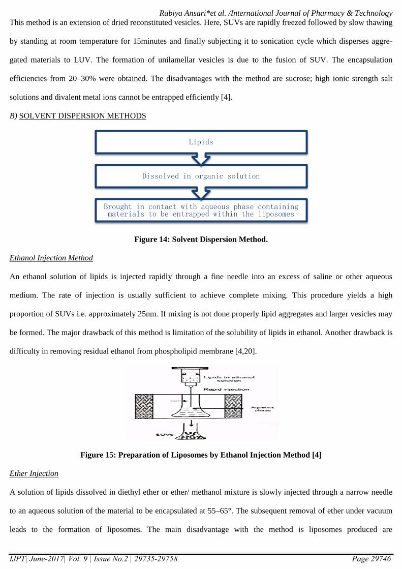
B) SOLVENT DISPERSION METHODS
Figure 14: Solvent Dispersion Method.
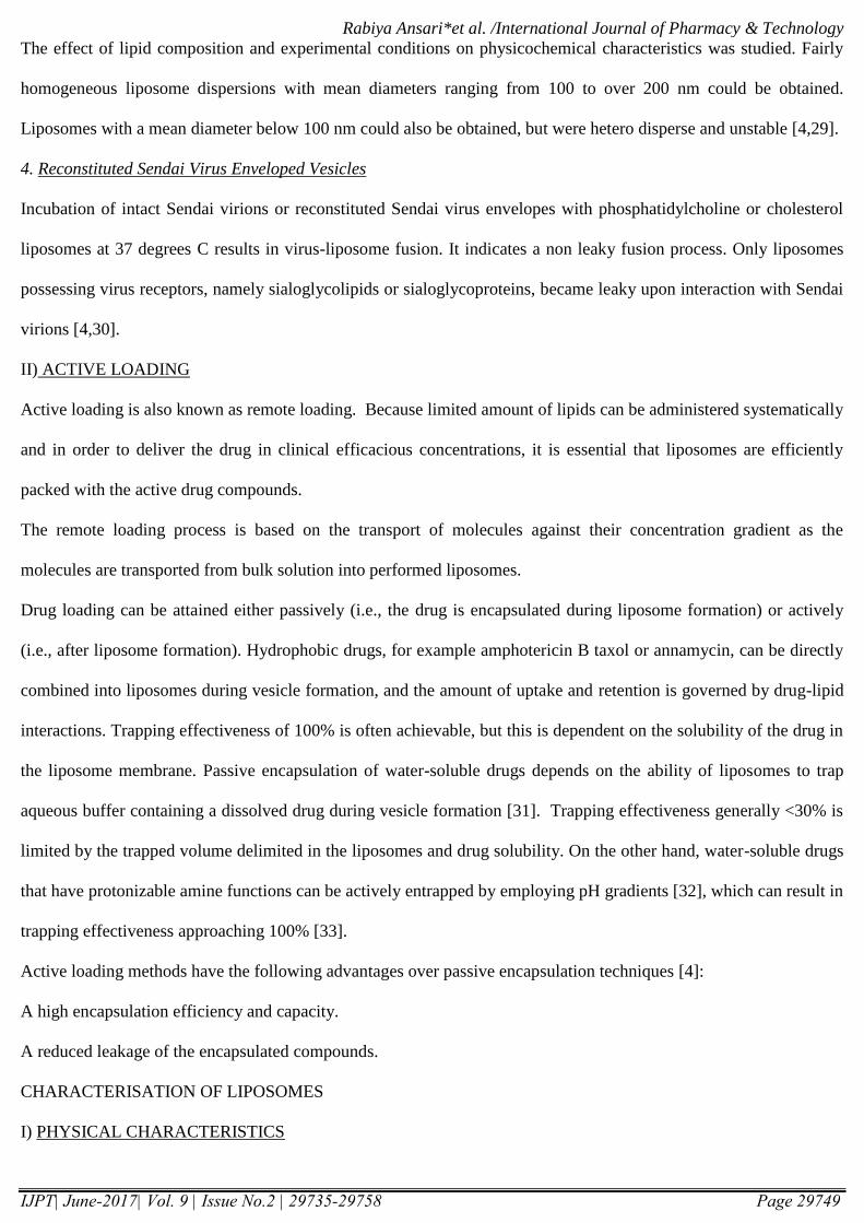
Ethanol Injection Method
An ethanol solution of lipids is injected rapidly through a fine needle into an excess of saline or other aqueous
medium. The rate of injection is usually sufficient to achieve complete mixing. This procedure yields a high
proportion of SUVs i.e. approximately 25nm. If mixing is not done properly lipid aggregates and larger vesicles may
be formed. The major drawback of this method is limitation of the solubility of lipids in ethanol. Another drawback is
difficulty in removing residual ethanol from phospholipid membrane [4,20].
Figure 15: Preparation of Liposomes by Ethanol Injection Method [4]
Ether Injection
A solution of lipids dissolved in diethyl ether or ether/ methanol mixture is slowly injected through a narrow needle
to an aqueous solution of the material to be encapsulated at 55–65°. The subsequent removal of ether under vacuum
leads to the formation of liposomes. The main disadvantage with the method is liposomes produced are
Brought in contact with aqueous phase containing materials to be entrapped within the liposomes
Dissolved in organic solution
Lipids

Rabiya Ansari*et al. /International Journal of Pharmacy & Technology
IJPT| June-2017| Vol. 9 | Issue No.2 | 29735-29758 Page 29747
heterogeneous in nature i.e. 70–190 nm and the material to be encapsulated will be exposed to higher temperature.
The efficiency of material encapsulated is relatively low. The captured volume per mole of lipid is high i.e. 8-17
litre/mol[4,20].
Figure 16: Preparation of Liposomes by Ether Injection Method [4]
3. Double Emulsion
The double emulsion is prepared by rapidly injecting the dispersion of micro droplets into hot aqueous solution of tris
buffer with the help of 22 gauge hypodermic needle under vigorous stirring. The organic solvent is evaporated using
strong jet of nitrogen thus forming double emulsion.
The traces of organic solvent are removed evaporation and finally the volume is adjusted by adding extra distilled
water and then the product is centrifuged at 20 degree Celsius for 30 minutes at 37000g to remove lipid aggregates
[4].
4. Reverse Phase Evaporation Vesicles
First water in oil emulsion is formed by sonication of a two phase system containing phospholipids in organic solvent
i.e. diethylether or isopropylether or mixture of isopropyl ether and chloroform and aqueous buffer. The organic
solvents are removed under reduced pressure, resulting in the formation of a viscous gel.
The liposomes are formed when residual solvent is removed by continued rotary evaporation under reduced pressure.
With this method high encapsulation efficiency up to 65% can be obtained in a medium of low ionic strength for
example 0.01M NaCl. The method has been used to encapsulate small and large macromolecules. The main
disadvantage of the method is the exposure of the materials to be encapsulated to organic solvents and to brief
periods of sonication [28].
5. Stable Plurilamellar Vesicles
Rabiya Ansari*et al. /International Journal of Pharmacy & Technology
IJPT| June-2017| Vol. 9 | Issue No.2 | 29735-29758 Page 29748
Here, water inorganic phase dispersion is prepared with an excess of lipid followed by drying under continued bath
sonication with a discontinuous stream of nitrogen. The redistribution and equilibrium of aqueous solvent and solute
occurs in between the various bilayers in each plurilamellar vesicle. The percent entrapment is around 30% [4].
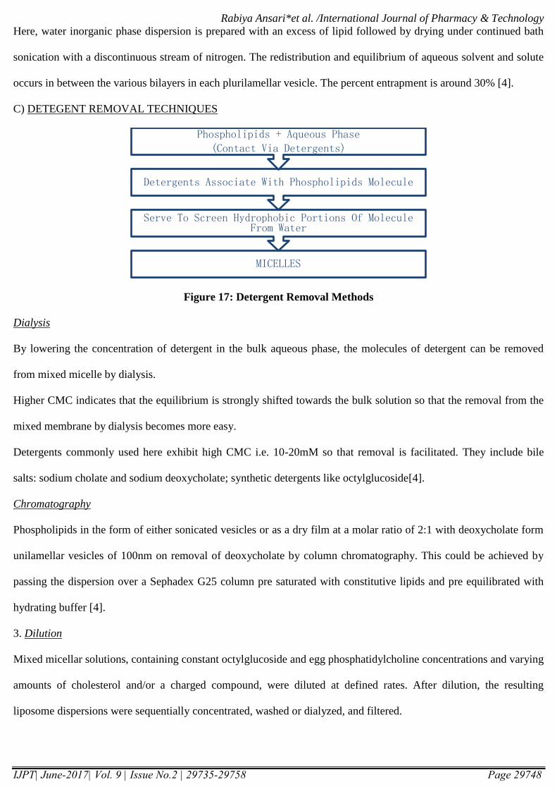
C) DETEGENT REMOVAL TECHNIQUES
Figure 17: Detergent Removal Methods
Dialysis
By lowering the concentration of detergent in the bulk aqueous phase, the molecules of detergent can be removed
from mixed micelle by dialysis.
Higher CMC indicates that the equilibrium is strongly shifted towards the bulk solution so that the removal from the
mixed membrane by dialysis becomes more easy.
Detergents commonly used here exhibit high CMC i.e. 10-20mM so that removal is facilitated. They include bile
salts: sodium cholate and sodium deoxycholate; synthetic detergents like octylglucoside[4].
Chromatography
Phospholipids in the form of either sonicated vesicles or as a dry film at a molar ratio of 2:1 with deoxycholate form
unilamellar vesicles of 100nm on removal of deoxycholate by column chromatography. This could be achieved by
passing the dispersion over a Sephadex G25 column pre saturated with constitutive lipids and pre equilibrated with
hydrating buffer [4].
3. Dilution
Mixed micellar solutions, containing constant octylglucoside and egg phosphatidylcholine concentrations and varying
amounts of cholesterol and/or a charged compound, were diluted at defined rates. After dilution, the resulting
liposome dispersions were sequentially concentrated, washed or dialyzed, and filtered.
MICELLES
Serve To Screen Hydrophobic Portions Of Molecule From Water
Detergents Associate With Phospholipids Molecule
Phospholipids + Aqueous Phase(Contact Via Detergents)
Rabiya Ansari*et al. /International Journal of Pharmacy & Technology
IJPT| June-2017| Vol. 9 | Issue No.2 | 29735-29758 Page 29749
The effect of lipid composition and experimental conditions on physicochemical characteristics was studied. Fairly
homogeneous liposome dispersions with mean diameters ranging from 100 to over 200 nm could be obtained.
Liposomes with a mean diameter below 100 nm could also be obtained, but were hetero disperse and unstable [4,29].
4. Reconstituted Sendai Virus Enveloped Vesicles
Incubation of intact Sendai virions or reconstituted Sendai virus envelopes with phosphatidylcholine or cholesterol
liposomes at 37 degrees C results in virus-liposome fusion. It indicates a non leaky fusion process. Only liposomes
possessing virus receptors, namely sialoglycolipids or sialoglycoproteins, became leaky upon interaction with Sendai
virions [4,30].
II) ACTIVE LOADING
Active loading is also known as remote loading. Because limited amount of lipids can be administered systematically
and in order to deliver the drug in clinical efficacious concentrations, it is essential that liposomes are efficiently
packed with the active drug compounds.
The remote loading process is based on the transport of molecules against their concentration gradient as the
molecules are transported from bulk solution into performed liposomes.
Drug loading can be attained either passively (i.e., the drug is encapsulated during liposome formation) or actively
(i.e., after liposome formation). Hydrophobic drugs, for example amphotericin B taxol or annamycin, can be directly
combined into liposomes during vesicle formation, and the amount of uptake and retention is governed by drug-lipid
interactions. Trapping effectiveness of 100% is often achievable, but this is dependent on the solubility of the drug in
the liposome membrane. Passive encapsulation of water-soluble drugs depends on the ability of liposomes to trap
aqueous buffer containing a dissolved drug during vesicle formation [31]. Trapping effectiveness generally <30% is
limited by the trapped volume delimited in the liposomes and drug solubility. On the other hand, water-soluble drugs
that have protonizable amine functions can be actively entrapped by employing pH gradients [32], which can result in
trapping effectiveness approaching 100% [33].
Active loading methods have the following advantages over passive encapsulation techniques [4]:
A high encapsulation efficiency and capacity.
A reduced leakage of the encapsulated compounds.
CHARACTERISATION OF LIPOSOMES
I) PHYSICAL CHARACTERISTICS
Rabiya Ansari*et al. /International Journal of Pharmacy & Technology
IJPT| June-2017| Vol. 9 | Issue No.2 | 29735-29758 Page 29750
VESICLE SHAPE AND LAMELLARITY
Vesicle shape can be assessed using various electron microscopic techniques. Lamellarity of vesicles i.e. the number
of bilayers present in liposomes is determined using freeze-fracture electron microscopy and 31P nuclear magnetic
resonance analysis [4,34].
Freeze Fracture and Freeze-Etch Electron Microscopy: Freeze fracture electron microscopy can be used to assess the
shape, lamellarity and surface morphology of liposomes. In this technique, the fracture plan passes through the
vesicles, which are randomly positioned in the frozen state. The observed distribution profile depends on the distance
of vesicle centre from the plane of fracture [4,34].
31p Nuclear Magnetic Resonance Analysis: This technique exploits 31P nuclear magnetic resonance NMR to
monitor the phospholipid phosphorus signal intensity. A reduction of 50% in NMR signal intensity indicates a
unilamellar vesicle whereas subsequent reduction indicates multilamellar vesicular preparation [4,34].
VESICLE SIZE AND DISTRIBUTION
Microscopic Techniques
Optical Microscopy: Vesicular dispersion are diluted and wet mounted on a haemocytometer and photographed with
a phase contrast microscope. The negatives are then projected on a piece of calibrated paper using a photographic
enlarger x1250. Diameters of approximately 500 vesicles are measured. This microscopic method includes the use of
Bright field, Phase contrast microscope and Fluorescent microscope. It is useful in evaluating the vesicle size of large
vesicles > 1 micrometer [4,8,34].
Negative Stain Transmission Electron Microscopy: Liposomes are embedded in this method in a thin film of electron
dense heavy metal stain. Negative Stain Transmission Electron Microscopy visualizes relatively electron transparent
liposomes as bright areas against a dark background [4,8,34].
Cryo Transmission Electron Microscopy Technique: This method involves freeze fracturing of the samples followed
by their visualization. Using transmission electron microscopy. Thin sample films are prepared under controlled
temperature 25 degree C and humidity conditions within a custom-built environment chamber. The films are
thereafter vitrified by quick freezing in liquid ethanol and transferred to TEM analysis [4,8,34].
Freeze Fracture Electron Microscopy
Scanning Electron Microscopy: This technique is less frequently used due to the distortion caused during the sample
preparation [4].
Rabiya Ansari*et al. /International Journal of Pharmacy & Technology
IJPT| June-2017| Vol. 9 | Issue No.2 | 29735-29758 Page 29751
Diffraction And Scattering Techniques
Laser Light Scattering Techniques: It is a laser-based technique; quasi-elastic light scattering techniques are useful in
analyzing the homogenous colloids. This technique can be applied to unimodel system with diameter less than 1
micrometer [4,34].
Hydrodynamic Techniques
Field Flow Fractionation Techniques (FFF): Because of fundamental difference in driving forces, sedimentation FFF
and flow FFF measure different vesicle properties [4,34].
Gel Permeation: The ability to separate various components of the heterodispersed preparations could be exploited to
estimate various sized particles present in the dispersion. It is also used for the separation of various heterodispersed
liposomal preparations [4].
Ultracentrifuge: It yields valuable data on size distribution of liposomes. It is also used for analytical purposes [4].
3) SURFACE CHARGE
In general two methods are used to assess the charge [4],
Free flow electrophoresis
Zeta potential measurement
The surface charge can be calculated by estimating the mobility of the liposomal dispersion in a suitable buffer
determined using Helmholtz– Smolochowski equation.
4)ENCAPSULATION EFFECIENCY
It determines the percent of drug in aqueous phase is usually expressed as % entrapment/mg lipid. Encapsulation
efficiency is assessed using two techniques including mini column centrifugation method and protamine aggregation
method [4,8].
% Entrapment efficiency= Entrapped Drug (mg) X 100
Total Drug added (mg)
5) ENTRAPPED VOLUME
The entrapped volume of a population of liposomes in μl/mg phospholipids can often be deduced from measurements
of total quantity of solute entrapped inside liposome assuring that the concentration of solute in the aqueous medium
inside liposome is the same after separation from entrapped material [4].
6) PHASE RESPONSE AND TRANSITIONAL BEHAVIOR

Rabiya Ansari*et al. /International Journal of Pharmacy & Technology
IJPT| June-2017| Vol. 9 | Issue No.2 | 29735-29758 Page 29752
Phase behavior of liposomal membrane determines properties such as permeability, fusion, aggregation and protein
binding. The phase transition has been evaluated using freeze fracture electron microscopy. They are more
comprehensive verified by differential scanning calorimeter analysis. Moreover these phase behaviors are important
to characterize while formulating the liposomes with the lipids having different phase transition temperatures and
polymer PEG grafted liposomes where polymer grafting as such provides a steal thing effect deterring them from
macrophagic uptake and hence long circulation [4,8].
7) DRUG RELEASE
The liposome-based formulations can be assisted by employing in vitro assays to predict pharmacokinetics and
bioavailability of the drug before employing in vivo studies, as the in vivo studies are costly and time consuming
[4,8].
II) CHEMICAL CHARACTERISTICS
PHOSPHOLIPID IDENTIFICATION AND ASSAY
Bartlett assay, Stewart assay and thin layer chromatography can be used to estimate the phospholipid concentration in
the liposomal formulation [4].
In Bartlett assay, the phospholipid phosphorus in the sample is first hydrolyzed to inorganic phosphates. This is
converted to phospho molybdic acid by addition of ammonium molybdate. Phosphor molybdic acid is quantitatively
reduced to blue colored compound by amino naphthylsulphonic acid. The intensity of blue color is measured
spectrophotometrically and is compared with the standard curve of phospholipids. The disadvantage of this method is
Bartlett assay is very sensitive to inorganic phosphates and creates problems in measurement of phospholipids.
In Stewart assay phospholipids form a complex with ammonium ferrothiocynate in organic solution. The standard
curve is first prepared by adding 0.1M ammonium ferrothiocynate solution with different concentrations of
phospholipids in chloroform. Similarly the samples are treated and optical density of the solutions is measured at
485nm and the absorbance of samples compared with standard curve of phospholipids. The advantage of this method
is that the presence of inorganic phosphates does not interfere with the assay. The disadvantage of this method is that
it is not applicable to samples where mixtures of unknown phospholipids may be present.
Thin layer chromatography is employed for determining the purity and concentration of lipids. If the compound is
pure, it should run as a single spot in all elution. Phospholipids, which are degraded, can be observed as a long smear
with a tail trailing to the origin compared to the pure material, which runs as one clearly defined spot.
Rabiya Ansari*et al. /International Journal of Pharmacy & Technology
IJPT| June-2017| Vol. 9 | Issue No.2 | 29735-29758 Page 29753
2) CHOLESTROL ANALYSIS
Cholesterol oxidase assay or ferric perchlorate method and Gas liquid chromatography techniques can be used to
determine the cholesterol concentration [4].
Cholesterol is qualitatively analyzed using capillary column of flexible fused silica.
Cholesterol is quantitatively estimated by measuring the absorbance of purple complex produced with the iron upon
reaction with the combined agent containing ferric perchlorate, ethyl acetate and sulphuric acid at 610nm.
STABILITY OF LIPOSOMES
A stable dosage forms is the one which maintains the physical stability and chemical integrity of the active molecule
during its developmental procedure and storage. Hence a stability protocol is essential to study the physical and
chemical integrity of the drug product in its storage.
1. PHYSICAL STABILITY
During the storage of liposomes, the vesicles tend to aggregate and increase in size to attain thermodynamically
favorable state. Drug leakage from the vesicles can occur due to fusion and breaking of vesicles, which deteriorates
the physical stability of the liposomal drug product. Hence morphology, size and size distribution of the vesicles are
important parameters to assess the physical stability [4,10,11,16].
2. CHEMICAL STABILITY
Phospholipids are prone to oxidation and hydrolysis, which may alter the stability of the drug product. Ionic strength,
pH, solvent system and buffered species also play a major role in maintaining a liposomal formulation. Chemical
reaction can also be induced by light, oxygen, temperature and heavy metal ions. Oxidation deterioration involves the
formation of cyclic peroxides and hydroxyl peroxidases due to the result of free radical generation in the oxidation
process.
Liposomes can be prevented from oxidative degradation by protecting them from light, by adding anti-oxidants such
as alpha – tocopherol or butylated hydroxyl toluene (BHT), producing the product in an inert environment (presence
of nitrogen or Argon) or by adding EDTA to remove trace heavy metals. Hydrolysis of the ester bond at carbon
position of the glycerol moiety of phospholipids leads to the formation of lysophosphatidylcholine (lysoPC), which
enhances the permeability of the liposomal contents. Hence, it becomes necessary to control the limit of lysoPC
within the liposomal drug product. This can be achieved by formulating liposomes with phosphatidylcholine free
from lysoPC[4,10,11,16].
Rabiya Ansari*et al. /International Journal of Pharmacy & Technology
IJPT| June-2017| Vol. 9 | Issue No.2 | 29735-29758 Page 29754
APPLICATIONS OF LIPOSOMES
APPLICATIONS OF LIPOSOMES IN THE SCIENCES [35]
Mathematics: Topology of two-dimensional surfaces in three-dimensional space governed only by bilayer elasticity
Physics: Aggregation behavior, fractals, soft and high-strength materials
Chemistry: Photochemistry, artificial photosynthesis, catalysis, micro compartmentalization
Biochemistry: Reconstitution of membrane proteins into artificial membranes
Biology: Model biological membranes, cell function, fusion, recognition
Pharmaceutics: Model biological membranes, cell function, fusion, recognition
Medicine: Drug-delivery and medical diagnostics, gene therapy
LIPOSOMES IN PHARMACEUTICAL INDUSTRY [35]
For solubilization amphotericin B, minoxidil are used in treatment of fungal infections.
For site-avoidance, Amphotericin B is used to reducednephro toxicity, and doxorubicin is used to decrease cardio
toxicity in fungal infections and cancer.
For sustained-release action of systemic antineoplastic drugs, hormones, corticosteroids, drug depot in the lungs in
cancer and biotherapeutics
3) LIPOSOMES AS DRUG DELIVERY VEHICLES[4]
Enhanced drug solubilization, e.g., Amphotericin B, Minoxidil, Cyclosporine
Protection of sensitive drug molecules, e.g., Cytosine arabinose, DNA, RNA
Enhanced intracellular uptake, e.g., Anticancer drugs, Antiviral drugs, Antimicrobial drugs
Altered pharmacokinetics and biodistribution i.e. prolonged or sustained release of drugs with short circulatory half-
lives.
Increased therapeutic index, e.g., Antitumor drugs i.e. cytosine arabinoside tri-phosphate (ara-CTP), Actinomycin D
4) LIPOSOMES AS A LYSOSOMOTROPIC CARRIER [4]
Liposomes have been used as lysosomotropic carriers therapeutically in enzyme diseases like Gaucher’s disease i.e.
beta glycosidase deficiency and Pompe’s disease i.e. alpha glycosidase deficiency
A variety of lysosomal enzymes can be entrapped in liposomes and administered to patients suffering from lysosomal
storage disorders.
Liposomes is also used in the treatment of metal poisoning, e.g., liposomal EDTA

Rabiya Ansari*et al. /International Journal of Pharmacy & Technology
IJPT| June-2017| Vol. 9 | Issue No.2 | 29735-29758 Page 29755
5) LIPOSOMES IN ANTICANCER THERAPY[4,35]
DaunoXome®: Daunorubicin containing DSPC/chol liposomes is used in Advanced Kaposi's sarcoma
Doxil®/Caelyx®: Doxorubicin containing HSPC/chol/PEG-DSPE liposomes is used in Metastatic ovarian cancer and
advanced Kaposi's sarcoma
MyocetTM: Doxorubicin containing EPC/chol liposomes is used in Metastatic breast cancer
6) LIPOSOME AS ANTI-INFECTIVE AGENTS [20]
Active Targeting Approach
Anamycin is used in treatment of Leishmianiasis
Asiaticoside is used in treatment of Tuberculosis and Leprosy
Rifampicin is used in treatment of Tuberculosis
Passive targeting approach
Amphotericin B is used in treatment of Meningitis, Leishmianiasis, Candidiasis
Praziquantal is used in treatment of Macrophage activation
Gentamycin is used in treatment of Staphylococcal pneumonias
7) LIPOSOME IN EYE DISORDERS
Liposome has been widely used to treat disorder of both anterior and posterior segment. The disease of eye includes
dry eyes; Keratitis, an inflammation of the cornea; Corneal transplant rejection; Uveitis, An inflammation of the
middle layer of the eye; Endophthalmitis, an inflammation of the internal coats of the eye; and Proliferative
vitreoretinopathy (PVR), a disease that develops as a complication of rhegmatogenous retinal detachment i.e. retinal
separation associated with a break, a hole, or a tear in the sensory retina [20].
The liposomal drugs currently approved are ‘verteporfin’ for the use in the eye.
8) LIPOSOMES AS RADIODIAGNOSTIC CARRIERS
Liposomes are used in different imaging modalities to locate the sites specifically. Their radio diagnostic applications
include Liver and spleen imaging, Lymphatic imaging, Tumor imaging, Blood pool imaging, Brain imaging, Imaging
cardiovascular pathologies, Visualization of inflammation and infection sites, bone marrow and eye vasculature [4].
LIPOSOMES IS DERMATOLOGY AND COSMETOLOGY [4, 20]
CaptureTM
marketed by Christian Dior contains liposomes in gel with ingredients
PlenitudeTM
marketed by L’Oreal contains tanning agents in liposomes

Rabiya Ansari*et al. /International Journal of Pharmacy & Technology
IJPT| June-2017| Vol. 9 | Issue No.2 | 29735-29758 Page 29756
NactosomesTM
marketed by Lancome (L’Oreal) contains vitamins, Retinol acetate, in liposomes
10) TUMOR-TARGETED ANTISENSE LIPOSOMAL THERAPY
The use of antisense oligonucleotides (asODNs) to selectively target tumor-associated genes without affecting the
normal ones appears to be a promising new approach [36].
11) FOOD APPLICATION
The ability of liposomes to solubilize compounds with demanding solubility properties, sequester compounds from
potentially harmful medium, and release incorporated molecules in a sustained and predictable way can be used in
food processing industry. A classical example is cheese making [37].
Conclusion: Since the discovery in the last six decades, liposome as a novel drug delivery systems has played a
significant role in reformulation of potent drugs to improve the therapeutics and stability. A number of drugs, which
are highly potent and have low therapeutic indication, can be targeted to the required diseased site using the
liposomal drug delivery system. Liposomal approach can be successfully utilized to improve the pharmacokinetics
and therapeutic efficacy, simultaneously reducing the toxicity of various highly potent drugs. Liposomes can cross
the blood brain barrier (BBB) because of the lipophilic nature of the phospholipids, so even the hydrophilic drugs,
which cannot easily cross the BBB, can be formulated as liposomes. Liposomes are prepared by various methods in
which the most common method applied for research purpose is film method and many more. Liposomes have a
broad range of pharmaceutical applications. Nowadays the liposomal topical formulations are mostly used in the
cosmetic and hair technologies. Liposomes are widely used as radio diagnostic agents. Liposomes are giving a good
and encouraging result in the anticancer therapy and human therapy. They are also widely used as drug delivery
vehicles, lysosomotropic carriers, anti-infective agents and in treatment of eye disorders. Liposomes are one of the
unique and most promising drug delivery system.
References
1. Mansoori and Agrawal, ‘A Review On Liposome’IJARPB, 2012; 2(4), 453-464.
2. Rutuja R D et al. ‘A Review On Liposomes’.World Journal Of Pharmacy And Pharmaceutical Sciences, 2016;
5(3), 506-517.
3. Maurer, Fenske&Cullis, ‘Developments in liposomal drug delivery systems’, Expert Opin. Biol. Ther. 2001;
1(6), 01-25.
4. Vyas SP, Khar RK Targeted and Controlled Drug Delivery: Novel Carrier Systems; 2004; 173-248.

Rabiya Ansari*et al. /International Journal of Pharmacy & Technology
IJPT| June-2017| Vol. 9 | Issue No.2 | 29735-29758 Page 29757
5. Bangham A D, Horne R W. Negative Staining of Phospholipids and Their Structural Modification by Surface-
Active Agents As Observed in the Electron Microscope, Journalof Molecular Biology, 1964; 8(5), 660-668.
6. Anwekar H, Patel S, Singhai AK. Liposome as drug carriers.Int J of Pham & Life Sci 2011; 2(7); 945-51.
7. ShobhaRani,RHiremath and Vanaja K. Textbook of Industrial Pharmacy: Drug Delivery Systems, and Cosmetic
and Herbal Drug Technology 97- 120.
8. KanthShashi et al. ‘A Complete Review On Liposomes’. International Journal Of Pharmacy, 2012; 7 (3), 10-16.
9. Mazafari, M.R. Liposomes: an overview of Manufacturing Techniques, Cell Mol Bio Lett, 2005: 10: 711-719.
10. Sanjay K Jain and N K Jain, Liposomes As Drug Carriers. Controlled and Novel Drug Delivery 304-348.
11. KalepuSandeep et al, publisher. ‘Liposomal drug delivery system - A Comprehensive Review’. Int. J. Drug Dev.
& Res., 2013; 5(4) 62-75.
12. Cancer Treatment- ‘Conventional And Innovative Approaches’, Book Edited By Leticia Range 2013
13. Hiemenz PC, Rajagopalan R. Principles of colloid and surface chemistry, 3rd edition, New York: Marcel
Dekker: 1997; 120-156.
14. Kumar KS, Jyothi, Nagendar S, Devipriya S, Kumar GV, Babu S. Stability of Liposomes. Int J Adv in Pham Sci
2011; 2(4): 301-7.
15. Stryer S. Biochemistry, 1981; 213.
16. Loveleenpreetkauret al. ‘Liposome as a drug carrier – a review’.International Journal of Research in Pharmacy
and Chemistry, 2013; 3(1) 121-128.
17. Horne R W, Bangham A D, Whittaker VP.Negatively Stained Lipoprotein Membranes, Nature, 200(4913), 1963,
1340.
18. Alving CR Macrophages, as targets for delivery of liposome encapsulated antimicrobial agents, Adv Drug
Delivery Rev, 1998; 2.
19. Emanuel et al. ‘Preparation And Characterisation Of Doxorubicin Loaded Sterically Stabilized
Immunoliposomes’. Pham. Res. 1996; 13, 352-359.
20. Deepthi V and Kavitha AN. ‘Liposomal Drug Delivery System-A Review’. RGUHS J Pharm Sci. 2014; 4 (2) 47-
56.
21. Yash Roy R C. ‘Lamellar Dispersion And Phase Separation Of Chloroplast Membrane Lipids By Negative
Staining Electron Microscopy’. Journalof Biosciences, 1990; 15(2), 93-98.

Rabiya Ansari*et al. /International Journal of Pharmacy & Technology
IJPT| June-2017| Vol. 9 | Issue No.2 | 29735-29758 Page 29758
22. Hope, M.Y.; Bally, M.B; Mayer, L.D. Chem. Phys. Lipids., 1986; 40, 89.
23. Samad et al.‘Liposomal Drug Delivery Systems: An Update Review’. Current Drug Delivery, 2007, 4(4), 297-
305.
24. Lasic, D.D. The mechanism of vesicle formation.Biochem J. 1988; 256: 1-11.
25. Lasic, D.D et al. ‘Spontaneous vesiculation’.Adv Colloid Interfac Sci. 2001; 89-90: 337-349.
26. Remington. The Science and Practice of Pharmacy. Vol I, 21st edition, BI Publishers Pvt Ltd; 314–6.
27. Riaz M. Review: Liposomes preparation methods. Pak J Pharm Sci. 1996; 19: 65–77.
28. J.S. Dua1, Prof. A. C. Rana, Dr. A. K. Bhandari. ‘Liposome: Methods Of Preparation And Applications’
International Journal Of Pharmaceutical Studies And Research 2012; 3(2): 14–20.
29. Jiskoot W et al ‘Preparation of liposomes via detergent removal from mixed micelles by dilution; The effect of
bilayer composition and process parameters on liposome characteristics.’ Pharm Weekbl Sci. 1986 Oct 17;8(5):
259-65.
30. V Citovsky and A Loyter. Fusion of Sendai Virions or reconstituted Sendai virus envelopes with liposomes or
erythrocyte membranes lacking virus receptors by., JBC, the journal of Biological Chemistry.
31. Akbarzadeh et al. ‘Liposome: Classification, Preparation, And Applications’. Nanoscale Res Lett.2013; 8(1):
102.
32. Mayer ID et al. 'Liposome Technology. 2. Gregoriadis G, editor. Boca Raton: CRC Press;.pH gradient-mediated
drug entrapment in liposomes; 1993; 27–44.
33. Arcadio C, Cullis PR. ‘Recent advances in liposomal drug-delivery systems’. CurrOpin Biotechnol.1995; 6, 698–
708.
34. SaraswathiMarripati. et al. ‘A Review On Liposomes’. International Journal of Research in Pharmaceutical and
Nano Sciences. 2014, 3(3), 159 - 169.
35. D.D. Lasic. Liposome Technology. ©1995 Elsevier Science B.V. Handbook of Biological Physics.Volume 1,
Edited By R. Lipowskyand E. Sackmann.Chapter 10, 491-519.
36. D Di Paolo, F Pastorino, C Brignole et al. ‘Drug Delivery Systems: Application of Liposomal Anti-Tumor
Agents to Neuroectodermal Cancer Treatment’. Tumori, 2008; 94, 246-253.
37. Laouini et al. ‘Preparation, Characterization and Applications of Liposomes: State of the Art’ Journal of Colloid
Science and Biotechnology. 2012; 1, 147–168.