issn 0355-1180 university of helsinki department of food ... · tiivistelmä referat abstract...
TRANSCRIPT

ISSN 0355-1180
UNIVERSITY OF HELSINKI
Department of Food and Environmental Sciences
EKT Series 1645
ROTAVIRUS IN DRINKING WATER
– MOLECULAR METHODS FOR MEASUREMENT OF
INFECTIVITY
Hyejeong Lee
Helsinki 2014

HELSINGIN YLIOPISTO HELSINGFORS UNIVERSITET UNIVERSITY OF HELSINKI
Tiedekunta/Osasto Fakultet/Sektion Faculty
Faculty of Agriculture and Forestry
Laitos Institution Department
Department of Food Technology
Tekijä Författare Author
Hyejeong Lee
Työn nimi Arbetets titel Title
Rotavirus in drinking water – molecular methods for measurement of infectivity
Oppiaine Läroämne Subject
Food safety (food microbiology)
Työn laji Arbetets art Level
M.Sc Thesis Aika Datum Month and year
June 2014
Sivumäärä Sidoantal Number of pages
62 Tiivistelmä Referat Abstract
Quantitative reverse transcription PCR (RT-qPCR) assay is widely used for the detection
of RNA viruses in environmental water samples. However, a major limitation of using RT-
qPCR assay to quantify virus titers is its inability to discriminate between infectious and
non-infectious viruses, resulting in overestimation of viral infectivity. Thus, the aim of this
study was to develop a reliable molecular method for rotavirus detection with information
on viral infectivity, and which may contribute to the development of molecular detection
methods for correct estimation of infectivity of non-cultivable viruses.
In experimental work, the potential of using propidium monoazide (PMA) or RNase
treatment prior to RT-qPCR assay was evaluated to measure the infectivity of human
rotavirus. In brief, original human rotavirus (HRV) stock was produced by propagating
viruses in MA-104 cells. The virus stocks (including HRV stock A and B) were thermally
treated at 80 °C at different time points. The virus titer was measured by (1) cell culture-
based infectivity assay, (2) RT-qPCR assay, and (3) RT-qPCR assay with PMA or RNase
pretreatment. The result of cell culture-based infectivity assay showed that heat exposure
for 5 min at 80 °C was sufficient to inactivate the HRV, while RT-qPCR assay alone
overestimated the viral infectivity. The results of RT-qPCR assay with pre-treatments
showed that, for thermally-inactivated HRV stock A, similar level of false-positive results
was reduced with PMA treatment regardless of inactivation time (ranges from 1.04 to 1.18
log10 PCR-units), while higher reduction level was observed with RNase treatment (ranges
from 2.64 to 2.89 log10 PCR-units). On the other hand, the effects of both pre-treatments on
thermally-inactivated HRV stock B were negligible.
In conclusion, both PMA and RNase pre-treatments eliminated the false-positive results of
RT-qPCR assay to some extent in defined conditions, while the discrepancy between the
infectivity assay and RT-qPCR assay even with PMA or RNase treatment was observed. In
order to confirm the potential of using RT-qPCR assay combined with pre-treatments to
measure the infectivity of rotavirus, further studies on optimization of PMA and RNase
treatments and production of optimal virus stock would be necessary.
Avainsanat Nyckelord Keywords
Rotavirus, RT-qPCR, PMA, RNase, Viral infectivity
Säilytyspaikka Förvaringsställe Where deposited
Viikki Campus Library
Muita tietoja Övriga uppgifter Further information
EKT series 1645, Public 31.12.2015, Funding source: Aquavalens project no 311846

PREFACE
This study was carried out from September 2013 to May 2014 in the Department of Food
Hygiene and Environmental Health in the Faculty of Veterinary Medicine. This study was
financially supported by European Union, as a part of research project – the ‘Aquavalens’
project number 311846 which aims to improve the safety of European drinking water
through developing more rapid methods of detecting viruses, bacteria and parasites in
water.
I would like to thank my supervisor Dr. Leena Maunula for giving me a chance to work in
her research group and her support. I want to extend my gratitude to PhD student Satu
Oristo for her support and her guidance on experimental research, and also to research
assistant Kirsi Söderberg for her practical advices during experimental research.
Last but not least, I wish to thank my dearest parents for their support, encouragement and
love throughout my life. Thank you for giving me opportunity to chase my dreams. I am
grateful to my friends in Finland, South Korea and elsewhere for their encouragement and
support during my master studies.

ABBREVIATIONS
ATCC American Type Culture Collection
CDC Centers for Disease Control and Prevention
CPE Cytopathogenic effect
Cq Quantification cycle
EMA Ethidium Monoazide
HRV Human Rotavirus
ICC-PCR Integrated Cell Culture-Polymerase Chain Reaction
IMS-PCR Immunoseperation-Polymerase Chain Reaction
NSP Non-structural protein
PCR Polymerase Chain Reaction
PMA Propidium Monoazide
qPCR Quantitative Polymerase Chain Reaction
RCWG Rotavirus Classification Working Group
RT-PCR Reverse Transcriptase-Polymerase Chain Reaction
RT-qPCR Reverse Transcriptase-Quantitative Polymerase Chain Reaction
TCID50 50% tissue culture infectious dose
VP Viral protein
WHO World Health Organization

TABLE OF CONTENTS
1. INTRODUCTION ............................................................................................................. 1
2. LITERATURE REVIEW .................................................................................................. 3
2.1 Rotavirus ...................................................................................................................... 3
2.1.1 Historical background ........................................................................................... 3
2.1.2 Structure and genome ............................................................................................ 3
2.1.3 Classification ......................................................................................................... 5
2.1.4 Diversity and Evolution ........................................................................................ 7
2.2 Rotavirus disease ......................................................................................................... 8
2.2.1 Burden of rotavirus disease ................................................................................... 8
2.2.2 Clinical aspects of rotavirus disease...................................................................... 9
2.2.3 Epidemiological aspects of rotavirus disease ...................................................... 11
2.3 Rotavirus in drinking water ....................................................................................... 12
2.3.1 Waterborne transmission ..................................................................................... 12
2.3.2 Prevalence of rotavirus contamination in water .................................................. 13
2.3.3 Waterborne outbreaks ......................................................................................... 15
2.4 Molecular detection of rotavirus in water .................................................................. 16
2.4.1 Current methods and challenges ......................................................................... 16
2.4.2 Promising methods to measure viral infectivity by PCR-based assays .............. 20
3. EXPERIMENTAL RESEARCH ..................................................................................... 26
3.1 Aims of the present study .......................................................................................... 26
3.2 Materials and methods ............................................................................................... 26
3.2.1 Virus and host cell ............................................................................................... 26
3.2.2 Infectivity assay................................................................................................... 28
3.2.3 PMA treatment .................................................................................................... 28
3.2.4 PMA treatment of viral RNA .............................................................................. 28
3.2.5 RNase treatment .................................................................................................. 29
3.2.6 Heat treatment ..................................................................................................... 29
3.2.7 Viral RNA extraction .......................................................................................... 30
3.2.8 VP2 gene specific primers and probes ................................................................ 30
3.2.9 RT-qPCR assay ................................................................................................... 31
3.2.10 Statistical analysis ............................................................................................. 32
3.3 Results ........................................................................................................................ 32
3.3.1 Standard curve ..................................................................................................... 32

3.3.2 Validation of PMA treatment .............................................................................. 32
3.3.3 Validation of RNase treatment ............................................................................ 34
3.3.4 Thermal inactivation curve.................................................................................. 35
3.3.5 PMA treatment to monitor thermal inactivation of HRV stock A ...................... 36
3.3.6 RNase treatment to monitor thermal inactivation of HRV stock A .................... 36
3.3.7 Comparison of PMA and RNase treatment on HRV stock A ............................. 37
3.3.8 PMA treatment to monitor thermal inactivation of HRV stock B ...................... 38
3.3.9 RNase treatment to monitor thermal inactivation of HRV stock B .................... 38
3.3.10 Comparison of PMA and RNase treatment on HRV stock B ........................... 39
3.4 Discussion .................................................................................................................. 40
4 CONCLUSION ................................................................................................................ 51
REFERENCES .................................................................................................................... 52

1
1. INTRODUCTION
Human rotavirus is a leading cause of viral gastroenteritis in infants and children
worldwide, and it is responsible for 37% of all diarrhea deaths in children under five and
50-60% of acute gastroenteritis cases of hospitalized children throughout the world (WHO
2011; Tate and others 2012). WHO estimates that approximately 453 000 children aged
under 5 years died during 2008 due to rotavirus infection; the vast majority (95%) of these
children live in developing countries with poor hygienic situations (WHO 2012). On the
other hand, the deaths from rotavirus infection in developed countries are rare, but it
remains the one of most common causes of hospitalization for acute gastroenteritis in
children (Leung and others 2005; Widdowson and others 2005).
It is generally known that the transmission of rotavirus mainly occurs by faecal-oral routes.
Humans become infected by person-to-person contact and the inhalation of airborne
human rotaviruses as well as the ingestion of water or food contaminated with human
rotaviruses (WHO 2011b). Although the ingestion of drinking water is not the most
common way of exposure, the presence of human rotaviruses in drinking water poses a
public health risk due to high morbidity rates at low infectious doses (WHO 2011b).
Waterborne transmission may be facilitated due to the stability of rotavirus in
environmental water and its resistance to disinfection treatments (He and others 2009).
Occasional waterborne outbreaks have been caused by consumption of drinking water
contaminated with human rotaviruses (Hopkins and others 1984; Villena and others 2003;
Koroglu and others 2011; Mellou and others 2014).
Accordingly, the availability of a reliable and reproducible method for detection of
rotavirus in environmental samples is crucial to identify the infectious risk for public
health and to reduce their impact on public health (Rosa and Muscillo 2013). However, the
virological analysis of environmental water samples has been historically challenging
mainly due to the low concentration of target viruses as well as the presence of inhibitors
in environmental water (Hamza and others 2011; Rosa and others 2012; Gensberge and
Kostic 2013). Particularly, the environmental analysis of rotavirus has more difficulties
and requires different methodological protocols than other enteric viruses (Ruggeri and
Fiore 2013).

2
Basically cell culture is the gold standard method to examine the infectivity of rotavirus
(Hamza and others 2011). However, it is not sufficiently sensitive for all rotavirus strains,
and it is difficult to perform and time consuming (Ruggeri and Fiore 2013). Currently, the
detection of viral genome using PCR-based molecular assays is the only way to identify
the infectious risk for the population (Gassilloud and others 2003). Especially, quantitative
PCR assays (qPCR or RT-qPCR) have become the method of choice for the detection and
quantitation of health-significance viruses including rotavirus, and this approach is widely
used in the field of food and environmental virology and continuously evolving (Yeh and
others 2009; Bosch and others 2011; Rodriguez-Lazaro and others 2012). However, one
major limitation of using these assays is that they detect and quantify both infectious and
non-infectious viral genomes, and in consequence they do not provide correct information
of viral infectivity (Fittipaldi and others 2011). Indeed, numerous studies have showed that
PCR-based assays resulted in overestimation of viral infectivity (Gassilloud and others
2003; Duizer and others 2004; Bosch and others 2008, 2011).
In order to overcome the limitations of current methodologies, some promising methods to
measure the viral infectivity has been proposed. Among several approaches, pre-treatments
of viral sample using dyes (EMA and PMA) or enzymes (Proteinase K and RNase)
combined with qPCR assays would appear to be relatively easy and rapid to perform. In
addition, the applicability of both pre-treatments to certain human viruses has found to be
successful in some cases to discriminate infectious and inactivated viruses. However, the
applicability of such assays to measure the infectivity of rotavirus has not been thoroughly
investigated yet.
The present study aimed to develop a reliable and rapid molecular method to quantify the
infectivity of human rotavirus as health-significance virus in drinking water, and further to
contribute to the development of molecular methods for correct estimation of infectivity of
non-cultivable health-significant viruses such as human norovirus.
In the first part of the thesis, the general aspects of rotavirus and rotavirus disease, the
significance of rotavirus in drinking water, as well as current and promising methods for
rotavirus detection in environmental water samples are reviewed. In the second part, the
potential of using RT-qPCR assay combined with enzymatic treatment or dye treatment to
assess the infectivity of human rotavirus are investigated and evaluated.

3
2. LITERATURE REVIEW
2.1 Rotavirus
2.1.1 Historical background
In 1973, Bishop and colleagues discovered new virus particles in the epithelial cells of
duodenal mucosa from children with acute gastroenteritis (Bishop and others 1973). The
viruses with similar morphological appearance had been found in the intestinal epithelium
of infant mice with diarrhea in 1963 (Adams and others 1963). A year after Bishops’
discovery in 1974, Flewett and colleagues named rotavirus after the Latin word rota
(=wheel) plus virus, since the shape of virus particles resembled the spokes of wheel under
electron microscopy as described in Figure 1 (Flewett and others 1974).
Figure 1. Wheel-like shape of rotaviruses under electron microscopy. Adapted from CDC 2011.
2.1.2 Structure and genome
Rotaviruses are the members of the Reoviridae family (Matthews and Maurin 1979). The
virion of rotaviruses is characterized as a 70-nm non-enveloped icosahedral particle with a
capsid (Wilhelmi and others 2003). As shown in Figure 2A, the virion possesses a viral
genome consisting of 11 segments of double-stranded RNA (dsRNA), and each RNA
segment encodes one protein, except the segment 11 which encodes two proteins
(Greenberg and Estes 2009). There are 12 proteins encoded: 6 structural viral proteins
(VP1, VP2, VP3, VP4, VP6 (VP5+VP6), and VP7) and 6 non-structural proteins (NSP1,
NSP2, NSP3, NSP4, NSP5 and NSP6), and each of which plays a different role during
virus life cycle; including cell entry, transcription and replication (Jayaram and others 2004;
Hu and others 2012).

4
(A) (B)
Figure 2. Structure and proteins of rotavirus. (A) The viral genome of 11 double-stranded RNA segments is
analyzed by polyacrylamide gel electrophoresis, and each of 11 gene segments encodes one protein except
the segment 11, and thus there are 12 proteins encoded: 6 structural viral proteins (VP1, VP2, VP3, VP4,
VP6 (VP5+VP6), and VP7) and 6 non-structural proteins (NSP1, NSP2, NSP3, NSP4, NSP5 and NSP6). (B)
The structure of rotavirus virion is determined by image reconstruction after electron cryomicroscopy, and
viral proteins (VP) construct three concentric protein layers of rotavirus capsid and the capsid of rotavirus is
structured to protect its genome. Adapted from Greenberg and Estes 2009.
The capsid of rotavirus is structured to protect its genome and deliver it successfully into a
suitable host cell, in which the genome is replicated and the virus particles make copies
(Jayaram and others 2004). As depicted in Figure 2B, viral proteins (VP) construct three
concentric protein layers of rotavirus capsid, and they include an outermost layer, an
intermediate layer and an inner layer (Jayaram and others 2004). The outermost layers are
implicated in cell attachment, membrane penetration and cell entry (Patton 2013). They are
mostly composed of glycoprotein VP7 and the spikes of protease-activated attachment
protein VP4 (Patton 2013). VP7 is a Ca2+
binding protein and a key mediator of Ca2+
driven uncoating of the outermost layer, initiating the replication cycle (Trask and others
2012). VP4 is susceptible to proteolysis; e.g. trypsin, so that it is cleaved into VP8 and VP5,
enhancing viral infectivity by several folds and facilitating virus entry into cells (Clark and
others 1981; Carter and Saunders 2007). The sole component of the intermediate layer is
VP6, and it surrounds the inner layer (Leung and others 2005). VP6 maintains structural
integrity during the process of endogenous transcription, and provides mRNA exit
channels (Jayaram and others 2004). The inner layer of the virion is composed of the core
shell protein VP2. The viral RNA-dependent RNA-polymerase VP1 and capping enzyme
VP3 are attached to the inside of the VP2 layer (Patton 2013). Together VP1, VP2 and
VP3 represent the core of rotavirus virion (Leung and others 2005).

5
The ds RNA genome encodes its own enzymes necessary for transcription and it is capable
of endogenously transcribing its genome (Jayaram and others 2004). The capped (+) RNA
transcripts encode the viral proteins and function as templates for the production of (-)
RNA to make the progeny ds RNA (Jayaram and others 2004). The mode of replication is
conservative since the ds RNA of the infecting virion remains intact (Carter and others
2007). Once enough viral proteins have been made in cells, the RNA genome is replicated
and packaged into newly made double-layer particle in specialized structures called
viroplasms (Jayaram and others 2004). At the final stage of replication, virus assembly
takes place by the addition of outer layer of the capsid and the spikes, and then virions are
released from the cells either by lysis or by exocytosis (Carter and others 2007).
2.1.3 Classification
Rotaviruses have been classified into five serological groups A to E and two tentative
groups F and G, mainly on the basis of antigenic specificity of VP6, or more recently
sequence analysis of VP6 (Kindler and others 2013). Rotaviruses belonging to group A, B
and C are known to induce infections in both humans and animals, whereas group D, E, F
and G are only in animals (Matthijnssens and others 2010). Additionally, another group of
rotaviruses originally named ‘new adult diarrhea viruses or ADRV-N was discovered, and
recently renamed as group H (Matthijnssens and others 2010; Kindler and others 2013).
Group A rotaviruses are the leading causes of viral diarrhea, accounting for nearly all
rotavirus-associated mortality and morbidity, especially in children less than 5 years of age
(Patton 2013). In consequence, group A rotaviruses have been extensively studied and
have been classified further using various approaches (Table 1). Currently available
vaccines are directed against the common group A human rotaviruses (Matthijnssens and
others 2010; Ghosh and Kobayashi 2011). Group B rotaviruses are genetically and
antigenicallly distinct from group A rotaviruses, and they causes severe, cholera-like
diarrhea mostly in adults (Yamamoto and others 2010). They were first identified as adult
diarrhea rotaviruses (ADRV), which have caused large outbreaks of severe diarrhea
involving thousands of adults in China (Hoshino and Kapikian 2000). It has been only
detected in China, India, Bangladesh, and recently in Myanmar (Yamamoto and others
2010). Group C rotaviruses tend to cause sporadic outbreaks and they have been
occasionally associated with food-borne contamination (Patton 2013). Group D and E
rotaviruses are known to infect avian species (Patton 2013). Group F and G rotaviruses
have been originally identified in chicken in 1984, and have been only found in birds so far

6
(Kindler and others 2013). Group H rotaviruses (also called ADRV-N) have been only
isolated in a large outbreak among adults in China, with a sporadic case in Bangladesh
(Matthijnssens and others 2010).
Table 1. Classification of group A human rotaviruses. Group A rotaviruses have been categorized based on (i)
whole-genome RNA hybridization patterns (genogroups); (ii) the antigenic properties of VP6, VP7 and VP4
(subgroups, G-serotypes and P-serotypes, respectively); (iii) the nucleotide sequence analysis of VP7 and
VP4 (G-genotypes and P-genotypes); and (iv) the migration pattern of the RNA genome segments when
subjected to polyacrylamide gel electrophoresis (long, short, supershort or atypical electropherotypes).
Adapted from Maunula 2001.
Classification Types Based on identification of
Within Group A
Genogroup WA-like, DS-1-like,
and AU-1-like
RNA-RNA hybridization
Subgroup I, II Antigenic specificities of VP6
G-serotypes
(G:glycoprotein)
or G-genotypes
G1, G2…etc
Antigenic properties of VP7
or sequence analysis of VP7
P-serotypes
(P:proteinase)
P1, P2…etc Antigenic properties of VP4
P-genotypes P[1], P[2] etc Sequence analysis of VP4
Electropherotypes long, short, supershort
or atypical types (e-types)
Patterns of 11 genes after gel
electrophoresis of genomic RNA
Recently, a whole genome-based genotyping scheme of group A rotaviruses, also known
as the Rotavirus Classification Working Group (RCWG) genotyping system has proposed
(Matthijnssens and others 2008). This RCWG system has recommended to use a uniform
nomenclature in defining the complete genotype constellation of group A rotaviruses, with
notation Gx-P[x]-Ix-Rx-Cx-Mx-Ax-Nx-Tx-Ex-Hx (x representing the genotype number)
being used to denote the genotype of VP7-VP4-VP6-VP1-VP2-VP3-NSP1-NSP2-NSP3-
NSP4-NSP5 genes respectively (Matthijnssens and others 2011). Until 2011, the whole
genomes of at least 167 group A human rotaviruses, a limited number of group B and C
have been analyzed (Ghosh and Kobayashi 2011). In addition, only one complete genome
sequence exists for group D and two genome sequences are available for group H, but no
sequence available for group E yet (Ghosh and Kobayashi 2011). Recently, first complete
genome sequences of group F and G have been described (Kindler and others 2013).

7
2.1.4 Diversity and Evolution
Human rotaviruses display a considerable genetic diversity (Ghosh and Kobayashi 2011).
Among different groups A, B and C, the sequence identity of 11 gene segments is
generally less than 60 % (Alam and others 2007; Nagashima and others 2008). The high
level of genetic diversity within a group and different level of sequence diversity in each
11 gene segment have been observed (Matthijnssens and others 2008). Moreover, there are
at least four mechanisms which generate and increase the overall diversity of rotaviruses
and by which rotaviruses evolve: (i) point mutation or “drift”, (ii) genomic reassortment or
“shift”, (iii) gene rearrangement, and (iv) interspecies transmission (Iturriza-Gomara and
others 2001; Matthijnssens and others 2009; Ghosh and Kobayashi 2011).
The accumulation of point mutations is believed to occur frequently in human rotaviruses
due to the error-prone nature of viral RNA-dependent polymerase (Matthijnssens and
others 2009). It results in changes in the gene sequence and thus may affect the function of
the viral proteins (Ghosh and Kobayashi 2011). Genomic reassortment is an exchange and
substitution of RNA segments between different rotavirus strains (Ghosh and Kobayashi
2011). Due to the segmented nature of rotavirus genome, it is a major evolutionary
mechanism in commonly circulating rotavirus strains (Matthijnssens and others 2009;
McDonald and others 2009). Most reassortment events across genogroups occurs in the
genes encoding VP4 and VP7, resulting in the formation of new strains with unusual G/P
combinations and contributing to overall diversity (Iturriza-Gomara and others 2001;
Cunliffe and others 2002).
In addition, gene arrangement within single RNA segment, such as deletions, duplications
and insertions, causes a change in the size of the RNA segment. However, the overall
contribution of gene arrangement to the diversity of rotaviruses seems to be lower than
other mechanisms (Matthijnssens and others 2009). Moreover, the interspecies
transmission between animal and human rotaviruses is known to be another major
mechanism generating the diversity of human rotaviruses (Gentsch and others 2005).
Previous studies have detected several human rotaviruses which have close relations to
animal strains; for example, feline-like rotaviruses in children in Japan (Nakagomi and
Nakagomi 1989), porcine serotype G5 rotavirus in Brazilian children (Gouvea and others
1994), and bovine-like serotype G8 in children in Malawi (Cunliffe and others 2001).

8
2.2 Rotavirus disease
2.2.1 Burden of rotavirus disease
Nearly all children by 5 years of age have been infected by rotavirus at least once, in both
developed and developing countries (WHO 2013). It accounts for 37% of all diarrhea
deaths in children under five and 50-60% of acute gastroenteritis cases of hospitalized
children throughout the world (WHO 2011; Tate and others 2012). The rotavirus infections
result in estimated 25 million outpatient visits and 2 million hospitalizations each year
worldwide (Parashar and others 2003, 2009). WHO estimated that globally around 453 000
(420 000 - 494 000) child deaths occurred during 2008 due to rotavirus infections, and
most deaths (95%) occurred in malnourished infants living in socioeconomically
disadvantaged rural regions in low-income countries, where access to healthcare is poor,
particularly located in sub-Saharan Africa and Southeast Asia as shown in Figure 3 (WHO
2012). On the other hand, the deaths from rotavirus infections are rare in developed
countries, in Australia, East Asia, Europe and North America, but the incidence of disease
in young children is similar to that of developing countries (Chen and others 2012).
Soriano-Gabarro and others (2006) estimated that rotavirus accounted for 231 deaths,
87,000 hospitalizations, almost 700,000 outpatient visits as well as 3.6 million episodes of
rotavirus disease among the 23.6 million children less than 5 years of age each year from
2000 to 2003 in the European Union. Thus, in developed countries, rotavirus infection
remains the one of most common causes of hospitalization for acute gastroenteritis in
children and leads to major medical costs (Leung and others 2005; Widdowson and other
2005).
Figure 3. Estimated rotavirus deaths in 2008. National estimates of rotavirus attributable deaths among
children under five years of age ranged from 98 621 (India) to fewer than 5 deaths (74 countries). Twenty-
two percent of all rotavirus deaths under five years of age occurred in India. Five countries (India, Nigeria,
the Democratic Republic of the Congo, Ethiopia and Pakistan) accounted for more than half of all rotavirus
deaths under age five in 2008. Adapted from WHO 2012.

9
2.2.2 Clinical aspects of rotavirus disease
The primary site of rotavirus infection is mature enterocytes of the apical portion of villi in
the small intestine causing gastroenteritis, and thus the main clinical manifestation is
diarrhea (Greenberg and Estes 2009; Kindler and others 2013). Multifactorial and
malabsorptive diarrheas may occur due to the virus-mediated destruction of absorptive
enterocytes, the virus-induced suppression of absorptive enzymes, and functional changes
in tight junctions between enterocytes leading to paracellular leakage (Greenberg and Estes
2009). In addition, the activation of enteric nervous system (ENS) and the effects of
enterotoxin NSP4 (virus-encoded enterotoxin) seem to mediate secretory components of
rotavirus diarrhea (Ramig 2004; Greenberg and Estes 2009). However, the mechanism of
diarrhea induced by rotavirus is not fully understood (Ramig 2004; Greenberg and Estes
2009).
However, rotavirus infection is not always limited to the intestine (Greenberg and Estes
2009). Rotaviruses have been detected in the extra-intestinal sites of the liver, kidney, and
central nervous system (CNS) (Ramig 2004; Dickey and others 2009; Greenberg and Estes
2009). Few cases include the finding of virus in the liver following fatal disease (Carlson
and others 1978), the finding of elevated liver enzymes associated virus infection
(Kitamoto and others 1993), and the demonstration of viral replication in the liver and
kidneys of immune-deficient children (Gilger and others 1992). In addition, some clinical
reports of rotavirus have shown that rotavirus spread and pathogenesis may play a potential
role to cause viremia (Blutt and others 2003; Lynch and others 2003).
Rotavirus infections can result in asymptomatic infection, mild diarrhea or severe
gastroenteritis, after the incubation period of 1 to 3 days (Bernstein 2009; Chen and others
2012). Rotaviral gastroenteritis is more severe than other causes of gastroenteritis, and it
often results in dehydration, hospitalization and even death (Bernstein 2009). Although
rotavirus disease can occur at any age, rotavirus gastroenteritis is most common and severe
in children 3 to 36 months of age (Dennehy and others 2008; Chen and others 2012). The
major symptoms in young children include mild-to-severe watery diarrhea, vomiting, and
low-grade fever, and symptoms usually last for up to 4–8 days (Staat and others 2002; Lee
and others 2008). Among young children, the first rotavirus infection is most likely to
produce moderate-to-severe diarrhea disease, but the incidence of moderate-to-severe
diarrhea decreases with second infections, and third infections are typically asymptomatic
(Bernstein 2009).
10
Among adults, rotavirus infections have been associated with a wide spectrum of disease
severity and manifestations; symptoms included diarrhea, fever, headache, malaise, nausea
or cramping (Anderson and Weber 2004). Particularly, rotavirus infections in adults have
been often related to epidemic outbreaks (Meurman and others 1977; Foster and others
1980; Griffin and others 2002) or traveler’s diarrhea (Bolivar and others 1978; Vollet and
others 1979; Steffen and others 1999). In addition, adults who are in contact with children
are shown to be at particularly high risk of infection since transmission of rotavirus within
families from children to parents seems to be a common event (Wenman and others 1979;
Grimwood and others 1983).
Treatments primarily aim at symptom relief and the restoration of normal physiological
functions, as rotavirus disease is usually self-limited (Anderson and Weber 2004). The
rehydration and maintenance of proper fluid and electrolyte balance remains the mainstay
of treatment (Leung and Robson 1989). WHO recommends the use of oral rehydration
formula for children who are mildly to moderately dehydrated, whereas intravenous
rehydration therapy is recommended for those who have severe diarrhea, intractable
vomiting or for those too sick to take oral feedings (WHO 2006). In addition, resumption
of a normal, age-appropriate diet is essential for maintaining the nutritional status of child,
and it can reduce the morbidity and mortality of rotavirus gastroenteritis (Leung and
Robson 1989; Nutrition committee 2003). The use of probiotics (Lactobacillus GG,
Bifidobacterium bifium etc) early in the course of diarrhea is shown to reduce the duration
of diarrhea and rotavirus shedding in affected patients (Saavedra and others 1994;
Guandalini and others 2000; van Niel and others 2002).
In principle, the prevention of rotavirus infection can be achieved by avoiding exposures
and fecal-oral spread (Anderson and Weber 2004). Contact with sick children and
potentially contaminated food and water should be avoided, and contaminated objects and
surfaces should be properly disinfected (Leung and others 2005). General measures such as
personal hygiene and frequent hand washing may help control outbreaks in hospitals and
child care center (Leung and others 2005). In addition, breast-feeding is encouraged, as it
may be associated with milder disease in affected infants (Clemens and others 1993).
Fortunately, rotavirus infection is regarded as the single most frequent vaccine-preventable
disease among children, and currently universal rotavirus vaccination is frequently used to
control the disease (Widdowson and others 2005; Soriano-Gabarro and others 2006). Two
effective rotavirus vaccines, RotaTeq (Merck) and Rotarix (GlaxoSmithKline) were

11
licensed for use in various countries of the world, beginning in 2004-2005 (Yen and others
2011). WHO recommends the routine immunization of all infants, and has introduced the
rotavirus vaccine programs to countries with a high incidence of rotavirus mortality but
lack of infrastructure or financial resources to develop such programs themselves (WHO
2009).
2.2.3 Epidemiological aspects of rotavirus disease
Human rotaviruses are mainly transmitted by faecal-oral route, person-to-person contact
and the inhalation of airborne human rotaviruses or aerosols containing the viruses (WHO
2011b). In consequence, they can generate small epidemic outbreaks in all age groups,
particularly within schools, hospitals, nursing homes, and care centers (Ruggeri and Fiore
2013). In addition, human rotaviruses are transmitted through faecally contaminated food
and water, and thus results in occasional waterborne and foodborne outbreaks (WHO
2011b).
Earlier study on the seasonality of rotavirus disease found that rotavirus infection was
certainly more common in the cooler months in temperate regions, whereas seasonal peaks
of the infections can vary broadly and occur from autumn to spring in warmer tropical
regions (Cook and others 1990). More recently, however, the seasonality of the rotavirus
infection is shown to have stronger relation to the level of country development than
latitude or geographic location (Pitzer and others 2011; Patel and others 2013). Patel and
others (2013) found that poorer countries, particularly those in Africa, Asia, and South
America have shown year-round circulation and had lesser seasonal variation in disease
than more developed countries like Europe, North America and Oceania. In addition, one
study by Pitzer and others (2011) suggested that the high birth rates and transmission rates
typical of developing countries may be the reason for the relative lack of rotavirus
seasonality observed in many tropical countries rather than being driven primarily by
environmental conditions.
In accordance with increasing global burden of rotavirus disease and development of
rotavirus vaccines, the introduction of rotaviruses surveillances program by WHO and
others helps describing the diversity of rotavirus strains in different countries and their
regions, and identifying the emerging strains (Chen and others 2012; Patel and others
2013). Currently, 27 G genotypes and 35 P genotypes have been described, and at least 73
G/P genotype combinations of group A rotaviruses have been detected (Matthijnssens and
others 2012; Patton 2013). Globally, G1, G2, G3, G4 and G9 are the most prevalent VP7

12
serotypes; P[4], P[6] and P[8] are the most common prevalent VP4 genotypes (Chen and
others 2012). Five rotavirus strains of G/P combination; G1P[8], G2P[4], G3P[8] , G4P[8]
or G9P[8], are the major causes of children rotavirus diseases globally (Patel and others
2011). Among these strains, G1P[8] rotaviruses are consistently primary cause in North
America, Austrailia and Europe (70% of infections), whereas less in South America, Asia
and Africa (20-30%) (Soriano-Gabarro and others 2006; Iturriza-Gomara and others 2009).
Recently, G12 have emerged and spread globally over the past decade (Matthijnssens and
others 2009; Patton 2013).
In developing countries, uncommon G/P combinations may be also a frequent cause of
disease in young children, and the types of uncommon strains varies from one region to
another region (Armah and others 2010; Binka and others 2011). WHO surveillance
program has revealed that predominant uncommon strains are G12P[8], G12P[6] in the
Southeast Asia, G2P[6], G3P[6] and G1P[6] in the sub-Saharan Africa, G1P[4], G2P[8] in
the Western Pacific, and G9P[4] in the Americas (WHO 2011a). The chance of rotavirus
co-infection to generate reassortant viruses seems to be higher in developing countries than
in developed countries, contributing the greater strain diversity in developing countries
(Patton 2013).
2.3 Rotavirus in drinking water
2.3.1 Waterborne transmission
Human rotaviruses are excreted by patients in large quantities up to 1011
particles per gram
of faeces for periods of about 8 days (WHO 2011b). It means that wastewater receiving
faecal matter and any environments polluted with human faeces are likely to contain high
numbers of human rotaviruses (Bosch 1998; WHO 2011b; Rosa and others 2012). A
significant viral load can be released in effluent discharge and spread to the aquatic
environment such as estuarine water, seawater and river (Rosa and others 2012). In
consequence, all types of water including raw or treated sewage, river water, recreational
water, and drinking water, are potential vehicles of virus transmission as described in
Figure 4 (Bosch and others 2011). The high stability of rotaviruses in environmental water
and their resistance to disinfection treatments may facilitate the waterborne transmission of
rotaviruses (He and others 2009). In addition, rotaviruses are highly contagious and
infectious dose is very low since as few as 10 particles can cause infection (Chen and
others 2012). Ultimately, the consumption of contaminated drinking water or fruits and
vegetables being in contacted with contaminated water poses public health risk, although
13
waterborne transmission of human rotaviruses is a not major route of exposure (Bosch and
others 1998, 2008, 2011; WHO 2011b).
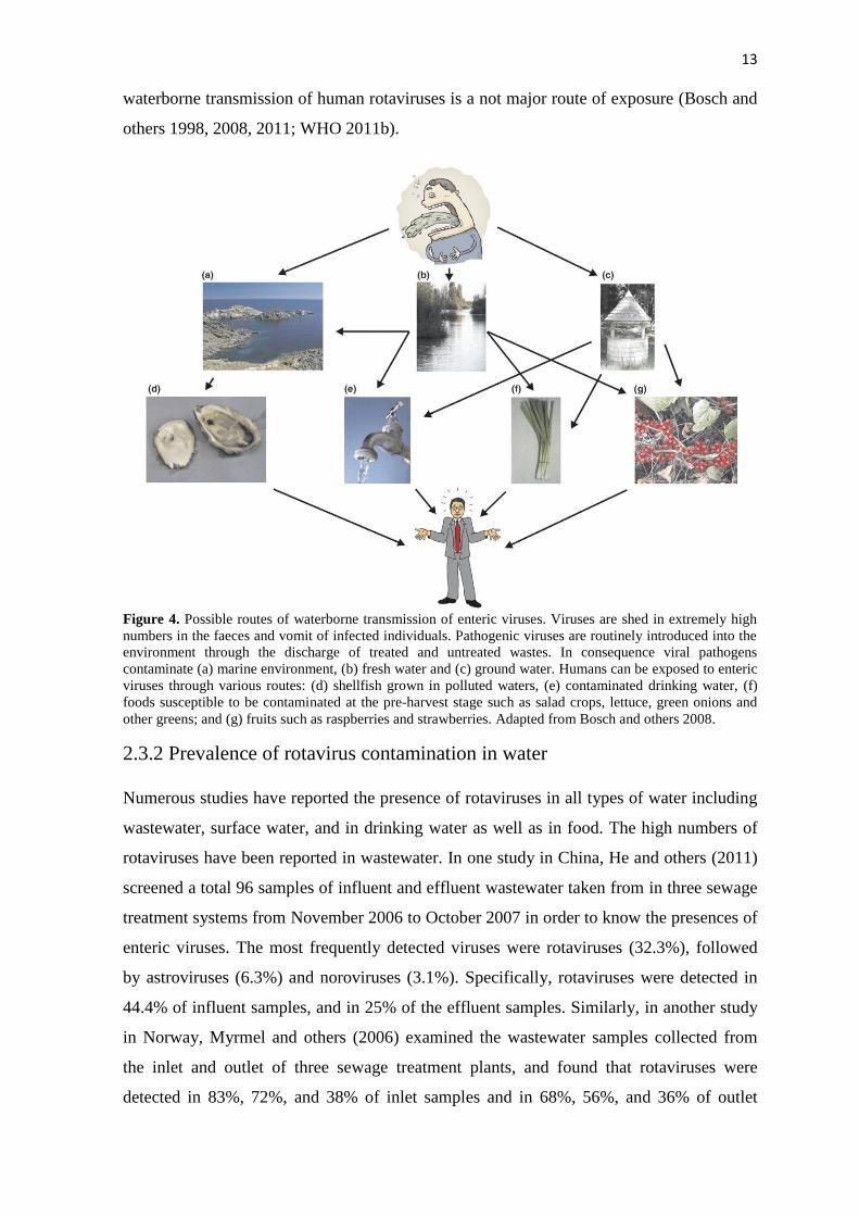
Figure 4. Possible routes of waterborne transmission of enteric viruses. Viruses are shed in extremely high
numbers in the faeces and vomit of infected individuals. Pathogenic viruses are routinely introduced into the
environment through the discharge of treated and untreated wastes. In consequence viral pathogens
contaminate (a) marine environment, (b) fresh water and (c) ground water. Humans can be exposed to enteric
viruses through various routes: (d) shellfish grown in polluted waters, (e) contaminated drinking water, (f)
foods susceptible to be contaminated at the pre-harvest stage such as salad crops, lettuce, green onions and
other greens; and (g) fruits such as raspberries and strawberries. Adapted from Bosch and others 2008.
2.3.2 Prevalence of rotavirus contamination in water
Numerous studies have reported the presence of rotaviruses in all types of water including
wastewater, surface water, and in drinking water as well as in food. The high numbers of
rotaviruses have been reported in wastewater. In one study in China, He and others (2011)
screened a total 96 samples of influent and effluent wastewater taken from in three sewage
treatment systems from November 2006 to October 2007 in order to know the presences of
enteric viruses. The most frequently detected viruses were rotaviruses (32.3%), followed
by astroviruses (6.3%) and noroviruses (3.1%). Specifically, rotaviruses were detected in
44.4% of influent samples, and in 25% of the effluent samples. Similarly, in another study
in Norway, Myrmel and others (2006) examined the wastewater samples collected from
the inlet and outlet of three sewage treatment plants, and found that rotaviruses were
detected in 83%, 72%, and 38% of inlet samples and in 68%, 56%, and 36% of outlet
14
sample in three sewage treatment plants, respectively. In the study conducted in Germany,
Pusch and others (2005) found that rotaviruses were detected in 3–24% of 123 treated
wastewater samples collected from three sewage treatment plants. These studies
demonstrated that untreated and treated wastewater may be a source of viral dissemination,
and they are responsible for the environmental spread of rotaviruses.
The presence of rotaviruses has been also reported in surface water and drinking water. In
one study in China, He and others (2012) collected a total 108 urban surface water samples
from September 2007 to August 2007, and examined for the presence of enteric viruses.
Among 63 virus strains identified, the most predominant viruses were rotaviruses (48%),
followed by astroviruses (5.6%) and noroviruses (4.6%). In another study in Slovenia,
Steyer and others (2011) screened surface water and drinking water for the presence of
enteric viruses. Group A rotaviruses were detected in only 17.5% of the total 63 surface
water samples, while noroviruses were more prevalent (41.3%) in surface water samples.
On the other hand, among 72 drinking water samples, group A rotaviruses were the most
prevalent (37.5%), followed by noroviruses (2.8%) and astroviruses (1.4%). These studies
demonstrated the high prevalence of rotaviruses in surface water and drinking water, and
highlighted the possibility to get rotavirus infection through the consumption of water.
Although rotaviruses were estimated to cause only 1% of all food-related illness and death
(Mead and others 1999), the presence of rotaviruses has been occasionally reported in
food. In the case of foodborne transmission, the main source of contamination is
considered to be polluted water that has been in contact with food or inadequately treated
or untreated sewage used for irrigation (Bosch and others 2008). In one study in Costa
Rica, Hernandez and others (1997) investigated the presence of rotaviruses in lettuce
bought in farm markets. Those samples collected during months (from December to
January) of high incidence of rotavirus diarrhea were positive for rotavirus, and they
suggested that the lettuce might have been contaminated with sewage. In addition, one
study in Canada, Mattison and others (2010), they claimed the possible foodborne
transmission of rotaviruses through the packaged leafy greens, although only 0.4% of
samples were positive for rotaviruses. In another study in Canada, Brassard and others
(2012) detected two positive samples of group A rotaviruses in 60 strawberries samples,
and here irrigation water was suspected to be the source of contamination. These findings
implied that the possible risk of rotavirus to contaminate food through waterborne
transmission should be taken into account, even if the prevalence of rotavirus in food is
expected to be relatively lower than that of other enteric viruses.

15
2.3.3 Waterborne outbreaks
Waterborne rotavirus infections are responsible for significant morbidity and mortality
(Glass and others 2001; Villena and others 2003; Divizia and others 2004). Occasional
waterborne outbreaks mainly due to the consumption of drinking water contaminated with
rotaviruses have been reported (Table 2). Thus, the presence of human rotaviruses in
drinking water is directly related to public health risk (WHO 2011b).
Table 2. Rotavirus related waterborne outbreaks due to the consumption of contaminated drinking water.
Month/Year Place/Country Etiological
agents
Predominant
symptoms
Reference
March
2012
Elassona, Greece
Rotavirus
*AGI; diarrhea,
abdominal pain,
vomiting and fever
Mellou and others
2014
November
2007 Nokia, Finland
Norovirus,
Astrovirus
Adenovirus,
Rotavirus,
Enterovirus
AGI; diarrhea and
vomiting
Maunula and others
2009
November
2005 Malatya, Turkey Rotavirus
AGI; diarrhea,
abdominal pain, fever,
and vomiting
Koroglu and others
2011
December
2000 Tirane, Albania
Rotavirus,
Astrovirus,
Adenovirus,
Norovirus
AGI Villena and others
2003
August
2000
Gourdon,France
Rotavirus,
Norovirus
Co-infection
AGI; diarrhea,
abdominal pain
and nausea
Gallay and others
2006
April
1994
Noormarkku,
Finland
Norovirus, small
round virus,
Rotavirus
AGI; abdominal pain,
severe vomiting, in some
cases high fever,
headache and diarrhea.
Kukkula and others
1997
March
1981
Eagle-Vail,
Colorado, USA Rotavirus
AGI; diarrhea and
vomiting
Hopkins and others
1984
*AGI = acute gastrointestinal illness

16
2.4 Molecular detection of rotavirus in water
2.4.1 Current methods and challenges
Environmental virology research has been focused on aquatic environment including
wastewater, surface water, and drinking water, mainly owing to public health concerns of
viral related waterborne diseases and outbreaks (Bosch and others 2008; Gensberge and
Kostic 2013). Historically, the virological analysis of environmental water samples has
been challenging due to (i) the low concentration of target viruses; (ii) the presence of PCR
inhibitors in water; (iii) diverse and evolving nature of viruses (Hamza and others 2011;
Gensberge and Kostic 2013). Particularly, the environmental analysis of rotavirus has more
difficulties and requires different methodological protocols than other enteric viruses
(Ruggeri and Fiore 2013). Thus, there is a need for the reliable and reproducible analytical
methods for the detection of waterborne viruses such as human rotavirus in environmental
samples, in order to identify the infectious risk for public health and to reduce their impact
(Rosa and Muscillo 2013).
The virological analysis of environmental water is a complex process that can be divided
into two main steps: virus concentration and virus detection (Bosch and others 2008;
Hamza and others 2011). As mentioned before, the concentration of viruses in water is
usually very low, and they are distributed heterogeneously in environmental water samples
(Rodriguez-Lazaro and others 2012). Thus, a desirable virus concentration method should
be able to concentrate only viral particles while avoiding co-concentration of inhibitory
compounds in water samples (Rosa and Muscillo 2013). Currently, three concentration
techniques are commonly used: adsorption/elution, ultrafiltration, and ultracentrifugation
(Rosa and Muscillo 2013). After the concentration of target virus from the samples, virus
detection can be performed with either cell culture which is based on the observation of
cytopathogenic effects (CPE) caused by viruses to cells, or molecular techniques such as
PCR or qPCR assays which basically detect the target viral genomes by molecular
amplification after viral nucleic acid extraction and purification (Bosch and others 2011;
Rodriguez-Lazaro and others 2012).
Cell culture
Cell culture is the standard method to isolate human viruses in environmental samples
based on the ability of viruses to produce visible cytopathogenic effects (CPE) (Rosa and
Muscillo 2013). After infectious viruses are propagated in suitable cell culture, the

17
cytopathogenic effects (CPE) can be quantified with plaque assay (plaque forming unit
PFU) or 50 % tissue culture infections dose (TCID50) (Theron and Cloete 2002; Bosch and
others 2011). However, the cell culture is basically difficult to perform and time-
consuming, and more importantly, it is not universally applicable to all viruses since some
viruses are non-cultivable or grown with difficulty (Yeh and others 2009; Rosa and
Muscillo 2013). In addition, inoculated cell culture often deteriorates before the presence
of CPE, making it difficult to obtain reliable and reproducible results (Ko and others 2003).
Nevertheless, to date, it is considered to be the only reliable method to detect and quantify
infectious viral particles (Yeh and others 2009; Bosch and others 2011).
Human rotaviruses are difficult to propagate as they are fastidious and may require more
than 1 week to produce clear CPE (Li and others 2009). Fortunately, several cell lines have
found to be efficient to some extent, for example, the cell-culture adapted rotavirus strains
such as the human strain WA (Wyatt and others 1980) or the simian strain SA-11 (Estes
and others 1979). However, cell culturing is not sufficiently sensitive for all human
rotavirus strains, especially wild rotaviruses naturally contaminating water (Ruggeri and
Fiore 2013).
Molecular techniques
With the development of molecular biology techniques, the application of PCR-based
assays which detect the genome of target virus has considerably improved the ability to
detect viruses in environmental samples (Mackay 2002). In brief, the PCR is a procedure
by which specific sequence of DNA can be copied and amplified a billion-fold by
exploiting DNA polymerase and using short sequence-specific primers (Valasek and others
2005). As PCR assay generally must use DNA as a template and some viral genomes such
as rotaviruses are solely composed of RNA, reverse transcription-PCR (RT-PCR) assay is
used (Valasek and others 2005). This assay utilizes reverse transcriptase which generates a
complementary DNA (cDNA) from a RNA template and then the cDNA can be amplified
by PCR (Bustin 2000; Valasek and others 2005). The relative amount of a given cDNA
generated by reverse transcription is proportional to the relative amount of its RNA
template (Valasek and others 2005). The RT-PCR assay has been applied for rotavirus
detection in environmental samples in several studies (Gratacap-Cavallier and others 2000;
He and others 2009; Yang and others 2011a). It has shown to have higher specificity and
sensitivity for the detection of rotavirus compared to electron microscopy and
immunoassays (Buesa and others 1996; Tang 2000). On the other hand, conventional PCR
18
assays are time-consuming, labor-intensive and non-quantitative, and they have substantial
probability of cross-contamination due to post-PCR handling steps (Valasek and others
2005).
More recently, real-time quantitative PCR (qPCR) assay has been developed with the
application of fluorescence techniques to the conventional PCR assay (Bustin 2000). The
development of quantitative PCR (qPCR) assay has enabled rapid, sensitive and specific
virus detection as well as quantitation of viral load (Bustin and others 2005; Valasek and
others 2005). In principle, the qPCR assay integrates both amplification and detection by
using fluorescent indicators such as double-stranded DNA dyes or fluorescently labelled
probes, and instrumentations to detect emitted fluorescent signal (Wittwer and Farrar 2011).
The amount of emitted fluorescence is proportional to the amount of PCR product (Klein
2002). Since the fluorescence level is detected after each cycle, it is possible to monitor the
progress of PCR reaction in real-time and measure the quantity of PCR product during
“exponential phase” in every cycle (Wittwer and Farrar 2011). Thus, the qPCR assay
enables the accurate estimation of the quantity of initial template (Bustin 2000; Valasek
and others 2005). Similarly in the RT-PCR assay, reliably generated cDNA from RNA is
used as the template for qPCR (Valasek and others 2005).
In addition to the inherent quantitative potential of PCR, the qPCR or RT-qPCR assays
represent technological advance over conventional PCR assays for several reasons: (i),
high sensitivity as they have ability to detect less than 5 copies of target sequence and
quantify the target sequence with a wide dynamic range (7–8 log units) (Klein 2002); (ii)
minimized cross-contamination as they are performed in a close-tube reaction (Bustin and
others 2005; Valasek and others 2005); and (iii) rapidness due to reduced cycle time and
high-throughput automation system (Mackay and others 2002; Valasek and others 2005).
The RT-qPCR assay has become the method of choice for the detection of RNA viruses,
and currently, this approach is widely used in the field of food and environmental virology
and continuously evolving (Yeh and others 2009; Bosch and others 2011; Rodriguez-
Lazaro and others 2012). The RT-qPCR assay has been applied for rotavirus detection in
environmental water samples in several studies (Verheyen and others 2009; Ganime and
others 2012; Ye and others 2012).
However, qPCR or RT-qPCR assays have some limitations in routine virological analysis
(Bosch and others 2008). The majority of these limitations are also present in conventional
PCR or RT-PCR assays. First, they are susceptible to obtain either false-positive results

19
due to cross-contamination, or false-negative results due to inefficient nucleic acid
extraction or due to the presence of inhibitory substance in RT or PCR reaction (Gassilloud
and others 2003; Yeh and others 2009; Bosch and others 2011). It is generally known that
RNA is extremely labile compared with DNA, and therefore isolation must be carefully
performed to ensure both the integrity of the RNA itself and the removal of contaminating
nucleases, genomic DNA, and RT or PCR inhibitors (Valasek and others 2005). In addition,
quality control (QC) measures by using positive and negative controls are critical (Bosch
and others 2011; Rodriguez-Lazaro and others 2012). Moreover, a careful selection for
highly conserved sequences targeting primers and probes is required for effective detection
and absolute quantification in spite of the genomic diversity of viruses and continuous
emergence of new virus variants (Bosch and others 2008).
In addition, it is critical to know the information on the number of viral particles with
infective capacity in the field of environmental virology (Rodriguez-Lazaro and others
2012). However, the detection of the viral genome by itself does not provide any
information about the infectious nature of the viruses (Duizer and others 2004; Bosch and
others 2008; Yeh and others 2009). In consequence, the PCR-based molecular methods
often lead to false-positive results and overestimation of viral infectivity (Gassilloud and
others 2003; Duizer and others 2004). Duizer and others (2004) found that for most
disinfection methods applied at levels where viral infectivity could no longer detected,
viral RNA remained detectable by PCR assay. They demonstrated that the detection of
viral RNA using PCR-based assays underestimated the reduction in viral infectivity. Choi
and Jian (2005) also observed the discrepancy between the high number of genome copies
of viruses detected by RT-qPCR and absence of infectivity detected by cell culture. They
suggested that PCR results significantly overestimated the occurrence of infectious viruses
in environment. These studies demonstrated that positive PCR results do not allow a
definitive evaluation of the infectious capability of the viral genomes detected, although
negative PCR results obtained with well standardized quality controls and highly sensitive
PCR assays can provide robust evidence for the absence of pathogens or indicators in the
samples (Rodriguez-Lazaro and others 2012).
Overall, the detection of viral genomes, especially for non-cultivable viruses, may be
necessary to identify infectious risk for the human population but it is not sufficient for
assessment of infectious risk (Glassilloud and others 2003). Thus, utmost caution should be
taken in directly extrapolating positive PCR results of human viruses to assess public
health risks (Choi and Jiang 2005).

20
2.4.2 Promising methods to measure viral infectivity by PCR-based assays
Viral infectivity can be defined as the capacity of viruses to enter the host cell and use the
cell resource to replicate and produce infectious viral particles, which may lead to infection
and subsequent disease in the human host (Black 1996; Rodriguez and others 2009;
Rodriguez-Lazaro and others 2012). For viral infectivity, the functional integrity of two
components; viral capsid and viral genome, is required (Strauss and Strauss 2002;
Rodriguez and others 2009). An undamaged viral capsid is critical for the initiation of a
successful infection, while at the same time the replication and translation of the viral
genome to viral proteins and enzymes are important for the successful production of new
viral particles (Rodriguez and others 2009). PCR-based molecular assays have been used to
determine the presence of amplifiable undamaged genome which may indicate the good
condition of viral capsid protecting viral genome (Rodriguez and others 2009). However,
current limitations of using PCR based assays to determine viral infectivity have led to the
recent development of the PCR assays combined with other techniques such as (i) pre-
sample treatments with dyes or enzymes, (ii) immunocapture of the virus from the sample,
(iii) cell culture and (iv) oxidative stress marker.
Dye treatment prior to PCR-based assays (EMA- or PMA-PCR)
One of the promising approaches to measure viral infectivity is pre-treatment of viral
sample with a viability dye, such as ethidium monoazide (EMA) or propidium monoazide
(PMA), prior to PCR assay (Fittipaldi and others 2011). EMA is a photosensitive analog of
ethidium bromide (EB) which has been used as a DNA intercalating agent (Yielding and
others 1984). PMA is identical to propidium iodide (PI) except the presence of an
additional azide group allowing cross-linkage to DNA upon light exposure (Nocker and
others 2006). In theory, both dyes can ideally only penetrate membrane-compromised dead
cells and suppress its amplification, but not intact cells (Fittipaldi and others 2012). The
mechanism of amplification signal suppression is not fully understood (Fittipaldi and
others 2012). One possible mechanism was suggested that the azide groups that both dyes
have in common, may be converted into highly reactive nitrene radicals and they allow
covalent cross-linkage to DNA upon light exposure (Fittipaldi and others 2012). Such a
binding event is assumed to modify DNA and inhibit its amplification by PCR (Nocker and
others 2006). On the other hand, the excess dyes may react with water molecules and may
be converted into hydroxylamine, and in consequence they may be no longer reactive
(Nocker and others 2008).

21
The concept of EMA-PCR assay was first introduced by Nogva and others (2003) to
differentiate viable and dead cells in bacterial culture. The EMA-PCR assays were shown
to selectively amplify and quantify target DNA of viable cells of bacteria in some studies
(Rudi and others 2005; Wang and Levin 2006). However, EMA has shown to also
penetrate to viable cells of some bacterial species in other studies (Nocker and Camper
2006; Kobayashi and others 2009). Few studies have applied the EMA-PCR assays on
enteric viruses, and the effect of EMA treatment has shown to be different depending on
the virus species: Graiver and others (2010) found the ineffective binding of EMA to avian
influenza viral genome, while Kim and others (2011) claimed the potential of using EMA
treatment for selective detection of polioviruses. The lack of specificity for intact cells or
capsid has remained the greatest concern with the application of EMA treatment (Fittipaldi
and others 2012).
As the alternative molecules of EMA, PMA was invented later by Nocker and others
(2006), and the higher impermeability of PMA than EMA through intact cells of bacteria
was shown in their study. Since their invention, the PMA-PCR assays have been
successfully applied in a wide range of microorganisms including bacteria (Yang and
others 2012; Kaushik and others 2013; Zhang and others 2013), bacterial spores
(Mohapatra and others 2012), fungi (Vesper and others 2008), and yeast (Andorra and
others 2010). More recently, few studies have employed the PMA-PCR assays to different
types of viruses (Table 3).
PMA-PCR assay is considered to be a promising tool as it is easy and rapid to perform and
it can provide viability information (Fittipaldi and others 2012). In addition, it can
potentially applicable to non-cultivable viruses to examine viral infectivity (Hamza and
others 2011). On the other hand, the generation of false-positive signals due to incomplete
signal suppression remains to be the greatest concern with the application of PMA
treatment (Fittipaldi and others 2012). For further evaluation of this assay, see the
discussion section.

22 Table 3. Overview of the publications where PCR-based assays combined with PMA pre-treatment were
employed to discriminate between infectious and inactivated viruses.
Detection
method
Viruses Inactivation method Reference
PMA-RT-qPCR Bacteriophage T4 Heating at 85 °C or 110 °C,
Proteolysis
Fittipaldi and others 2010
PMA-RT-qPCR Coxsackievirus,
Poliovirus, Echovirus,
Norwalk virus
Heating at 19 °C, 37 °C,
or 72 °C,
Hypochlorite.
Parshionikar
and others 2010
PMA-RT-qPCR Bacteriophage MS2,
Murine norovirus
Heating at 72 °C or 80 °C Kim and others 2012
PMA-RT-qPCR Hepatitis A virus Hypochlorite,
High-pressure,
Heating at 99 °C
Sanchez and others 2012
PMA&surfactants
- RT-qPCR
Hepatitis A virus,
Simian rotavirus,
Human rotavirus
Heating 37 °C, 68°C, 72°C or
80 °C
Coudray-Meunier
and others 2013
Enzymatic treatment prior to PCR-based assays
Another promising approach to assess viral infectivity is enzymatic pre-treatment of viral
sample prior to PCR assays (Nuanualsuwan and Cliver 2002). This approach is based on
the ability of viral capsid to protect the genomes from protease and nuclease. In principle,
viral capsids of infectious viruses must be sufficiently intact to protect the viral genome
from degradation. Nuanualsuwan and Cliver first proposed the pre-treatment of viral
sample with both proteinase K and RNase prior to PCR to discriminate between infectious
and inactivated viruses. They hypothesized that viral capsids of inactivated viruses can be
more easily degraded by enzyme such as protease. Then the degraded capsids may allow
unprotected nucleic acid to be exposed and degraded by nuclease, yielding a negative PCR
result. On the other hand, intact capsids may protect nucleic acid from protease and
nuclease, resulting in a positive PCR result. Later, this approach has been applied to
different types of viruses (Table 4). Similarly as dye treatment, enzymatic treatment is easy
and rapid to perform, and it can be potentially applicable to non-cultivable viruses to assess
the viral infectivity (Hamza and others 2011). However, the capsid integrity alone as the
criterion for viral infectivity may be limited (Pecson and others 2009; Hamza and others
2011). For further evaluation of this assay, see the discussion section.

23 Table 4. Overview of the publications where PCR-based assays combined with enzymatic pre-treatment
were used to discriminate between infectious and inactivated viruses.
Detection
method
Viruses Inactivation method Reference
Proteinase K and
RNase-RT-PCR
Hepatitis A virus,
Poliovirus,
Feline Calicivirus
Ultraviolet light,
Hypochlorite,
Heating at 72 °C.
Nuanualsuwan
and Cliver 2002
Proteinase K and
RNase-RT-PCR
Human Picornavirus,
Feline Calicivirus
Ultraviolet light,
Hypochlorite,
Heating at 37 °C, 72 °C
Nuanualsuwan
and Cliver 2003
Proteinase K and
RNase-RT-q PCR
Murine norovirus Heating at 80 °C Baert and others 2008
RNase-RT-q PCR Feline Calicivirus,
Human norovirus
Heating at 20 – 80 °C Topping and others 2009
Proteinase K and
RNase-RT-q PCR
Bacteriophage MS2 Ultraviolet light,
Singlet oxygen,
Heating at 72 °C
Pecson and others 2009
RNase-RT-q PCR Hepatitis A virus Hypochlorite,
High-pressure,
Heating at 99 °C
Sanchez and others 2012
Pronase and
RNase-RT-q PCR
Human norovirus,
Murine norovirus
Ultraviolet light Rönnqvist and others
2014
Immunomagnetic separation prior to PCR-based assays (IMS-PCR)
Immunomagnetic separation technique utilizes paramagnetic beads coupled to a virus-
specific antibody targeting viral antigen, allowing the separation of virus from
contaminating materials and virus concentration in a single step (Gilpatrick and others
2000). The combination of immunomagnetic separation method and PCR assays (IMS-
PCR) was first developed by Grinde and others (1995) for rotavirus detection. They found
that this assay provided a better correlation with viral infectivity than either method alone.
This assay enabled the detection of target viral genome packed in capsid proteins, not just
the presence of proteins or of naked viral genome (Grinde and others 1995). Later, this
method has been applied for the detection of enteric viruses such as enterovirus (Casas and
Sunen 2002), hepatitis A virus (Casas and Sunen 2002), human norovirus (Gilpatrick and
others 2000; Myrmel and others 2000), human rotavirus (Grinde and others 1995), and
simian rotavirus (Casas and Sunen 2002; Yang and others 2011b).

24
Casas and Sunen (2002) suggested that this method was relatively rapid and easy to
perform and it enabled efficient, sensitive and specific detections of enteric viruses in
environmental samples despite the presence of complex inhibitory substances. In addition,
Yang (2011b) found that this assay had higher virus recovery efficiency by removing the
PCR inhibitors in complex sewage concentrates, and the results showed a good correlation
with cell culture. On the other hand, this method seems to be highly dependent on
antigenic properties of the viral capsids, so that the conformational changes in the viral
proteins could inhibit the interaction with antibodies (Rodriguez and others 2009). In
addition, it may require a specific assay for each virus strain since certain antibody may not
able to target all possible strains of the viruses (Hamza and others 2011).
Integrated cell culture-PCR-based assays (ICC-PCR)
Integrated cell culture with PCR (ICC-PCR) is an approach to overcome most of the
disadvantages of both cell culture assay and PCR assay (Rodriguez and others 2009).
Detection is based on an initial biological amplification of viral nucleic acid using cell
culture and followed by PCR amplification (Hamza and others 2011). In consequence, it
enables the selective enumeration of infectious virus with rapid detection (Rigotto and
others 2010). Reynolds and others (1996) first introduced the ICC-PCR assay for detection
of human enteric viruses in environmental samples. Since then, the ICC-PCR assays have
been applied for the detection of human enteric viruses including adenovirus (Greening
and others 2002; Cheong and others 2009; Amdiouni and others 2011), enterovirus
(Reynolds and others 2001; Greening and others 2002; Shieh and others 2008), hepatitis A
virus (Reynolds and others 2001), poliovirus (Blackmer and others 2000), human rotavirus
(Rutjes and others 2009; Li and others 2009, 2010, 2011), and simian rotavirus (Li and
others 2009, 2010, 2011) in environmental water samples.
Reynolds and others (2001) found that ICC-PCR was useful for the evaluation of viral
infectivity in accordance with cell culture assay with shorter incubation time. In addition,
Li and others (2009) showed that ICC-RT-qPCR was more effective, sensitive and faster
than direct RT-PCR for rotavirus detection with the information of infectivity. Moreover, a
cell culture step may eliminate or reduce inhibitory compounds in environmental samples
(Rodriguez and others 2009; Gensberger and Kostic 2013). However, it may require
multiple cell lines and it is not applicable for non-cultivable viruses, such as human
norovirus (Hamza and others 2011).

25
Oxidative stress marker
A novel approach to assess the infectivity of non-cultivable viruses was recently proposed
by Sano and others (2010). In theory, some amino acids such as lysine, arginine, proline,
and threonine can form carbonyl groups by the oxidative reaction with different chemicals,
and then this carbonylation on protein molecules could result in the loss of protein
functions (Levine 2002; Temple and others 2006). Non-enveloped enteric viruses could be
injured by exogenous stress in natural environment, and damages on viral capsid would
lead to loss of infectivity (Sano and others 2010). Accordingly, cumulative carbonyl
groups on viral particles created by oxidative stress may be detected by labeling with a
biotin that can bind to the carbonyl group, and then damaged virus particles (biotinylated)
could be separated from intact virions (non-biotinylated) using avidin-immobilized affinity
chromatography (Mirzaei and Regneler 2005; Tojo and others 2013). Thus intact and
damaged virions can be separately quantified by PCR assays (Tojo and others 2013). This
approach has been only applied to the detection of human astrovirus and norovirus (Sano
and others 2010) and rhesus rotavirus (Tojo and others 2013).
Sano (2010) claimed that the oxidative products on viral capsid proteins might be
quantitatively detected as an indication of viral particle integrity, which has a significant
correlation with viral infectivity, and thus the direct detection of oxidative damage by this
approach seemed to be a powerful tool for the evaluation of viral infectivity of non-
cultivable viruses. Later study by Tojo (2013) confirmed the ability to determine the
reduction level of viral infectivity using this method, and the infectivity reduction level
was equivalent to that achieved using the plaque assay. However, the absence of oxidative
damage may not ensure that viruses are still infectious, in case other mechanisms have led
to virus inactivation in the environment, and furthermore it may not be applicable in
routine basis due to the high cost (Hamza and others 2011).

26
3. EXPERIMENTAL RESEARCH
3.1 Aims of the present study
The aim of the study was to develop a reliable molecular method for rotavirus detection
with information on viral infectivity, and which may also contribute to the development of
molecular detection methods for non-cultivable health-significant viruses such as human
norovirus. The specific aim was to evaluate the potential of using RT-qPCR assay
combined with PMA or RNase treatment to assess the infectivity of human rotavirus
(HRV), in comparison with using RT-qPCR assay without any pre-treatment. The cell
culture-based infectivity assay was used as a reference method to measure the infectivity of
HRV. It was hypothesized that using RT-qPCR assay with pre-treatments would be able to
selectively distinguish between infectious and thermally-treated HRV.
3.2 Materials and methods
3.2.1 Virus and host cell
MA-104 African green monkey epithelial cell line (ATCC® CRL-2378.1) was obtained
from Professor Lennart Svensson at the Linköping University (Linköping, Sweden). MA-
104 cells were grown in Eagle’s Minimum Essential Medium (EMEM) (Sigma-Aldrich
Co. Saint Louis, MO, USA), containing 10 % heat-inactivated foetal bovine serums (FBS)
(Thermo Fisher Scientific Inc. Waltham, MA, USA), 10 mM HEPES (4-(2-hydroxyethyl)-
1-piperazineethanesulfonic acid) solution (Sigma-Aldrich Co.) and 1 % glutamine-
penicillin streptomycin (Sigma-Aldrich Co.). The cells were grown at 37 °C in an
atmosphere containing 5 % CO2. Cells from passage 5 to passage 35 were used for the
experiments.
Human Rotavirus (HRV) WA strain G1P[8] was obtained from Professor Lennart
Svensson at the Linköping University (Linköping, Sweden), and was propagated in MA-
104 cells. To produce virus stock, HRV WA strain was cultivated on confluent (80 %) cell
monolayer for 2 – 3 days, and after appearance of the cytopathogenic effect (CPE) using a
light microscope, the infected cells were frozen and thawed three times and then
centrifuged at 4.5 x 10³ g for 10 minutes at 4 °C to remove residual debris. The supernatant
was subjected to ultrafiltration (Amicon Ultra-15; Millipore, Billerica, MA, USA) at 4.5 x
103 g for 10 minutes at 4 °C and the supernatant from the ultrafiltration unit was recovered,
and adjusted to 2 ml with 1 x PBS, and stored in aliquots at – 70 °C.

27
The original HRV stock (containing 3.29 x 10⁵ TCID50/ml) produced as above was used in
cell culture-based infectivity assay. Two different conditions of the HRV stocks were
prepared for RT-qPCR assay as following; HRV stock A (concentration of 3.29 x 10³
TCID50/ml) which was diluted to 10-2
of the original virus stock due to the much higher
sensitivity of RT-qPCR assay than cell culture-based infectivity assay; and HRV stock B
(concentration not measured) which was diluted 10-2
of the original virus stock and
subjected to RNase treatment (described in detail in section 3.2.6) in order to remove the
possible presence of external RNA. Figure 5 describes the flow chart of experiments.
Figure 5. Flow chart of the experimental protocols; (1) original HRV stock (containing 3.29 x 10⁵
TCID50/ml) was thermally treated at 80 °C for different time points, and the infectivity was measured by cell
culture-based infectivity assay, (2) HRV stock A (containing 3.29 x 10³ TCID50/ml) was thermally treated at
80 °C for different time points, and the virus titer was measured by RT-qPCR assay, RT-qPCR assay with
PMA or RNase treatment, and (3) HRV stock B was thermally treated at 80 °C for different time points, and
the virus titer was measured by RT-qPCR assay, RT-qPCR assay with PMA or RNase treatment.
Human Rotavirus HRV WA strain G1P[8]
propagated in MA-104 cells
Heat treatment at 80 °C
for 0, 1, 5, 10, 20, 30, and 60 min
Infectivity assay
(cell culture method)
RNase treatment
Heat treatment at 80 °C
for 0, 1, 5, 10, 20, 30, and 60 min
PMA or RNase treatment
RNA extraction and RT-qPCR assay
RNA extraction and RT-qPCR assay
3
2 1

28
3.2.2 Infectivity assay
The infectivity of original HRV stock was measured by determining the 50 % tissue
culture infectious dose (TCID50). In brief, MA 104 cells were diluted in EMEM containing
10 % FBS to a concentration of 1.0 x 10⁶ cells/ml, and 500 µl of cell suspension was
seeded into each well in a 24-well plate. After 24 h of incubation at 37 °C under 5 % CO2,
each well was washed with PBS once, and EMEM containing 0 % FBS was added. After
24 h of incubation, 200 µl of each 10-fold serial dilution of trypsin-activated HRV were
added to four parallel wells on the plate per dilution. The plates were incubated at 37 °C
under 5 % CO2 and checked daily for the presence of CPE using a light microscope. The
wells with CPE were recorded as positive. The final recording was performed after 7 days
and TCID50/ml was calculated using by Spearman and Kärber formula as described in
Hierholzer and Killington (1996).
3.2.3 PMA treatment
PMA (propidium monoazide) was purchased from Biotium, Hayward, CA, USA at 20 mM
in H2O and diluted in sterile H2O to obtain the solutions used in this study. The PMA
solution was stored at – 20 °C in the dark. PMA was added to aliquots of 140 µl of HRV
stock A or B to chosen concentrations, and incubated for 5 min in the dark at room
temperature prior to light exposure for 15 min using the PMA-Lite LED (Light-emitting
diode) Photolysis Device (Biotium, Hayward, CA, USA).
Prior to the optimal concentration (100 µM) of PMA being chosen for further studies, the
effects of light exposure and different concentrations (50 and 100 µM) of PMA on
infectious HRV stock A (3.29 x 10³ TCID50/ml) were preliminarily tested. Two different
final concentrations of PMA were added to infectious HRV stock A. HRV stock A without
PMA treatment was used as a control. Two identical sets of samples were prepared: one set
of samples was exposed to light and the other set was not.
3.2.4 PMA treatment of viral RNA
The binding of PMA to viral RNA was tested to check whether the final concentration of
100 µM was sufficient enough to bind viral RNA and inhibit its amplification by RT-q
assay. In detail, viral RNA was extracted from six aliquots of 140 µl of HRV stock A and
eluted in 60 µl of elution buffer, according to the manufacturer’s instructions with QIAmp
Viral RNA Mini Kit (QIAGEN, Hilden, Germany). The eluted viral RNAs were pooled to
obtain homogeneous RNA stock. First, three different viral RNA samples were prepared as

29
following: one sample without any treatment, another one treated with PMA at a final
concentration of 100 µM followed by light exposure, and the other treated with PMA at a
final concentration of 100 µM followed by no light exposure. In addition, in order to
remove the potential inhibitory effect of unbound PMA to RT-qPCR assay, identical
samples were prepared as above, but then the samples were purified by using QIAquick
PCR purification kit according to manufacturer’s instructions. Finally, all samples were
analyzed with RT-qPCR assay.
3.2.5 RNase treatment
RNase was purchased from Roche Diagnostics, Indianapolis, IN, USA and diluted in
sterile Tris-HCI, H2O and glyserol to obtain the solutions used in this study. The RNase
solution was stored at – 20 °C. The chosen doses of RNase were added to aliquots of 140
µl of HRV stock A and B, and incubated at 37 °C for 30 min, after which 80 U of RNase
inhibitor (RiboLock, Thermo Fisher Scientific Inc, Waltham, MA, USA) was added.
Prior to the optimal dose (0.08 mg) of RNase being chosen for further studies, the effects
of different doses (0.04 and 0.08 mg) of RNase on infectious HRV stock A (3.29 x 10³
TCID50/ml) was preliminarily tested. Two different doses of RNase were added to the
HRV stock A. HRV stock A without RNase treatment was used as a control.
3.2.6 Heat treatment
Heat treatment was conducted to study the potential of using PMA or RNase treatment to
discriminate between infectious and thermally-treated HRV. In the preliminary tests, heat
treatment at 37, 80, and 97 °C was selected to monitor the potential of both pre-treatments.
HRV stock A (3.29 x 10³ TCID50/ml) was incubated for 30 min at each temperature using
a dry bath set. For PMA experiment, the effects of different concentrations (50 and 100
µM) of PMA on thermally-treated HRV stock A were tested. Two different final
concentrations of PMA were added to thermally-treated HRV stock A. The HRV stock A
without PMA treatment was used as a control. For RNase experiment, the effects of
different doses (0.04 and 0.08 mg) of RNase on thermally-treated HRV stock A were
tested. Two different doses of RNase were added to thermally-treated HRV stock A. The
HRV stock A without RNase treatment was used as a control.
Heat treatment at 80 °C was chosen for further studies. First, the original HRV stock (3.29
x 105 TCID50/ml) was thermally treated by using a dry bath set at 80 °C for 0, 1, 5, 10, 20,
30, and 60 min, after which the samples were immediately placed on ice. The infectivity of

30
the thermally-treated HRV stocks at different time points was measured using infectivity
assay as already described in section 3.2.2 in order to obtain thermal inactivation curve at
80 °C.
Second, HRV stock A (3.29 x 10³ TCID50/ml) was thermally treated as above. For PMA
experiment, two identical HRV stock A samples were prepared at each time point; one
sample was treated with 100 µM of PMA while the other was not. All samples were
incubated for 5 min in the dark at room temperature and then exposed to light for 15 min.
For RNase experiment, two HRV stock A samples were prepared at each time point; one
sample was treated with 0.08 mg of RNase while the other was not. All samples were
incubated for 30 min at 37 °C, and after which 80 U of RNase inhibitor was added only to
the samples with RNase treatment. In each experiment, the non-pre-treated HRV stock A
stored at 4 °C was used as a control. Finally, all samples were subjected to RNA extraction
and analyzed with RT-qPCR assay. Each experiment was performed twice.
Lastly, HRV stock B was prepared after the addition of RNase (0.08 mg) to aliquots of 140
µl of HRV stock A and incubated at 37 °C for 30 min, after which 80 U of RNase inhibitor
was added. A control was prepared by adding sterile H2O instead of RNase and RNase
inhibitor. HRV stock B was also thermally treated as above. PMA and RNase experiments
were performed as described above. In each experiment, the non-pre-treated HRV stock B
stored at 4 °C was used as a control. Finally, all samples were subjected to RNA extraction
and analyzed with RT-qPCR assay. Each experiment was performed twice.
3.2.7 Viral RNA extraction
RNA extraction was performed according to the manufactures’ instructions with QIAamp
Viral RNA Mini Kit (QIAGEN, Hilden, Germany). Aliquots of 140 µl of each sample
were applied for nucleic acid extraction, and nucleic acids were eluted in 60 µl of elution
buffer and stored at – 20 °C.
3.2.8 VP2 gene specific primers and probes
The specific primers and probes targeting VP2 gene of HRV WA strain G1P[8] was used
(Gutierrez-Aguirre and others 2008). The sequence of the primer pairs and probe was as
follows: the forward primer (VP2-F2): 5’-CAGACACGGTTGAACCCATTAA-3, the
reverse primer (VP2-R1): 5’-GTTGGCGTTTACAGTTCGTTCAT-3’ and the VP2
Taqman probe: 5’-FAM-ATGCGCATRTTRTCAAAHGCAA-MGB-3’. The target gene
sequence can be found with accession numbers X14942.1 in Nucleotide Database, in

31
National Center for Biotechnology Information (NCBI, Bethesda MD, USA). It generated
amplification products of 84 bp.
3.2.9 RT-qPCR assay
One step RT-qPCR amplifications were performed in duplicate using Rotor-gene 3000
thermal cycler (Corbett Life science, Sydney, Australia). Each reaction was performed in a
20 µl reaction mixture containing 5 µl of template, 0.9 µM each forward and reserve
primers (VP2-F2 and VP2-R1), 0.26 µM of VP2 Taqman probe, 10 µl of 1x QuantiTect
Probe RT-PCR Master Mix (QIAGEN, Hilden, Germany), 0.2 µl of QuantiTect RT Mix
(QIAGEN, Hilden, Germany), and 2.4 µl of RNase-Free water. The reaction conditions
were as follows: reverse transcription of RNA for 60 min at 50 °C followed by PCR initial
activation step for 10 min at 95 °C, and finally the 45 cycles of 2-step cycling step
including; denaturation for 15 sec at 95 °C and combined annealing and extension for 60
sec at 60 °C.
The PCR titer of HRV stock A was measured using a standard curve. The standard curve
was generated by amplifying 10-fold serial dilutions of original HRV stock from 10-2
to
10-8
by RT-qPCR assay in duplicate. The quantification cycle (Cq) at which target gene
was amplified at exponential phase was determined by fluorescence threshold of 0.03. The
Cq obtained from each dilution was used to plot the standard curve in the way that the
highest dilution (10-7
) showing a positive results with Cq (< 40) was assigned a
concentration of 10 RT-qPCR detectable virus units (PCR-units) and progressively 10-fold
higher values to the lower dilutions, respectively. The equation of linear regression and the
correlation coefficient (r2) values were then obtained for the standard curve, and from the
slope of the regression curve, PCR amplification efficiency was calculated according to
following equation: ⁄ , where a value of 1 corresponds to 100 %
efficiency. The PCR titer of unknown samples was determined by plotting the
corresponding Cq value against the standard curve obtained as above. All unknown
samples were analyzed with RT-qPCR assay in duplicates, and in every set of samples, two
positive controls, one negative control for RNA extraction, one negative PCR control and
one non-template control (NTC) were included.

32
3.2.10 Statistical analysis
Student’s t test was used to evaluate the significance of difference among the mean
numbers of virus titers between treated and non-treated samples, with a significance level
of P < 0.05 (Microsoft Office Excel, Microsoft, Redmond, USA).
3.3 Results
3.3.1 Standard curve
The standard curve of HRV RT-qPCR assay was generated (Figure 6). The last dilution
10-7
showing positive Cq value (< 40) was assigned to the concentration of 10 RT-qPCR
units which was the lowest level of detection per each reaction. As each reaction contained
5 µl of template, the detection limit of RT-qPCR assay was estimated to be 2 PCR-units
per 1 µl of template. The PCR titer of HRV stock A was determined as 1.0 x 10⁶ PCR-
units per 5 µl of template. The linear dynamic ranges of detection were from 101 to 10
6
PCR-units. The linear regression curve was obtained using following equation:
. The correlation coefficient (r2) was 0.983 and the amplification
efficiency was 0.69.
Figure 6. Standard curve of HRV RT-qPCR assay. The Cq obtained from each dilution was used to plot a
standard curve in the way that the highest diluton (10-7
) with a Cq (< 40) was assigned a concentration of 10
PCR-units per 5 µl, and progressively, 10-fold higher values to the lower dilutions 10-6
, 10-5
, 10-4
, 10-3
and
10-2
respectively.
3.3.2 Validation of PMA treatment
The effects of light exposure and different PMA concentrations (50 and 100 µM) on
infectious HRV stock A were investigated (Table 5). The effect of light exposure alone on
infectious HRV stock A was negligible. PMA treatment without light exposure to
infectious HRV stock A had no effect on virus titer detected by RT-qPCR assay regardless

33
of the PMA concentrations. On the other hand, after PMA treatment upon light exposure,
the virus titer of infectious HRV stock A was decreased by slightly more than 1 log10 PCR-
unit. The reduction level was slightly higher with PMA at a final concentration of 100 µM
than at 50 µM.
Table 5. The effects of light exposure and different PMA concentrations (50 and 100 µM) on infectious
HRV stock A.
Level of HRV stock A (3.29 x 10³ TCID50/ml)
without light exposure with light exposure
Quantification
1
Log10 (PCR-units) Reduction
2
Quantification1
Log10 (PCR-units) Reduction
2
Infectious control 5,70 ± 0,05 5,66 ± 0,09
Infectious + 50 µM PMA 5,72 ± 0,01 0,01 4,50 ± 0,33 1,16
Infectious + 100 µM PMA 5,71 ± 0,08 0,00 4,37 ± 0,34 1,29
1. HRV titers (PCR-units) were calculated by plotting the samples against the standard curve, and standard deviations
were calculated among the biological replicates.
2. Reduction in quantification between infectious viruses before and after PMA treatment.
The effects of different PMA concentrations (50 and 100 µM) were preliminarily tested on
thermally-treated HRV stock A at three different temperatures (37, 80, and 97 °C) for 30
min (Table 6). PMA treatment to thermally-treated HRV stock A decreased the virus titers
about 1 log10 PCR-unit regardless of temperature (data at 97 °C not shown). The reduction
level was slightly higher with PMA at a final concentration of 100 µM than 50 µM. Even
after PMA treatment (100 µM), the titer of thermally-treated HRV stock A at 80 °C for 30
min (4.49 log10 PCR-units) was still as high as the titer of infectious HRV stock A (4.37
log10 PCR-units) (Table 5).
Table 6. The effects of PMA concentrations (50 and 100 µM) on thermally-treated HRV stock A.
Level of HRV stock A (3.29 x 10³ TCID 50/ml)
37 °C for 30 min 80 °C for 30 min
Quantification
1
Log10 (PCR-units) Reduction
2
Quantification1
Log10 (PCR-units) Reduction
2
Thermally-treated
control 5,61 5,70
Thermally-treated
+ 50 µM PMA 4,67 0,94 4,80 0,90
Thermally-treated
+ 100 µM PMA 4,44 1,13 4,49 1,21
1. HRV titers (PCR-units) were calculated by plotting the samples against the standard curve, and standard deviations
were calculated among the biological replicates.
2. Reduction in quantification between inactivated viruses before and after PMA treatment.

34
The binding of PMA (100 µM) to extracted viral RNA was investigated (Table 7). PMA
treatment to non-purified viral RNA upon light exposure decreased virus titer by 4 log10
PCR-units. After RNA purification with QiaQuick PCR purification kit, the reduction level
was decreased by 3 log10 PCR-units. In addition, PMA treatment to non-purified viral RNA
without light exposure decreased virus titer by 5.12 log10 PCR-units, while the virus titer
rendered similar as the titer of control viral sample after the purification.
Table 7. The binding of PMA on viral RNA with and without purification.
Level of HRV stock A (3.29 x 10³ TCID50/ml)
Without purification With purification
Quantification1
Log10 (PCR-units)
Reduction2 Quantification
1
Log10 (PCR-units)
Reduction2
Viral RNA control 5,50 ± 0,04 5,47 ± 0,00
Viral RNA
+ 100 µM PMA3
1,42 ± 0,29 4,08 2,45 ±0,22 3,02
Viral RNA
+ 100 µM PMA4
0,38 ± 0,54 5,12 5,36 ±0,05 0,11
1. HRV titers (PCR-units) were calculated by plotting the samples against the standard curve, and standard deviations
were calculated among the biological replicates.
2. Reduction in quantification between viral RNA before and after PMA treatment.
3. With light exposure and analyzed with RT-qPCR assay.
4. Without light exposure and analyzed with RT-qPCR assay.
3.3.3 Validation of RNase treatment
The effects of different RNase doses (0.04 and 0.08 mg) on infectious HRV stock A were
evaluated (Table 8). RNase treatment decreased virus titers slightly more than 1 log10 PCR-
unit. The reduction level was slightly higher with 0.08 mg of RNase than 0.04 mg.
Table 8. The effects of two different doses (0.04 and 0.08 mg) of RNase on infectious HRV stock A.
Level of HRV stock A (3.29 x 10³ TCID50/ml)
Quantification1
Log10 (PCR-units)
Reduction2
Infectious control 5,65 ± 0,14
Infectious + 0,04 mg RNase 4,61 ± 0,37 1,04
Infectious + 0,08 mg RNase 4,39 ± 0,62 1,26
1. HRV titers (PCR-units) were calculated by plotting the samples against the standard curve, and standard deviations
were calculated among the biological replicates.
2. Reduction in quantification between infectious virus before and after RNase treatment.
The effects of different RNase doses (0.04 and 0.08 mg) were preliminarily tested on
thermally-treated HRV stock A at different temperature (37, 80, and 97 °C) for 30 min
(Table 9). RNase treatment decreased the titer of virus incubated at 37 °C for 30 min about
1 log10 PCR-unit. On the other hand, the titer of thermally-treated virus at 80 °C for 30 min

35
was decreased by 3 log10 PCR-units, and the similar reduction level was also observed for
thermally-treated virus at 97 °C for 30 min (data not shown). The reduction levels of virus
titer were similar for both RNase doses (0.04 and 0.08 mg)
Table 9. The effects of two different doses (0.04 and 0.08 mg) of RNase on thermally-treated HRV stock A.
Level of HRV (3.29 x 10³ TCID50/ml)
37 °C for 30 min 80 °C for 30 min
Quantification
1
Log10 (PCR-units) Reduction
2
Quantification1
Log10 (PCR-units) Reduction
2
Thermally-treated
control 5,64 5,56
Thermally-treated
+ 0,04 mg RNase 4,59 1,05 2,60 2,95
Thermally-treated
+ 0,08 mg RNase 4,51 1,13 2,62 2,94
1. HRV titers (PCR-units) were calculated by plotting the samples against the standard curve, and standard deviations
were calculated among the biological replicates.
2. Reduction in quantification between inactivated viruses before and after PMA treatment.
3.3.4 Thermal inactivation curve
The infectivity of original HRV stock was measured by cell culture-based infectivity assay
after heat treatment at 80 °C at different time points, and thermal inactivation curve was
generated (Figure 7). The initial infectivity of HRV without any heat treatment was 3.29 x
105 TCID50/ml. After 1 min, the infectivity level of HRV was dramatically decreased to
1.04 x 10 TCID50/ml. After 5 min, HRV was completely inactivated. The detection limit of
infectivity assay was estimated to be 2.18 TCID50/ml.
Figure 7. Thermal inactivation curve of original HRV stock at 80°C. x (horizontal) axis stands for different
time points applied at 80 °C, and y (vertical) axis stands for the infectivity level of virus by log (TCID50/ml).
0
1
2
3
4
5
6
7
0 1 5 10 20 30 60
Log
(TC
ID50
/ml)
Time (min)

36
3.3.5 PMA treatment to monitor thermal inactivation of HRV stock A
The potential of PMA treatment to distinguish infectious and thermally-treated HRV stock
A was investigated (Figure 8). Using RT-qPCR assay, similar virus titers (black column in
Figure 8) were observed for both infectious and thermally-treated HRV regardless of
inactivation time. On the other hand, virus titers measured by RT-qPCR assay with PMA
treatment (gray column in Figure 8) were significantly (P < 0.05) different from the titers
measured by RT-qPCR assay alone. After PMA treatment, the titer of infectious HRV was
decreased by 1.29 log10 PCR-units. The reduction levels were similar for thermally-treated
HRV regardless of inactivation time (ranges from 1.04 to 1.24 log10 PCR-units).
Figure 8. Quantification of infectious and thermally-treated HRV stock A (containing 3.29 x 10
3 TCID50/ml)
with and without PMA treatment; RT-qPCR without PMA treatment (black column) and RT-qPCR with
PMA treatment (gray column). x (horizontal) axis stands for different time points (min) applied at 80 °C, and
y (vertical) axis stands for virus titers (PCR-units) detected by RT-qPCR assay and calculated to log10 (PCR-
units).
3.3.6 RNase treatment to monitor thermal inactivation of HRV stock A
The potential of RNase treatment to discriminate infectious and thermally-treated HRV
stock A was evaluated (Figure 9). The titers of both infectious and thermally-treated HRV
measured by RT-qPCR assay with RNase treatment (gray column in Figure 9) were
significantly (P < 0.05) different from the titers measured by RT-qPCR assay alone (black
column in Figure 9). After RNase treatment, the titer of infectious HRV was decreased by
0.81 log10 PCR-unit. The reduction level was similar after 1 min of heat treatment, whereas
the reduction level was increased by 2.6 log10 PCR-units after 5 min and slightly more after
10 min, and maximum decrease (2.9 log10 PCR-units) was obtained after 60 min of heat
treatment.
0,00
1,00
2,00
3,00
4,00
5,00
6,00
0 1 5 10 20 30 60
Log
10
(PC
R-u
nit
s)
Time (min)

37
Figure 9. Quantification of infectious and thermally-treated HRV stock A (containing 3.29 x 10
3 TCID50/ml)
with and without RNase treatment; RT-qPCR without RNase treatment (black column) and RT-qPCR with
RNase treatment (gray column). x (horizontal) axis stands for different time points (min) applied at 80 °C,
and y (vertical) axis stands for virus titers (PCR-units) detected by RT-qPCR assay and calculated to log10
(PCR-units).
3.3.7 Comparison of PMA and RNase treatment on HRV stock A
The efficiencies of PMA and RNase treatments were compared for their potential to
discriminate infectious and thermally-inactivated HRV for 60 min at 80 °C, which
treatment completely destroyed viral infectivity according to the infectivity assay (Table
10). PMA (100 µM) treatment of inactivated HRV stock A decreased the virus titer by 1
log10 PCR-unit, whereas RNase (0.08 mg) treatment reduced the titer by 2.89 log10 PCR-
units. The higher reduction level of inactivated HRV for 60 min at 80 °C was observed by
RNase treatment than PMA treatment.
Table 10. Comparison of PMA and RNase treatment between infectious and inactivated HRV stock A at
80 °C for 60 min.
PMA RNase
PMA or
RNase
Quantification a
Log10 (PCR-units)
Reduction Quantification a
Log10 (PCR-units)
Reduction
Infectious HRV - 5,68 ± 0,25 5,51 ± 0,12
+ 4,39 ± 0,05 1,29b 4,70 ± 0,35 0,81
b
Inactivated HRV (60 min at 80 °C)
- 5,63 ± 0,28 5,52 ± 0,11
+ 4,57 ± 0,21 1,07c 2.63 ± 0,33 2,89
c
a. HRV titers (PCR-units) were calculated by plotting the samples against the standard curve, and standard deviations
were calculated among the biological replicates.
b. Reduction in quantification between infectious virus before and after PMA or RNase treatment.
c. Reduction in quantification between inactivated virus before and after PMA or RNase treatment.
0,00
1,00
2,00
3,00
4,00
5,00
6,00
0 1 5 10 20 30 60
Log
10 (
PC
R-u
nit
s)
Time (min)

38
3.3.8 PMA treatment to monitor thermal inactivation of HRV stock B
The potential of PMA treatment to HRV stock B was also evaluated (Figure 10). First,
using RT-qPCR assay alone, the titers of both infectious and thermally-treated HRV stock
B (black column in Figure 10) were significantly (P < 0.05) different from the titers of
those HRV stock A (black column in Figure 8). In comparison to the titer of infectious
HRV stock A, the titer of infectious HRV stock B was decreased by 1 log10 PCR-unit. The
titers of thermally-treated HRV stock B were gradually decreasing from 1 min to 10 min,
and remained constant until 60 min of heat treatment. Moreover, the titers of both
infectious and thermally-treated HRV stock B measured by RT-qPCR assay with PMA
treatment (gray column in Figure 10) were significantly different (P < 0.05) from the titers
of those HRV stock B measured by RT-qPCR assay alone (black column in Figure 10).
After PMA treatment, the titer of HRV stock B without heat treatment reduced by 1.77
log10 PCR-units, while the reduction level was not higher for thermally-treated HRV stock
B; it gradually decreased from 1 min to 10 min, and it rendered similar (less than 1 log10
PCR-unit) for the viral samples inactivated more than 10 min of heat treatment.
Figure 10. Quantification of infectious and thermally-treated HRV stock B with and without PMA treatment;
RT-qPCR without PMA treatment (black column) and RT-qPCR with PMA treatment (gray column). x
(horizontal) axis stands for different time points (min) applied at 80 °C, and y (vertical) axis stands for virus
titers (PCR-units) detected by RT-qPCR assay and calculated to log10 (PCR-units).
3.3.9 RNase treatment to monitor thermal inactivation of HRV stock B
The potential of RNase treatment to HRV stock B was also evaluated (Figure 11). The
titers of both infectious and thermally-treated HRV stock B measured by RT-qPCR assay
with RNase treatment (gray column in Figure 11) were not significantly (P > 0.05)
different from the titers of those HRV stock B measured by RT-qPCR assay alone (black
0,00
1,00
2,00
3,00
4,00
5,00
6,00
0 1 5 10 20 30 60
Log
10
(PC
R-u
nit
s)
Time (min)

39
column in Figure 11). In contrast to the previous result, the effect of RNase treatment to
HRV stock B was negligible as it decreased the titers of both infectious and thermally-
treated HRV by less than 1 log10 PCR-unit.
Figure 11. Quantification of infectious and thermally-treated HRV stock B with and without RNase
treatment; RT-qPCR without RNase treatment (black column) and RT-qPCR with RNase treatment (gray
column). x (horizontal) axis stands for different time points (min) applied at 80 °C, and y (vertical) axis
stands for virus titers (PCR-units) detected by RT-qPCR assay and calculated to log10 (PCR-units).
3.3.10 Comparison of PMA and RNase treatment on HRV stock B
The efficiencies of these two treatments were also compared for their potential to
discriminate infectious and thermally-inactivated HRV stock B for 60 min at 80 °C (Table
11). PMA (100 µM) treatment reduced the titer of infectious HRV stock B by almost 2
log10 PCR-units, whereas less than 1 log10 PCR-unit reduction was obtained for thermally-
inactivated HRV stock B. RNase (0.08 mg) treatment decreased the titers of both
infectious and inactivated HRV stock B by less than 1 log10 PCR-unit. Both treatments
decreased the titers of both infectious and inactivated virus by less than 1 log10 PCR-unit.
Table 11. Comparison of PMA and RNase treatment between infectious and inactivated HRV stock B at
80 °C for 60 min.
PMA RNase
PMA or
RNase
Quantification a
Log10 (PCR-units)
Reduction Quantification a
Log10 (PCR-units)
Reduction
Infectious HRV - 4,67 ± 0,13 4,74 ± 0,04
+ 2.90 ± 0,03 1,77b 4,44 ± 0,24 0,30
b
Inactivated HRV (60 min at 80 °C)
- 2,66 ± 0,05 2,70 ± 0,16
+ 1.87 ± 0.08 0,79c 2.06 ± 0.21 0,64
c
a. HRV titers (PCR-units) were calculated by plotting the samples against the standard curve, and standard deviations
were calculated among the biological replicates.
b. Reduction in quantification between infectious virus before and after PMA or RNase treatment.
c. Reduction in quantification between inactivated virus before and after PMA or RNase treatment.
0,00
1,00
2,00
3,00
4,00
5,00
6,00
0 1 5 10 20 30 60
Log
10 (
PC
R-u
nit
s)
Time (min)

40
3.4 Discussion
Despite the significance of rotavirus in public health, current methods for rotavirus
detection are limited mainly due to the lack of information on viral infectivity which is
critical to assess infectious risk to population (Gassilound and others 2003; Duizer and
others 2004; Choi and Jian 2005; Rodriguez-Lazaro and others 2012). In order to establish
a reliable and reproducible molecular method for rotavirus detection with information of
viral infectivity, the potential of using RT-qPCR assay combined with PMA or RNase
treatment to measure the infectivity of human rotavirus (HRV) was investigated.
The validation of PMA and RNase treatments
In order to assess the viral infectivity, several approaches have recently been developed on
the basis of using PCR assays. First, one of the promising approaches is pre-treatment of
virus sample with a viability dye such as ethidium monoazide (EMA) or propidium
monoazide (PMA), prior to PCR-based assay (Fittipaldi and others 2011). Both EMA and
PMA are DNA intercalating dyes that can possibly only penetrate to the damaged or
compromised capsid of inactivated or non-infectious viruses, and covalently bind to viral
RNA upon light exposure, and inhibit its amplification by PCR assay (Gensberger and
Kostic 2013). On the other hand, the dyes possibly do not penetrate to intact capsid of
infectious viruses, and thus those viral RNA of infectious viruses can successfully amplify
by PCR assay (Fittipaldi and others 2012). Due to the lack of specificity of EMA treatment
for intact cells and higher permeability of PMA than EMA to intact cells of bacteria, PMA
treatment seems to be more promising. Some studies have evaluated the applicability of
PMA treatment combined with PCR-based assays on enteric viruses; bacteriophage T4
(Fittipaldi and others 2010), poliovirus (Parshionikar and others 2010), hepatitis A virus
(Sanchez and others 2012), murine norovirus (Kim and others 2012) and recently human
rotavirus (Coundray-Meunier and others 2013). Most studies claimed that PMA treatment
combined with PCR-based assay would be a useful tool to assess the viral infectivity under
defined condition, as the applicability of this assay was depending on virus species and
inactivation methods applied. The generation of false-positive signals remains to be the
greatest concern.
In this study, the effects of PMA treatment on infectious and thermally-treated HRV stock
A were preliminarily tested. First, the light exposure and PMA itself without light exposure
had no effect on infectious virus. On the other hand, PMA treatment (50 and 100 µM)
upon light exposure decreased the titer of infectious virus slightly more than 1 log10 unit

41
(reduction levels from 1.16 to 1.29 log10 units). The similar reduction levels (from 0.09 to
1.21 log10 units) were observed when PMA was treated to thermally-treated viruses
regardless of PMA concentrations and temperatures used for inactivation. These results
were in disagreement with the ones from previous studies; the addition of PMA to
infectious virus did not have strong effects on virus titer detected by PCR assays, whereas
higher reduction level of virus titer were obtained when PMA was treated to inactivated
viruses. For instance, Coundray-Meunier and others (2013) found that PMA treatment to
infectious rotavirus decreased the virus titer ranging from 0.05 log10 to 0.63 log10 units
depending on PMA concentration used (50, 75 and 100 µM), whereas they observed the
maximum decrease (about 1.45 log10 units) for thermally-inactivated rotavirus (for 10 min
at 80 °C) with PMA at a final concentration of 100 µM. Fittipaldi and others (2010)
observed almost 7 log10 units reduction for thermally-inactivated bacteriophage T4 viruses
(for 15 min at 110 °C) using PMA treatment (100 µM) whereas only 0.66 log10 unit
reduction for infectious virus. In addition, Sanchez and others (2012) observed that PMA
treatment to infectious hepatitis A virus reduced its titer less than 0.35 log10 unit, while
more than 2.5 log10 unit reduction was obtained with thermally-inactivated viruses (for 5
min at 99 °C). Thus, the validity of PMA treatment on our HRV stock A was doubtful due
to similar reduction levels observed between infectious and inactivated viruses.
Accordingly, it was hypothesized that PMA concentration chosen for this study may not be
sufficient enough to bind to all of viral RNA in HRV stock A, so that the binding of PMA
to viral RNA was tested. As PMA treatment of 100 µM to thermally-treated viruses
yielded a slightly higher reduction level (0.03 log10 units) than 50 µM, PMA at a final
concentration of 100 µM was chosen for this experiment. In contrast to our hypothesis, 4
log10 units reduction was obtained with PMA treatment to the given amount of viral RNA
(1.0 x 10⁶ PCR-units) upon light exposure. The reduction level of 3 log10 units was
observed after RNA purification, implying the inhibitory effect of unbound PMA or excess
PMA. The reduction levels in our study were even higher than the ones in the previous
study of Coundray-Meunier and others (2013) where the reduction level of 2 log10 units
was observed when 108 genome copies of viral RNA were treated with PMA at
concentration of 100 µM. Our result indicated that PMA at a final concentration at 100 µM
was able to sufficiently bind to all of viral RNA. In addition, another minor finding in this
experiment was that PMA (100 µM) had inhibitory effect on viral RNA without light
exposure, as only 0.38 log10 PCR-unit was detectable which was below the detection limit
of RT-qPCR assay. However, the inhibitory effect was efficiently removed by RNA

42
purification step. This finding corresponded to the study where PMA concentrations
ranging from 50 to 200 µM totally inhibited RT-qPCR amplification of viral RNA
(Coundray-Meunier and others 2013). In consequence, PMA at final concentration of 100
µM was chosen for further studies in order to investigate its potential to assess the viral
infectivity in comparison to RT-qPCR assay alone and cell culture-based infectivity assay.
Next, another promising approach to assess the viral infectivity is enzymatic treatment
combined with RT-qPCR assay. This approach was first introduced by Nuanualsuwan and
Cliver (2002). They used the pre-treatment of virus samples with Proteinase K and RNase
prior to RT-PCR assays. Based on the fact that the viral infectivity requires the functional
integrity of viral capsid and viral genome, they added Proteinase K to degrade capsid
proteins damaged by inactivation, and then RNase to reach viral RNAs released from the
degraded viral capsids. In their study, hepatitis A virus, poliovirus, and feline calicivirus
were thermally inactivated at 72 °C, and both enzymes were treated prior to RT-PCR.
Enzymes treated viruses gave negative PCR results, while positive result was obtained
with PCR without any enzymes digestion. Later, some studies have investigated the
applicability of enzymatic treatment combined with PCR-based assays on enteric viruses;
bacteriophage MS2 (Pecson and others 2009), hepatitis A virus (Sanchez and others 2012),
human norovirus (Rönnqvist and others 2014), and murine norovirus (Baert and others
2008; Rönnqvist and others 2014). Most studies suggested that this approach could be
useful to track infectivity at some degree, while the applicability of this assay may be
limited due to the generation of false-positive results.
In this study, the effects of RNase treatment on infectious and thermally-treated HRV stock
A were preliminarily tested. Although the use of both Proteinase and RNase had
synergistic effect on signal reduction (Baert and others 2008; Rönnqvist and others 2013),
RNase treatment alone was used in this study. This was because the purpose of using
Proteinase in previous studies was mainly to attack the viral capsids, so that high
concentration of Proteinase K may also affect intact capsids. Thus, the absence of
Proteinase K was expected to be negligible. RNase treatment (0.04 and 0.08 mg) decreased
the titer of infectious virus by slightly more than 1 log10 unit (reduction levels from 1.04 to
1.26 log10 units). This reduction level was slightly higher than previous studies; Baert and
others (2008) found that the use of RNase treatment alone did not have strong impact on
the number of genome copies of the unheated murine noroviruses, and Rönnqvist and
others (2013) observed that the loss of RNA was less than one log unit after the enzymatic

43
treatment of infectious viruses. The reason for obtaining higher reduction level in this
study may imply the presence of external viral RNA in our virus stock.
Moreover, the similar reduction level (about 1 log10 unit reduction) was observed for the
viruses incubated at 37 °C for 30 min. On the other hand, maximum decrease (3 log10 unit
reduction) was obtained for thermally-treated virus at 80 °C and 97 °C for 30 min. Similar
results were found in previous studies; Nuanualsuwan and Cliver (2003) found that capsid
of those inactivated viruses at 37 °C were still detectable after RNase treatment, while that
of those inactivated at 72 °C was no longer detectable. Since the thermal inactivation at 72
°C significantly attacks the viral capsids, the capsids became susceptible to both enzymes
and can no longer protect viral RNA. Moreover, Pecson and others (2009) observed that
the elimination of false-positive signals using enzymatic treatment differed between
inactivating treatments. Accordingly, this corresponding result may demonstrate the
limitation of using this method in that capsid integrity as criterion for viral infectivity may
be limited, while at the same time, it indicated that RNase treatment may be able to attack
RNA in case of thermal inactivation at 80 °C.
Even though the similar reduction level was observed for both RNase doses, 0.08 mg of
RNase was chosen for further studies to ensure sufficient amount of RNase to degrade the
external RNA released from damaged virus particles. This was based on the previous study
by Rönnqvist and others (2013) where they observed that too high concentration of
proteinase reduced the number of presumably viable viruses whereas too low concentration
of either enzyme was not enough to digest the damaged virus particles. They suggested the
importance of enzyme concentration and balance between proteinase and RNase enzyme.
PMA and RNase treatments to monitor thermal inactivation of HRV stock A
From preliminary test, heat treatment at 80 °C as one of disinfection methods was selected
as it was expected to cause the damage on viral capsid, and different time points (0, 1, 5,
10, 20, 30, and 60 min) were employed to investigate the potential of PMA and RNase
treatment to discriminate between infectious and thermally-treated rotavirus. First, thermal
inactivation curve of original HRV stock was generated using the cell culture-based
infectivity assay. After 1 min of heat exposure at 80 °C, the virus titer was dramatically
decreased from 3, 29 x 105 TCID50/ml to 1, 04 x 10 TCID50/ml (4 log10 reduction). After 5
min, infectivity was completely destroyed (> 5 log10 reduction). It demonstrated that the
heat exposure at 80 °C for 5 min was sufficient to inactivate the rotavirus. In previous
study, Ojeh and others (1995) found that autoclaving for 15 min at 121°C completely

44
destroyed the infectivity of rotavirus as well as amplifiable viral RNA by PCR assay.
Moreover, Estes and others (1979) found that heating at 50 °C for 30 min inactivates 99%
of the rotavirus infectivity. Mahony and others (2000) observed more than 7 log10
reduction of rotavirus infectivity after the heat exposure at 60 °C 10 min.
Next, the RT-qPCR assay with and without pre-treatments were applied to measure the
titers of HRV stock A before and after heat treatment. Due to the higher sensitivity of RT-
qPCR assay than cell culture-based infectivity assay, HRV stock A was prepared by
diluting to 10-2
of original HRV stock. Using RT-qPCR assay without any pre-treatments,
the titer of infectious virus was about 5.5 log10 units, while the titer of thermally-treated
virus remained constant even after heat treatment for 60 min at 80 °C. Although the results
between cell culture (original HRV stock) and RT-qPCR assay (HRV stock A) were not
equivalently comparable in this study due to different concentrations of HRV stocks, the
discrepancy of results between cell culture and RT-qPCR assays demonstrated that RT-
qPCR assay without any pretreatment overestimated the infectivity of thermally-treated
viruses. Similar result was observed in previous study by Baert and others (2008) where
found that after heat exposure for 1 hour at 80 °C, more than 6 log10 genomic copies of
murine norovirus was still detected by qPCR assay whereas more than 6 log10 unit
reduction was observed using plaque assay only after 150 s at 80 °C. This result can be
partially explained by the mechanism of heat inactivation. Volkin and others (1997)
suggested that the mechanism of thermal inactivation above 65 °C might cause large
irreversible structural change due to extensive protein unfolding. Nuanualsuwan and Cliver
(2003) observed that heat treatment at 72 °C caused the conformational change in viral
protein of human picornavirus and feline calicivirus, and enormously diminished the
functions of capsid. They suggested that the target of thermal inactivation seemed to be
viral capsid although it was temperature-dependent. In addition, Gassillound and others
(2003) found a great difference in heat resistance between infectious viruses and viral
genomes in mineral water. Higher temperature could modify the viral protein capsid,
leading to inactivation while it had little effect on the viral genome, and the infectious
particles were degraded more rapidly than viral genomes. Baert and others (2008) also
claimed that heat inactivation seemed to have a much stronger detrimental effect on viral
infectivity than on the integrity of viral genome.
Moreover, several studies found the inconsistency between the number of infectious virus
and the number of viral genome detected by PCR assays, leading to the overestimation of
viral infectivity and possible viral risk (Gassilloud and others 2003; Duizer and others

45
2004; Choi and Jian 2005; Bosch and others 2008; Yeh and others 2009; Rodriguez-Lazaro
and others 2012). These studies argued a need of interpretation of positive PCR results
with caution, and claimed that detection of viral genomes may be necessary but not
sufficient for assessment of the infectious risk for human population.
Using RT-qPCR assay with PMA treatment, the titers of thermally-treated viruses (ranges
from 4.52 to 4.67 log10 units) were similar as the titer of infectious viruses (4.39 log10 units)
regardless of inactivation time. Although PMA treatment decreased the titers of both
infectious and thermally-treated viruses by 1 log10 units compared to one without any pre-
treatment, the results were in disagreement with the one from cell culture assay. It may
imply that PMA (100 µM) did not efficiently bind to inactivated viral particles after heat
treatment, although the concentration of 100 µM seemed enough to bind the viral RNA in
the preliminary test.
In previous studies, the applicability of PMA treatment to assess the viral infectivity was
shown to be depending on inactivation methods as well as virus species. Fittipaldi and
others (2010) found that PMA could clearly differentiate non-infectious bacteriophage T4
viruses from infectious viruses in case of extreme damage to viral capsid such as 110 °C of
heat treatment but not in case of moderate heating (85 °C). They claimed that disinfection
methods which did not directly damage viral capsid might lead to loss of infectivity, but
render its capsid uncompromised, and therefore PMA failed to reach viral RNA. Likewise,
Parshionikar and others (2010) found PMA treatment did discriminate non-infectious
viruses when hypochlorite treatment, heat treatment at 37 °C and 72 °C were applied, but
not for heat treatment at 19 °C. They highlighted that PMA can be used to assess the
infectivity level under the conditions defined. Moreover, Coundray-Meunier and others
(2013) found that the quantity of rotavirus remained constant in heat treatment at 37 °C,
although the genome titers of rotavirus following heat treatment at 68 °C to 80 °C became
similar to infectious titers when PMA treatment was used. In addition, Kim and others
(2012) observed that PMA treatment could distinguish between infectious and thermally-
inactivated bacteriophage MS2, but not with murine norovirus. They claimed that the
reason for difference in results may be due to difference in the size and secondary structure
of nucleic acid of target virus, since the secondary structure of the nucleic acid can affect
the binding affinity of PMA (Parshionikar and others 2010).
However, in our case, PMA treatment showed similar levels of reduction (ranges from 1.04
to 1.29 log10 PCR-units) for both infectious and thermally-treated viruses regardless of

46
temperatures and inactivation time used. Coundray-Meunier and others (2013) proved the
potential of using PMA treatment to discriminate between infectious and thermally-
inactivated human rotaviruses at 80 °C, indicating that thermal inactivation at 80 °C was
supposed to damage the capsids of human rotaviruses. Accordingly, the content of virus
stock was thought not to be optimal for this study. Although virus stock was newly
produced for this study, our rotavirus strain had been stored for more than 20 years. In
consequence, one reason may be that several freezing-thawing during experiments might
have easily caused the damage on viral capsid of our stock and had led to release viral
RNA. In addition, it might have been that the presence of dead cells (MA-104 cells) which
were also collected during production of our virus stock might have disrupted the binding
of PMA on our virus stock. Some studies with bacteria have suggested that the presence of
high number of dead cells seems to influence the false-positive results of PMA treatment
(Wang and others 2009; Fittipaldi and others 2012). Although the reason for influence of
dead cells is not currently clear, it may be that high numbers of dead cells might take up
the dye resulting in the lower concentration of available dyes, and it is likely that the
effectiveness of PMA may be reduced by increasing cell number (Varma and others 2009;
Fittipaldi and others 2012).
On the other hand, the results of using RT-qPCR assay with RNase treatment were
different from the one with PMA treatment. The titer of heat-treated viruses for 1 min
(4.69 log10 units) was similar as the titer of infectious virus (4.70 log10 units) after RNase
treatment. Then, the titer of thermally-treated viruses for 5 min was decreased to 2.94 log10
units, and the titer of 2.63 log10 units was observed for thermally-treated virus for 60 min
(maximum decrease of 2.9 log10 units). Although the virus titers were gradually decreasing
as longer inactivation time applied, this result was not completely in agreement with the
one of cell culture assay. While the infectivity of virus after 1 min of heat treatment was
reduced about 4 log10 TCID50 units in cell culture assay, the titer rendered similar with
RNase treatment. It indicated that RNase treatment was not able to degrade the viral RNA
in this condition. In consequence, it was hypothesized that 1 min of heat treatment might
not be sufficient enough to damage the viral capsid and sufficiently expose RNA, although
this condition caused the loss of viral infectivity to some extent. Although it never
completely eliminated false-positive results of thermally-treated viruses even after 60 min
of heat treatment, RNase treatment did decrease false-positive results of thermally-treated
viruses to some extent, and reduction level was correlated to some degree to viral
inactivation observed in cell culture assay.

47
Similar results were found in previous studies. Baert and others (2008) observed the
reduction of heat-treated norovirus titer using enzymatic treatment, but high number of
genome copies (more than 6 log10 units) was still detected even after enzyme treatment in
the absence of infectious virus. Pecson and others (2009) also found that enzymatic
treatment reduced the inactivated bacteriophage MS2 titer (more than 5 log10 units) but did
not completely eliminate the false-positive signals. They considered this phenomenon as
plateau effect, and they suggested that this incomplete degradation (plateau effect) may be
related to both the secondary structure of genome and its association with the capsid,
which may result in less RNA degradation due to the decreased access between the RNase
and the genome. The plateau effect was also observed by Rönnqvist and others (2013).
They found that enzymatic treatment reduced false-positive signals of damaged norovirus
particles but it did not reach a level at which all inactivated virus particles would have been
enzymatically digested. Thus, in accordance with the previous findings, the result in this
study indicated that RNase could degrade the inactivated viral particles to some extent,
although it could not completely exclude the inactivated viruses.
So far, the RNase treatment seemed to be more effective than PMA treatment to
discriminate the infectious and thermally-inactivated HRV stock A in defined condition.
This was because thermally-inactivated HRV titer (for 60 min at 80 °C) was decreased by
almost 3 log10 PCR-units with RNase treatment, and only 1 log10 PCR-units with PMA
treatment. In case of Hepatitis A viruses, Sanchez and others (2012) found that PMA
treatment was more effective than RNase treatment for differentiating infectious and
thermally-inactivated hepatitis A viruses (for 5 min at 99 °C), as the virus titer reduced by
more than 2.4 log10 units with PMA treatment whereas only 0.55 log10 units with RNase
treatment. These finding may suggest that the applicability of both pre-treatments may
depend on virus species.
PMA and RNase treatments to monitor thermal inactivation of HRV stock B
Yet, in order to confirm the effects of PMA and RNase treatments, further studies were
necessary. This was mainly because the result of using RT-qPCR with PMA treatment
raised the question whether our HRV stock was optimal for this study. In consequence,
HRV stock B was prepared by treating RNase to the HRV stock A in order to remove the
possible external RNA or dead cells. The RT-qPCR assay with and without pre-treatments
were applied to measure the titers of HRV stock B before and after heat treatment. Using
RT-qPCR assay without any pre-treatment, the titer of non-heat treated HRV stock B (4.67
log10 units) was decreased about 1 log10 unit compared to the titer of non-heat treated HRV

48
stock A (5.65 log10 units). It implied the presence of external RNA outside intact capsid in
the original virus stock as expected, and thus only complete viral particles which ideally
contained intact capsid protecting viral genome (also called virion), were expected to
remain in the HRV stock B. Due to the limitation of PCR assays previously described, it
was assumed that the titers of HRV stock B with or without thermal inactivation would be
similar using RT-qPCR assay without any pre-treatment. However, the titer of HRV stock
B was gradually decreased from 1 min to 10 min of heat treatment, and remained constant
after 10 min. This result can be explained by the co-occurrences of two possible
hypotheses. First, although RNase was added on the purpose of removing the free external
RNA, it was possible that the viral capsids became more vulnerable after RNase treatment
with subsequent heat treatment, and in consequence the viral genome might have been
released. At the same time, it might be that RNase inhibitor did not properly inhibit the
activity of the RNase so that they were still able to bind the released viral genome during
heat treatment, although enzyme was supposed to be inactivated after few minutes in 80
°C. These hypotheses could be partially supported by a previous study. Gassillound and
others (2003) suggested the slight decrease of viral genome quantities using RT-PCR assay
alone after 140 days at 35 °C could have been due to the possible presence of RNase in
which might have originated from the environmental or stool samples where virus stock
was isolated, and the protection of capsid might have been partially disappeared with time
at 35 °C, and in consequence RNase were able to digest viral RNA as soon as it became
available outside the capsid. Thus, if these hypotheses above would have been correct, RT-
qPCR assay alone might have been discriminating between the infectious and thermally-
treated viruses to some extent, and would lead to difficulties in interpreting the true effect
of PMA and RNase treatment in further studies.
Using RT-qPCR with PMA treatment, the titer of HRV stock B without heat treatment was
reduced by 1.77 log10 units after PMA treatment only. If these hypotheses above would
have been accepted, PMA might have also excluded RNA from more vulnerable capsids of
the infectious virus synergistically with the remained activity of RNase. Otherwise, it could
have been that HRV stock B became non-infectious after RNase treatment, although it
could not be confirmed as the infectivity titer of HRV stock B was not measured. On the
other hand, the reduction levels of heat-treated HRV stock B (range from 0.52 to 1.62 log10
units) were lower than those of non-heat treated (1.77 log10 units): the titer of heat-treated
HRV stock B was gradually decreased from 1 to 10 min, and rendered similar (less than 1
log10 unit) after 10 min of heat treatment. It might have been that PMA could not yield any

49
higher reduction level after longer time of heat treatment, as it was hypothesized that RT-
qPCR assay alone would have been already discriminating between the infectious and
thermally-treated viruses to some extent.
In contrast to the previous result of RT-qPCR assay with RNase treatment on HRV stock
A, the effect of RNase treatment on HRV stock B was negligible. RT-qPCR assay with
RNase treatment decreased the virus titers by less than 1 log10 unit for both HRV stocks B
with and without heat treatment. The results of statistical analysis showed that the titers of
HRV stock B measured by RT-qPCR assay with or without RNase treatment were
significantly correlated. Although RNase treatment was applied twice before and after heat
treatment in HRV stock B, it was likely that RNase treatment had the plateau effect due to
the decreased access between the RNase and the genome as discussed before.
Nevertheless, due to the lack of information on infectivity assay of HRV stock B, the true
effect of PMA and RNase treatment to assess the viral infectivity is hard to compare in this
condition.
Overall evaluation of PMA and RNase treatments to human rotavirus
All in all, PMA treatment seemed to bind rotavirus RNA to some extent by decreasing the
false-positive results, although the high numbers of false-positive signals from thermally-
inactivated viruses were still detected even with PMA treatment. One of the main reasons
for false-positive results in this study might be the shortness of the target gene region (84
bp) amplified. In previous studies, Coundray-Meunier and others (2013) observed that the
reduction level of virus titer was different depending on the RT-qPCR assays where
different lengths of target gene were amplified. Moreover, Fittipaldi and others (2012)
suggested that the amplification of longer target gene sequence correlates with a higher
probability of discriminating between damaged and non-damaged gene, resulting in a
stronger suppression of signals from dead cells.
Indeed, the generation of false-positive signals due to incomplete signal suppression has
been considered as the greatest concern using this assay, and in consequence, the need of
optimizing the PMA treatment has been suggested for a successful application (Fittipaldi
and others 2010). Fittipaldi and others (2012) claimed the several important factors to
optimize this method; dye concentration, the incubation condition, the light source, the
presence of high number of dead cells, the salt concentration or pH of the reaction mix, the
length of target gene, and the sequence of the target gene etc. In this study, although RNase
treatment was performed to remove the presence of external RNA or dead cells in our virus

50
stock, this treatment was still unsuccessful to obtain the optimal virus stock. Thus, further
studies may be necessary to focus on optimizing the assays such as using different primers,
and producing the optimal virus stock in order to extensively evaluate the potential of
PMA treatment to assess the infectivity of rotavirus. In addition, studies on the functional
changes of rotavirus capsid and genome upon thermal inactivation at 80 °C would be
helpful in understanding the effect of PMA on rotavirus.
To our knowledge, this is the first study where RNase treatment was employed to assess
the infectivity of human rotavirus. Although RNase treatment seemed to discriminate
between infectious and thermally-treated HRV stock A in the early stage of the
experiments, the results using HRV stock B gave conflicting findings. Thus it was not
possible to confirm its effects on HRV. Nevertheless, RNase treatment seemed to degrade
rotavirus RNA to some extent by eliminating the false-positive results of thermally-
inactivated rotaviruses, although the discrepancy between infectivity assays and enzymatic
treatment with PCR assays was observed. Pecson and others (2009) claimed that the main
advantage of using this assays is that it caused a significant decrease of false-positive
signals and never contributed an additional false-positive signals, so that this assay would
be a valuable tool regarding viral infectivity as the degree of reduction responded to
inactivation in manner proportional to cell culture-based assays. In addition, they
suggested that the relationship of inactivation curve between the cell culture and enzymatic
treatment with PCR assays would need to be established for each virus and inactivating
treatment, for the practical application of this assay.
In order to confirm the applicability of RNase treatment to human rotavirus, further studies
are necessary to produce the optimal virus stock and inactivation curve for that virus stock.
The moderate heating (for 1 min for 80 °C) was not enough to cause the damage on viral
capsids. Therefore, attempt to use proteinase prior to RNase treatment as most of other
studies would be necessary. Moreover, it would be worth trying to investigate the potential
of this assay to discriminate between infectious and inactivated rotavirus after other
inactivation methods such as hypochlorite or ultraviolet light that are commonly used in
disinfection treatments for drinking water.

51
4 CONCLUSION
The purpose of the present study was to develop a robust molecular method for rotavirus
detection with information of viral infectivity, and which may also contribute to the
development of molecular methods for correct estimation of infectivity of non-cultivable
health-significant viruses. First, the significance of rotavirus in public health and the need
of reliable molecular methods for virus detection and promising methods were reviewed.
Second, the potential of using PCR-based molecular assay combined with dye or
enzymatic treatments to assess the infectivity of human rotavirus were evaluated.
In summary, this study demonstrates that heat exposure at 80 °C for 5 min was sufficient to
inactivate human rotavirus based on the infectivity assay, and shows that RT-qPCR assay
did not distinguish between infectious and inactivated viral genomes, resulting in the
overestimation of viral infectivity. Moreover, this study shows that RT-qPCR assay
combined with PMA and RNase pre-treatment eliminated the false-positive results of RT-q
PCR assay to some extent in defined conditions. However, this study still finds
discrepancy between the infectivity assay and RT-qPCR assays even with PMA or RNase
treatment. The current investigation was limited by the use of possibly non-optimal HRV
stock.
Thus, in order to confirm the potential of using PCR-based assay combined with PMA or
RNase treatment to measure the infectivity of human rotavirus, further studies on
optimization of PMA and RNase treatment as well as production of the optimal HRV stock
would be necessary. Further studies on the functional changes of capsid and genomes of
HRV by thermal inactivation would help to evaluate the effects of PMA and RNase
treatments on thermally treated human rotavirus. Nevertheless, this study confirms the
previous findings and contributes additional evidence that suggests the potential of using
these assays to assess the infectivity of non-cultivable viruses in defined condition.

52
REFERENCES
Adams WR, Kraft LM. 1963. Epizootic diarrhea of infant mice: Identification of the etiologic agent. Science
141(3578):359-60.
Alam MM, Kobayashi N, Ishino M, Ahmed MS, Ahmed MU, Paul SK, Muzumdar BK, Hussain Z, Wang,
YH, Naik TN. 2007. Genetic analysisof an ADRV-N-like novel rotavirus strain B219 detected in a sporadic
case of adult diarrhea in Bangladesh. Arch.Virol. 152(1): 199–208.
Amdiouni H, Faouzi A, Fariat N, Hassar M, Soukri A, Nourlil J. 2012. Detection and molecular
identification of human adenoviruses and enteroviruses in wastewater from Morocco. Lett Appl Microbiol
54(4):359-66.
Anderson EJ, Weber SG. 2004. Rotavirus infection in adults. Lancet Infect Dis 4(2):91-9.
Andorrà I, Esteve-Zarzoso B, Guillamón JM, Mas A. 2010. Determination of viable wine yeast using DNA
binding dyes and quantitative PCR. Int J Food Microbiol 144(2):257-62.
Armah GE, Sow SO, Breiman RF, Dallas MJ, Tapia MD, Feikin DR, Binka FN, Steele AD, Laserson KF,
Ansah NA, Levine MM, Lewis K, Coia ML, Attah-Poku M, Ojwando J, Rivers SB, Victor JC, Nyambane G,
Hodgson A, Schödel F, Ciarlet M, Neuzil KM. 2010. Efficacy of pentavalent rotavirus vaccine against severe
rotavirus gastroenteritis in infants in developing countries in sub-Saharan Africa: A randomised, double-
blind, placebo-controlled trial. Lancet 376(9741):606-14.
Baert L, Wobus CE, Van Coillie E, Thackray LB, Debevere J, Uyttendaele M. 2008. Detection of murine
norovirus 1 by using plaque assay, transfection assay, and real-time reverse transcription-PCR before and
after heat exposure. Appl Environ Microbiol 74(2):543-6.
Bernstein DI. 2009. Rotavirus overview. Pediatr Infect Dis J 28(SUPPL. 3):S50-3.
Binka E, Vermund SH, Armah GE. 2011. Rotavirus diarrhea among children less than 5 years of age in
Urban Ghana. Pediatr Infect Dis J 30(8):716-8.
Bishop RF, Davidson GP, Holmes IH, Ruck BJ. 1973. Virus particles in epithelial cells of duodenal mucosa
from children with acute non-bacterial gastroenteritis. Lancet 2(7841):1281-3.
Black JG. 1996. Microbiology: principles and applications. 3rd ed. Upper Saddle River, NJ: Prentice Hall.
Reference from: Rodríguez RA, Pepper IL, Gerba CP. 2009. Application of PCR-based methods to assess the
infectivity of enteric viruses in environmental samples. Appl Environ Microbiol 75(2):297-307.
Blackmer F, Reynolds KA, Gerba CP, Pepper IL. 2000. Use of integrated cell culture-PCR to evaluate the
effectiveness of poliovirus inactivation by chlorine. Appl Environ Microbiol 66(5):2267-8.
Blutt S E, Kirkwood CD, Parreno V, Warfield KL, Ciarlet M, Estes MK, Bok K, Bishop RF, Conner ME.
2003. Rotavirus antigenaemia and viraemia: a common event?. Lancet 362(9394):1445–1449.
Bolivar R, Conklin RH, Vollet JJ, Pickering LK, DuPont HL, Walters DL, Kohl S. 1978. Rotavirus in
travelers' diarrhea: study of an adult student population in Mexico. J Infect Dis 137(3):324-7.
Bosch A, Guix S, Sano D, Pintó RM. 2008. New tools for the study and direct surveillance of viral pathogens
in water. Curr Opin Biotechnol 19(3):295-301.
Bosch A, Sánchez G, Abbaszadegan M, Carducci A, Guix S, Le Guyader FS, Netshikweta R, Pintó RM, van
der Poel WHM, Rutjes S, Sano D, Taylor MB, van Zyl WB, Rodríguez-Lázaro D, Kovac K, Sellwood J.
2011. Analytical methods for virus detection in water and food. Food Anal Method 4(1):4-12.
Bosch A. 1998. Human enteric viruses in the water environment: a minireview. Int Microbiol 1(3):191-6.

53 Brassard J, Gagné M-, Généreux M, Côté C. 2012. Detection of human food-borne and zoonotic viruses on
irrigated, field-grown strawberries. Appl Environ Microbiol 78(10):3763-6.
Bustin SA, Benes V, Nolan T, Pfaffl MW. 2005. Quantitative real-time RT-PCR - a perspective. J Mol
Endocrinol 34(3):597-601.
Bustin SA. 2000. Absolute quantification of mRNA using real-time reverse transcription polymerase chain
reaction assays. J Mol Endocrinol 25(2):169-93.
Carlson JA, Middleton PJ, Szymanski MT, Huber J, Petric M. 1978. Fatal rotavirus gastroenteritis: an
analysis of 21 cases. Am J Dis Child 132(5):477–479.
Carter JB and Saunders VA. 2007. Virology: principles and applications. West Sussex, UK: John Wiley &
Sons, Ltd. p148-155.
Casas N, Sunén E. 2002. Detection of enteroviruses, hepatitis a virus and rotaviruses in sewage by means of
an immunomagnetic capture reverse transcription-PCR assay. Microbiol Res 157(3):169-75.
CDC. 2011. Photos of rotaviruses. GA, USA. Available at: http://www.cdc.gov/rotavirus/about/photos.html.
Chen S-, Tan L-, Huang L-, Chen K-. 2012. Rotavirus infection and the current status of rotavirus vaccines. J
Formosan Med Assoc 111(4):183-93.
Cheong S, Lee C, Song SW, Choi WC, Lee CH, Kim S-. 2009. Enteric viruses in raw vegetables and
groundwater used for irrigation in South Korea. Appl Environ Microbiol 75(24):7745-51.
Choi S, Jiang SC. 2005. Real-time PCR quantification of human adenoviruses in urban rivers indicates
genome prevalence but low infectivity. Appl Environ Microbiol 71(11):7426-33.
Clark SM, Roth JR, Clark ML, Barnett BB, Spendlove RS. 1981. Trypsin enhancement of rotavirus
infectivity: mechanism of enhancement. J Virol 39(3):816-22.
Clemens J, Rao M, Ahmed F, Ward R, Huda S, Chakraborty J, Yunus M, Khan MR, Ali M, Kay B, Van
Loon F, Sack D. 1993. Breast-feeding and the risk of life-threatening rotavirus diarrhea: Prevention or
postponement? Pediatrics 92(5):680-5.
Cook SM, Glass RI, LeBaron CW, Ho M-. 1990. Global seasonality of rotavirus infections. Bull World
Health Organ 68(2):171-7.
Coudray-Meunier C, Fraisse A, Martin-Latil S, Guillier L, Perelle S. 2013. Discrimination of infectious
hepatitis A virus and rotavirus by combining dyes and surfactants with RT-qPCR. BMC Microbiol 13(1):
Cunliffe NA, Bresee JS, Gentsch JR, Glass RI, Hart CA. 2002. The expanding diversity of rotaviruses.
Lancet 359(9307):640-1.
Cunliffe NA, Gondwe JS, Graham SM, Thindwa BDM, Dove W, Broadhead RL, Molyneux ME, Hart
CA.2001. Rotavirus strain diversity in Blantyre, Malawi, from 1997 to 1999. J Clin Microbiol. 39(3):836–43.
Dennehy PH. 2008. Rotavirus vaccines: an overview. Clin Microbiol Rev 21(1):198-208.
Dickey M, Jamison L, Michaud L, Care M, Bernstein DI, Staat MA. 2009. Rotavirus meningoencephalitis in
a previously healthy child and a review of the literature. Pediatr Infect Dis J 28(4):318-21.
Divizia M, Gabrieli R, Donia D, Macaluso A, Bosch A, Guix S, Sánchez G, Villena C, Pintó RM, Palombi L,
Buonuomo E, Cenko F, Leno L, Bebeci D, Bino S. 2004. Waterborne gastroenteritis outbreak in Albania.
Water Sci Technol 50(1):57-61.

54 Duizer E, Bijkerk P, Rockx B, De Groot A, Twisk F, Koopmans M. 2004. Inactivation of caliciviruses. Appl
Environ Microbiol 70(8):4538-43.
Estes MK, Graham DY, Gerba CP, Smith EM. 1979. Simian rotavirus SA11 replication in cell cultures. J
Virol 31(3):810-5.
Fittipaldi M, Codony F, Adrados B, Camper AK, Morató J. 2011. Viable Real-Time PCR in Environmental
Samples: can all data be interpreted directly? Microb Ecol 61(1):7-12.
Fittipaldi M, Nocker A, Codony F. 2012. Progress in understanding preferential detection of live cells using
viability dyes in combination with DNA amplification. J Microbiol Methods 91(2):276-89.
Fittipaldi M, Rodriguez NJP, Codony F, Adrados B, Peñuela GA, Morató J. 2010. Discrimination of
infectious bacteriophage T4 virus by propidium monoazide real-time PCR. J Virol Methods 168(1-2):228-32.
Flewett TH, Davies H, Bryden AS, Robertson MJ. 1974. Acute gastroenteritis associated with reovirus like
particles. J Clin Pathol 27(8):608-14.
Foster SO, Palmer EL, Gary Jr. GW, Martin ML, Herrmann KL, Beasley P, Sampson J. 1980. Gastroenteritis
due to rotavirus in an isolated Pacific island group: An epidemic of 3,439 cases. J Infect Dis 141(1):32-9.
Gallay A, De Valk H, Cournot M, Ladeuil B, Hemery C, Castor C, Bon F, Mégraud F, Le Cann P, Desenclos
JC. 2006. A large multi-pathogen waterborne community outbreak linked to faecal contamination of a
groundwater system, France, 2000. Clin Microbiol and Infec 12(6):561-70.
Ganime AC, Carvalho-Costa FA, Mendonça MCL, Vieira CB, Santos M, Costa Filho R, Miagostovich MP,
Leite JPG. 2012. Group A rotavirus detection on environmental surfaces in a hospital intensive care unit. Am
J Infect Control 40(6):544-7.
Gassilloud B, Schwartzbrod L, Gantzer C. 2003. Presence of viral genomes in mineral water: a sufficient
condition to assume infectious risk? Appl Environ Microbiol 69(7):3965-9.
Gensberger ET, Kostic T. 2013. Novel tools for environmental virology. Curr Opin Virol 3(1):61-8.
Gentsch JR, Laird AR, Bielfelt B, Griffin DD, Bányai K, Ramachandran M, Jain V, Cunliffe NA, Nakagomi
O, Kirkwood CD, Fischer TK, Parashar UD, Bresee JS, Jiang B, Glass RI. 2005. Serotype diversity and
reassortment between human and animal rotavirus strains: Implications for rotavirus vaccine programs. J
Infect Dis 192(SUPPL. 1):S146-59.
Ghosh S, Kobayashi N. 2011. Whole-genomic analysis of rotavirus strains: Current status and future
prospects. Future Microbiol 6(9):1049-65.
Gilger MA, Matson DO, Conner ME, Rosenblatt HM, Finegold MJ, Estes MK. 1992. Extraintestinal
rotavirus infections in children with immunodeficiency. J. Pediatr. 120(6):912–917.
Gilpatrick SG, Schwab KJ, Estes MK, Atmar RL. 2000. Development of an immunomagnetic capture reverse
transcription-PCR assay for the detection of Norwalk virus. J Virol Methods 90(1):69-78.
Glass RI, Bresee J, Jiang B, Gentsch J, Ando T, Fankhauser R, Noel J, Parashar U, Rosen B, Monroe SS.
2001. Gastroenteritis viruses: An overview. Novartis Found Symp 2385-25.
Gouvea V, de Castro L, Do Carmo Timenetsky C, Greenberg H, Santos N.1994. Rotavirus serotype G5
associated with diarrhea in Brazilian children. J Clin Microbiol. 32(5): 1408–09.
Graiver DA, Saunders SE, Topliff CL, Kelling CL, Bartelt-Hunt SL. 2010. Ethidium monoazide does not
inhibit RT-PCR amplification of nonviable avian influenza RNA. J Virol Methods 164(1-2):51-4.

55 Gratacap-Cavallier B, Genoulaz O, Brengel-Pesce K, Soule H, Innocenti-Francillard P, Bost M, Gofti L,
Zmirou D, Seigneurin JM. 2000. Detection of human and animal rotavirus sequences in drinking water. Appl
Environ Microbiol 66(6):2690-2.
Greenberg HB, Estes MK. 2009. Rotaviruses: from pathogenesis to vaccination. Gastroenterology
136(6):1939-51.
Greening GE, Hewitt J, Lewis GD. 2002. Evaluation of integrated cell culture-PCR (C-PCR) for virological
analysis of environmental samples. J Appl Microbiol 93(5):745-50.
Griffin DD, Fletcher M, Levy ME, Ching-Lee M, Nogami R, Edwards L, Peters H, Montague L, Gentsch JR,
Glass RI. 2002. Outbreaks of adult gastroenteritis traced to a single genotype of rotavirus. J Infect Dis
185(10):1502-5.
Grimwood K, Abbott GD, Fergusson DM, Jennings LC, Allan JM. 1983. Spread of rotavirus within families:
a community based study. Br Med J 287(6392):575-7.
Grinde B, Jonassen TØ, Ushijima H. 1995. Sensitive detection of group A rotaviruses by immunomagnetic
separation and reverse transcription-polymerase chain reaction. J Virol Methods 55(3):327-38.
Guandalini S, Pensabene L, Zikri MA, Dias JA, Casali LG, Hoekstra H, Kolacek S, Massar K, Micetic-Turk
D, Papadopoulou A, De Sousa JS, Sandhu B, Szajewska H, Weizman Z. 2000. Lactobacillus GG
administered in oral rehydration solution to children with acute diarrhea: A multicenter European trial. J
Pediatr Gastroenterol Nutr 30(1):54-60.
Gutie´rrez-Aguirre I, Steyer A, Boben J, Gruden K, Poljsˇak-Prijatelj M, Ravnikar M. 2008. Sensitive
detection of multiple rotavirus genotypes with a single reverse transcription–real-time quantitative PCR
assay. J Clin Microbiol 46(8):2547-2554.
Hamza IA, Jurzik L, Überla K, Wilhelm M. 2011. Methods to detect infectious human enteric viruses in
environmental water samples. Int J Hyg Environ Health 214(6):424-36.
He X, Wei Y, Cheng L, Zhang D, Wang Z. 2012. Molecular detection of three gastroenteritis viruses in urban
surface waters in Beijing and correlation with levels of fecal indicator bacteria. Environ Monit Assess
184(9):5563-70.
He XQ, Cheng L, Zhang DY, Li W, Xie XM, Ma M, Wang ZJ. 2009. First molecular detection of group a
rotaviruses in drinking water sources in Beijing, China. Bull Environ Contam Toxicol 83(1):120-4.
He XQ, Cheng L, Zhang DY, Xie XM, Wang DH, Wang Z. 2011. One-year monthly survey of rotavirus,
astrovirus and norovirus in three sewage treatment plants in Beijing, China and associated health risk
assessment. Water Sci Technol 63(1):191-8.
Hernández F, Monge R, Jiménez C, Taylor L. 1997. Rotavirus and hepatitis A virus in market lettuce (Latuca
sativa) in Costa Rica. Int J Food Microbiol 37(2-3):221-3.
Hierholzer JC and Killington RA. 1996. Virus isolation and quantitation. In: Mahy BWJ, Kangro HO.
Virology Methods Manual. London: Academic Press. p 25-46.
Hopkins RS, Gaspard GB, Williams Jr. FP, Karlin RJ, Cukor G, Blacklow NR. 1984. A community
waterborne gastroenteritis outbreak: evidence for rotavirus as the agent. Am J Public Health 74(3):263-5.
Hoshino Y, Kapikian AZ. 2000. Rotavirus serotypes: classification and importance in epidemiology,
immunity, and vaccine development. J Health Popul Nutr 18(1):5-14.
Hu L, Crawford SE, Hyser JM, Estes MK, Prasad BVV. 2012. Rotavirus non-structural proteins: structure
and function. Curr Opin Virol 2(4):380-8.
56 Iturriza-Gómara M, Dallman T, Bányai K, Böttiger B, Buesa J, Diedrich S, Fiore L, Johansen K, Korsun N,
Kroneman A, Lappalainen M, László B, Maunula L, Matthinjnssens J, Midgley S, Mladenova Z, Poljsak-
Prijatelj M, Pothier P, Ruggeri FM, Sanchez-Fauquier A, Schreier E, Steyer A, Sidaraviciute I, Tran AN,
Usonis V, Van Ranst M, De Rougemont A, Gray J. 2009. Rotavirus surveillance in Europe, 2005-2008: web-
enabled reporting and real-time analysis of genotyping and epidemiological data. J Infect Dis 200(SUPPL.
1):S215-21.
Iturriza-Gómara M, Isherwood B, Desselberger U, Gray J. 2001. Reassortment in vivo: Driving force for
diversity of human rotavirus strains isolated in the United Kingdom between 1995 and 1999. J Virol
75(8):3696-705.
Jayaram H, Estes MK, Prasad BVV. 2004. Emerging themes in rotavirus cell entry, genome organization,
transcription and replication. Virus Res 101(1):67-81.
Kaushik R, Balasubramanian R. 2013. Discrimination of viable from non-viable Gram-negative bacterial
pathogens in airborne particles using propidium monoazide-assisted qPCR. Sci Total Environ 449237-43.
Kim K, Katayama H, Kitajima M, Tohya Y, Ohgaki S. 2011. Development of a real-time RT-PCR assay
combined with ethidium monoazide treatment for RNA viruses and its application to detect viral RNA after
heat exposure. Water Sci Technol 63(3):502-7.
Kim SY, Ko G. 2012. Using propidium monoazide to distinguish between viable and nonviable bacteria,
MS2 and murine norovirus. Lett Appl Microbiol 55(3):182-8.
Kitamoto N, Mattion NM, Estes MK. 1993. Alterations in the sequence of the gene 4 from a human
rotavirus after multiple passages in HepG2 cells. Arch. Virol. 130(1-2):179–185.
Kindler E, Trojnar E, Heckel G, Otto PH, Johne R. 2013. Analysis of rotavirus species diversity and
evolution including the newly determined full-length genome sequences of rotavirus F and G. Infect Genet
Evol 14(1):58-67.
Klein D. 2002. Quantification using real-time PCR technology: applications and limitations. Trends Mol
Med 8(6):257-60.
Ko G, Cromeans TL, Sobsey MD. 2003. Detection of infectious adenovirus in cell culture by mRNA reverse
transcription-PCR. Appl Environ Microbiol 69(12):7377-84.
Kobayashi H, Oethinger M, Tuohy MJ, Hall GS, Bauer TW. 2009. Unsuitable distinction between viable and
dead Staphylococcus aureus and Staphylococcus epidermidis by ethidium bromide monoazide. Lett Appl
Microbiol 48(5):633-8.
Koroglu M, Yakupogullari Y, Otlu B, Ozturk S, Ozden M, Ozer A, Sener K, Durmaz R. 2011. A waterborne
outbreak of epidemic diarrhea due to group a rotavirus in Malatya, Turkey. New Microbiol 34(1):17-24.
Kukkula M, Arstila P, Klossner M-, Maunula L, Bonsdorff C-V, Jaatinen P. 1997. Waterborne outbreak of
viral gastroenteritis. Scand J Infect Dis 29(4):415-8.
La Rosa G, Fratini M, Della Libera S, Iaconelli M, Muscillo M. 2012. Emerging and potentially emerging
viruses in water environments. Ann Ist Super Sanita 48(4):397-406.
Lee J-, Lee C-, Shao P-, Chang L-, Lu C-, Lee P-, Chen C-, Lee C-, Huang L-. 2008. Clinical characteristics
of nosocomial rotavirus infection in children in Taiwan. J Formosan Med Assoc 107(10):791-7.
Leung AKC, Kellner JD, Dele Davies H. 2005. Rotavirus gastroenteritis. Adv Ther 22(5):476-87.
Leung AKC, Robson WLM. 1989. Acute diarrhea in children: what to do and what not to do. Postgrad Med
86(8):161-74.

57 Levine RL. 2002. Carbonyl modified proteins in cellular regulation, aging, and disease. Free Radical Bio
Med 32(9):790-6.
Li D, Gu AZ, He M, Shi H-, Yang W. 2009. UV inactivation and resistance of rotavirus evaluated by
integrated cell culture and real-time RT-PCR assay. Water Res 43(13):3261-9.
Li D, Gu AZ, Yang W, He M, Hu X-, Shi H-. 2010. An integrated cell culture and reverse transcription
quantitative PCR assay for detection of infectious rotaviruses in environmental waters. J Microbiol Methods
82(1):59-63.
Li D, Gu AZ, Zeng S-, Yang W, He M, Shi H-. 2011. Monitoring and evaluation of infectious rotaviruses in
various wastewater effluents and receiving waters revealed correlation and seasonal pattern of occurrences. J
Appl Microbiol 110(5):1129-37.
Lynch M, Sheih WJ, Tatti K, Gentsch JR, Ferebee-Harris T, Jiang B, Guarner J, Bresee JS, Greenwald M,
Cullen S, Davies HD, Trevens C, Zaki SR, Glass RI. 2003. The pathology of rotavirus-associated deaths
using new molecular diagnostics. Clin Infect. Dis. 37(10):1327–1333.
Mackay IM, Arden KE, Nitsche A. 2002. Real-time PCR in virology. Nucleic Acids Res 30(6):1292-305.
Matthews REF, Maurin J. 1979. Classification and nomenclature of viruses. Ann Microbiol (Paris) 130
A(1):133-6.
Matthijnssens J, Bilcke J, Ciarlet M, Martella V, Bányai K, Rahman M, Zeller M, Beutels P, Van Damme P,
Van Ranst M. 2009. Rotavirus disease and vaccination: impact on genotype diversity. Future Microbiol
4(10):1303-16.
Matthijnssens J, Ciarlet M, Rahman M, Attoui H, Bányai K, Estes MK, Gentsch JR, Iturriza-Gómara M,
Kirkwood CD, Martella V, Mertens PPC, Nakagomi O, Patton JT, Ruggeri FM, Saif LJ, Santos N, Steyer A,
Taniguchi K, Desselberger U, Van Ranst M. 2008. Recommendations for the classification of group a
rotaviruses using all 11 genomic RNA segments. Arch Virol 153(8):1621-9.
Matthijnssens J, Martella V, Van Ranst M. 2010. Genomic evolution, host-species barrier, reassortment and
classification of rotaviruses. Future Virol 5(4):385-90.
Matthijnssens J, Van Ranst M. 2012. Genotype constellation and evolution of group A rotaviruses infecting
humans. Curr Opin Virol 2(4):426-33.
Mattison K, Harlow J, Morton V, Cook A, Pollari F, Bidawid S, Farber JM. 2010. Enteric viruses in ready-
to-eat packaged leafy greens. Emerg Infect Dis 16(11):1815-7.
Maunula L, Klemola P, Kauppinen A, Söderberg K, Nguyen T,Pitkänen T, Kaijalainen S, Simonen LM,
Miettinen IT,Lappalainen M, Laine J, Vuento R, Kuusi M, Roivainen M. 2009. Enteric viruses in a large
waterborne outbreak of acute gastroenteritis in Finland. Food Environ Virol 1:31–36.
Maunula L. 2001. Molecular Epidemiology of Human Rotavirus- a Study in genetic diversity. PhD thesis.
University of Helsinki.
McDonald SM, Matthijnssens J, McAllen JK, Hine E, Overton L, Wang S, Lemey P, Zeller M, Van Ranst M,
Spiro DJ, Patton JT. 2009. Evolutionary dynamics of human rotaviruses: balancing reassortment with
preferred genome constellations. PLoS Pathogens 5(10): e1000634.
Mead PS, Slutsker L, Dietz V, McCaig LF, Bresee JS, Shapiro C, Griffin PM, Tauxe RV. 1999. Food-related
illness and death in the United States. Emerg Infect Dis 5(5):607-25.
Mellou K, Katsioulis A, Potamiti-Komi M, Pournaras S, Kyritsi M, Katsiaflaka A, Kallimani A, Kokkinos P,
Petinaki E, Sideroglou T, Georgakopoulou T, Vantarakis A, Hadjichristodoulou C. 2014. A large waterborne
gastroenteritis outbreak in central Greece, March 2012: challenges for the investigation and management.
Epidemiol Infect. 142(1):40-50.

58 Meurman OH, Laine MJ. 1977. Rotavirus epidemic in adults. N Engl J Med 296(22):1298-9.
Mirzaei H, Regnier F. 2005. Affinity chromatographic selection of carbonylated proteins followed by
identification of oxidation sites using tandem mass spectrometry. Anal Chem 77(8):2386-92.
Mohapatra BR, La Duc MT. 2012. Rapid detection of viable Bacillus pumilus SAFR-032 encapsulated
spores using novel propidium monoazide-linked fluorescence in situ hybridization. J Microbiol Methods
90(1):15-9.
Myrmel M, Rimstad E, Berg EMM, Grinde B. 2006. Enteric viruses in inlet and outlet samples from sewage
treatment plants. J Water Health 4(2):197-209.
Myrmel M, Rimstad E, Wasteson Y. 2000. Immunomagnetic separation of a Norwalk-like virus (genogroup
I) in artificially contaminated environmental water samples. Int J Food Microbiol 62(1-2):17-26.
Nagashima S, Kobayashi N, Ishino M, Alam MM, Ahmed MU, Paul SK, Ganesh B, Chawla-Sarkar M,
Krishnan T, Naik TN, Wang YH. 2008. Whole genomic characterization of a human rotavirus strain B219
belonging to a novel group of the genus Rotavirus. J. Med. Virol.80(11): 2023–2033.
Nakagomi T, Nakagomi O. 1989. RNA-RNA hybridization identifies a human rotavirus that is genetically
related to feline rotavirus. J Virol; 63(3): 1431–34.
Nocker A, Camper AK. 2006. Selective removal of DNA from dead cells of mixed bacterial communities by
use of ethidium monoazide. Appl Environ Microbiol 72(3):1997-2004.
Nocker A, Cheung C-, Camper AK. 2006. Comparison of propidium monoazide with ethidium monoazide
for differentiation of live vs. dead bacteria by selective removal of DNA from dead cells. J Microbiol
Methods 67(2):310-20.
Nocker A, Sossa-Fernandez P, Burr MD, Camper AK. 2007. Use of propidium monoazide for live/dead
distinction in microbial ecology. Appl Environ Microbiol 73(16):5111-7.
Nogva HK, Drømtorp SM, Nissen H, Rudi K. 2003. Ethidium monoazide for DNA-based differentiation of
viable and dead bacteria by 5'-nuclease PCR. BioTechniques 34(4):804-13.
Nuanualsuwan S, Cliver DO. 2002. Pretreatment to avoid positive RT-PCR results with inactivated viruses. J
Virol Methods 104(2):217-25.
Nuanualsuwan S, Cliver DO. 2003. Capsid functions of inactivated human picornaviruses and feline
calicivirus. Appl Environ Microbiol 69(1):350-7.
Nutrition committee, Canadian Paediatric Society. 2003. Treatment of diarrheal disease. Paediatr Child
Healt. 8:455-457.
Ojeh CK, Cusack TM, Yolken RH. 1995. Evaluation of the effects of disinfectants on rotavirus RNA and
infectivity by the polymerase chain reaction and cell-culture methods. Mol Cell Probes 9(5):341-6.
O'Mahony J, O'Donoghue M, Morgan JG, Hill C. 2000. Rotavirus survival and stability in foods as
determined by an optimised plaque assay procedure. Int J Food Microbiol 61(2-3):177-85.
Parashar UD, Burton A, Lanata C, Boschi-Pinto C, Shibuya K, Steele D, Birmingham M, Glass RI. 2009.
Global mortality associated with rotavirus disease among children in 2004. J Infect Dis 200(SUPPL. 1):S9-
S15.
Parashar UD, Hummelman EG, Bresee JS, Miller MA, Glass RI. 2003. Global illness and deaths caused by
rotavirus disease in children. Emerg Infect Dis 9(5):565-72.

59 Parshionikar S, Laseke I, Fout GS. 2010. Use of propidium monoazide in reverse transcriptase PCR to
distinguish between infectious and noninfectious enteric viruses in water samples. Appl Environ Microbiol
76(13):4318-26.
Patel MM, Pitzer VE, Alonso WJ, Vera D, Lopman B, Tate J, Viboud C, Parashar UD. 2013. Global
seasonality of rotavirus disease. Pediatr Infect Dis J 32(4):e134-47.
Patel MM, Steele D, Gentsch JR, Wecker J, Glass RI, Parashar UD. 2011. Real-world impact of rotavirus
vaccination. Pediatr Infect Dis J 30(SUPPL. 1):S1-5.
Patton JT. 2013. Rotavirus diversity and evolution in the post-vaccine world. Discovery Med 13(68):85-97.
Pecson BM, Martin LV, Kohn T. 2009. Quantitative PCR for determining the infectivity of bacteriophage
MS2 upon inactivation by heat, UV-B radiation, and singlet oxygen: Advantages and limitations of an
enzymatic treatment to reduce false-positive results. Appl Environ Microbiol 75(17):5544-54.
Pitzer VE ,Viboud C, Lopman BA, Patel MM, Parashar UD, Grenfell BT. 2011. Influence of birth rates and
transmission rates on the global seasonality of rotavirus incidence. J. R. Soc. Interface. 8(64):1584-93.
Pusch D, Oh D-, Wolf S, Dumke R, Schröter-Bobsin U, Höhne M, Röske I, Schreier E. 2005. Detection of
enteric viruses and bacterial indicators in German environmental waters. Arch Virol 150(5):929-47.
Ramig RF. 2004. Pathogenesis of intestinal and systemic rotavirus infection. J Virol 78(19):10213-20.
Ramig RF. 2007. Systemic rotavirus infection. Expert Review of Anti-Infective Therapy 5(4):591-612.
Reynolds KA, Gerba CP, Abbaszadegan M, Pepper IL. 2001. ICC/PCR detection of enteroviruses and
hepatitis A virus in environmental samples. Can J Microbiol 47(2):153-7.
Reynolds KA, Gerba CP, Pepper IL. 1996. Detection of infectious enteroviruses by an integrated cell culture-
PCR procedure. Appl Environ Microbiol 62(4):1424-7.
Rigotto C, Victoria M, Moresco V, Kolesnikovas CK, Corrêa A, Souza DSM, Miagostovich MP, Simões
CMO, Barardi CRM. 2010. Assessment of adenovirus, hepatitis A virus and rotavirus presence in
environmental samples in Florianopolis, South Brazil. J Appl Microbiol 109(6):1979-87.
Rodríguez RA, Pepper IL, Gerba CP. 2009. Application of PCR-based methods to assess the infectivity of
enteric viruses in environmental samples. Appl Environ Microbiol 75(2):297-307.
Rodríguez-Lázaro D, Cook N, Ruggeri FM, Sellwood J, Nasser A, Nascimento MSJ, D'Agostino M, Santos
R, Saiz JC, Rzezutka A, Bosch A, Gironés R, Carducci A, Muscillo M, Kovac K, Diez-Valcarce M,
Vantarakis A, von Bonsdorff C-, de Roda Husman AM, Hernández M, van der Poel WHM. 2012. Virus
hazards from food, water and other contaminated environments. FEMS Microbiol Rev 36(4):786-814.
Rönnqvist M, Mikkelä A, Tuominen P, Salo S, Maunula L. 2014. Ultraviolet light inactivation of murine
norovirus and human norovirus GII: PCR may overestimate the persistence of noroviruses even when
combined with pre-PCR treatments. Food Environ Virol 6(1):48-57.
Rosa GL, Muscillo M. 2013. Molecular detection of viruses in water and sewage. In: Viruses in food and
water: risks, surveillance and control. Cambridge, UK: Woodhead Publishing Limited. p 97-115.
Rudi K, Moen B, Drømtorp SM, Holck AL. 2005. Use of ethidium monoazide and PCR in combination for
quantification of viable and dead cells in complex samples. Appl Environ Microbiol 71(2):1018-24.
Ruggeri FM, Fiore L. 2013. Advances in understanding of rotaviruses as food- and waterborne pathogens and
progress with vaccine development. In: Viruses in food and water: risks, surveillance and control.
Cambridge, UK:Woodhead Publishing Limited. p 362-382.

60 Rutjes SA, Lodder WJ, Van Leeuwen AD, De Roda Husman AM. 2009. Detection of infectious rotavirus in
naturally contaminated source waters for drinking water production. J Appl Microbiol 107(1):97-105.
Saavedra JM, Bauman NA, Oung I, Perman JA, Yolken RH. 1994. Feeding of Bifidobacterium bifidum and
Streptococcus thermophilus to infants in hospital for prevention of diarrhoea and shedding of rotavirus.
Lancet 344(8929):1046-9.
Sánchez G, Elizaquível P, Aznar R. 2012. Discrimination of infectious hepatitis A viruses by propidium
monoazide real-time RT-PCR. Food Environ Virol 4(1):21-5.
Sano D, Pintó RM, Omura T, Bosch A. 2010. Detection of oxidative damages on viral capsid protein for
evaluating structural integrity and infectivity of human norovirus. Environ Sci Technol 44(2):808-12.
Shieh YC, Wong CI, Krantz JA, Hsu FC. 2008. Detection of naturally occurring enteroviruses in waters
using direct RT-PCR and integrated cell culture-RT-PCR. J Virol Methods 149(1):184-9.
Soriano-Gabarró M, Mrukowicz J, Vesikari T, Verstraeten T. 2006. Burden of rotavirus disease in European
Union countries. Pediatr Infect Dis J 25(1 SUPPL.):S7-S11.
Staat MA, Azimi PH, Berke T, Roberts N, Bernstein DI, Ward RL, Pickering LK, Matson DO. 2002. Clinical
presentations of rotavirus infection among hospitalized children. Pediatr Infect Dis J 21(3):221-7.
Steffen R, Collard F, Tornieporth N, Campbell-Forrester S, Ashley D, Thompson S, Mathewson JJ, Maes E,
Stephenson B, DuPont HL, Von Sonnenburg F. 1999. Epidemiology, etiology, and impact of traveler's
diarrhea in Jamaica. J Am Med Assoc 281(9):811-7.
Steyer A, Torkar KG, Gutiérrez-Aguirre I, Poljšak-Prijatelj M. 2011. High prevalence of enteric viruses in
untreated individual drinking water sources and surface water in Slovenia. Int J Hyg Environ Health
214(5):392-8.
Strauss EG and Strauss JH. 2002. Viruses and human disease. San Diego, CA: Academic Press. Reference
from: Rodríguez RA, Pepper IL, Gerba CP. 2009. Application of PCR-based methods to assess the infectivity
of enteric viruses in environmental samples. Appl Environ Microbiol 75(2):297-307.
Tate JE, Burton AH, Boschi-Pinto C, Steele AD, Duque J, Parashar UD. 2012. 2008 estimate of worldwide
rotavirus-associated mortality in children younger than 5 years before the introduction of universal rotavirus
vaccination programmes: a systematic review and meta-analysis. Lancet Infect Dis 12(2):136-41.
Theron J, Cloete TE. 2002. Emerging waterborne infections: contributing factors, agents, and detection tools.
Crit Rev Microbiol 28(1):1-26.
Tojo K, Sano D, Miura T, Nakagomi T, Nakagomi O, Okabe S. 2013. A new approach for evaluating the
infectivity of noncultivatable enteric viruses without cell culture. Water Sci Technol 67(10):2236-40.
Topping JR, Schnerr H, Haines J, Scott M, Carter MJ, Willcocks MM, Bellamy K, Brown DW, Gray JJ,
Gallimore CI, Knight AI. 2009. Temperature inactivation of Feline calicivirus vaccine strain FCV F-9 in
comparison with human noroviruses using an RNA exposure assay and reverse transcribed quantitative real-
time polymerase chain reaction- a novel method for predicting virus infectivity. J Virol Methods 156(1-
2):89-95.
Trask SD, Ogden KM, Patton JT. 2012. Interactions among capsid proteins orchestrate rotavirus particle
functions. Curr Opin Virol 2(4):373-9.
Valasek MA, Repa JJ. 2005. The power of real-time PCR. Am J Physiol 29(3):151-9.
van Niel CW, Feudtner C, Garrison MM, Christakis DA. 2002. Lactobacillus therapy for acute infectious
diarrhea in children: a meta-analysis. Pediatrics 109(4):678-84.

61 Verheyen J, Timmen-Wego M, Laudien R, Boussaad I, Sen S, Koc A, Uesbeck A, Mazou F, Pfister H. 2009.
Detection of adenoviruses and rotaviruses in drinking water sources used in rural areas of benin, west africa.
Appl Environ Microbiol 75(9):2798-801.
Vesper S, McKinstry C, Hartmann C, Neace M, Yoder S, Vesper A. 2008. Quantifying fungal viability in air
and water samples using quantitative PCR after treatment with propidium monoazide (PMA). J Microbiol
Methods 72(2):180-4.
Villena C, Gabrieli R, Pintó RM, Guix S, Donia D, Buonoma E, Palombi L, Cenko F, Bino S, Bosch A,
Divizia M. 2003. A large infantile gastroenteritis outbreak in Albania caused by multiple emerging rotavirus
genotypes. Epidemiol Infect 131(3):1105-10.
Volkin DB, Burke CJ, Marfia KE, Oswald CB, Wolanski B, Middaugh CR. 1997. Size and conformational
stability of the hepatitis A virus used to prepare VAQTA, a highly purified inactivated vaccine. J Pharm Sci
86(6):666-73.
Vollet JJ, Ericsson CD, Gibson G, Pickering LK, DuPont HL, Kohl S, Conklin RH. 1979. Human rotavirus
in an adult population with travelers' diarrhea and its relationship to the location of food consumption. J Med
Virol 4(2):81-7.
Wang L, Li Y, Mustapha A. 2009. Detection of viable Escherichia coli O157:H7 by ethidium monoazide
real-time PCR. J Appl Microbiol 107(5):1719-28.
Wang S, Levin RE. 2006. Discrimination of viable Vibrio vulnificus cells from dead cells in real-time PCR. J
Microbiol Methods 64(1):1-8.
Wenman WM, Hinde D, Feltham S, Gurwith M. 1979. Rotavirus infection in adults. results of a prospective
family study. N Engl J Med 301(6):303-6.
WHO. 2006. Oral Rehydration Salts - Production of the new ORS. 2.p. Available at:
http://www.who.int/maternal_child_adolescent/documents/fch_cah_06_1/en/.
WHO. 2009. Meeting of the strategic advisory group of experts on immunization, October 2009—
conclusions and recommendations. Weekly epidemiological record. 84(50):518.
WHO. 2011a. Global rotavirus information and surveillance bulletin. Reporting period: January through
December 2010. Available at:
http://www.who.int/immunization/diseases/rotavirus/rota_info_surv_bulletin/en/.
WHO. 2011b. Guidelines for drinking-water quality, fourth edition.267-268.p. Available at:
http://www.who.int/water_sanitation_health/publications/2011/dwq_guidelines/en/.
WHO. 2013. Rotavirus vaccines WHO position paper – January 2013. Weekly epidemiological record.
88(5):50. Available at: http://www.who.int/wer/2013/wer8805/en/.
WHO.2012.World Health Organization estimates for January 2012. Available at:
http://www.who.int/immunization/monitoring_surveillance/burden/estimates/rotavirus/en/.
Widdowson M-, Bresee JS, Gentsch JR, Glass RI. 2005. Rotavirus disease and its prevention. Curr Opin
Gastroenterol 21(1):26-31.
Wilhelmi I, Roman E, Sánchez-Fauquier A. 2003. Viruses causing gastroenteritis. Clinical Microbiology and
Infection 9(4):247-62.
Wittwer and Farrar. 2011. Magin in solution: an introduction and brief history of PCR In: PCR
troubleshooting and optimization, the essential guide. Norfolk, UK: Caister Academic Press. p 1-21.

62 Wyatt RG, James WD, Bohl EH, Theil KW, Saif LJ, Kalica AR, Greenberg HB, Kapikian AZ, Chanock RM.
1980. Human rotavirus type 2: Cultivation in vitro. Science 207(4427):189-91.
Yamamoto D, Ghosh S, Ganesh B, Krishnan T, Chawla-Sarkar M, Alam MM, Aung TS, Kobayashi N. 2010.
Analysis of genetic diversity and molecular evolution of human group B rotaviruses based on whole genome
segments. J Gen Virol 91(7):1772-81.
Yang M, Qiu W, Shen Y, Wu M. 2011a. Detection of rotaviruses in river water and sewage water in
Shanghai by RT-PCR. ISWREP 2011 - Proceedings of 2011 International Symposium on Water Resource
and Environmental Protection 32007-9.
Yang W, Gu AZ, Zeng S-, Li D, He M, Shi H-. 2011b. Development of a combined immunomagnetic
separation and quantitative reverse transcription-PCR assay for sensitive detection of infectious rotavirus in
water samples. J Microbiol Methods 84(3):447-53.
Yang Y, Wan C, Xu H, Lai W, Xiong Y, Xu F, You X, Xu H, Aguilar ZP, Sun J, Wei H. 2012. Development
of a multiplexed PCR assay combined with propidium monoazide treatment for rapid and accurate detection
and identification of three viable Salmonella enterica serovars. Food Control 28(2):456-62.
Ye XY, Ming X, Zhang YL, Xiao WQ, Huang XN, Cao YG, Gu KD. 2012. Real-time PCR detection of
enteric viruses in source water and treated drinking water in Wuhan, China. Curr Microbiol 65(3):244-53.
Yeh H-, Yates MV, Chen W, Mulchandani A. 2009. Real-time molecular methods to detect infectious
viruses. Semin Cell Dev Biol 20(1):49-54.
Yen C, Tate JE, Patel MM, Cortese MM, Lopman B, Fleming J, Lewis K, Jiang B, Gentsch J, Steele D,
Parashar UD. 2011. Rotavirus vaccines: update on global impact and future priorities. Hum Vaccines
7(12):1282-90.
Yielding LW, Yielding KL, Donoghue JE. 1984. Ethidium binding to deoxyribonucleic acid:
Spectrophotometric analysis of analogs with amino, azido, and hydrogen substituents. Biopolymers 23(1):83-
110.
Zhang Z, Wang L, Xu H, Aguilar ZP, Liu C, Gan B, Xiong Y, Lai W, Xu F, Wei H. 2014. Detection of non-
emetic and emetic Bacillus cereus by propidium monoazide multiplex PCR (PMA-mPCR) with internal
amplification control. Food Control 35(1):401-6.