isotope effects the crotonase reaction? - university of · pdf file · 2004-06-21is...
TRANSCRIPT

Biochemistry 1989, 28, 4 173-4 18 1 4173
Isotope Effects on the Crotonase Reaction? Brian J. Bahnson and Vernon E. Anderson*
Department of Chemistry, Brown University, Providence, Rhode Island 0291 2 Received December 8, 1988; Revised Manuscript Received February 17, 1989
ABSTRACT: The primary, a-secondary, @-secondary, and @’-secondary deuterium and primary l 8 0 kinetic isotope effects on V / K for the dehydration of [ (3S)-3-hydroxybutyryl] pantetheine by bovine liver crotonase (enoyl-CoA hydratase, EC 4.2.1.17) have been determined by the equilibrium perturbation method. The primary deuterium and I8O kinetic isotope effects are 1.61 and 1.05 1, respectively. The secondary deuterium effects at C-2, C-3, and C-4 are 1.12, 1.13, and 1 .OO per H, respectively. The large l80 isotope effect suggests C-0 bond cleavage is largely rate determining but is consistent with either an E l c b or E2 mechanism with a large amount of carbanion character. The @-secondary effect is a factor of 1.05 greater than the equilibrium isotope effect, indicating that this C-H bond is less stiff in the affected transition state or that its motion is coupled to the reaction coordinate motion. Analytical solutions to the differential equations describing uni-uni equilibrium perturbations are presented.
C r o t o n a s e (enoyl-CoA hydratase, EC 4.2.1.17) catalyzes the syn addition of water across the double bond of a-6-un- saturated CoA thioesters. The enzyme accepts a wide variety of unsaturated thioesters including acryloyl-CoA and p-sub- stituted cinammoyl-CoAs (Person, 198 1) besides the normal straight-chain thioesters. The requirement for the CoA portion is far more restricted, the pantetheine esters being utilized with less than 0.1% of the efficiency of the CoA thioesters (Waterson et al., 1972). Some of this efficiency may be returned by use of analogues of the adenosine portion of CoA as a cosubstrate with the pantetheine thioesters.
Far less is known about the chemical mechanisms of en- zymatic syn elimination or addition than about the corre- sponding anti additions and eliminations. It is apparent that those substrates for enzyme-catalyzed elimination reactions that have more acidic hydrogens prefer the syn pathway. The a-hydrogens of CoA thioesters are deprotonated nearly as fast as the a-hydrogens of acetone (Lienhard & Wang, 1968) and fall into this class. The enhanced acidity of the a-hydrogens is believed to stabilize an enolate form of the carbanion at the enzymes active site and result in an Elcb mechanism. The tight binding of the enolate form of acetoacetyl-CoA (Acac- CoA)’ to the enzyme (Waterson & Hill, 1972) suggests there may be an enolate intermediate. Thibblin and Jencks (1979) have made the contrary suggestion that the kinetic difficulty of deprotonating weak acids such as CoA thioesters may lead to a concerted elimination.
The crotonase-catalyzed elimination is an ideal system to examine the mechanism of syn eliminations. The reaction has an equilibrium constant near unity, and the a-/3-unsaturated thioester provides a chromophoric substrate, which makes the equilibrium perturbation methodology (Schimerlik et al., 1975) available for the precise determination of small isotope effects. The structure of the substrate permits not only the primary kinetic isotope effects (KIEs) to be measured, but three sep- arate secondary 2H KIEs are available to aid in characterizing the hybridization state of the carbon atoms in the transition state (Cleland, 1987). The very large V / K of 5 X lo7 M-’
and small primary 2H KIE on dehydration of 3-hydroxy- butyryl-CoA (Person, 198 1) suggested that to prevent rate-
‘This work was supported in part by grants from the National In- stitute of General Medical Sciences (GM-36562), the Research Corpo- ration (5-29658), and the Brown Biomedical Research Support Grant.
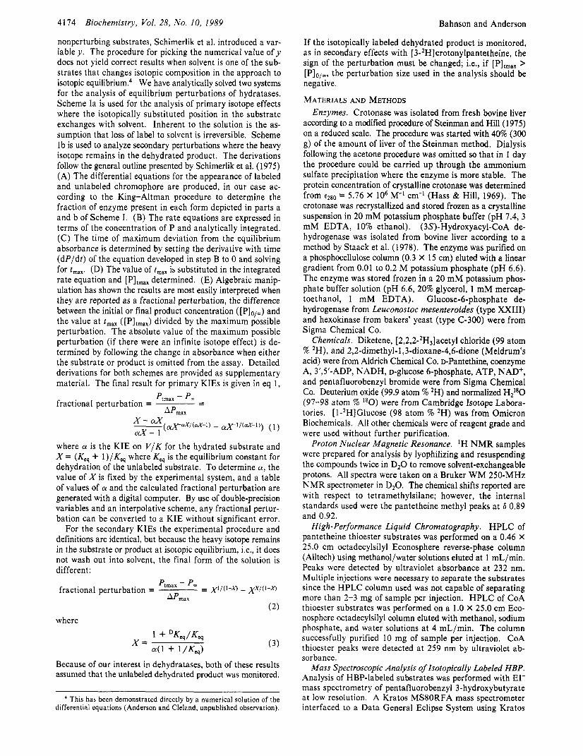
Scheme I
k, k, E4 - EP*H20 E4 6 EP
k, k A
k A s k<P E
“X k , F’ EA’ EP’ k3’
k4’ k4’ EA’ - EP*H,O’
Ia Ib
determining diffusion from masking the KIEs, a slower al- ternative substrate should be employed in the studies. In this paper we report the primary 2H, primary I8O, and secondary a, /3, and p’ KIEs2 on the dehydration of [(3S)-3-hydroxy- butyryllpantetheine (HBP) in the presence of the activator
THEORY Perturbation Derivations. The equilibrium perturbation
method for determining isotope effects was introduced by Schimerlik et al. (1975). The derivation presented used an ordered bi-ter reaction as the model. The adaptation to hy- dratases for primary isotope effects employed solvent at either 55 M for oxygen effects on 110 M for 2H effects as one of the perturbants. To correct for the commitments3 introduced by
3’,5’-ADP.
’ Abbreviations: Acac-CoA, acetoacetyl-CoA; KIE, kinetic isotope effect; HBP, [(3S)-3-hydroxybutyryl]pantetheine; EDTA, ethylenedi- aminetetraacetic acid; AcacP, S-(acetoacety1)pantetheine; DTNB, 5,s’- dithiobis(2-nitrobenzoic acid); TAPS, 3-[ [tris(hydroxymethyl)methyl] - amino]propanesulfonic acid; CrP, trans-crotonylpantetheine; MOPS, 3-(N-morpholino)propanesulfonic acid; HB-CoA, (3S)-3-hydroxy- butyryl-CoA; Cr-CoA, trans-crotonyl-CoA; Tris, tris(hydroxymethy1)- aminomethane.
In elimination reactions the a-secondary deuterium effect is for isotopic substitution on the carbon bonded to the heavy atom leaving group, the @-secondary effect is for isotopic substitution at the carbon whose C-H bond is broken, and the p’-secondary effect is for substitution at the nonreacting @-carbon (Cook, 1976).
Northrop (1977) introduced the terminology of commitments to catalysis to reflect the partitioning of enzyme-substrate complexes be- tween catalysis and dissociation of the substrate. If the commitment is generated by the addition of a second substrate, the commitment is termed external. An internal commitment is generated by nonisotope- dependent steps, e.g., product dissociation, being rate determining.
0006-2960/89/0428-4173$01.50/0 0 1989 American Chemical Society
4174
nonperturbing substrates, Schimerlik et al. introduced a var- iable y . The procedure for picking the numerical value of y does not yield correct results when solvent is one of the sub- strates that changes isotopic composition in the approach to isotopic eq~ilibrium.~ We have analytically solved two systems for the analysis of equilibrium perturbations of hydratases. Scheme Ia is used for the analysis of primary isotope effects where the isotopically substituted position in the substrate exchanges with solvent. Inherent to the solution is the as- sumption that loss of label to solvent is irreversible. Scheme Ib is used to analyze secondary perturbations where the heavy isotope remains in the dehydrated product. The derivations follow the general outline presented by Schimerlik et al. (1975) (A) The differential equations for the appearance of labeled and unlabeled chromophore are produced, in our case ac- cording to the King-Altman procedure to determine the fraction of enzyme present in each form depicted in parts a and b of Scheme I . (B) The rate equations are expressed in terms of the concentration of P and analytically integrated. (C) The time of maximum deviation from the equilibrium absorbance is determined by setting the derivative with time ( d P / d t ) of the equation developed in step B to 0 and solving for t,,,. (D) The value oft,,, is substituted in the integrated rate equation and [PIt,,, determined. (E) Algebraic manip- ulation has shown the results are most easily interpreted when they are reported as a fractional perturbation, the difference between the initial or final product concentration ([PI,/,) and the value at t,,, ([PItmax) divided by the maximum possible perturbation. The absolute value of the maximum possible perturbation (if there were an infinite isotope effect) is de- termined by following the change in absorbance when either the substrate or product is omitted from the assay. Detailed derivations for both schemes are provided as supplementary material. The final result for primary KIEs is given in eq 1,
Biochemistry, Vol. 28, No. 10, 1989 Bahnson and Anderson
If the isotopically labeled dehydrated product is monitored, as in secondary effects with [3-*Hlcrotonylpantetheine, the sign of the perturbation must be changed; i.e., if [PItmax > [PIo/_, the perturbation size used in the analysis should be negative.
MATERIALS AND METHODS Enzymes. Crotonase was isolated from fresh bovine liver
according to a modified procedure of Steinman and Hill (1 975) on a reduced scale. The procedure was started with 40% (300 g) of the amount of liver of the Steinman method. Dialysis following the acetone procedure was omitted so that in I day the procedure could be carried up through the ammonium sulfate precipitation where the enzyme is more stable. The protein concentration of crystalline crotonase was determined from t280 = 5.76 X lo6 M-' cm-' (Hass & Hill, 1969). The crotonase was recrystallized and stored frozen as a crystalline suspension in 20 mM potassium phosphate buffer (pH 7.4, 3 mM EDTA, 10% ethanol). (3S)-Hydroxyacyl-CoA de- hydrogenase was isolated from bovine liver according to a method by Staack et al. (1978). The enzyme was purified on a phosphocellulose column (0.3 X 15 cm) eluted with a linear gradient from 0.01 to 0.2 M potassium phosphate (pH 6.6). The enzyme was stored frozen in a 20 mM potassium phos- phate buffer solution (pH 6.6, 20% glycerol, 1 mM mercap- toethanol, 1 mM EDTA). Glucose-6-phosphate de- hydrogenase from Leuconostoc mesenteroides (type XXIII) and hexokinase from bakers' yeast (type C-300) were from Sigma Chemical Co.
Chemicals. Diketene, [2,2,2-2H3]acetyl chloride (99 atom % 2H), and 2,2-dimethyl-l,3-dioxane-4,6-dione (Meldrum's acid) were from Aldrich Chemical Co. DPantethine, coenzyme A, 3',5'-ADP, NADH, D-glucose 6-phosphate, ATP, NAD', and pentafluorobenzyl bromide were from Sigma Chemical Co. Deuterium oxide (99.9 atom % 2H) and normalized H2'*0 (97-98 atom % I8O) were from Cambridge Isotope Labora- tories. [ l-2H]Glucose (98 atom % 2H) was from Omicron Biochemicals. All other chemicals were of reagent grade and were used without further purification.
Proton Nuclear Magnetic Resonance. 'H NMR samples were prepared for analysis by lyophilizing and resuspending the compounds twice in D20 to remove solvent-exchangeable protons. All spectra were taken on a Bruker WM 250-MHz NMR spectrometer in D20. The chemical shifts reported are with respect to tetramethylsilane; however, the internal standards used were the pantetheine methyl peaks at 6 0.89 and 0.92.
High-Performance Liquid Chromatography. HPLC of pantetheine thioester substrates was performed on a 0.46 X 25.0 cm octadecylsilyl Econosphere reverse-phase column (Alltech) using methanol/water solutions eluted at 1 mL/min. Peaks were detected by ultraviolet absorbance at 232 nm. Multiple injections were necessary to separate the substrates since the HPLC column used was not capable of separating more than 2-3 mg of sample per injection. HPLC of CoA thioester substrates was performed on a 1.0 X 25.0 cm Eco- nosphere octadecylsilyl column eluted with methanol, sodium phosphate, and water solutions at 4 mL/min. The column successfully purified 10 mg of sample per injection. CoA thioester peaks were detected at 259 nm by ultraviolet ab- sorbance.
Mass Spectroscopic Analysis of Isotopically Labeled HBP. Analysis of HBP-labeled substrates was performed with EI- mass spectrometry of pentafluorobenzyl 3-hydroxybutyrate at low resolution. A Kratos MS80RFA mass spectrometer interfaced to a Data General Eclipse System using Kratos
- - Pmax - Pm APmx
fractional perturbation =
where CY is the KIE on V / K for the hydrated substrate and X = (Keq + l ) / K e q where Kq is the equilibrium constant for dehydration of the unlabeled substrate. To determine CY, the value of X is fixed by the experimental system, and a table of values of a and the calculated fractional perturbation are generated with a digital computer. By use of double-precision variables and an interpolative scheme, any fractional pertur- bation can be converted to a KIE without significant error.
For the secondary KIEs the experimental procedure and definitions are identical, but because the heavy isotope remains in the substrate or product at isotopic equilibrium, i.e., it does not wash out into solvent, the final form of the solution is different:
fractional perturbation = = XI/('-" - xX/(I-"
(2)
Ptmax - P- AP,,,
where
1 + DKeq/Keq 4 1 + l/Keq) X = (3)
Because of our interest in dehydratases, both of these results assumed that the unlabeled dehydrated product was monitored.
This has been demonstrated directly by a numerical solution of the differential equations (Anderson and Cleland, unpublished observation).
Crotonase Isotope Effects
DS-55 software was tuned with perfluorokeroscene in the EI- mode. Isotopically labeled HBP was hydrolyzed in 0.5 mL of 0.1 M sodium phosphate, pH 10.5. The solution was titrated to pH 6.5 with 1 M H3P04, and a 5-fold excess of penta- fluorobenzyl bromide was added. Ethanol (0.5 mL) was added as a cosolvent, and the reaction was stirred at 60 OC for 30 min. The pentafluorobenzyl ester was extracted into ethyl acetate and brought to dryness under a stream of dry nitrogen. Samples were injected into a gas chromatography fused silica capillary column interfaced to the mass spectrometer. Fol- lowing the injection of sample, the oven temperature was kept at 60 OC for 3 min; then it was increased to 250 OC in 9.5 min with a linear gradient. 3-Hydroxybutyric acid was derivatized with pentafluorobenzyl bromide and used as a standard. The mass spectrum showed an intense fragment peak at 103 amu and a peak 4% the size at 104 amu corresponding to I3C natural abundance of the four-carbon fragment. No other fragment peaks were observed. The isotopic composition of the labeled substrates was determined by comparison of the m through m + 3 peak intensities, which were corrected for 13C natural abundance.
S-(Acet0acetyf)pantetheine (AcacP). Pantethine (3.6 mmol) was reduced to pantetheine with an excess of sodium borohydride (10 mmol) in 0.2 M sodium phosphate, pH 9. The solution was acidified to pH 4.0 with 1 M H3P04 to quench unreacted sodium borohydride and then titrated to pH 8.5 with 1 M NaOH and reacted with an excess of diketene (10 mmol). The reaction was stirred at 25 "C until there was no detectable free thiol as assayed by 1.0 mM 5,Y-dithio- bis(2-nitrobenzoic acid) (DTNB) in 0.1 M 3- [ [tris(hydroxy- methyl)methyl]amino]propanesulfonic acid (TAPS), pH 8.5 (Ellman, 1959). The solution was titrated to pH 5 with 1 M H3PO4, saturated with NaCI, and then extracted five times with equal volumes of ethyl acetate. The ethyl acetate was removed by rotary evaporation, and AcacP was purified on a silica gel column (3 X 15 cm) eluted with chloroform/ methanol (90/10 v/v). AcacP concentration was assayed by use of ~ 3 0 2 ~ ~ = 16900 M-' cm-' in 0.1 M TAPS, pH 8.5, with 25 mM Mg2+ (Middleton, 1972). 'H NMR: 6 0.89 (s, 3 H), 0.92 (s, 3 H), 2.30 (s, 3 H), 2.47 (t, 2 H), 3.06 (t, 2 H), 3.45 (m, 6 H), 3.99 (s, 1 H).
HBP. AcacP (140 pmol), glucose 6-phosphate (150 pmol), and NADH (catalytic amount) were added to 1 mL of 0.1 M sodium phosphate, pH 7.5. The absorbance at 380 nm was monitored during the following two steps to determine when the reaction had been driven to completion by following the NADH concentration (e380 = 900 M-I cm-'). (3S)-3- Hydroxyacyl-CoA dehydrogenase, which catalyzes the ste- reospecific reduction of (3-ketoacy1)pantetheine to [(3S)-3- hydroxyacyllpantetheine (Wakil, 1963), was added, which resulted in the rapid decrease of the measured absorbance until equilibrium was reached. Glucose-6-phosphate dehydrogenase, which regenerates NADH by catalyzing the reduction of NAD' by glucose 6-phosphate, was added, which resulted in a gradual increase to a steady-state absorbance. The reaction was determined 100% complete when the absorbance increased rapidly from the steady-state absorbance to an absorbance corresponding to the complete regeneration of NADH. HBP was purified by HPLC eluted isocratically with 25% methanol. The HBP peak was collected at 14 min following the pan- tetheine peak at 9 min. 'H NMR: 6 0.89 (s, 3 H), 0.92 (s, 3 H), 1.23 (d, 3 H), 2.47 (t, 2 H), 2.79 (d, 2 H), 3.06 (t, 2 H), 3.45 (m, 6 H), 3.99 (s, 1 H), 4.27 (m, 1 H).
trans-Crotonylpantetkeine (CrP). HBP was dehydrated to CrP by crotonase. The equilibrium was attained in 1 mL of
Biochemistry, Vol. 28, No. 10, 1989 4175
0.1 M 3-(N-morpholino)propanesulfonic acid (MOPS), pH 7.0, with 100 pM 3',5'-ADP and 20 nM crotonase. HPLC eluted isocratically with 35% methanol gave the CrP peak at 20 min. 'H NMR: 6 0.89 (s, 3 H), 0.92 (s, 3 H), 1.90 (d, 3 H), 2.47 (t, 2 H), 3.06 (t, 2 H), 3.45 (m, 6 H), 3.99 (s, 1 H), 6.31 (d, 1 H), 7.04 (m, 1 H). Crotonylpantetheine that was synthesized by the acylation of pantetheine with crotonic anhydride contained contaminating cis isomer. The 'H NMR spectrum of crotonic anhydride from Aldrich Chemical Co. indicated the presence of 7% cis isomer. CrP that was syn- thesized enzymatically from AcacP showed no cis isomer by 'H NMR.
(2R)-[2-2H]HBP. Deuterium oxide (0.5 mL) was added to pure HBP to remove residual water. The solution was evaporated and then resuspended in 1 mL of D 2 0 to which 20 nM crotonase and 100 pM 3',5'-ADP were added. (2R)-[2-2H]HBP was separated from CrP by HPLC after 3 h was allowed for the exchange of deuterium into the C-2 primary position. The 'H NMR spectrum of (2R)-[2-2H]HBP differed from the spectrum of HBP at 2.79 ppm where the doublet corresponding to the C-2 protons integrated to 1 h due to the 2H incorporation at that position. The spin-spin cou- pling constant of the doublet was reduced to 4.3 Hz, which is consistent with an erythro coupling constant (Mohrig et al., 1980; Anderson & Hammes, 1984). The multiplet peak at 4.27 ppm corresponding to the C-3 proton was noticeably different due to the loss of the threo spin-spin coupling upon primary deuteration of HBP. The extent of crotonase-cata- lyzed deuterium exchange into the C-2 primary position was determined to be 91 f 1 atom % 2H by low-resolution mass spectrometry.
(3s) -3- Hydroxybutyryf- CoA (HB-CoA) , trans- Crotonyl- CoA (Cr-CoA), and (2R)-[2-2H]HB-C0A. Coenzyme A (50 pmol) was reacted with an excess of diketene (1 50 pmol) in 1 mL of sodium phosphate (100 mM, pH 8.0), until the ab- sence of free thiol was detected (Ellman, 1959). The solution was titrated to pH 5 with 1 M H3PO4 and extracted two times with 1-mL aliquots of ethyl acetate. The solution was titrated to pH 7 with 1 M NaOH. The Acac-CoA was enzymatically reduced to HB-CoA (see above). HB-CoA was purified by HPLC eluted isocratically with an 11 % methanol and 10 mM sodium phosphate solution (pH 4.5). The HB-CoA peak was collected at 8-12 min. HB-CoA was equilibrated with cro- tonase, and the product Cr-CoA was purified by HPLC eluted isocratically with a 16% methanol and 10 mM sodium phos- phate solution (pH 4.5). The Cr-CoA peak was collected at 17-20 min. HB-CoA was equilibrated in 1 mL of DzO with crotonase to exchange 2H into the primary C-2 position. (2R)- [2-2H]HB-CoA was purified from Cr-CoA by HPLC. The 'H NMR spectrum of (2R)-[2-2H]HB-CoA indicated >95 atom % 2H incorporation by integration of the C-2 proton peak at 2.7 ppm.
[3-I80]HBP. AcacP (140 pmol) was dissolved and evapo- rated twice from 1 mL of anhydrous ethyl acetate to azeotrope away water. H2I80 (400 pL, 97-98 atom % ISO) and 1 pL of concentrated HCl were combined, which made the solution acidic (pH 2) in order to exchange the 3-keto oxygen of AcacP. After 3 days was allowed for the exchange, the solution was titrated to pH 7.0 with tris(hydroxymethy1)aminomethane (Tris). The [3-'80]AcacP was reduced to [3-I80]HBP en- zymatically as was done for the unlabeled AcacP to HBP reduction except the reaction was run in H2I80. The H2180 was recovered following the reduction by lyophilizing the sample under high vacuum and capturing the H2I80 in a trap cooled with liquid nitrogen. Mass spectroscopic analysis in-
4176
dicated 93.0 f 1.0 atom % I8O incorporation. [2,2-2H2]HBP. The C-2 protons of AcacP (140 pmol) were
exchanged with deuterium in less than 5 min when dissolved in 1.0 mL of D20. The [2,2-2H2]AcacP was enzymatically reduced to [2,2-2H2]HBP (as above). The 'H NMR spectrum of [2,2-2H2]HBP was used to determine an enrichment of 97 f 1 atom % 2H at C-2 by integration of the C-2 proton peak at 2.79 ppm. Mass spectroscopic analysis of the [2,2-2H2]HBP indicated that during prolonged (2 days) exchange of the C-2 protons of AcacP there was partial exchange of the C-4 methyl protons (> lo atom % 2H), by the presence of m + 3, m + 4, and m + 5 peaks in addition to the expected m , m + 1, and m + 2 peaks.
[ 2-2H] CrP and (2s)- [ 2-2H] HBP. [ 2-2H] CrP was obtained by the crotonase-catalyzed dehydration of [2,2-2H2]HBP to [2-2H]CrP followed by HPLC isolation. Subsequent hydration of [2-2H]CrP by crotonase followed by HPLC isolation yielded (2S)-[2-2H]HBP. The 'H N M R spectrum of [2-2H]CrP differed from the spectrum of CrP by the lack of a peak at 6.31 ppm, which corresponds to a deuterated secondary C-2 position, and the multiplet at 7.04 ppm became a broadened singlet. The 'H NMR spectrum of (2S)-[2-2H]HBP differed from the spectrum of (2R)-[2-2H]HBP only by the value of the spin-spin coupling constant. A JHCCH of 8.3 Hz for the C-2 proton peak at 2.79 ppm is consistent with a threo coupling constant (Mohrig, 1980; Anderson & Hammes, 1984).
[3-2H] HBP and [ 3-2H] CrP. AcacP ( 140 pmol) was reduced stereospecifically with (4R)-[4-2H]NADH. [ l-2H]Glucose (98 atom % 2H, 150 pmol), ATP (150 pmol), 5 mg of mag- nesium acetate, hexokinase, and a catalytic amount of NAD' (5 pmol) were added instead of the glucose 6-phosphate and NADH which were used for the unlabeled AcacP to HBP reduction. A rapid increase in absorbance at 380 nm indicated the reaction was complete. [3-2H]HBP was obtained pure by HPLC. [3-2H]CrP was obtained by HPLC following equil- ibration of the [3-2H]HBP with crotonase. In the 'H NMR spectrum of [3-*H]HBP there was no observable peak at 4.27 ppm, corresponding to >98 atom % 2H incorporation. 'H NMR peaks at 1.23 and 2.79 ppm, which correspond to C-4 and C-2 protons, respectively, were singlets due to 2H incor- poration at (2-3.
A solution of [2,2,2-2H3]acetyl chloride (13.75 mmol) in 10 mL of methy- lene chloride was added dropwise over 30 min to a mixture of Meldrum's acid (1 2.5 mmol) and pyridine (25 mmol) in 20 mL of methylene chloride stirring at 0 OC. The solution was stirred an additional 30 min at 25 OC. Rotary evaporation of the solvent gave acylated Meldrum's acid in 70% yield. Pantetheine (1 mmol), from the reduction of pantethine by sodium borohydride, was extracted into ethyl acetate. The solution was dissolved and evaporated from anhydrous ethyl acetate under a stream of dry N2 twice to azeotrope away residual water. The anhydrous pantetheine was added to acylated Meldrum's acid (1 mmol) in 10 mL of anhydrous THF and refluxed for 2 h. [4,4,4-2H3]AcacP (20% yield) was separated on a silica gel column (3 X 10 cm) eluted with chloroform/methanol (90/ 10 v/v). [4,4,4-2H3]HBP and [4,4,4-2H3]CrP were obtained by the same route described for the unlabeled substrates, starting with [4,4,4-2H3]AcacP. The 'H NMR spectra of [4,4,4-2H3]HBP and [4,4,4-2H3]CrP in- dicated 60 f 3 atom % 2H by integration of the C-4 methyl peak relative to the pantetheine methyl peaks. Mass spec- troscopic analysis of [4,4,4-2H3]HBP indicated 57 f 1 atom % 2H.
Biochemistry, Vol. 28, No. 10, 1989
[4,4,4-2H3]HBP and [4,4,4-2H3]CrP.
Bahnson and Anderson
Kinetic Isotope Effects. KIEs reported were measured according to the equilibrium perturbation technique of Schimerlik et al. (1975). A Perkin-Elmer A-3B UV/vis spectrophotometer thermostated at 25 f 0.1 OC interfaced to an IBM PC computer using ASYST software was used for data acquisition. Substrate concentrations were measured enzymatically by measuring absorbance changes at 280 nm where the b i 2 8 0 corresponding to the hydration of a,P-un- saturated CrP has an extinction coefficient of 3600 M-' cm-' (Lynen & Ochoa, 1953). Typically, equilibrium perturbations were run in 0.1 M MOPS (pH 7.00,O.l M ionic strength with potassium salt, 1 mM EDTA) with 100 pM 3',5'-ADP and 20 nM crotonase in a total volume of 600 pL unless otherwise noted. Under these conditions equilibrium perturbations reached their maximum or minimum absorbance in 2-5 min, and final equilibrium was reached within 20-50 min. Typical substrate concentrations resulted in a u 2 8 0 of +0.2 OD for HBP and -0.2 OD for CrP upon addition of crotonase. By use of the Kq value of 0.29 for the dehydration reaction, Mza0 of 0.2 OD, and the extinction coefficient for the b i 2 8 0 , sub- strate concentrations are calculated to be approximately 250 pM HBP and 70 pM CrP. Initial substrate concentrations were fine tuned so that the final absorbance was equal to the initial absorbance of the perturbation (Cleland, 1980).
HBP and CrP, with one substrate labeled and one substrate unlabeled, were added to a 1-mL quartz cuvette from separate stock solutions so that each substrate had an equal and opposite A A 2 8 0 . The perturbation solution was temperature equilibrated to 25 OC for 5 min in the spectrophotometer. Prior to starting the perturbation, the initial absorbance was recorded, and the absence of a background change in absorbance was confirmed. At higher pH or over long storage times (days), HBP is prone to hydrolysis to produce pantetheine. The product thiol forms a Michael adduct by attacking CrP at the C-3 position. The presence of free thiol consequently results in a constant de- crease in absorbance, which makes measurement of the equilibrium perturbations less precise. To avoid this problem, it was necessary to purify the substrates by HPLC immediately before use. The equilibrium perturbation was initiated by the addition of a small volume of crotonase (25 pL, 25 "C) fol- lowed by three inversions of the cuvette to mix the solution.
The fractional perturbation is reported as the u 2 8 0 of the perturbation (AApn) divided by the maximum M 2 8 0 possible (AA,), The AA- is the difference between the perturbation maximum or minimum and the initial or final absorbance. In cases when the initial and the final absorbance of a pertur- bation were not equal and the difference was not large com- pared to the AA,,, the initial and final absorbance values were averaged. The AA, is defined as the of either substrate from its initial to its equilibrium absorbance upon addition of crotonase since each substrate should have an equal and op- posite M 2 8 0 . The estimated errors are derived from an es- timated error of fO.001 absorbance unit in the perturbation size. Duplicate perturbations were always obtained within the indicated error limits. Differences between this paper and our preliminary results (Bahnson & Anderson, 1988) came from systematic errors, the most important being a failure to exclude all free thiols from the reaction mixture.
For the interpretation of secondary ZH kinetic isotope effects it was necessary to accurately estimate the 2H equilibrium isotope effect (DKq)5 due to the dependence of the calculated
The notation for isotope effects is from Cleland (1982). A leading superscript of the heavier isotope indicates an isotope effect on the fol- lowing parameter. The nomenclature for discussing isotope effects on enzyme reactions is from the same source.

Crotonase Isotope Effects
DV/K on DK (eq 2 and 3). The DK, used was estimated from
molecules resembling HBP and CrP at the secondary labeled position to water (Cleland, 1980).
RESULTS Equilibrium Perturbation Control Experiment. To ensure
that observed equilibrium perturbations were the result of an isotope effect of the labeled substrate, a control was run with unlabeled substrates. HBP and CrP, which were at concen- trations that correspond to a AA,,, of 0.2 OD, were added together. There was no equilibrium perturbation detectable (AApcrt < 0.002). This rules out the possibility that something other than an isotope effect of the labeled substrate is re- sponsible for the equilibrium perturbations reported below. One potential problem that was discovered by this control was a 7% cis contaminant in CrP which had been synthesized from crotonic anhydride. Crotonase catalyzes the hydration of cis-crotonyl-CoA at about ' I3 of the rate of trans-crotonyl-CoA (Stern, 1961). Using CrP that had contaminating cis isomer in the equilibrium perturbation control experiment gave a positive perturbation as expected for a slower alternative substrate (Anderson, unpublished observation). This problem was corrected by synthesizing CrP enzymatically starting from AcacP.
Primary DV/K with (2R)-[2-*H]HBP. The primary DV/K was measured with a mixture of (2R)-[2-*H]HBP and CrP which were at equilibrium concentrations prior to and following the equilibrium perturbation. Equilibrium perturbations measured at pH 7.00 had a fractional perturbation of -25.0 f 0.5% with a AA,,, of 0.226 OD. The mass spectroscopic analysis of (2R)-[2-2H]HBP, which indicated 91.0 f 1.0 atom % 2H, was used to correct the fractional perturbation to -27.5 f 0.9%. From eq 1, which relates the D V / K to the fractional perturbation for a primary KIE, the primary D V / K was cal- culated to be 1.61 f 0.03. The equilibrium perturbation was repeated at pH 5.92 with the same substrate solutions, and an identical fractional perturbation was measured, showing the lack of a pH dependence on the D V / K between pH 5.92 and pH 7.00.
Primary D V / K with (2R)-[2-2H]HB-CoA. The primary D V / K was measured with a mixture of (2R)-[2-2H]HB-CoA and Cr-CoA. Equilibrium perturbations measured at pH 7.00 with no added 3',5'-ADP and a crotonase concentration of 10 pM resulted in a fractional perturbation of 8.8 f 1.7%, where the AA,,, was 0.1 OD. From eq 1, the primary D V / K was calculated to be 1.15 f 0.03.
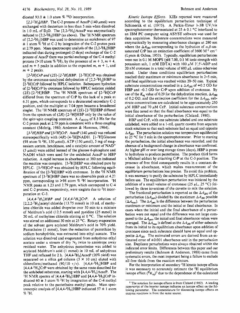
Primary 1 8 ( V/K). The primary 1 8 ( V/K) was measured with a mixture of [3-I80]HBP and CrP. A typical perturbation with [3-I80]HBP as perturbant is shown in Figure 1. Equilibrium perturbations measured at pH 7.00 had a frac- tional perturbation of -3.0 f 0.2%. The equilibrium pertur- bations were measured at X = 263 nm to give a larger value of AA,,, (0.5270 OD) with all other conditions of the ex- periments being standard. The mass spectroscopic analysis of [3-180]HBP, which indicated 93.0 f 1.0 atom % l 8 0 , was used to correct the fractional perturbation to -3.23 f 0.24%. From eq 1, which relates the 18( V / K ) to the fractional per- turbation for a primary KIE, the primary 18( V / K ) was cal- culated to be 1.051 f 0.004. In preliminary experiments slightly larger values (1.060) were obtained but with greater uncertainty due to the smaller absolute perturbation size and less I8O enrichment in the [3-I80]HBP.
@-Secondary DV/K. During the dehydration reaction, the substituted hydrogen changes from being on an sp3 carbon a to a carbonyl to being on an sp2 carbon. The "Kq value for
the ratio of 3 Kq values for the transfer of deuterium from
1 -1 60 I
0 P
3 -2.40
Biochemistry, Vol. 28, No. 10, 1989 4177
--
--
0.00 1 i
t - 3 . 2 0
,-- 1
T J , - , - + - , - , - , - , - L
0.0 600 1200 1800 2400 TIME (sec)
FIGURE 1: Spectrophotometric time course of an equilibrium per- turbation. Crotonase is added at time = 0 to a temperature-equil- ibrated assay mix containing equilibrium concentrations of CrP and [3-'*0]HBP in the presence of 100 pM 3',5'-ADP. The absorbance of the CrP has been normalized by the maximum possible perturbation size (AAmaX) and is reported as a percentage. The perturbation size (AA,,,) was 0.016 absorbance unit.
this reaction was estimated to be 1.07 from the ratio of the "Keg values of 0.93 for the transfer of 2H from [3JH]keto- glutarate to water and 0.87 for the transfer of 2H from [2- *H]fumarate to water (Cleland, 1980).
The @-secondary D V / K was measured with a mixture of [2-2H]CrP and HBP that were at concentrations that corre- spond to a AA,,, of 0.300 OD. The fractional perturbation measured at pH 7.00 was 2.10 f 0.33%. From the 'H N M R analysis of 97 f 1 atom 9% 2H at C-2, the fractional pertur- bation was corrected to 2.17 f 0.36%. By use of the "Keg value of 1.07 in eq 2, which relates the D V / K to the fractional perturbation measured for a secondary DV/K, the D V / K for the dehydration reaction was calculated to be 1.12 f 0.01. The equilibrium perturbation was repeated at one-fifth the activator concentration (20 pM 3',5'-ADP) in the same buffer, enzyme, and substrate solutions. An identical fractional perturbation of 2.10% was measured, showing a lack of an activator con- centration dependence on the @-secondary V / K between 20 and 100 pM 3',5'-ADP.
Alternatively, the @-secondary D V / K was measured with a mixture of (2S)-[2-2H]HBP and CrP that were at concen- trations that correspond to a AA,,, of 0.187 OD. The frac- tional perturbation measured at pH 7.00 was -1.93 f 0.53%. The fractional perturbation was -2.02 f 0.55% after correction for the 2H enrichment at C-2. By use of the DKq value of 1.07 in eq 2, the D V / K for the dehydration reaction was calculated to be 1.1 1 f 0.02. Errors in the estimate of DKq will be reflected proportionately in the measured isotope effect (see below) leaving the more important ratio, (DV/K) /DKq, un- changed. These two complementary perturbations are shown in Figure 2.
a-Secondary DV/K. During dehydration the substituted carbon-hydrogen bond is converted from an sp3 bond with an a C-0 bond to an sp2 bond. The "K? value for this reaction was estimated to be 1.33 from the ratio of the DKq values of 1.16 for the transfer of 2H from [2-2H]-2-propanol or [ l- 2H]cyclohexanol to water and 0.87 for the transfer of [2- 2H]fumarate to water (Cleland, 1980).
The a-secondary D V / K was measured with a mixture of [ 3-2H]HBP and CrP that were at concentrations that corre- spond to a AA,,, of 0.276 OD. The fractional perturbation measured at pH 7.00 with 100 pM 3',5'-ADP was 3.99 f 0.36%. By use of the DKes value of 1.33 in eq 2, the D V / K for the dehydration reaction was calculated to be 1.13 f 0.01.

4178
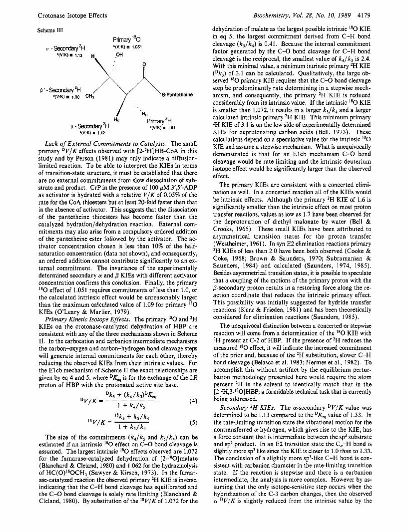
Scheme I1
Biochemistry, Vol. 28, No. 10, 1989 Bahnson and Anderson
E l carbocation intermediate
E2 concerted
El cb carbanion intermediate
1 00 1 k! t 3 o o o t .
-1 "" 00
-2 00 +
0.0 400 800 1200 1600 TIME (sec)
FIGURE 2: @-Secondary D V / K determination by equilibrium per- turbation. Same conditions as in Figure 1 exce t the isotopic label
perturbations were +0.0063 and -0.0036 absorbance unit, respectively. was introduced as [2-*H]CrP (A) or (2S)-[2- P HIHBP (B). The
The equilibrium perturbation was repeated at 20 pM 3',5'- ADP in the same buffer, enzyme, and substrate solutions. An identical fractional perturbation of 3.99% was measured, showing the lack of an activator concentration dependence on the D V / K between 20 and 100 pM 3',5'-ADP.
The a-secondary D V / K was additionally measured with a mixture of [3-2H]CrP and HBP that were at concentrations that correspond to a AA,,, of 0.204 OD. The fractional perturbation measured at pH 7.00 was -4.12 f 0.49%. By use of the DKcp value of 1.33 in eq 2, the D V / K for the deh- ydration reaction was calculated to be 1.12 f 0.02. The ratio of ( D V / K ) / D K , of 0.846 f 0.015 is independent of the esti- mated value for DKq.
@'-Secondary D V / K . The "Kq value for the reaction be- tween [4,4,4-2H3]HBP and [4,4,4-2H3]CrP was estimated to be 1.00, which means that 2H at that position has no effect on the equilibrium constant. Equilibrium perturbations were measured first with [4,4,4-2H3]HBP and CrP and then with [4,4,4-2H3]CrP and HBP that were at concentrations that correspond to a AA,,, of 0.200 absorbance unit. In no case was a fractional perturbation measured that was greater than 0.50%. After correction for 57 f 1 atom % 2H, the fractional perturbation became 0.00 f 0.88%. By use of the DKq value of 1 .OO in eq 2, this range in the fractional perturbation cor- responds to a 4,4,4-2H3 secondary D V / K of 1.00 f 0.03 or (1 .OO f O.0l)I2H.
DISCUSSION A priori the C-H and C-X bond cleavages in a-@ elimi-
nation reactions can occur in either order or in a concerted fashion as shown in Scheme 11. When a carbanion can be stabilized @ to the leaving group, base-promoted E2 and Elcb reactions predominate. Gandler and Jencks (1982) have ar- gued that there is a distinct transformation from an E2 mechanism to an Elcb mechanism as the stability of the carbanion is enhanced within a fixed substrate structure and that the two mechanisms cannot both be operative for the same molecule. The argument is physically rationalized on the basis of the similar location in More O'Ferrall diagrams (More O'Ferrall, 1970) of the two different transition states and the assumption that if there is an accessible well for the carbanion intermediate, the E2 reaction pathway could not avoid it. Mayer et al. (1984) studied a-@ elimination reactions in @- decalone systems and concluded that the strained lactone ring of 1 l-oxatricycl0[4.4.3.0~~~] tridecan-3-one promotes an E2 elimination despite the activation of the eliminated @-hydrogen by the ketone. These two arguments frame the mechanistic questions for the crotonase-promoted CY-@ elimination from 3-hydroxy thioesters: Has the enzyme stabilized the enol(ate) of the thioester sufficiently for an Elcb-type mechanism to be operative, or has the leaving group been activated to a greater extent, resulting in a concerted elimination?
For crotonase the thioester functionality has been invoked as providing the necessary stabilization for the formation of a carbanion intermediate. The submicromolar binding of the carbanionic form of Acac-CoA (Waterson & Hill, 1972) is the only experimental evidence for such an enolate interme- diate in the crotonase-catalyzed reaction. However, the affinity of crotonase for this inhibitor may arise from stabilization of the negative charge at the C-3 oxygen where OH- is eliminated in the normal reaction. Additional support for enol(ate) in- termediates in reactions of coenzyme A thioesters has come from theoretical calculations on the condensation of malo- nyl-CoA with thioesters (Dewar & Dieter, 1988), from dou- ble-isotope fractionation studies on malate synthase (Clark et al., 1988) and from direct observation of the enethiol(ate) of acetyldithio-CoA bound to citrate synthase (Wlassics & An- derson, 1989). As discussed below individually, the KIEs reported here and summarized in Scheme I11 are consistent with both the Elcb and E2 mechanisms but confirm the carbanionic character of the transition state and provide the necessary background for the unequivocal demonstration of the concerted or stepwise nature of the reaction by a subse- quent double-isotope fractionation study.
Crotonase Isotope Effects Biochemistry, Vol. 28, No. 10, 1989 4179
dehydration of malate as the largest possible intrinsic I8O KIE in eq 5 , the largest commitment derived from C-H bond cleavage ( k 5 / k 4 ) is 0.41. Because the internal commitment factor generated by the C-0 bond cleavage for C-H bond cleavage is the reciprocal, the smallest value of k4/k5 is 2.4. With this minimal value, a minimum intrinsic primary 2H KIE (Dk3) of 3.1 can be calculated. Qualitatively, the large ob- served I 8 0 primary KIE requires that the C-0 bond cleavage step be predominantly rate determining in a stepwise mech- anism, and consequently, the primary 2H KIE is reduced considerably from its intrinsic value. If the intrinsic l 8 0 KIE is smaller than 1.072, it results in a larger k5/k4 and a larger calculated intrinsic primary 2H KIE. This minimum primary 2H KIE of 3.1 is on the low side of experimentally determined KIEs for deprotonating carbon acids (Bell, 1973). These calculations depend on a speculative value for the intrinsic I8O KIE and assume a stepwise mechanism. What is unequivocally demonstrated is that for an Elcb mechanism C-0 bond cleavage would be rate limiting and the intrinsic deuterium isotope effect would be significantly larger than the observed effect.
The primary KIEs are consistent with a concerted elimi- nation as well. In a concerted reaction all of the KIEs would be intrinsic effects. Although the primary 2H KIE of 1.6 is significantly smaller than the intrinsic effect on most proton transfer reactions, values as low as 1.7 have been observed for the deprotonation of diethyl malonate by water (Bell & Crooks, 1965). These small KIEs have been attributed to asymmetrical transition states for the proton transfer (Westheimer, 1961). In syn E2 elimination reactions primary 2H KIEs of less than 2.0 have been both observed (Cooke & Coke, 1968; Brown & Saunders, 1970; Subramanian & Saunders, 1984) and calculated (Saunders, 1974, 1985). Besides asymmetrical transition states, it is possible to speculate that a coupling of the motions of the primary proton with the 0-secondary proton results in a restoring force along the re- action coordinate that reduces the intrinsic primary effect. This possibility was initially suggested for hydride transfer reactions (Kurz & Frieden, 1981) and has been theoretically considered for elimination reactions (Saunders, 1985).
The unequivocal distinction between a concerted or stepwise reaction will come from a determination of the I8O KIE with 2H present at C-2 of HBP. If the presence of ZH reduces the measured I8O effect, it will indicate the increased commitment of the prior and, because of the 2H substitution, slower C-H bond cleavage (Belasco et al. 1983; Hermes et al., 1982). To accomplish this without artifact by the equilibrium pertur- bation methodology presented here would require the atom percent 2H in the solvent to identically match that in the [2-2H,3-'80]HBP a formidable technical task that is currently being addressed.
Secondary 2H KIEs. The a-secondary D V / K value was determined to be 1.1 3 compared to the DKcs value of 1.33. In the rate-limiting transition state the vibrational motion for the nontransferred a-hydrogen, which gives rise to the KIE, has a force constant that is intermediate between the sp3 substrate and sp2 product. In an E2 transition state the C,-H bond is slightly more sp3 like since the KIE is closer to 1 .O than to 1.33. The conclusion of a slightly more sp3-like C-H bond is con- sistent with carbanion character in the rate-limiting transition state. If the reaction is stepwise and there is a carbanion intermediate, the analysis is more complex. However by as- suming that the only isotope-sensitive step occurs when the hybridization of the C-3 carbon changes, then the observed a D V / K is slightly reduced from the intrinsic value by the
Scheme 111
Primary l80 a - ~ e c o n d a r y ~ ~ 'O(V/K) 1.051
'(VIK) 1.13 H OH
\: i; +Pantetheine V / K ) 1.00 CH3
P 1 - Secondary 'H
HS Primary 2H p -Secondary2H W / K ) = 1.61
O(V/K) = 1112
Lack of External Commitments to Catalysis. The small primary D V / K effects observed with [2-2H]HB-CoA in this study and by Person (1981) may only indicate a diffusion- limited reaction. To be able to interpret the KIEs in terms of transition-state structure, it must be established that there are no external commitments from slow dissociation of sub- strate and product. CrP in the presence of 100 pM 3',5'-ADP as activator is hydrated with a relative V / K of 0.05% of the rate for the CoA thioesters but at least 20-fold faster than that in the absence of activator. This suggests that the dissociation of the pantetheine thioesters has become faster than the catalyzed hydration/dehydration reaction. External com- mitments may also arise from a compulsory ordered addition of the pantetheine ester followed by the activator. The ac- tivator concentration chosen is less than 10% of the half- saturation concentration (data not shown), and consequently, an ordered addition cannot contribute significantly to an ex- ternal commitment. The invariance of the experimentally determined secondary a and ,d KIEs with different activator concentration confirms this conclusion. Finally, the primary I 8 0 effect of 1.05 1 requires commitments of less than 1 .O, or the calculated intrinsic effect would be unreasonably larger than the maximum calculated value of 1.09 for primary I8O KIEs (O'Leary & Marlier, 1979).
Primary Kinetic Isotope Effects. The primary I8O and 2H KIEs on the crotonase-catalyzed dehydration of HBP are consistent with any of the three mechanisms shown in Scheme 11. In the carbocation and carbanion intermediate mechanisms the carbon-oxygen and carbon-hydrogen bond cleavage steps will generate internal commitments for each other, thereby reducing the observed KIEs from their intrinsic values. For the Elcb mechanism of Scheme I1 the exact relationships are given by eq 4 and 5 , where DK, is for the exchange of the 2R proton of HBP with the protonated active site base.
(4)
The size of the commitments ( k 4 / k 5 and k 5 / k 4 ) can be estimated if an intrinsic I8O effect on C-0 bond cleavage is assumed. The largest intrinsic I8O effects observed are 1.072 for the fumarase-catalyzed dehydration of [2-'80]malate (Blanchard & Cleland, 1980) and 1.062 for the hydrazinolysis of HC(0)I80CH3 (Sawyer & Kirsch, 1973). In the fumar- ase-catalyzed reaction the observed primary 2H KIE is inverse, indicating that the C-H bond cleavage has equilibrated and the C-0 bond cleavage is solely rate limiting (Blanchard & Cleland, 1980). By substitution of the 1 8 V / K of 1.072 for the
4180
internal commitment introduced by the C-H bond cleavage step. As noted above, an estimate of the largest commitment possible is 0.41, allowing us to estimate the largest intrinsic a-secondary 2H KIE to be 1.18. The magnitude of this effect together with the large primary '*O KIE is not consistent with a carbocation intermediate. In the SN1 solvolysis of isopropyl tosylate where C-0 bond cleavage is rate limiting, an a-sec- ondary D V / K of 1.22 is observed (Streitweiser & Dafforn, 1969).
The @-secondary D V / K value of 1.12 f 0.01 compared to the DKeq value of 1.07 is the most intriguing KIE measured in this study. A D V / K in excess of the DK, for the dehydration indicates that in the rate-limiting transition state there is a vibrational mode for the nontransferred @-hydrogen which has a reduced force constant compared to either substrate or product. This phenomenon cannot be explained solely by the extent of rehybridization of the @-carbon in the transition state. Errors in the estimated "Keg value also cannot account for this difference. Equations 2 and 3 can be simplified to eq 6 and 7 when Keg << 1 .O, indicating that the observed perturbation size really determines the ratio of D V / K and DKes. For the
Biochemistry, Vol. 28, No. 10, 1989
X = D K e s / a (6)
- - Ptmm - P m APInax
fractional perturbation =
(D~,~/,)1/(1-~Kcq/a) - ( D K, , / (~ ) (~K. s / " ) / (1 -~4 /u ) (7)
crotonase-catalyzed dehydration of HBP, Kq = 0.29, and the relationships in eq 6 and 7 are not exact. Assuming DKep = 1.12 (instead of 1.07) yielded a D V / K of 1.17, numerically indicating that the perturbation has established (DV/K) /DK,9 = 1 .OS f 0.01. The equal and opposite perturbations shown in Figure 2 are unequivocal evidence that D V / K is greater than DKq. If D V / K were equal to DKq, the absolute value of both fractional perturbations would be less than 0.5%. Any error present is limited to the magnitude that D V / K exceeds DKep.
If the reaction is stepwise, the C-0 bond cleavage step will generate a significant reverse internal commitment. Using the minimum k 4 / k 5 of 2.4 estimated above and "Kq = 1 .076 in eq 4 yields a minimum intrinsic &secondary KIE of 1.24. This value is not too large to be explained by a coupled motion in the transition state. In formate dehydrogenase the ratio of ( D V / K ) / D K , q for the secondary effect at C-4 of the nicotin- amide ring is 1.38 (Hermes et al., 1984).
Subramanian and Saunders (1 984) reported @-secondary KIEs that exceed the equilibrium isotope effect in the elim- ination reactions of 2-arylethyl derivatives. They observed that an increase of carbanion character in the transition state re- sulted in the measured @-secondary KIE being larger than DKeq. Saunders (1984, 1985) calculated KIEs for E2 elimi- nation reactions which predict a tunneling correction is nec- essary when the bending motion of the nontransferred @-hy- drogen is coupled to the stretching motion of the transferred hydrogen. These calculations also predict small primary 2H KIEs. This is consistent with the small primary 2H KIE of 1.61 measured in this study, which would be an intrinsic effect if an E2 mechanism were operative. The P-secondary D V / K
Bahnson and Anderson
DK, should ideally be for the conversion of HBP to the enolate. We were unable to locate any suitable fractionation factors for hydrogens on enolate carbanions. A crystal structure of a model thioester enolate (P. W. Willard and M. Hintze, personal communication) indicates that the a-carbon-carbonyl carbon bond distance is 1.348 A and the a-carbon substituents are nearly coplanar with the C(0-)S; i.e., the a-carbon is crystallographically indistinguishable from a normal sp2 carbon, sup- porting the validity of using a DK, for the overall reaction.
measured in the crotonase reaction could be explained by coupling of the nontransferred 0-hydrogen with the transfer of the primary hydrogen in the rate-limiting transition state. The accepted method to study coupled motions of protons in the transition state (Kurz & Frieden, 1980; Hermes et al., 1984) is to determine if deuteration at one of the coupled sites reduces the deuterium KIE at the second site. These studies are in progress in our laboratory.
The /3'-secondary D V / K value of unity unequivocally elim- inates the possibility of a partial positive charge developing on C-3 in the rate-limiting transition state. A carbocation stepwise mechanism or concerted mechanism with carbocation character would be expected to have a /3'-secondary DV/K of 1.1 /'H due to hyperconjugation resonance effects that pref- erentially stabilize the undeuterated transition state (Westa- way, 1987). The lack of positive charge in the rate-limiting transition state indicates that the increase in electron density due to C-C double bond formation must nearly balance the loss of electron density associated with C-0 bond cleavage that occurs in the rate-determining transition state.
SUPPLEMENTARY MATERIAL AVAILABLE
information is given on any current masthead page.
REFERENCES Anderson, V. E., & Hammes, G. G. (1984) Biochemistry 23,
Bahnson, B. J., & Anderson, V. E. (1988) Biochemistry 27,
Belasco, J. G., Albery, W. J., & Knowles, J. R. (1983) J. Am.
Bell, R. P. (1973) in The Proton in Chemistry, pp 263-265,
Bell, R. P., & Crooks, J. E. (1965) Proc. R . SOC. London, A
Blanchard, J. S. , & Cleland, W. W. (1980) Biochemistry 19,
Brown, K. C., & Saunders, W. H. (1970) J. Am. Chem. SOC.
Clark, J. D., O'Keefe, S . J., & Knowles, J. R. (1988) Bio-
Cleland, W. W. (1980) Methods Enzymol. 64, 104-125. Cleland, W. W. (1982) CRCCrit. Rev. Biochem. 13, 385-428. Cleland, W. W. (1987) Biorg. Chem. 15, 303-327. Cook, D. (1976) J . Org. Chem. 41, 2173-2179. Cooke, M. P., Jr., & Coke, J. L. (1968) J . Am. Chem. SOC.
Dewar, M. J. S . , & Dieter, K. M. (1988) Biochemistry 27,
Ellman, G. L. (1959) Arch. Biochem. Biophys. 82, 70-77. Gandler, J. R., & Jencks, W. P. (1982) J . Am. Chem. SOC.
Hass, G. M., & Hill, R. L. (1969) J . Biol. Chem. 244, 6080. Hermes, J. D., Roeske, C. A., O'Leary, M. H., & Cleland,
Hermes, J . D., Morrical, S . W., O'Leary, M. H., & Cleland,
Kurz, L. C., & Frieden, C. (1980) J . Am. Chem. SOC. 102,
Lienhard, 0. E., & Wang, T.-C. (1968) J . Am. Chem. SOC.
Lynen, F., & Ochoa, S . (1953) Biochim. Biophys. Acta 12,
Mayer, B. J., Spencer, T. A,, & Onan, K. D. (1984) J . Am.
Derivation of eq 1-3 from Scheme I (9 pages). Ordering
2088-2094.
3104 (Abstract 138).
Chem. SOC. 105, 2475-2477.
Cornell University Press, Ithaca, NY.
286, 285.
4506.
92, 4292-4295.
chemistry 27, 5961.
90, 5556-5561.
3302.
104, 1937-1951.
W. W. (1982) Biochemistry 21, 5106-5114.
W. W. (1984) Biochemistry 23, 5479-5488.
4198-4203.
90, 3781-3787.
299.c.
Chem. SOC. 106, 6343-6348.

Biochemistry 1989,
Melander, L., & Saunders, W. H., Jr. (1980) in Reaction Rates of Isotropic Molecules, pp 172-174, Wiley, New York.
Middleton, B. (1972) Biochem. J . 139, 109. Mohrig, J. R., Vreede, P. J., Schulz, S. C., & Fierke, C. A.
(1981) J . Org. Chem. 46, 4655-4658. More O'Ferrall, R. A. (1970) J . Chem. SOC. B, 274. Northrop, D. B. (1977) in Isotope Effects on Enzyme-Cata-
lyzed Reactions (Cleland, W. W., O'Leary, M. H., & Northrop, D. B., Eds.) p 122, University Park Press, Bal- timore, MD.
OLeary, M. H., & Marlier, J. F. (1979) J. Am. Chem. SOC.
Person, N. B. (1981) Ph.D. Dissertation, State University of
Saunders, W. H. (1974) Chem. Scr. 8, 27-36. Saunders, W. H. (1984) J. Am. Chem. Soc. 106,2223-2224. Saunders, W. H. (1985) J. Am. Chem. SOC. 107, 164. Sawyer, C. B., & Kirsch, J. F. (1973) J. Am. Chem. SOC. 95,
Schimerlik, M. I., Rife, J. E., & Cleland, W. W. (1975)
101, 3300-3306.
New York at Buffalo.
7375.
Biochemistry 14, 5347-5354.
28, 4181-4187 4181
Staack, H., Binstock, J. F., & Schulz, H. (1978) J. Biol. Chem.
Steinman, H. M., & Hill, R. L. (1975) Methods Enzymol.
Stern, J. R. (1961) Enzymes, 2nd Ed. 5, 511. Streitweiser, A., Jr., & Dafforn, G. A. (1969) Tetrahedron
Subramanian, Rm., & Saunders, W. H. (1984) J. Am. Chem.
Thibblin, A., & Jencks, W. P. (1979) J. Am. Chem. SOC. 101,
Wakil, S . J. (1963) Enzymes, 2nd Ed. 7 , 97. Waterson, R. M., & Hill, R. L. (1972) J . Biol. Chem. 247,
Waterson, R. M., Hass, G. M., & Hill, R. L. (1972) J . Biol. Chem. 247, 5252-5257.
Westaway, K. C. (1987) in Isotopes in Organic Chemistry (Buncel, E., & Lee, C. C., Eds.) Vol. 7, pp 283-288, El- sevier, New York.
253, 1827.
35, 136-151.
Lett., 1263.
SOC. 106, 7887-7890.
4963-4973.
5258-5265.
Westheimer, F. H. (1961) Chem. Rev. 61, 265. Wlassics, I. D., & Anderson, V. E. (1989) Biochemistry 28,
1627-1633.
Phospholipid Asymmetry in Large Unilamellar Vesicles Induced by Transmembrane pH Gradients?
Michael J. Hope,***!$ Tom E. Redelmeier,' Kim F. Wong,' Wendi Rodrigueza,' and Pieter R. Cullis*,# Biochemistry Department, The University of British Columbia, Vancouver, BC, Canada V6T 1 W.5, and The Canadian Liposome
Company Ltd., Suite 308, 267 West Esplanade, North Vancouver, BC, Canada V7M lA.5 Received June 21, 1988; Revised Manuscript Received October 17, 1988
ABSTRACT: The influence of membrane pH gradients on the transbilayer distribution of some common phospholipids has been investigated. We demonstrate that the transbilayer equilibrium of the acidic phospholipids egg phosphatidylglycerol (EPG) and egg phosphatidic acid (EPA) can be manipulated by membrane proton gradients, whereas phosphatidylethanolamine, a zwitterionic phospholipid, remains equally distributed between the inner and outer monolayers of large unilamellar vesicles (LUVs). Asymmetry of EPG is examined in detail and demonstrated by employing three independent techniques: ion-exchange chromatography, 13C NMR, and periodic acid oxidation of the (exterior) EPG headgroup. In the absence of a transmembrane pH gradient (ApH) EPG is equally distributed between the outer and inner monolayers of LUVs. When vesicles composed of either egg phosphatidylcholine (EPC) or DOPC together with 5 mol % EPG are prepared with a transmembrane ApH (inside basic, outside acidic), EPG equilibrates across the bilayer until 80-90% of the EPG is located in the inner monolayer. Reversing the pH gradient (inside acidic, outside basic) results in the opposite asymmetry. The rate a t which EPG equilibrates across the membrane is temperature dependent. These observations are consistent with a mechanism in which the protonated (neutral) species of EPG is able to traverse the bilayer. Under these circumstances EPG would be expected to equilibrate across the bilayer in a manner that reflects the transmembrane proton gradient. A similar mechanism has been demonstrated to apply to simple lipids that exhibit weak acid or base characteristics [Hope, M. J., & Cullis, P. R. (1987) J . Biol. Chem. 262, 4360-43661.
Phospholipid asymmetry is now well established for many biological membranes (Op den Kamp, 1979; Zwaal, 1978; Michaelson et al., 1983; Higgins & Pigott, 1982; Herbette et al., 1984; Houslay & Stanley, 1982). The most commonly
studied systems are mammalian plasma membranes in which the amino-containing phospholipids phosphatidylethanolamine (PE) and phosphatidylserine (PS) are observed to be pre- dominantly located in the cytoplasmic side of the bilayer [for review see Op den Kamp (1 979)l.
The mechanism whereby phospholipid asymmetry is gen- erated and maintained is not understood. However, several recent papers provide compelling evidence for the existence
This research was supported by the Medical Research Council (MRC) of Canada.
*The Universitv of British Columbia. 8 The Canadian' Liposome Co. Ltd. of a transport mechanism that exhibits specificity for PE and
0006-2960/89/0428-418 1$01.50/0 0 1989 American Chemical Society