isolation of tumor infiltrating lymphocytes (til) using ttdr
TRANSCRIPT
Isolation of Tumor Infiltrating Lymphocytes (TIL) Using TTDR
Cancer development involves the selection of less immunogenic cancer cells and the gain of immune escape mechanisms, such as the expression of immune checkpoint molecules, including PD-1 and CTLA-4, leading to the suppression of anti-tumor immune responses. Cancer immunotherapy, which augments anti-tumor immune responses by targeting these molecules, has been used in the clinic during the last decade. However, the clinical efficacy of cancer immunotherapy is limited (around 20% as monotherapy and 40% in combination with other modalities). In order to obtain better clinical outcomes, it is critical to fully understand the immune status of each patient to provide optimal cancer immunotherapy.
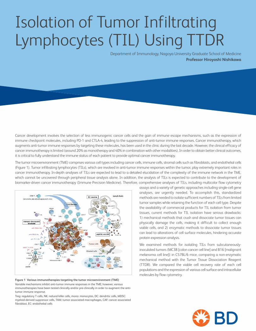
The tumor microenvironment (TME) comprises various cell types including cancer cells, immune cells, stromal cells such as fibroblasts, and endothelial cells (Figure 1). Tumor infiltrating lymphocytes (TILs), which are involved in anti-tumor immune responses within the tumor, play extremely important roles in cancer immunotherapy. In-depth analyses of TILs are expected to lead to a detailed elucidation of the complexity of the immune network in the TME, which cannot be uncovered through peripheral tissue analysis alone. In addition, the analysis of TILs is expected to contribute to the development of biomarker-driven cancer immunotherapy (Immune Precision Medicine). Therefore, comprehensive analyses of TILs, including multicolor flow cytometry
assays and a variety of genetic approaches including single-cell gene analyses, are urgently needed. To accomplish this, standardized methods are needed to isolate sufficient numbers of TILs from limited tumor samples while retaining the function of each cell type. Despite the availability of commercial products for TIL isolation from tumor tissues, current methods for TIL isolation have serious drawbacks: 1) mechanical methods that crush and dissociate tumor tissues can physically damage the cells, making it difficult to collect enough viable cells, and 2) enzymatic methods to dissociate tumor tissues can lead to alterations of cell surface molecules, hindering accurate protein expression analysis.
We examined methods for isolatlng TILs from subcutaneously-inoculated tumors (MC38 [colon cancer cell line] and B16 [malignant melanoma cell line]) in C57BL/6 mice, comparing a non-enzymatic mechanical method with the Tumor Tissue Dissociation Reagent (TTDR). We compared the viable cell recovery rate of each cell populations and the expression of various cell surface and intracellular molecules by flow cytometry.
Figure 1 Various immunotherapies targeting the tumor microenvironment (TME)Variable mechanisms inhibit anti-tumor immune responses in the TME; however, various immunotherapies have been tested clinically and/or pre-clinically in order to augment the anti-tumor immune response. Treg: regulatory T cells, NK: natural killer cells, mono: monocytes, DC: dendritic cells, MDSC: myeloid-derived suppressor cells, TAM: tumor associated macrophages, CAF: cancer associated fibroblast, EC: endothelial cells
Department of Immunology, Nagoya University Graduate School of Medicine Professor Hiroyoshi Nishikawa

Preparation of TILs from Tumor TissuesTumor tissues were excised from the mice 14 days after tumor inoculation. Blood vessels were carefully removed and the excised tissues were cut into pieces of approximately 5 mm3. The tissue pieces were divided into two groups, each weighing about 100 mg. Each sample was minced with scissors to a size not exceeding 1 to 2 mm3, and then TILs were extracted using either the mechanicalmethod or the TTDR method as shown in Figure 2. For the TTDR method, a solution of 2X TTDR was added to the finely-minced sample to a final concentration of 1X TTDR and the sample was incubated with gentle agitation.
Figure 2
Tumor tissue Tumor tissue
TTDR reaction (with gentle agitation)
Mechanical dissociation
Mechanical (non-enzymatic)
TTDR (enzymatic)
Filtration Filtration
Addition of reaction-halting solution
Centrifugation (TIL recovery) Centrifugation (TIL recovery)
Staining TILs and Multicolor Analysis by Flow Cytometry (FACS)Isolated TILs were stained with Fixable Viability Stain 780 (FVS780) to identify viable cells and fluorochrome-conjugated antibodies in the presence of BD Horizon™ Brilliant Stain Buffer. The cell surface markers used are as follows; CD3, CD4, CD8, PD-1 (immune checkpoint molecule expressing on activated or exhausted T cells), Tim-3, CD44 and CD62L (cell adhesion molecules), CXCR4 (chemokine receptor), CD69 (early activation marker), CD25, and GARP (highly expressed by FoxP3+ regulatory T [Treg] cells). CD4+ T cells that express both CD25 and FoxP3 were considered to be Treg cells for this analysis. After washing stained samples with BD Pharmingen™ Stain Buffer (BSA), cells were fixed and permeabilized using the BD Pharmingen™ Transcription Factor Buffer Set, and then the anti-FoxP3 antibody was used for intracellular staining. After washing, the samples were passed through a cell strainer into BD Trucount™ Tubes to calculate the absolute number of cells per tumor tissue mass (mg), and analyzed using a BD LSRFortessa™ X-20 flow cytometer.
Viable Cell Recovery Rate in Lymphocytes and SubpopulationsCompared with the mechanical method, the TTDR method showed a higher cell recovery rate for lymphocytes, approximately 3- and 1.4-fold for MC38 and B16 tumors, respectively. Similarly, the number of cells in the CD3+, CD4+, CD8+ and Treg subpopulations were 2- to 3-fold higher in samples isolated using the TTDR method. The ratio of CD4+T/CD8+T/Treg was comparable for both methods (Figure 3).
MC38 B16
Figure 3 Comparison of cell recovery rate in lymphocytes and each sub-population by the mechanical and the TTDR methods (left: MC38, right: B16)The value in the data represents cell number per 10 mg of the tumor tissue (mean ± SD: n=3).
0
20000
40000
60000
80000
Lymphocytes
0
5000
10000
15000
20000
25000
CD3+ cells
0
200
400
600
800
1000
CD4+T/CD8+T/Treg cellsCD4+ CD8+ Treg
Mechanical TTDR
Mechanical TTDR
Mechanical TTDR
events/10 mg (tissue)
events/10 mg (tumor tissue)
13137
54004
316 342
90
810 779
198
events/10 mg (tumor tissue)
6602
17936
0
500
1000
1500
2000
0
1000
500
1500
2000
Lymphocytes CD3+ cells
0
200
400
600
800
1000
CD4+T/CD8+T/Treg cellsCD4+ CD8+ Treg
Mechanical TTDR
Mechanical TTDR
Mechanical TTDR
events/10 mg (tissue)
events/10 mg (tumor tissue)
989
1398
362
1990
222
598
41
events/10 mg (tumor tissue)
836
1674

MC38
Mechanical
TTDR
Live Lym CD3+ TregCD4+/CD8+
MC38
CD4+T
Treg
CD8+T
FoxP3 CD44 CD62L CD25 Tim-3 PD-1 CXCR4 GARPCD69— Mechanical— TTDR
B16
CD4+T
Treg
CD8+T
FoxP3 CD44 CD62L CD25 Tim-3 PD-1 CXCR4 GARPCD69— Mechanical— TTDR
Mechanical method
CD4+T
Treg
CD8+T
CD62L/CD44 CD69 Tim-3 GARPPD-1 CXCR4
TTDR Method
CD4+T
Treg
CD8+T
Note: Y-axis not labeled; all the Y-axes show FVS780 (Viability).
CD62L/CD44 CD69 Tim-3 GARPPD-1 CXCR4
Mechanical
TTDR
B16Live Lym CD3+ TregCD4+/CD8+
CD4+T
Treg
CD8+T
Mechanical methodCD62L/CD44 CD69 Tim-3 GARPPD-1 CXCR4
CD4+T
Treg
CD8+T
TTDR Method
Note: Y-axis not labeled; all the Y-axes show FVS780 (Viability).
CD62L/CD44 CD69 Tim-3 GARPPD-1 CXCR4
Expression of Cell Surface Markers and Intracellular MoleculeFigure 4 shows scatter-grams of the expression of CD3, CD4/CD8 and CD25/FoxP3 in TILs isolated using the mechanical method and the TTDR method. The TTDR method was able to obtain larger numbers of viable cells than the mechanical method in both MC38 and B16 models. Additionally, CD4+T cells, Treg cells and CD8+T cells were analyzed for the expression of CD62L/CD44, CD69, Tim-3, PD-1, CXCR4 and GARP. The expression of these molecules was comparable in samples prepared using the TTDR method versus the mechanical method, as measured by the identification of positive cells and the fluorescence intensity, shown by the overlay histograms (Figure 5).
Figure 4 Comparison of the expression of cell surface and intracellular molecules in TILs isolated by the mechanical and the TTDR methods (left: MC38, right: B16)Analysis of the viable cells identified by FVS: lymphocytes (lym) > CD3+> CD4+/CD8+. Treg cells are identified as a subset of CD4+ T cells expressing CD25 and FoxP3.
Figure 5 Comparison of molecular expression in TILs (CD4+T cells, Treg cells, CD8+T cells) isolated by the mechanical and the TTDR methodExpression of nine molecules in CD4+T cells, Treg cells and CD8+ T cells were analyzed by FACS (top: scatter-gram, bottom: overlay histograms).

Catalog Number Product Name Quantity Storage Conditions Expiry Period
661563 BD Horizon™ Dri Tumor & Tissue Dissociation Reagent (TTDR) 15 vials/box 2 to 8°C 12 Mo from
manufacture
Catalog Number Product Name Quantity554656 BD Pharmingen™ Stain Buffer (FBS) 500mL562574 BD Pharmingen™ Transcription Factor Buffer Set 100 Tests340334 BD Trucount™ Absolute Counting Tube 50 Tests
Catalog Number Antigen Name Clone Fluorochrome553236 CD69 H1.2F3 FITC551892 CD279/PD-1 J43 PE565019 CD184/CXCR4 2B11/CXCR4 PE-CF594563902 FoxP3 R16-715 PerCP-Cy™5.5559250 CD44 IM7 APC557984 CD3 500A2 Alexa Fluor®700562910 CD62L MEL-14 BV421563068 CD8a 53-6.7 BV510563061 CD25 (IL-2 Receptor α) PC61 BV605563727 CD4 RM4-5 BV786747622 CD366 (TIM-3) 5D12/TIM3 BV711565388 Fixable Viability Dye FVS780 — —
Antibody Reagents Used in This Study
Antibody Reagents
Support Products
Although the TTDR method is enzymatic, it can gently isolate cells from tumor tissues with less damage compared with the mechanical method, which might physically crush tissues. TILs continually interacted with various types of cells, including cancer cells, and are considered to be extremely fragile cells. The TTDR method is a promising tool for isolating lymphocytes from tissue samples with a high recovery of viable cells, retaining their function as in the tumor. Some optimization is required, including the medium, temperature, and incubation time, in order to obtain enough cells of interest for downstream research applications. TTDR is expected to enable the isolation of relatively large cells, such as cancer cells and fibroblasts, with a high yield of viable cells. Thus, the TTDR method can contribute to cancer research, making possible a wide range of analytical approaches, for example, using cell sorting of TILs to enable single-cell gene analysis using RNAseq approaches, in addition to protein expression analysis by FACS, as demonstrated here.
Department of Immunology, Nagoya University Graduate School of Medicine Professor Hiroyoshi Nishikawa, M.D., Ph.D.Prof. H. Nishikawa graduated from Mie University Graduate School of Medicine in 1995. After residency program at the Mie University Hospital and some other institutions, he joined a doctoral course in Mie University Graduate School of Medicine in 1998, and completed the course in 2002 (PhD degree). After the completion of his thesis, he continuously studied the role of immune suppressive mechanisms, particularly regulatory T cells in tumor immunity in Dr. Lloyd J Old’s lab at Memorial Sloan Kettering Cancer Center (New York, NY). He then came back to Japan as an Assistant Professor in 2006 at Mie University Graduate School of Medicine. In 2010, he became as an Associate Professor at Osaka University Immunology Frontier Research Center, having taken up a joint position as an Adjunct Associate Professor at Roswell Park Cancer Institute in 2012. He is now the chief of Division of Cancer Immunology, Research Institute /Exploratory Oncology Research and Clinical Trial Center, at National Cancer Center Japan since 2015, and is involved in translational research developing novel cancer immunotherapies targeting regulatory T cells. Additionally, in 2016, he is cross-appointed as Professor and Chair, Department of Immunology, Nagoya University Graduate School of Medicine to expand his research, particularly pursuing cancer immunology from the basic to translational research. In recent years, he has been working to elucidate the nature of the immunosuppressive network in the TME to develop novel cancer immunotherapies.
Note: Antigen name: GARP (LRRC32) BioLegend, Inc.
23-22161-00
BD Life Sciences, San Jose, CA, 95131, USA
bdbiosciences.comBD, the BD Logo and all other trademarks are property of Becton, Dickinson and Company or its affiliates. © 2019 BD. All rights reserved.
For Research Use Only (RUO). Not for use in diagnostic or therapeutic procedures.