isolation of human genomic dna for genetic analysis from premature neonates: a comparison between...
TRANSCRIPT

METHODOLOGY ARTICLE Open Access
Isolation of human genomic DNA for geneticanalysis from premature neonates: a comparisonbetween newborn dried blood spots, wholeblood and umbilical cord tissueShavanthi Rajatileka1, Karen Luyt2, Manal El-Bokle3, Maggie Williams4, Helena Kemp5, Elek Molnár6
and Anikó Váradi1*
Abstract
Background: Genotyping requires biological sample collection that must be reliable, convenient and acceptablefor patients and clinicians. Finding the most optimal procedure of sample collection for premature neonates whohave a very limited blood volume is a particular challenge. The aim of the current study was to evaluate the use ofumbilical cord (UC) tissue and newborn dried blood spot (DBS)-extracted genomic DNA (gDNA) as an alternative tovenous blood-derived gDNA from premature neonates for molecular genetic analysis.All samples were obtained from premature newborn infants between 24-32 weeks of gestation. Paired blood andUC samples were collected from 31 study participants. gDNA was extracted from ethylenediaminetetraacetic acid(EDTA) anticoagulant-treated blood samples (~500 μl) and newborn DBSs (n = 723) using QIAamp DNA Micro kit(Qiagen Ltd., Crawley, UK); and from UC using Qiagen DNAeasy Blood and Tissue kit (Qiagen Ltd., Crawley, UK).gDNA was quantified and purity confirmed by measuring the A260:A280 ratio. PCR amplification and pyrosequencingwas carried out to determine suitability of the gDNA for molecular genetic analysis. Minor allele frequency of twounrelated single nucleotide polymorphisms (SNPs) was calculated using the entire cohort.
Results: Both whole blood samples and UC tissue provided good quality and yield of gDNA, which wasconsiderably less from newborn DBS. The gDNA purity was also reduced after 3 years of storage of the newbornDBS. PCR amplification of three unrelated genes resulted in clear products in all whole blood and UC samples and86%-100% of newborn DBS. Genotyping using pyrosequencing showed 100% concordance in the paired UC andwhole blood samples. Minor allele frequencies of the two SNPs indicated that no maternal gDNA contaminationoccurred in the genotyping of the UC samples.
Conclusions: gDNAs from all three sources are suitable for standard PCR and pyrosequencing assays. Given that UCprovide good quality and quantity gDNA with 100% concordance in the genetic analysis with whole blood, it canreplace blood sampling from premature infants. This is likely to reduce the stress and potential side effectsassociated with invasive sample collection and thus, greatly facilitate participant recruitment for genetic studies.
Keywords: Premature newborns, Single nucleotide polymorphism, Newborn dried blood spots, Umbilical cord,Genomic DNA, Pyrosequencing, Sample storage
* Correspondence: [email protected] for Research in Biosciences, Department of Biological, Biomedicaland Analytical Sciences, Faculty of Health and Applied Sciences, University ofthe West of England, Bristol BS16 1QY, UKFull list of author information is available at the end of the article
© 2013 Rajatileka et al.; licensee BioMed Central Ltd. This is an open access article distributed under the terms of the CreativeCommons Attribution License (http://creativecommons.org/licenses/by/2.0), which permits unrestricted use, distribution, andreproduction in any medium, provided the original work is properly cited.
Rajatileka et al. BMC Genetics 2013, 14:105http://www.biomedcentral.com/1471-2156/14/105

BackgroundThe reliability and performance of the molecular assayssuch as polymerase chain reaction (PCR) and pyro-sequencing are strongly influenced by the quality andquantity of the starting template. The availability of highquality gDNA from a large number of well characterisedpatients and healthy controls is a prerequisite for the suc-cess of genetic variation studies. Conventionally, gDNAfor use in clinical epidemiological studies is obtained fromperipheral blood samples because it provides high qualityand a good yield of gDNA [1-4]. However, obtaining per-ipheral blood is invasive and unsuitable for certain cohortssuch as very low birthweight preterm infants because theyhave a small circulating blood volume (~85 ml/kg); [5-7]and it is not considered to be ethical to sample more than1 ml for research purposes. Recalling these infants at alater stage when suitable amount of whole blood can becollected is problematic because the neonatal mortalityrate in the very low birthweight cohort is significant, par-ticularly in the high risk preterm group [8].
An alternate source of gDNA, which is now used fre-quently in molecular genetic studies, is newborn driedblood spots (DBS) [9-13]. The blood is usually collected bya heel-prick and applied on special filter paper, a conveni-ent medium for transport and storage [14]. These newbornDBS are used for neonatal metabolic screening and thenstored in repositories for follow-up testing and publichealth research [15-18]. Using newborn DBS would be anideal replacement for the use of fresh human tissue forgDNA extraction, as it is routinely carried out at birtheliminating the need for additional needle pricks for samplecollection and for specialist storage conditions. The draw-back of using newborn DBS for genetic analysis is the min-iscule amount of blood available. The amount of gDNAthat can be extracted from a 3.2 mm punch of a newbornDBS is about 60 ng [19]. In reality, only about one to max-imum three 3.2 mm punches are available for academicresearch purposes, which is not sufficient for large scalesingle nucleotide polymorphisms (SNP) detection studies.
This problem can be overcome by using umbilicalcord blood, aspirated from the placenta after birth, forgDNA preparation. The practice of delayed cord clamp-ing is advantageous to the preterm infant [20-22], facili-tating an autotransfusion from the placenta. However,this means that the volume of infant blood remaining inthe cut umbilical cord and placenta is significantly re-duced. Cord blood is frequently required for clinical in-dications, such as blood group haemoglobin and serumbilirubin analysis, taking priority over research samples.When cord blood is aspirated, there is also a potentialrisk of contamination by maternal blood [23]. However,umbilical cord tissue which would usually be discardedas clinical waste following birth [24] can be collectedeasily and potentially used for gDNA extraction [25].
In this study we compared three different sources forgDNA extraction from very premature babies where alarge volume of whole blood or umbilical cord blood isnot available. The suitability of newborn DBS and umbil-ical cord tissue for PCR and pyrosequencing was investi-gated and the concordance of paired umbilical cordtissue gDNA and whole blood from the same individualwas assessed. Our study showed that umbilical cord tis-sue can effectively be used for genetic analysis of prema-ture babies.
MethodsSample collection and processingBlood, newborn DBS and umbilical cord tissue were col-lected from a subset of patients participating in an asso-ciation study to investigate the genetic background ofpremature infants to white matter brain injury. Thestudy received ethical approval in April 2008 from theNational Research Ethics Service, UK (REC referencenumber 10/H0106/10). For the use of whole blood andumbilical cord written informed consent was obtainedfrom the parents of eligible infants participating in thestudy. Similarly, informed written consent was obtainedfrom healthy adult volunteers for the use of whole bloodsamples. The archived newborn blood spot samples usedfor the study were fully anonymised according to theHuman Tissue Act and MRC Guidance and used for re-search without individual informed consent as permittedby the UK newborn screening programme Code of Prac-tice for the retention and Storage of Residual Spots(April 2005, ISBN 0955013801).
Blood samplesWhole blood samples (~500 μl) were obtained from pre-term infants between 24-32 week gestation during thefirst week of life when stable on intensive care. Sampleswere collected in K2-EDTA tubes, mixed by inversion8-10 times after being drawn and stored at 4°C for up toa month prior to gDNA isolation.
Dried blood spots (DBS)Newborn DBS were collected from heel prick blood sam-pling on blood spot screening cards prepared routinelywithin 5-8 days of birth as part of the UK NewbornScreening Programme [http://newbornbloodspot.screen-ing.nhs.uk]. Samples, collected from infants 24-32 weeksgestation within the past 3-22 years, were used in thestudy. The newborn DBS samples from the participantsthat were stored in the biobank were 3-5 years old (n =25); 6-10 years old (n = 25); 11-15 years old (n = 20); and16-22 years old (n = 30). Newborn DBS obtained withinthe last three years were not available for analysis becausethese samples might need to be recalled by the pathologylaboratories for further tests for up to 3 years after birth.
Rajatileka et al. BMC Genetics 2013, 14:105 Page 2 of 9http://www.biomedcentral.com/1471-2156/14/105

All blood spot screening cards were stored in boxes atroom temperature. To compare yields and quality frommore recently prepared dried blood spots, 25 μl of wholeblood from volunteer adults was spotted onto a blood spotscreening card, air dried and stored at room temperaturefor a period of one month (n = 20).
Umbilical cord tissueA 5-10 cm long segment collected from the mid portionof each cord was obtained immediately following deliv-ery, washed in sterile water and stored in sterile 30 mlspecimen containers at−20°C until required for DNAisolation (Figure 1).
DNA isolationDNA from 100 μl of whole blood and all newborn DBS wasisolated using the QIAamp DNA Micro Kit (QIAGEN Ltd.,Crawley, UK) following manufacturer’s guidelines. For new-born DBS, one to three 3.2 mm disks were punched fromeach card for DNA extraction. Umbilical cord DNA was ex-tracted from the inner layer of the cord tissue including theWharton’s jelly and blood vessels (0.5 g wet tissue) usingthe DNeasy Blood & Tissue Kit (QIAGEN Ltd., Crawley,
UK) according to the manufacturer’s instructions. Umbilicalcords were thawed for 5 min at room temperature (~20°C),and during the thawing whilst semi frozen, the outer layer(Figure 1A&B) was removed with a sterile scalpel. This wasdone to prevent cross-contamination of the infant genomicDNA with maternal or other external DNA due to handlingfollowing birth. 0.5 g of the inner layer of the cord tissue in-cluding the Wharton’s jelly and blood vessels were used forDNA extraction (Figure 1A&B). DNA was stored at −20°Cuntil analysis. The key steps of the DNA isolation protocolsare summarised in Table 1.
gDNA quantificationDNA concentration was measured at 260 nm againstnuclease free water using a NanoDrop ND-1000 (Lab-tech International Ltd, Ringmer East Sussex, UK). Thepurity of gDNA was determined by measuring the 260-280 nm absorbance ratio (A260:A280; Table 2). Optimalpurity is expected to be in the range of 1.7-2.0.
gDNA quality assessment by gel electrophoresisThe integrity of the gDNA samples were assessed by ana-lysing the samples (10-50 ng) for evidence of degradation
Figure 1 Isolation of Umbilical cord tissue for DNA extraction. (A) A schematic structure of the umbilical cord (cross sectional view).a maternal sheath, b Wharton’s jelly; c umbilical vein; d allantoic duct; e umbilical arteries. (B) Umbilical cord preparation for gDNA extraction.i) Cord tissue was cut across as indicated with the white line. ii) Cross-section of the umbilical cord. The outer maternal sheath was removed asindicated with the white line. iii) The internal Wharton’s jelly with umbilical vein and arteries was used for gDNA extraction.
Rajatileka et al. BMC Genetics 2013, 14:105 Page 3 of 9http://www.biomedcentral.com/1471-2156/14/105

using agarose gel electrophoresis. Genomic DNA sampleswere run on an agarose gel (0.75% agarose) containing0.5 μg/ml ethidium bromide alongside a DNA ladder,lamda-HindIII (Thermoscientific, Massachusetts, USA)for 90 min. Samples were visualised under ultravioletlight (Gel Doc 1000, Bio Rad Laboratories Ltd, HemelHempstead, UK). The size of the gDNA was determinedby comparison with the DNA ladder. Appropriate qual-ity gDNA is expected to migrate predominantly above10kb on agarose gels.
Assessment of genomic DNA by PCR amplificationTo evaluate the gDNA quality, PCR amplification was per-formed first on two randomly selected samples from eachgroup of DNA source and from each storage length, usingprimers for human β-actin (GeneBank accession numberX00351) a house-keeping gene with the following primers5′-TGCCCATCTACGAGGGGTATG-3′ and 5′-GAAATCGTGCGTGACATTAAGGAG-3′. To compare amplifi-cation rates for gDNA extracted from different sources(whole blood (n = 31), umbilical cord (n = 31) and newbornDBS (n = 723)), amplification was also carried out flankingtwo unrelated SNPs: rs1835740 [26] and rs4354668 [27].All PCR assays were carried out for 35 cycles in a total vol-ume of 25 μl, containing 1× high fidelity reaction buffer -(100 mM Tris–HCl, 500 mM KCl pH 8.3), 1 mM ofMgCl2, 200 μM of each dNTP, 100 pmol of each oligo-nucleotide primer, 1 unit of high fidelity Taq Polymerase(FastStart High Fidelity Taq Polymerase, Roche DiagnosticsLimited, West Sussex, UK) and 2 μl (~1-30 ng) of gDNA.
Assessment of the fidelity of gDNA obtained fromumbilical cordsTo assess the fidelity of the gDNA obtained from umbil-ical cords, two single nucleotide polymorphisms (SNPs)rs1835740 [26] and rs4354668 [27] were genotyped bypyrosequencing (Qiagen Ltd., Crawley, UK) using pairedgDNA isolated from both whole blood and umbilicalcords from the same individual (n = 31).
PyrosequencingSingle-stranded biotinylated PCR products were pre-pared for the pyrosequencing reaction using a VacuumPrep Tool (Qiagen Ltd., Crawley, UK). The biotinylatedPCR products were immobilised onto high performancestreptavidin sepharose beads (Streptavidin Sepharose™HP, GE Healthcare, Chalfont St Giles, Buckinghamshire,UK). For a single sample, 3 μl of streptavidin sepharosewere added to 40 μl binding buffer (10 mM Tris–HCl,2 M NaCl, 1 mM EDTA, 0.1% TweenTM 20, pH 7.6;Qiagen Ltd., Crawley, UK) and mixed with 20 μl PCRproduct and 17 μl deionised water on a mechanicalshaker for 5 min at room temperature (~20°C) in a 96-well plate. The beads containing the immobilised tem-plates were isolated by filter probes using vacuum andthen washed with 70% ethanol, denaturizing solution(0.2 M NaOH; Qiagen Ltd., Crawley, UK) and thenwashing buffer (10 mM Tris-acetate pH 7.6; QiagenLtd., Crawley, UK) for 5 s each. Beads were released intoa PSQTM 96 well plate (Qiagen Ltd., Crawley, UK) con-taining 38.4 μl annealing buffer (20 mM Tris-acetate,5 mM magnesium acetate, pH 7.6; Qiagen Ltd., Crawley,
Table 1 Comparison of the key steps of gDNA extraction protocols from the three different starting materials
DNA isolation step Starting material
3.2 mm DBS 100 μl WB 0.5 g UC
1. DNA binding column Silica-based Silica-based Silica-based
2. Use of carrier RNA* Yes Yes No
3. Duration of lysis step 1 h 1 h ~12 h
4. Elution volume 60 μl 100 μl 1 ml
5. Solution used for elution Nuclease free water Nuclease free water Nuclease free water
*Carrier RNA was added into the lysis buffer at manufacturer’s recommended concentrations (DBS: 0.01 μg/μl and WB: 0.005 μg/μl). DBS–dried blood spots;WB–whole blood; UC–umbilical cord.
Table 2 gDNA concentration measured at 260 nm
Sample DBS DBS average WB UC
Storage duration 1 monthn = 20
3-5 yearsn = 25
6-10 yearsn = 25
11-15 yearsn = 15
15-22 yearsn = 30
3-22 yearsn = 95
(<1 month)n = 31
(<1 month)n = 31
ng/μl 9.2 ± 1.5 6.2 ± 8.7 11.4 ± 6.9 7.9 ± 2 7.6 ± 4.2 8.3 ± 10.7 40.3 ± 10.9 117.3 ± 112.9
Total DNA (μg) 0.55 0.37 0.68 0.48 0.46 0.49 4.03 70.4
A260:A280 1.7 2.0 2.2 2.5 2.4 2.3 1.8 1.9
p < 0.01 WB vs UC, p < 0.001 DBS (3-22 years average) vs UC total DNA.
Rajatileka et al. BMC Genetics 2013, 14:105 Page 4 of 9http://www.biomedcentral.com/1471-2156/14/105

UK) and 1.6 μl of the sequencing primer, rs1835740-PyroSeq (0.4 μM final concentration). Annealing wasachieved by heating the samples to 80°C for 5 minfollowed by cooling to room temperature (~20°C). Pyro-sequencing reactions were performed on the PyroMark™Q96 ID (Qiagen Ltd., Crawley, UK) according to themanufacturer’s instructions using the PSQTM 96 SNPReagent Kit (Qiagen Ltd., Crawley, UK; Table 3) and thegenotype was determined using PyroMark™ ID program(Qiagen Ltd., Crawley, UK).
DNA sequencingGenotypes from pyrosequencing were confirmed by Sangersequencing (using ABI 3730xl 96 capillary DNA Analyzers)at Eurofins MWG Operon (Ebesberg, Germany). Tensamples were randomly selected for sequencing and PCRproducts were purified using Wizard® SV Gel and PCRClean-Up System (Promega, Southampton, UK) followingthe manufacturer’s instructions.
Statistical analysisBasic statistical data (mean, standard deviation, standarderror) were derived using MS Excel. Statistical analysiswas carried out using a standard student’s t-test inMicrosoft Excel™.
ResultsAssessment of gDNA quantity and qualityGenomic DNA concentration and quality was deter-mined in 177 samples using spectrophotometry (Table 1).The average concentration of gDNA was the highest inthe umbilical cord extractions (UC) followed by thewhole blood (WB) and then newborn DBS. A significantdifference was observed between the three groups(Table 1; p < 0.01 WB versus UC; p < 0.001 newborn DBS
versus UC; p < 0.001 WB versus newborn DBS). Therewas no significant correlation between the storage lengthand gDNA concentration in the DBS samples (Table 2).
The quality of gDNA was comparable between wholeblood, umbilical cords and DBS prepared 1 month priorto extraction. The average A260:A280 ratio of the gDNAin these samples (1.7-1.9) fell within the optimal rangefor gDNA purity (1.8-2.0). However, the purity of gDNAin the DBS samples decreased with the storage lengthfrom A260:A280 1.7 to 2.4 (p < 0.05) over the 22 year-period (Table 2).
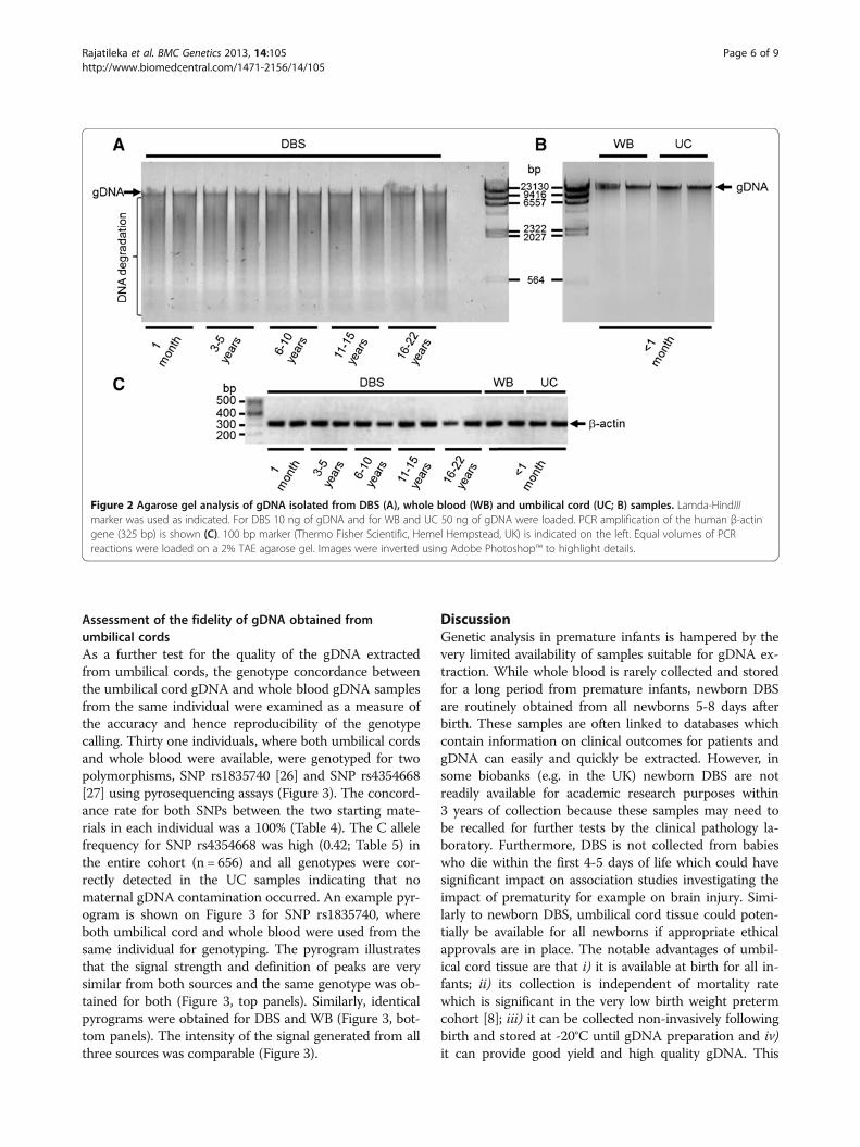
Analysis of DNA quality by agarose gel electrophoresisTo detect gDNA degradation in various samples, agarosegel electrophoresis was carried out (Figure 2A&B). Rep-resentative samples from each group were analysed forgDNA purity. All WB and UC samples showed uniformelectrophoretic mobility and gDNA appeared as a single,high-molecular-weight band >10 kb (Figure 2B) with nolow-molecular-weight fragmented bands present whichwould indicate sample degradation. In contrast, therewas no clearly defined band at 10 kb visible in the DBSsamples and the DNA produced a smear of low-molecular-weight fragmented bands on the gel indicat-ing DNA degradation (Figure 2A). High quality gDNA isexpected to be mostly >10 kb.
PCR amplification of gDNAPCR was performed to confirm the integrity of the gDNAand to determine if any inhibitory materials (e.g. guani-dium, RNA or proteins) were present in the extractions.For this purpose a 325 bp fragment of a house keepinggene, β-actin was amplified which showed a clear specificband with the expected size (Figure 2C). All tested sam-ples produced an amplicon at the expected size.
Table 3 Pyrosequencing primers and conditions used in the study
Oligonucleotide Sequence 5′-3′ Product size (bp) T (°C) Modifications
rs1835740PyroF CTCATTCGTTTTCTGCCTGTTG 300 60 None
rs1835740PyroR-BIO TCTTGCATATTTGAGCAGACTTTG 5′Biotin
rs1835740PyroSeq CACAACTTGATTCCAATCT N/A None
Target sequence GC/TGTATGTAGATT
Nucleotide dispensation order AGCTCGTAT
rs4354668PyroF-BIO GGGGCTAAACCTTGCAATC 166 60 5′Biotin
rs4354668PyroR GAGTGGCGGGAGCAGAGA None
rs4354668PyroSeq GGGTGTGTGCGCGCC N/A None
Target sequence T/GGGGGAGGCGGTGGAGGCC
Nucleotide dispensation order CGTGCAGCGTGAGCGTGC
Primer pair rs1835740PyroF/rs1835740PyroR-BIO and rs4354668PyroF-BIO/rs4354668PyroR were used to generate biotinylated PCR products flanking SNPsrs1835740 and rs4354668, respectively. Primers rs1835740PyroSeq and rs4354668PyroSeq were used for pyrosequencing. The target sequence and the order ofnucleotide dispensation for each pyrosequencing assay are listed. In the dispensation order the nucleotides used as negative controls for pyrosequencing areunderlined. In optimal pyrosequencing conditions these nucleotides are not incorporated into the target DNA sequence and thus their addition do not generatepeak on the pyrogram (see also Figure 2). The nucleotide change in the target sequence is indicated in bold.
Rajatileka et al. BMC Genetics 2013, 14:105 Page 5 of 9http://www.biomedcentral.com/1471-2156/14/105
Assessment of the fidelity of gDNA obtained fromumbilical cordsAs a further test for the quality of the gDNA extractedfrom umbilical cords, the genotype concordance betweenthe umbilical cord gDNA and whole blood gDNA samplesfrom the same individual were examined as a measure ofthe accuracy and hence reproducibility of the genotypecalling. Thirty one individuals, where both umbilical cordsand whole blood were available, were genotyped for twopolymorphisms, SNP rs1835740 [26] and SNP rs4354668[27] using pyrosequencing assays (Figure 3). The concord-ance rate for both SNPs between the two starting mate-rials in each individual was a 100% (Table 4). The C allelefrequency for SNP rs4354668 was high (0.42; Table 5) inthe entire cohort (n = 656) and all genotypes were cor-rectly detected in the UC samples indicating that nomaternal gDNA contamination occurred. An example pyr-ogram is shown on Figure 3 for SNP rs1835740, whereboth umbilical cord and whole blood were used from thesame individual for genotyping. The pyrogram illustratesthat the signal strength and definition of peaks are verysimilar from both sources and the same genotype was ob-tained for both (Figure 3, top panels). Similarly, identicalpyrograms were obtained for DBS and WB (Figure 3, bot-tom panels). The intensity of the signal generated from allthree sources was comparable (Figure 3).
DiscussionGenetic analysis in premature infants is hampered by thevery limited availability of samples suitable for gDNA ex-traction. While whole blood is rarely collected and storedfor a long period from premature infants, newborn DBSare routinely obtained from all newborns 5-8 days afterbirth. These samples are often linked to databases whichcontain information on clinical outcomes for patients andgDNA can easily and quickly be extracted. However, insome biobanks (e.g. in the UK) newborn DBS are notreadily available for academic research purposes within3 years of collection because these samples may need tobe recalled for further tests by the clinical pathology la-boratory. Furthermore, DBS is not collected from babieswho die within the first 4-5 days of life which could havesignificant impact on association studies investigating theimpact of prematurity for example on brain injury. Simi-larly to newborn DBS, umbilical cord tissue could poten-tially be available for all newborns if appropriate ethicalapprovals are in place. The notable advantages of umbil-ical cord tissue are that i) it is available at birth for all in-fants; ii) its collection is independent of mortality ratewhich is significant in the very low birth weight pretermcohort [8]; iii) it can be collected non-invasively followingbirth and stored at -20°C until gDNA preparation and iv)it can provide good yield and high quality gDNA. This
Figure 2 Agarose gel analysis of gDNA isolated from DBS (A), whole blood (WB) and umbilical cord (UC; B) samples. Lamda-HindIIImarker was used as indicated. For DBS 10 ng of gDNA and for WB and UC 50 ng of gDNA were loaded. PCR amplification of the human β-actingene (325 bp) is shown (C). 100 bp marker (Thermo Fisher Scientific, Hemel Hempstead, UK) is indicated on the left. Equal volumes of PCRreactions were loaded on a 2% TAE agarose gel. Images were inverted using Adobe Photoshop™ to highlight details.
Rajatileka et al. BMC Genetics 2013, 14:105 Page 6 of 9http://www.biomedcentral.com/1471-2156/14/105

study aimed to assess the suitability of newborn DBS andumbilical cord tissue extracted gDNA as an alternative tovenous blood-derived gDNA from premature neonates forgenetic analysis.
The yields of gDNA extracted from whole blood andumbilical cords are comparable to previous studieswhere ∼6 μg gDNA/200 μl whole blood [28], ~100 μggDNA/ 200 μl umbilical cord blood [29] were obtained(Table 2). The gDNA yield of DBS 180 ng/3.2 mm punchhowever was higher than previously published ~60 nggDNA/3.2 mm punch [19] or 19-40 ng/3.0 mm punch[30]. These differences are most likely due to the non-uniform distribution of the blood on the card and thetype of filter paper used for blood collection [30]. In-deed, blood spots which were not correctly collectedhad to be used for research purposes leaving the cor-rectly collected blood spots for further clinical path-ology investigations. It is unlikely that a pathologicalincrease in white blood cells in the premature infantswould be responsible for the increased gDNA yield
observed in our study because a similar yield wasachieved for both adults and newborns (Table 2,1 month versus DBS samples 3-22 years). The gDNA ex-traction method can also have a significant impact onthe yield. Carrier RNA was added to Buffer AL (Table 1),which enhances gDNA binding to the QIAamp columnmembrane, especially if there are very few target mole-cules in the sample. To further enhance gDNA binding,the column membrane was equilibrated with nucleasefree water and the bound gDNA was eluted in two stepsby adding 30 μl of nuclease free water twice.
The quality of gDNA from umbilical cord and newbornDBS was comparable to whole blood gDNA (1.7 and 1.9versus 1.8, Table 1). A good quality gDNA sample shouldhave an A260:A280 ratio between 1.7-2.0 [31,32]. In addi-tion to measuring the A260:A280 ratio, a random selec-tion of samples were analysed on agarose gels toeliminate the possibility of contaminants in the samples(i.e. guanidium, RNA or proteins; [31-34]; Figure 2A&B).
Table 4 The number of samples successfully genotyped/the total number of samples attempted for each SNPtested
SNP Sample call rateDBS
Sample call rateWB
Sample call rateUC
rs1835740 682/723 (94%) 31/31 (100%) 31/31 (100%)
rs4354668 625/723 (86%) 31/31 (100%) 31/31 (100%)
Table 5 Distribution of alleles in the sample cohort
SNP WT/WT WT/MT MT/MT Mutant allele frequency
rs1835740 60% 33% 7% 0.23
rs4354668 30% 57% 13% 0.42
Distribution of the three genotypes (WT/WT, WT/MT, MT/MT in %) for SNPsrs1835740 and rs4354668 is shown. The frequency of the mutant allele(T allele for rs1835740 and C allele for rs4354668) is indicated in thelast column.
Figure 3 Pyrograms showing genotyping of SNP rs1835470 using paired umbilical cord (UC) and whole blood (WB) gDNA (top panels)or dried blood spot (DBS) and whole blood (WB) gDNA (bottom panels) from the same individual. The position of the SNP is highlightedin yellow boxes. Peak height is shown on the y-axes and the first nucleotide A and the fifth nucleotide C are negative controls and should not beincorporated into the target DNA sequence. E and S indicate enzyme and substrate, respectively.
Rajatileka et al. BMC Genetics 2013, 14:105 Page 7 of 9http://www.biomedcentral.com/1471-2156/14/105

These contaminants as well as degraded gDNA migrateat different rates compared to intact gDNA and thuscan be detected on an agarose gel. No obvious contam-ination of gDNA was observed in the WB and UC sam-ples (Figure 2B).
The length of storage of the dried blood spots did notsignificantly affect the total amount of gDNA recovered(Table 2). In contrast, the purity reduced significantlywith storage length from 1.7 to 2.4 (Table 2). This is inline with previous studies that showed reduced gDNAquality following 25 years storage [35,36]. Similarly toour observation, even after 6 years of storage at roomtemperature the gDNA quality was reduced [35,36].However, others reported that gDNA is stable for at least11 years at ambient tropical conditions [37]. It is welldocumented that there are several factors that may com-promise sample integrity which includes high humidity,temperature, persistence of nucleases and other chemicalagents as well as other sub-optimal conditions that mayoccur not only during transport, but also within storagefacilities [38]. Dry storage of nucleic acids has been rec-ommended to eliminate the need for cold storage basedon the assumption that nucleic acids are stable whendry. However there are numerous examples where deg-radation occurs during storage, in the cold or at ambientconditions, that can irreversibly damage samples in solu-tion or even those that are dehydrated [39]. Althoughdried blood spots provide a valuable bioresource for re-search, DNA from this source has been shown to deteri-orate with prolonged storage [40] which is in line withour observation. It has also been reported that the col-lection filter paper might have an impact on gDNA qual-ity [30,35], but unfortunately there is no informationavailable on the type of filter paper used for the collec-tion of our samples or whether more than one type hasbeen used.
To test the ability to detect the short DNA fragment ofthe β-actin gene in the samples, PCR amplification wasused (Figure 2C). All whole blood, umbilical cord andDBS samples amplified β-actin successfully. All of thesesamples were then used to detect two unrelated SNPs bypyrosequencing (Figure 3). No direct link was observedbetween storage length and positive outcome with eitherPCR or pyrosequencing. While all of the samples fromwhole blood or umbilical cord produced conclusive pyro-grams (Table 4), 6% and 14% of the DBS samples wereunsuccessful for the detection of rs1835740 [26] andrs4354668 [27], respectively (Table 4). However, differentsamples failed the two PCR and pyrosequencing assayssuggesting that the source of gDNA played an importantrole in the success of the analysis and the storage lengthdid not seem to have a major impact. This is in line withprevious observations [37,41] that gDNA fragmentationover time with storage has little impact on short DNA
detection (200-700 bp). The variation observed in thePCR success rate might be dependent on the amount ofnatural PCR inhibitors (protein, haemoglobin, iron)present in the newborn DBS [40]. The concordance ratefor both SNPs in gDNA prepared from umbilical cord tis-sue and whole blood was 100% (Table 4). The minor allelefrequency for SNP rs4354668 was high in our prematureinfant cohort (0.42; Table 5) and all genotypes were cor-rectly detected in the UC samples indicating that no ma-ternal gDNA contamination occurred.
ConclusionsThis study established that both umbilical cord tissueand newborn DBS can be used as alternatives to wholeblood for gDNA extraction from premature infants withsuitable quality and fidelity for standard PCR andpyrosequencing-based genotyping. Considering the nu-merous advantages of using umbilical cord tissue forgDNA extraction, as discussed above, this could poten-tially improve recruitment to clinical studies and reduceethical and logistical challenges associated with bloodsample collection across multicentre studies. The qualityand yield of gDNA from umbilical cord tissue makes ithighly suitable for genome wide studies.
AbbreviationsDBS: Dried blood spots; DNA: Deoxyribonucleic acid; gDNA: Genomicdeoxyribonucleic acid; PCR: Polymerase chain reaction; kb: Kilobase;RNA: Ribonucleic acid; SNP: Single nucleotide polymorphism;K2-EDTA: Potassium ethylene diamine tetraacetic acid; Tris-HCI: Tris(hydroxymethyl)aminomethane hydrochloride; KCl: Potassium chloride;MgCl2: Magnesium chloride; dNTP: Deoxyribonucleotide triphosphate.
Competing interestsThe authors declare that they have no competing interests.
Authors’ contributionsSR designed and carried out all experimental work. KL organised researchethics and NHS R&D permissions, parent information and consentingprocesses and clinical data collection. KL and ME collected the whole bloodand umbilical cord tissue; KL and HK provided the DBS; MW assisted withthe pyrosequencing analysis. EM and AV advised on experimental design. SRand AV wrote the manuscript and all authors reviewed the manuscript priorto submission. All authors read and approved the final manuscript.
AcknowledgementsWe would like to thank all the parents, children and clinicians whoparticipated in this study. This project was funded by the University of theWest of England, Bristol, UK (Grants awarded to AV and SR). The blood spotretrieval was funded by the David Telling Charitable Trust (Grant awarded toKL). EM is supported by the Biotechnology and Biological Sciences ResearchCouncil, UK (grants BB/F011326/1 and BB/J015938/1).
Author details1Centre for Research in Biosciences, Department of Biological, Biomedicaland Analytical Sciences, Faculty of Health and Applied Sciences, University ofthe West of England, Bristol BS16 1QY, UK. 2Neonatal Neuroscience, Schoolof Clinical Sciences, University of Bristol, St Michael’s Hospital, SouthwellStreet, Bristol BS2 8EG, UK. 3Southmead Hospital, Bristol BS10 5NB, UK. 4BristolGenetics Laboratory, Pathology Sciences, Blood Sciences and Bristol Genetics,Southmead Hospital, Bristol BS10 5NB, UK. 5Department of ChemicalPathology, Southmead Hospital, Bristol BS10 5NB, UK. 6Centre for SynapticPlasticity, School of Physiology and Pharmacology, University of Bristol,Medical Sciences Building, University Walk, Bristol BS8 1TD, UK.
Rajatileka et al. BMC Genetics 2013, 14:105 Page 8 of 9http://www.biomedcentral.com/1471-2156/14/105

Received: 15 March 2013 Accepted: 17 September 2013Published: 29 October 2013
References1. Parker SP, Cubit WD: The use of dried blood spot samples in
epidemiological studies. J Clin Pathol 1999, 52:633–639.2. Steinberg K, Beck J, Nickerson D, Garcia-Closas M, Gallagher M, Caggana M,
Reid Y, Cosentino M, Ji J, Johnson D, Hayes RB, Earley M, Lorey F, Hannon H,Khoury MJ, Sampson E: DNA banking for epidemiologic studies: a reviewof current practices. Epidemiol 2002, 13:246–254.
3. Holland NT, Smith MT, Eskenazi B, Bastaki M: Biological sample collectionand processing for molecular epidemiological studies. Mutat Res/RevMutat Res 2003, 543:217–234.
4. Beckett SM, Laughton SJ, Pozza LD, McCowage GB, Marshall G, Cohn RJ,Milne E, Ashton LJ: Buccal swabs and treated cards: methodologicalconsiderations for molecular epidemiologic studies examining pediatricpopulations. Am J Epidemiol 2008, 167:1260–1267.
5. Usher R, Shepard M, Lind J: The blood of the newborn infant andplacental transfusion. Acta Paediatr 1963, 52:497–512.
6. Bauer K, Linderkamp O, Versmold HT: Systolic blood pressure and bloodvolume in preterm infants. Arch Dis Child 1993, 69:521–522.
7. Aladangady N, Leung T, Costeloe K, Delpy D: Measuring circulating bloodvolume in newborn infants using pulse dye densitometry andindocyanine green. Paediatr Anaesth 2008, 18:865–871.
8. Tyson JE, Parikh NA, Langer J, Green C, Higgins RD: Intensive care forextreme prematurity: moving beyond gestational age. N Engl J Med 2008,358:1672–1681.
9. Edwards JR, Ulrich PP, Weintrub PS, Cowan MJ, Levy JA, Wara DW, Vyas GN:Polymerase chain reaction compared with concurrent viral cultures forrapid identification of human immunodeficiency virus infection amonghigh-risk infants and children. J Pediatr 1989, 115:200–203.
10. McCabe ER: Utility of PCR for DNA analysis from dried blood spots onfilter paper blotters. Genome Res 1991, 1:99–106.
11. Cassol S, Salas T, Gill MJ, Montpetit M, Rudnik J, Sy CT, O’Shaughnessy MV:Stability of dried blood spot specimens for detection of humanimmunodeficiency virus DNA by polymerase chain reaction. J ClinMicrobiol 1992, 30:3039–3042.
12. Harding D, Dhamrait S, Marlow N, Whitelaw A, Gupta S, Humphries S,Montgomery H: Angiotensin-converting enzyme DD genotype isassociated with worse perinatal cardiorespiratory adaptation in preterminfants. J Pediatr 2003, 143:746–749.
13. Malikova J, Votava F, Vrzalova Z, Lebl J, Cinek O: Genetic analysis of theCYP21A2 gene in neonatal dried blood spots from children with transientlyelevated 17-hydroxyprogesterone. Clin Endocrinol 2012, 77:187–194.
14. Mei JV, Alexander JR, Adam BW, Hannon WH: Use of filter paperfor thecollection and analysis of human whole blood specimens. J Nutr 2001,131:1631S–1636S.
15. Rubin EM, Andrews KA, Kan YW: Newborn screening by DNA analysis ofdried blood spots. Hum Genet 1989, 82:134–136.
16. Green A: Neonatal screening: current trends and quality control in theUnited Kingdom. Rinsho Byori 1998, 46:211–216.
17. Aoki K: Newborn screening in Japan. Southeast Asian J Trop Med PublicHealth 2003, 34(Suppl 3):80.
18. Olney RS, Moore CA, Ojodu JA, Lindegren ML, Hannon WH: Storage anduse of residual dried blood spots from state newborn screeningprograms. J Pediatr 2006, 148:618–622.
19. Hannelius U, Lindgren CM, Melen E, Malmberg A, Von Dobeln U, Kere J:Phenylketonuria screening registry as a resource for population geneticstudies. J Med Genet 2005, 42:e60.
20. Rabe H, Wacker A, Hulskamp G, Homig-Franz I, Jorch G: Late cord clampingbenefits extrauterine adaptation. Pediatr Res 1998, 44:454.
21. Rabe H, Reynolds G, Diaz-Rossello J: Early versus delayed umbilical cordclamping in preterm infants. Cochrane Database Syst Rev 2004, 4:CD003248.
22. Mercer JS, Vohr BR, McGrath MM, Padbury JF, Wallach M, Oh W: Delayedcord clamping in very preterm infants reduces the incidence ofintraventricular hemorrhage and late-onset sepsis: a randomized,controlled trial. Pediatrics 2006, 117:1235–1242.
23. Armson BA: Umbilical cord blood banking: implications for perinatal careproviders. J Obstet Gynaecol Can 2005, 27:263–290.
24. Tong CK, Vellasamy S, Tan BC, Abdullah M, Vidyadaran S, Seow HF,Ramasamy R: Generation of mesenchymal stem cell from human
umbilical cord tissue using a combination enzymatic and mechanicaldisassociation method. Cell Biol Int 2011, 35:221–226.
25. Godfrey KM, Sheppard A, Gluckman PD, Lillycrop KA, Burdge GC, McLean C,Rodford J, Slater-Jefferies JL, Garratt E, Crozier SR, Emerald BS, Gale CR, Inskip HM,Cooper C, Hanson MA: Epigenetic gene promoter methylation at birth isassociated with child’s later adiposity. Diabetes 2011, 60:1528–1534.
26. Anttila V, Stefansson H, Kallela M, Todt U, Terwindt GM, Calafato MS, Nyholt DR,Dimas AS, Freilinger T, Müller-Myhsok B, Artto V, Inouye M, Alakurtti K,Kaunisto MA, Hämäläinen E, de Vries B, Stam AH, Weller CM, Heinze A,Heinze-Kuhn K, Goebel I, Borck G, Göbel H, Steinberg S, Wolf C, Björnsson A,Gudmundsson G, Kirchmann M, Hauge A, Werge T, et al: Genome-wideassociation study of migraine implicates a common susceptibility varianton 8q22.1. Nat Genet 2010, 42:869–873.
27. Mallolas J, Hurtado O, Castellanos M, Blanco M, Sobrino T, Serena J,Vivancos J, Castillo J, Lizasoain I, Moro MA, Dávalos A: A polymorphism inthe EAAT2 promoter is associated with higher glutamate concentrationsand higher frequency of progressing stroke. J Exp Med 2006, 203:711–717.
28. Santella RM: Approaches to DNA/RNA extraction and whole genomeamplification. Cancer Epidemiol Biomarkers Prev 2006, 15:1585–1587.
29. Lehmann AS, Haas DM, McCormick CL, Skaar TC, Renbarger JL: Collection ofhuman genomic DNA from neonates: a comparison between umbilicalcord blood and buccal swabs. Am J Obstetrics Gynecol 2011, 204:362–372.
30. Halsall A, Ravetto P, Reyes Y, Thelwell N, Davidson A, Gaut R, Little S: Thequality of DNA extracted from liquid or dried blood is not adverselyaffected by storage at 4°C for up to 24h. Int J Epidemiol 2008, 37:7–10.
31. Glasel JA: Validity of nucleic acid purities monitored by 260nm/280nmabsorbance ratios. Biotechniques 1995, 18:62–63.
32. Teare JM, Islam R, Flanagan R, Gallagher S, Davies MG, Grabau C: Measurementof nucleic acid concentrations. Biotechniques 1997, 22:1170–1117.
33. Sambrook J, Russell D: Molecular cloning: a laboratory manual. 3rd edition.Long Island, New York, USA: Cold Spring Harbor Laboratory Press; 2001.
34. Tataurov AV, You Y, Owczarzy R: Predicting ultraviolet spectrum of singlestranded and double stranded deoxyribonucleic acids. Biophys Chem2008, 133:66–70.
35. Hollegaard MV, Thorsen P, Norgaard-Pedersen B, Hougaard DM:Genotyping whole-genome-amplified DNA from 3-to 25-year-oldneonatal dried blood spot samples with reference to fresh genomicDNA. Electrophoresis 2009, 30:2532–2535.
36. Hollegaard MV, Grove J, Grauholm J, Kreiner-Møller E, Bønnelykke K,Nørgaard M, Benfield TL, Nørgaard-Pedersen B, Mortensen PB, Mors O,Sørensen HT, Harboe ZB, Børglum AD, Demontis D, Ørntoft TF, Bisgaard H,Hougaard DM: Robustness of genome-wide scanning using archiveddried blood spot samples as a DNA source. BMC Genet 2011, 12:58–64.
37. Chaisomchit S, Wichajarn R, Janejai N, Chareonsiriwatana W: Stability ofgenomic DNA in dried blood spots stored on filter paper. Southeast AsianJ Tropical Med Public Health 2005, 36:270–273.
38. Bonnet J, Colotte M, Coudy D, Couallier V, Portier J, Morin B, Tuffet S: Chainand conformation stability of solid-state DNA: implications for roomtemperature storage. Nucleic Acids Res 2010, 38:1531–1546.
39. Anchordoquy TJ, Molina MC: Preservation of DNA. Cell Preserv Technol2007, 5:180–188.
40. Makowski GS, Davis EL, Hopfer SM: The effect of storage on Guthrie cards:implications for deoxyribonucleic acid amplification. Ann Clin Lab Sci1996, 26:458–469.
41. Rossmanith P, Röder B, Frühwirth K, Vogl C, Wagner M: Mechanisms ofdegradation of DNA standards for calibration function during storage.Appl Microbiol Biotechnol 2011, 89:407–417.
doi:10.1186/1471-2156-14-105Cite this article as: Rajatileka et al.: Isolation of human genomic DNA forgenetic analysis from premature neonates: a comparison betweennewborn dried blood spots, whole blood and umbilical cord tissue. BMCGenetics 2013 14:105.
Rajatileka et al. BMC Genetics 2013, 14:105 Page 9 of 9http://www.biomedcentral.com/1471-2156/14/105