isolation of bacteria by dilution plating. pure vs mixed culture pure: originate from 1 bacteria...
TRANSCRIPT

Isolation of bacteria by dilution plating
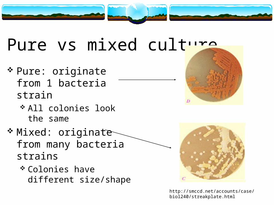
Pure vs mixed culture Pure: originate from 1
bacteria strain All colonies look the same
Mixed: originate from many bacteria strains Colonies have different
size/shape
http://smccd.net/accounts/case/biol240/streakplate.html
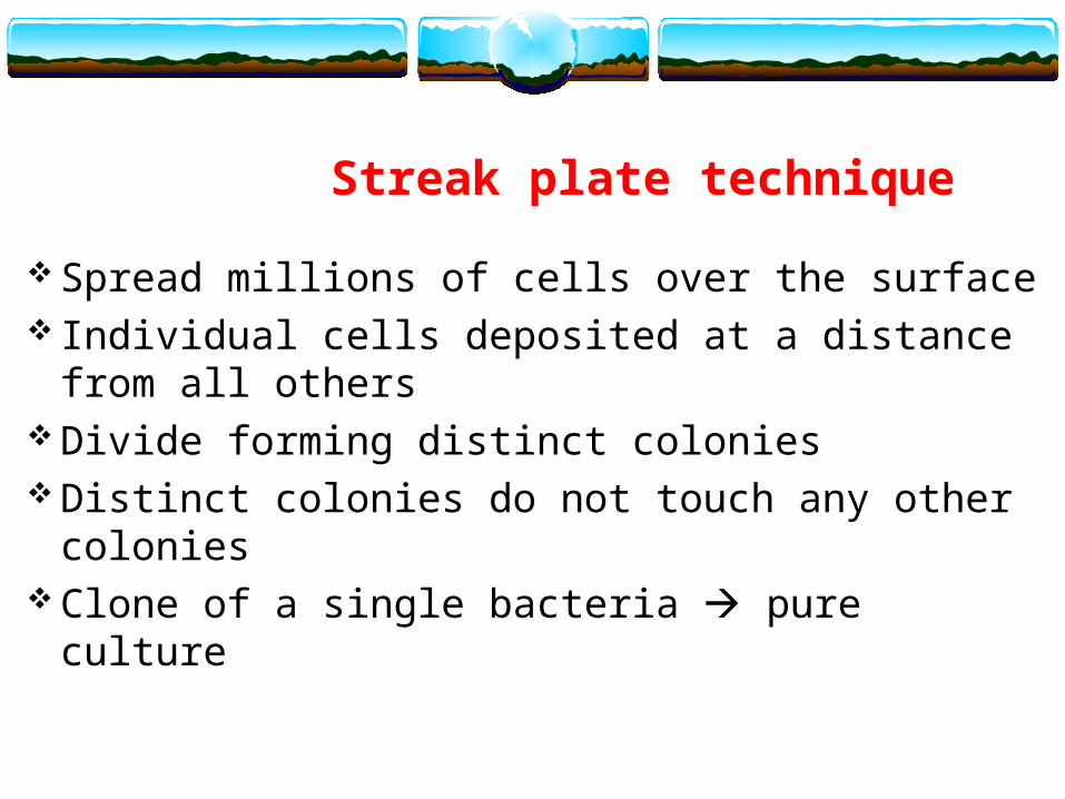
Spread millions of cells over the surface Individual cells deposited at a distance from all
others Divide forming distinct colonies Distinct colonies do not touch any other colonies Clone of a single bacteria pure culture
Streak plate technique
Disinfect your bench, wash your hands and wear gloves
Label the bottom of a NA plateNA = nutrient agar (general high nutrient media)
Keep lid closed when the plate is not in use You streak the plate on 3 different portion
You can draw the section that you will streak on the bottom of your plate (why not top?)

Objective 1: Streak plate
http://www.rlc.dcccd.edu/MATHSCI/reynolds/MICRO/lab_manual/streakplate3.jpg

Streak plate Using a sterile loop take a
loopful of your bacteria from the broth
Streak a vertical line Then streak gently across
section 1 Zig-zag pattern until a 1/3
of the plate is covered Do not dig into the agar
http://www.rlc.dcccd.edu/MATHSCI/reynolds/MICRO/lab_manual/streakplate2.jpg

Streak plate Sterilize the loop let it cool Rotate the plate about 90 degrees and spread the
bacteria from the first streak into a second area Do only one streak (or very few) in the first area
and once you are in the second area do not go back to the first
Do a zig-zag pattern until the 2nd area is covered Sterilize again do the same for 3rd area
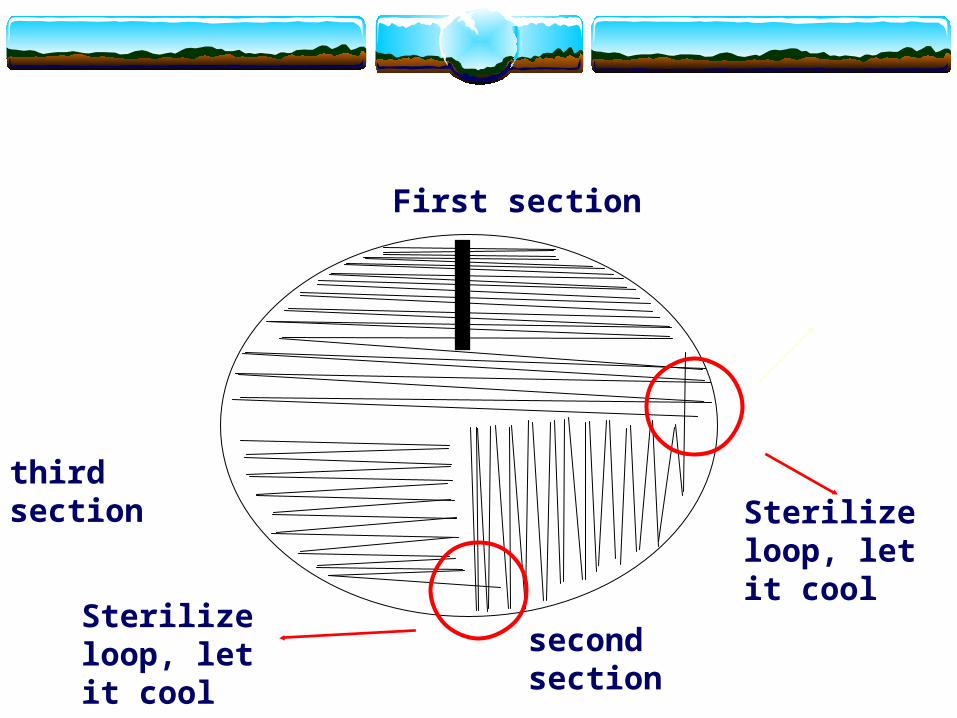
First section
second section
third sectionSterilize loop, let it cool
Sterilize loop, let it cool
Sterilized loop
Make sure that your red hot loop is cool enough prior to touch the bacteria
After you waited a few seconds Stab it into the agar in a position away from
bacteria will cool it If you stab where bacteria are production of
aerosol
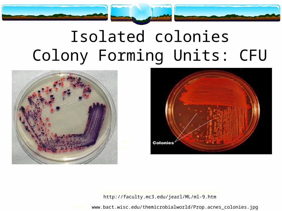
Isolated coloniesColony Forming Units: CFU
http://faculty.mc3.edu/jearl/ML/ml-9.htm
http://www.bact.wisc.edu/themicrobialworld/Prop.acnes_colonies.jpg
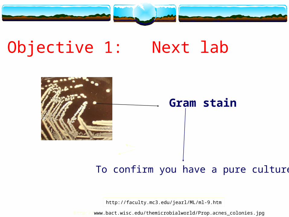
Objective 1: Next lab
http://faculty.mc3.edu/jearl/ML/ml-9.htm
http://www.bact.wisc.edu/themicrobialworld/Prop.acnes_colonies.jpg
To confirm you have a pure culture
Gram stain
Objective 2: Spread plate technique and dilutions
Label four plates for this exercise Name, date, dilution Pipette 1 ml from the bacteria culture into 99ml saline 1:100 dilution
0.1 ml of this into your first plate 1:1000 dilution Pipette 1ml of your 100 ml dilution of bacteria in saline and put into a 9 ml
tube 1:1000 0.1 ml of this into your 2nd plate 1:10000 dilution
Pipette 1ml of your 10 ml dilution of bacteria in saline and put into another 9ml tube 1:10000
0.1 ml of this into your 3rd plate 1: 100000 dilution Pipette 1ml of your 10 ml dilution of bacteria in saline and put into another
9ml tube 1:100000 0.1 ml of this into your 4th plate 1: 1000000 dilution
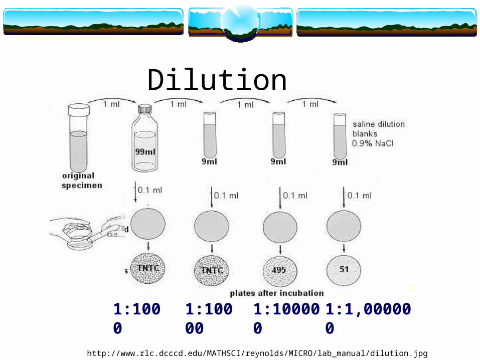
Dilution
http://www.rlc.dcccd.edu/MATHSCI/reynolds/MICRO/lab_manual/dilution.jpg
33
1:1000 1:10000 1:100000 1:1,000000
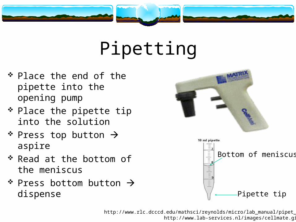
Pipetting Place the end of the pipette
into the opening pump Place the pipette tip into the
solution Press top button aspire Read at the bottom of the
meniscus Press bottom button
dispense
http://www.lab-services.nl/images/cellmate.gif
Pipette tip
Bottom of meniscus
http://www.rlc.dcccd.edu/mathsci/reynolds/micro/lab_manual/pipet_dilut.html
Pipette other considerations Always change pipette when going from a more
concentrated solution to a less concentrated solution avoid carry over
Dilution are additive 1:10 dilution is 1 ml into 9 ml, not 1 ml into 10
(that will be a 1:11 dilution) Mix well (pipette up and down or swirl gently)
prior to take your sample for your dilution
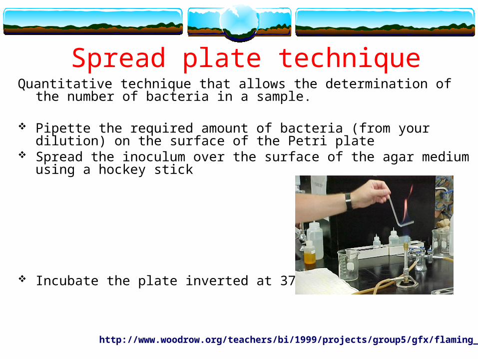
Spread plate techniqueQuantitative technique that allows the determination of the number of bacteria in a
sample.
Pipette the required amount of bacteria (from your dilution) on the surface of the Petri plate
Spread the inoculum over the surface of the agar medium using a hockey stick
Incubate the plate inverted at 37oC
http://www.woodrow.org/teachers/bi/1999/projects/group5/gfx/flaming_stick.jpg

http://www.bact.wisc.edu/Microtextbook/images/book_3/chapter_10/10-4.jpg
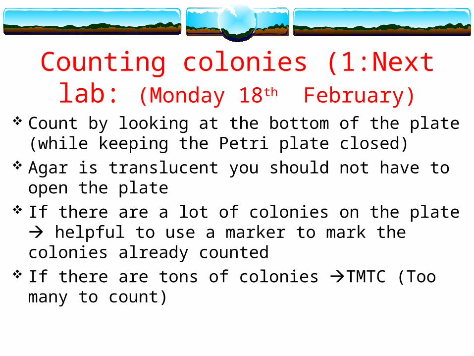
Counting colonies (1:Next lab: (Monday 18th February)
Count by looking at the bottom of the plate (while keeping the Petri plate closed)
Agar is translucent you should not have to open the plate If there are a lot of colonies on the plate helpful to use
a marker to mark the colonies already counted If there are tons of colonies TMTC (Too many to count)
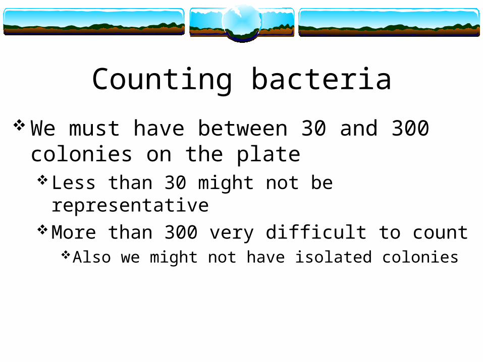
Counting bacteria
We must have between 30 and 300 colonies on the plateLess than 30 might not be representative More than 300 very difficult to count
Also we might not have isolated colonies
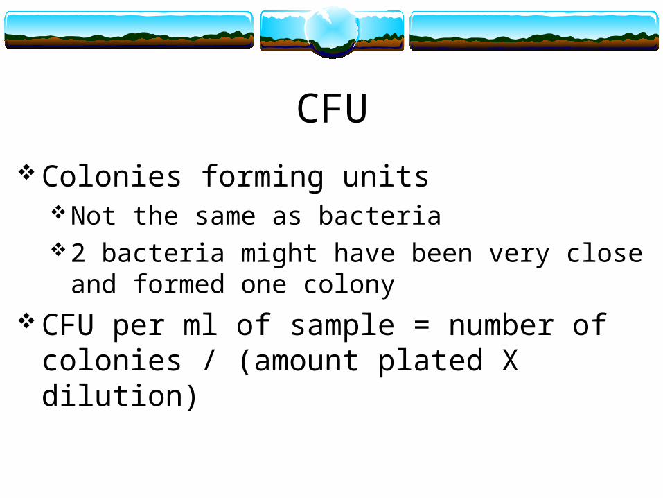
CFU
Colonies forming unitsNot the same as bacteria2 bacteria might have been very close and formed one
colony CFU per ml of sample = number of colonies /
(amount plated X dilution)
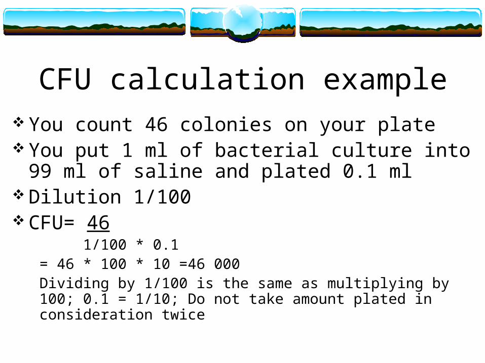
CFU calculation example You count 46 colonies on your plate You put 1 ml of bacterial culture into 99 ml of
saline and plated 0.1 ml Dilution 1/100 CFU= 46
1/100 * 0.1= 46 * 100 * 10 =46 000Dividing by 1/100 is the same as multiplying by 100; 0.1 = 1/10; Do not take amount plated in consideration twice