isolation of astragalin from cressa cretica cultivated in iraq
TRANSCRIPT
Isolation of Astragalin from Cressa cretica cultivated in Iraq Farah Fawzi1, Monther F. Mahdi2, Ibrahim S.Abaas1
1 Department of Pharmacognosy and Medicinal Plants, College of Pharmacy, Al-Mustansiriya University, Baghdad, Iraq. 2Department of Pharmaceutical Chemistry, College of Pharmacy, Ashur University, Baghdad, Iraq.
Abstract Cressa cretica L. Fam: Convolvulaceae (morning glory). Is a halophytic dwarf shrub grows in sandy, saline land. The aims of the study were isolation, quantification, and identification of kaempferol-3-O glucoside (astragalin) of Cressa cretica aerial parts .the presence of Kaempferol 3- O- β- glucoside (astragalin) was detected as the major glycoside in the polar fraction by TLC, HPTLC and HPLC comparing with astragalinstandard. Astragalin was isolated from Iraqi plant as yellow powder by preparative TLC and then it was determined by HPTLC. Theidentification and the structural elucidation of isolated astragalin were performed by1 H-nuclear magnetic resonance, 13C-nuclear magneticresonance), infrared, and ultraviolet.Results showed that the quantity of Kaempferol3-O- glucoside (astragalin) is 0.3% which is extractedunder optimal conditions (80% aqueous ethanol and 80°c for 3 hrs.).
Keywords: Astragalin, Cressa cretica, Halophytic, Isolation.
INTRODUCTION According to the World Health Organization (WHO), more than 70% of the population uses medicinal plants in their primary health care, mainly of herbal sources, This is especially the case in third world countries where the cost of consulting a doctor and the price of medication cannot be afforded by many People.1,2 despite herbal medicine has been used since the beginning of life, however, one of the greatest challenges is the fact that our knowledge of how plants actually affect human physiology is poor. Therefore many researchers are going on in this field and plants are claiming various medicinal uses is increased.3 Cressa cretica is a halophytic dwarf shrub grows in sandy, saline land. It is a small plant with a woody base and many stems. The leaves of the plant are small and compressed. The flowers are small and their color is white.4 From the phytochemical point of view, the plant was reported to contain: Coumarins, sterols, alkaloids, tannins, glycosides (cardiac glycoside, anthraquinone glycoside), protein, carbohydrate, flavonoids, unidentified sugars, and high salt content.5–9 Kaempferol-3-O-glucoside (Astragalin) is one of the major flavonoid found in a variety of plants.10,11 astragalin is receiving increasing attention due to its varies health benefiting and biological activities including anti-oxidative, anti-inflammatory anti-HIV, anti-allergic effects, besides this astragalin is responsible for the color of different beans and has the potential to extract it and market as a nutritionally important food supplement.12 The aerial parts of Cressa cretica L. yielded five flavonoids that were identified as quercetin (1), quercetin-3-O-glucoside(2), kaempferol-3-O-glucoside (3), kaempferol-3-Orhamnoglucoside (4), and rutin (5).13 Chemically flavonoids arebased upon a fifteen-carbon skeleton consisting of two benzenerings (A and B) linked via a heterocyclic pyrane ring (C).14
The widespread uses of Cressa cretica in traditional medicinehave resulted in considerable chemical analysis of the plant and itsactive principles. Available information indicates thathydroalcoholic extract of Cressa cretica was assayed againstdifferent carcinoma cell lines.15
The aim of this study is to investigate the presence of astragalin inthe Iraqi Cressa cretica L.To the best of our knowledge, this study is the first work studiedthe qualitative-quantitative analysis and isolation with thestructural elucidation of astragalin of Cressa cretica L. cultivatedin Iraq
METHODS General procedures Ultraviolet (UV) spectra were recorded in MeOH using a CAMAG system, IR spectra in KBR disk on Fourier-transform IR (FTIR) (Jasco-6100), H-NMR spectrum was measured on BRUKER AVANCE III 500 MHz apparatus high-performance
thin-layer chromatography (HPTLC) analysis was carried out using CAMAG system (Switzerland), pre-coated silica gel GF254 (aluminum TLC) from Merck co., and the standard astragalin from Sigma-Aldrich chemicals Co. All the solvents were from Sigma-Aldrich chemicals Co and Merk chemical company. Plant material The aerial parts (stems and leaves) of Cressa cretica were collected from area Al-Msiab and identified by the national herbarium in Abu-Ghraib, Baghdad. The plant material was collected during October and dried at room temperature in the shade, then grinded as powder and weighed. Preparation of aqueous methanolic extract The powdered aerial parts of Cressa cretica (180 g) were defatted with hexane (1200mL), using soxhlet extractor 250 x 6 units. The defatted plant material was further extracted with ethanol 80% (1500 mL) using soxhlet extractor at extraction temperature in the range (60-80°C) for 3 hours. The ethanolic extract was concentrated to (250 mL) by evaporation under reduced pressure using a rotary evaporator at (50°C). Then distilled water (100mL) was added to the ethanolic extract, and the extract partitioned with ethyl acetate (350mL) and allowed to settle overnight. The lower aqueous layer (300mL) was collected and labeled as fraction A, while the upper ethyl acetate layer was collected and labeled as fraction B. Chromatographic analysis for the detection of astragalin TLC TLC analysis of extract in comparison with astragalin standard was done by development with (ethyl acetate: methanol: water, 15:1.25:1) as a mobile phase.16 The HPTLC analysis was conducted to detect the presence of kaempferol 3-O-β- glucoside in the extract prepared with the same concentration The HPTLC analysis was conducted to detect the presence of astragalin. The HPTLC analysis was performed by using HPTLC silica gel 60 GF 254s (10x20 cm), the layer thickness was 0.5 mm. The standard astragalin 2 μL and the sample (2μL) fromextract were applied automatically on the plate by CAMAGLinomat 5. The plate was automatically submerged into anautomatic developing chamber (ADC2 CAMAG) using solventsystem (ethyl acetate: methanol: water, 15:1.25:1, v/v/v), withmigration distance about 7.5 cm. The plates were air-dried afterdevelopment and scanned under UV (366 and 245 nm) usingCAMAG TLC scanner 4. The data were processed using winCATS software.HPLCHPLC analysis was performed for detection and estimation ofAstragalin in the ethyl acetate extract fraction (B). The extractwas analyzed by (HLPC) method with UV detection. The HPLCanalysis was carried out by prominence HPLC system (SYKAM)
Farah Fawzi et al /J. Pharm. Sci. & Res. Vol. 11(1), 2019, 185-190
185
and the separation was performed in a reversed phase (RP) ODS- C18 column (25 cm x4.6 mm x5μ m). The mixture was passed through a 0.45 μm PVDF membrane and then 20 μL of each sample was injected into the HPLC system. The separation was done by elution with isocratic mixtures, 80% methanol as solvent A and 20%(water with 0.1% acetic acid) as solvent (B). A flow rate was set as 0.8ml/min for 10 minutes, detected by UV at 360. The astragalin was detected according to the retention time of the standard astragalin.17 Isolation and purification of flavonoid glycoside The ethyl acetate layer (fraction B) purified by preparative TLC. The purification technique performed on (20x20 cm) glass plates pre-coated with silica gel GF 254; prepared manually with 0.5 mm thickness. After that the sample (fraction B) applied as a concentrated solution in a raw spots by glass pasture pipette four times on each plate, one should wait after each application until all the solvent is evaporated, the elution system was (ethyl acetate: methanol: water, 15:1.25:1), the separated bands were visualized under UV light (254nm) as shown in the figure (3), bands at Rf= 0.5 were scrapped off with comparison with standard, and then eluted with acetone and methanol. The solvent was evaporated by rotary evaporator and weighed it. Then the band is further purified on another glass plates with 0.25 mm thickness with another solvent system( chloroform: methanol, 9:1)18 Identification of the isolated compound Spectrometric analysis Chemical structure elucidation was obtained by IR, UV, 1H NMR, and 13C-NMR.
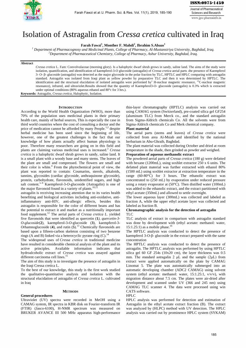
RESULT AND DISCUSSIONS Chromatographic analysis for detection of astragalin. The extract of aerial parts was analyzed by HPLC and HPTLC. TLC The result showed the best separation of compounds in the ethyl acetate extract and a fluorescent blue spot of astragalin was showed under 254 nm of UV light with Rf value 0.50 in comparison with standard astragalin as shown in ( fig:1) and table (1).
Table (1): Rf values of Astragalin in Cressa cretica extract and standard
Solvent system Value of Rf of astragalin standard Rf value of extract
Ethyl acetate: methanol, water,
(15:1.25:1) 0.52 0.50
Figure (1): Analytical TLC of astragalin standard and Cressa
cretica extract.
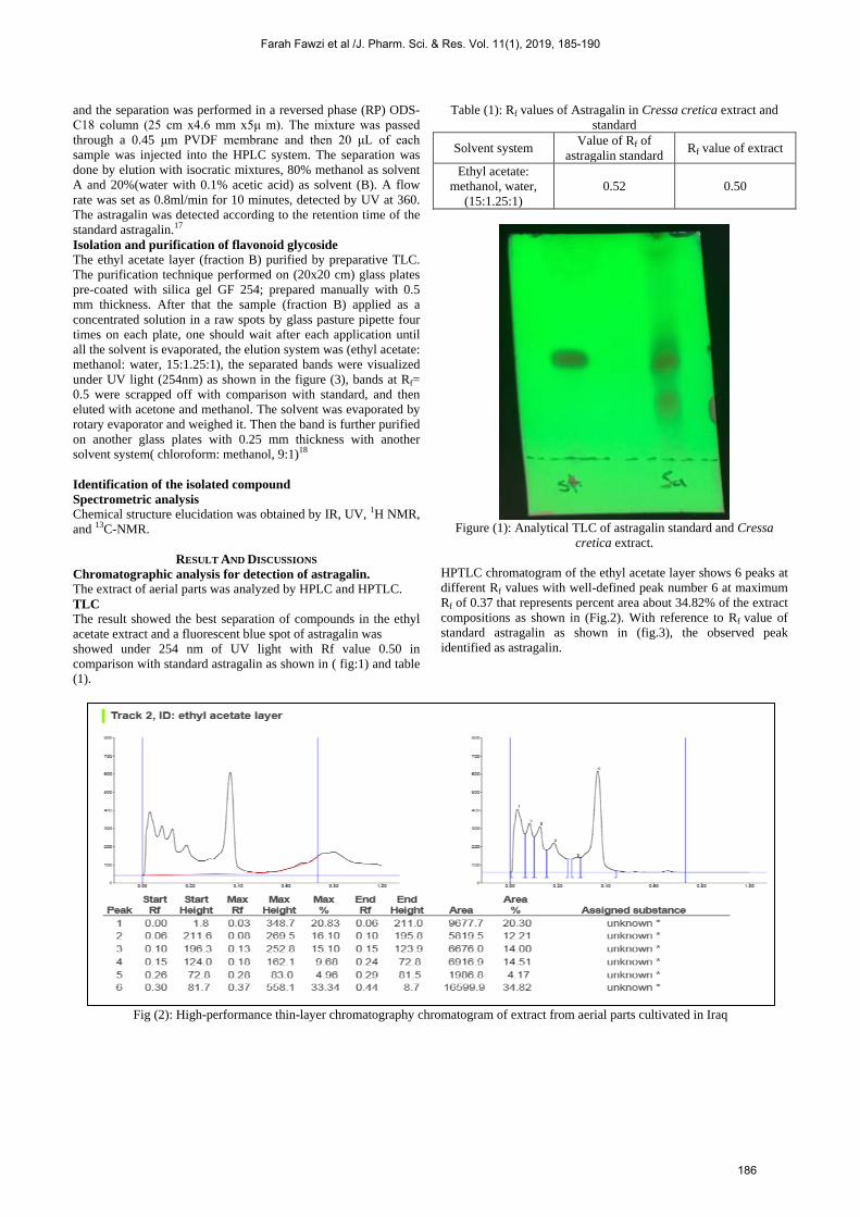
HPTLC chromatogram of the ethyl acetate layer shows 6 peaks at different Rf values with well-defined peak number 6 at maximum Rf of 0.37 that represents percent area about 34.82% of the extract compositions as shown in (Fig.2). With reference to Rf value of standard astragalin as shown in (fig.3), the observed peak identified as astragalin.
Fig (2): High-performance thin-layer chromatography chromatogram of extract from aerial parts cultivated in Iraq
Farah Fawzi et al /J. Pharm. Sci. & Res. Vol. 11(1), 2019, 185-190
186
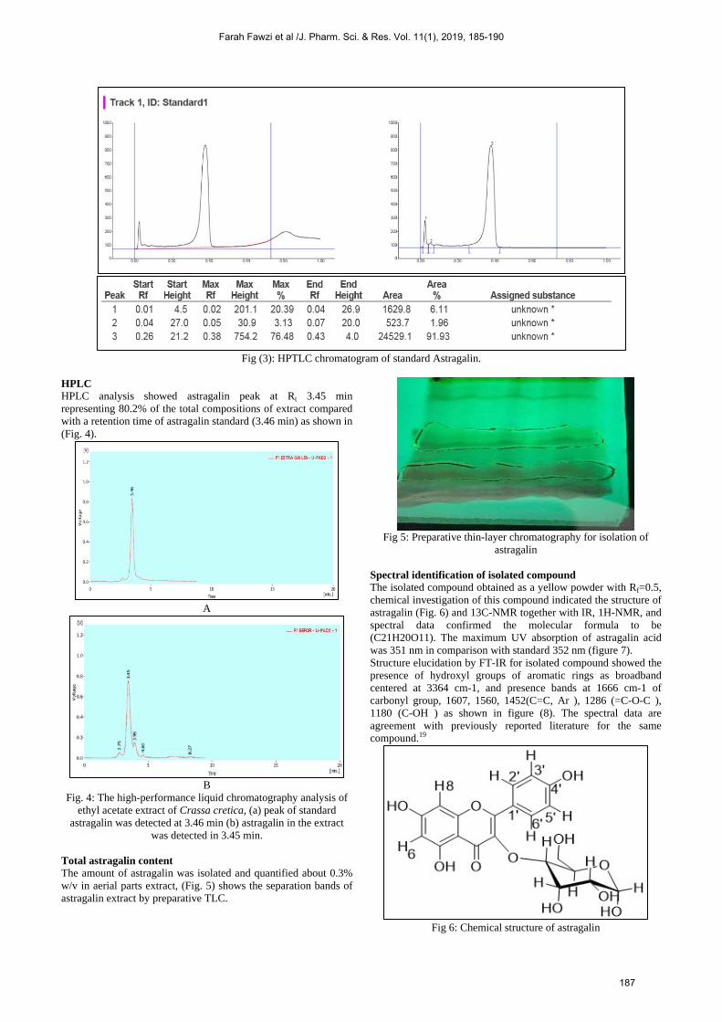
Fig (3): HPTLC chromatogram of standard Astragalin.
HPLC HPLC analysis showed astragalin peak at Rt 3.45 min representing 80.2% of the total compositions of extract compared with a retention time of astragalin standard (3.46 min) as shown in (Fig. 4).
A
B
Fig. 4: The high-performance liquid chromatography analysis of ethyl acetate extract of Crassa cretica, (a) peak of standard
astragalin was detected at 3.46 min (b) astragalin in the extract was detected in 3.45 min.
Total astragalin content The amount of astragalin was isolated and quantified about 0.3% w/v in aerial parts extract, (Fig. 5) shows the separation bands of astragalin extract by preparative TLC.
Fig 5: Preparative thin-layer chromatography for isolation of
astragalin Spectral identification of isolated compound The isolated compound obtained as a yellow powder with Rf=0.5, chemical investigation of this compound indicated the structure of astragalin (Fig. 6) and 13C-NMR together with IR, 1H-NMR, and spectral data confirmed the molecular formula to be (C21H20O11). The maximum UV absorption of astragalin acid was 351 nm in comparison with standard 352 nm (figure 7). Structure elucidation by FT-IR for isolated compound showed the presence of hydroxyl groups of aromatic rings as broadband centered at 3364 cm-1, and presence bands at 1666 cm-1 of carbonyl group, 1607, 1560, 1452(C=C, Ar ), 1286 (=C-O-C ), 1180 (C-OH ) as shown in figure (8). The spectral data are agreement with previously reported literature for the same compound.19
Fig 6: Chemical structure of astragalin
Farah Fawzi et al /J. Pharm. Sci. & Res. Vol. 11(1), 2019, 185-190
187
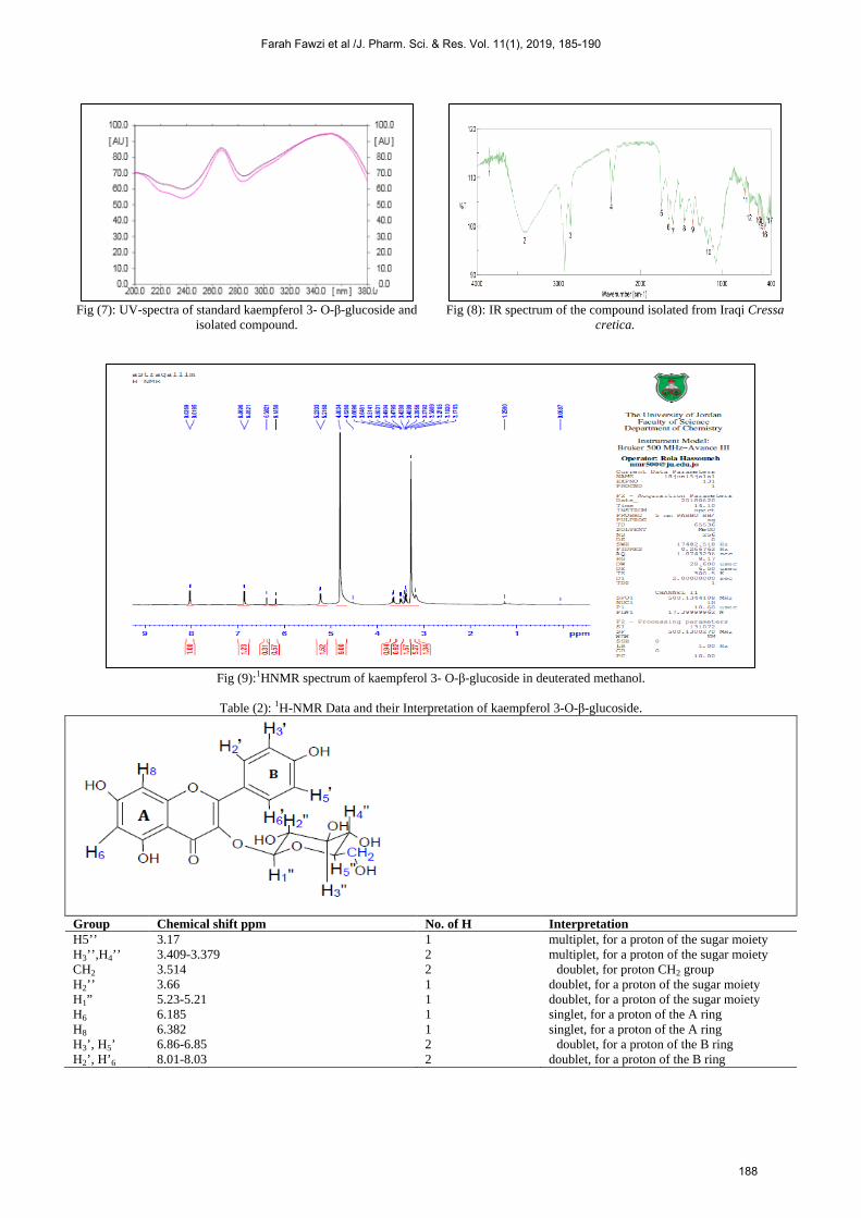
Fig (7): UV-spectra of standard kaempferol 3- O-β-glucoside and
isolated compound.
Fig (8): IR spectrum of the compound isolated from Iraqi Cressa
cretica.
Fig (9):1HNMR spectrum of kaempferol 3- O-β-glucoside in deuterated methanol.
Table (2): 1H-NMR Data and their Interpretation of kaempferol 3-O-β-glucoside.
Group Chemical shift ppm No. of H Interpretation H5’’ 3.17 1 multiplet, for a proton of the sugar moiety H3’’,H4’’ 3.409-3.379 2 multiplet, for a proton of the sugar moiety CH2 3.514 2 doublet, for proton CH2 group
H2’’ 3.66 1 doublet, for a proton of the sugar moiety H1” 5.23-5.21 1 doublet, for a proton of the sugar moiety H6 6.185 1 singlet, for a proton of the A ring H8 6.382 1 singlet, for a proton of the A ring H3’, H5’ 6.86-6.85 2 doublet, for a proton of the B ring
H2’, H’6 8.01-8.03 2 doublet, for a proton of the B ring
Farah Fawzi et al /J. Pharm. Sci. & Res. Vol. 11(1), 2019, 185-190
188

Fig (10):13CNMR spectrum of kaempferol 3- O-β-glucoside in deuterated methanol.
The 1H-NMR spectrum of the isolated compound indicated that B ring protons, H2' and H6' signals were given doublet at 8.01- 8.03 ppm. H3' and H5' proton signals were observed at 6.86-6.85 ppm as a doublet.H8 and H6 protons were at 6.382 and 6.185 ppm, respectively as a singlet. Based on these findings and by comparison of NMR data, the aglycone identified as kaempferol. The aromatic proton (H-1") of the hexose appeared as a doublet at 523ppm. H2” and CH2 appeared as a doublet at 3.669 ppm and 3.514 ppm respectively, H3”, H4”gave signal at 3.409-3.379 ppm as multiplet and H5” appeared as a multiplet at 3.175 ppm.20 Therefore the isolated compound was assumed to be kaempferol 3-O-β- glucoside (Astragalin). as shown in (fig:9) & (table 2)
13C-NMR shows characteristic peaks in which broadband decoupling present 19 different signals, with highest chemical shift for (C=O) at 178.15 and C=C attached to (OH) groups at range 173.42-160.9, other C=C signals at range 157.69-102.67, while those carbons attach to (OH) group at range 76.65-61.23ppm. The peaks assigned in 13C NMR corresponded to the Ref.21 as shown in (fig.10).
CONCLUSION
The quantity of Kaempferol 3- O- β- glucoside (astragalin) is (0.3% W/W) present in the Iraqi plant as a major glycoside. Astragalin was extracted from Cressa cretica under optimal conditions (80% aqueous ethanol and 80°c for 3 hrs.). We suggest that these isolated compounds are a suitable candidate for further clinical and pharmacological study Acknowledgments Sincere gratitude to the department of pharmacognocy and medicinal, college of pharmacy, Mustansyria University, for providing the laboratory and experimenter facilities for performing this work.
REFERENCES 1. Chan K. Some aspects of toxic contaminants in herbal medicines.
Chemosphere. 2003;52(9):1361-1371. doi:10.1016/S0045-6535(03)00471-5
2. Myers N, Mittermeier RA, Mittermeier CG, Fonseca da GAB, Kent J. Biodiversity hotspots for conservation priorities. Nature.
2000;403:853-858. 3. Priyashree S, Jha S, Pattanayak SP. A review on Cressa cretica
Linn.: A halophytic plant. Pharmacogn Rev. 2010;4(8):161-166. doi:10.4103/0973-7847.70910
4. Prajapati ND, Purohit SS, Sharma AK KT. A handbook of medicinal plants, a complete source book Agrobios. India: Eastern Book Corporation. 2004:173.
5. Khare P, Chaudhary S, Kumar A, Yadav G, Thakur N. A study on the standardization parameters of a halophytic plant (Cressa cretica L.). Middle-East J Sci Res. 2013;15(10):1472-1477.
6. H VM, D GN, L NS, et al. Pharmacognostic profile and antimicrobial potential of fruits of Cressa cretica L. Int J Phytopharm Res. 2012;3(2):72-75.
7. Chaudhary Sudhir, R.L Khosa, Priyank RS. A report on Pharmacognostical and quality control parameters of stem and root of Cressa cretica Linn, Convolvulaceae. J Pharm Res. 2012;5(1):616-621.
8. Chaudhary S, Khosa RL, Jha KK, Verma N. Evaluation of antidiabetic activity of Cressa cretica linn in alloxan induced diabetes in rats. Pharmacologyonline. 2010;3:181-188.
9. Gupta RS, Kachhawa JBS, Khushalani V, Tanwar K, Joshi YC. Effect of Cressa cretica methanol extract on testicular function of albino rats. Pharm Biol. 2006;44(5):382-388.
10. Winkel-shirley B. Flavonoid Biosynthesis . A Colorful Model for Genetics , Biochemistry , Cell Biology , and Biotechnology 1. Plant Physiol. 2001;126(2):485-493.
11. Wei M, Mahady GB, Liu D, Zheng ZS, Lu Y. Astragalin, a flavonoid from Morus alba (mulberry) increases endogenous estrogen and progesterone by inhibiting ovarian granulosa cell apoptosis in an aged rat model of menopause. Molecules. 2016;21(5). doi:10.3390/molecules21050675
12. Parveen Z, Deng Y, Saeed MK, et al. Optimizations of conditions for maximum recovery of astragalin from Thesium chinense Turcz. J Appl Sci. 2006;6(13):2829-2832.
13. Shahat AA, Abdel-Azim NS, Pieters L, Vlietinck AJ. Flavonoids from Cressa cretica. Pharm Biol. 2004;42(4-5):349-352.
14. Kumar S, Pandey AK. Chemistry and biological activities of flavonoids: An overview. Sci World J. 2013;2013.
15. Mutlag SH, Hamad MN, Abbas IS, Ismael SH. The Evaluation of Ethyl Acetate Fraction of Cressa cretica Effect on Mitotic Index and Micronucleous Frequency in Mice 1. Int J Pharm Sci Rev Res. 2017;45(28):147-150.
16. Ahmed FA, Abd El-Wahab Khamis IM, Desoukey SY. Flavonoids of Neotorularia aculeolata plant. J Pharm Nutr Sci. 2011;1(2):134-139. doi:10.6000/1927-5951.2011.01.02.08
Farah Fawzi et al /J. Pharm. Sci. & Res. Vol. 11(1), 2019, 185-190
189
17. Gupta M, Sasmal S, Majumdar S, Mukherjee A. HPLC Profiles ofStandard Phenolic Compounds Present in Medicinal Plants. Vol 4.;2012.
18. Thorburn Burns D. Plant Drug Analysis: A Thin LayerChromatography Atlas. Anal Chim Acta. 1984:126-142.
19. Saito S, Silva G, Santos RX, Gosmann G, Pungartnik C, Brendel M.Astragalin from cassia alata induces DNA adducts in vitro andrepairable DNA damage in the yeast saccharomyces cerevisiae. Int JMol Sci. 2012;13(3):2846-2862. doi:10.3390/ijms13032846
20. Deng S, Deng Z, Fan Y, et al. Isolation and purification of three
flavonoid glycosides from the leaves of Nelumbo nucifera (Lotus) by high-speed counter-current chromatography. J Chromatogr B Anal Technol Biomed Life Sci. 2009;877(24):2487-2492. doi:10.1016/j.jchromb.2009.06.026
21. Wei Y, Xie Q, Fisher D, Sutherland IA. Separation of patuletin-3-o-glucoside, astragalin, quercetin, kaempferol and isorhamnetin fromflaveria bidentis (L.) kuntze by elution-pump-out high-performancecounter-current chromatography. J Chromatogr A.2011;1218(36):6206-6211. doi:10.1016/j.chroma.2011.01.058
Farah Fawzi et al /J. Pharm. Sci. & Res. Vol. 11(1), 2019, 185-190
190