isolation characterizationof human muscle cells - pnas · mostbiochemical studies ofhumanmuscle in...
TRANSCRIPT

Proc. NatL Acad. Sci. USAVol. 78, No. 9, pp. 5623-5627, September 1981Cell Biology
Isolation and characterization of human muscle cells(differentiation/contractile protein synthesis)
HELEN M. BLAU AND CECELIA WEBSTERDepartment of Pharmacology, Stanford University School of Medicine, Stanford, California 94305
Communicated by Robert T. Schimke, May 11, 1981
ABSTRACT We have developed an in vitro system for thestudy of postnatal human muscle under standardized conditions.The technique utilizes cloning to isolate pure populations ofmusclecells. By manipulating culture conditions we can maximize eitherproliferation or differentiation of individual clones or of clonespooled to yield mass cultures of muscle cells. The muscle pheno-type is stable; cells can be stored in liquid nitrogen for long-termuse without loss of proliferative or differentiative potential. Wehave determined proliferative capacity of muscle cells from ananalysis of clonal growth kinetics; we determined differentiativecapacity from morphological evidence (cell fusion, striations, con-tractions, and the appearance of acetylcholine receptors) and bio-chemical analysis of muscle protein synthesis (creatine kinase, a-actin, tropomyosin, and myosin light chains). Our approach elim-inates the variability in cellular composition that has complicatedstudies of primary muscle to date. We can now study in a con-trolled fashion the interactions and contributions of different celltypes to the development of normal and genetically dystrophichuman muscle.
Most biochemical studies of human muscle in vitro have usedeither explants in organ culture or dissociated monolayers ofprimary cells. With both techniques, muscle cells are inevitablycontaminated by diverse cell types including nerve, adipocytes,and fibroblasts (see refs. 1 and 2 for reviews). It is well knownthat the ratio of muscle to nonmuscle cells influences the be-havior ofthe muscle cells present (3) and varies in disease states(ref. 4; unpublished data). The proportion of muscle cells canbe increased to 90% ofthe total cell population by preincubationon substrates not coated with collagen (5). Nonetheless, quan-titative experiments using such mixed cultures remain ex-tremely difficult. Unlike rat and mouse for which several celllines exist (3-6), there are no established human muscle lines.As a result, previous studies of pure muscle cell populationsfrom humans have utilized individual clones derived from singlecells to analyze clonal growth kinetics and morphology (4, 7-9).Because attempts to produce differentiation of these clonedmuscle cells in mass cultures have met with little success (7, 10),biochemical studies of the differentiation process have beenlimited.To date, the majority of studies of human muscle in tissue
culture have used embryonic muscle, a source with distinctdisadvantages. First, because definitive diagnosis ofgeneticallydetermined muscular dystrophies is only possible postnatally,cells from fetuses have the uncertainty of, at best, a 50% riskfor a disease. Second, cells isolated from the muscle of individ-uals of different ages have been shown to differ in the differ-entiative properties they express when cultured in vitro (11).Consequently, muscle from embryonic sources may not displaythe characteristic pathological abnormalities of the dystrophyin culture. This influence of developmental stage on cellular
differentiative potential in culture is an important considerationin the analysis of normal and dystrophic myogenesis.
In this report, we describe the isolation, growth, and expan-sion of clones ofhuman muscle cells of postnatal origin and theconditions that maximize the proliferative or differentiative ca-pacity of these muscle cells, either as individual clones or aspooled clones or mass cultures. The pure populations of my-ogenic cells can be analyzed separately or in cell mixtures ofknown composition in investigations ofinduction during muscledevelopment, of cell-cell interactions at the neuromuscularjunction, and of the etiology of human muscular dystrophies.
MATERIALS AND METHODSSource of Human Muscle. Muscle samples were obtained
from 17 normal patients during surgical treatment for or-thopedic nonmuscle problems in accordance with the guide-lines ofthe Human Subjects Committee ofStanford University.
Cell Culture Conditions. Growth medium (GM) containedHam's nutrient mixture F-10 with 0.5% chicken embryo extractand either 20% (vol/vol) fetal calfserum (GM-1) or horse serum(GM-2). Fusion medium (FM) contained Dulbecco's modifiedEagle's medium and 2% (vol/vol) horse serum. Conditionedmedium (CM) was GM-2 exposed to confluent human muscle-derived fibroblast cultures for 24 hr. filtered through a 0.2-pymNalgene filter, and diluted 1:1 with fresh GM-2. CM can bestored at 40C for 2 weeks or at -70'C for 6 months. All cultureswere grown in the presence of penicillin G (200 units/ml) andstreptomycin sulfate (200 pug/ml) on a collagen substrate (calfskin collagen, Calbiochem; 1.4 mg/ml in distilled water, au-toclaved). All cells were grown at 37°C in a humidified Formaincubator containing 5% CO2 and 95% air. Cells were storedfrozen in 10% dimethyl sulfoxide (Mallinkrodt) in horse or fetalcalf serum.
F-10 and Dulbecco's modified Eagle's media, chicken em-bryo extract, and fetal calf serum were obtained from GIBCO,and horse serum was from Kansas City Biologicals. Horseserum lots were pretested for those that would best supportfusion. Tissue culture dishes, flasks, and multiwells were fromFalcon Plastics, and penicillin and streptomycin were fromGIBCO.
Assays for Muscle Gene Expression. Creatine kinase (CPK)activity was assayed as described (12). Synthesis of a-actin wasdetermined by a modification ofthe method ofBlau and Epstein(12). Cells were labeled for 3 hr in methionine-free mediumsupplemented with [3S]methionine (250 4Ci/ml; 1102 Ci/mmol; Amersham) and then harvested; 0.60-2.7 X 10r cpm in35 1Lg of protein was layered in 20 p.1 on each gel. y-, f3-, anda-actin species were resolved by fractionating labeled proteinson isoelectric focusing polyacrylamide gels, followed by auto-radiography. Relative rates of actin synthesis were quantitated
Abbreviations: CPK, creatine kinase; mU, milliunits; GM, growth me-
dium; FM, fusion medium; CM, conditioned medium.
The publication costs ofthis article were defrayed in part by page chargepayment. This article must therefore be hereby marked "advertise-ment" in accordance with 18 U. S. C. §1734 solely to indicate this fact.
5623

5624 Cell Biology: Blau and Webster
by scanning autoradiograms with a densitometer (E. C. Appa-ratus, St. Petersburg, FL) and cutting out and weighing the areaunder the relevant peaks. Other major contractile proteins wereidentified on two-dimensional gels (13) by comigration of la-beled proteins with human contractile proteins purified directlyfrom adult human muscle (14).
The distribution of acetylcholine receptors was assayed byautoradiographic analysis of the binding of '"I-labeled-a-bun-garotoxin to intact myotubes in culture (15, 16). Cells were la-beled for 45 min in FM with '"I-labeled-a-bungarotoxin (spe-cific activity, 95-140 Ci/mmol; New England Nuclear) at 50nM, a concentration determined to be sufficient to saturate thereceptors. Specificity of binding was controlled by preincubat-ing replicate dishes of cells at each time point with unlabeleda-bungarotoxin (5 juM) for 20 min prior to labeling. For assaysof total receptor, cells were solubilized in 1 M NaOH and as-sayed in a Micromedic 4/600 gamma counter with 75% effi-ciency. Total protein was determined by the method of Lowryet al. (17).
Isolation and Selection of Muscle Cells from Adult Tissues.A relatively small number of cells positioned between the base-ment membrane and the sarcoplasmic reticulum ofadult musclefibers are myoblasts capable of proliferation (18). After tissuedissociation, it is these satellite cells that give rise to clones inculture. Postnatal muscle samples can be stored at 40C in F-10medium for up to 24 hr prior to dissociation without adverseeffects on the yield and viability of satellite cells. For dissocia-tion, a 0.1- to 0.3-cm3 piece of skeletal muscle tissue in F-10medium at 40C is carefully dissected to remove as much con-nective tissue as possible and minced to obtain fragmentssmaller than 1 mm . To remove residual debris, the fragmentsare washed with F-10 three times at 40C and once at 370C. Thetissue is then dissociated for a total of40-60 min by two or threesuccessive treatments with- 25 ml of 0.05% trypsin-EDTA(GIBCO) at 370C in a Wheaton graduated trypsinization flaskwith constant stirring. The cells collected in the supernatantafter each trypsin treatment are pooled and cooled to 4°C onice. Horse serum is added to a final concentration of 10% (vol/vol) to terminate further protease activity. The dissociated cellsare then centrifuged (2 min; 25°C); the cell pellet is resuspendedin CM and either plated in culture or frozen in liquid nitrogenat a density of 0.1 cm3 of tissue per ml for future use.
Enrichment ofthe cell population for muscle is accomplishedby preplating the cells at 37C for 20 min on a non-collagen-coated dish, a substrate to which fibroblasts preferentially ad-here (5). Because cell counts and efficiency of plating cannotbe accurately determined for human postnatal muscle cells dueto debris resulting from myotube destruction during tissue dis-sociation, unattached cells are plated at a range of densities ap-proximated from the expected satellite cell number, 107/cm3ofhuman muscle (19). Those yielding between 5 and 50 clonesper 60-mm collagen-coated plate are used. Cultures are main-tained in 2-3 ml of CM; they are not fed for the first 3-6 daysand are fed only every 4 days thereafter. For future use, clonedcells can be frozen at a density of 5 x 10⁶/ml. The efficacy ofthe isolation and selection procedure is demonstrated by thefact that 96-100% of 828 clones from four different sampleswere myogenic (see Table 1).
RESULTS
Clonal Analysis: Generation of Pure Muscle Cell Popula-tions. A major problem in the study of postnatal human cellsin culture is their limited longevity-approximately 45 dou-blings compared to 60-70 for muscle cells from 80-day fetuses(4). Our approach works within the time frame imposed by se-
nescence. Clones containing 1000-2000 cells are harvestedprior to fusion; groups of three are pooled, grown to 60-80%confluence in GM-1 in order to prevent initiation of myoge-nesis, and then frozen for long-term storage at a density of 5X 10⁶ cells per ml. Simultaneously, individual clones are testedseparately for their myogenic potential by plating a few cells ofeach in 16-mm tissue culture wells and scoring these for fusion.This approach ensures that the frozen cells are homogeneouslymuscle, have not initiated differentiation, and spend a mini-mum of time in culture prior to use.
Potential Yield of Muscle Cells per Biopsy. The yield ofmuscle cells from a small biopsy is sufficient for many kinds ofbiochemical and morphological analyses. From a 0.1-cm3 pieceof tissue, of which 50% is connective tissue and fat, 5 x 10³viable, proliferative satellite cells can be obtained. In our ex-perience, each satellite cell is capable of giving rise to at least1 X 107 cells, equivalent to one confluent T-75 flask or ap-proximately 2 mg of protein.
Proliferative Capacity of Frozen Stored Muscle. The pro-liferative capacity of frozen cells was compared with that offresh cells. Clonal growth kinetics were determined by ran-domly selecting and circling clones and then visually countingthe number of cells in each on subsequent days by using an in-verted microscope with phase-contrast optics. Although therange in cell number per clone on day 4 suggests a high degreeofheterogeneity, the growth curves are remarkably similar (Fig.1). The apparent heterogeneity simply reflects the time in cul-ture required for individual cells to become established andbegin proliferation. For example, the clones indicated by A and4 have lag times of2 and 4 days, respectively, but similar growthkinetics.
Table 1 shows data for the growth properties of clones offreshly dissociated and frozen-thawed cells derived from fourmuscle biopsy specimens. The ranges in doubling times (12-20hr) and in the lag times prior to the initiation of proliferation(2-4 days) suggest that there is variability among clones of thesame sample, of different anatomic muscles, and of differentindividuals with respect to these growth values. However, ofimportance is the observation that, on average, fresh and frozencells behave similarly. In addition, cells frozen at the same den-sity (0.1 cm3 of dissociated tissue per ml) but in small volumes(0.2 ml), are not adversely affected with respect to either theirgrowth or their differentiative properties and exhibit only aslight decrease in total cell yield (data not shown). These resultsdemonstrate that dissociated cells can be frozen in numerousaliquots for use at different times.
a 1000 b7 ~~~~~~500-
6 /
6 5 - 416 0 2Oi4 ~~~50-3
/
0 I~~~~~~~~1
1~ ~~~~~~~1
14] A~~~~~~~~~nm
0 24 6 810121416 0 2 4 6 8
Cells/clone Time in culture, days
FIG. 1. Kinetics of growth of clones of fresh cells. The number ofcells in 20 individual clones was counted on day4 (a) and daily there-after (b); growth curves for six representatives are shown. The cloneswith the least and most cells are indicatd by 4 and A, respectively.
Proc. Nad Acad. Sci. USA 78 (1981)
Proc. NatL Acad. Sci. USA 78 (1981) 5625
Table 1. Clonal growth and differentiation of fresh andfrozen-thawed cellsMuscle Aliquot Doubling Lag Fusion,sample* size, mlt time, hrt time, hrt % offFresh:
1 - 20.7 ±0.8 42.2± 4.7 -2 - 16.8 ± 0.5 89.2 ± 2.6 97 (151)3 - 14.3 ± 0.6 55.0 ± 3.6 98(98)4 - 13.5 ± 0.4 69.0 ± 4.4 100 (107)
Mean 16.3 ± 1.6 63.9 ± 15.8 98 ± 1Frozen:
2 1.0 14.4 ± 0.8 62.9 ± 0.8 100 (50)2 0.2 14.9 ± 0.7 70.9 ± 3.9 96 (53)3 1.0 12.8 ± 0.4 72.4 3.8 100 (156)3 0.5 13.1 ± 0.4 70.8 ± 4.6 99 (121)3 0.2 12.6 ± 0.7 61.8 ± 3.8 99 (92)
Mean 13.6 ± 0.5 67.8 2.2 99 ± 1
* Each sample was from a separate muscle biopsy. Growth kinetics forindividual clones of samples 3 and 4, two biopsies from the same in-dividual, are shown in Fig. 1. Sample 2 was from the biceps femoris;all other samples were from the vastus lateralis.
t Cells were frozen at the same density (0.1 cm3 of dissociated tissueper ml) but in different volumes (0.2, 0.5, 1.0 ml).
t Cell doubling and lag times prior to initiation of proliferation areexpressed as mean ± SEM for at least 10 clones in each case.
§ The percentage fusion is the proportion of total colonies that differ-entiated as muscle. The number of clones scored is in parentheses.
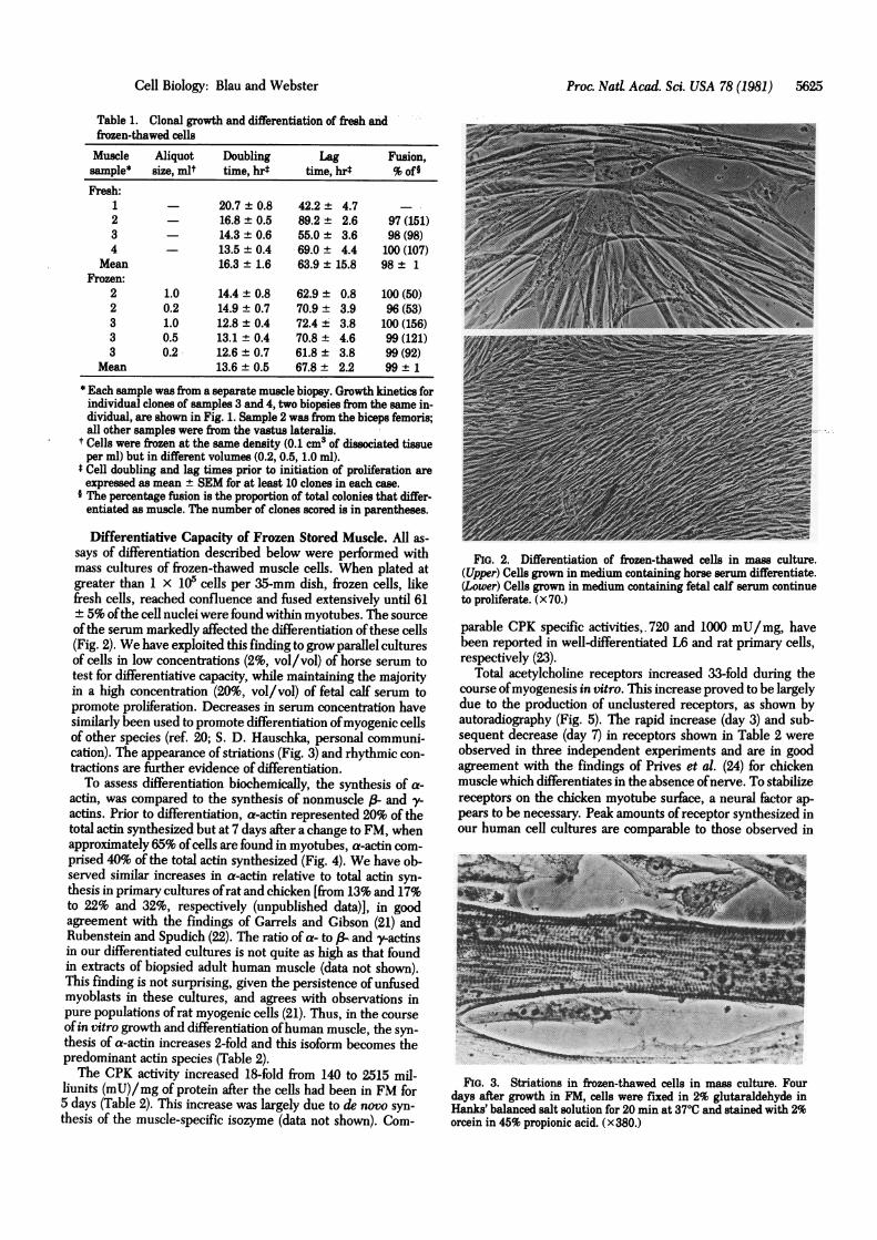
Differentiative Capacity of Frozen Stored Muscle. All as-says of differentiation described below were performed withmass cultures of frozen-thawed muscle cells. When plated atgreater than 1 x 10⁵ cells per 35-mm dish, frozen cells, likefresh cells, reached confluence and fused extensively until 61± 5% ofthe cell nuclei were found within myotubes. The sourceof the serum markedly affected the differentiation ofthese cells(Fig. 2). We have exploited this finding to grow parallel culturesof cells in low concentrations (2%, vol/vol) of horse serum totest for differentiative capacity, while maintaining the majorityin a high concentration (20%, vol/vol) of fetal calf serum topromote proliferation. Decreases in serum concentration havesimilarly been used to promote differentiation ofmyogenic cellsof other species (ref. 20; S. D. Hauschka, personal communi-cation). The appearance of striations (Fig. 3) and rhythmic con-tractions are further evidence of differentiation.To assess differentiation biochemically, the synthesis of a-
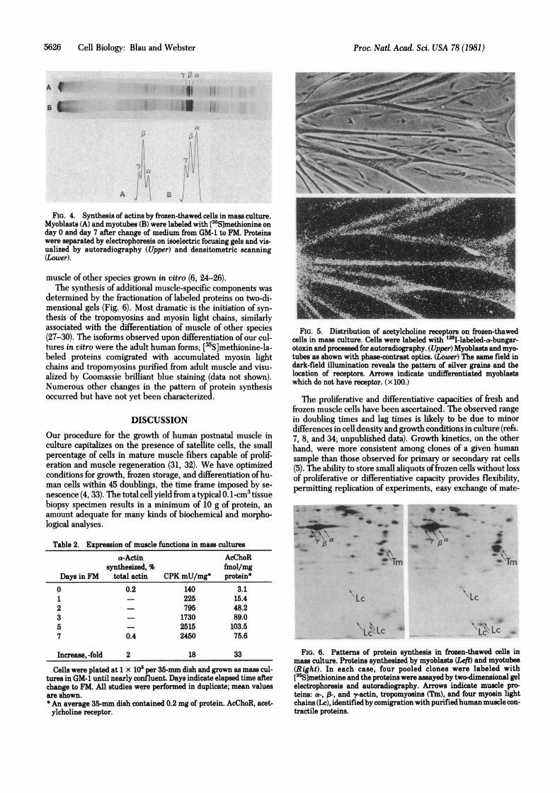
actin, was compared to the synthesis of nonmuscle (& and y-actins. Prior to differentiation, a-actin represented 20% of thetotal actin synthesized but at 7 days after a change to FM, whenapproximately 65% ofcells are found in myotubes, a-actin com-prised 40% of the total actin synthesized (Fig. 4). We have ob-served similar increases in a-actin relative to total actin syn-thesis in primary cultures ofrat and chicken [from 13% and 17%to 22% and 32%, respectively (unpublished data)], in goodagreement with the findings of Garrels and Gibson (21) andRubenstein and Spudich (22). The ratio of a- to (3 and y-actinsin our differentiated cultures is not quite as high as that foundin extracts of biopsied adult human muscle (data not shown).This finding is not surprising, given the persistence of unfusedmyoblasts in these cultures, and agrees with observations inpure populations of rat myogenic cells (21). Thus, in the courseofin vitro growth and differentiation ofhuman muscle, the syn-thesis of a-actin increases 2-fold and this isoform becomes thepredominant actin species (Table 2).The CPK activity increased 18-fold from 140 to 2515 mil-
liunits (mU)/mg of protein after the cells had been in FM for5 days (Table 2). This increase was largely due to de novo syn-thesis of the muscle-specific isozyme (data not shown). Com-
FIG. 2. Differentiation of frozen-thawed cells in mass culture.(Upper) Cells grown in medium containing horse serum differentiate.(Lower) Cells grown in medium containing fetal calf serum continueto proliferate. (x70.)
parable CPK specific activities, 720 and 1000 mU/mg, havebeen reported in well-differentiated L6 and rat primary cells,respectively (23).
Total acetylcholine receptors increased 33-fold during thecourse ofmyogenesis in vitro. This increase proved to be largelydue to the production of unclustered receptors, as shown byautoradiography (Fig. 5). The rapid increase (day 3) and sub-sequent decrease (day 7) in receptors shown in Table 2 wereobserved in three independent experiments and are in goodagreement with the findings of Prives et al. (24) for chickenmuscle which differentiates in the absence of nerve. To stabilizereceptors on the chicken myotube surface, a neural factor ap-pears to be necessary. Peak amounts of receptor synthesized inour human cell cultures are comparable to those observed in
.---*-..................................,Fri
FIG. 3. Striations in frozen-thawed cells in mass culture. Fourdays after growth in FM, cells were fixed in 2% glutaraldehyde inHanks' balanced salt solution for 20 min at 37TC and stained with 2%orcein in 45% propionic acid. (x380.)
-. .-i
. 'I-3.0-1
Cell Biology: Blau and Webster
5626 Cell Biology: Blau and Webster
B iL
A
FIG. 4. Synthesis of actins by frozen-thawed cells in mass culture.Myoblasts (A) and myotubes (B) were labeled with [55S]methionine onday 0 and day 7 after change of medium from GM-1 to FM. Proteinswere separated by electrophoresis on isoelectric focusing gels and vis-ualized by autoradiography (Upper) and densitometric scanning(Lower).
muscle of other species grown in vitro (6, 24-26).The synthesis of additional muscle-specific components was
determined by the fractionation of labeled proteins on two-di-mensional gels (Fig. 6). Most dramatic is the initiation of syn-thesis of the tropomyosins and myosin light chains, similarlyassociated with the differentiation of muscle of other species(27-30). The isoforms observed upon differentiation of our cul-tures in vitro were the adult human forms; ['S]methionine-la-beled proteins comigrated with accumulated myosin lightchains and tropomyosins purified from adult muscle and visu-alized by Coomassie brilliant blue staining (data not shown).Numerous other changes in the pattern of protein synthesisoccurred but have not yet been characterized.
DISCUSSIONOur procedure for the growth of human postnatal muscle inculture capitalizes on the presence of satellite cells, the smallpercentage of cells in mature muscle fibers capable of prolif-eration and muscle regeneration (31, 32). We have optimizedconditions for growth, frozen storage, and differentiation ofhu-man cells within 45 doublings, the time frame imposed by se-nescence (4, 33). The total cell yield from a typical 0.1-cm3 tissuebiopsy specimen results in a minimum of 10 g of protein, anamount adequate for many kinds of biochemical and morpho-logical analyses.
Table 2. Expression of muscle functions in mass culturesa-Actin AcChoR
synthesized, % fmol/mgDays in FM total actin CPK mU/mg* protein*0 0.2 140 3.11 225 15.42 795 48.23 - 1730 89.05 2515 103.57 0.4 2450 75.6
Increase, -fold 2 18 33
Cells were plated at 1 x 10⁵ per 35-mm dish and grown as mass cul-tures in GM-1 until nearly confluent. Days indicate elapsed time afterchange to FM. All studies were performed in duplicate; mean valuesare shown.* An average 35-mm dish contained 0.2 mg of protein. AcChoR, acet-ylcholine receptor.
FIG. 5. Distribution of acetylcholine receptors on frozen-thawedcells in mass culture. Cells were labeled with "2'-labeled-a-bungar-otoxin and processed for autoradiography. (Upper) Myoblasts and myo-tubes as shown with phase-contrast optics. (Lbwer) The same field indark-field illumination reveals the pattern of silver grains and thelocation of receptors. Arrows indicate undifferentiated myoblastswhich do not have receptor. (x 100.)
The proliferative and differentiative capacities of fresh andfrozen muscle cells have been ascertained. The observed rangein doubling times and lag times is likely to be due to minordifferences in cell density and growth conditions in culture (refs.7, 8, and 34; unpublished data). Growth kinetics, on the otherhand, were more consistent among clones of a given humansample than those observed for primary or secondary rat cells(5). The ability to store small aliquots offrozen cells without lossof proliferative or differentiative capacity provides flexibility,permitting replication of experiments, easy exchange of mate-
?tTm
\Lc
A
*Tm
ALc
Lc\Lc C\\; Lc
FIG. 6. Patterns of protein synthesis in frozen-thawed cells inmass culture. Proteins synthesized by myoblasts (Left) and myotubes(Right). In each case, four pooled clones were labeled with[(5S]methionine and the proteins were assayed by two-dimensional gelelectrophoresis and autoradiography. Arrows indicate muscle pro-teins: a-, 3-, and y-actin, tropomyosins (Tm), and four myosin lightchains (Lc), identifiedby comigration with purifiedhuman muscle con-tractile proteins.
Proc. Natl. Acad. Sci. USA 78 (1981)

Cell Biology: Blau and Webster
rial among laboratories, and comparative studies of propertiesof normal and dystrophic muscle under identical conditions.
Although the specific reasons why our conditions are effec-tive remain unknown, it is clear that our procedure results ina degree of differentiation of mass cultures previously only ob-served in individual clones (7, 10). Our method differs fromothers in that F-10 and CM are used only to promote prolif-eration and not for differentiation. For optimal differentiation,we have defined a low nutrient medium with a high calciumconcentration which routinely results in the formation ofstriated, contractile myotubes at incubator CO2 levels of 5%.Nutrient deprivation appears to enhance fusion. When F-10 isreplaced by Dulbecco's modified Eagle's medium and theserum concentration is decreased from 20% to 2%, extensivedifferentiation occurs. In addition, the 6-fold higher calciumconcentration of the latter medium compared to F-10 may becritical. pH also affects differentiation. CO2 levels >5% are in-hibitory; again, the 3-fold higher bicarbonate concentration inDulbecco's modified Eagle's medium relative to F-10 may aidin maintaining a stable pH.
Under our conditions, human muscle, like muscle of otherspecies (6, 20-29), exhibited marked increases in the synthesisof a-actin, CPK, and acetylcholine receptor. In addition, syn-thesis ofthe myosin light chains and tropomyosin were initiatedin the course of myogenesis in vitro. It should be noted thatthese proteins, which are characteristic of differentiated mus-cle, are the adult human forms because they comigrate on gelswith contractile proteins purified directly from adult humanmuscle tissue. -In addition, the observed increase in CPK ac-tivity is due to the de novo appearance of the muscle-specificisozyme.A distinct advantage of our approach is that it uses postnatal
muscle, permitting application of the methods to dystrophicmuscle obtained from individuals with diagnosed genetic mus-cle disease rather than from fetuses at risk for a disorder. Post-natal satellite cells are also more likely to have matured suffi-ciently in vivo to express the relevant muscle properties in vitro(11).
In summary, we have determined the requirements for areproducible procedure for the isolation, growth, and differ-entiation of human muscle cell populations for in vitro study.These isolated human muscle cells will be invaluable to studiesof cell-cell interactions, permitting identification of functionsintrinsic to muscle and those induced or contributed by nerve,fibroblasts, or the extracellular matrix in the course of normaland genetically dystrophic human muscle development.
We are grateful to Dr. D. Yaffe and Ms. B. Dieckmann for encour-agement and advice early in this work, to Drs. E. Bleck and L. Rinskyfor muscle samples, to Drs. S. Guttman, S. Packman, and P. Byers forcritical-reading of the manuscript, and to Ms. C. Spain for expert sec-retarial assistance. This work was supported by grants to H. B. from the
Proc. NatL Acad. Sci. USA 78 (1981) 5627
Muscular Dystrophy Association of America, National Institutes ofHealth Grant GM 26717, and Basil O'Connor Starter Grant 5-179 fromthe March of Dimes-National Foundation.
1. Rowland, L. P. (1976) Arch. Neurol. 33, 315-321.2. Rowland, .L. P. (1980) Muscle and Nerve 3, 3-20.3. Yaffe, D. & Saxel, 0. (1977) Nature (London) 270, 725-727.4. Hauschka, S. D., Linkhart, T. A., Clegg, C. & Merfill, G. (1979)
in Muscle Regeneration, ed. Mauro, A. (Raven, New York), pp.311-322.
5. Richler, C. & Yaffe, D. (1970) Dev. BioL 23, 1-22.6. Noble, M. D., Brown, T. H. & Peacock, J. H. (1978) Proc. Nati
Acad. Sci. USA 75, 3488-3492.7. Hauschka, S. D. (1974) Dev. Biol 37, 329-344.8. Hauschka, S. D. (1974) Dev. Biot 37, 345-368.9. Hauschka, S. D., Hainey, C., Angello, J. C., Linkhart, T. A.,
Bonner, P. H. & White, N. K. (1977) in Pathogenesis ofHumanMuscular Dystrophies, International Congress Series, ed. Row-land, L. P. (Excerpta Medica, Amsterdam), pp. 834-855.
10. Holtzer, H., Jones, K. W. & Yaffe, D. (1975)J. Neurot Sci. 26,115-124.
11. Koenig, J. & Vigny, M. (1978) Nature (London) 271, 75-77.12. Blau, H. M. & Epstein, C. J. (1979) Cell 17, 95-108.13. O'Farrell, P. H. (1975) 1. BioL Chem. 250, 4007-4021.14. Kielley, W. W. & Harrington, W. F. (1960) Biochem. Biophys.
Acta 41, 401-421.15. Blau, H. M. & Kafatos, F. C. (1978)J. Cell Biol 78, 131-151.16. Teng, N. N. H. & Fiszman, M. Y. (1976)J. Supramot Struct. 4,
381-387.17. Lowry, 0. H., Rosebrough, N. J., Farr, A. L. & Randall, R. J.
(1951)J. Biot Chem. 193, 265-275.18. Mauro, A. (1961)J. Biophys. Biochem. Cytot 9, 493-495.19. Muir, A. R. (1970) in Regeneration of Striated Muscle and My-
ogenesis, International Congress Series, eds. Mauro, A., Shafiq,S. A. & Milhorat, A. T. (Excerpta Medica, Amsterdam), pp.91-100.
20. Yaffe, D. & Saxel, 0. (1977) Differentiation 7, 159-166.21. Garrels, J. I. & Gibson, W. (1976) Cell 9, 793-805.22. Rubenstein, P. A. & Spudich, J. A. (1977) Proc. NatL Acad. Sci.
USA 74, 120-123.23. Shainberg, A., Yagil, G. & Yaffe, D. (1971) Dev. Biol 25, 1-29.24. Prives, J., Silman, I. & Amsterdam, A. (1976) Cell 7, 543-550.25. Betz, H. & Changeux, J.-P. (1979) Nature (London) 278,
749-752.26. Libby, P., Bursztajn, S. & Goldberg, A. L. (1980) Cell 19,
481-491.-27. Garrels, J. I. (1979) Dev. BioL 73, 134-152.28. Devlin, R. B. & Emerson, C. P., Jr. (1978) Cell 13, 599-611.29. Devlin, R. B. & Emerson, C. P., Jr. (1979) Dev. BioL 69,
202-216.30. Stockdale, F. E., Raman, N. & Baden, H. (1981) Proc. Natl Acad.
Sci. USA 78, 931-935.31. Konigsberg, U. R., Lipton, B. H. &,Konigsberg, I. R. (1975) Dev.
Biol 45, 260-275.32. Bischoff, R. (1974) Anat. Rec. 180, 645-662.33. Hayflick, L. (1965) Exp. Cell Res. 37, 614-636.34. Konigsberg, I. R. & Hauschka, S. D. (1965) in Reproduction:
Molecular, Subcellular, and Cellular, ed. Locke, M. (Academic,New York), pp. 243-290.