isolation characterization cdna ysubunitof · nucleic acid sequence correspondingtothe carboxyl...
TRANSCRIPT

Proc. Natl. Acad. Sci. USAVol. 81, pp. 6948-6952, November 1984Biochemistry
Isolation and characterization of a cDNA clone for the y subunit ofbovine retinal transducin
(photoreceptor biochemistry/ras/signal transduction/molecular evolution)
JAMES B. HURLEY, HENRY K. W. FONG, DAVID B. TEPLOW, WILLIAM J. DREYER, AND MELVIN I. SIMON
Division of Biology, California Institute of Technology, Pasadena, CA 91125
Communicated by Lubert Stryer, July 23, 1984
ABSTRACT We have isolated and characterized a cDNAclone that encodes the y subunit of transducin, the guaninenucleotide binding regulatory protein found in vertebrate pho-toreceptors. The y subunit was separated from the a and Psubunits of transducin and purified to homogeneity by re-versed-phase high performance liquid chromatography. Thesequence of the first 45 amino acids at the amino terminus ofthis polypeptide was then determined by automated Edmandegradation. Oligodeoxynucleotide probes corresponding totwo nonoverlapping regions of this sequence were synthesizedand then used to screen a bovine retinal cDNA library. Oneprobe, T1.1, was a mixture of 32 different heptadecamers com-plementary to all possible mRNA sequences that could encodea portion of the T., sequence; the other probe, Ty2, was a mix-ture of 128 different heptadecamers. Thirteen clones that hy-bridized with T.1 were selected. Only one of these had an in-sert that also hybridized with T.2. The DNA sequence of thisinsert encodes a 73-amino acid polypeptide that corresponds tothe transducin y subunit on the basis of amino-terminal se-quence, amino acid composition, and carboxyl-terminal se-quence. The molecular weight of the mature y subunit is 8400.It appears to be synthesized as a discrete polypeptide and notas a domain of a larger precursor polyprotein. The transduciny subunit is very hydrophilic and acidic; it has 19 acidic and 11basic amino acids as well as three cysteine residues. Further-more, significant homology was found in comparisons of thenucleic acid sequence corresponding to the carboxyl terminusof the y transducin transcript with the sequences correspond-ing to the carboxyl terminus of ras oncogene products, suggest-ing a possible ancestral relationship between these genes.
Photolysis of rhodopsin triggers an enzymatic cascade invertebrate photoreceptors that results in the rapid hydrolysisof cyclic GMP (see ref. 1 for a review). This process is medi-ated through transducin (T), a guanine nucleotide bindingprotein found specifically in rod outer segments. Photolyzedrhodopsin stimulates hundreds of molecules of transducin tobind GTP, after which each transducin-GTP complex acti-vates a cyclic GMP phosphodiesterase. The resulting hydrol-ysis of cyclic GMP appears to be involved in visual transduc-tion.
Transducin is a member of a family of membrane-associat-ed guanine nucleotide binding proteins referred to as G pro-teins (2-4). G proteins all act through a common mechanism;they bind GTP in response to stimulation by specific recep-tor proteins and then regulate the activity of an enzyme.Transducin is stimulated by photolyzed rhodopsin and acti-vates a phosphodiesterase, whereas other G proteins inter-act with a variety of hormone or neurotransmitter receptorsand either activate or inhibit adenylate cyclase (5). G pro-teins that stimulate adenylate cyclase are referred to as Gs
(6), whereas those that inhibit adenylate cyclase are calledGI (7).
Transducin, like all of the well-characterized G proteins, ismade up of three polypeptide subunits (8-10). Ta has an ap-parent molecular weight (Mr) of 39,000 and contains sites forguanine nucleotide binding (9) and for ADP-ribosylation cat-alyzed by either cholera toxin (3) or pertussis toxin (11, 12).To and Ty are polypeptides of Mr 36,000 and 6,000-10,000,respectively. Gs, GI, and transducin each have unique a sub-units (ref. 4; unpublished data), whereas the 83 subunits of allthe G proteins that have been examined are nearly identical(4).
In the inactive state, transducin is a complex of Taff, To,and Ty (1). The presence of the P3 and y subunits is absolutelyrequired for photolyzed rhodopsin to stimulate Ta to bindGTP (13). When Ta binds GTP, it dissociates from the com-plex of To and Ty subunits (Tg,9 ) and activates the phospho-diesterase (9) by relieving the inhibitory constraints imposedon the phosphodiesterase by its own inhibitory subunit (14).We have recently begun to isolate cDNA clones for the
subunits of bovine transducin in order to determine their pri-mary structures and the genetic relationship between trans-ducin and other members of the G protein family. The ap-proach we have used is to first purify the individual transdu-cin subunits and determine part of their amino acidsequence. Oligodeoxynucleotide probes based on these se-quences have been synthesized and used to select phageclones from a bovine retinal cDNA library. The DNA se-quences of the isolated clones have then been determinedand compared to the respective amino acid sequences.
This report describes the isolation and characterization ofa cDNA clone for the y subunit of transducin. The isolationof cDNA clones for the y subunit has been more straightfor-ward than for the other subunits because the amino acid se-quence of 'y can be determined directly by Edman degrada-tion, whereas the a and /3 subunits have modified amino ter-mini and cannot be sequenced directly.* We have recentlydetermined partial amino acid sequences from proteolyticfragments of Ta and To (unpublished data), and this informa-tion can be used to isolate cDNA clones corresponding tothese subunits.
MATERIALS AND METHODSMaterials. Bovine retinas were obtained from Hormel
(Austin, MN). Guanosine 5'-[,3,y]triphosphate (p[NH]ppG)was purchased from Sigma. Restriction enzymes were ob-
Abbreviations: G proteins, a family of guanine nucleotide bindingproteins; GI, inhibitory G protein; Gs, stimulatory G protein;p[NH]ppG, guanosine 5'-[,3,y]imidotriphosphate; kb, kilobases; T,transducin.*We have attempted to determine amino acid sequence from allthree purified subunits by using a gas/liquid solid-phase proteinsequenator. TY was the only subunit to yield to Edman degrada-tion.
6948
The publication costs of this article were defrayed in part by page chargepayment. This article must therefore be hereby marked "advertisement"in accordance with 18 U.S.C. §1734 solely to indicate this fact.
Dow
nloa
ded
by g
uest
on
Feb
ruar
y 18
, 202
0

Proc. Natl. Acad ScL USA 81 (1984) 6949
tained from Boehringer Mannheim and Bethesda ResearchLaboratories and [y32P]ATP (>5000 Ci/mmol; 1 Ci = 37GBq), from ICN. T4 polynucleotide kinase was purchasedfrom New England Nuclear. The Vydac C4 reversed-phaseHPLC column was purchased from Western Analytic; tri-fluoroacetic acid, carboxypeptidase Y, and 6 M constant-boiling HCl was from Pierce; and acetonitrile was Burdickand Jackson (Muskegon, MI) HPLC grade.
Purification of TV. Transducin was extracted from purifiedbovine rod outer segment membranes as described else-where (9) except that 10 gM p[NH]ppG, rather than GTP,was used to elute the transducin from the membranes. Thetransducin extract was concentrated to 2 mg/ml by ultrafil-tration, dialyzed into 20 mM 4-morpholinepropanesulfonicacid (Mops), pH 7.2/1 mM MgCl2/1 mM dithiothreitol andstored at -70'C.TY was purified from this extract on a reversed phase 300-
A pore size Vydac C4 HPLC column, using two Altex 110Apumps and an AXXIOM model 711 controller. The concen-trated extract was injected directly onto the column and theprotein was eluted with a gradient of increasing acetonitrileconcentration in 0.1% trifluoroacetic acid (15). Elution ofprotein was monitored by absorbance at 214 nm with a Beck-man model 160 detector. The gradient conditions are de-scribed in the legend of Fig. 1. To obtain enough material foramino acid sequence determination, approximately 2 mg oftransducin extract was loaded onto the column, and about 50gg of purified T. was recovered. T. was also purified byNaDodSO4/polyacrylamide gel electrophoresis followed byelectroelution from the gel by the method of Hunkapiller etal. (16). From 600 tkg of transducin, about 5 tig of purified TYwas obtained by this method.
Protein Sequence Analysis. T. purified by reversed-phaseHPLC or by electroelution was dried, redissolved in approx-imately 50 1.d of water, and loaded directly onto a gas/liquidsolid-phase protein sequenator. Phenylthiohydantoin deriva-tives were analyzed by reversed-phase HPLC as describedby Hunkapiller and Hood (17). Residue assignments weremade by manual inspection of the HPLC chromatographs.Carboxypeptidase Y Digestion and Amino Acid Analysis.
Approximately 55 Ag (7 nmol) of purified T. in 0.5% Na-DodSO4/0.1 M 2-(N-morpholino)ethanesulfonic acid (Mes),pH 6.3, were digested with 0.43 ,ag (7 pmol) of carboxypepti-dase Y at 23TC. Norleucine (4 nmol) was included as an inter-nal standard. Aliquots (1.8 nmol) were taken at 5, 20, 60, and100 min, treated as described by Martin et al. (18) to removeNaDodSO4, and analyzed on a Durrum D-500 amino acidanalyzer. Amino acid analysis was performed by hydrolyz-ing 1-nmol aliquots of HPLC-purified T. in constant-boiling6 M HCl for 12, 24, and 48 hr. An additional aliquot was firsttreated with performic acid to convert cysteine to cysteicacid and then hydrolyzed. The amino acid compositions ofthese hydrolysates were then determined. Values for senineand threonine were determined by extrapolation to zero timeto correct for their destruction during hydrolysis.Gel Electrophoresis of Proteins and DNA. NaDodSO4/
polyacrylamide gel electrophoresis of proteins was per-formed as described by Laemmli (19), using a 16% gel. Themolecular weight markers used were ovalbumin, 43,000; car-bonic anhydrase, 30,000; soybean trypsin inhibitor, 21,000;lysozyme, 14,000; and bovine pancreatic trypsin inhibitor,6000.DNA samples were digested with restriction enzymes,
electrophoresed in agarose, and blotted to nitrocellulose ac-cording to standard procedures (20).DNA Sequence Analysis and Preparation of Oligodeoxynu-
cleotides. DNA sequencing was performed by the dideoxymethod of Sanger et al. (21).
Oligodeoxynucleotides were synthesized on an automatedDNA synthesizer using phosphoramidite chemistry (22).
Screening of the Bovine Retinal cDNA Library. A bovineretinal cDNA library in the XgtlO phage vector was con-structed and generously supplied to us by Jeremy Nathans(Stanford University School of Medicine) (23). Replicate ni-trocellulose filter blots (24) of the cDNA library were hybrid-ized to mixed oligodeoxynucleotide probes that were labeledat their 5' ends by using tv32PIATP. Hybridizations wereperformed in 6x SET (0.9 M NaCl/150 mM Tris HCl, pH8.0/6 mM EDTA), 5x Denhardt's solution (0.1% bovine se-rum albumin/0. 1% Ficoll/0.1% polyvinylpyrrolidone), and0.1% NaDodSO4 at 420C for 2-4 hr. Filters were washedthree times for 5 min in 0.9 M NaCl/90 mM sodium citrate,pH 7.2/0.2% NaDodSO4 at room temperature and autora-diographed for 1-4 days at -70'C, using Kodak XAR-5 filmand an intensifying screen (Kodak X-Omatic regular).
RESULTSPurification of Transducin and Its y Subunit. To isolate Ty
cDNA clones, it was first necessary to determine a portionof the amino acid sequence of the y subunit. Transducin wasextracted from bleached bovine rod outer segment mem-branes by using a standard protocol (9). The extract con-tained the three polypeptide subunits of transducin as judgedby NaDodSO4/polyacrylamide gel electrophoresis (resultsnot shown).Tywas then purified from the extract by reversed-phase
HPLC on a Vydac C4 column using a gradient of increasingacetonitrile concentration in 0.1% trifluoroacetic acid. Anexample of a purification of Ty by this method is shown inFig. 1 along with a NaDodSO4/polyacrylamide electropho-resis gel of the purified Typ Approximately 6 nmol of Ty wasprepared by this method for amino acid sequence determina-tion and amino acid analysis. Approximately 0.6 nmol of Tywas also prepared from the transducin extract by electroelu-tion from a NaDodSO4/polyacrylamide electrophoresis gelof the transducin extract.Amino Acid Sequence of To. Fig. 2 shows the 45 amino acid
sequence that was determined by automated Edman degra-dation of approximately 3 nmol of HPLC-purified Ty, Thesequence of Ty purified by electroelution was also deter-mined and it was identical. The sequence in Fig. 2 does not,however, represent the complete amino acid sequence of Ty.
0.01 A220 J
fl.43.000
- 21 .000
- 14.000
- 6.000
20 30 40
Elution time. min
FIG. 1. Separation of the transducin subunits by reversed-phaseHPLC. An analytical separation of the transducin subunits isshown. The transducin (50 ,ug) was injected onto a Vydac C4 column(4 x 25 mm) equilibrated with 0.1% trifluoroacetic acid in water at a
flow rate of 1.0 ml/min. An increasing gradient of acetonitrile was
used to elute the polypeptides. Five minutes after the injection, theacetonitrile concentration was raised to 40o over 5 min, then to 50%over 5 min, to 60%o over 10 min, and finally to 70%o over 15 min.Protein elution was monitored by absorbance at 220 nm. In the pre-parative separation 2 mg of transducin was applied. The purified TYis shown on a NaDodSO4/16% polyacrylamide electrophoresis gelon the right along with molecular weight markers.
Biochemistry: Hurley et aL
T,, - T;3
Dow
nloa
ded
by g
uest
on
Feb
ruar
y 18
, 202
0

6950 Biochemistry: Hurley etaLP
Ty2 TylNH2-Pro Val Ile Asn Ile Glu Asp Leu Thr Glu Lys Asp Lys Leu Lys Met Glu Val Asp Gin Leu Lys Lys
5 10 15 20
Glu Val Thr Leu Glu Arg Met Leu Val Ser Lys ? ? Glu Glu Phe Arg Asp Tyr Vol Glu Glu25 30 35 40
Tyl
Ty2
32 x 17mersLys Met Glu Val Asp Gln
3' TTT TAC CTT CAA CTA GT 5C C G G
TC
Thr3' TGA
GTC
128 x i7mersGlu Lys Asp Lys LeuCTT TTT CTA TTT AA 5
C C G CG
FIG. 2. Partial amino acid sequence of the bovine retinal transducin -y subunit. The oligodeoxynucleotide probes T'Y and T.2 that were usedto screen the bovine retinal cDNA library are shown in the lower portion of the figure.
HPLC-purified TY was digested with carboxypeptidase Yand was found to have a carboxyl-terminal serine residue,not a glutamic acid. Furthermore, the amino acid composi-tion of purified TY determined by amino acid analysis did notcorrespond to the composition of the sequence shown in Fig.2, suggesting that the sequence was incomplete.
Isolation and Characterization of a T, cDNA Clone. Twononoverlapping TY-specific oligodeoxynucleotide probeswere synthesized (22) and are shown in Fig. 2. Probe TI1 is amixture of 32 different heptadecamers that is complemen-tary to all possible mRNA sequences that could encode thatregion. Similarly, T.2 is a mixture of 128 different heptade-camers. Probe T' 1 had the lowest level of redundancy, so itwas first 5'-end-labeled by using T4 polynucleotide kinaseand then used to probe nitrocellulose filter replicas of a XgtlObovine retinal cDNA library. This library has been used forisolation of rhodopsin cDNA clones (23). From a screeningof approximately 30,000 plaques, 13 were selected that hy-bridized with the Ty1 probe. DNA was isolated from each ofthese clones, digested with BamHI and Bgl II, and analyzedafter transfer to nitrocellulose by hybridization with the 5'-etlo-32P-labeled TY1 and TY2 oligodeoxynucleotides (Fig. 3).All of the selected clones had inserts that hybridized withTY1 (Fig. 3A); however, only one of these also hybridizedwith probe TY2 (Fig. 3B). The size of the insert in this phageis about 1.2 kilobases (kb), as estimated by its restrictiondigest pattern (not shown).
A
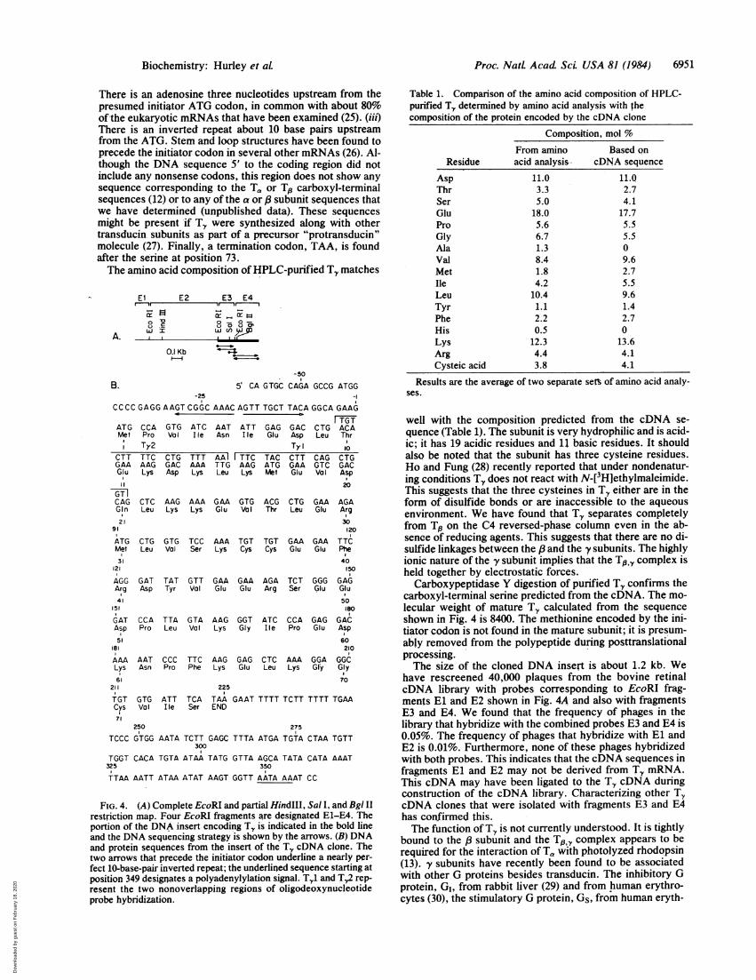
A restriction map and partial DNA sequence of the clonedinsert are shown in Fig. 4. Translation in one reading frameproduces a protein sequence that agrees precisely with theamino-terminal 45 amino acid sequence of T, and also clari-fies the assignment of two cysteines at positions 35 and 36.In addition, the open reading frame continues until an in-phase termination signal (TAA) is reached at nucleotide 223.The sequence encodes a polypeptide that is 73 amino acidslong, beginning with the proline at position 1. The amino acidcomposition of the sequence agrees very closely with thecomposition of TY determined by amino acid analysis ofHPLC-purified Ty (Table 1). The termination codon is fol-lowed by a 134-base-pair 3' untranslated region that is about76% A+T and contains the mRNA 3' consensus polyadenyl-ylation signal (24) A-A-T-A-A-A starting at position 349.However, no poly(A) tract was found, which suggests thatthis cDNA clone may be incomplete at its 3' terminus.
DISCUSSIONWe have isolated a cDNA clone coding for the y subunit ofbovine retinal transducin and deduced from it the completeamino acid sequence of this subunit. TY appears to be syn-thesized as a discrete polypeptide rather than as a domain ofa polyprotein because many of the features of a eukaryotictranslation initiation site are found. (i) An initiator codon(ATG) immediately precedes the amino-terminal proline. (ii)
1 2 3 4 5 6 7 81 . 1 1 1
T- 1
B
kb
- 6.6
- 4.3
1 2 3 4 5 6 7 8
6.6
- 4.3T.2 :4.: *- . t,2.. , , . ,e;,S. ,,_ . ., , . ,$ ,9 ,.,^ x, ^~~~~~~~~~~~~~~~~~~~P-A
go
T,:2~~~~~~~~~~~r I... .\.2: a u',
* .... ~~~~~~~~.~~~~W' .Sws ,
FIG. 3. Southern blot analysis of cDNA clones that hybridized to oligodeoxynucleotide probe TY1. DNA from each of these clones wasdigested with BamHI and BgI II, separated on a 1.0%o agarose gel, transferred to nitrocellulose, and probed with the designated 32P-labeledoligodeoxynucleotide probes T.Y (A) and Ty2 (B). The fragments corresponding to the inserts of 8 of the 13 selected phages are shown.
Proc. NatL Acad ScL USA 81 (1984)
Dow
nloa
ded
by g
uest
on
Feb
ruar
y 18
, 202
0
Proc. NatL Acad. Sci. USA 81 (1984) 6951
There is an adenosine three nucleotides upstream from thepresumed initiator ATG codon, in common with about 80%of-the eukaryotic mRNAs that have been examined (25). (iii)There is an inverted repeat about 10 base pairs upstreamfrom the ATG. Stem and loop structures have been found toprecede the initiator codon in several other mRNAs (26). Al-though the DNA sequence 5' to the coding region did notinclude any nonsense codons, this region does not show anysequence corresponding to the Ta or To carboxyl-terminalsequences (12) or to any of the a or a subunit sequences thatwe have determined (unpublished data). These sequencesmight be present if Ty were synthesized along with othertransducin subunits as part of a precursor "protransducin"molecule (27). Finally, a termination codon, TAA, is foundafter the serine at position 73.The amino acid composition of HPLC-purified TY matches
A.
B.
El E2 E3 E4
0 00
0 'o 0 80<,I] I LL ) W
0.1 Kb L*
-50
5' CA GTGC CAGA GCCG ATGG-25 -I
CCCC GAGG AAGT CGGC AAAC AGTT TGCT TACA GGCA GAAG< ~ ~~~~~~~~~~~~~~Dro
ATGMet
CTTGAAGlu
1l
GTICAGGin2 1
91
ATGMet31
121
AGGArg41
151
GATAsp51
181
AAALys
~l61211
TGTCys71
CCAProTy2
TTCAAGLys
GTG ATC AAT ATT GAGVolI lie Asn Ile Glu
CTG TTT AA TTC TACGAC AAA TTG AAG ATGAsp Lys Leu Lys Met
GACAspTyCTTGAAGlu
TGTCTG ACALeu Thr
10
CAG CTGGTG GACVa I Asp
20
CTC AAG AAA GAA GTG ACG CTG GAALeu Lys Lys Glu Va Thr Leu Glu
CTG GTG TCC AAA TGT TGT GAA GAALeu Val Ser Lys Cys Cys Glu Glu
GAT TAT GTT GAA GAA AGA TCT GGGAsp Tyr Val Glu Glu Arg Ser Glu
CCA TTA GTA AAG GGT ATC CCA GAGPro Leu Val Lys Gly Ile Pro Glu
AAT CCC TTC AAG GAG CTC AAA GGAAsn Pro Phe Lys Glu Leu Lys Gly
GTG ATT TCAVal Ile Ser
AGAArg30120
TTGPhe40150
GAGGlu50
180
GAGAsp60210
GGGGly70
225
TAA GAAT TTTT TCTT TTTT TGAAEND
?50 2751 ~~~~~~~~~~~~~~.I
TCCC GTGG AATA TCTT GAGC TTTA ATGA TGTA CTAA TGTT300
TGGT CACA TGTA ATAA TATG GTTA AGCA TATA CATA AAAT325 350
TTAA AATT ATAA ATAT AAGT GGTT AATA AAAT CC
FIG. 4. (A) Complete EcoRI and partial HindIII, Sal I, and Bgl II
restriction map. Four EcoRI fragments are designated E1-E4. Theportion of the DNA insert encoding T, is indicated in the bold lineand the DNA sequencing strategy is shown by the arrows. (B) DNAand protein sequences from the insert of the T, cDNA clone. Thetwo arrows that precede the initiator codon underline a nearly per-fect 10-base-pair inverted repeat; the underlined sequence starting atposition 349 designates a polyadenylylation signal. Tyl and Ty,2 rep-resent the two nonoverlapping regions of oligodeoxynucleotideprobe hybridization.
Table 1. Comparison of the amino acid composition of HPLC-purified T. determined by amino acid analysis with thecomposition of the protein encoded by the cDNA clone
Composition, mol %
Residue
AspThrSerGluProGlyAlaValMetIleLeuTyrPheHisLysArgCysteic acid
From amino Based onacid analysi&-- cDNA sequence
11.03.35.0
18.05.66.71.38.41.84.2
10.41.12.20.5
12.34.43.8
11.02.74.1
17.75.55.509.62.75.59.61.42.70
13.64.14.1
Results are the average of two separate seM of amino acid analy-ses.
well with the composition predicted from the cDNA se-quence (Table 1). The subunit is very hydrophilic and is acid-ic; it has 19 acidic residues and 11 basic residues. It shouldalso be noted that the subunit has three cysteine residues.Ho and Fung (28) recently reported that under nondenatur-ing conditions Ty does not react with N-[3H]ethylmaleimide.This suggests that the three cysteines in Ty either are in theform of disulfide bonds or are inaccessible to the aqueousenvironment. We have found that Ty separates completelyfrom To on the C4 reversed-phase column even in the ab-sence of reducing agents. This suggests that there are no di-sulfide linkages between the p and the y subunits. The highlyionic nature of the y subunit implies that the Tp, y complex isheld together by electrostatic forces.Carboxypeptidase Y digestion of purified Ty confirms the
carboxyl-terminal serine predicted from the cDNA. The mo-lecular weight of mature To calculated from the sequenceshown in Fig. 4 is 8400. The methionine encoded by the ini-tiator codon is not found in the mature subunit; it is presum-ably removed from the polypeptide during posttranslationalprocessing.The size of the cloned DNA insert is about 1.2 kb. We
have rescreened 40,000 plaques from the bovine retinalcDNA library with probes corresponding to EcoRI frag-ments El and E2 shown in Fig. 4A and also with fragmentsE3 and E4. We found that the frequency of phages in thelibrary that hybridize with the combined probes E3 and E4 is0.05%. The frequency of phages that hybridize with El andE2 is 0.01%. Furthermore, none of these phages hybridizedwith both probes. This indicates that the cDNA sequences infragments El and E2 may not be derived from Ty mRNA.This cDNA may have been ligated to the Ty cDNA duringconstruction of the cDNA library. Characterizing other TycDNA clones that were isolated with fragments E3 and E4has confirmed this.The function of Ty is not currently understood. It is tightly
bound to the p subunit and the T,3,y complex appears to berequired for the interaction of Ta with photolyzed rhodopsin(13). y subunits have recently been found to be associatedwith other G proteins besides transducin. The inhibitory Gprotein, GI, from rabbit liver (29) and from human erythro-cytes (30), the stimulatory G protein, Gs, from human eryth-
Biochemistry: Hurley et aL
Dow
nloa
ded
by g
uest
on
Feb
ruar
y 18
, 202
0

6952 Biochemistry: Hurley et al.
rocytes (30), and the "other" G protein from bovine brain(31), Go, all have small molecular weight (6,000-10,000) sub-units that remain tightly associated with their 3 subunitsthroughout several purification steps. Reversed-phaseHPLC, polyacrylamide gel electrophoresis, and silver stain-ing of two of these y subunits show that they are substantial-ly different from one another.t Thus, some of the specificityfor a particular class of G protein may reside in the y subunitsequence,Gilman (5) has recently suggested that the G proteins are
related to the GTP binding ras family of proteins on the basisof homology between a short stretch of amino acids at thecarboxyl terminus of the a subunit of transducin and a se-quence in the middle of the ras protein. There is also somehomology between the carboxyl-terminal sequences of TYand of the ras proteins. Powers et al. (32) have noted that rasproteins from different sources all terminate with the aminoacid sequence C-A-A-X, in which A represents an aliphaticamino acid, C represents cysteine, and X is a carboxyl-ter-minal serine, methionine, or cysteine. The carboxyl-terminalsequence of TY fits this pattern. Furthermore, there is 44%homology between the DNA sequences starting at exon 4 ofhuman c-Ha-rasl protooncogene (nucleotides 451-570 in ref.33) and nucleotides 107-225 of To. To maximize the homolo-gy, the sequences were aligned by introducing only one gap,consisting of a single nucleotide between nucleotides 162 and163 of TY. It is possible that these two families of proteins(ras and the G proteins) are derived from common ancestralgenes. Domain functions in the ras protein may be embodiedin the different subunits of transducin and both proteins mayfunction in transducing signals at the cellular level. Recentevidence suggests that ras interacts directly with cell-surfacereceptor proteins (34, 35).The existence of TY cDNA clones now makes possible di-
rect sequence comparison of the y subunits of transducinand other G proteins. Homologous cDNAs can be isolatedfrom cDNA libraries constructed from other tissues to makethese comparisons. In addition, this TY cDNA clone can beused along with cDNA clones for Ta and Tp to study thelocation and arrangement of genes for transducin and theother G proteins.
tAn unpublished comparison of Ty with the y subunit derived fromrabbit liver GI, done in collaboration with G. Bokoch and A. G.Gilman.
We thank Jeremy Nathans for supplying us with his bovine retinalcDNA library, Dr. Suzanna Horvath and Carol Graham for synthe-sizing the oligodeoxynucleotides, Chin Sook Kim for preparation ofreagents for the protein sequenator, Vince Farnsworth for perform-ing the amino acid analyses, and Dr. R. Bruce Wallace for advice.This work was supported by a grant from the National ScienceFoundation to M.I.S., a Helen Hay Whitney Fellowship to J.B.H.,National Institutes of Health Block Grant GM06965 to W.J.D., andNational Institutes of Health Training Grant GM07401 to D.B.T.
1. Stryer, L., Hurley, J. B. & Fung, B. K.-K. (1981) Curr. Top.Membr. Transp. 15, 93-108.
2. Bitensky, M. W., Wheeler, G. L., Yamazaki, A., Rasenick,M. M. & Stein, P. J. (1981) Curr. Top. Membr. Transp. 15,273-290.
3. Abood, M. E., Hurley, J. B., Pappone, M. C., Bourne, H. R.& Stryer, L. (1982) J. Biol. Chem. 257, 10540-10543.
4. Manning, D. R. & Gilman, A. G. (1983) J. Biol. Chem. 258,7059-7063.
5. Gilman, A. G. (1984) Cell 36, 577-579.6. Northrup, J. K., Sternweis, P. C., Smigel, M. D., Schleifer,
L. S., Ross, E. M. & Gilman, A. G. (1980) Proc. NatI. Acad.Sci. USA 77, 6516-6520.
7. Bokoch, G. M., Katada, T., Northrup, J. K., Hewlett, E. L.& Gilman, A. G. (1983) J. Biol. Chem. 258, 2072-2075.
8. Kuhn, H. (1980) Nature (London) 283, 587-589.9. Fung, B. K.-K., Hurley, J. B. & Stryer, L. (1981) Proc. Natl.
Acad. Sci. USA 78, 152-156.10. Baehr, W., Morita, E. A., Swanson, R. J. & Applebury, M. L.
(1982) J. Biol. Chem. 257, 6452-6460.11. Van Dop, C., Yamanaka, G., Steinberg, F., Sekura, R. D.,
Manclark, C. D., Stryer, L. & Bourne, H. R. (1984) J. Biol.Chem. 259, 23-26.
12. Manning, D. R., Fraser, B. A., Kahn, R. A. & Gilman, A. G.(1984) J. Biol. Chem. 259, 749-756.
13. Fung, B. K.-K. (1983) J. Biol. Chem. 258, 10495-10502.14. Hurley, J. B. & Stryer, L. (1982) J. Biol. Chem. 257, 11094-
11099.15. Mahoney, W. C. & Hermodson, M. A. (1980) J. Biol. Chem.
255, 11199-11203.16. Hunkapiller, M. W., Lujon, E., Ostrander, F. & Hood, L. E.
(1983) Methods Enzymol. 91, 227-236.17. Hunkapiller, M. W. & Hood, L. E. (1983) Methods Enzymol.
91, 486-493.18. Martin, B., Svendsen, I. & Ottesen, M. (1977) Carlsberg Res.
Comm. 42, 99-102.19. Laemmli, U. K. (1970) Nature (London) 227, 680-685.20. Southern, E. M. (1975) J. Mol. Biol. 98, 503-517.21. Sanger, F., Nicklen, S. & Coulsen, A. R. (1977) Proc. Natl.
Acad. Sci. USA 74, 5463-5467.22. Hunkapiller, M., Kent, S., Caruthers, M., Dreyer, W., Firca,
J., Giffin, C., Horvath, S., Hunkapiller, T., Tempst, P. &Hood, L. (1984) Nature (London) 310, 105-111.
23. Nathans, J. & Hogness, D. S. (1983) Cell 34, 807-814.24. Benton, W. D. & Davis, R. W. (1977) Science 196, 180-182.25. Fitzgerald, M. & Shenk, T. (1981) Cell 24, 251-260.26. Kozak, M. (1984) Nature (London) 308, 241-246.27. Lomedico, P., Rosenthal, N., Efstratiadis, A., Gilbert, W.,
Kolodner, R. & Tizard, R. (1979) Cell 18, 545-558.28. Ho, Y.-K. & Fung, B. K.-K. (1984) J. Biol. Chem. 259, 6694-
6699.29. Bokoch, G. M., Katada, T., Northrup, J. K., Ui, M. & Gil-
man, A. G. (1984) J. Biol. Chem. 259, 3560-3567.30. Hildebrandt, J. D., Codina, J., Risinger, R. & Birnbaumer, L.
(1984) J. Biol. Chem. 259, 2039-2042.31. Sternweis, P. C. & Robishaw, J. D. (1984) J. Biol. Chem. 259,
in press.32. Powers, S., Kataoka, T., Fasano, O., Goldfarb, M., Strathern,
J., Broach, J. & Wigler, M. (1984) Cell 36, 607-612.33. Taparowski, E., Shimuzu, K., Goldfarb, M. & Wigler, M.
(1983) Cell 34, 581-586.34. Finkel, T. & Cooper, G. M. (1984) Cell 36, 1115-1121.35. Kamata, T. & Feramisco, J. R. (1984) Nature (London) 310,
147-149.
Proc. Natl. Acad Sci. USA 81 (1984)
Dow
nloa
ded
by g
uest
on
Feb
ruar
y 18
, 202
0