isolation, characterization and application of indigenous ... · isolation, characterization and...
TRANSCRIPT
415 Nawaz et al.
Int. J. Biosci. 2016
RESEARCH PAPER OPEN ACCESS
Isolation, characterization and application of indigenous lactic
acid bacteria in milk fermentation
Farah Nawaz1, Sadia Mehmood
1, Shakira Ghazanfar
3, S.S. Tahir
2, Naseem Rauf
2,
Muhammad Imran1*
1Department of Microbiology,Quaid-i-Azam University, Islamabad, Pakistan
2Pakistan Council for Scientific and Industrial Research (PCSIR), Islamabad, Pakistan
3Institute of Microbial Culture Collection of Pakistan, National Agricultural Research
Centre, Park Road, Islamabad-45500, Pakistan
Key words: Indigenous starters, Milk, Lactic acid bacteria, Nutrition.
http://dx.doi.org/10.12692/ijb/9.6.415-430 Article published on December 31, 2016
Abstract
Lactic acid bacteria are essential part of milk fermentation impart characteristic attributes on product physio-
chemistry, nutrition and sensory properties. The present study was designed to characterize the Lactic acid
bacteria isolated from fermented milk product Dahi for potential application in milk fermentation. All isolates
were Gram positive rods and cocci. Most of the isolates were heterofermentative shown carbon dioxide
production. Isolates shown higher lipolytic, proteolytic and fair amylolytic activity. Physiological characterization
revealed that cocci were showing optimum growth at 2 and 4% NaCl while rods at 6.5% followed by 4% salt.
Agglomerative hierarchical clustering (AHC) was done based on growth rate and alteration in media pH at
differenttemperatures. Identification of representative isolates from each group was confirmed by 16SrRNA gene
partial sequencing.Selective strains were identified as Lactobacillus delbrueckii QAULb01(KT021869),
Streptococcus thermophilusQauSt1(KT021870)andLactobacillus delbrueckii Qaulbd16, and Enterococcus
mundtii QAUEM02. Comparative fermentation experiments of commercial starter culture and indigenous
isolates combination (QauSt1+Qaulb01) was done. The local strain combination shown more aroma as compare
to commercial starter that was also confirmed by change in FTIR spectrum. Comparative physiochemical
analysis shown that local cultures in term less syneresis, high solid content, stability in pH and acidity and
higher viscosity.The nutrition of fermented milk produced by local strain in term mineral contents was
comparable with significantly higher zinc and iron contents (K :720 mg/kg ), Na: 642.5 mg/kg, Ca: 245.2 mg/kg,
Fe: 2.7 mg/kg and 2 mg/kg of Zinc). Hence these strains can be successfully used to replace starter cultures at
commercial scale for fermented milks.
* Corresponding Author: Muhammad Imran [email protected]
International Journal of Biosciences | IJB |
ISSN: 2220-6655 (Print), 2222-5234 (Online)
http://www.innspub.net
Vol. 9, No. 6, p. 415-430, 2016

416 Nawaz et al.
Int. J. Biosci. 2016
Introduction
Lactic acid bacteria (LAB) are generally recognized as
safe (GRAS) microorganisms in food fermentations
worldwide. In dairy and food industry, they are
enormously used as a starter cultures because they
are known to improve technological, nutritional,
organoleptic, and shelf-life properties in diverse
fermented foods and beverages(Wouters et al.,
2002;Capozzi et al., 2012; Patel et al., 2013;). These
are categorized as gram positive cocci or rods, aero
tolerant but anaerobic, produce energy and lactic acid
by utilizing carbohydrates. The metabolic pathways
include homofermentative in which two molecules of
lactate are produced e.g. Lactococcus and
Streptococcus. While in heterofermentation, lactic
acid, ethanol and carbon dioxide are produced e.g.
Lactobacilli and Leuconostoc(Mahmoudi et al.,
2013).
Now a day much focus is paid on the technological
potential of LAB for the bio control of plant, food and
soil born fungus and bacteria (Fhoula et al., 2013). In
dairy ecosystem LAB efficiently consume milk
constituents, principally lactose and caseins, that’s
why they are used as starter cultures in the
production of dairy products. Previous studies show
affiliation between fast growth, rate of acid
production and efficient milk casein degradation
(Courtin, 2002). Amino acid and peptides are not
freely available in milk for LAB (Zourari, 1992; Abu-
Tarboush, 1996). LAB need an exogenous supply of
amino acid and peptide which is provided by the
proteolysis of milk casein. That’s why they are called
fastidious microorganism.Glucose containing mono
and disaccharides that are normally present in
natural food are readily available for amylolytic LAB
(Guyot et al., 2000).
Milk is enriched with indigenous lipoprotein lipase
(LPL) and estrases. Lactococcus strains are the
examples of producing intracellular Lipases (Casey,
1992; Fox et al., 1996; Holland and Coolbear, 1996).
During the lipolysis hydrolysis of triglycerides into
short, intermediate fatty acid chains and glycerol is
produced especially in cheese fermentation. Quality
of cheese is directly influenced by the fats present in
it, which affect the taste and texture of that cheese.
Free fatty acids (FFA) formation occurs as a result of
lipolysis affect the flavor compounds such as methyl
ketones, alcohols and lactones present in cheese.
Co-inoculation of different species in food
fermentations to improve the quality of existing
products is widely applied now days. Also, industries
are taking keen interest in using the molecular
approaches for the survival and improvement of
selected strains; by closer examination, namely the
protocooperation between S. thermophiles and L.
bulgaricus, phage resistance, utilization of prebiotic
carbohydrates (Goin, 2010). Fermented foods in
which the interactions, like protocooperation,
mutualism and synergism, are important for the
growth rate e.g. cultures consist of filamentous fungi,
LAB and yeast (Sieuwerts et al., 2008).
The present study was designed to Identify and
characterize the lactic acid bacteria isolated from
indigenous fermented milk product Dahi and evaluate
the technological and physiochemical attributes of the
LAB isolates, so that they could be successfully used
for Milk fermentation.
Materials and methods
Isolation of lactic acid bacteria
Fifty Dahi samples were collected from different
locations of Islamabad and Rawalpindi available at
the retail under sterile conditions. Isolation of lactic
acid bacteria from Dahi was done by Serial dilution
method on MRS and M17 agar plates. The media
plates were incubated at 37°C for 48 hrs. Isolates
were maintained on MRS and M17 media at 4°C for
further studies.
Morphological and biochemical identification
For morphological and biochemical characterization
of the isolated strains several tests were performed:
Gram staining, Oxidase test, Catalase test, and carbon
dioxide (CO2) production.For CO2 production, MRS
and M17 broth containing Durham tubes were used.
Citrate excluding MRS and M17 broth were used

417 Nawaz et al.
Int. J. Biosci. 2016
because citrate can produce CO2. 100 µl of 24 hour
old cultures were inoculated into 5ml citrate minus
MRS and M17 broth and incubated for 72 hours at 37
°C.
Technological characteristics
Evaluation of enzymatic activity of lactic acid
bacteria
For amylolytic activity,1 gram of starch was added in 1
gram of nutrient agar in 100 ml of distilled water.
Surface dried plates of starch were streaked with 24
hours old culture of LAB and incubated at 37°C for 48
hours. These plates were then flooded with gram’s
iodine for 15 to 30 minutes. Then clear zone around
the colonies were observed.
For proteolytic activity, Skim milk agar plate was
prepared (10 g of skim milk and 2.5 gram of agar)
(Gordon et al., 1973). Surface dried plates of milk
agar were streaked with 24 hours old culture LAB and
incubated at 37°C for 48 hours. After incubation clear
zone around the streak were observed.
Media used for the lipolytic activity was described by
(Sierra, 1957) in this method tween 80 was used as a
lipid substrate. Clear zone of hydrolysis was observed.
Physiological parameters
To check the survival of presumed Lactic acid bacteria
isolates, different NaCl concentration 2%, 4 % for
cocci and 4%, 6.5% for rods were used. To perform
the experiment, 100 µl of overnight cultures were
inoculated into 5ml of MRS with 2% 4 % and 6.5 %
NaCl concentration.
Growth and pH of isolates was determined at three
different temperatures 15°C, 30 °C and 45°C. 100µl of
overnight activated cultures were inoculated into
MRS broth for bacilli and M17 broth for cocci.
Acidification potential was also determined at the
same temperature by inserting probe of pH meter.
Growth pattern was observed with the help of optical
density at 600nm.
Identification of selected isolates by partial
sequencing of 16SrDNA
For DNA extraction, bacterial isolates were inoculated
in TSB broth and placed at 37 oC shaker for 24hr.
DNA extraction of isolates was carried out using the
CTAB method as described by Kate Wilson (Wilson,
1975). The gel was run at 100 volts, 400 milli-
amperes current to visualize the extracted DNA
quality.
Phylogenetic analysis
After DNA extraction, isolated DNA was partially
sequenced for 16S rRNA gene. Sequencing was
carried out by Macrogen Commercial Seoul, South
Korea. 16S rRNA gene sequences of the isolated
strains and the most similar sequences from Gen
Bank were identified through BLAST from NCBI. The
alignments were then thoroughly analyzed and
corrected. This was followed by construction of
Phylogenetic trees for isolates with Bootstrap values
using neighbor joining method.
Analysis of isolates for the milk fermentation
For the milk fermentation cultures were activated
into the tryptone soya broth(TSB) broth for 24hours.
Then inoculated into pasteurized milk for 24 hours.
Fermentation was performed in two batches, one with
starter culture that act as a control, was introduced
into pasteurized milk, and other batch, with
combination of (QauSt1+QauLb01) were inoculated.
Both of these combinations were examined for pH,
titratable acidity, total solids, syneresis, and FTIR
analysis at different intervals that described the
biochemical changes occurred during the
fermentation process. The experimental samples were
also analysed for viscosity and Mineral content.
(A.O.A.C, 1990) method number 981.12 for the
determination of pH was followed. (A.O.A.C, 1990)
method number 967.16 was followed for determine
the titratable acidity. Similarly, (A.O.A.C, 1990)
method number 925.23 was used to calculate the total
solid content present in yogurt. After separation of
solid content, the resulting whey was used to
determine percentage of syneresis.

418 Nawaz et al.
Int. J. Biosci. 2016
Frontier transformed infrared spectroscopy was used
in order to analyse the chemical changes taking place
in yogurt fermentation. FTIR was done by Perkin
ELMER spectrum 65FTIR spectroscopy equipped
with ATR. For each sample the spectrum range was
650-4000𝑐 𝑚 . In order to observe different peaks, an
overlay was formed which showed the new peak.
Changes occurred in the sample were compared to the
control sample.
Measurement of viscosity
Viscosity was measured using a viscometer model
LVDVE, DV-E Viscometer (Brookfield Engineering
Laboratories Inc., Stoughton, MA) using spindle
number 64 which was set clockwise at 100rpm. Every
experiment was repeated 3 times to have some
meaningful results after a statistical analysis.
Viscosity was expressed as milli poises (m.p.s).
Determination of ash contents
The ash content was determined by dried ash method
in a muffle furnace (Carbolite, Model No. CWF 1200)
at 500˚C for 6 hours (overnight) as described by
(Hernandez and Park, 2014). Ash contents were
determined by the following formula:
Digestion of ash and mineral analysis
The digestion of ash samples was carried out to
determine mineral concentration in Dahi. Ash was
dissolved in 15-20 ml of nitric acid (1: 1 ratio) and
heated on hot plate at 800C till the colour of the
fumes changed from brown to yellow; this process
was performed in the fume hood. After dissolution
2M nitric acid was used to make the volume upto
25ml. minerals including Ca, Mg were determined by
using the scientific Nov 300 Flame Atomic
Absorption Spectrophotometer Fe, zinc concentration
was determined by the Analytik Jena AAS vario 6
Atomic absorption spectrophotometer While Na and
potassium were determined by AFP 100 flame
photometer using their standards, and concentrations
were noted.
Results and discussion
Morphological and biochemical identification
In current study, selective media such as MRS and
M17 were used keeping in view the specific nutritionl
requirements of LAB and twenty isolates were
selected for identification and characterization from
indigenous fermented milk product Dahi.
Table 1. Morphological and biochemical identification of the isolates.
Isolates Colony
Surface
Colony
Margin
Colony colour Gram
staining
Cell
Shape
Catalase
Test
Oxidase
test
CO2
Production
QauLac 1 Pin point Regular Pale yellow G (+) Cocci - - -
QauLeu3 Pin point Regular Pale yellow G (+) Cocci - - +
QauLeu4 Pin point Regular Pale yellow G (+) Cocci - - +
QauSt 9 Pin point Regular Pale yellow G (+) Cocci - - +
QauEm02 Pin point Regular Pale yellow G (+) Cocci - - +
QauLeu11 Pin point Regular Creamy G (+) Cocci - - +
QauLac12 Pin point Regular Pale yellow G (+) Cocci - - +
QauLeu14 Pin point Regular Pale yellow G (+) Cocci - - +
QauSt1 Pin point Regular Pale yellow G (+) Cocci - - +
QauLeu18 Pin point Regular Pale yellow G (+) Cocci - - +
QauLbd16 Smooth Irregular Light grey G (+) Rods - - +
QauLb01 Smooth Irregular Light grey G (+) Rods - - +
QauLabf Smooth Irregular transparent G (+) Rods - - +
QauLabe smooth Irregular Whitish G (+) Rods + - -
QauLabf1 Smooth Regular Grey G (+) Rods - - +
QauLabmo Smooth Irregular Grey/white G (+) Rods + + -
QauLabf3 Smooth Irregular transparent G (+) Rods - - -
QauLabw2 Smooth Regular Transparent G (+) Rods - - -
QauLabw3 Smooth Regular Transparent G (+) Rods - - +
QauLabw5 Smooth Irregular Creamy G (+) Rods + + -

419 Nawaz et al.
Int. J. Biosci. 2016
The colonies were pinheaded shiny and transparent
on MRS agar.
The results of gram staining showed the presence of
purple rods individually and in chains which
confirmed that they were gram positive. While on
M17 agar the colonies were yellowish creamy and pin
point in appearance. Gram staining indicated the
presence of purple cocci.
Table 2. Evaluation of enzymatic potential of presumed Lactic acid bacteria.
Isolates Lipolytic activity Proteolytic activity Amylolytic activity
QAULAC 1 + + -
QAULEU3 + + -
QAULEU4 + - +
QAU ST 9 + - -
QAUEM02 + + -
QAULEU11 + + +
QAULAC12 + + -
QAULEU14 - - -
QAUST1 + + -
QAULEU18 + + -
QAULAB16 + + +
QAULB01 + + -
QAULABF + + +
QAULAB E + + +
QAULABF1 + + +
QAULABMO + + +
QAULABF3 + + +
QAULABW2 - + +
QAULABW3 + + +
QAULABW5 + + -
The dominance of catalase and oxidase negative gram
positive rods and cocci among the isolates is
consistent with the finding of (Naeem et al., 2012)
and (Maqsood et al., 2013) who observed the
presence of these bacteria from locally available dahi.
Similar results were also presented by (Gad et al.,
2014) who confirmed the presence of these bacteria in
dairy and pharmaceutical products.
Isolates were further analysed for the biochemical
test. Results for CO2 production test shown that six
strains were homofermentative i.e. end product was
lactic acid and fourteen were heterofermentative i.e.
along with lactic acid other aromatic compounds and
CO2 were also formed. The results for morphological
and biochemical identification are summarized in
Table 1.
Technological characteristics
The results for enzymatic activity of isolates are
summarized in the Table 2. Proteolytic activity
(degradation of casein) was observed in seventeen
isolates, when tested on skim milk agar. It is the most
abundant protein in the milk as a main source of
amino acids. Proteolytic properties are important for
the development of organoleptic properties of
different fermented products. During proteolysis, the
amino acids which are produced, serve as a precursor
for the development of flavouring compounds
especially aldehydes in the product. Our study is in
agreement with the earlier findings (Dagdemir and
Ozdemir, 2008; Hassaïne, 2008).
Lipolytic activity was examined in eighteen isolates
out of twenty. The isolates showed hollow zone

420 Nawaz et al.
Int. J. Biosci. 2016
around the colonies which was an indication of lipid
degradation while QauLeu14 and QauLabw2 showed
negative results for lipolytic activity. It is predicted
that such activities can be manifested in vivo for the
reduction of cholesterol level in humans, as a starter
and adjunct culture (Ramakrishnan 2012). Our study
is contrary to the findings of (Bridget, 2011) where
less lipolytic and amylolytic strains were screened in
dairy industry these can be used in the hydrolysis of
milk fat. Similar findings have been reported by
(Fortune Akabanda, 2014).
Table 3. Physiological profiling of Lactic acid bacteria Isolates.
Isolates Growth at different temperatures pH at different temperatures Growth at diff. NaCl concentrations
OD at 600
15°C 30°C 45°C 15°C 30 °C 45°C 2% 4% 6.5%
QauLac1 0.89±0.48 3.67±0.37 3.70±0.93 7.33±0.49 6.58±0.43 6.91±0.16 0.34±0.04 1.69±0.05 -
QauLeu3 1.14±0.36 3.83±1.20 4.29±1.53 7.36±0.42 7.49±0.39 6.51±0.25 0.43±0.01 3.47±0.33 -
QauLeu4 1.17±0.35 5.32±1.88 3.66±1.06 7.09±0.27 7.11±0.27 6.63±0.32 0.34±0.03 2.78±0.13 -
QauSt 9 1.46±0.43 5.38±1.31 2.86±0.69 7.25±0.16 7.34±0.65 6.67±0.14 1.56±0.02 0.60±0.14 -
QauEm02 4.27±1.01 6.45±2.8 0.68±0.07 7.04±0.09 6.81±0.10 6.64±0.19 1.43±0.01 3.39±0.01 -
QauLeu11 0.97±0.18 3.14±0.69 3.78±1.54 7.24±0.23 7.55±0.72 6.76±0.40 0.51±0.01 2.28±0.09 -
QauLac12 1.31±0.57 4.13±2.61 4.93±1.54 7.36±0.44 7.47±0.44 6.81±0.34 2.28±0.04 0.29±0.20 -
QauLeu14 2.04±1.46 3.55±2.0 1.39±0.47 7.07±0.24 7.40±062 6.67±0.48 2.40±0.01 1.65±0.04 -
QauSt1 0.82±0.16 3.81±1.62 3.91±02.9 7.24±0.18 6.92±0.16 6.60±0.24 3.44±0.32 2.46±0.42 -
QauLeu18 1.44±0.16 4.45±1.50 4.64±1.23 6.81±0.18 6.89±1.07 6.45±0.25 2.43±0.22 0.57±0.05 -
QauLab16 1.03±0.2 2.41±0.40 2.62±0.46 5.42±0.55 5.55±0.46 4.83±0.15 - 4.39±0.23 6.96±0.12
QauLb01 0.98±0.04 4.58±2.87 4.59±2.34 6.18±1.17 6.11±0.98 4.89±0.12 - 0.80±0.22 3.31±0.19
QauLabf 1.19±0.41 3.23±1.67 1.02±0.64 5.86±0.50 5.16±0.53 4.83±0.15 - 2.67±0.15 2.58±0.10
QauLabe 0.87±0.15 3.90±1.49 5.15±0.78 7.07±0.42 6.94±0.86 4.88±0.49 - 5.41±0.14 3.36±0.35
QauLabf1 1.10±0.53 6.90±0.24 2.91±1.22 5.48±0.5 6.12±1.0 5.01±0.20 - 2.47±0.26 7.09±0.23
QauLabmo 0.83±0.47 4.48±3.95 3.31±2.15 7.27±0.37 6.21±0.21 6.56±1.7 - 5.08±0.26 6.62±0.10
QauLabf3 0.51±0.27 1.45±0.31 1.47±0.72 5.78±1.2 5.94±0.74 4.98±0.18 - 0.91±0.05 4.61±0.29
QauLabw2 0.54±0.43 3.43±1.88 3.83±1.88 6.06±0.21 6.63±0.3 5.07±0.68 - 1.16±0.01 4.22±0.19
QauLabw3 0.85±0.05 2.80±1.36 4.49±2.36 5.83±0.47 6.37±1.3 5.13±0.29 - 1.22±0.29 6.56±0.31
QauLabw5 2.84±1.18 3.73±1.85 5.53±2.0 5.43±0.60 6.55±1.6 4.82±0.24 - 1.10±0.15 1.49±0.10
Amylolytic Lactobacillus strains hydrolyse the starch
granules into low molecular weight sugar causing the
acidification of media (Giraud, 1994). This trait of the
strains to convert directly the cheaply available
starchy substances to lactic acid is quite economical
for agriculture. Screening of amylolytic activity is in
line with these reports (Reddy et al., 2008; Tchekessi
et al., 2014; Vishnu, 2006). Ten Isolates had shown
clear zone around the colonies which indicated
degradation of starch. While other isolates showed
negative results for amylolytic activity, this could be
due to the absence of amylase enzyme (Table 2).
Physiological characteristics of isolates
Growth and pH at different temperatures indicated
that six isolates were found to be mesophiles and
showed maximum growth rate at 30°C and decline in
pH at 30°C was observed only in one isolate
(QauLabmo) as shown in (Table 3). Thermophilic
isolates showed better logarithmic phase and
increased acidification at 45°C. Some isolates showed
increase in growth rate and rapid decrease in pH at
two different temperatures i.e. at 30°C and 45°C.
These represent meso/thermophiles. It is concluded
that LAB had a growth survival better at 30°C and
45°C. Our finding is conclusive with the reports
mentioned by the following (Mallesha SR, 2010;
Nikita and Hemangi, 2012). Minor growth was
observed at 15°C, however most of the rod shaped
bacteria also shown a decline in pH at this

421 Nawaz et al.
Int. J. Biosci. 2016
temperature. The obtained results revealed that the
presumed Lactobacilli species were fast acid
producers as compared to
Lactococci/Enterococci/Streptococci species. pH of
Lactobacilli decreased to 4 and remained 4 after the
4th day. However, heterofermentative bacteria are
sensitive to low pH.
Fig. 1. Agglomerative hierarchical clustering (AHC) for the grouping of isolates based technological properties.
Fig. 2. Dendeogram of phylogenetic analysis of Lactobacillus bulgaricus subsp. Delbrueckii.
This study is in agreement with the finding of (Abd El
Gawad et al., 2010; Hassaïne, 2008; Khedid, 2009).
Rapid acidification is the good candidate for the
development of starter culture. Slow acid producers
can be used as adjunct starter i.e. in the production of
flavour compounds, playing the role of nonstarter
culture in order to inhibit other strains as well as
accelerate the ripening process (Herreros, 2003).
Strains were tested for their growth at different NaCl
concentrations i.e. 2%, 4% and 6.5%. For cocci 2 and
4% NaCl was used, for rods 4 and 6.5% NaCl
concentrations were used. Mostly Lactobacilli isolates
showed better survival at 6.5%. rather than 4% of

422 Nawaz et al.
Int. J. Biosci. 2016
NaCl concentration. Survival of Lactococci isolates in
2% and 4% NaCl concentrations was of equal ratio.
When LAB are confronted with the decreased water
activity i.e. during drying over the long period of time,
salt tolerant LAB can accumulate betaine and
carnitine. These solutes help in the survival in harsh
environment. Such strains are important in food and
feed industry where dried frozen starter cultures are
used (Edwin et al., 1996).
Fig. 3. Dendrogram of phylogenetic analysis of Streptococcus thermophilus Qaust1.
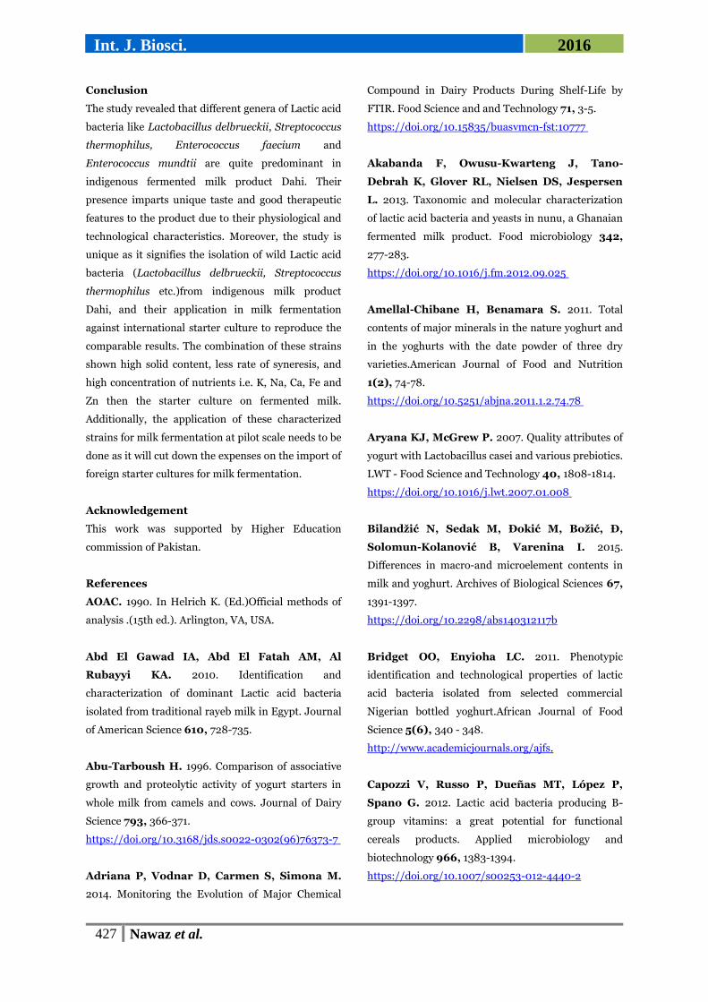
Fig. 4. Graph showing the change in pH, Acidity, Total Solid content, Syneresis after fermentation for 96 hr.
Selection of isolates for phylogenetic identification
based on their growth rate and pH
Agglomerative hierarchical clustering (AHC)was used
to group the isolates into different groups based on
their growth rate and pH.
Partial sequencing 16SrRNA Gene and phylogenetic
analysis of selected lactic acid bacteria
Molecular identification of selected isolates was
carried out to confirm the identity of isolated strains
through 16S DNA sequencing. The result of 16S
423 Nawaz et al.
Int. J. Biosci. 2016
sequencing indicated that isolated strains were
Lactobacillus bulgaricus subsp. delbrueckii and
Streptococcusthermophilus, Enterococcus mundtii
etc.
Phylogenetic analysis of 16S rRNA gene showed that
the selected isolate belonged to genus
Lactobacillus.Our isolate is more homologous to the
ATCC strain 11842 dairy products; Bulgarian yogurt.
The sequence for Lactobacillus
delbrueckii QAULB01is submitted with accession no
KT021869 (Fig. 2).
Phylogenetic Analysis showed thatour isolate was
more homologous to the S. thermophilus TUS5
isolated from dairy ecosystem in Taif Saudi Arabia.
The sequence forStreptococcus thermophilus Qaust01
is submitted in the gene bank under the accession no.
KT021870in NCBI (Fig. 3).
Comparative fermentation ability of isolated
strainswith commercial starter culture
On the basis of biochemical and technological
characterization two strains were selected and
inoculated in the milk in combination (Lactobacillus
delbrueckii QauLb01+ Streptococcus thermophilus
QauSt1). There affect was studied in comparaison
withcommercial starter (Clerici-Sacco, Italy). Milk
after fermentation was subjected to following
Rheological parameters: pH, Solid content, Syneresis,
titratable acidity, FTIR analysis, Sensory evaluation,
Viscosity, Proteolysis and Mineral analysis etc.
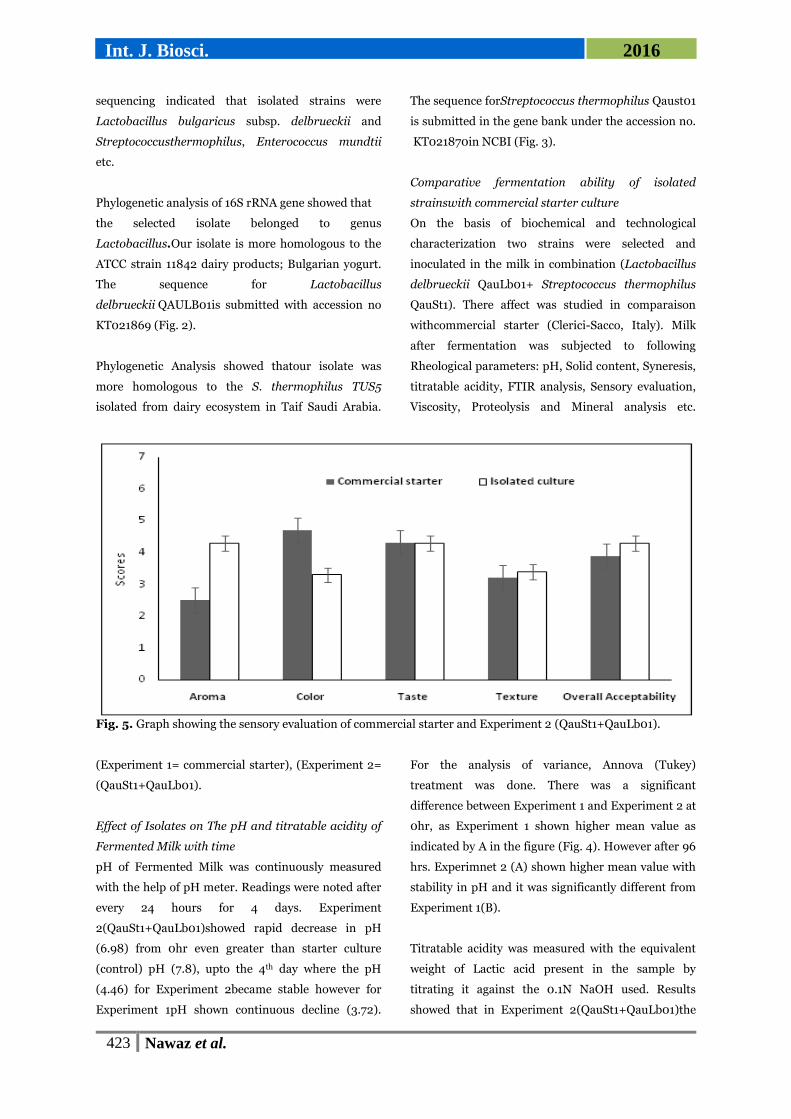
Fig. 5. Graph showing the sensory evaluation of commercial starter and Experiment 2 (QauSt1+QauLb01).
(Experiment 1= commercial starter), (Experiment 2=
(QauSt1+QauLb01).
Effect of Isolates on The pH and titratable acidity of
Fermented Milk with time
pH of Fermented Milk was continuously measured
with the help of pH meter. Readings were noted after
every 24 hours for 4 days. Experiment
2(QauSt1+QauLb01)showed rapid decrease in pH
(6.98) from 0hr even greater than starter culture
(control) pH (7.8), upto the 4th day where the pH
(4.46) for Experiment 2became stable however for
Experiment 1pH shown continuous decline (3.72).
For the analysis of variance, Annova (Tukey)
treatment was done. There was a significant
difference between Experiment 1 and Experiment 2 at
0hr, as Experiment 1 shown higher mean value as
indicated by A in the figure (Fig. 4). However after 96
hrs. Experimnet 2 (A) shown higher mean value with
stability in pH and it was significantly different from
Experiment 1(B).
Titratable acidity was measured with the equivalent
weight of Lactic acid present in the sample by
titrating it against the 0.1N NaOH used. Results
showed that in Experiment 2(QauSt1+QauLb01)the

424 Nawaz et al.
Int. J. Biosci. 2016
acidity was significantly increased after 6 hours and
48 hours but on 4th day the acidity was same as
compared to Experiment 1 (starter culture). Annova
shown that there was no statistically significant
difference between the acidity of both experiments
(Fig. 4). With the help of titratable acidity increase in
acid production was also conformed. The presence of
lactic acid is responsible for the sour taste, safety and
improves microbial stability.
Solid content was assessed by the centrifugation
followed by successive drying of yogurt sample in dry
oven. The results shown that there was no statistically
significant difference between solid content of both
experiments for 6hrs as shown by (A). After 6 hrs.
there was a significant difference between solid
content of Experiment 2 (QauSt1+QauLb01). (A) (Fig.
4). However, at 96hrs although mean value for
Experiment 2 was high than Experiment 1 but there
was less significant difference between Experiment 2
and 1. The increase in solid content led to more thick
consistency in milk fermented with isolated strains as
compared to control sample. Increase in solid content
could be related to the increase in proteolytic activity
that contribute towards the taste, texture and
flavoured compounds formation (Akabanda et al.,
2013).
Fig. 6. FTIR spectrum of Commercial starter (blue line) and Experiment 2 as (QauSt1 + QauLb01; Red line); A:
After inoculation at start of fermentation. B: after 6hrs of fermentation.

425 Nawaz et al.
Int. J. Biosci. 2016
Liquid content that is present during the yogurt
formation is called Syneresis. Syneresis is calculated
after every 24 hours for about 4days. Syneresis was
less in Experiment 2 as compared to the Experiment 1
and remained less until the 4th day.
There was statistically significant difference between
syneresis of Experiment 1, as indicated by its higher
mean value (A) and experiment 2 with less mean
value (B) until 96 hrs. (Fig. 4). Study revealed that
syneresis and whey separation is the result of
instability of weak yogurt gel network. In this way
protein molecule is unable to trap the water in three
dimensional networks. Our finding is in agreement
with the results that syneresis usually increases with
decrease in pH (Olson and Aryana, 2008).
Sensory evaluation
The end products of Experiment 1 and Experiment 2
were tasted by trained Judges to check the
organoleptic characteristics of fermented milk of both
treatments. Though there was a huge significant
difference in taste as compared to commercial starter.
Relatively higher scores given to aroma, texture and
taste of the product fermented by isolated cultures as
compared to starter culture. In colour, more score
was given to the stater culture (Fig. 5). The white
colour of yogurt sample in Experiment 1 is caused by
the light scattering of fat globules and casein micelles.
While the yellowish colour of the product was due to
the presence of riboflavin in the Experiment 2
(Walstra, 2006). Our results were contrary to the
finding of (Aryana and McGrew, 2007).
Fig. 7. Concentration of macronutrient (A) and oligo nutrients (B) in fermented milk produced by commercial
starter culture and isolated cultures.

426 Nawaz et al.
Int. J. Biosci. 2016
Viscosity
Viscosity of yogurt samples was calculated at sixth
hour of incubation. Starter culture showed 400 to
final viscosity: 200 milli poises. Experiment 2
(QauSt1 + QauLb01), 550 to final viscosity: 400 milli
poiseswas observed. Significant increase in sample
was quite evidentwhich means that Experiment2must
had higher stabilityas compared to Starter culture.
Increase in apparent viscosity might be due to
rearrangement of the fat globules, more proteins
(mainly caseins) from the serum get attached. As a
result, the number of structure building components
increase (Robinson, 2006). Increase in protein matrix
as well as the formation of bigger number of bonds
that are resistant to flow.
Estimation of biochemical changes during
fermentation by FTIR
ATR-FTIR of Experiment 1 (commercial starter
culture) and Experiment 2 (QauSt1 + QauLb01), were
carried out at time of inoculation. Two main stretches
and one in the minor stretch fingerprinting region
were observed. The first wave number obtained is
3357cm-1 indicated the presence of alcohols and
phenols. Peak at 1640 cm-1 indicated the presence
primary amines or alkenes. Peak at 1076 cm-1
represented aliphatic amines (Fig. 6A).
However, after 6th hour of yogurt formation, peak at
1745.11cm-1 indicated the presence of esters. Similar
ester groups within 1740-1750 cm-1 were also
observed (Naumann, 2000). Ethyl esters, originated
from the enzymatic or chemical esterification of acids
with ethanol possess sweet and fruity notes and
contribute to the aroma of dairy products (Molimard
and Spinnler, 1996). Peaks at 2923.74 cm-1 and
12853.54cm-1 indicated the presence of alkanes group.
Spectral region between 2850 and 3000cm-1 was
dominated by –CH3>CH2 and >CH groups usually
present in fatty acid components (Naumann, 2000;
Nicolaou et al., 2010; Sakhamuri, 2004). Peak at
1455.11cm-1 was only evident in Experiment 2 (Fig.
6B) represents the aromatics group as compared to
commercial starter. These aroma compounds could
be the result of microbial, enzymatic, or chemical
transformations of lactose, lipids, citric acid, and
proteins/amino acids present in milk. One major
pathwayis through lipolysis or oxidation of fatty acids
in milk fat, while another is microbiological
transformations of lactose by lactic acid bacteria
whichproduce flavour e.g. lactic acid, acetaldehyde,
diacetyl, acetoin, and ethanol (Adriana et al., 2014).
Impact of starter on Mineral contents
The average % age of ash for Experiment 2 was
(0.765±0.035) which was higher than control
(Experiment 1) (0.478 ± 0.072). The ash content
determines the degree of mineralization indirectly
that’s why the digested ash was analyzed further by
flame photometry and Atomic absorption
spectrometer.
The digested ash samples were subjected to Flame
Photometer and Atomic absorption spectrometer to
analyze the Na, K and Ca, Mg, Fe, Zn respectively. In
the present study, Experiment 2 shown the highest
concentration of K720 mg/kg as compared to the
Experiment 1 (starter culture) 645 mg/kg followed by
other macronutrients i.e. Na, Ca and Mg. Also
Experiment 2 (QauSt1 + QauLb01), showed higher
concentration of all these nutrients in contrast to
Experiment 1 (starter culture), except for Mg which
was slightly higher for starter culture (Fig. 17A).
These results are in accordance with the study of
(Amellal-Chibane and Benamara, 2011) on natural
yogurt in Algeria.
They reported that the potassium, calcium, sodium
and magnesium concentrations were up to the mean
values of 540.58 ± 38.33, 1950.41 ± 67.90, 684.72 ±
8.92 and 132.16 ±16.36 mg/L respectively while the
highest concentration of these minerals was reported
by (Bilandžić et al., 2015) in Croatia. The oligo
nutrients containing Fe and Zn content were also
measured within a range of 2.7 mg/L and 2 mg/L
respectively in the Experiment 2 greater than
Experiment 1 (Fig. 7B) and these results again
confirmed the findings of (González-Martín et al.,
2011; Amellal-Chibane and Benamara, 2011; Chekri et
al., 2012).
427 Nawaz et al.
Int. J. Biosci. 2016
Conclusion
The study revealed that different genera of Lactic acid
bacteria like Lactobacillus delbrueckii, Streptococcus
thermophilus, Enterococcus faecium and
Enterococcus mundtii are quite predominant in
indigenous fermented milk product Dahi. Their
presence imparts unique taste and good therapeutic
features to the product due to their physiological and
technological characteristics. Moreover, the study is
unique as it signifies the isolation of wild Lactic acid
bacteria (Lactobacillus delbrueckii, Streptococcus
thermophilus etc.)from indigenous milk product
Dahi, and their application in milk fermentation
against international starter culture to reproduce the
comparable results. The combination of these strains
shown high solid content, less rate of syneresis, and
high concentration of nutrients i.e. K, Na, Ca, Fe and
Zn then the starter culture on fermented milk.
Additionally, the application of these characterized
strains for milk fermentation at pilot scale needs to be
done as it will cut down the expenses on the import of
foreign starter cultures for milk fermentation.
Acknowledgement
This work was supported by Higher Education
commission of Pakistan.
References
AOAC. 1990. In Helrich K. (Ed.)Official methods of
analysis .(15th ed.). Arlington, VA, USA.
Abd El Gawad IA, Abd El Fatah AM, Al
Rubayyi KA. 2010. Identification and
characterization of dominant Lactic acid bacteria
isolated from traditional rayeb milk in Egypt. Journal
of American Science 610, 728-735.
Abu-Tarboush H. 1996. Comparison of associative
growth and proteolytic activity of yogurt starters in
whole milk from camels and cows. Journal of Dairy
Science 793, 366-371.
https://doi.org/10.3168/jds.s0022-0302(96)76373-7
Adriana P, Vodnar D, Carmen S, Simona M.
2014. Monitoring the Evolution of Major Chemical
Compound in Dairy Products During Shelf-Life by
FTIR. Food Science and and Technology 71, 3-5.
https://doi.org/10.15835/buasvmcn-fst:10777
Akabanda F, Owusu-Kwarteng J, Tano-
Debrah K, Glover RL, Nielsen DS, Jespersen
L. 2013. Taxonomic and molecular characterization
of lactic acid bacteria and yeasts in nunu, a Ghanaian
fermented milk product. Food microbiology 342,
277-283.
https://doi.org/10.1016/j.fm.2012.09.025
Amellal-Chibane H, Benamara S. 2011. Total
contents of major minerals in the nature yoghurt and
in the yoghurts with the date powder of three dry
varieties.American Journal of Food and Nutrition
1(2), 74-78.
https://doi.org/10.5251/abjna.2011.1.2.74.78
Aryana KJ, McGrew P. 2007. Quality attributes of
yogurt with Lactobacillus casei and various prebiotics.
LWT - Food Science and Technology 40, 1808-1814.
https://doi.org/10.1016/j.lwt.2007.01.008
Bilandžić N, Sedak M, Đokić M, Božić, Đ,
Solomun-Kolanović B, Varenina I. 2015.
Differences in macro-and microelement contents in
milk and yoghurt. Archives of Biological Sciences 67,
1391-1397.
https://doi.org/10.2298/abs140312117b
Bridget OO, Enyioha LC. 2011. Phenotypic
identification and technological properties of lactic
acid bacteria isolated from selected commercial
Nigerian bottled yoghurt.African Journal of Food
Science 5(6), 340 - 348.
http://www.academicjournals.org/ajfs.
Capozzi V, Russo P, Dueñas MT, López P,
Spano G. 2012. Lactic acid bacteria producing B-
group vitamins: a great potential for functional
cereals products. Applied microbiology and
biotechnology 966, 1383-1394.
https://doi.org/10.1007/s00253-012-4440-2

428 Nawaz et al.
Int. J. Biosci. 2016
Casey LS, Lichtman AH, Boothby M. 1992. IL4
induces IL-2 receptor ~75 B-chain gene expression
and IL-2dependent proliferation in mouse T
lymphocytes. Journal of Immunology. 148, 3418-
3428.
Chekri R, Noël L, Millour S, Vastel C, Kadar A,
Sirot V, Leblanc JC, Guérin T. 2012. Calcium,
magnesium, sodium and potassium levels in
foodstuffs from the second French Total Diet Study.
Journal of Food Composition and Analysis 252, 97-
107.
https://doi.org/10.1016/j.jfca.2011.10.005
Courtin CM, Delcour JAl. 2002. Arabinoxylans
and endoxylanases in wheat flour bread-making.
Journal of cereal science 35(3), 225-243.
https://doi.org/10.1006/jcrs.2001.0433
Dagdemir E, Ozdemir S. 2008. Technological
characterization of the natural lactic acid bacteria
of artisanal Turkish White Pickled cheese.
Interbational Journal of Dairy Technology. 61(2),
133-140.
ttps://doi.org/10.1111/j.1471-0307.2008.00394.x
Edwin PWK, Teunissen PJM, DBJAM. 1996.
Effect of Compatible Solutes on Survival of Lactic
Acid Bacteria Subjected to Drying . Applied and
environmental microbiology. 62(1), 259-261.
Fhoula I, Najjari A, Turki Y, Jaballah S,
Boudabous A, Ouzari H. 2013. Diversity and
antimicrobial properties of lactic acid bacteria
isolated from rhizosphere of olive trees and desert
truffles of Tunisia. BioMed research international
2013, 1-14
https://doi.org/10.1155/2013/405708
Fortune Akabanda JOK, Kwaku Tano-Debrah,
Charles Parkouda 3, Lene Jespersen. 2014. The
Use of Lactic Acid Bacteria Starter Culture in the
Production of Nunu, a Spontaneously Fermented
Milk Product in Ghana. International Journal of Food
Science., 2014, 1-11.
https://doi.org/10.1155/2014/721067
Fox P, Wallace J, Morgan S, Lynch C, Niland,
E, Tobin J. 1996. Acceleration of cheese ripening.
Antonie van Leeuwenhoek 702(4), 271-297.
https://doi.org/10.1007/bf00395937
Gad GFM, Abdel-Hamid AM, Farag ZSH. 2014.
Antibiotic resistance in lactic acid bacteria isolated
from some pharmaceutical and dairy products.
Brazilian Journal of Microbiology 45(1), 25-33.
https://doi.org/10.1590/s151783822014000100005
Giraud E, Champailler A, Raimbault. 1994.
Degradation of raw starch by a wild amylolytic strain
of Lactobacillus plantarum.Applied and
Environmental Microbiology 60, 4319 - 4323.
Goin CD. 2010. Growth of Lactic Acid Bacteria:
Infuence of Protocooperation, Bacteriophage
Infection, and Prebiotic Carbohydrates.
González-Martín I, Hernández-Hierro J,
Revilla I, Vivar-Quintana A, Ortega IL. 2011.
The mineral composition (Ca, P, Mg, K, Na) in
cheeses (cow’s, ewe’s and goat’s) with different
ripening times using near infrared spectroscopy with
a fibre-optic probe. Food chemistry 127(1), 147-152.
https://doi.org/10.1016/j.foodchem.2010.12.114
Gordon RE, Haynes WC, Pang CHN. 1973. The
genus bacillus. Agricultural Research Service, US
Department of Agriculture.
Guyot JP, Calderon M, Morlon-Guyot J. 2000.
Effect of pH control on lactic acid fermentation of
starch by Lactobacillus manihotivorans LMG
18010T. Journal of Applied Microbiology 88, 176–
182.
Hassaïne O, Zadi-Karam H, Karam NE. 2008.
Phenotypic identification and technological
properties of lactic acid bacteria isolated from three
breeds dromedary raw milks in south Algeria. Journal
of Food and Agriculture 20(1), 46-59.

429 Nawaz et al.
Int. J. Biosci. 2016
http://dx.doi.org/10.9755/ejfa.v12i1.5180
Hernandez K, Park YW. 2014. Evaluation of 20
Macro and Trace Mineral Concentrations in
Commercial Goat Milk Yogurt and Its Cow Milk
Counterpart. Food and Nutrition Sciences 5(10),
889-895.
http://dx.doi.org/10.4236/fns.2014.510098
Herreros MA, Fresno JM, Gonzalez Prieto M.
J, Tornadijo ME. 2003. Technological
characterization of lactic acid bacteria isolated from
Armada cheese (a Spanish goats’ milk cheese).
International Dairy Journal 13(6), 469-479.
http://dx.doi.org/10.1016/s0958-6946(03)00054-2
Holland R, Coolbear T. 1996. Purification of
tributyrin esterase from Lactococcus lactis subsp.
cremoris E8. Journal of Dairy Research 630(1), 131-
140.
http://dx.doi.org/10.1017/s0022029900031605
Khedid K, Faid M, Mokhtari A, Soulaymani A,
Zinedine A. 2009. Characterization of lactic acid
bacteria isolated from the one humped camel milk
produced in Morocco. Microbiological Research.
164(1), 81-91.
http://dx.doi.org/10.1016/j.micres.2006.10.008
Mahmoudi F, Hadadji M, Guessas B, Kihal M.
2013. Identification and Physiological Properties of
Bifidobactérium Strains Isolated from Different
Origin. Journal of food Science and Engineering 3,
196-206.
Mallesha SR, SD, Jagannath JH. 2010.
Isolation and identification of lactic acid bacteria
from raw and fermented products and their
antibacterial activity. Recent Research in Science and
Technology 2(6), 42-46.
Maqsood S, Hasan F, Masud T. 2013.
Characterization of lactic acid bacteria isolated from
indigenous dahi samples for potential source of
starter culture. African journal of Biotechnology
12(33), 5226-5231
http://dx.doi.org/10.5897/ajb09.1172
Molimard P, Spinnler H. 1996. Review:
Compounds involved in the flavor of surface mold-
ripened cheeses: Origins and properties. Journal of
Dairy Science 792, 169-184.
http://dx.doi.org/10.3168/jds.s00220302(96)76348-
8
Naeem M, Ilyas M, Haider S, Baig S, Saleem
M. 2012. Isolation characterization and identification
of lactic acid bacteria from fruit juices and their
efficacy against antibiotics. Pakistan Journal of
Botany 44, 323-328.
Naumann K. 2000. Influence of chlorine
substituents on biological activity of chemicals: a
review. Pest management science 56(1), 3-21
http://dx.doi.org/10.1002/(sici)15264998(200001)5
6:1<3::aid-ps107>3.3.co;2-g
Nicolaou SA, Gaida SM, Papoutsakis ET. 2010.
A comparative view of metabolite and substrate stress
and tolerance in microbial bioprocessing: from
biofuels and chemicals, to biocatalysis and
bioremediation. Metabolic engineering 12(4), 307-
331.
http://dx.doi.org/10.1016/j.ymben.2010.03.004
Nikita C, Hemangi D. 2012. Isolation,
identification and characterization of lactic acid
bacteria from dairy sludge sample. Journal of
Environmental Research And Development Vol
7(1A).
Olson D, Aryana K. 2008. An excessively high
Lactobacillus acidophilus inoculation level in yogurt
lowers product quality during storage. LWT-Food
Science and Technology 415, 911-918.
Patel A, Shah N, Prajapati J. 2013. Biosynthesis
of vitamins and enzymes in fermented foods by lactic
acid bacteria and related genera-A promising
approach. Croatian Journal of Food Science and

430 Nawaz et al.
Int. J. Biosci. 2016
Technology 52, 85-91.
Ramakrishnan V, Balakrishnan B, Rai AK,
Narayan B, Halami PM. 2012. Concomitant
production of lipase, protease and enterocin by
Enterococcus faecium NCIM5363 and Enterococcus
durans NCIM5427 isolated from fish processing
waste. . International Aquatic Research. 4, 14.
Reddy G, Altaf M, Naveena B, Venkateshwar
M, Kumar EV. 2008. Amylolytic bacterial lactic
acid fermentation—a review. Biotechnology advances
26(1), 22-34.
http://dx.doi.org/10.1016/j.biotechadv.2007.07.004
Robinson BH. 2006. Lactic acidemia and
mitochondrial disease. Molecular genetics and
metabolism 89(1-2), 3-13.
http://dx.doi.org/10.1016/j.ymgme.2006.05.015
Sakhamuri SB, Jamie Irudayaraj, Joseph
Demirci Ali Z. 2004. Simultaneous determination
of multiple components in nisin fermentation using
FTIR spectroscopy.
Sierra G. 1957. A simple method for the detection of
lipolytic activity of micro-organisms and some
observations on the influence of the contact between
cells and fatty substrates. Antonie van Leeuwenhoek
23(1), 15-22.
http://dx.doi.org/10.1007/bf02545855
Sieuwerts S, De Bok FA, Hugenholtz J, Van
Hylckama Vlieg JE. 2008. Unraveling microbial
interactions in food fermentations: from classical to
genomics approaches. Applied and Environmental
Microbiology 7416, 4997-5007.
Tchekessi CKC, IYB, Azokpota P, Agbangla C,
Daube G, Scippo ML, Korsak N, Gotcheva V,
Blagoeva G, Angelov A. 2014. Isolation and
Quantification of Lactic Acid Bacteria from
Traditional Fermented Products in Benin.
International Journal of Current Microbiology and
Applied Sciences 11(1-8), 2319-7706.
ISSN: 2319-7706.
http://www.ijcmas.com
Vishnu C, Naveena BJ, Altaf MD,
Venkateshwar M, Reddy G. 2006.
Amylopullulanase: a novel enzyme of L.
amylophilusGV6 indirect fermentation of starch to
L(+) lactic acid. Enzyme and Microbial Technology
38(3-4), 545-550.
http://dx.doi.org/10.1016/j.enzmictec.2005.07.010
Walstra P, Wouters JTM, Geurts TJ. 2006.
Dairy Science and Technology. Taylor and Francis,
Abingdon, Oxford, UK.
http://dx.doi.org/10.1016/j.fm.2007.02.003
Wouters JTM, Ayad EHE, Hugenholtz J, Smit
G. 2002. Microbes from raw milk for fermented dairy
products. International Dairy Journal 12(2-3), 91-
109.
http://dx.doi.org/10.1016/s0958-6946(01)001510
Zourari AAJP, Desmazeaud MJ. 1992.
Metabolism and biochemical characteristics of yogurt
bacteria. A review. Le lait, Dairy Science and
Technology 72(1), 1-34.
http://dx.doi.org/10.1051/lait:199211