isolation and study of the chemical structure of a disaccharide from micrococcus zysodeikticus
TRANSCRIPT

THE JOURNAL OF BIOLOGICAL CHEMISTRY Vol. 241, No. 1, Issue of January 10, 1966
Printed in U.S.A.
Isolation and Study of the Chemical Structure of a Disaccharide
from Micrococcus Zysodeikticus Cell Walls*
(Received for publication, June 17, 1965)
NATHAN SHARoN,t TOSHIAKI OSAWA,~: HAROLD M. FLOWERS,? AND ROGER W. JEANLOZ
From the Laboratory for Carbohydrate Research, Departments of Biological Chemistry and Medicine, Harvard Medical School and the Massachusetts General Hospital, Boston, Massachusetts
SUMMARY
A large scale isolation of a disaccharide from a dialyzable fraction cf Micrococcus Zysodeikticus cell wall is described. This fraction was obtained by degradation of isolated walls with egg white lysozyme. After purification by chromatog- raphy on charcoal-Celite 2nd Dowex 1, the compound was characterized by a crystalline heptaacetyl methyl ester. Comparison with synthetic 0-2-acetamido-2-deoxy-O-D- glucopyranosyl-(l-6)-2-acetamido-3 -O-(o-l-carboxyethyl)- 2-deoxy-D-glucose and with synthetic 2-acetamido-1,4-di-O- acetyl-2-deoxy-3-0-[n-1-(methyl carboxylate)ethyl]-6-0-(2- acetamido-3,4,6-tri-O-acetyl-2-deoxy-P-~-glucopyranosyl)- cY-n-glucopyranose, respectively, showed these compounds to be not identical, and a (l-4) linkage is proposed for the natural disaccharide.
The preparation of bacterial cell walls devoid of cytoplasmic or membrane material opened the way to the study of their chemi- cal structure. Thus Salton (2, 3) and Ghuysen and Salton (4) and, independently, Perkins and Rogers (5-7) were able to iso- late low molecular weight fragments, after degradation by lyso- zyme or acid, of Micrococcus lysodeikticus cell walls. In addition to fragments containing amino acids, a disaccharide and a tetra- saccharide were obtained. A similar disaccharide was obtained by Hoshino (8) from a lysozyme digest of a “muco-complex” extracted from whole cells of M. lysodeikticus. Chemical struc- tures were proposed by the three groups of investigators (8-11). Because only small amounts of material were available, the struc-
* This is the 44th publication of the series dealing with amino sugars, and Publication 403 of the Robert W. Lovett Memorial Unit for the Study of Crippling Diseases, Harvard Medical School, at the Massachusetts General Hospital. This investigation was supported by grants from the National Institute of Allergy and Infectious Diseases, National Institutes of Health, United States Public Health Service (Grant E-4282), from the Medical Founda- tion, Boston, and from the National Science Foundation (Grant GB-i20). A’preliminary report has been published (1).
t Present address, Weizmann Institute of Science; Rehovoth, Israel.
$ Fulbright Fellow from Tokyo Medical and Dental University, 3-Chome, Yushima, Bunkyo-Ku, Tokyo, Japan.
tures of the disaccharide and tetrasaccharide were based mainly on paper chromatographic evidence obtained by reduction with sodium borohydride, by oxidation with iodine, and by periodate oxidation, and on color formation in the Morgan-Elson test (12). Since the structure proposed by Hoshino (8) conflicted with the one proposed by the two other groups, and since the interpreta- tion of the various analytical data can be different from the one given by Salton and Ghuysen (9, 10) and Perkins (II), a rein- vestigation of the structure of the disaccharide was undertaken. It was based on a large scale preparation of the natural disac- charide, on the preparation of crystalline derivatives, and on the comparison of the natural disaccharide with a synthetic disac- charide possessing one of the proposed structures.
EXPERIMENTAL PROCEDURE
d[ethods-Melting points were taken on a hot stage, equipped with a microscope, and correspond to “corrected melting point.” Rotations were determined in semimicro or micro (for amounts smaller than 3 mg) tubes with lengths of 100 or 200 mm, with the use of a Rudolph photoelectric polarimeter attachment model 200; the chloroform used was analytical reagent grade and contained approximately 0.75% ethanol. Infrared spectra were determined on a Perkin-Elmer spectrophotometer, model 237. The mass spectra were determined on an AEI NS 9 mass spectrometer, with direct insertion and a resolution of 1000. Chromatograms on silica gel were made with the flowing method on “silica gel Davison,” from the Davison Company, Baltimore (Grade 950; 60 to 200 mesh), used without pretreatment. When deactivation by contact with moist air occurred, reactivation was obtained by heating to 170-200” (manufacturer’s instruc- tions). The sequence of eluents was hexane, benzene or dichloro- ethane, ether, ethyl acetate, acetone, and methanol individually or in binary mixtures. The proportion of weight of substance to be adsorbed to weight of adsorbent was 1:50 to 1 :lOO. The proportion of weight of substance, in grams, to volume of frac- tion of eluent, in milliliters, was 1: 100. The ratio of diameter to length of the column was 1:20. Evaporations were carried out under reduced pressure, with an outside bath temperature kept below 45”. Amounts of volatile solvent smaller than 20 ml were evaporated under a stream of dry nitrogen. The micro- analyses were done by Dr. M. Manser, Zurich, Switzerland.
Paper Chromatography-Unless specified otherwise, paper chromatography was carried out by the descending method on
223
by guest on January 22, 2019http://w
ww
.jbc.org/D
ownloaded from

224 Disaccharide ;frona ill. lysodeikticus Cell Walls Vol. 241, So. 1
Whatman Fo. 1 paper with a mixture of I-butanol, acetic acid, and water (4 : 1: 5, v/v, upper phase). The sugars were revealed with the silver nitrate reagent, the amino sugars and amino acids with ninhydrin, and the disaccharides with the Morgan-Elson reagent, as modified by Salton (3).
Calorimetric Determination of N-Acetylamino Sugars-Color formation of N-acetylamino sugars in the Morgan-Elson test (12) was determined with the modifications of Aminoff, Morgan, and Watkins (13) and Reissig, Strominger, and Leloir (14) and with an additional modification of the latter procedure. Ghuysen and Salton (4) used the modification of Reissig et al. (14), apply- ing various heating times, and they observed that the disac- charide gave a maximum color yield at 40 min of heating time. In our hands, no such maximum was observed, the color still increasing after 45 min of heating time; a time of 35 min was therefore arbitrarily selected. The color values were calculated on a molar basis.
RESULTS
Preparation of Dialyzable Fraction of Lysozyme Digest of M. lysodeikticus Cell Walls-Commercially available cells of M. lysodeikticus were disintegrated in the Sorvall Omni-Mixer high speed homogenizer, and the walls were isolated according to the method of Sharon and Jeanloz (15). The amino acid composi- tion, after hydrolysis in 6 N hydrochloric acid for 18 hours at llO”, was determined with an automatic amino acid analyzer. The results are reported in Table I. Glucosamine (2-amino-2- deoxyglucose) and muramic acid (2-amino-2-deoxy-3-@(n-l-car- boxyethyl)-n-glucose) were shown by paper chromatography to be present in the approximate molar ratio of 1: 1, but were not determined quantitatively.
TABLE I
Amino acid composition of isolated cell walls of M. lysodeikticus
The determination was carried out in an automatic amino acid analyzer by the method of Piez and Morris (16).”
Amino acid Composition
mmoles/g
Alanine .._.. ._... 1.56 Glutamic acid 0.58 Glycine 0.73 Lysine....... .__.. __ ., ,. 0.58
a The determination was performed by Dr. G. L. Mechanic.
400 -80
- -i-
1 300 - I -60 -
t? - ii
5 200 - -40 s
E g
5: z
m IOO- a
-20 )5 w
.\”
0, -r),
*-
0 0 40 80 120
TUBE NO.
FIG. 1. Isolation of the natural disaccharide by charcoal chromatography of the dialyzable fraction. Details of chromato- graphic techniques are reported in “Experimental Procedure.”
The lyophilized cell walls were then digested with crystalline egg white lysozyme (1 to 2 mg of enzyme per g of walls in 40 ml of 0.05 N ammonium acetate solution). The digestion was car- ried out under t,oluene for 24 hours at 37”. The resulting clear solution was dialyzed against 3 to 4 volumes of distilled water at 3-4” for 24 hours, and the dialysate was lyophilized. The dialysis was repeat.ed three more times against similar volumes of fresh water, and each of the dialysates was lyophilized. The resulting white powders were combined, dissolved in a small volume of water, filtered, and lyophilized again. The solution retained in the dialysis bag was centrifuged at 9000 x g for 15 min to remove a small amount of suspended impurities, and was lyophilized. From 1 g of cell walls, 250 to 300 mg of dialyzable material and 650 to 700 mg of nondialyzable material were ob- tained.
Preparation of Disaccharide by Charcoal Chromatography of Dialyzable &action-A column (20 X 2.5 cm) of charcoal (Darco G-60) and Celite 535, 1: 1 (w/w), was prepared according to Whistler and Durso (17). It was washed extensively with 3 N
hydrochloric acid, followed by distilled water. A solution of 500 mg of the dialyzable fraction in 2 ml of water was added to the column, and the elution was started with 500 ml of water. This eluate was lyophilized, giving 52 mg, which consisted essentially of ammonium acetat,e and of a small amount of monosaccharides which were not further investigated. The column was then connected to a mixing chamber with 500 ml of water, to which a reservoir containing a mixture of ethanol and water, 2:8 (v/v), was attached. Fractions of 13 ml were collected at a rate of 2.5 ml per hour, and 140 fractions (approximately 1800 ml) were collected in this fashion. Aliquots of 0.5 ml of every other frac- tion were assayed by the Reissig et al. (14) modification of the Morgan-Elson procedure, with a heating time of 35 min; crystal- line 2-acet.amido-2-deoxyglucose was used as standard. The alcohol concentration was measured gravimetrically in selected tubes. A typical elution curve is given in Fig. 1. The fractions giving a positive Morgan-Elson reaction were pooled and lyo- philized; after elution of 6 mg of material (tubes 1 to 45), a first peak (C-l) weighing 98 mg was eluted at an ethanol concentra- tion of 12 to 15%; a second peak (C-2) emerging at an ethanol concentration of 16 to 18% weighed 30 mg; and a third peak (C-3) emerging at an ethanol concentration of 18 to 19% gave an additional 20 mg, which was followed by 13 mg of material. Further elution with 500 ml of ethanol and water (1: 1, v/v) gave 107 mg of material, and a final elution with 500 ml of pyridine- ethanol-water (2:49:49, v/v) gave 5 mg. The total recovery of material from the column was thus 330 mg (66%).
Composition of Fractions from Charcoal Chromatography- Samples of 10 mg of the pooled fractions eluted from the charcoal column were hydrolyzed with 1 ml of 2 N hydrochloric acid for 4 hours at 100” for analysis of the carbohydrate components, and with 6 N hydrochloric acid for 18 hours at 110” for the amino acids. In all cases the acid was removed under vacuum in the presence of potassium hydroxide after completion of the hydrolysis. The dried samples were dissolved in 0.2 ml of water and examined by paper chromatography. Fractions C-l and C-2 gave mainly spots corresponding to 2-amino-2-deoxyglucose and 2-amino-3- O-(n-l-carboxyethyl)-2-deoxy-n-glucose, with only trace amounts of amino acids. Later fractions contained the above two amino sugars together with lysine, glutamic acid, glycine, and alanine.
Fraction C-l showed on paper chromatogram one major com- ponent, with R2-rteetamido-2-deoxyClueose 1.0, together with several
by guest on January 22, 2019http://w
ww
.jbc.org/D
ownloaded from

other contaminants. This major component did not give a IO00 2.0 -i- reaction with ninhydrin, and reacted only weakly with the silver nitrate reagent. It gave a positive Morgan-Elson reaction on - 1
I 600- - 1.6 paper, however, and its chromatographic properties and color z reactions suggested that it was the cell wall disaccharide previ-
n - 600- - 1.2 3
ously described by Salton and Ghuysen (3,4) and by Perkins and 2 Rogers (5-7). Since this disaccharide contained a free carboxyl -0.6 group, purification on ion exchange resins was attempted.
D 4”
Purification of Disaccharide by Chromatography on Dowex l- Dowex 1 was converted into the acetate form, and then poured
- 0.4 E onto a column 1.6 x 50 cm (bed volume, approximately 100 ml). I Fractions C-l (418 mg) pooled from several charcoal columns 0 40
0 60 120 160
were dissolved in 2 ml of water and added to the column. A TUBE NO
mixing chamber containing 2 liters of water, into which was FIG. 2. Purification of the natural disaccharide by chromatog-
passed 0.8 N acetic acid from a reservoir, was attached to the raphy on Dowex 1. Details of chromatographic techniques are
column. Fractions of 13 ml were collected, at a rate of 80 ml per reported in “Experimental Procedure.”
hour. Aliquots (1 ml each) of every other fraction were dried, under vacuum, in the presence of potassium hydroxide and ana- 500, II.0 ‘;
lyzed for their N-acetylamino sugar content; a heating time of 35 I
- .!. min was used. For determination of the acetic acid concentra- I 400 06 tion, l-ml samples of every 10th tube were titrated with 0.1 N
L u
sodium hydroxide with phenolphthalein as indicator. ;; 300 06 2
The elution pattern is shown in Fig. 2. The major peak (tubes s
2
2
75 to 105, about 0.3 N acetic acid) was lyophilized to give 242 mg 200
(58 %) of substance which was homogeneous on paper rhromato- E 04 s
cn ::
grams. The material obtained by lyophilization of tube 80 was : 100 02 0
identical on a paper chromatogram with that from tube 100. k
Acid hydrolysis of a sample of this peak with 2 N hydrochloric :
0 0 40 60
0 u
acid for 4 hours at loo”, followed by paper chromatography, re- 120 160
vealed the presence of only glucosamine and muramic acid. The TUBE NO.
total recovery from the column was 295 mg (70%). Similar FIG. 3. Isolation of the nat’ural disaccharide by chromatog-
purification of Peak C-2 of the charcoal-Celite column showed raphy of the dialyzable fraction on Dowex 1. Details of chro-
that it contained only 3 y0 of disaccharide. matographic techniques are reported in “Experimental Proce- dure.”
Direct Isolation of Disaccharide by Chromatography of Dialyxable Fraction on Dowex 1 -The method described above for the isola- tion of the disaccharide is tedious, since it involves two chromato-
13% of the dialyzable fraction) was identical with the disac- charide described above.
graphic separations, first on charcoal-Celite and then on Dowex 1. Accordingly, the possibility of isolating the pure disaccharide
A fourth peak (tubes 100 to 120, 0.5 to 0.55 x acetic acid) was also isolated.
from the dialyzable fraction directly by chromatography on It reacted very weakly with the Morgan-
Dowex 1 was investigated. Elson reagent, and gave 4 mg of material (2.57, of the dialyzable
To a column (1 x 32 cm) of Dowex l-acetate, prepared as material) on lyophilization. On paper chromatograms it migrated as an
above, a solution of 150 mg of dialyzable material in 1 ml of essentially homogeneous material
CR water was added, and a gradient elution with acetic acid was
2 acetamido-2-deoxyglucose 0.6), and gave a very weak reaction
applied. The mixing chamber contained 1 liter of water, and with the silver nitrate reagent and no reaction with ninhydrin. Acid hydrolysis revealed only the presence in equimolar ratio of
the reservoir, 0.8 N acetic acid. Fractions of 10 ml were col- glucosamine and muramic acid. This product was further
lected at a rate of 20 ml per hour and assayed as described above. purified by preparative paper chromatography in the butanol-
The results are shown in Fig. 3. Three major Morgan-Elson- acetic acid-water solvent. The purified material, examined by
positive peaks were isolated and analyzed by paper chromatogra- ultracentrifuge analysis (2 W0 solution in 0.2 M ?iaCl) by the
phy before and after acid hydrolysis. Before hydrolysis, the method of Yphantis (18), showed a molecular weight of 889. It
first two peaks gave several slowly moving spots which reacted was therefore most probably the tetrasaccharide composed of
positively in the ninhydrin and Morgan-Elson tests and reacted 2-acetamido-3-0-(o-l-carboxyethyl)-2-deoxyglucose and 2-aceta-
weakly with the silver nitrate reagent. After acid hydrolysis, mido-2-deoxyglucose previously described (10). It could be
spots corresponding to glucosamine, muramic acid, lysine, glu- hydrolyzed further with lysozyme to the disaccharide, under
tamic acid, glycine, and alanine appeared. The third peak the conditions used by Ghuysen and Salton (4). The color de-
(tubes 52 to 64, about 0.3 N acetic acid) gave essentially a single veloped in the Morgan-Elson reaction is reported in Table II.
spot showing Rt- Properties of Natural Disaccharide-The product was obtained
ace amide-2-deoxyglucose t 1.0 on paper chromato- grams, with traces of slowly moving impurities. Upon acid
in an amorphous state. It was homogeneous on paper chromato- It showed
hydrolysis, equimolar amounts of glucosamine and muramic grams in four different solvent systems (Table III).
acid, as judged by visual estimation of paper chromatograms, mutarotation in water, from [cy]i4 f10” to f4.5’ (after 2 hours, at equilibrium, c = 1.55). Prior to analysis, it was dried for 2 to
were liberated. The material obtained from this peak (20 mg, 3 hours at 50-60” in high vacuum.
Issue of January 10, 1966 M. Sharon, T. Osawa, $1. 111. Flowers, and R. W. Jeanlox 225
by guest on January 22, 2019http://w
ww
.jbc.org/D
ownloaded from
226 Disaccharide ~“rom Jl. lysodeikticus Cell Walls Vol. 241, so. 1
c1,H,ex?Ola
Calculated: C 45.94, II 6.50, S 5.64 Found : C 45.79, H 6.68, S 5.58
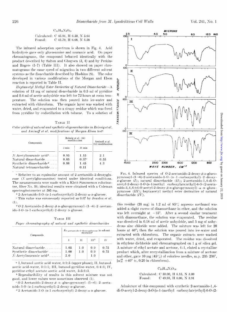
The infrared adsorption spectrum is shown in Fig. 4. Acid hydrolysis gave only glucosamine and muramic acid. On paper chromatograms, the compound behaved identically with the product described by Salton and Ghuysen (3, 4) and by Perkins and Rogers (5-i) (Table III). It also showed on paper rhro- matograms the same speed of migration in two different solvent systems as the disaccharide described by Hoshino (8). The c*olor developed in various modifirations of the Morgan and Elson reaction is reported in Table II.
Ileptaacetyl dlcthyl Ester Derivative of Satural Disaccharide--A solution of 13 mg of natural disaccharide in 0.5 ml of pyridice and 0.4 ml of acetic anhydride was left for 72 hours at room tem-
MICRONS 2.5 4.0 loo 5.0 a.0 12.0 18.0
I 1 ___-_- --.- :v *
perature. The solution was then poured into ice-water and 3 loo extracted uith chloroform. The organic layer was washed with $ water, dried, and evaporated to a sirupg residue which was freed k from pyridine by eodistillation with toluene. To a solution of $
2
TABLE II i? Color yields of natural and synthetic oligosxccharides in Reissig et al.
and Amino$ et al. modijkations of Morgan-Elson test”
Reissig et al. (14) modification Aminoff et al.
(13) modification
3 min 35 min __ --
S-Acetylmuramir aridb 0.95 1.15 2.1 Satural disaccharide 0.05 0.37c 0.55 Synthetic disacrharided 0.88 1.45 4.3 Satural tetrasacrharide 0.11
a Relative to au equimolar amount of 2-acetamido-2-deoxyglu- rose (,~-acet4’lglucosamine) tested under identical conditions. The measuremeuts were made with a Klett-Summerson colorime- ter, filter l;o. 56; ideutical results were obtained with a Coleman spectrophotometer at 585 mp.
6 2-Acetamido-3-O-(n-l-carbox~ethgl)-2-deox~-~-n-g~u~ose. c This value was erroneously reported as 0.67 by Jeauloz et al.
(1). d: 0-2.Acetarnido-2-deoxy-p-o-gluropyranosyl- (l-+6)-2-aretam-
ido-3.0-(n-1-carboxyeth~l).2.deoxy-u-glucose.
TABLE III Paper chromatography of natural and synthetic disaccharides
I II III* IY -..
Satural disaccharide. 1.05 1.0 0.9 0.75 Synthetic disarrharidec. 1.00 1.0 0.9 0.75 ,V-Aretyhnuramic acidd. 2.0 1.0
a I, butanol-acetic acid-wat,er, 4: 1:5 (upper phase); II, but,anol- aretic acid-water, 3:l:l; III, butanol-pyridine-water, 6:4:3; IV, pyridine-ethyl acetate-aretic acid-water, 5:5:1:3.
* Reproducibility of results in this solvent mixture was not good, and lower values were sometimes observed (1).
r 0-2-Acetamido-2-deoxy-p-D-glucopyranosyl- (l-+6) -2-areta- rrlido-3.0.(n-I-rarboxyethZ-1).2.deoxy-o-glurose.
d 2-Aretan~ido-3-O-(~-l-rarbox~ethyl)-2-deoxy-~-n-glu~ose.
01’ I 4000 2500 2ooo 1600 1200 800
WA YE NUMBER. CM-~
FIG. 4. Infrared spectra of O-2-aretamido-2-deoxy$-n-gluro- pyranosyl-(l-+6)-2-acetamido-3-0 - (D -1 -rarboxyethyl) -2-deoxy- n-glurose (I); natural disaccharide (II); 2-acetanndo-1,4-di-O- acetyl-2-deoxy-3-O-[n-l(methy1 carboxylate)ethyl]-6-O-(2-aceta- mido-3,4, (i-tri-O-acetyl-2-deoxy-p-o-glucopyranosyl) -(Y -D -gluco - pyranose (111); heptaacetpl methyl ester derivative of natural disarrharide (IV).
this residue (20 mg) in 1.2 ml of 859; aqueous met,hanol was added a slight excess of diazomethane in ether, and the solution was left overnight at -15’. After a serond similar treatment with diazomethane, the solution was evaporated. The residue was dissolved in 0.18 ml of aretic anhydride, and 5 mg of anhy- drous zinc chloride were added. The mixture was left for 20 hours at 40’; then the solution was poured into ice-water and extracted with chloroform. The organic extracts were washed with water, dried, and evaporated. The residue was dissolved in et,hylene dirhloride and chromatographed on 1 g of silica gel. A mixture of et’hyl acet’ate and acetone, 4 : 1, eluted a crystalline product which, after recrystallization from a mixture of acetone and ether, gave 10 mg (45’0) of colorless needles, m.p. 235 236”, [CC]: +40” (c, 0.20 in chloroform).
Calculated: C 50.00, H 6.15, N 3.89 Found : c 49.93, II 6.06, 1; 3.91
Admixture of this compound with synthetic 2-acetamido-1,4- di-O-acetyl-2-deoxy-3-0-[n-1-(methyl earbosylate)ethyl]-6-0-(2-
by guest on January 22, 2019http://w
ww
.jbc.org/D
ownloaded from

Issue of January 10, lQ66 S. Sharon, T. Osawa, H. M. Flowers, and R. Jr. Jeanlox 227
acetamido-3,4,6-tri-O-awtyl-2-deoxy-P -o-glucopyranosyl) - WD-
glwopgranose (19) depressed the melting point to 210-220”. The infrared adsorption spectrum is reported in Fig. 4, the x-ray diffraction data in Table V, and the mass spectrum in Fig. 6.
Synthesis of 0-6-4cetamido-2-deoxy-P-D-glucopyranosyl-(1 -+ 6)--d-acetan~ido-S-O-(D-l-carboxyethyl)-d-deoxy-D-glz~cose (Corn -
pound II)- Benzyl 2-awtamido-4-0-awtyl-2-deoxy-3-O-[u-l-
TABLE IV
Physical properties of natural and synthetic disaccharides and of their crystalline derivatives
Compounds
JSatural disacrha- ride.
Synthetic disacrha- ride5
Derivative of natu- ral disaccharideb
Derivative of syn- thetic disaccha- ride?.
Melting point
Amorphous
Amorphous
235-236”
240- 241
(methyl carboxylate)ethyl]-6-0-(2-acetamido-3,4,6-tri-O-acetyl- 2-deoxy-fl-n-glucopyranosyl)-a-n-glucopyranoside (Compound 1) (20) (70 mg) was saponified with sodium methoxide at room temperature as previously described (20). The solution was acidified with aretic acid and diluted with 10 ml of absolute ethanol. It was then hydrogenolyzed at room temperature and normal pressure for 6 hours in the presence of an excess (30 to 50
TABLE V
X-ray diffraction data of crystalline heptaacetyl methyl ester derivatives of natural and s!lnthatic disaccharides
Optical rotation, [& Compounds I
X-ray diffraction dataa
+10-t +4.5<
+I6 --f +14
+49
+49
- I- --~- -- _
Solvent
Natural disaccharide, heptaace- t,yl methyl ester
Wat’er
Water
A
13.00H @), 11.33s (2), 7.9ow, 6.71W, 6.42VW, 5.22VH (I), 4.87M (3), 4.53VW, 4.33VW, 4.08&I, 3.90M, 3.71vw, 1.99w, 1.82W
Chloroform
Chloroform
2.Acetamido-1,4-di-O-acetyl-2. 14.73&I, 12.8&I, 10.78W, 9.315 deoxy-3.0-[n-1-(methyl car- (l), 8.04M, 6.6lM, 5.57W. boxylate)ethyl]-6-0-(2-a&a 5.04AM, 4.96M, 4.46s (2), 4.27M mido-3,4,6-tri-0-acetyl-2-d+ (3), 3.83W, 3.71W, 3.53W, oxy-p-D-glucopyranosyl)-ol-n- 3.44M, 3.3OVW, 2.75VW, 2.49
a 0-2.Acet,amido-2-deoxy-p - D - glucopyranosyl - (l+(i) -2 - aceta- glucopyranose mido-3-0-(o-l-carboxyethyl)-2-deoxy-o-gmrose.
) VW, 2.27\W, 2.OOW.
b Heptaacetyl methyl ester. a Data give inderplanar spacings for C&X, radiation with rela- c 2-Aretamido-1,4-di-O-aretyl-2-deoxy-3-O-[n-l-(n~ethyl ear- tive intensities estimated visually. S, strong; M, moderate; W,
boxylate)ethyl]-6-0-(2-aretan~ido-3,4,6-tri-O-acetyI-2-deoxy-~- weak; F’, very; strongest lines are numbered in order of decreas- n-gluropyranosyl)-ol-o-glucopyranose (19). ing intensities (1, strongest).
100 496
r I t I
00-
60 t
602
Ll 600 650
m/e
562
700
FIG. 5. Mass spect’ra between m/e 450 and 700 of synthetic 2-acet~arnido-1,4-di-O-acetyl-2-deoxy-3-O-[n-l-(n~ethyl carboxyl- ate)-ethy1]-6-O-(%acetamido-3,4,6-tri-O-acetyI-2-deoxy-~-n -glucopyranosyl)-a-o-glucose (19) (upper pattern) and of the heptaacetyl methyl ester derivative of the natural disacrharide (lower pattern). The relative abundance is based on wl/e 498 and 602 for the upper and lower patterns, respectively.
by guest on January 22, 2019http://w
ww
.jbc.org/D
ownloaded from
228 Disaccharide from AI. lysodeikticus Cell Walls Vol. 231, X0. 1
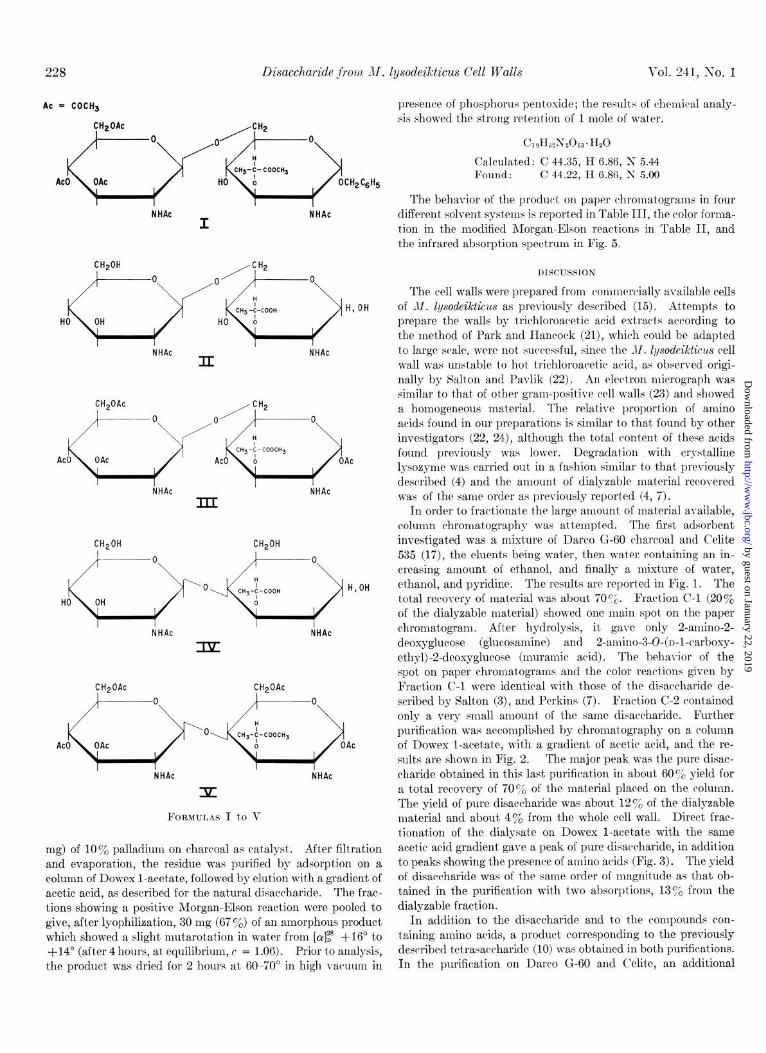
AC = COCH3
NHAc NHAc I
CH20H /CHz
NHAc II
NHAc
kti AC I
III
CH,OH CH,OH
NHAc NHAc
CH20Ac CH,OAc
FORMULAS I to V
mg) of loo/; palladium on charcoal as catalyst. After filtration and evaporation, the residue was purified by adsorption on a column of Dowex l-acetate, followed by elution with a gradient of acetic acid, as described for the natural disaccharide. The frac- tions showing a positive Morgan-Elson reaction were pooled to give, after lyophiliaation, 30 mg (67%) of an amorphous product which showed a slight mutarotation in water from [ar]z* $16” to +14” (after 4 hours, at equilibrium, c = 1.06). Prior to analysis, the product was dried for 2 hours at 60-70” in high vacuum in
presence of phosphorus pent)oxide; the results of chemical analy- sis showed the strong retention of 1 mole of water.
Calculated: C 44.35, H 6.86, S 5.44 Found : C 44.22, II 6.86, S 5.00
The behavior of the product on paper chromatograms in four different solvent systems is reported in Table III, the color forma- tion in the modified Morgan-Elson react,ions in Table II, and the infrared absorption spectrum in Fig. 5.
DISCUSSION
The cell walls were prepared from commercially available cells of M. Zgsodeikticus as previously described (15). Attempts to prepare the walls by trichloroacetic acid extracts according to the method of Park and Hancock (al), which could be adapted to large scale, were not successful, since the M. Zysodeikticus cell wall was unstable to hot trichloroacetic acid, as observed origi- nally by Salton and Pavlik (22). iin electron micrograph was similar to that of other gram-positive cell walls (23) and showed a homogeneous material. The relative proportion of amino acids found in our preparations is similar to that found by other investigators (22, 24), although the total content of these acids found previously was lower. Degradation with crystalline lysozyme was carried out in a fashion similar to that previously described (4) and the amount of dialyzable material recovered was of the same order as previously reported (4, 7).
In order to fract’ionate t,he large amount of mat’erial available, column chromatography was attempted. The first adsorbent investigated was a mixture of Darco G-60 charcoal and C’elite 535 (17), the eluents being water, then water containing an in- creasing amount of ethanol, and finally a mixt’ure of wat.er, ethanol, and pyridine. The results are reported in Fig. 1. The total recovery of material was about 7076. Fraction C-l (20% of the dialyzable material) showed one main spot on the paper chromatogram. After hydrolysis, it gave only 2-amino-2- deoxyglucose (glucosamine) and 2-amino-3-@(n-l-carboxy- ethyl) -2-deoxyglucose (muramic acid). The behavior of the spot on paper chromat.ograms and the color reactions given by Fraction C-l were identical with those of the disaccharide de- scribed by Salt’on (3), and Perkins (7). Fraction C-2 contained only a very small amount of the same disaccharide. Further purification was accomplished by chromatography on a column of Dowex l-acetate, with a gradient of acetic acid, and the re- sults are shown in Fig. 2. The major peak was the pure disac- charide obtained in this last purification in about 60% yield for a total recovery of 70 g/, of the material placed on the column. The yield of pure disaccharide was about 12% of the dialyzable material and about 4% from the whole cell wall. Direct frac- tionation of the dialysate on Dowex l-acetate with the same acetic arid gradient gave a peak of pure disaccharide, in addition to peaks showing the presence of amino acids (Fig. 3). The yield of disaccharide was of the same order of magnitude as that ob- tained in the purification with two absorptions, 13% from the dialyzable fraction.
In addition to the disaccharide and to the compounds con- taining amino arids, a product corresponding to the previously described tetrasaccharide (10) was obtained in both purifications. In the purification on Darco G-60 and Celite, an additional
by guest on January 22, 2019http://w
ww
.jbc.org/D
ownloaded from
Issue of January 10, 1966 N. Sharon, T. Osawa, H. M. Flowers, and R. W. Jeanlox 229
amorphous compound was isolated in a small yield (0.1 y0 of the weight of the cell wall) ; on paper chromatograms its speed of migration corresponded to that of a disaccharide and it showed the presence of only 2-amino-2-deoxyglucose after hydrolysis.
The glucosamine-muramic acid disaccharide was shown to be homogeneous on paper chromatograms in four different solvent systems (Table III). It migrated on paper in a manner identi- cal with the major spot of a product kindly provided by Dr. 0. Hoshino. It was amorphous, but showed a slight mutarotation in water. The elemental analysis corresponded to that of a disaccharide composed of 2-acetamido-2-deoxyglucose and 2- acetamido-3-O-(o-1-carboxyethyl)-2-deoxyglucose. It was com- pared with synthetic 0-2-acetamido-2-deoxy-p-D-glucopyranosyl- (l&6)-2-acetamido-3-0- (~-1 -carboxyethyl) -2-deoxy-n-glucose (Compound II). This compound was obtained as an amorphous product in 67 y0 yield by alkaline hydrolysis followed by catalytic hydrogenolysis of benzyl 2-acetamido-4-O-acetyl-2-deoxy-3-0- [o-l-(methyl carboxylate)ethyl]-6-O-(2-acetamido-3,4,6-tri-O- acetyl-2-deoxy-P-n-glucopyranosyl)-cr-n-glucopyranoside (Com- pound I) (20). Structure II had been proposed for the disaccharide by Salton and Ghuysen (9, 10) and by Perkins (11).
Both disaccharides showed similar speeds of migration on paper chromatograms in three solvent systems, but the natural product was slightly faster on development with the upper phase of butanol-acetic acid-water, 4: 1:5 (Table III). The optical rotations at equilibrium were slightly different (Table IV), but the infrared spectra (Fig. 4) showed no significant discrepancy. Both products showed significant differences in the Morgan-Elson test, however, either in the modification of Reissig et al. (14) with a 3-min or a 35-min heating time or in the modification of Aminoff et al. (13). The X,,, (545 and 585 rnp) and Xmin (565 rnp) of the absorption curves were identical for the colors formed by both compounds. When the colors obtained for both prod- ucts in the three modifications were compared on a molar basis with the colors formed by crystalline 2-acetamido-3-0-(n-l- carboxyethyl)-2-deoxy-ar-n-glucose (N-acetylmuramic acid) (20)) the intensity of the color developed by the natural disaccharide was approximately 5,30, and 25 %, whereas that of Compound II was 95, 125, and 200%, respectively (Table II). The low yield in color produced by the natural disaccharide has already been observed by other investigators (8-11).
Since both the natural disaccharide and Compound II were amorphous, the preparation of various crystalline derivatives was attempted. Finally the 01 anomer of the heptaacetyl methyl ester derivative of the natural disaccharide was obtained in crystalline form after treatment with acetic anhydride in pyridine solution, diazomethane, and then acetic anhydride and zinc chloride. Since a sufficient amount of Compound II was not available to prepare the cr anomer of the heptaacetyl methyl ester by the same method, a new synthesis of 2-acetamido-1,4-di- 0-acetyl-2-deoxy-3-O- [D - 1 - (methyl carboxylate)ethyl] - 6- 0-(2- acetamido-3 ,4, 6-tri-0-acetyl-2-deoxy-@-n-glucopyranosyl)-cu-n- glucopyranose (Compound III) was devised and is reported elsewhere (19). The optical rotations of both derivatives were identical (Table IV) ; their melting points (Table IV), and their infrared adsorption curves (Fig. 4) were nearly identical. In admixture, however, both products gave a sharp depression of the melting point. Further evidence for the nonidentity of the peracetylated natural disaccharide and Compound III was ob- tained by comparing the x-ray powder diffraction patterns of
both products recrystallized from the same solvent mixture (Ta- ble V) and their mass spectra (Fig. 5). In the latter case, be- cause of the complexity of the starting materials, no attempt was made to identify the resulting fragments. The striking difference of the patterns of fragmentation clearly indicates that the natural and synthetic disaccharides are not identical.
Since Structure II proposed for the natural disaccharide by Salton and Ghuysen (9, 10) and by Perkins (11) is not correct, it is necessary to re-examine their results and interpretations. The structure 0-2-acetamido-3-0-(n-l-carboxyethyl)-2-deoxy-n- glucopyranosyl-(1 --) 4)2-acetamido-2-deoxy-n-glucopyranose, or N-acetylmuraminyl-(1 + 4)N-acetylglucosamine, proposed by Hoshino (8), is not acceptable in view of the results of the alka- line iodine oxidation obtained by Perkins (11) and of the reduc- tion with sodium borohydride obtained by Salton and Ghuysen (9, 10). The latter reduction is unfortunately not conclusive when applied to 2-acetamido-3-0-(n-1-carboxyethyl)-2-deoxy-n- glucose, since, in addition to a reduction, epimerization can take place at C-2 (25) as well as @ elimination of the n-1-carboxyethyl group at C-3 under alkaline conditions (26). Attempts to ob- tain the pure 2-amino-3-O-(n-l-carboxyethyl)-2-deoxy-r,-glucitol or its N-acetyl derivative by sodium borohydride reduction have not been successful as yet.’
Since the 2-acetamido-2-deoxy-n-glucose is the nonreducing moiety of the natural disaccharide, the only possible structure is that of 0-2-acetamido-2-deoxy-/3-n-glucopyranosyl-(l+4)-2- acetamido-3-0-(n-l-carboxyethyl)-2-deoxy-n-glucose (IV) and, for its heptaacetyl methyl ester derivative, Structure V.
Salton and Ghuysen (9, 10) based Structure II on the failure of the natural disaccharide to release formaldehyde after periodate oxidation. This behavior of a product possessing Structure IV can be explained by the disaccharide being in the pyranose form. In this case no formaldehyde is formed, as was shown in the oxi- dation of 2,3,4-tri-O-methyl-n-glucose (27) or of 2,4-di-O- methyl-n-galactose (28). Another possibility would be stabiliza- tion of the formyl ester at C-5 as a result of the oxidation of the C-l to C-2 link in the 2-acetamido-3-0-(o-1-carboxyethyl)-2- deoxy-n-glucose moiety, and no further oxidation would take place between C-5 and C-6. This possibility was demonstrated in the oxidation of 3-O-methyl-n-glucose, 3,4-di-o-methyl-n- galactose, and 3,4-di-0-methyl-n-mannose (28). It is of interest that most of the derivatives of glucose, galactose, and mannose just mentioned are substituted at C-3 and C-4, as is the 2-aceta- mido-3-O-(n-1-carboxyethyl)-2-deoxy-n-glucose moiety of Com- pound IV.
A (1 ---f 6) linkage for the natural disaccharide was proposed by Perkins (11) on the basis of a positive Morgan-Elson reaction, since 2-acetamido-2-deoxyglucose derivatives substituted at C-4 had been shown to give no color in this reaction (26, 29). When the color formed by the natural disaccharide is compared with that formed by crystalline 2-acetamido-3-O-(o-l-carboxyethyl)- 2-deoxy-n-glucose or by the synthetic disaccharide II, however, very low values are obtained under the usual conditions (Table II). The value was much higher after a 35-min heating time, but under these conditions N , N’-diacetylchitobiose, a disac- charide with (1 + 4) linkage, also shows a positive reaction as reported by Salton and Ghuysen (10). A C-4-substituted amino sugar, 2-4-diacetamido-2,4-dideoxy-n-glucose, has also been
1 R. W. Jeanloz and E. Walker, unpublished results.
by guest on January 22, 2019http://w
ww
.jbc.org/D
ownloaded from
230 Disaccharide jrom M. lysodeikticus Cell Walls Vol. 241, No. 1
shown to give a positive Morgan-Elson reaction after a prolonged time of reaction (30).
It can thus be concluded that Structure IV is the most likely one for the disaccharide isolated from M. Zysodeikticus cell wall after digestion with egg white lysozyme. Final evidence will have to await the synthesis of Compound IV, in which we are presently engaged.
BcIcnowZecZgments-The authors wish to thank Dr. G. L. Mechanic for the quantitative determination of the amino acids, Dr. D. Horton for the x-ray diffraction data, Dr. J. Dudek for the mass spectra, and Dr. 0. Hoshino for a sample of disac- charide isolated from M. Zysodeikticus cell walls.
1.
20.
21.
22.
2. 3. 4.
JEANLOZ, R. W., SHARON, N., AND FLOWERS, H. M., Biochem. and Biophys. Research Communs., 13, 20 (1963).
SALTON, M. R. J., Biochim. et Biophys. Acta, 22, 495 (1956). SALTON, M. R. J., Biochim. et Biophys. Acta, 34, 308 (1959). GHUYSEN, J. M., .IND SALTON, M. Ii.. J., Biochim. et Biophys.
Acta, 40, 462 (1960). 5. PERKINS, H. R., AND ROGERS, H. J., Biochem. J., 69, 15P
(1958).
23.
24.
25.
6. PERKINS, H. R., AND ROGERS, H. J., Biochem. J., 72, 647 26. (1959).
7. 8. 9.
10.
PERKINS, H. R., Biochem. J., 74, 186 (1960). HOSHINO, O., Chem. Pharm. Bull. (Tokyo), 8, 405,411 (1960). SALTON, M. R. J.. AND GHUYSEN, J. M.. Biochim. et BioDhus.
Acta, ‘36, 552 (1959). I ”
SALTON, M. R. J., AND GHUYSEN, J. M., Biochim. et Biophys. Acta, 46, 355 (1960).
27. JEANLOZ, R., Helv. Chim. Acta, 27, 1509 (1944). 28. BELL, D. J.; J. Chem. Sot., 992 (1948). 29. JEANLOZ. R. W.. END TRBMAGE. M.. Federation Proc.. 15. 282
30.
REFERENCES
11. 12.
13.
14.
15. 16. 17.
18. YPHANTIS, D., Ann. iv. Y. Acad. Sci., 88, 586 (1960). 19. &.4wa, T.. AND JESNLOZ. R. W.. Carbohudrate Research. 1.
PERKINS, H. R., Biochem. J., 74, 182 (1960). MORGAN, W. T. J., AND ELSON, L. A.: Biochem. J., 26, 988
(1934). AMINOFF, D., MORG.IN, W. T. J., AND WATKINS, W. M., Bio-
them. J., 51, 379 (1952). REISSIG, J. L., STROMINGER, J. L., AND LELOIR, L. F., J.
Biol. Chem., 217, 959 (1955). SHARON, N., AND JEANLOZ, R. W., Ezperientia, 20, 253 (1964). PIEZ, K. A., AND MORRIS, L., Anal. Biochem., 1, 187 (1960). WHISTLER, R. L., .\ND DURSO, D. F., J. Am. Chem. Sot., 72,
677 (1950).
181 (i965j. FLOWERS, H. M., AND JE‘INLOZ, R. W., J. Org. Chem., 28, 2983
(1963). P.4RK, J. T., AND HANCOCK, R., J. Gen. Microbial., 22, 249
(1960). SALTON, M. R. J., .-END P~VLIK, J. G., Biochim. et Biophys.
Acta, 39, 398 (1960). SALTON, M. R. J., The bacterial cell wall, American Elsevier
Publishing Company, Inc., New York, 1964, p. 66. CZERKAWSKI, J. W., PERKINS, H. It., .IND ROGERS, H. J.,
Biochem. J., 86, 468 (1963). RoSEM.~N. S.. AND COMB. D. G.. J. Am. Chem. Sot.. 80. 3166
(1958). ’ ’ I
KUHN, R., GAUHE, A., IIND BAER, H. H., Chem. Ber., 87, 1138 (1954).
(1956).’ ’ I I I
JEANLOZ, R. W., 1\~~ RAPIN, A. M. C., J. Org. Chem., 28, 2978 (1963).
by guest on January 22, 2019http://w
ww
.jbc.org/D
ownloaded from
Nathan Sharon, Toshiaki Osawa, Harold M. Flowers and Roger W. Jeanloz Cell WallsMicrococcus lysodeikticus
Isolation and Study of the Chemical Structure of a Disaccharide from
1966, 241:223-230.J. Biol. Chem.
http://www.jbc.org/content/241/1/223Access the most updated version of this article at
Alerts:
When a correction for this article is posted•
When this article is cited•
to choose from all of JBC's e-mail alertsClick here
http://www.jbc.org/content/241/1/223.full.html#ref-list-1
This article cites 0 references, 0 of which can be accessed free at
by guest on January 22, 2019http://w
ww
.jbc.org/D
ownloaded from