isolation and screening of effective trichoderma spp. against the … jat2010_102fj.pdf ·...
TRANSCRIPT

Journal of Agricultural Technology 2011, Vol.7(3): 623-635
623
Isolation and screening of effective Trichoderma spp. against the root rot pathogen Macrophomina phaseolina Sreedevi, B., Charitha Devi, M.* and Saigopal, D.V.R. Sri Venkateswara University, Tirupati -517501. A.P, India. Sreedevi, B., Charitha Devi, M. and Saigopal, D.V.R. (2011). Isolation and screening of effective Trichoderma spp. against the root rot pathogen Macrophomina phaseolina. Journal of Agricultural Technology 7(3): 623-635. The main emphasis of the present study was screened and evaluation of effective Trichoderma spp. for biocontrol of Macrophomina phaseolina the causative agent of root rot of groundnut. Five Trichoderma spp. were isolated from the rhizosphere soil of healthy groundnut plants, identified using morphological and microscopic characteristics and were evaluated for in vitro antifungal activity against M. phaseolina by dual culture plate technique and bioassay methods (in vitro antibiosis). Scanning electron microscopy was used to study the conidial surface of T. harzianum. Among the five isolates T. harzianum (T3), T.viride (T1) had maximum antifungal activity against M.phaseolina compared to the other Trichoderma spp. In dual culture technique T. viride and T. harzianum reduced mycelial growth by 61.1% and 64.4% respectively. Based on the dual culture technique, T. harzianum (T3), T. viride (T1) were selected for further research. Metabolites released from T. harzianum, T.viride were tested in culture medium against M.phaseolina. Cell free metabolites of T. viride, T. harzianum inhibited the growth of M. phaseolina in vitro and appeared to be fungicidal. The inhibition varied depending on the Trichoderma species producing metabolites, T. viride inhibited fungal growth upto 69% and T. harzianum upto 72.7% in nonvolatile and 47%, 64.7% in volatile metabolites respectively. The medium was optimized for mass multiplication of Trichoderma spp. T. harzianum, T. viride were tested for their ability to protect groundnut plants from disease caused by M. phaseolina in pot culture experiment. The growth of groundnut plants with the antagonist alone or in combination with pathogen was greater than in plants inoculated with pathogen alone. Key words: T. harzianum, T. viride, M. phaseolina, biological control, groundnut Introduction
Groundnut (Arachis hypogaea .L) is one of the chief commercial crops of India. It is the primary source of vegetable oil and contributes to about 43% of the total oil production in India. Species of Trichoderma, Gliocladium and Aspergillus etc. have been found effective in reducing the sheath blight and extensively explored for the control of soil borne plant pathogens (Khan and * Corresponding author: M. Charitha Devi; e.mail: [email protected]
Journal of Agricultural Technology 2011 Vol. 7(3): 623-635 Available online http://www.ijat-aatsea.com
ISSN 1686-9141

624
Sinha, 2005). Trichoderma sp. is one of the most important biocontrol agent used for management of different diseases (Harman, 2004). Trichoderma spp. are free living fungi that are common in soil and root ecosystems and promote plant growth (Yedidia, 2001). Trichoderma spp. are effective in control of soil/seed-borne fungal diseases in several crop plants (Kubicek et al., 2001). Different isolates of P. fluorescens and Trichoderma spp. were identified as biocontrol agents of groundnut stem rot and other soil-borne diseases (Podile and Kishore, 2002). These free-living fungi are ubiquitous in the soil environment and are being successfully used and commercialized to combat a broad range of phytopathogenic fungi such as Rhizoctonia solani, Pythium ultimum, and Botrytis cinerea (Hjeljord et al., 2000; Desai et al., 2002; Fravel, 2005). Trichoderma spp. can directly impact other fungi, after sensing a suitable fungal host, Trichoderma sp. responds with the production of antibiotic compounds, formation of specialized structures, and degradation of the host’s cell wall, followed by the assimilation of its cellular content, a process known as mycoparasitism (Chet and Chernin, 2002; Steyaert et al., 2003; Benitez et al., 2004). The mechanisms of mycoparasitism, antibiosis and competition afforded by Trichoderma spp. have been widely studied (Howell, 2003; Harman et al., 2004b). T. harzianum has been evaluated for the control of charcoal stem root rot of melon in Egypt (Khalifa and Linddel, 1995). Trichoderma harzianum (Rifai) is a most potent biocontrol agent which inhibits the growth of F. solani, causal organism of wilt of brinjal, efficiently both in vitro and in field condition (Chakraborty, 2005). Seed treatment with T. harzianum and T.viride was more effective incontrolling R. solani in Phaseolus vulgaris both under greenhouse and field condition ( Robert et al. 1993). Recently, several attempts have been undertaken to survey Trichoderma spp. promotion of seedling establishment, enhancement of plant growth and elicit plant defense reaction in some crops such as cotton (Shanmugaiah et al., 2009), bean (Hoyos-Carvajal et al., 2009). Using T. harzianum control of soil borne plant pathogens has been reported by many investigators (Ulkhede 1992, Ziedan et al., 2005) but there is little information was available on the use of T. viride and T. harzianum as biocontrol agents against M.phaseolina in groundnut plants. The objective of the present investigation was isolation and screening of effective Trichoderma spp. against the root rot pathogen M. phaseolina and optimization of cultural conditions for the growth of Trichoderma spp.
Materials and methods
Isolaton of Biological control agents from soil by serial dilution method: Different strains of Trichoderma were isolated from rhizosphere soil of healthy groundnut plants by serial dilution technique on Trichoderma specific medium.

Journal of Agricultural Technology 2011, Vol.7(3): 623-635
625
(MgSO4.7H2O-0.2g, K2HPO4-0.9g, KCl-0.15g, NH4NO3-3.0g, Glucose-3.0g, Agar-15g, Rosebengal-0.15g, Chloramphenicol-0.25g, Distilled water-1000ml, pH-6.5) (Elad, 1980). Isolation of Macrophomina phaseolina
Grounndnut (Arachis hypogaea .L) plants showing root rot symptoms were identified, collected for the isolation of the pathogen . The isolation of the pathogen from diseased plant roots was performed on potato dextrose agar (Potatoes-200g, Dextrose- 20g, Agar- 20g, Distilled water-1000ml) medium and identified according to its morphology and colony characteristics.
Scanning electron microscopy: To study the surface of conidia of T. harzianum: Material for SEM examination of conidial surfaces was obtained from cultures that were grown on PDA for 10 days at 30oC. Agar blocks were cut into small pieces with conidiating hyphae and fixed in 2% glutaraldehyde in 0.1 M NaPO4 buffer and post fixed in buffered 1% O5O4 for 2 hours. The material was then dehydrated in an ethanol series (10, 25, 60, 75, 95 and 100%) with 15 min per change. The specimens were dried in a critical point drying apparatus and viewed using a scanning electron microscope (Itamar Soares de Melo, 2004). Dual culture technique
Dual culture plate technique (Webster, 1971b) was used to study the antagonistic effects of the Trichoderma isolates on M.phaseolina. All antagonistic pathogen combinations were examined on 20ml of PDA in 9-cm petriplates, with four replicate plates per treatment. For dual culture technique, a mycelial plug (0.5cm in diameter), taken from actively growing 3 day old culture of M. phaseolina and Trichoderma isolates placed 8cm apart from each other on the PDA. For control treatments, a plug of M. phaseolina was placed on the PDA medium. The plates were incubated at 28oC. Observations on the antagonistic activities of Trichoderma isolates on M. phaseolina were recorded after every 24hr for 5days and inhibition percentage was calculated using the following formula (Edington, 1971).
Inhibition percentage (%) = A1-A2/A1 x 100
Where, A1 is the colony area of uninhibited M. phaseolina in the control, and A2 is the colony area of M. phaseolina in dual culture.

626
Effect of non-volatile compounds produced by antagonist(s) on the radial growth of Macrophomina phaseolina
The effect of culture filtrate of the fungal antagonist(s) on the growth of M. phaseolina was studied by method described by (Bruce, 1984; Dennis, Webster, 1971b). Trichoderma isolates were grown in potato dextrose broth at 27oC with intermittent shaking at 150rpm. Metabolites were collected after 10days and filtered through the Whatmann No.1 filter paper and centrifuged at 2000 rpm for 10 min. The supernatant was filtered through Sartorius Millipore (0.4µ) filter. Filtrates were amended in PDA to make 5%, 10%, 20%, 40% concentration in petriplates. Solidified agar plates were inoculated at the centre with 6mm diameter mycelial disc of pathogen and incubated at 28oC for 5 days. Plates without filtrate served as control.
Effect of volatile compounds produced by antagonist(s) on the radial growth of Macrophomina phaseolina
The effects of volatile metabolites from Trichoderma isolates were tested. Fungal biocontrol agent(s) were grown on petriplates containing PDA medium for 48hr. The lid of each petriplate was replaced with the bottom position of a petriplate dispensed with PDA and inoculated with M. phaseolina at the centre. The bottom of the petriplate containing centrally inoculated mycelial disc of test fungus (M. phaseolina) was kept inverted to the petriplate containing PDA media only which served as control. The pairs of each plate were attached together with cellophane adhesive tape. Observations on the radial growth of the test pathogen were recorded after 24, 48 and 96 hr of incubation at 28+ 1oC. The colony diameter of the test fungus in the treatment in comparison with that of control gave growth inhibition percent.
Influence of various nutrients on the growth of Trichoderma species
In all nutritional studies aliquots of 50ml of the medium were taken into 250ml conical flasks, plugged with cotton and sterilized. The basal medium used was Czapek-Dox medium of the above composition. The initial pH of all the media was adjusted to pH 5.0 and the cultures were incubated for 10 days at room temperature as stationary cultures. A 0.5cm disc of actively growing mycelium cut from the periphery of a 7 day old culture with the help of a sterile cork borer and inoculated on to czapek dox medium. The flasks were incubated for 10 days at 28+2oC as stationary culture. A number of carbon, nitrogen, sulphur and phosphorus compounds were tested for their effect on growth of the Trichoderma species (Grondona, 1997).

Journal of Agricultural Technology 2011, Vol.7(3): 623-635
627
Pot culture technique
Oatmeal sand was prepared in 250ml conical flasks. Each flask inoculated with T. viride, T. harzianum, M. phaseolina separately and incubated at 28+ 2oCfor 10 days. 1kg of soil and 1kg of sand was taken into polythene bags and sterilized at 121oC for 30min at 15lbs pressure for two successive days. 9”earthenware pots were taken; sterilized sandy soil was added into the pots. Surface sterilized groundnut seeds were sown in pots filled with sandy soil containing M. phaseolina, T. viride, T. harzianum separately and in combinations. Replicates were maintained for each treatment. Control pots also maintained without any fungal cultures. On germination of seeds symptoms, root/shoot length, dry weights were recorded.
Results and discussion Isolation and Identification of Trichoderma isolates
Trichoderma species were isolated according to Elad et al. (1981) method using Trichoderma selective medium (TSM) and then subcultured on potato dextrose agar. Based on the conidiophore morphology, the isolates were designated as T1, T2, T3, T4, and T5. Based on the mycelial characteristics, the fungus was identified as Macrophomina phaseolina (Dhingra and Sinclair, 1973). Under scanning electron microscope the fine structure of T. harzianum conidial surfaces and conidial masses on a phialide was observed (Fig. 1). The ability of Trichoderma isolates to inhibit the mycelial growth of M. phaseolina in dual culture was determined on PDA medium.
Fig. 1. Scanning microscopy of fixed conidia and conidial masses of T.harzianum.

628
Effect of Trichoderma species on Mycelial growth of Macrophomina phaseolina in vitro
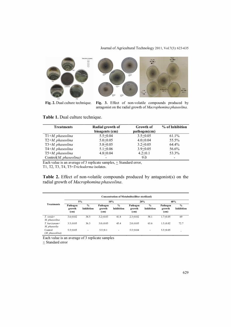
All the five isolates of Trichoderma spp. exhibited antibiotic potential against M. phaseolina by inhibiting its mycelial growth (Fig. 2). Five days after inoculation, growth of M.phaseolina was found to be inhibited by T1, T2, T3, T4, T5 isolates and attained a growth of 5.5cm, 5.0cm, 5.8cm, 5.1cm, and 4.8 cm respectively. This indicates that T3 (T. harzianum) and T1 (T. viride) isolates had maximum antifungal activity against M. phaseolina compared to the other Trichoderma spp (Table 1). Based on microscopic characters, conidiophore branching patterns and conidium morphology T3 and T1 isolates indentified as T. harzianum, T. viride respectively (Rifai, 1969). Majumdar et al. (1996) observed that T. viride and T. harzianum were inhibitory to M. phaseolina causing blight of mothbean. Based on dual culture technique, T. harzianum, T. viride species were used for further research. Culture metabolites of T.viride at 40% inhibited the growth of M. phaseolina upto 69% where as the culture metabolites of T. harzianum inhibited M.phaseolina by 72.7% at 40% (Fig. 3). Antibiotic substances were produced in sufficient concentration to affect the growth of soil fungi M. phaseolina by Trichoderma spp (Table 2). The effect of non volatile culture metabolites on M. phaseolina revealed that T. harzianum, T. viride isolates were effective for inhibition of mycelial growth of M. phaseolina (Dubey and Dwivedi, 1988; Mathur and Bhatnagar, 1994). 24hrs old culture of T. harzianum produced maximum volatile metabolites which accounted for 64.7% inhibition of growth of M. phaseolina followed by T. viride (47%) (Fig.4, Table 3). Volatile and non volatile antibiotics produced by T. harzianum are responsible for the inhibitor action against root pathogen, Fusarium culmorum (Iqbal et al., 1994) and F. oxysporum (Michrina et al., 1995).
Optimization of cultural conditions for the growth of Trichoderma species
All Trichoderma isolates were able to grow in the presence of different carbon sources. Of different carbon sources tested, sucrose was proved to be the best source of carbon, followed by glucose, cellulose, starch, and lactose. Poor growth was observed on fructose, glycerol and pectin. Among different nitrogen sources tested, sodium nitrate and potassium nitrate supported good growth of the Trichoderma isolates; where as moderate growth was observed in ammonium nitrate, magnesium nitrate and urea. Among the different sulphur sources, sodium sulphate and potassium sulphate yielded maximum growth of the Trichoderma isolates, while the others supported only moderate growth (Table 4).
Journal of Agricultural Technology 2011, Vol.7(3): 623-635
629
Fig. 2. Dual culture technique. Fig. 3. Effect of non-volatile compounds produced by antagonist on the radial growth of Macrophomina phaseolina.
Table 1. Dual culture technique.
Treatments Radial growth of bioagents (cm)
Growth of pathogen(cm)
% of Inhibition
T1+M. phaseolina 5.5+0.04 3.5+0.05 61.1% T2+M. phaseolina 5.0+0.05 4.0+0.04 55.5% T3+M. phaseolina 5.8+0.05 3.2+0.05 64.4% T4+M. phaseolina 5.1+0.06 3.9+0.05 56.6% T5+M. phaseolina 4.8+0.04 4.2+0.1 53.3% Control(M. phaseolina) - 9.0 -
Each value is an average of 3 replicate samples, + Standard error, T1, T2, T3, T4, T5=Trichoderma isolates.
Table 2. Effect of non-volatile compounds produced by antagonist(s) on the radial growth of Macrophomina phaseolina.
Each value is an average of 3 replicate samples + Standard error
Treatments
Concentration of Metabolite(filter sterilized)
5% 10% 20% 40% Pathogen
growth (cm)
% Inhibition
Pathogen growth (cm)
% Inhibition
Pathogen growth (cm)
% Inhibition
Pathogen growth
(cm)
% Inhibition
T. viride+ M. phaseolina
3.6+0.02 34.5 3.2+0.03 41.8 2.3+0.02 58.1 1.7+0.05 69
T. harzianum+ M. phaseolia
3.5+0.05 36.3 3.0+0.05 45.4 2.0+0.05 63.6 1.5+0.02 72.7
Control (M. phaseolina)
5.5+0.05 - 5.5+0.1 - 5.5+0.04 - 5.5+0.05 _

630
Table 3. Volatile compounds produced by Trichoderma spp. against Macrophomina phaseolina.
Treatments Radial growth of Macrophomina
phaseolina (cm) % inhibition
24 hr 48 hr 72 hr 24 hr 48 hr 72 hr T.harzianum+M.phaseolina 0.6+0.1 1.9+0.1 2.6+0.1 64.7 42.4 39.5 T.viride+ M.phaseolina 0.9+0.05 2.5+0.1 3.7+0.05 47 21.2 13.9 Control(M.phaseolina) 1.7+0.05 3.2+0.05 4.3+0.05 - - - Each value is an average of 3 replicate samples, + Standard error. Table 4. Effect of Carbon compounds on the growth of Trichoderma isolates.
Carbon sources Average dry weight in mg/50ml T1 T2 T3 T4 T5
Glucose 650 570 980 800 120 Lactose 490 120 100 50 280 Fructose 160 90 60 110 60 Cellulose 890 650 590 580 880 Starch 610 280 650 160 350 Pectin 20 40 80 30 70 Glycerol 220 60 120 160 50 Sucrose 1000 10 1230 480 260
Nitrogen sources Ammonium nitrate 170 100 300 200 60 Potassium nitrate 240 200 450 140 250 Sodium nitrate 1000 560 1200 480 260 Urea 140 120 110 150 120 Magnesium nitrate 180 145 185 137 140
Sulphur sources Magnesium sulphate 880 530 780 830 970 Copper sulphate 300 340 450 690 620 Sodium sulphate 860 900 890 980 810 Potassium sulphate 910 840 850 910 710 Phosphorus sources
Potassium phosphate 540 900 870 850 620 Sodium phosphate 870 880 820 600 880 Calcium phosphate 400 700 880 530 670 Ammonium phosphate 840 730 670 790 640
T1, T2, T3, T4, T5=Trichoderma isolates.

Journal of Agricultural Technology 2011, Vol.7(3): 623-635
631
Fig. 4. Volatile compounds produced by Trichoderma spp. against Macrophomina phaseolina. Biological control of Macrophomina phaseolina on groundnut in plot culture technique
Maximum root length was recorded in T. harzianum, T. harzianum+M. phaseolina, T. viride, and T. viride+M. phaseolina treated plants compared to M. phaseolina inoculated plants (Table 5). Maximum shoot length was recorded in T. harzianum, T. harzianum+M. phaseolina, T. viride, and T. viride+M. phaseolina treated plants compared to M. phaseolina inoculated plants (Table 6). Similarly, T. harzianum and T. viride and their fusants enhanced rice and tomato shoot and root lengths (Balasubramanian, 2003).
Table 5. Effect of Trichoderma spp. and M.phaseolina on root length (cm) of groundnut at different stages of growth.
Treatments Stage1 Stage2 Stage3 T.viride 6.5+0.05 14.5+0.04 18.3+0.1 T.viride+M.phaseolina 7.2+0.05 12.7+0.1 17.4+0.05 T.harzianum 9.3+0.01 15.8+0.05 23.2+0.06 T.harzianum+M.phaseolina 8.6+0.1 14.3+0.1 20.6+0.05 M.phaseolina 3.5+0.05 6.8+0.08 14.7+0.04 Control 6.5+0.05 14.2+0.05 16.3+0.05
Each value is an average of 3 replicate samples, + Standard error.
Table 6. Effect of Trichoderma spp.and M.phaseolina on shoot length(cm) of groundnut at different stages of growth.
Treatments Stage1 Stage2 Stage3 T.viride 16.4+0.2 27.3+0.7 37.4+0.5 T.viride+M.phaseolina 14.3+0.05 24.2+0.5 38.3+0.9 T.harzianum 19.2+0.1 28.5+0.3 39.2+0.4 T.harzianum+M.phaseolina 17.5+0.4 26.3+0.5 38.3+0.7 M.phaseolina 7.5+0.7 16.2+0.08 17.3+0.5 Control 16.2+0.5 22.4+0.4 27.5+0.09
Each value is an average of 3 replicate samples, + Standard error.

632
Biological control of plant pathogens by microorganisms has been considered as a natural and environmentally acceptable alternative to the existing chemical treatment methods (Baker, Paulitz, 1996). Intensified use of fungicides resulted in the accumulation of toxic compounds potentially hazardous to humans and environment (Cook, Baker, 1983). The T. harzianum (T3), T. viride (T1) isolates were better potential biocontrol agents against M. phaseolina in vitro compared to other isolates of Trichoderma spp. According to Hermosa et al (2000) T. harzianum had a potential biocontrol activity in a dual culture studies against the phytopathogenic fungi of Phoma betae, Rosellinia necatrix, Botrytis cinerea and Fusarium oxyporum f. sp. dianthia in the three different media. The present work of in vitro plate assays showed that T. harzianum is more effective in suppressing the growth of M. phaseolina followed by T.viride. With the increase in concentration of culture filtrates of the bioagent, the radial growth of test pathogen was proportionally decreased. Maximum inhibition of the mycelial growth of M. phaseolina was observed with the culture filtrate of T. harzianum used as 40% concentration. Seshagiri and Eswaran (2002) reported that mycelial growth of pathogen decreases with the increase in the concentration of the culture filtrate produced by fungal antagonists from 10 to 40 percent and no growth at 50%. T. harzianum overgrew M. phaseolina and suppressed its growth in vitro (Patel and Anahosur 2001).Volatile toxic substances produced by antagonists could diffuse easily through their filled pores of the soil and inhibited the soil borne pathogen M. phaseolina specially suppressing the sclerotia formation. Kukuk and Kvanc (2004) reported that T. harzianum strains produced a metabolite that inhibit growth of plant pathogenic fungi. Similar results reported that seed treatment with T. harzianum and T.viride was more effective in controlling Rhizoctonia solani in Phaseolus vulgaris under both green house and field conditions (Prashanthi et al 1997). Biological activity of antagonistic fungi and bacteria may partially be associated with production of antibiotics (Etebarian et al., 2000; Faull et al., 1994; Pusey and Wilson, 1984). The present investigation suggests that metabolites released by this Trichoderma spp. are toxic and fungistatic to M. phaseolina. The growth of the groundnut plants in the T. viride + Macrophomina, T. harzianum, T. harzianum + M. phaseolina was greater than for M. phaseolina alone and demonstrated the best result in the control of root rot in groundnut. In pot culture assay soil treatment with T.harzianum, T. viride was found to enhance the root/shoot length. Increased growth by Trichoderma sp. was also induced by a diffusible growth-regulating factor (Windham et al., 1986). R. solani infection was completely inhibited by T. harzianum (Malik et al., 2005). Spraying of Talc based formulations of bioagents T. harzianum and T.virens were found quite effective against sheath

Journal of Agricultural Technology 2011, Vol.7(3): 623-635
633
blight (Khan and Sinha, 2006). In conclusion, the Trichoderma isolates reduced disease severity in groundnut plants.
References: Baker, R.; Paulitz, T.C. (1996). Theoretical basis for microbial interactions leading to biological
control of soilborne plant pathogens In: Principles and Practice of Managing Soilborne Plant Pathogens. Hall, R. (ed.). The American Phytopathol. Soc. St. Paul, MN. pp. 50-79.
Balasubramanian, N. (2003). Strain improvement of Trichoderma spp. by protoplast fusion for enhanced lytic enzyme and biocontrol potential. Ph.D thesis, University of Madras, Chennai, India.
Benitez, T. (2004). Biocontrol mechanisms of Trichoderma strains. International microbiology. 7: 249-260.
Bruce, A., Austin, W.J. and King, B. (1984). Control of growth of Lentimus lepideus by volatiles and from Trichoderma. Trans. Br. Mycol.Soc.82:423-428.
Chakraborty, M.R. (2005). Studies in Fusarial wilt of Brinjal (Solanum melongena) and its integrated management. Ph.D. Thesis. Burdwan University, India, 80-92.
Chet, I. and Chernin, L. (2002). Microbial enzymes in the biocontrol of plant pathogens and pests. In Enzymes in the environment, R.G. Burns and R.P. Dick, Eds (New York: Marcel Dekker, Inc.), pp. 171-226.
Cook, R. J., Baker, K. F. (1983). The nature and practice of biological control of plant pathogens. Amer. Phytopathol. Soc., Minnesotap. 539.
Dennis, C., Webster, J. (1971b). Antagonistic properties of species groups of Trichoderma III, Hyphal interactions.Trans Br. Mycol Soc. 57: 363-369.
Desai, S., Reddy, M.S., and Kloepper, J.W. (2002). Comprehensive testing of biological agents. In Biological control of crop diseases, S.S. Gnanamnickam, ed (New York: Marcel Dekker), pp. 387-420.
Dhingra, O.D. and Sinclair, J.B. (1973).Variation among isolates of Macrophomina phaseolina from different regions. Phytopathology 76: 2000-2004.
Dubey R.C., Dwivedi, R.S. (1988). Antagonism between Macrophomina phaseolina and some rhizosphere micro fungi of soybean.Int.J.Tropical plant Disease.6:89-94.
Edington, L.V., Khew, K.L. and Barren, G.I. (1971). Fungitoxic spectrum of benzimidazole compounds. Phyto path. 61:42-44.
Elad, Y., Chet, I. and Henis, Y. (1981). A selective medium for improving quantitative isolation Of Trichoderma spp. from soil. Phytoparasitica 9, 59-67.
Elad, Y., Chet, I. and Katan, Y. (1980). Trichoderma harzianum a biocontrol agent effective against Sclerotium rolfsii and Rhizoctonia solani. Phyto path.70: 119-121.
Etebarian, H.R., Scott, E.S. and Wicks, T.J. (2000). Trichoderma harzianum T39 and T. virens DAR 74290 as potential biological control agents for Phytophthora erythroseptica. European Journal of Plant Pathology 106: 329-337.
Faull, J. L. and Craene-Cook, K.A. (1994). Production of an isonitrin antibiotic by a U-V induced mutant of Trichoderma harzianum. Phytochemistry 36: 1273-1276.
Fravel, D.R. (2005). Commercialization and implementation of biocontrol (1). Annu. Rev. Phytopathol. 43: 337-359.
Grondona, I., Hermosa, R. and Tejada, M. (1997). Physiological and biochemical characterization of Trichoderma harzianum, a biological control agent against soil borne fungal plant pathogens.Appl.Environ. Microbiol.63: 3189-3198.

634
Harman, G.E. (2004). Trichoderma species opportunistic, avirulent plant symbionts. Nature Reviews Microbiology 2: 43-56.Henis, Y.(1984). Biological control. In “Current perspectives in microbial ecology”. eds. Klug, MJ, Reddy CA, American society of microbiology, Washington. pp.353-361.
Harman, G. E. (2004b). Trichoderma species opportunistic, avirulent plant symbionts. Nature Reviews Microbiology 2: 43-56.
Hermosa, M. R. (2000). Molecular characterization and identification of biocontrol isolates of Trichoderma spp. Appl. Environ. Microbiol. 66: 1890–1898.
Hjeljord, L.G., Stensvand, A., and Tronsmo, A. (2000). Effect of temperature and nutrient stress on the capacity of commercial Trichoderma products to control Botrytis cinerea and Mucor piriformis in greenhouse strawberries. Biol. Control 19: 149-160.
Howell, R.C. (2003). Mechanisms employed by Trichoderma species in the biological control of plant diseases, the history and evolution of current concepts. Plant Disease 87: 4-10.
Hoyos-Carvajal, L., Ordua, S. and Bissett, J. (2009). Growth stimulation in bean (Phaseolus vulgaris L.) by Trichoderma. Biol. Control 51: 409-416.
Iqbal, S. M. (1994). Antagonism to Colletotrichum falcatum Went, the cause of sugarcane red rot. Sarhad Journal of Agriculture 10: 575-579.
Itamar, S.M. (2004). Scanning electron microscopy of conidia of Trichoderma stromaticum, a biocontrol agent of Witches’broom disease of cocoa, Brazilian journal of microbiology 35: 330-332.
Khalifa, E.Z. and Lindell, C. M, (1995). Biological management of Macrophomina phaseolina. Plant protection congress, The Hague, Netherlands. p.521.
Khan, A.A. and Sinha, A.P. (2006). Influence of formulations, doses and time of application of fungal bioagents on rice sheath blight. Annl.Pl. Protec. Sci. 14: 157-161.
Khan, A.A. and Sinha, A.P. (2005). Influence ofdifferent factors on the affectivity of fungal bioagents to manage sheath blight of rice, in nursery. Indian phytopath. 58: 289-293.
Kubicek, C.P. (2001). Trichoderma: from genes to biocontrol. J. Plant Path. 83: 11-23. Kukuk, C. and Kvanc, M. (2004). In vitro antifungal activity of strains of Trichoderma
harzianum.Turkish J. Biology. 28: 111-115. Majumdar, V. L., Jat, J. R. and Gour, H. N. (1996). Effects of biocontrol agents on the growth
of Macrophomina phaseolina, the incitant of blight of mothbean. Indian J Mycol Pl Pathol 26: 201-203.
Malik, G., Dawar, S., Sattar, A. and Dawar, A. (2005). Efficacy of Trichoderma harzianum after multiplication on different substrates in the control of root rot fungi. International J. Biology and Biotech. 2: 237-242.
Mathur, K. and Bhatnagar, K. (1994). Effect of volatile and non volatile substances produced by Trichoderma viride on stem blight pathogen of cowpea. J.Biol.control 8: 64.
Michrina, J., Michalikova, A., Konacik, T. and Kulichora, R. (1995). Antibiosis a possible mechanism of antagonistic action of Trichoderma harzianum against Fusarium culmorum. Ocharna Rostlin 31: 177-184.
Patel, S. T. and Anahosur, K. H. (2001) Potential antagonism of Trichoderma harzianum against Fusarium spp., Macrophomina phaseolina and Sclerotium rolfsii. J Mycol Pl Pathol 31: 365-366.
Podile, A. R. and Kishore, G. K. (2002). Biological control of peanut diseases. Biological control of crop diseases, ed. By S.S. Gnanamanickam. Marcel Dekker, Inc., New York, pp.131-160.
Prashanthi, S. K., Sreekanth, K., Sreenivasa, M. N. and Kulkarni, S. (1997). Integrated management of root rot disease of Phaseolus vulgaris caused by Rhizoctonia solani. Environmental Ecology 15: 800-802.

Journal of Agricultural Technology 2011, Vol.7(3): 623-635
635
Pusey, P. L. and Wilson, C. L. (1984). Post harvest biological control of stone fruit brown rot by Bacillus subtilis. Plant Dis. 70: 587-590.
Rifai, M. (1969). A Revision of Genus Trichoderma. Mycological papers pp: 116-156. Robert, R., Ghisellini, L., Pisi, A., Flori, P. and Allippini, G. (1993). Efficacy of two species of
Trichoderma as a biological control against Rhizoctonia khun. isolated from string bean root rot in Italy. Advances in Horticultural Sciences, 7: 19-25.
Seshagiri, E. and Eswaran, A. (2002). Effect of culture filtrates of contaminants on the diametric growth of Calocybe indica. J. Mycopathol. Res. 40: 213-214.
Shanmugaiah, V., Balasubramanian, N., Gomathinayagam, S., Monoharan, P.T., Rajendran, A. (2009). Effect of single application of Trichoderma viride and Pseudomonas fluorences on growth promotion in cotton plants. Afr. J. Agric. Res. 4(11): 1220-1225.
Steyaert. (2003). Genetic basis of mycoparasitism: a mechanism of biological control by species of Trichoderma. N. Z. J. Crop Hortic. Sci. 31: 281-291.
Ulkhede, R.S. (1992). Biological control of soil borne pathogens of fruit trees and grapevines.Can J Plant Pathol.14 (1): 100-110.
Windham, M. T., Elad, Y. and Baker, R. (1986). A mechanism for increased plant growth induced by Trichoderma spp. Phytopathology 26: 518-521.
Yedidia, I. and Chet, I. (2001). Effect of Trichoderma harzianum on microelement concentrations and increased growth of cucumber plants. Plant Soil 235: 235-242.
Zeidan, E.H., Saad, M. and Farage, S. (2005). Biological controls of grapevine root rot by antagonistic microorganism’s . African Mycol Biotechnol. 13(3): 19-36.
(Received 1 October 2010; accepted 26 March 2011)