isolation and partial characterization of cronobacter ... · enterobacter. e.sakazakii was ....
TRANSCRIPT

International Journal of Scientific & Engineering Research, Volume 5, Issue 4, April-2014 671 ISSN 2229-5518
IJSER © 2014 http://www.ijser.org
Isolation and partial characterization of Cronobacter sakazakii by 16S rRNA
sequence analysis isolated from milk of dairy cows with Mastitis in Chikmagalur,
Karnataka, INDIA Vineeth B, Priyanka B, Ramesh B S, Makari Hanumanthappa K*, Vinay Suvarna M N and Vivek
Chandramohan
Abstract
Mastitis is a chronic inflammation of milk glands which is caused by the bacteriological variations in the milk and other changes in the glandular tissue. Mastitis occurs throughout the world wherever dairy farms are found. Cronobacter sakazakii is one of the causative organism and also it is known as Enterobacter sakazakii. Cronobacter sakazakii enters the cattle through the udder, where the cattle are reared in the unhygienic condition. The bacteria were isolated from Mastitis infected raw milk around the Chikmagalur District. Bacteria were isolated with the standard protocols and were maintained in Lowry broth (LB) media. All the isolates were subjected to RAPD analysis. Further DNA of the bacteria was isolated by CTAB method and subjected to 16s ribosomal sequence analysis using 16S rRNA FU8 universal primers (16s rRNA F-5’AGAGTTTGATCCTGGCTCAG 3’, 16s rRNA R-5’ACG GCTACCTTGTTA3’) and phylogenetic analysis were used to molecular relatedness of the isolated animal pathogenic bacteria.16S rRNA sequences were submitted to GenBank, NCBI and accession number was allotted. The obtained 16S rRNA sequences were subjected in-silico analysis by genomics workbench software for Sequence information, atomic composition, nucleic acid distribution, nucleotide distribution, histogram and secondary structure prediction. The findings of this research greatly anticipated for the identification and characterization of the animal pathogen Cronobacter sakazakii was first time reported in Chikmagalur, Karnataka, India. Key words: Mastitis, Cronobacter sakazakii, RAPD analysis, 16S rRNA sequence and phylogenetic analysis.
Introduction:
Mastitis is a chronic inflammation of milk
glands which is caused by the bacteriological
variations in the milk and other changes in the
glandular tissue (Radostitis et al., 2000)1. Mastitis is
an endemic disease which makes highly economic
loss. Mastitis is characterized by sudden change,
swelling, redness and pain in teat of the udder and
reduces milk secretion from the affected quarters
(M.Z. Khan et al., 2006)2. The causative bacteria
classification is done as major and minor
pathogens (Harmon, 1994)3. The major pathogens
are further classified into environmental (E. coli,
Streptococcus dysgalactiae and Streptococcus uberis) or
contagious (S. aureus and S. agalactiae) based on
their pimary reservoir (BramLey et al., 1996 and
Riffon et al.,2001)4,5. Enterococci is also known as
Enterobacter. E.sakazakii was defined as a species by
Farmer et al in 1980. Enterobacter sakazakii species is
now named as Cronobacter sakazakii (Farmer et al
1980)6, along with the description of the new
species. It is a Gram negative, rod shaped,
pathogenic bacteria. The majority
IJSER
International Journal of Scientific & Engineering Research, Volume 5, Issue 4, April-2014 672 ISSN 2229-5518
IJSER © 2014 http://www.ijser.org
of Cronobacter cases are in cattles and additionally
it is associated with a rare cause of invasive
infection of infants with historically high case
fatality rates (40–80%). Mastitis impairs the quality
of milk and milk products (Philpot., 2003)7 In
infants it can cause bacteraemia, meningitis and
necrotizing enterocolitis. Some neonatal
Cronobacter (E.sakazakii) infections have been
associated with the use of powdered infant
formula with some strains able to survive in a
desiccated state for more than two years.
Diagnosis Clinical findings like abnormalities of
secretions, abnormalities of size, consistency and
temperature of mammary gland were examined by
visual inspection and palpation. Pain reaction
upon palpation, changes in the milk (blood tinged
milk, watery secretions, clots, pus), and changes in
consistency of udder were considered as
indications of the presence of clinical mastitis.
• **1Vineeth B, Department of UG and PG Applied Zoology, IDSG Government College, Chikmagalur- 577102, Karnataka, INDIA.
• 2Priyanka B, Department of UG and PG Applied Zoology, IDSG Government College, Chikmagalur- 577102, Karnataka, INDIA.
• 3Ramesh B S Department of UG and PG Applied Zoology, IDSG Government College, Chikmagalur- 577102, Karnataka, INDIA.
• *4Makari Hanumanthappa K, Department of Biotechnology, IDSG Government College, Chikmagalur- 577102, Karnataka, INDIA.
*corresponding author: [email protected]. • 5Vinay Suvarna M N, Research and Development
Centre, Bharathiar University, Coimbatore- 641046, Tamil Nadu, INDIA.
• 6Vivek Chandramohan, Department of Biotechnology,
Siddaganga Institute of Technology, Tumkur -572103, Karnataka, INDIA.
Materials and Methods
A total of 10 dairy cattles at different
villages of Chikmaglur District were investigated
in the month of January. Among these cattle, 8
were with clinical or subclinical mastitis and 10
raw milk samples were obtained from dairy cattle.
Milk samples were taken with the help veterinary
practitioner of nearby veterinary hospital. Before
sampling, teat ends mastitis infected cows were
disinfected with cotton swabs soaked in 70%
alcohol and allowed to dry and the first streams of
milk were discarded. Sterile tubes were filled with
samples about 5 mL by the veterinarian and
transported in icebox to the Laboratory of
Biotechnology, IDSG Government College,
Chikmaglur, Karnataka for further studies.
Isolation and Identification of Microorganism:- All positive samples were analyzed
microbiologically as described previously (Quinn
PJ et al,. 1994)8, 0.01 mL milk was plated onto 7%
sheep blood agar, as well as on Mac Conkey agar.
Strains were maintained in LB broth for further
analysis. The plates were incubated at 37ºC for 72
hr under aerobic conditions. The classical
characteristics (colony morphology, heamolysis,
Gram stain, catalase, coagulase, potassium
hydroxide (KOH 3%) and oxidase test) were
investigated for the isolated microorganisms.
IJSER

International Journal of Scientific & Engineering Research, Volume 5, Issue 4, April-2014 673 ISSN 2229-5518
IJSER © 2014 http://www.ijser.org
Biochemical test and phenotypic
characterization was done by standard protocols.
The phenotypic characteristics, based on 18
individual biochemical tests and a commercial
identification test correspond to those reported for
cronobacter sakazakii by other investigators
(Devriese et al., 1986 and Soedarmanto et al.,
1996)9, 10. It is interesting that on primary culture all
C.sakazakii isolates appeared to be amplicillin and
cephalothin negative. Meanwhile, the isolates
uniformly gave positive results for amplicillin and
cephalothin in after they were sub cultured. This
effect could affect the routine diagnosis of mastitis.
The probability of identification of all isolates was
99.9%, on the basis of the biochemical reaction
implemented with the Bioscience test kit.
Susceptibilities to antimicrobial agents The antibiotic sensitivity test was
performed as follows. All the identical colonies
were incubated in LB broth for 72 hrs at 370c. Then
0.1mL of bacterial suspension was placed on
macconkeys media, followed by addition of
antibiotic disks ampicillin, cephalothin,
spectinomycin and nalidixic acid.
RAPD analysis of isolated strain All the isolates were subjected to RAPD is
generated by single primer OPA-02 ‘3-
TGCCGAGCTG-5’ PCR were used to compare the
relatedness of the isolates. For each isolates data
record was constructed in which each band of a
particular molecular weight, as generated by each
primer.
RAPD-PCR profiles obtained (1b, 2b) with primer
OPA-02 ‘3-TGCCGAGCTG-5’
Cronobacter sakazakii isolated from mastitis infected milk
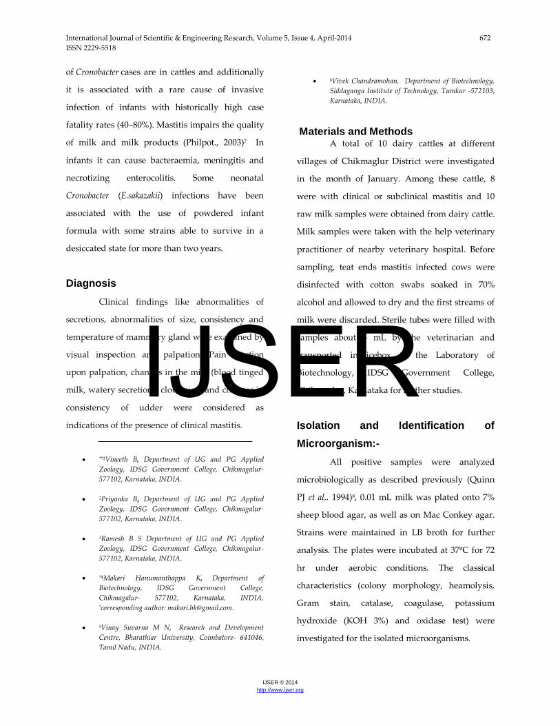
Antibiotic sensitivity test • Mueller - Hinton (MH) agar plates were
prepared and the cultures of bacterial
isolates 1b, 2a, 2b and also 1a were spread
on respectively labeled plates.
• The antibiotic discs of cephalothin,
spectinomycin, nalidixic acid and
ampicillin were placed on the spread
culture.
• For antibiotics in solution- kanamycin,
tetracycline, streptomycin and
chloramphenicol wells were punched and
antibiotics were added in the wells.
• The plates were incubated for 18 hours
and zones formed were observed.
IJSER
International Journal of Scientific & Engineering Research, Volume 5, Issue 4, April-2014 674 ISSN 2229-5518
IJSER © 2014 http://www.ijser.org
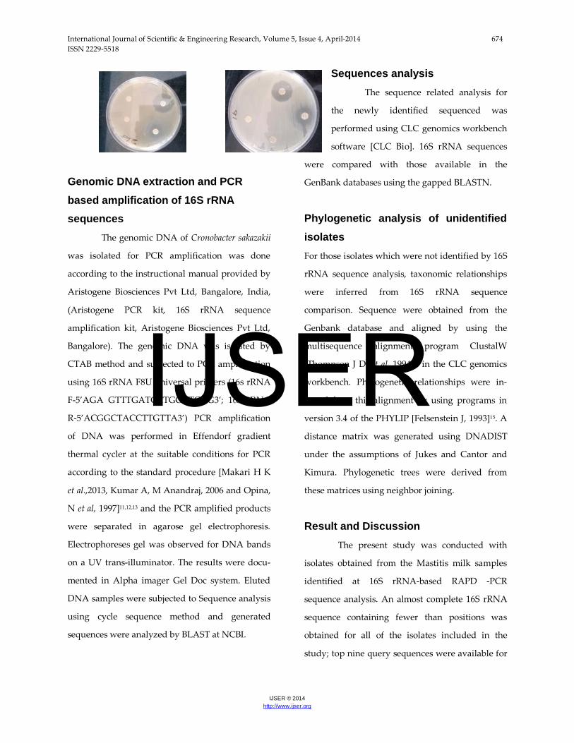
Genomic DNA extraction and PCR based amplification of 16S rRNA sequences
The genomic DNA of Cronobacter sakazakii
was isolated for PCR amplification was done
according to the instructional manual provided by
Aristogene Biosciences Pvt Ltd, Bangalore, India,
(Aristogene PCR kit, 16S rRNA sequence
amplification kit, Aristogene Biosciences Pvt Ltd,
Bangalore). The genomic DNA was isolated by
CTAB method and subjected to PCR amplification
using 16S rRNA F8U universal primers (16s rRNA
F-5’AGA GTTTGATCCTGGCTCAG3’; 16S rRNA
R-5’ACGGCTACCTTGTTA3’) PCR amplification
of DNA was performed in Effendorf gradient
thermal cycler at the suitable conditions for PCR
according to the standard procedure [Makari H K
et al.,2013, Kumar A, M Anandraj, 2006 and Opina,
N et al, 1997]11,12,13 and the PCR amplified products
were separated in agarose gel electrophoresis.
Electrophoreses gel was observed for DNA bands
on a UV trans-illuminator. The results were docu-
mented in Alpha imager Gel Doc system. Eluted
DNA samples were subjected to Sequence analysis
using cycle sequence method and generated
sequences were analyzed by BLAST at NCBI.
Sequences analysis The sequence related analysis for
the newly identified sequenced was
performed using CLC genomics workbench
software [CLC Bio]. 16S rRNA sequences
were compared with those available in the
GenBank databases using the gapped BLASTN.
Phylogenetic analysis of unidentified isolates
For those isolates which were not identified by 16S
rRNA sequence analysis, taxonomic relationships
were inferred from 16S rRNA sequence
comparison. Sequence were obtained from the
Genbank database and aligned by using the
multisequence alignment program ClustalW
[Thompson J D, et al, 1994]14 in the CLC genomics
workbench. Phylogenetic relationships were in-
ferred from this alignment by using programs in
version 3.4 of the PHYLIP [Felsenstein J, 1993]15. A
distance matrix was generated using DNADIST
under the assumptions of Jukes and Cantor and
Kimura. Phylogenetic trees were derived from
these matrices using neighbor joining.
Result and Discussion The present study was conducted with
isolates obtained from the Mastitis milk samples
identified at 16S rRNA-based RAPD -PCR
sequence analysis. An almost complete 16S rRNA
sequence containing fewer than positions was
obtained for all of the isolates included in the
study; top nine query sequences were available for
IJSER
International Journal of Scientific & Engineering Research, Volume 5, Issue 4, April-2014 675 ISSN 2229-5518
IJSER © 2014 http://www.ijser.org
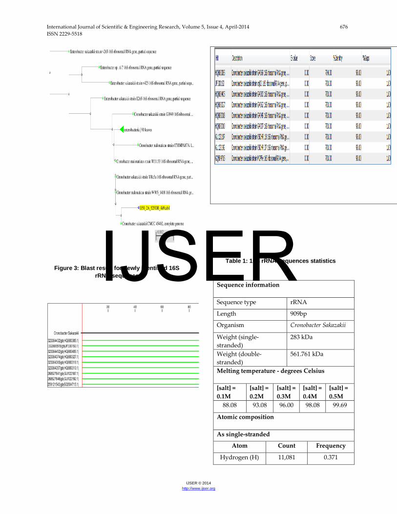
comparison (Figure 3). For this isolates belonging
to the organism Cronobacter sakazakii (Earlier name
was Enterobacter). The Phylogenetic tree was
produced using PHYLogeny inference package
(PHYLIP) with neighbor joining method shows
some of the closely related to identified bacteria
previously described in dairy environments the re-
sult as shown in the (Figure 2). Another interesting
observation was that more than one pathogen was
found in some mastitis samples reported earlier by
others (EL-Khodery S A., et al., 2008)16. It is
accepted that bacterial, environmental,
management and cow factors may affect the
occurrence and severity of mastitis, some reports
have indicated that mastitis mainly depends on
cow factors as shown by cases where cows were
infected by the same species (Buvernich C., et al.,
2003)17.The CLC genomics technique with
improved workbench software has generated the
following information for the input sequence.
Sequence information, melting temperature(0C),
atomic composition and nucleotide distribution.
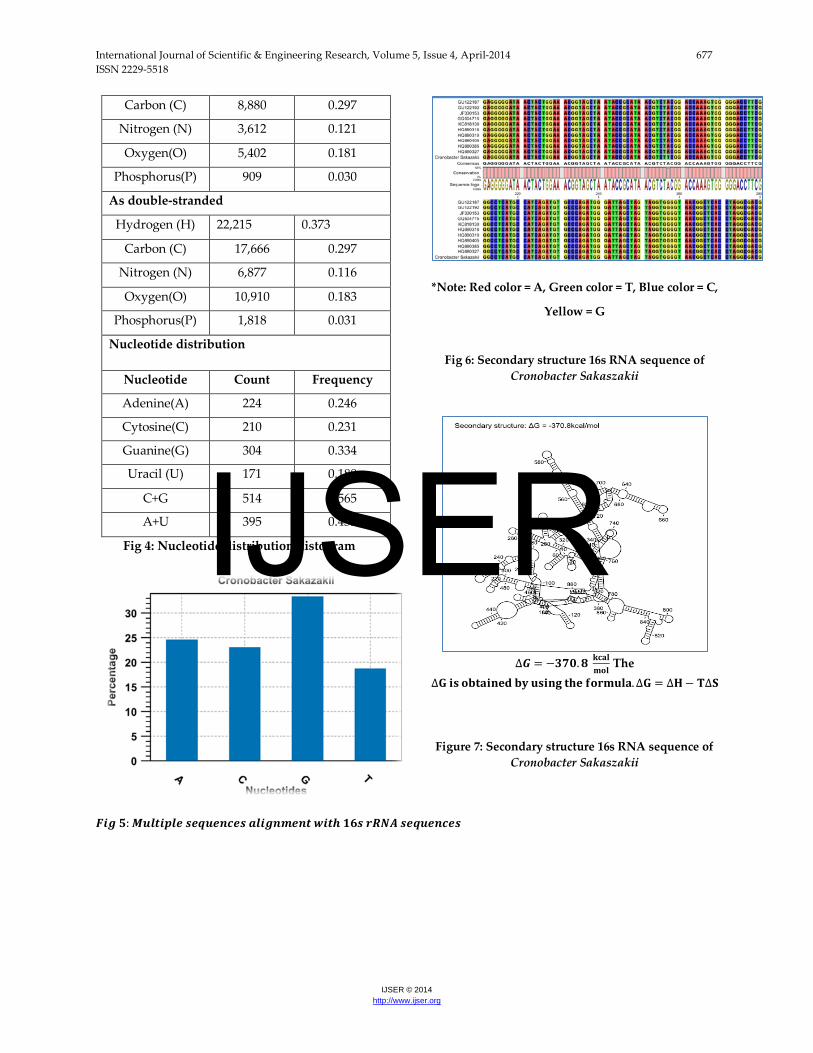
The results are shown in the (Table 1). Nucleotide
Guanine has the maximum number of occurrence
(304) and Uracil being the lowest (171).
From the table it is clear that C+G
combination is more (514) compared to A+G (395).
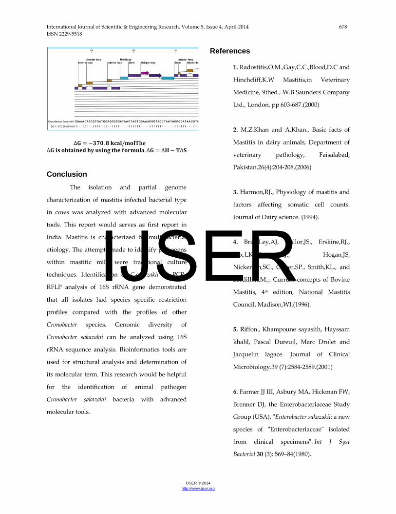
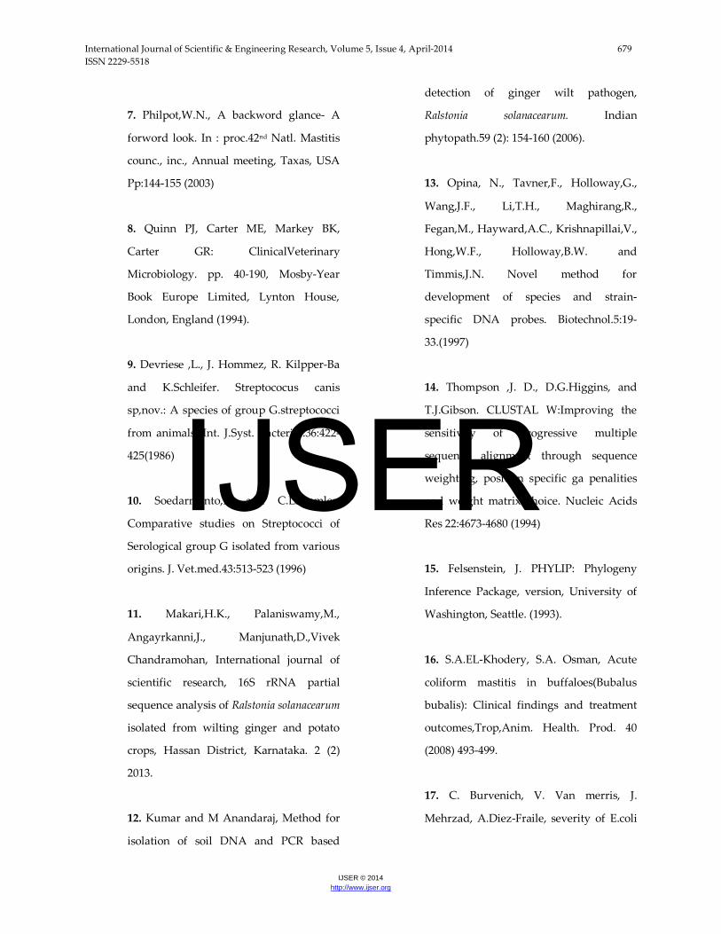
The RNA structure prediction results are shown in
the (Figure 6 and Figure 7). In that result gives the
information about stem, multi loop bulge loop and
hairpin loop. Nucleotide distribution histogram is
shown in (Figure 4). 16S rRNA sequencing is a
powerful tool for rapid identification and
phylogenetic analysis of bacterial species. This
method gives increasingly comprehensive and
more precise picture of the bacterial group
associated with the mastitis milk sample. The
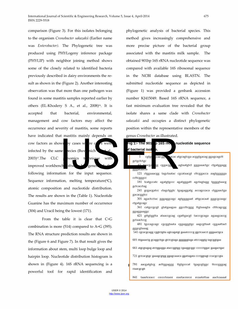
obtained 901bp 16S rRNA nucleotide sequence was
compared with available 16S ribosomal sequence
in the NCBI database using BLASTN. The
submitted nucleotide sequence as depicted in
(Figure 1) was provided a genbank accession
number KJ415049. Based 16S rRNA sequence, a
fast minimum evaluation tree revealed that the
isolate shares a same clade with Cronobacter
sakazakii and occupies a distinct phylogenetic
position within the representative members of the
genus Cronobacter as illustrated.
Fig 1:- The 901 bp 16S rRNA nucleotide sequence of bacterial isolate.
Fig 2: Phylogenetic analysis for newly identified 16S rRNA sequence
1 cgtgcggcaa ggcctaacac atgcagtcga acggtgacag ggagcagctt gctgctctgc 61 tgacgagtgg cggacgggtg agtaatgtct gggaaactgc ctgatggagg gggataacta 121 ctggaaacgg tagctaatac cgcataacgt cttcggacca aagtggggga ccttcgggcc 181 tcatgccatc agatgtgccc agatgggatt agctagtagg tggggtaacg gctcacctag 241 gcgacgatcc ctagctggtc tgagaggatg accagccaca ctggaactga gacacggtcc 301 agactcctac gggaggcagc agtggggaat attgcacaat gggcgcaagc ctgatgcagc 361 catgccgcgt gtatgaagaa ggccttcggg ttgtaaagta ctttcagcgg ggaggaaggc 421 gttgtggtta ataaccgcag cgattgacgt tacccgcaga agaagcaccg gctaactccg 481 tgccagcagc cgcggtaata cggagggtgc aagcgttaat cggaattact gggcgtaaag 541 cgcacgcagg cggtctgtta agtcagatgt gaaatccccg ggctcaacct gggaactgca 601 tttgaaactg gcaggcttga gtctcgtaga ggggggtaga attccaggtg tagcggtgaa
661 atgcgtagag atctggagga ataccggtgg cgaaggcggc ccccctggac gaagactgac
721 gctcacgtgc gaaagcgtgg ggagcaaaca ggattagata ccctggtagt ccacgccgta
781 aacgatgtcg acttggaggg ttgtgcccat tgagcgtggc ttcccgggag ctaacgcgtt
841 taagtcgacc cgccctgagg gagtacggcg gcaatgttaa aactcaaaat
IJSER
International Journal of Scientific & Engineering Research, Volume 5, Issue 4, April-2014 676 ISSN 2229-5518
IJSER © 2014 http://www.ijser.org
Figure 3: Blast result for newly identified 16S rRNA sequence
Table 1: 16S rRNA Sequences statistics
Sequence information
Sequence type rRNA
Length 909bp
Organism Cronobacter Sakazakii
Weight (single-stranded)
283 kDa
Weight (double-stranded)
561.761 kDa
Melting temperature - degrees Celsius
[salt] = 0.1M
[salt] = 0.2M
[salt] = 0.3M
[salt] = 0.4M
[salt] = 0.5M
88.08 93.08 96.00 98.08 99.69
Atomic composition
As single-stranded
Atom Count Frequency
Hydrogen (H) 11,081 0.371
IJSER
International Journal of Scientific & Engineering Research, Volume 5, Issue 4, April-2014 677 ISSN 2229-5518
IJSER © 2014 http://www.ijser.org
Carbon (C) 8,880 0.297
Nitrogen (N) 3,612 0.121
Oxygen(O) 5,402 0.181
Phosphorus(P) 909 0.030
As double-stranded
Hydrogen (H) 22,215 0.373
Carbon (C) 17,666 0.297
Nitrogen (N) 6,877 0.116
Oxygen(O) 10,910 0.183
Phosphorus(P) 1,818 0.031
Nucleotide distribution
Nucleotide Count Frequency
Adenine(A) 224 0.246
Cytosine(C) 210 0.231
Guanine(G) 304 0.334
Uracil (U) 171 0.188
C+G 514 0.565
A+U 395 0.435
Fig 4: Nucleotide distribution histogram
𝑭𝒊𝒈 𝟓: 𝑴𝒖𝒍𝒕𝒊𝒑𝒍𝒆 𝒔𝒆𝒒𝒖𝒆𝒏𝒄𝒆𝒔 𝒂𝒍𝒊𝒈𝒏𝒎𝒆𝒏𝒕 𝒘𝒊𝒕𝒉 𝟏𝟔𝒔 𝒓𝑹𝑵𝑨 𝒔𝒆𝒒𝒖𝒆𝒏𝒄𝒆𝒔
*Note: Red color = A, Green color = T, Blue color = C,
Yellow = G
Fig 6: Secondary structure 16s RNA sequence of Cronobacter Sakaszakii
∆𝑮 = −𝟑𝟕𝟎.𝟖 𝐤𝐜𝐚𝐥𝐦𝐨𝐥
The ∆𝐆 𝐢𝐬 𝐨𝐛𝐭𝐚𝐢𝐧𝐞𝐝 𝐛𝐲 𝐮𝐬𝐢𝐧𝐠 𝐭𝐡𝐞 𝐟𝐨𝐫𝐦𝐮𝐥𝐚.∆𝐆 = ∆𝐇− 𝐓∆𝐒
Figure 7: Secondary structure 16s RNA sequence of
Cronobacter Sakaszakii
IJSER
International Journal of Scientific & Engineering Research, Volume 5, Issue 4, April-2014 678 ISSN 2229-5518
IJSER © 2014 http://www.ijser.org
∆𝐆 = −𝟑𝟕𝟎.𝟖 𝐤𝐜𝐚𝐥/𝐦𝐨𝐥The ∆𝐆 𝐢𝐬 𝐨𝐛𝐭𝐚𝐢𝐧𝐞𝐝 𝐛𝐲 𝐮𝐬𝐢𝐧𝐠 𝐭𝐡𝐞 𝐟𝐨𝐫𝐦𝐮𝐥𝐚.∆𝐆 = ∆𝐇− 𝐓∆𝐒 Conclusion
The isolation and partial genome
characterization of mastitis infected bacterial type
in cows was analyzed with advanced molecular
tools. This report would serves as first report in
India. Mastitis is characterized by multibacterial
etiology. The attempts made to identify pathogens
within mastitic milk were traditional culture
techniques. Identification of C.sakazakii by PCR-
RFLP analysis of 16S rRNA gene demonstrated
that all isolates had species specific restriction
profiles compared with the profiles of other
Cronobacter species. Genomic diversity of
Cronobacter sakazakii can be analyzed using 16S
rRNA sequence analysis. Bioinformatics tools are
used for structural analysis and determination of
its molecular term. This research would be helpful
for the identification of animal pathogen
Cronobacter sakazakii bacteria with advanced
molecular tools.
References
1. Radostitis,O.M.,Gay,C.C.,Blood,D.C and
Hinchcliff,K.W Mastitis,in Veterinary
Medicine, 9thed., W.B.Saunders Company
Ltd., London, pp 603-687.(2000)
2. M.Z.Khan and A.Khan., Basic facts of
Mastitis in dairy animals, Department of
veterinary pathology, Faisalabad,
Pakistan.26(4):204-208.(2006)
3. Harmon,RJ., Physiology of mastitis and
factors affecting somatic cell counts.
Journal of Dairy science. (1994).
4. BramLey,AJ, Cullor,JS., Erskine,RJ.,
Fox,LK.,Harmon,RJ., Hogan,JS,
Nickerson,SC., Oliver,SP., Smith,KL., and
Sordillo,LM.,: Current concepts of Bovine
Mastitis, 4th edition, National Mastitis
Council, Madison,WI.(1996).
5. Riffon., Khampoune sayasith, Hayssam
khalil, Pascal Dureuil, Marc Drolet and
Jacquelin lagace. Journal of Clinical
Microbiology.39 (7):2584-2589.(2001)
6. Farmer JJ III, Asbury MA, Hickman FW,
Brenner DJ, the Enterobacteriaceae Study
Group (USA). "Enterobacter sakazakii: a new
species of "Enterobacteriaceae" isolated
from clinical specimens". Int J Syst
Bacteriol 30 (3): 569–84(1980).
IJSER
International Journal of Scientific & Engineering Research, Volume 5, Issue 4, April-2014 679 ISSN 2229-5518
IJSER © 2014 http://www.ijser.org
7. Philpot,W.N., A backword glance- A
forword look. In : proc.42nd Natl. Mastitis
counc., inc., Annual meeting, Taxas, USA
Pp:144-155 (2003)
8. Quinn PJ, Carter ME, Markey BK,
Carter GR: ClinicalVeterinary
Microbiology. pp. 40-190, Mosby-Year
Book Europe Limited, Lynton House,
London, England (1994).
9. Devriese ,L., J. Hommez, R. Kilpper-Ba
and K.Schleifer. Streptococus canis
sp,nov.: A species of group G.streptococci
from animals. Int. J.Syst. Bacteriol.36:422-
425(1986)
10. Soedarmanto,I., and C.La..mmler,
Comparative studies on Streptococci of
Serological group G isolated from various
origins. J. Vet.med.43:513-523 (1996)
11. Makari,H.K., Palaniswamy,M.,
Angayrkanni,J., Manjunath,D.,Vivek
Chandramohan, International journal of
scientific research, 16S rRNA partial
sequence analysis of Ralstonia solanacearum
isolated from wilting ginger and potato
crops, Hassan District, Karnataka. 2 (2)
2013.
12. Kumar and M Anandaraj, Method for
isolation of soil DNA and PCR based
detection of ginger wilt pathogen,
Ralstonia solanacearum. Indian
phytopath.59 (2): 154-160 (2006).
13. Opina, N., Tavner,F., Holloway,G.,
Wang,J.F., Li,T.H., Maghirang,R.,
Fegan,M., Hayward,A.C., Krishnapillai,V.,
Hong,W.F., Holloway,B.W. and
Timmis,J.N. Novel method for
development of species and strain-
specific DNA probes. Biotechnol.5:19-
33.(1997)
14. Thompson ,J. D., D.G.Higgins, and
T.J.Gibson. CLUSTAL W:Improving the
sensitivity of progressive multiple
sequence alignment through sequence
weighting, position specific ga penalities
and weight matrix choice. Nucleic Acids
Res 22:4673-4680 (1994)
15. Felsenstein, J. PHYLIP: Phylogeny
Inference Package, version, University of
Washington, Seattle. (1993).
16. S.A.EL-Khodery, S.A. Osman, Acute
coliform mastitis in buffaloes(Bubalus
bubalis): Clinical findings and treatment
outcomes,Trop,Anim. Health. Prod. 40
(2008) 493-499.
17. C. Burvenich, V. Van merris, J.
Mehrzad, A.Diez-Fraile, severity of E.coli
IJSER

International Journal of Scientific & Engineering Research, Volume 5, Issue 4, April-2014 680 ISSN 2229-5518
IJSER © 2014 http://www.ijser.org
mastitis is mainly determined by cow
factors, Vet.Res. 34(2003)521-564.
IJSER